Apoferritin–CeO2 nano-truffle that has excellent artificial redox ...
Apoferritin–CeO2 nano-truffle that has excellent artificial redox ...
Apoferritin–CeO2 nano-truffle that has excellent artificial redox ...

You also want an ePaper? Increase the reach of your titles
YUMPU automatically turns print PDFs into web optimized ePapers that Google loves.
Downloaded by Institute of Process Engineering, Chinese Academy of Sciences on 23 May 2012<br />
Published on 01 December 2011 on http://pubs.rsc.org | doi:10.1039/C1CC15815E<br />
Chemical Communications<br />
www.rsc.org/chemcomm Volume 48 | Number 26 | 28 March 2012 | Pages 3141–3248<br />
ISSN 1359-7345<br />
COMMUNICATION<br />
Ding Ma et al.<br />
Apoferritin–CeO 2 <strong>nano</strong>-truffl e <strong>that</strong> <strong>has</strong> <strong>excellent</strong> artifi cial <strong>redox</strong> enzyme<br />
activity<br />
View Online / Journal Homepage / Table of Contents for this issue<br />
1359-7345(2012)48:26;1-8
Downloaded by Institute of Process Engineering, Chinese Academy of Sciences on 23 May 2012<br />
Published on 01 December 2011 on http://pubs.rsc.org | doi:10.1039/C1CC15815E<br />
ChemComm<br />
Cite this: Chem. Commun., 2012, 48, 3155–3157<br />
Apoferritin–CeO 2 <strong>nano</strong>-<strong>truffle</strong> <strong>that</strong> <strong>has</strong> <strong>excellent</strong> <strong>artificial</strong> <strong>redox</strong><br />
enzyme activityw<br />
Xiangyou Liu,z ab Wei Wei,z c Quan Yuan, a Xin Zhang, d Ning Li, e Yuguang Du, b<br />
Guanghui Ma, c Chunhua Yan a and Ding Ma* a<br />
Received 20th September 2011, Accepted 1st November 2011<br />
DOI: 10.1039/c1cc15815e<br />
4.5 nm <strong>nano</strong>ceria particles are successfully encapsulated into the<br />
apoferritin cavity via a dissociation–reconstruction route. The<br />
apoferritin coating not only improves the biocompatibility and<br />
changes the cellular uptake route of <strong>nano</strong>ceria, but also manipulates<br />
the electron localization at the surface of the <strong>nano</strong>particle thereby<br />
ameliorating the ROS-scavenging activity.<br />
Incomplete removal of reactive oxygen species (ROS), which<br />
cause oxidative damage to human beings, leads to various<br />
detrimental effects on human health. 1 Natural antioxidant<br />
enzymes including endogenous superoxide dismutase (SOD) 2<br />
may not be sufficient to protect cells from sudden oxidative<br />
damage. There is therefore intense interest in developing an<br />
active <strong>artificial</strong> enzyme with a high ROS-scavenging activity<br />
but low cytotoxicity. Some <strong>nano</strong>materials were reported to be<br />
able to act as antioxidants, 3,4 among which <strong>nano</strong>ceria particles<br />
(<strong>nano</strong>-CeO2) drew much more attention due to their SOD<br />
mimetic activity and their reversibility and auto-regenerative<br />
properties. 5–7 However, it is still a challenge to construct a<br />
highly active <strong>artificial</strong> enzyme (even more active than SOD)<br />
with outstanding biocompatibility. Here we have shown <strong>that</strong><br />
reconstructing a cage protein, apoferritin, to encapsulate the<br />
4.5 nm <strong>nano</strong>-CeO 2 could fulfill this goal. Apoferritin not<br />
only improved the biocompatibility of the <strong>nano</strong>-CeO 2, but<br />
also manipulated the electron localization at the surface of<br />
the <strong>nano</strong>particle thereby ameliorating the ROS-scavenging<br />
activity of the apoferritin–CeO2 hybrid <strong>nano</strong>composite<br />
(AFt–CeO2).<br />
a Beijing National Laboratory for Molecular Sciences, College of<br />
Chemistry and Molecular Engineering, Peking University,<br />
Beijing 100871, China. E-mail: dma@pku.edu.cn;<br />
Fax: +86-10-62758603; Tel: +86-10-62758603<br />
b Dalian Institute of Chemical Physics, Chinese Academy of Sciences,<br />
Dalian 116023, China<br />
c National Key Laboratory of Biochemical Engineering,<br />
Institute of Process Engineering, Chinese Academy of Sciences,<br />
Beijing 100190, China<br />
d Department of Molecular and Experimental Medicine,<br />
The Scripps Research Institute, 10550 North Torrey Pines Road,<br />
La Jolla, CA 92037, USA<br />
e Lab of Applied Biocatalysis, South China University of Technology,<br />
Guangzhou 510640, China<br />
w Electronic supplementary information (ESI) available: Experimental<br />
details and supplementary results. See DOI: 10.1039/c1cc15815e<br />
z These authors contributed equally to this work.<br />
Dynamic Article Links<br />
View Online<br />
www.rsc.org/chemcomm COMMUNICATION<br />
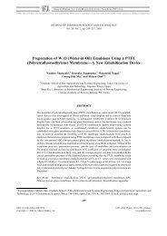
Fig. 1 (a) Schematic presentation of the assemblage process of<br />
AFt–CeO2. (b) TEM image of negatively stained AFt–CeO2. Theinset<br />
shows an enlarged single AFt–CeO 2 <strong>nano</strong>composite. (c) HRTEM image<br />
of a single AFt–CeO2 <strong>nano</strong>particle. (d) Energy dispersive spectrum of<br />
AFt–CeO 2. The signal of Cu is from the Cu grid. (e) Native PAGE gel<br />
of ferritin, AFt–CeO2 and apoferritin (AFt) (from left to right).<br />
The cage protein, apoferritin (the soft shell), was reconstructed<br />
into the <strong>nano</strong>-complex with the <strong>nano</strong>-CeO2 (the hard<br />
core) wrapped into the central cavity of apoferritin via a<br />
dissociation–reconstruction process (Fig. 1a). 8,9 To the best<br />
of our knowledge, the <strong>nano</strong>-CeO2, with all the faces being the<br />
most chemically active (100) facet and a narrow size distribution<br />
centered around 4.5 nm (Fig. S1, ESIw), have not been<br />
synthesized before. A transmission electron microscopy<br />
(TEM) image of negatively stained AFt–CeO 2 showed <strong>that</strong><br />
all the <strong>nano</strong>-crystals were encapsulated inside the apoferritin<br />
cage (Fig. 1b). These discrete <strong>nano</strong>-crystals were further<br />
proved to be <strong>nano</strong>-CeO2 by high-resolution TEM (HRTEM)<br />
(Fig. 1c) and energy dispersive spectroscopic measurements<br />
(Fig. 1d). Circular dichroism (CD) spectra revealed no<br />
This journal is c The Royal Society of Chemistry 2012 Chem. Commun., 2012, 48, 3155–3157 3155
Downloaded by Institute of Process Engineering, Chinese Academy of Sciences on 23 May 2012<br />
Published on 01 December 2011 on http://pubs.rsc.org | doi:10.1039/C1CC15815E<br />
significant changes in the secondary structure of apoferritin<br />
after the encapsulation of <strong>nano</strong>ceria (Fig. S2, ESIw). The<br />
integrality of AFt–CeO 2 was further validated by native polyacrylamide<br />
gel electrophoresis (PAGE) analysis which showed<br />
identical mobilities for ferritin, apoferritin, and AFt–CeO 2<br />
(Fig. 1e). Inductively coupled plasma optical emission spectrometry<br />
(ICP-OES) analysis of the protein bands in Fig. 1e<br />
showed <strong>that</strong> cerium was detected in the band of AFt–CeO2<br />
but not in the other two bands. Taken together, these results<br />
confirmed the successful reconstitution of apoferritin in which<br />
<strong>nano</strong>-CeO2 were integrated.<br />
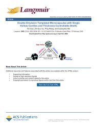
We then asked whether AFt–CeO2 would possess an altered<br />
ROS-scavenging activity. To this end, we measured the ability of<br />
AFt–CeO 2 to quench ROS and compared its activity to other<br />
materials including natural SOD. Notably, AFt–CeO 2 was able to<br />
clear more than 70% of superoxide anions (O 2 ), while the<br />
natural SOD could only quench B20% at most (Fig. 2a and b).<br />
Apoferritin-encapsulated Pt <strong>nano</strong>particles (AFt–Pt) were also able<br />
to quench superoxide anions, 10 however, the efficiency of AFt–Pt<br />
in ROS scavenging was much lower than <strong>that</strong> of AFt–CeO2. As<br />
shown in Fig. S3 (ESIw) and Fig. 2a, the IC50 of AFt–Pt (56 mM)<br />
was more than 160 fold higher than <strong>that</strong> of the AFt–CeO2<br />
(0.34 mM).y Interestingly, <strong>nano</strong>-CeO2, AFt alone or the mixture<br />
of these two materials did not possess the same ROS-scavenging<br />
activity as AFt–CeO 2. At fixed cerium and protein concentrations,<br />
pristine <strong>nano</strong>-CeO 2 and apoferritin could quench less than<br />
B10% of superoxide anions, while the direct mixture of <strong>nano</strong>-<br />
CeO 2 andapoferritin(AFt+CeO 2) merely quenched no more<br />
than 15% of superoxide anions (Fig. 2b). These observations<br />
directly demonstrated <strong>that</strong> the bio-conjugation of <strong>nano</strong>-CeO2<br />
with apoferritin conferred a superb ROS scavenging activity.<br />
Why did the AFt–CeO2 prove to be such a highly efficient<br />
<strong>artificial</strong> <strong>redox</strong> enzyme? It <strong>has</strong> been suggested <strong>that</strong> the reactivity<br />
of ceria in a <strong>redox</strong> process is largely dependent on the surface<br />
defects or vacancies on the crystals. 11 We then reasoned <strong>that</strong> the<br />
apoferritin modification improved the reducing activity of the<br />
<strong>nano</strong>-CeO 2. To test this hypothesis, electron energy loss<br />
Fig. 2 (a) Dose-dependent O 2 scavenging by AFt–CeO 2. (b)<br />
Comparison of O2 scavenging activity. The protein concentrations<br />
of AFt, AFt–CeO 2, ‘‘AFt + CeO 2’’ and SOD are all 0.01 mM, while<br />
the cerium concentrations of CeO2, AFt–CeO2 and ‘‘AFt + CeO2’’ are<br />
1 mM. (c) EELS profiles of pristine <strong>nano</strong>-CeO 2 and AFt–CeO 2.<br />
View Online<br />
spectroscopy (EELS) analysis was used to characterize the<br />
chemical state of <strong>nano</strong>-CeO2 with and without an apoferritin<br />
corona (Fig. 2c). EELS in the M-edge region of cerium carries<br />
information on the initial state 4f occupancy. The spectra were<br />
characterized by sharp white lines of 3d 3/2 - 4f 5/2 (M 4)and<br />
3d 5/2 - 4f 7/2 (M 5) associated with spin–orbit splitting. The<br />
relative intensities of the white lines were associated with the<br />
4f-shell occupancy of cerium, and thus could be used to<br />
determine its valence states. 12 A method for calculating the<br />
valence state of Ce can be found in ref. 12. The result of this<br />
calculation was <strong>that</strong> pristine <strong>nano</strong>-CeO2 had a 100% content of<br />
Ce 4+ while the AFt–CeO2 was a mixture of Ce 3+ and Ce 4+ ,<br />
with an estimated 70% fraction of Ce 3+ . This fact strongly<br />
indicated the existence of a charge transfer at the interface of the<br />
protein corona and the <strong>nano</strong>-CeO 2, resulting in a valence change<br />
of the oxide (Fig. S4, ESIw). A similar valence change <strong>has</strong> also<br />
been observed in the process where native ferritin sequesters and<br />
releases iron in the living biological systems, during which the<br />
valency of Fe oscillates between +2 and +3. 13,14 The charge<br />
transfer process changed the localization of electrons on<br />
the surface of <strong>nano</strong>-CeO2 inside the apoferritin, and more<br />
importantly, induced the formation of surface defects and<br />
vacancies on the <strong>nano</strong>-CeO2. 15 It is well-established <strong>that</strong> more<br />
defects or vacancies on the surface result in a much higher<br />
<strong>redox</strong> activity, 7,16 which explains the superior reactivity of<br />
AFt–CeO 2 in the ROS-scavenging assay.<br />
The superb ROS-scavenging activity of AFt–CeO 2 made it a<br />
promising candidate to serve as an <strong>artificial</strong> <strong>redox</strong> enzyme to<br />
relieve the oxidative stress of living cells. To test its activity in<br />
living cells, we used a ROS-sensitive fluorescent dye, DCFH-DA<br />
(2,7-dichlorofluorescein diacetate), which could be oxidized to a<br />
highly fluorescent compound DCF by intracellular ROS. 17<br />
HepG2 cells were preincubated with either <strong>nano</strong>-CeO2 or<br />
AFt–CeO2 before they were subjected to H2O2 treatment.<br />
Subsequently, the fluorescent level of DCF was used as an<br />
indicator of the residual ROS level. From several independent<br />
experiments, AFt–CeO 2 was convincingly shown to scavenge<br />
intracellular ROS more efficiently than <strong>nano</strong>-CeO 2 (Fig. 3a).<br />
Quantitatively, the IC50 of AFt–CeO 2 was 6.8 mM, B20 times<br />
lower than <strong>that</strong> of <strong>nano</strong>-CeO2 (132.2 mM). This result was<br />
further confirmed by corresponding images of confocal laser<br />
scanning microscopy (CLSM) (Fig. 3b). In the absence of<br />
<strong>nano</strong>-CeO2 and AFt–CeO2, the cells were strongly fluorescent,<br />
indicating a high ROS level. However, the fluorescent signal of<br />
AFt–CeO2-pretreated HepG2 cells was significantly diminished,<br />
suggesting an efficiently reduced ROS level. Notably, HepG2<br />
cells pretreated with <strong>nano</strong>-CeO 2 still exhibited a significant level<br />
of fluorescent intensity, indicating a rather limited reducing<br />
activity of <strong>nano</strong>-CeO 2 (Fig. 3b).<br />
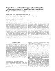
To test whether cells adapted readily to AFt–CeO 2 treatment,<br />
we examined the cellular internalization and cytotoxicity of<br />
AFt–CeO2. As shown in Fig. 4a, the amount of internalized<br />
AFt–CeO2 in HepG2 cells was about 2 fold higher than <strong>that</strong> of<br />
<strong>nano</strong>-CeO2, implying <strong>that</strong> the AFt–CeO2 was preferentially<br />
taken by HepG2 cells. Both CLSM and TEM images also<br />
confirmed <strong>that</strong> more AFt–CeO2 <strong>nano</strong>complexes could be internalized<br />
in HepG2 cells than <strong>nano</strong>-CeO2 (Fig. 4b–e). Interestingly,<br />
although AFt–CeO 2 entered HepG2 cells efficiently, cells<br />
were still viable in the presence of a high concentration of<br />
3156 Chem. Commun., 2012, 48, 3155–3157 This journal is c The Royal Society of Chemistry 2012
Downloaded by Institute of Process Engineering, Chinese Academy of Sciences on 23 May 2012<br />
Published on 01 December 2011 on http://pubs.rsc.org | doi:10.1039/C1CC15815E<br />
Fig. 3 (a) Scavenging of ROS by AFt–CeO2 and <strong>nano</strong>-CeO2 in<br />
HepG2 cells. (b) Representative CLSM images of H 2O 2-treated<br />
HepG2 cells after staining with DCFH-DA. From left to right:<br />
control, cells preincubated with <strong>nano</strong>-CeO 2 (25 mM) and AFt–CeO 2<br />
(25 mM). All the scale bars represent 10 mm.<br />
Fig. 4 (a) Quantification of AFt–CeO 2 and <strong>nano</strong>-CeO 2 internalized<br />
by HepG2 cells. (b–e) Representative CLSM and TEM images of<br />
HepG2 cells with <strong>nano</strong>-CeO 2 (b, d) or AFt–CeO 2 (c, e) internalized. In<br />
CLSM images (b) and (c), the bright spots in the cytoplasm around the<br />
nuclei are the internalized <strong>nano</strong>-CeO 2 (b) or AFt–CeO 2 (c) particles.<br />
(f) TEM image showing the typical macropinocytosis movement for<br />
<strong>nano</strong>-CeO 2 particles to enter HepG2 cells. (g and h) Schematic<br />
illustration of the macropinocytosis for <strong>nano</strong>-CeO2 and the clathrinmediated<br />
endocytosis for AFt–CeO 2. The scale bars in (b) and (c)<br />
represent 10 mm, while those in (d–f) represent 500 nm.<br />
AFt–CeO 2 (Fig. S5, ESIw). How did cells adapt to the AFt–CeO 2<br />
with a high internalized amount and a low cytotoxicity? We<br />
speculated <strong>that</strong> this distinct feature of AFt–CeO2 was partially<br />
attributed to the apoferritin corona, which could potentially<br />
promote the cellular internalization process mediated via specific<br />
ferritin receptors and prevent the adverse interactions between<br />
the <strong>nano</strong>-CeO 2 and biomolecules in the cytoplasm. 18 Further<br />
study (Fig. S6, ESIw) also suggested <strong>that</strong> the internalization of<br />
AFt–CeO 2 was primarily regulated by clathrin-mediated endocytosis<br />
(Fig. 4h), while the internalization of the <strong>nano</strong>-CeO 2 was<br />
a macropinocytosis process (Fig. 4f and g).<br />
In conclusion, we have demonstrated a strategy, combining<br />
cage proteins with a novel synthetic <strong>nano</strong>-material, to construct a<br />
novel <strong>nano</strong>-complex (AFt–CeO2) <strong>that</strong> <strong>has</strong> proven to be by far the<br />
most active <strong>artificial</strong> <strong>redox</strong> enzyme with mimetic SOD activity.<br />
The modification of the surface of <strong>nano</strong>-CeO2 <strong>has</strong> changed the<br />
intrinsic properties of individual building blocks, conferring a<br />
superb <strong>redox</strong> activity to the AFt–CeO 2. In addition, the cage<br />
proteins carry their biological identities to the AFt–CeO 2 to<br />
initiate an endocytosis process and improve its biocompatibility.<br />
This is a step-forward for the rational design/construction of<br />
<strong>artificial</strong> enzymes with designated functions, which have the<br />
potential to treat incurable diseases like some types of amyotrophic<br />
lateral sclerosis due to the failure of defense against ROS.<br />
Acknowledgements. This work received financial support<br />
from the Natural Science Foundation of China (20603036,<br />
21073004). The authors gratefully thank Miss Hua Yue and<br />
Mr. Zhanguo Yue for their help.<br />
Notes and references<br />
View Online<br />
y All the concentrations of AFt–CeO2 mentioned in this study are the<br />
concentrations of Ce, unless otherwise stated.<br />
1 J. Nordberg and E. S. J. Arnér, Free Radical Biol. Med., 2001, 31,<br />
1287–1312.<br />
2 J. M. Mate´s, Toxicology, 2000, 153, 83–104.<br />
3 M. Kajita, K. Hikosaka, M. Iitsuka, A. Kanayama, N. Toshima<br />
and Y. Miyamoto, Free Radical Res., 2007, 41, 615–626.<br />
4 A. S. Karakoti, S. Singh, A. Kumar, M. Malinska, S. Kuchibhatla,<br />
K. Wozniak, W. T. Self and S. Seal, J. Am. Chem. Soc., 2009, 131,<br />
14144–14145.<br />
5 C. Korsvik, S. Patil, S. Seal and W. T. Self, Chem. Commun., 2007,<br />
1056–1058.<br />
6 A. S. Karakoti, N. A. Monteiro-Riviere, R. Aggarwal, J. P. Davis,<br />
R. J. Narayan, W. T. Self, J. McGinnis and S. Seal, JOM, 2008, 60,<br />
33–37.<br />
7 E. G. Heckert, A. S. Karakoti, S. Seal and W. T. Self, Biomaterials,<br />
2008, 29, 2705–2709.<br />
8 B. Hennequin, L. Turyanska, T. Ben, A. M. Beltra´n, S. I. Molina,<br />
M. Li, S. Mann, A. Patanè and N. R. Thomas, Adv. Mater., 2008,<br />
20, 3592–3596.<br />
9 E. Valero, S. Tambalo, P. Marzola, M. Ortega-Mun˜ oz, F. J.<br />
Lo´pez-Jaramillo, F. Santoyo-González, J. D. López, J. J. Delgado,<br />
J. J. Calvino, R. Cuesta, J. M. Domínguez-Vera and N. Ga´lvez,<br />
J. Am. Chem. Soc., 2011, 133, 4889–4895.<br />
10 L. B. Zhang, L. Laug, W. Mu¨nchgesang, E. Pippel, U. Go¨sele,<br />
M. Brandsch and M. Knez, Nano Lett., 2010, 10, 219–223.<br />
11 F. Esch, S. Fabris, L. Zhou, T. Montini, C. Africh, P. Fornasiero,<br />
G. Comelli and R. Rosei, Science, 2005, 309, 752–755.<br />
12 L. J. Wu, H. J. Wiesmann, A. R. Moodenbaugh, R. F. Klie, Y. M.<br />
Zhu, D. O. Welch and M. Suenaga, Phys. Rev. B, 2004, 69, 9.<br />
13 M. T. F. Telling and S. H. Kilcoyne, Phys. B, 2006, 374–375, 451–455.<br />
14 F. Bou-Abdallah, Biochim. Biophys. Acta, Gen. Subj., 2010, 1800,<br />
719–731.<br />
15 P. Dutta, S. Pal, M. S. Seehra, Y. Shi, E. M. Eyring and<br />
R. D. Ernst, Chem. Mater., 2006, 18, 5144–5146.<br />
16 E. Mamontov, T. Egami, R. Brezny, M. Koranne and S. Tyagi,<br />
J. Phys. Chem. B, 2000, 104, 11110–11116.<br />
17 C. P. Lebel, H. Ischiropoulos and S. C. Bondy, Chem. Res.<br />
Toxicol., 1992, 5, 227–231.<br />
18 X. Y. Liu, W. Wei, C. L. Wang, H. Yue, D. Ma, C. Zhu, G. H. Ma<br />
and Y. G. Du, J. Mater. Chem., 2011, 21, 7105–7110.<br />
This journal is c The Royal Society of Chemistry 2012 Chem. Commun., 2012, 48, 3155–3157 3157