Apoferritin–CeO2 nano-truffle that has excellent artificial redox ...
Apoferritin–CeO2 nano-truffle that has excellent artificial redox ...
Apoferritin–CeO2 nano-truffle that has excellent artificial redox ...

Create successful ePaper yourself
Turn your PDF publications into a flip-book with our unique Google optimized e-Paper software.
Downloaded by Institute of Process Engineering, Chinese Academy of Sciences on 23 May 2012<br />
Published on 01 December 2011 on http://pubs.rsc.org | doi:10.1039/C1CC15815E<br />
significant changes in the secondary structure of apoferritin<br />
after the encapsulation of <strong>nano</strong>ceria (Fig. S2, ESIw). The<br />
integrality of AFt–CeO 2 was further validated by native polyacrylamide<br />
gel electrophoresis (PAGE) analysis which showed<br />
identical mobilities for ferritin, apoferritin, and AFt–CeO 2<br />
(Fig. 1e). Inductively coupled plasma optical emission spectrometry<br />
(ICP-OES) analysis of the protein bands in Fig. 1e<br />
showed <strong>that</strong> cerium was detected in the band of AFt–CeO2<br />
but not in the other two bands. Taken together, these results<br />
confirmed the successful reconstitution of apoferritin in which<br />
<strong>nano</strong>-CeO2 were integrated.<br />
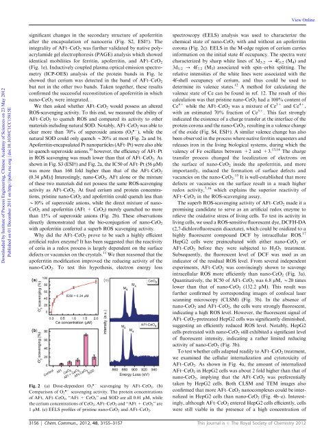
We then asked whether AFt–CeO2 would possess an altered<br />
ROS-scavenging activity. To this end, we measured the ability of<br />
AFt–CeO 2 to quench ROS and compared its activity to other<br />
materials including natural SOD. Notably, AFt–CeO 2 was able to<br />
clear more than 70% of superoxide anions (O 2 ), while the<br />
natural SOD could only quench B20% at most (Fig. 2a and b).<br />
Apoferritin-encapsulated Pt <strong>nano</strong>particles (AFt–Pt) were also able<br />
to quench superoxide anions, 10 however, the efficiency of AFt–Pt<br />
in ROS scavenging was much lower than <strong>that</strong> of AFt–CeO2. As<br />
shown in Fig. S3 (ESIw) and Fig. 2a, the IC50 of AFt–Pt (56 mM)<br />
was more than 160 fold higher than <strong>that</strong> of the AFt–CeO2<br />
(0.34 mM).y Interestingly, <strong>nano</strong>-CeO2, AFt alone or the mixture<br />
of these two materials did not possess the same ROS-scavenging<br />
activity as AFt–CeO 2. At fixed cerium and protein concentrations,<br />
pristine <strong>nano</strong>-CeO 2 and apoferritin could quench less than<br />
B10% of superoxide anions, while the direct mixture of <strong>nano</strong>-<br />
CeO 2 andapoferritin(AFt+CeO 2) merely quenched no more<br />
than 15% of superoxide anions (Fig. 2b). These observations<br />
directly demonstrated <strong>that</strong> the bio-conjugation of <strong>nano</strong>-CeO2<br />
with apoferritin conferred a superb ROS scavenging activity.<br />
Why did the AFt–CeO2 prove to be such a highly efficient<br />
<strong>artificial</strong> <strong>redox</strong> enzyme? It <strong>has</strong> been suggested <strong>that</strong> the reactivity<br />
of ceria in a <strong>redox</strong> process is largely dependent on the surface<br />
defects or vacancies on the crystals. 11 We then reasoned <strong>that</strong> the<br />
apoferritin modification improved the reducing activity of the<br />
<strong>nano</strong>-CeO 2. To test this hypothesis, electron energy loss<br />
Fig. 2 (a) Dose-dependent O 2 scavenging by AFt–CeO 2. (b)<br />
Comparison of O2 scavenging activity. The protein concentrations<br />
of AFt, AFt–CeO 2, ‘‘AFt + CeO 2’’ and SOD are all 0.01 mM, while<br />
the cerium concentrations of CeO2, AFt–CeO2 and ‘‘AFt + CeO2’’ are<br />
1 mM. (c) EELS profiles of pristine <strong>nano</strong>-CeO 2 and AFt–CeO 2.<br />
View Online<br />
spectroscopy (EELS) analysis was used to characterize the<br />
chemical state of <strong>nano</strong>-CeO2 with and without an apoferritin<br />
corona (Fig. 2c). EELS in the M-edge region of cerium carries<br />
information on the initial state 4f occupancy. The spectra were<br />
characterized by sharp white lines of 3d 3/2 - 4f 5/2 (M 4)and<br />
3d 5/2 - 4f 7/2 (M 5) associated with spin–orbit splitting. The<br />
relative intensities of the white lines were associated with the<br />
4f-shell occupancy of cerium, and thus could be used to<br />
determine its valence states. 12 A method for calculating the<br />
valence state of Ce can be found in ref. 12. The result of this<br />
calculation was <strong>that</strong> pristine <strong>nano</strong>-CeO2 had a 100% content of<br />
Ce 4+ while the AFt–CeO2 was a mixture of Ce 3+ and Ce 4+ ,<br />
with an estimated 70% fraction of Ce 3+ . This fact strongly<br />
indicated the existence of a charge transfer at the interface of the<br />
protein corona and the <strong>nano</strong>-CeO 2, resulting in a valence change<br />
of the oxide (Fig. S4, ESIw). A similar valence change <strong>has</strong> also<br />
been observed in the process where native ferritin sequesters and<br />
releases iron in the living biological systems, during which the<br />
valency of Fe oscillates between +2 and +3. 13,14 The charge<br />
transfer process changed the localization of electrons on<br />
the surface of <strong>nano</strong>-CeO2 inside the apoferritin, and more<br />
importantly, induced the formation of surface defects and<br />
vacancies on the <strong>nano</strong>-CeO2. 15 It is well-established <strong>that</strong> more<br />
defects or vacancies on the surface result in a much higher<br />
<strong>redox</strong> activity, 7,16 which explains the superior reactivity of<br />
AFt–CeO 2 in the ROS-scavenging assay.<br />
The superb ROS-scavenging activity of AFt–CeO 2 made it a<br />
promising candidate to serve as an <strong>artificial</strong> <strong>redox</strong> enzyme to<br />
relieve the oxidative stress of living cells. To test its activity in<br />
living cells, we used a ROS-sensitive fluorescent dye, DCFH-DA<br />
(2,7-dichlorofluorescein diacetate), which could be oxidized to a<br />
highly fluorescent compound DCF by intracellular ROS. 17<br />
HepG2 cells were preincubated with either <strong>nano</strong>-CeO2 or<br />
AFt–CeO2 before they were subjected to H2O2 treatment.<br />
Subsequently, the fluorescent level of DCF was used as an<br />
indicator of the residual ROS level. From several independent<br />
experiments, AFt–CeO 2 was convincingly shown to scavenge<br />
intracellular ROS more efficiently than <strong>nano</strong>-CeO 2 (Fig. 3a).<br />
Quantitatively, the IC50 of AFt–CeO 2 was 6.8 mM, B20 times<br />
lower than <strong>that</strong> of <strong>nano</strong>-CeO2 (132.2 mM). This result was<br />
further confirmed by corresponding images of confocal laser<br />
scanning microscopy (CLSM) (Fig. 3b). In the absence of<br />
<strong>nano</strong>-CeO2 and AFt–CeO2, the cells were strongly fluorescent,<br />
indicating a high ROS level. However, the fluorescent signal of<br />
AFt–CeO2-pretreated HepG2 cells was significantly diminished,<br />
suggesting an efficiently reduced ROS level. Notably, HepG2<br />
cells pretreated with <strong>nano</strong>-CeO 2 still exhibited a significant level<br />
of fluorescent intensity, indicating a rather limited reducing<br />
activity of <strong>nano</strong>-CeO 2 (Fig. 3b).<br />
To test whether cells adapted readily to AFt–CeO 2 treatment,<br />
we examined the cellular internalization and cytotoxicity of<br />
AFt–CeO2. As shown in Fig. 4a, the amount of internalized<br />
AFt–CeO2 in HepG2 cells was about 2 fold higher than <strong>that</strong> of<br />
<strong>nano</strong>-CeO2, implying <strong>that</strong> the AFt–CeO2 was preferentially<br />
taken by HepG2 cells. Both CLSM and TEM images also<br />
confirmed <strong>that</strong> more AFt–CeO2 <strong>nano</strong>complexes could be internalized<br />
in HepG2 cells than <strong>nano</strong>-CeO2 (Fig. 4b–e). Interestingly,<br />
although AFt–CeO 2 entered HepG2 cells efficiently, cells<br />
were still viable in the presence of a high concentration of<br />
3156 Chem. Commun., 2012, 48, 3155–3157 This journal is c The Royal Society of Chemistry 2012