Injection techniques for drug administration and methods of restraint
Injection techniques for drug administration and methods of restraint
Injection techniques for drug administration and methods of restraint

Create successful ePaper yourself
Turn your PDF publications into a flip-book with our unique Google optimized e-Paper software.
small ruminants<br />
dog <strong>and</strong> cat<br />
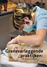
sphinx position<br />
Jos Ensink <strong>and</strong> Joris Robben 213<br />
In small ruminants, IV injections can be given in the external jugular vein. In fat animals, this vein<br />
is poorly visualised <strong>and</strong> in sheep this area needs to be clipped or shorn. For this reason, the cephalic<br />
vein is <strong>of</strong>ten used (<strong>for</strong> the technique, see below).<br />
In dogs <strong>and</strong> cats, the antebrachial part <strong>of</strong> the cephalic vein is preferred <strong>for</strong> IV injections as the<br />
animal accepts this with minimal <strong>restraint</strong>, <strong>and</strong> the vein is easy to locate. The animal is preferably<br />
placed on a table in the so-called “sphinx position” (a sitting or st<strong>and</strong>ing position is preferred by<br />
some animals <strong>and</strong> can there<strong>for</strong>e be more successful, but unexpected movements <strong>of</strong> the animal are<br />
more difficult to control).<br />
With one h<strong>and</strong>, the assistant restrains the head <strong>of</strong> the animal, turning it away to prevent the animal<br />
from biting the face <strong>of</strong> the person injecting. With the arm over the back <strong>of</strong> the animal, the other<br />
h<strong>and</strong> <strong>of</strong> the assistant grasps the opposite <strong>for</strong>elimb <strong>of</strong> the animal, at elbow level. In large dogs, the<br />
assistant thereby leans on the back <strong>of</strong> the animal, preventing it from sitting or st<strong>and</strong>ing up.<br />
The assistant compresses the vein with the thumb. In doing so, the entire h<strong>and</strong> is rotated outward:<br />
this causes the slightly medially running cephalic vein to be pulled “on top” <strong>of</strong> the <strong>for</strong>elimb, <strong>for</strong> a<br />
better presentation to the person injecting. To prevent the animal from pulling back its limb, the<br />
h<strong>and</strong> <strong>and</strong> lower arm is kept in contact with the tabletop. In cats, a second assistant may be necessary<br />
to control the hind limbs.<br />
The <strong>for</strong>elimb is grasped at the carpus (wrist) by the person injecting (right limb in left h<strong>and</strong><br />
<strong>and</strong> vice versa), whereby the thumb is positioned parallel to the cephalic vein to prevent lateral<br />
movement <strong>of</strong> the blood vessel. The person injecting should not grasp the limb too tightly in<br />
an ef<strong>for</strong>t to extend the limb (this should be per<strong>for</strong>med by the assistant), as this compresses the<br />
peripheral part <strong>of</strong> the cephalic vein, impeding circulation in the vessel. In preparation <strong>of</strong> the actual<br />
injection, the connection <strong>of</strong> the needle to the syringe should be checked, the calibration markings<br />
visible, the eccentric cone placed against the skin <strong>and</strong> the inside <strong>of</strong> the bevel visible. The needle is<br />
then advanced through the skin, into the blood vessel at an angle <strong>of</strong> 20-35°. The blood vessel can be<br />
punctured directly or the skin next to the blood vessel can be penetrated first, followed by puncture<br />
<strong>of</strong> the vein. The latter method prevents flattening <strong>of</strong> the vein at the time <strong>of</strong><br />
skin penetration, which leads to an increased risk <strong>of</strong> puncturing the opposite venous wall. Once the<br />
needle has penetrated the skin, the plunger <strong>of</strong> the syringe is pulled slightly backward to create slight<br />
negative pressure in the barrel, so that blood is aspirated the moment the needle punctures the vein.<br />
At this moment, the needle is advanced slightly further into the vessel to ensure that the needle<br />
does not slip out when the venous compression is released. Securing the syringe against the ball<br />
<strong>of</strong> the thumb after venipuncture allows the absorbtion <strong>of</strong> any unexpected limb movements. After<br />
releasing the compression (but not the limb!), the solution is slowly injected. After the injection,<br />
the skin is pulled taut over the injection site by the assistant after the needle is withdrawn, <strong>and</strong> kept<br />
in this position <strong>for</strong> a minute. The injection site may also be compressed with a b<strong>and</strong>age <strong>for</strong> 5 to 15<br />
minutes. An alternative location <strong>for</strong> IV injections is the saphenous vein in the hind limb.<br />
In dogs, the lateral, in the cat the median saphenous vein is routinely used. The dog is placed<br />
in lateral recumbency <strong>and</strong> the assistant restrains the upper hind limb at the stifle. The thumb is<br />
positioned over the patellar tendon, with the fingers around the popliteal area. Increased pressure<br />
<strong>of</strong> the h<strong>and</strong> will raise the lateral saphenous vein. The vein is better visualised than the cephalic vein,<br />
but is more difficult to puncture as proper fixation to reduce lateral movement is harder. In cats,<br />
the uppermost limb is held away by the assistant <strong>and</strong> the femoral vein is compressed in the inguinal<br />
area <strong>of</strong> the opposite (lower) limb. The median saphenous vein is clearly visible through the thin,<br />
nearly hairless skin. The external jugular vein is mainly used in small animals if (a large volume<br />
<strong>of</strong>) blood needs to be sampled, or if the injectable substance is too irritant <strong>for</strong> peripheral injection.<br />
For an IV injection in the external jugular vein, the animal is placed in the sphinx or in a sitting<br />
position. The assistant stretches the neck by lifting the head, turning it slightly to the contralateral<br />
side <strong>of</strong> which the jugular vein will be used. If this is done slowly <strong>and</strong> the neck is not overstretched,<br />
this generally leads to little resistance. In cats <strong>and</strong> small dogs it may be necessary to restrain the<br />
<strong>for</strong>elimbs, bringing them in a straight line with the head <strong>and</strong> neck, so that the person injecting has<br />
sufficient room to place the needle <strong>and</strong> syringe more parallel to the vein.<br />
The <strong>for</strong>elimbs may be restrained by holding them from the back in one h<strong>and</strong>, with a finger between<br />
the limbs. For this, the arm is placed over the back <strong>of</strong> the animal to the other side <strong>and</strong> brought