A Role for Piwi and piRNAs in Germ Cell ... - Hubrecht Institute
A Role for Piwi and piRNAs in Germ Cell ... - Hubrecht Institute
A Role for Piwi and piRNAs in Germ Cell ... - Hubrecht Institute

You also want an ePaper? Increase the reach of your titles
YUMPU automatically turns print PDFs into web optimized ePapers that Google loves.
while they were readily detectable <strong>in</strong> wild-type (Figure 2D).<br />
At 40 days of development no germ cells can be detected<br />
<strong>in</strong> ziwi mutants (Figure S2F; Table S1). Because vasa is expressed<br />
not only <strong>in</strong> PGCs but also <strong>in</strong> differentiat<strong>in</strong>g germ<br />
cells, these data argue that ziwi mutant germ cells do<br />
not enter a germ cell differentiation pathway.<br />
We next asked if germ cells <strong>in</strong> ziwi mutant gonads were<br />
disappear<strong>in</strong>g as a result of apoptosis. We assayed <strong>for</strong> the<br />
presence of apoptotic cells at 3 weeks of development <strong>in</strong><br />
both wild-type <strong>and</strong> mutant gonads by scor<strong>in</strong>g <strong>for</strong> the presence<br />
of Caspase-3 immunoreactivity. We observe apoptosis<br />
<strong>in</strong> mutant gonads but not <strong>in</strong> those isolated from wildtype<br />
fish (Figure 2E). This result suggests that loss of germ<br />
cells <strong>in</strong> ziwi mutants is a consequence of apoptosis. We cannot<br />
at this time, however, rule out the possibility that some<br />
ziwi mutant germ cells take on a somatic cell fate.<br />
We also exam<strong>in</strong>ed animals that had reduced levels of<br />
Ziwi function. We found that animals carry<strong>in</strong>g one mis-<br />
CELL 3253<br />
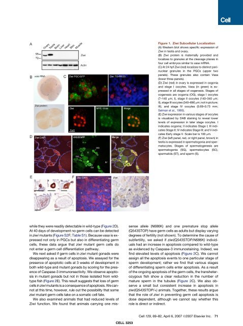
Figure 1. Ziwi Subcellular Localization<br />
(A) Western blot shows specific expression of<br />
Ziwi <strong>in</strong> testis <strong>and</strong> ovary.<br />
(B) Ziwi prote<strong>in</strong> is maternally provided <strong>and</strong><br />
localizes to granules at the cleavage planes <strong>in</strong><br />
four cell embryos similar to vasa mRNA.<br />
(C) At 24 hpf Ziwi (red) localizes to dist<strong>in</strong>ct per<strong>in</strong>uclear<br />
granules <strong>in</strong> the PGCs (upper two<br />
panels). These granules also conta<strong>in</strong> Vasa<br />
(lower three panels).<br />
(D) Ziwi (red) <strong>in</strong> ovary is expressed <strong>in</strong> oogonia<br />
<strong>and</strong> stage I oocytes. Vasa (<strong>in</strong> green) is expressed<br />
<strong>in</strong> all stages of oogenesis. Stages of<br />
oogenesis are oogonia (OG), stage I oocytes<br />
(7–140 mm; I), stage II oocytes (140–340 mm;<br />
II), stage III oocytes (340–690 mm; not <strong>in</strong> picture;<br />
III), <strong>and</strong> stage IV oocytes (0.69–0.73 mm;<br />
Selman et al., 1993).<br />
(E) Ziwi expression <strong>in</strong> various stages of oocytes<br />
is visualized by DAB sta<strong>in</strong><strong>in</strong>g to reveal lower<br />
levels of expression <strong>in</strong> later stage oocytes. I<br />
<strong>in</strong>dicates oogonia; II <strong>in</strong>dicates Stage I; III <strong>in</strong>dicates<br />
Stage II; IV <strong>in</strong>dicates Stage III; <strong>and</strong> V <strong>in</strong>dicates<br />
Early stage V. Scale bar is 100 mm.<br />
(F) Ziwi (left panel, red, or right panel, brown) <strong>in</strong><br />
testis is expressed <strong>in</strong> spermatogonia <strong>and</strong> spermatocytes.<br />
Stages of spermatogenesis are<br />
spermatogonia (SG), spermatocytes (SC),<br />
spermatids (ST), <strong>and</strong> sperm (S).<br />
sense allele (N686K) <strong>and</strong> one premature stop allele<br />
(Q543STOP) have germ cells as adults but display vary<strong>in</strong>g<br />
degrees of fertility (not shown). To determ<strong>in</strong>e the cause of<br />
subfertility, we asked if ziwi(Q543STOP/N686K) <strong>in</strong>dividuals<br />
had an <strong>in</strong>crease <strong>in</strong> apoptosis compared to wild-type<br />
as evidenced by Caspase-3 immunosta<strong>in</strong><strong>in</strong>g. Indeed, we<br />
f<strong>in</strong>d elevated levels of apoptosis (Figure 2C). We cannot<br />
assign all the apoptosis events to one particular stage of<br />
sperm development; rather we f<strong>in</strong>d that various stages<br />
of differentiat<strong>in</strong>g sperm cells enter apoptosis. As a result<br />
of the ongo<strong>in</strong>g apoptosis of the germ cells, the transheterozygous<br />
fish show a clear reduction <strong>in</strong> the number of<br />
mature sperm <strong>in</strong> the tubules (Figure 2C). We also observe<br />
a small but consistent <strong>in</strong>crease <strong>in</strong> apoptosis <strong>in</strong><br />
ziwi(Q543STOP/+) animals. Together, these results argue<br />
that the role of ziwi <strong>in</strong> prevent<strong>in</strong>g germ cell apoptosis is<br />
dose dependent, although we cannot say whether this<br />
role is direct or <strong>in</strong>direct.<br />
<strong>Cell</strong> 129, 69–82, April 6, 2007 ª2007 Elsevier Inc. 71