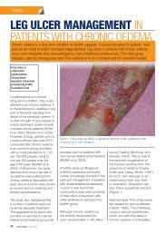
Management of Chronic Oedema in the Community - Wounds ...
Management of Chronic Oedema in the Community - Wounds ...
Management of Chronic Oedema in the Community - Wounds ...

Create successful ePaper yourself
Turn your PDF publications into a flip-book with our unique Google optimized e-Paper software.
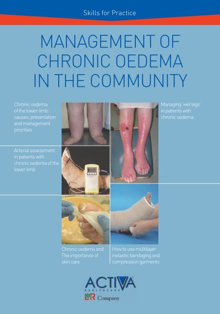
Skills for Practice<br />
MANAGEMENT OF<br />
CHRONIC OEDEMA<br />
IN THE COMMUNITY<br />
<strong>Chronic</strong> oedema<br />
<strong>of</strong> <strong>the</strong> lower limb:<br />
causes, presentation<br />
and management<br />
priorities<br />
Arterial assessment<br />
<strong>in</strong> patients with<br />
chronic oedema <strong>of</strong> <strong>the</strong><br />
lower limb<br />
<strong>Chronic</strong> oedema and<br />
The importance <strong>of</strong><br />
sk<strong>in</strong> care<br />
How to use multilayer<br />
<strong>in</strong>elastic bandag<strong>in</strong>g and<br />
compression garments<br />
Manag<strong>in</strong>g ‘wet legs’<br />
<strong>in</strong> patients with<br />
chronic oedema
© HealthComm UK Limited, trad<strong>in</strong>g as <strong>Wounds</strong> UK Limited, 2009<br />
All rights reserved. No reproduction, copy or transmission <strong>of</strong> this publication may be made without written<br />
permission from <strong>the</strong> publishers.<br />
This document was funded with an unrestricted educational grant from Activa Healthcare Ltd. The views<br />
expressed <strong>in</strong> this publication are those <strong>of</strong> <strong>the</strong> authors and do not necessarily reflect those <strong>of</strong> <strong>Wounds</strong> UK and<br />
Activa Healthcare Ltd<br />
To reference this document cite <strong>the</strong> follow<strong>in</strong>g:<br />
Skills for Practice: <strong>Management</strong> <strong>of</strong> <strong>Chronic</strong> <strong>Oedema</strong> <strong>in</strong> <strong>the</strong> <strong>Community</strong>. <strong>Wounds</strong> UK, Aberdeen, 2009
CONTENTS<br />
THE MANAGEMENT OF PATIENTS<br />
WITH CHRONIC OEDEMA<br />
IN THE COMMUNITY<br />
Paul<strong>in</strong>e Beldon, is Tissue Viability Nurse Consultant, Epsom and St Helier University Hospitals NHS Trust<br />
<strong>Chronic</strong> oedema <strong>of</strong> <strong>the</strong> lower<br />
limb is a prevalent condition<br />
which is frequently encountered<br />
and managed <strong>in</strong> <strong>the</strong> community. As<br />
<strong>the</strong> older adult population <strong>in</strong>creases<br />
it is reasonable to expect a parallel<br />
<strong>in</strong>crease <strong>in</strong> co-morbidities, many <strong>of</strong><br />
which are risk factors for chronic<br />
oedema and reduced mobility.<br />
Consequently it is important that any<br />
cl<strong>in</strong>ician car<strong>in</strong>g for patients <strong>in</strong> <strong>the</strong><br />
community has <strong>the</strong> ability to identify<br />
chronic oedema, understand its<br />
differ<strong>in</strong>g causes and presentations<br />
and <strong>in</strong>itiate early <strong>in</strong>terventions to<br />
both avoid complications and<br />
improve outcomes and quality <strong>of</strong><br />
life for <strong>the</strong> patient. The identification<br />
and management <strong>of</strong> any underly<strong>in</strong>g<br />
medical conditions which may<br />
be contribut<strong>in</strong>g to <strong>the</strong> condition<br />
is paramount. If <strong>the</strong> problem is<br />
complicated it should be identified<br />
as a multidiscipl<strong>in</strong>ary issue, so that<br />
both <strong>the</strong> patient and <strong>the</strong> community<br />
practitioner can receive <strong>the</strong> assistance<br />
required to manage <strong>the</strong> situation.<br />
Failure to identify chronic oedema<br />
or failure to refer <strong>the</strong> patient to an<br />
appropriate specialist leaves <strong>the</strong><br />
patient open to <strong>the</strong> development <strong>of</strong><br />
complications and <strong>the</strong> cl<strong>in</strong>ician open<br />
to allegations <strong>of</strong> poor care.<br />
The management <strong>of</strong> chronic oedema<br />
<strong>in</strong>volves several strands <strong>of</strong> care and<br />
requires a nurse with skills <strong>in</strong> <strong>in</strong>terpret<strong>in</strong>g<br />
assessment and tailor<strong>in</strong>g a package<br />
<strong>of</strong> leg care to suit <strong>the</strong> patient as an<br />
<strong>in</strong>dividual. A sk<strong>in</strong> care regimen should<br />
be developed to ma<strong>in</strong>ta<strong>in</strong> barrier<br />
function and vascular assessment is<br />
vital to exclude <strong>the</strong> presence <strong>of</strong> arterial<br />
disease. Once this is ascerta<strong>in</strong>ed,<br />
<strong>the</strong> cl<strong>in</strong>ician can proceed with <strong>the</strong><br />
use <strong>of</strong> an <strong>in</strong>dividualised application<br />
<strong>of</strong> compression, whe<strong>the</strong>r bandag<strong>in</strong>g<br />
or hosiery. Should <strong>the</strong> patient’s limbs<br />
Care <strong>of</strong> chronic oedema<br />
<strong>in</strong> <strong>the</strong> community<br />
CHRONIC OEDEMA OF THE LOWER LIMB: CAUSES, PRESENTATION AND MANAGEMENT PRIORITIES<br />
Vaughan Keeley ....................................................................................................................................................... 4<br />
ARTERIAL ASSESSMENT IN PATIENTS WITH CHRONIC OEDEMA OF THE LOWER LIMB<br />
Janice Bianchi ......................................................................................................................................................... 9<br />
CHRONIC OEDEMA AND THE IMPORTANCE OF SKIN CARE<br />
Carrie W<strong>in</strong>gfield .................................................................................................................................................... 13<br />
MANAGING WET LEGS IN PATIENTS WITH CHRONIC OEDEMA<br />
Paul<strong>in</strong>e Beldon ...................................................................................................................................................... 20<br />
HOW TO USE MULTILAYER INELASTIC BANADAGING AND COMPRESSION GARMENTS<br />
Kimby Osborne ...................................................................................................................................................... 24<br />
EDITORIAL<br />
become wet, <strong>the</strong>n wound management<br />
skills <strong>in</strong> select<strong>in</strong>g appropriate absorbent<br />
dress<strong>in</strong>gs for use under compression<br />
become important. This supplement<br />
provides relevant and practical<br />
<strong>in</strong>formation on all <strong>the</strong>se aspects<br />
<strong>of</strong> care to guide those cl<strong>in</strong>icians<br />
who are actively responsible for <strong>the</strong><br />
management <strong>of</strong> patients with chronic<br />
oedema <strong>in</strong> <strong>the</strong> community.<br />
F<strong>in</strong>ally, it should always be remembered<br />
that <strong>the</strong> patient and <strong>the</strong>ir family/carers<br />
also have a vital role to play <strong>in</strong> <strong>the</strong><br />
management <strong>of</strong> chronic oedema, s<strong>in</strong>ce<br />
if <strong>the</strong> appropriate efforts are not made<br />
to elevate <strong>the</strong> limb, wear compression<br />
hosiery or exercise (where possible)<br />
<strong>the</strong>n all <strong>the</strong> cl<strong>in</strong>ician’s efforts may be <strong>in</strong><br />
va<strong>in</strong>. Consequently <strong>the</strong> engagement<br />
<strong>of</strong> <strong>the</strong> patient by <strong>the</strong> cl<strong>in</strong>ician <strong>in</strong> a<br />
concordant relationship <strong>of</strong> trust and<br />
mutual respect and understand<strong>in</strong>g<br />
becomes vital to <strong>the</strong> successful<br />
management <strong>of</strong> chronic oedema.<br />
3
4<br />
Care <strong>of</strong> chronic oedema<br />
<strong>in</strong> <strong>the</strong> community<br />
CHRONIC OEDEMA OF THE LOWER<br />
LIMB: CAUSES, PRESENTATION<br />
AND MANAGEMENT PRIORITIES<br />
Vaughan Keeley is Consultant <strong>in</strong> Palliative Medic<strong>in</strong>e, Derby Hospitals NHS foundation Trust, Derby, UK<br />
<strong>Chronic</strong> oedema is an umbrella<br />
term for swell<strong>in</strong>g which has been<br />
present for at least three months <strong>in</strong><br />
<strong>the</strong> limbs and/or mid-l<strong>in</strong>e structures<br />
such as <strong>the</strong> trunk, head and neck<br />
or genitalia (M<strong>of</strong>fatt et al, 2003).<br />
From a practical, cl<strong>in</strong>ical po<strong>in</strong>t<br />
<strong>of</strong> view it is a useful term, as it<br />
covers oedema <strong>of</strong> a wide range<br />
<strong>of</strong> aetiologies. It is important to<br />
try to understand <strong>the</strong> cause <strong>of</strong> an<br />
<strong>in</strong>dividual patient’s chronic oedema<br />
<strong>in</strong> order to be able to determ<strong>in</strong>e <strong>the</strong><br />
best management.<br />
The aetiology <strong>of</strong> chronic oedema<br />
may be quite complex <strong>in</strong> some<br />
patients and <strong>the</strong>re may be a<br />
number <strong>of</strong> factors contribut<strong>in</strong>g to<br />
<strong>the</strong> condition. Common causes<br />
Figure 1.Mild dependency oedema<br />
<strong>in</strong> a patient with multiple sclerosis.<br />
are listed <strong>in</strong> Table 1. <strong>Oedema</strong> may<br />
also occur due to a number <strong>of</strong><br />
more ‘systemic’ problems such as<br />
heart failure, hepatic cirrhosis and<br />
hypothyroidism. It may be caused<br />
by drugs, such as <strong>the</strong> calcium<br />
channel antagonists amlodip<strong>in</strong>e<br />
and felodip<strong>in</strong>e, non-steroidal anti<strong>in</strong>flammatory<br />
drugs and steroids<br />
(Keeley, 2008).<br />
This article will describe <strong>the</strong><br />
symptoms and signs associated<br />
with some <strong>of</strong> <strong>the</strong> more common<br />
patterns <strong>of</strong> oedema likely to be<br />
encountered <strong>in</strong> <strong>the</strong> community, but<br />
will also give examples <strong>of</strong><br />
rarer problems.<br />
DEPENDENCY OEDEMA<br />
Cause<br />
This is usually associated with longstand<strong>in</strong>g<br />
immobility which may be<br />
due to neurological problems such<br />
as multiple sclerosis, respiratory<br />
problems such as chronic<br />
obstructive pulmonary disease or<br />
musculoskeletal problems such as<br />
rheumatoid arthritis.<br />
It arises because <strong>of</strong> failure <strong>of</strong> <strong>the</strong><br />
calf muscle pump which normally<br />
enhances venous and lymphatic<br />
dra<strong>in</strong>age <strong>in</strong> <strong>the</strong> legs.<br />
Presentation<br />
It typically presents as s<strong>of</strong>t<br />
oedema beg<strong>in</strong>n<strong>in</strong>g <strong>in</strong> <strong>the</strong> feet and<br />
work<strong>in</strong>g up <strong>the</strong> legs. It is usually<br />
bilateral (Figure 1), but may be<br />
unilateral, such as <strong>in</strong> patients with<br />
a hemiparesis. In <strong>the</strong> latter, arm<br />
swell<strong>in</strong>g can also occur. People<br />
affected by dependency oedema<br />
typically spend much <strong>of</strong> <strong>the</strong> day<br />
sitt<strong>in</strong>g and may even sleep <strong>in</strong> a<br />
sitt<strong>in</strong>g position at night.<br />
The oedema is usually very<br />
s<strong>of</strong>t and easily pits when it first<br />
presents, but can become firmer<br />
over time. Distortion <strong>of</strong> limb shape<br />
can develop (Figure 2) and sk<strong>in</strong><br />
breakdown can occur toge<strong>the</strong>r with<br />
ulceration.<br />
<strong>Management</strong> priorities and<br />
solutions<br />
Ideally, early management <strong>of</strong><br />
this problem is advisable before<br />
significant limb distortion occurs.<br />
Where possible, exercises and<br />
elevation <strong>of</strong> <strong>the</strong> limbs while sitt<strong>in</strong>g<br />
should be encouraged.<br />
Compression stock<strong>in</strong>gs are usually<br />
very helpful but <strong>the</strong>re can be<br />
difficulties <strong>in</strong> putt<strong>in</strong>g <strong>the</strong>m on and<br />
tak<strong>in</strong>g <strong>the</strong>m <strong>of</strong>f as many patients<br />
Table 1<br />
Common causes <strong>of</strong> chronic oedema<br />
l ‘Dependency’ oedema: associated with<br />
immobility<br />
l <strong>Oedema</strong> due to heart failure<br />
l Venous oedema: e.g. result<strong>in</strong>g from venous<br />
disease such as post-thrombotic<br />
syndrome or severe varicose ve<strong>in</strong>s<br />
l <strong>Oedema</strong> associated with obesity<br />
l Lymphoedema: primary and secondary<br />
l <strong>Oedema</strong> related to advanced cancer
with this type <strong>of</strong> oedema will not be<br />
able to do this <strong>the</strong>mselves and will<br />
require help.<br />
Particular care is needed <strong>in</strong><br />
patients with neurological problems<br />
and numbness <strong>in</strong> <strong>the</strong> limb as<br />
compression garments can cause<br />
tissue <strong>in</strong>jury which <strong>the</strong> patient may<br />
not be able to feel (Figure 3).<br />
If early <strong>in</strong>tervention is not<br />
<strong>in</strong>troduced, patients with<br />
dependency oedema can go on to<br />
develop chronic ‘leaky or wet legs’<br />
where pr<strong>of</strong>use leakage <strong>of</strong> lymph<br />
from breaks <strong>in</strong> <strong>the</strong> sk<strong>in</strong> requires<br />
longer-term bandag<strong>in</strong>g. Wet legs<br />
are discussed <strong>in</strong> detail on pgs.20–<br />
23 <strong>of</strong> this supplement.<br />
OEDEMA DUE TO HEART FAILURE<br />
Cause<br />
Heart failure is commonly due<br />
to ischaemic heart disease but<br />
could have o<strong>the</strong>r causes such as<br />
pulmonary heart disease, valvular<br />
disease and cardiomyopathy.<br />
Presentation<br />
Patients may develop peripheral<br />
oedema as a result <strong>of</strong> acute<br />
heart failure follow<strong>in</strong>g on from a<br />
myocardial <strong>in</strong>farction, but many<br />
patients have chronic heart failure<br />
which leads to persistently swollen<br />
legs. With reduced mobility,<br />
presentation will be similar to<br />
dependency oedema. Evidence<br />
for a cardiac cause <strong>of</strong> <strong>the</strong> oedema<br />
should be sought, such as ang<strong>in</strong>a,<br />
Figure 2. Dependency oedema <strong>in</strong> a<br />
patient with paraplegia.<br />
or a known history <strong>of</strong> ischaemic<br />
heart disease and known heart<br />
valve disease. Patients may also<br />
report breathlessness on exertion<br />
and have cl<strong>in</strong>ical signs <strong>of</strong> heart<br />
failure such as a raised jugular<br />
venous pressure, bilateral basal<br />
crackles on auscultation <strong>of</strong> <strong>the</strong><br />
chest and a triple rhythm on<br />
auscultation <strong>of</strong> <strong>the</strong> heart. <strong>Oedema</strong><br />
will typically be present <strong>in</strong> both<br />
legs, although it may not be<br />
symmetrical. The oedema may also<br />
be quite extensive and extend to<br />
<strong>the</strong> sacral region.<br />
<strong>Management</strong> priorities<br />
and solutions<br />
Heart failure is usually managed<br />
medically with drugs such as<br />
diuretics, ACE <strong>in</strong>hibitors and<br />
digox<strong>in</strong>. Never<strong>the</strong>less, some<br />
patients with chronic heart<br />
failure and immobility may<br />
benefit from light compression<br />
treatment <strong>of</strong> <strong>the</strong>ir legs. However,<br />
compression treatment may<br />
exacerbate acute heart failure so<br />
a medical assessment is required<br />
to determ<strong>in</strong>e whe<strong>the</strong>r this is<br />
be<strong>in</strong>g controlled and whe<strong>the</strong>r<br />
compression is advisable. Even<br />
if compression is <strong>in</strong>dicated,<br />
a modified reduced pressure<br />
regimen is usually applied, <strong>of</strong>ten<br />
commenc<strong>in</strong>g with only one leg<br />
to m<strong>in</strong>imise fluid movement<br />
(Lymphoedema Framework, 2006).<br />
Patients with ischaemic heart<br />
disease may also have peripheral<br />
vascular disease so it is particularly<br />
Figure 3. Tissue damage from a<br />
compression garment <strong>in</strong> a patient<br />
with peripheral neuropathy.<br />
Care <strong>of</strong> chronic oedema<br />
<strong>in</strong> <strong>the</strong> community<br />
important to assess peripheral<br />
arterial circulation <strong>in</strong> this group<br />
<strong>of</strong> patients before apply<strong>in</strong>g<br />
compression.<br />
If peripheral vascular disease<br />
is detected, it is recommended<br />
that <strong>the</strong> patient is assessed by a<br />
vascular surgeon. Full compression<br />
may <strong>the</strong>n be possible once <strong>the</strong><br />
vascular disease is treated.<br />
However, if <strong>the</strong> peripheral vascular<br />
disease cannot be improved,<br />
<strong>the</strong>n ei<strong>the</strong>r reduced compression<br />
<strong>the</strong>rapy or none at all may<br />
be <strong>in</strong>dicated.<br />
When monitor<strong>in</strong>g patients with<br />
chronic leg oedema <strong>of</strong> any cause,<br />
<strong>the</strong> development <strong>of</strong> heart failure<br />
should be considered if <strong>the</strong><br />
patient’s oedema becomes worse<br />
(Figure 4). Should this develop,<br />
appropriate medical treatment <strong>of</strong><br />
<strong>the</strong> heart failure is required and<br />
removal <strong>of</strong> compression garments<br />
or a reduction <strong>in</strong> <strong>the</strong> strength <strong>of</strong><br />
compression on <strong>the</strong> legs<br />
is advisable.<br />
VENOUS DISEASE AND OEDEMA<br />
Cause<br />
<strong>Chronic</strong> venous hypertension, e.g.<br />
due to post-thrombotic syndrome<br />
or severe varicose ve<strong>in</strong>s, can result<br />
<strong>in</strong> chronic oedema.<br />
Presentation<br />
As well as <strong>the</strong> oedema <strong>the</strong>re will usually<br />
be signs <strong>of</strong> chronic venous disease<br />
with venous dilation, e.g. submalleolar<br />
Figure 4. <strong>Chronic</strong> leg oedema <strong>in</strong> a<br />
patient who developed heart failure<br />
which exacerbated <strong>the</strong> oedema.<br />
5
6<br />
Care <strong>of</strong> chronic oedema<br />
<strong>in</strong> <strong>the</strong> community<br />
Figure 5. Lipodermatosclerosis <strong>in</strong><br />
<strong>the</strong> right leg <strong>of</strong> a patient with postthrombotic<br />
syndrome.<br />
venous flare, telangiectasia,<br />
varicose ve<strong>in</strong>s; sk<strong>in</strong> changes such<br />
as pigmentation, particularly <strong>in</strong> <strong>the</strong><br />
gaiter area; venous eczema and<br />
lipodermatosclerosis (Figure 5).<br />
Patients with chronic venous disease<br />
may go on to develop ulceration. In<br />
severe cases <strong>the</strong> limb may look like<br />
an <strong>in</strong>verted champagne bottle and<br />
may well display ulceration (Figure 6).<br />
<strong>Management</strong> priorities and<br />
solutions<br />
Most vascular surgical departments<br />
have local guidel<strong>in</strong>es for referral <strong>of</strong><br />
patients for consideration <strong>of</strong> varicose<br />
ve<strong>in</strong> surgery and <strong>the</strong>se should<br />
be followed.<br />
Us<strong>in</strong>g compression garments <strong>in</strong> <strong>the</strong><br />
earlier stages <strong>of</strong> venous disease<br />
is <strong>of</strong>ten very effective <strong>in</strong> eas<strong>in</strong>g <strong>the</strong><br />
discomfort associated with <strong>the</strong><br />
swell<strong>in</strong>g. This approach, toge<strong>the</strong>r<br />
with appropriate sk<strong>in</strong> care us<strong>in</strong>g<br />
moisturisers and topical steroid<br />
creams for varicose eczema where<br />
necessary, should reduce <strong>the</strong> chance<br />
<strong>of</strong> develop<strong>in</strong>g ulceration. Established<br />
ulcers are usually managed with<br />
ei<strong>the</strong>r an <strong>in</strong>elastic cohesive bandage<br />
system,short-stretch bandag<strong>in</strong>g<br />
or a four-layer elastic compression<br />
Figure 6. <strong>Chronic</strong><br />
lipodermatosclerosis and<br />
venous ulceration.<br />
bandage, followed by a compression<br />
garment once <strong>the</strong> ulcers are healed.<br />
OBESITY AND OEDEMA<br />
Cause<br />
Patients with severe problems with<br />
obesity quite commonly develop<br />
chronic oedema which may have <strong>the</strong><br />
cl<strong>in</strong>ical features <strong>of</strong> lymphoedema,<br />
but o<strong>the</strong>r patients have appearances<br />
more typical <strong>of</strong> venous disease<br />
(Figure 6). If a person becomes<br />
sufficiently immobile due to <strong>the</strong>ir<br />
obesity and associated complications<br />
such as arthritis, <strong>the</strong>y may develop<br />
dependency-type oedema.<br />
Presentation<br />
<strong>Oedema</strong> typically develops <strong>in</strong> <strong>the</strong><br />
lower parts <strong>of</strong> <strong>the</strong> legs, but may<br />
extend to <strong>in</strong>clude <strong>the</strong> whole leg.<br />
Some patients with severe obesity<br />
have an abdom<strong>in</strong>al ‘apron’ <strong>of</strong> fat,<br />
and lymphoedema may develop here<br />
(Figure 7). The typical appearances<br />
<strong>of</strong> chronic lymphoedema may<br />
develop <strong>in</strong> <strong>the</strong> sk<strong>in</strong> <strong>of</strong> <strong>the</strong> leg<br />
(Figure 8).<br />
Lymphoedema <strong>of</strong> <strong>the</strong> legs <strong>in</strong> obese<br />
patients can sometimes be confused<br />
with a separate condition called<br />
lipoedema (Figure 9). This is an<br />
Figure 7. <strong>Oedema</strong>tous ‘apron’.<br />
uncommon lipodystrophy <strong>in</strong> which<br />
<strong>the</strong>re is an <strong>in</strong>creased deposition<br />
<strong>of</strong> fat <strong>in</strong> <strong>the</strong> lower part <strong>of</strong> <strong>the</strong> body<br />
from <strong>the</strong> hips down to <strong>the</strong> ankles.<br />
Patients <strong>of</strong>ten describe hav<strong>in</strong>g had<br />
‘fat legs’ s<strong>in</strong>ce <strong>the</strong>ir teenage years.<br />
It occurs exclusively <strong>in</strong> women and<br />
typically presents with a non-pitt<strong>in</strong>g<br />
swell<strong>in</strong>g which does not usually affect<br />
<strong>the</strong> feet. However, if <strong>the</strong> condition<br />
has been present for some time,<br />
superadded oedema can develop,<br />
particularly <strong>in</strong> <strong>the</strong> feet and ankles<br />
(lipolymphoedema).<br />
<strong>Management</strong> priorities<br />
and solutions<br />
<strong>Management</strong> <strong>of</strong> <strong>the</strong> oedema <strong>in</strong><br />
this group <strong>of</strong> patients can pose<br />
significant challenges. There can<br />
<strong>of</strong>ten be practical difficulties <strong>in</strong><br />
apply<strong>in</strong>g compression bandages and<br />
garments. The oedema <strong>of</strong>ten adds<br />
to <strong>the</strong> immobility which <strong>the</strong> patient is<br />
already experienc<strong>in</strong>g and this <strong>in</strong> turn<br />
can lead to a fur<strong>the</strong>r exacerbation <strong>of</strong><br />
<strong>the</strong> swell<strong>in</strong>g. With reduced mobility,<br />
patients are unable to exercise<br />
and, <strong>the</strong>refore, <strong>the</strong>y tend to put on<br />
more weight. The development <strong>of</strong><br />
lymphoedema <strong>in</strong> <strong>the</strong> abdom<strong>in</strong>al<br />
apron is particularly difficult to<br />
manage as it is not an easy area to<br />
treat with compression <strong>the</strong>rapy.
Figure 8. Lymphoedematous sk<strong>in</strong><br />
changes with papillomatosis <strong>in</strong><br />
<strong>the</strong> legs.<br />
Cl<strong>in</strong>ical experience suggests that if<br />
obese patients are able to lose weight<br />
it can have a significant impact on<br />
reduc<strong>in</strong>g <strong>the</strong>ir oedema, which <strong>in</strong> turn<br />
may help <strong>the</strong>m become more mobile<br />
and enable <strong>the</strong>m to lose more weight.<br />
<strong>Management</strong> <strong>in</strong> this group <strong>of</strong> patients<br />
should <strong>the</strong>refore be focused upon<br />
weight loss as well as manag<strong>in</strong>g <strong>the</strong><br />
oedema with <strong>the</strong> usual compression<br />
techniques. Some patients will require<br />
referral for bariatric surgery.<br />
Patients with lipolymphoedema can<br />
benefit from compression treatments,<br />
but with pure lipoedema this<br />
approach is not particularly helpful,<br />
as compression does not reduce<br />
<strong>the</strong> fat component.<br />
LYMPHOEDEMA<br />
Causes — primary<br />
Primary lymphoedema occurs<br />
because <strong>of</strong> an abnormality <strong>in</strong> <strong>the</strong><br />
development <strong>of</strong> <strong>the</strong> lymphatic system,<br />
lead<strong>in</strong>g to poor dra<strong>in</strong>age <strong>of</strong> lymph.<br />
There are many types <strong>of</strong> primary<br />
lymphoedema, some <strong>of</strong> which are<br />
<strong>in</strong>herited. Although relatively little<br />
is known about <strong>the</strong> different types<br />
<strong>of</strong> primary lymphoedema, <strong>the</strong>re do<br />
seem to be a variety <strong>of</strong> abnormalities<br />
that can occur to cause <strong>the</strong> problem,<br />
Figure 9. Patient with<br />
lipoedema.<br />
such as distal hypoplasia <strong>in</strong> Milroy’s<br />
Disease and valvular problems <strong>in</strong><br />
<strong>the</strong> lymphatics <strong>in</strong> lymphoedemadistichiasis.<br />
Causes — secondary<br />
Secondary lymphoedema is<br />
due to lymphatic damage by<br />
some extr<strong>in</strong>sic process such as<br />
surgery, radio<strong>the</strong>rapy, <strong>in</strong>fection<br />
such as cellulitis, trauma and<br />
lymphadenopathy due to cancer.<br />
Presentation<br />
The pattern <strong>of</strong> presentation will depend<br />
upon <strong>the</strong> type <strong>of</strong> lymphoedema. Some<br />
rare primary lymphoedemas, such as<br />
Milroy’s disease, may present at birth<br />
(Figure 10).<br />
Primary lymphoedema is usually<br />
diagnosed on <strong>the</strong> basis <strong>of</strong> age <strong>of</strong><br />
onset, family history and <strong>the</strong> lack <strong>of</strong><br />
an identifiable cause <strong>of</strong> damage to <strong>the</strong><br />
lymphatic system. With secondary<br />
lymphoedema <strong>the</strong>re should be a clear<br />
cause <strong>of</strong> lymphatic damage<br />
(Figure 11).<br />
Initially, when lymphoedema presents<br />
<strong>the</strong> swell<strong>in</strong>g is usually s<strong>of</strong>t and<br />
easy to ‘pit’. As time goes by, <strong>the</strong><br />
subcutaneous tissues become firmer<br />
due to fibrosis and <strong>the</strong> deposition<br />
Care <strong>of</strong> chronic oedema<br />
<strong>in</strong> <strong>the</strong> community<br />
Figure 10. Infant with Milroy’s<br />
Disease.<br />
Figure 11. Lymphoedema <strong>of</strong> <strong>the</strong><br />
leg associated with pelvic and<br />
<strong>in</strong>gu<strong>in</strong>al lymphadenopathy due to<br />
melanoma with acute cellulitis <strong>of</strong><br />
<strong>the</strong> right thigh.<br />
<strong>of</strong> adipose tissue so <strong>the</strong> pitt<strong>in</strong>g is<br />
less obvious. Stemmer’s sign for<br />
lymphoedema is usually positive<br />
at this stage. This is def<strong>in</strong>ed as <strong>the</strong><br />
<strong>in</strong>ability to pick up a fold <strong>of</strong> sk<strong>in</strong> at<br />
<strong>the</strong> base <strong>of</strong> <strong>the</strong> second toe (see p17).<br />
Sk<strong>in</strong> changes such as hyperkeratosis,<br />
lymphangiectasia and papillomatosis<br />
7
8<br />
Care <strong>of</strong> chronic oedema<br />
<strong>in</strong> <strong>the</strong> community<br />
can occur as <strong>the</strong> condition progresses<br />
(Figure 12). These changes and care<br />
<strong>of</strong> <strong>the</strong> sk<strong>in</strong> are detailed on pgs.13–19.<br />
<strong>Management</strong> priorities<br />
and solutions<br />
Early treatment with compression<br />
bandages or garments, sk<strong>in</strong> care,<br />
exercises and massage (manual<br />
lymphatic dra<strong>in</strong>age [MLD] or selfadm<strong>in</strong>istered<br />
massage [SLD]) where<br />
<strong>in</strong>dicated is advisable. There is<br />
evidence that treat<strong>in</strong>g lymphoedema<br />
reduces <strong>the</strong> <strong>in</strong>cidence <strong>of</strong> cellulitis<br />
which is <strong>the</strong> most signifi cant<br />
complication (Ko et al, 1998).<br />
Cellulitis <strong>in</strong> lymphoedema typically<br />
presents as a fl u-like illness which<br />
usually precedes <strong>the</strong> development<br />
<strong>of</strong> <strong>in</strong>creased swell<strong>in</strong>g <strong>in</strong> <strong>the</strong> limb with<br />
redness, pa<strong>in</strong> and warmth <strong>in</strong> <strong>the</strong><br />
affected area (Figure 11). Prompt<br />
treatment with antibiotics is required.<br />
Once severe sk<strong>in</strong> changes have<br />
developed and <strong>the</strong> subcutaneous<br />
tissues become more fi brosed and<br />
fi rmer, conventional compression<br />
treatment is less effective.<br />
Therefore, <strong>the</strong> early identifi cation <strong>of</strong><br />
lymphoedema and <strong>the</strong> <strong>in</strong>troduction <strong>of</strong><br />
treatment to control <strong>the</strong> problem<br />
is advisable.<br />
CHRONIC OEDEMA OF<br />
MULTIPLE AETIOLOGY<br />
Cause<br />
For many patients <strong>the</strong>re is no one<br />
s<strong>in</strong>gle cause <strong>of</strong> <strong>the</strong>ir oedema. For<br />
example, a patient may have had<br />
surgery such as a hip or knee<br />
replacement, a deep ve<strong>in</strong> thrombosis<br />
<strong>in</strong> <strong>the</strong> same leg, become immobile<br />
as a result <strong>of</strong> arthritis, have varicose<br />
ve<strong>in</strong>s and be on medication which<br />
may exacerbate oedema.<br />
Presentation<br />
The multiple factors which contribute<br />
to <strong>the</strong> development <strong>of</strong> <strong>the</strong> oedema<br />
are usually evident from <strong>the</strong> patient’s<br />
medical history, but it is not normally<br />
possible to defi ne how much each<br />
factor contributes to <strong>the</strong> problem.<br />
Sk<strong>in</strong> appearances can be very<br />
variable and are <strong>of</strong>ten a mixture <strong>of</strong><br />
those described above (Figure 13).<br />
<strong>Management</strong> priorities<br />
and solutions<br />
The ma<strong>in</strong> priorities <strong>of</strong> treatment<br />
— as with o<strong>the</strong>r types <strong>of</strong> chronic<br />
oedema — are to reduce <strong>the</strong><br />
oedema, improve <strong>the</strong> sk<strong>in</strong> condition,<br />
reduce <strong>the</strong> <strong>in</strong>cidence <strong>of</strong> cellulitis<br />
and generally improve <strong>the</strong> patient’s<br />
quality <strong>of</strong> life. However, <strong>in</strong> this<br />
scenario it is important to consider<br />
o<strong>the</strong>r ‘reversible’ elements such as<br />
medication that may be exacerbat<strong>in</strong>g<br />
<strong>the</strong> problem and whe<strong>the</strong>r <strong>the</strong>re is a<br />
possibility <strong>of</strong> heart failure.<br />
In patients with advanced pelvic<br />
cancer, <strong>the</strong> leg oedema may<br />
have complex causes: metastatic<br />
lymphadenopathy, <strong>the</strong> aftermath<br />
<strong>of</strong> previous treatment, extr<strong>in</strong>sic<br />
venous compression or thrombosis,<br />
hypoalbum<strong>in</strong>aemia and reduced<br />
mobility. In <strong>the</strong>se situations <strong>the</strong><br />
oedema can become really quite<br />
extensive <strong>in</strong>volv<strong>in</strong>g <strong>the</strong> genitalia and<br />
trunk and, as a result, is <strong>of</strong>ten diffi cult<br />
to treat. This seems to be particularly<br />
<strong>the</strong> case where hypoalbum<strong>in</strong>aemia<br />
is a part <strong>of</strong> <strong>the</strong> aetiology. Although<br />
compression treatment can form<br />
part <strong>of</strong> <strong>the</strong> management, <strong>the</strong> focus<br />
is <strong>of</strong>ten on reliev<strong>in</strong>g symptoms ra<strong>the</strong>r<br />
than aim<strong>in</strong>g to control <strong>the</strong> swell<strong>in</strong>g<br />
fully. Thus, supportive bandag<strong>in</strong>g,<br />
particularly to control leakage <strong>of</strong> fl uid<br />
(lymphorrhoea), may be appropriate.<br />
In recent times <strong>the</strong>re has been <strong>in</strong>terest<br />
<strong>in</strong> <strong>the</strong> needle dra<strong>in</strong>age <strong>of</strong> severe<br />
oedema <strong>in</strong> patients with advanced<br />
cancer near<strong>in</strong>g <strong>the</strong> end <strong>of</strong> <strong>the</strong>ir<br />
lives, although this has not been<br />
established as a rout<strong>in</strong>e procedure<br />
(Cle<strong>in</strong> and Pugachev, 2004). For<br />
some patients <strong>the</strong> use <strong>of</strong> steroids and<br />
diuretics may play a part <strong>in</strong> controll<strong>in</strong>g<br />
<strong>the</strong> swell<strong>in</strong>g.<br />
CONCLUSION<br />
<strong>Chronic</strong> oedema covers a wide variety<br />
<strong>of</strong> different aetiologies and, <strong>in</strong>deed,<br />
<strong>in</strong> any one <strong>in</strong>dividual patient <strong>the</strong>re<br />
may be more than one cause <strong>of</strong> <strong>the</strong><br />
oedema. Careful cl<strong>in</strong>ical assessment<br />
is helpful <strong>in</strong> unravell<strong>in</strong>g <strong>the</strong>se<br />
causes and <strong>in</strong> determ<strong>in</strong><strong>in</strong>g <strong>the</strong> most<br />
appropriate management for<br />
<strong>the</strong> patient.<br />
Figure 12. Lymphoedema <strong>of</strong> <strong>the</strong> leg<br />
show<strong>in</strong>g severe papillomatosis.<br />
Figure 13. Leg oedema <strong>in</strong> a patient<br />
with mixed aetiology swell<strong>in</strong>g.<br />
REFERENCES<br />
Cle<strong>in</strong> LJ, Pugachev E (2004) Reduction<br />
<strong>of</strong> oedema <strong>of</strong> lower extremities by<br />
subcutaneous controlled dra<strong>in</strong>age: 8 cases.<br />
Am J Hospice Palliat Med 21(3): 228–32<br />
Keeley V (2008) Drugs that may exacerbate<br />
and those used to treat lymphoedema. J<br />
Lymphoedema 3(1): 57–65<br />
Ko DS, Lerner R, Klose G, Cosimi AB<br />
(1998) Effective treatment <strong>of</strong> lymphoedema<br />
<strong>of</strong> <strong>the</strong> extremities. Arch Surg 133(4): 452–8<br />
The Lymphoedema Framework (2006)<br />
Best Practice for <strong>the</strong> <strong>Management</strong> <strong>of</strong><br />
Lymphoedema. International Consensus.<br />
MEP LTD, London<br />
M<strong>of</strong>fatt CJ, Franks PJ, Doherty DC et al<br />
(2003) Lymphoedema: an underestimated<br />
health problem. QJM 96(10): 731–8
ARTERIAL ASSESSMENT IN<br />
PATIENTS WITH CHRONIC<br />
OEDEMA OF THE LOWER LIMB<br />
When treat<strong>in</strong>g patients with chronic<br />
oedema one <strong>of</strong> <strong>the</strong> key elements <strong>of</strong><br />
management is <strong>the</strong> application <strong>of</strong><br />
compression us<strong>in</strong>g ei<strong>the</strong>r multilayer<br />
<strong>in</strong>elastic bandag<strong>in</strong>g or hosiery,<br />
and this is discussed <strong>in</strong> detail on<br />
pgs. 24–31. National guidel<strong>in</strong>es<br />
recommend that vascular<br />
assessment should be carried out<br />
as part <strong>of</strong> an holistic assessment<br />
to exclude occult arterial disease<br />
before commenc<strong>in</strong>g compression<br />
<strong>the</strong>rapy for patients with leg<br />
ulceration (CREST, 1998; RCN,<br />
1998; SIGN, 1998), but <strong>the</strong>re is<br />
no such guidance for patients with<br />
chronic oedema.<br />
However, it is important that if<br />
<strong>the</strong> patient’s history and physical<br />
exam<strong>in</strong>ation <strong>in</strong>dicates <strong>the</strong> presence<br />
<strong>of</strong> arterial disease, vascular<br />
assessment should be carried<br />
out. With <strong>the</strong> prevalence <strong>of</strong> arterial<br />
disease <strong>in</strong>creas<strong>in</strong>g with age, it<br />
is likely to be present <strong>in</strong> a large<br />
number <strong>of</strong> patients (Burns et al,<br />
2003). O<strong>the</strong>r risk factors are listed<br />
<strong>in</strong> Table 1.<br />
WHY IS VASCULAR ASSESSMENT<br />
IMPORTANT?<br />
Necrosis caused by compression<br />
was reported as a possible<br />
complication <strong>in</strong> patients with<br />
occult arterial disease by Callam<br />
et al (1987). In this study 154<br />
surgeons were asked to complete<br />
a questionnaire concern<strong>in</strong>g<br />
<strong>the</strong> number <strong>of</strong> cases <strong>of</strong> ulcers<br />
or necrosis <strong>the</strong>y had seen <strong>in</strong><br />
<strong>the</strong> past five years that were<br />
Janice Bianchi is Nurse Lecturer, Glasgow Caledonian University, Glasgow<br />
specifically <strong>in</strong>duced or aggravated<br />
by compression bandages,<br />
compression stock<strong>in</strong>gs or antiembolic<br />
stock<strong>in</strong>gs. All <strong>of</strong> <strong>the</strong><br />
surgeons replied and 32% had<br />
seen at least one case <strong>of</strong> pressure<br />
damage while 32% had seen more<br />
than one case. In all, seven cases<br />
needed reconstruction and 12<br />
limbs were amputated.<br />
Although this study provides a<br />
useful rem<strong>in</strong>der <strong>of</strong> <strong>the</strong> hazards <strong>of</strong><br />
compression, it used a retrospective<br />
methodology and relied heavily on <strong>the</strong><br />
memories <strong>of</strong> <strong>the</strong> surgeons. Therefore<br />
it may not truly reflect <strong>the</strong> extent <strong>of</strong> <strong>the</strong><br />
problem as o<strong>the</strong>r factors could have<br />
<strong>in</strong>fluenced <strong>the</strong> results such as <strong>the</strong><br />
type <strong>of</strong> bandage used, <strong>the</strong> method<br />
<strong>of</strong> application and <strong>the</strong> skills <strong>of</strong> <strong>the</strong><br />
healthcare pr<strong>of</strong>essional perform<strong>in</strong>g<br />
<strong>the</strong> bandag<strong>in</strong>g. Indeed it is possible<br />
for <strong>in</strong>experienced and untra<strong>in</strong>ed<br />
nurses to produce reverse gradients<br />
when bandag<strong>in</strong>g and this can cause<br />
damage to <strong>the</strong> patient as reverse<br />
gradients can cause a tourniquet<br />
effect at <strong>the</strong> calf, lead<strong>in</strong>g to occlusion<br />
<strong>of</strong> sk<strong>in</strong> blood flow and necrosis. It<br />
is unclear how many patients truly<br />
develop necrosis as a complication<br />
<strong>of</strong> compression but <strong>in</strong> order to<br />
exclude significant arterial disease or<br />
ulceration that is o<strong>the</strong>r than venous<br />
<strong>in</strong> orig<strong>in</strong>, a detailed assessment<br />
should be carried out before apply<strong>in</strong>g<br />
compression <strong>the</strong>rapy.<br />
INVESTIGATIONS<br />
The key <strong>in</strong>vestigation for<br />
establish<strong>in</strong>g vascular status is <strong>the</strong><br />
Care <strong>of</strong> chronic oedema<br />
<strong>in</strong> <strong>the</strong> community<br />
Doppler ankle brachial pressure<br />
<strong>in</strong>dex (ABPI). Assessment <strong>of</strong> pedal<br />
pulses <strong>in</strong> patients with chronic<br />
oedema can be difficult due to<br />
<strong>the</strong> volumes <strong>of</strong> fluid present <strong>in</strong> <strong>the</strong><br />
tissue (Doherty et al, 2006). Simple<br />
palpation techniques can be carried<br />
out but are essentially flawed due<br />
to <strong>the</strong> distortion <strong>of</strong> <strong>the</strong> pulse signal<br />
through <strong>the</strong> oedematous tissue.<br />
This is also true when attempt<strong>in</strong>g<br />
to use Doppler <strong>in</strong> very oedematous<br />
limbs (Doherty et al, 2006).<br />
Where <strong>the</strong>re is difficulty carry<strong>in</strong>g<br />
out an ABPI, <strong>the</strong> Pulse Oximetry<br />
Lanarkshire Oximetry Index (LOI)<br />
may be a suitable alternative<br />
(Bianchi, 2005).<br />
Table 1<br />
Risk factors associated with arterial disease<br />
Systemic <strong>in</strong>dicators <strong>of</strong> arterial disease<br />
l History <strong>of</strong> ischemic heart disease<br />
l History <strong>of</strong> stroke<br />
l Transient ischaemic attacks<br />
l History <strong>of</strong> rest pa<strong>in</strong><br />
l History <strong>of</strong> arterial surgery<br />
l History <strong>of</strong> <strong>in</strong>termittent claudication<br />
Changes to <strong>the</strong> limb<br />
l Pale colourless limb<br />
l Dependent rubor<br />
l Th<strong>in</strong>, pale, yellow nails/thickened nails as a<br />
result <strong>of</strong> fungal <strong>in</strong>fection<br />
l Limb feels cold to <strong>the</strong> touch<br />
l Sh<strong>in</strong>y, taut, hairless sk<strong>in</strong><br />
l Dim<strong>in</strong>ished or absent foot pulses<br />
l Delayed capillary refill<br />
l Loss <strong>of</strong> colour on elevation<br />
9
10<br />
Care <strong>of</strong> chronic oedema<br />
<strong>in</strong> <strong>the</strong> community<br />
Figure 1.Excessive oedema <strong>of</strong> <strong>the</strong> lower leg.<br />
It is important to be aware that<br />
<strong>the</strong>se vascular <strong>in</strong>vestigations<br />
should be carried out by a tra<strong>in</strong>ed<br />
practitioner and <strong>the</strong> results viewed<br />
<strong>in</strong> <strong>the</strong> context <strong>of</strong> <strong>the</strong> patient’s<br />
overall presentation, medical history<br />
and f<strong>in</strong>d<strong>in</strong>gs. The follow<strong>in</strong>g section<br />
will describe both <strong>in</strong>vestigations.<br />
EQUIPMENT<br />
If <strong>the</strong> patient has excessive oedema<br />
(Figure 1) an ankle pressure read<strong>in</strong>g<br />
may be difficult to perform us<strong>in</strong>g<br />
standard equipment. It is <strong>the</strong>refore<br />
important to ensure that <strong>the</strong> correct<br />
equipment is available before<br />
beg<strong>in</strong>n<strong>in</strong>g <strong>the</strong> procedure.<br />
Blood pressure cuff<br />
For both LOI and ABPI, <strong>the</strong> size<br />
and shape <strong>of</strong> <strong>the</strong> oedamatous<br />
limb may mean that a normalsized<br />
blood pressure cuff cannot<br />
fit around <strong>the</strong> circumference <strong>of</strong> <strong>the</strong><br />
limb. It is imperative that nurses<br />
have access to a larger size cuff<br />
which will be more comfortable for<br />
<strong>the</strong> patients and make <strong>the</strong> overall<br />
assessment process more accurate<br />
and easier to carry out.<br />
The cuff sizes suggested below are<br />
guidel<strong>in</strong>es for arm circumference<br />
but may also be applied to leg<br />
circumference.<br />
l Limb circumference 22–26cm:<br />
‘small adult cuff’ 12x22cm<br />
l Limb circumference 27–34cm:<br />
‘adult cuff’ 16x30cm<br />
l Limb circumference 35–44cm:<br />
‘large adult cuff’ 16x36cm<br />
l Limb circumference 45–52cm:<br />
‘adult thigh cuff’ 16x42cm.<br />
Doppler probe<br />
Due to <strong>the</strong> signal distortion caused<br />
by <strong>the</strong> oedema <strong>in</strong> this patient<br />
group, it may be necessary to<br />
use a Doppler probe with a lower<br />
frequency than usual — 5mHz<br />
ra<strong>the</strong>r than 8mHz — to pick up<br />
<strong>the</strong> signal.<br />
Ultrasound gel<br />
A generous pea-sized amount <strong>of</strong><br />
gel should be used. If too little is<br />
used <strong>the</strong>re will not be enough to<br />
conduct <strong>the</strong> sound, too much and<br />
it will be difficult to ma<strong>in</strong>ta<strong>in</strong> <strong>the</strong><br />
position <strong>of</strong> <strong>the</strong> probe.<br />
CALCULATING THE ABPI<br />
ABPI is <strong>the</strong> <strong>in</strong>dex used to assign a<br />
numerical value to help objectively<br />
assess <strong>the</strong> level <strong>of</strong> arterial occlusion<br />
present <strong>in</strong> a patient. This is<br />
calculated by assess<strong>in</strong>g <strong>the</strong> systolic<br />
pressure <strong>of</strong> <strong>the</strong> brachial pulses<br />
and divid<strong>in</strong>g this with <strong>the</strong> systolic<br />
pressure read<strong>in</strong>g for <strong>the</strong> ankle.<br />
Doppler assessment should always<br />
be comb<strong>in</strong>ed with <strong>the</strong> cl<strong>in</strong>ical<br />
judgement <strong>of</strong> <strong>the</strong> practitioner before<br />
any garments or bandages are<br />
prescribed.<br />
The procedure<br />
The patient should lie flat for 20<br />
m<strong>in</strong>utes before <strong>the</strong> assessment.<br />
The follow<strong>in</strong>g steps should <strong>the</strong>n<br />
be taken:<br />
1) Place <strong>the</strong> correct size <strong>of</strong><br />
sphygmomanometer cuff around<br />
<strong>the</strong> arm <strong>of</strong> <strong>the</strong> patient and apply gel<br />
over <strong>the</strong> site <strong>of</strong> <strong>the</strong> brachial pulse.<br />
Apply doppler probe to brachial<br />
artery (Figure 2).<br />
2) Beg<strong>in</strong> <strong>in</strong>flat<strong>in</strong>g <strong>the</strong> cuff until <strong>the</strong><br />
signal disappears.<br />
3) Deflate <strong>the</strong> cuff slowly until <strong>the</strong><br />
signal returns. This figure is <strong>the</strong><br />
brachial systolic pressure.<br />
4) Repeat <strong>the</strong> procedure for <strong>the</strong><br />
Figure 2. Sphygmomanometer<br />
cuff around <strong>the</strong> upper arm with<br />
<strong>the</strong> Doppler probe placed over <strong>the</strong><br />
brachial pulse.
Figure 3.The sphygmomanometer<br />
cuff <strong>in</strong> position around <strong>the</strong> ankle<br />
o<strong>the</strong>r arm. The highest read<strong>in</strong>g <strong>of</strong><br />
<strong>the</strong> two is <strong>the</strong> one used to calculate<br />
<strong>the</strong> ABPI.<br />
5) Apply <strong>the</strong> correct size <strong>of</strong><br />
sphygmomanometer cuff around<br />
<strong>the</strong> ankle (Figure 3).<br />
6) Locate <strong>the</strong> Dorsalis pedis pulse<br />
by palpation <strong>the</strong>n apply ultrasound<br />
gel and <strong>the</strong> Doppler probe.<br />
7) Inflate <strong>the</strong> cuff until <strong>the</strong> signal<br />
disappears.<br />
8) Slowly deflate <strong>the</strong> cuff until<br />
<strong>the</strong> signal returns and record <strong>the</strong><br />
read<strong>in</strong>g.<br />
9) Locate <strong>the</strong> anterior tibial pulse<br />
and repeat <strong>the</strong> procedure. Use <strong>the</strong><br />
higher <strong>of</strong> <strong>the</strong> read<strong>in</strong>gs to calculate<br />
<strong>the</strong> ABPI.<br />
10) Repeat for <strong>the</strong> o<strong>the</strong>r leg.<br />
11) To calculate <strong>the</strong> ankle brachial<br />
pressure <strong>in</strong>dex for each leg divide<br />
<strong>the</strong> pressure at <strong>the</strong> ankle by <strong>the</strong><br />
pressure at <strong>the</strong> brachial and<br />
express as a decimal:<br />
l Normal = ABPI >0.8<br />
l Moderate arterial disease<br />
= ABPI 0.5–0.8<br />
l Severe arterial disease<br />
= ABPI
12<br />
Care <strong>of</strong> chronic oedema<br />
<strong>in</strong> <strong>the</strong> community<br />
When <strong>the</strong> pulse oximetry signal<br />
is lost, <strong>the</strong> pressure should be<br />
recorded at <strong>the</strong> measurement<br />
immediately below, e.g. if <strong>the</strong><br />
signal is lost at 180mmHg, <strong>the</strong><br />
nurse should record 160mmHg. If<br />
180mmHg is reached before <strong>the</strong><br />
signal is lost, <strong>the</strong> cuff should not<br />
be <strong>in</strong>flated fur<strong>the</strong>r and a maximum<br />
pressure <strong>of</strong> 180mmHg should be<br />
recorded. The measurement should<br />
be repeated on <strong>the</strong> o<strong>the</strong>r arm and<br />
<strong>the</strong> higher <strong>of</strong> <strong>the</strong> two read<strong>in</strong>gs used<br />
to calculate LOI.<br />
Measurement <strong>of</strong> toe oximetry<br />
pressure<br />
An appropriately-sized cuff<br />
is placed around <strong>the</strong> ankle<br />
immediately above <strong>the</strong> malleoli. Any<br />
fragile sk<strong>in</strong> or ulcer tissue should be<br />
protected beforehand. The oximetry<br />
sensor should be placed on one<br />
<strong>of</strong> <strong>the</strong> first three toes (Figure 5).<br />
The cuff should <strong>the</strong>n be <strong>in</strong>flated as<br />
above and <strong>the</strong> pressure at which<br />
<strong>the</strong> signal is lost recorded. Aga<strong>in</strong>, a<br />
measurement <strong>of</strong> 180mmHg should<br />
be recorded if reached without loss<br />
<strong>of</strong> signal.<br />
Calculat<strong>in</strong>g toe/f<strong>in</strong>ger oximetry<br />
<strong>in</strong>dex (ratio):<br />
The LOI for each leg is calculated<br />
by divid<strong>in</strong>g toe pressure by f<strong>in</strong>ger<br />
pressure and express<strong>in</strong>g it as a<br />
decimal, e.g. if <strong>the</strong> toe read<strong>in</strong>g is<br />
120mmHg and <strong>the</strong> f<strong>in</strong>ger read<strong>in</strong>g<br />
140mmHg, <strong>the</strong>n <strong>the</strong> LOI toe<br />
read<strong>in</strong>g/f<strong>in</strong>ger read<strong>in</strong>g which is<br />
140/120 =1.17. The read<strong>in</strong>gs are<br />
similar to ABPI read<strong>in</strong>gs:<br />
l Normal: LOI=0.8<br />
l Moderate arterial disease<br />
LOI=0.5–0.8<br />
l Severe arterial disease:<br />
LOI=0.5.<br />
Stage 3<br />
The nurse should apply an<br />
appropriate graduated compression<br />
bandage or stock<strong>in</strong>g to <strong>the</strong> leg<br />
and <strong>the</strong>n place <strong>the</strong> sensor on one<br />
<strong>of</strong> <strong>the</strong> first three toes. The signal<br />
should be checked with <strong>the</strong> leg<br />
horizontal (Figure 6), <strong>the</strong>n elevated<br />
for approximately 30 seconds<br />
(Figure 7).<br />
The test is carried out with<br />
patients <strong>in</strong> a semi-recumbent<br />
position, ra<strong>the</strong>r than sup<strong>in</strong>e as<br />
recommended for Doppler ABPI.<br />
The author’s cl<strong>in</strong>ical experience<br />
<strong>in</strong>dicates that many elderly patients<br />
with co-morbidities affect<strong>in</strong>g <strong>the</strong>ir<br />
cardiovascular or respiratory<br />
systems f<strong>in</strong>d ly<strong>in</strong>g flat particularly<br />
uncomfortable.<br />
THE LIMITATIONS OF LOI<br />
VASCULAR ASSESSMENT<br />
The limitations described here<br />
apply to a very small percentage <strong>of</strong><br />
patients but should be considered<br />
when carry<strong>in</strong>g out <strong>the</strong> vascular<br />
assessment by pulse oximetry.<br />
The signal may be difficult to detect<br />
if <strong>the</strong> patient has grossly dystrophic<br />
toenails or extreme cyanosis,<br />
or where <strong>the</strong>re is peripheral<br />
vasoconstriction such as with<br />
Raynaud’s disease. Additionally,<br />
LOI will not detect localised arterial<br />
disease where <strong>the</strong>re is adequate<br />
collateral circulation. If it is<br />
necessary to assess blood flow to<br />
<strong>in</strong>dividual arteries, Doppler should<br />
be used.<br />
OTHER CONSIDERATIONS<br />
Pa<strong>in</strong><br />
Pa<strong>in</strong> can be an <strong>in</strong>dicator <strong>of</strong>, among<br />
o<strong>the</strong>r conditions, <strong>the</strong> presence <strong>of</strong><br />
venous or arterial disease. Carry<strong>in</strong>g<br />
out a vascular assessment to<br />
assist <strong>in</strong> diagnosis is <strong>the</strong>refore<br />
important. It will be necessary<br />
to expla<strong>in</strong> <strong>the</strong> importance <strong>of</strong> <strong>the</strong><br />
assessment to <strong>the</strong> patient and to<br />
expla<strong>in</strong> that it will be carried out<br />
<strong>in</strong> a timely fashion. If <strong>the</strong> patient<br />
decides not to proceed, fur<strong>the</strong>r<br />
<strong>in</strong>vestigations may be required and<br />
a referral to a medical practitioner<br />
may be necessary.<br />
Unable to detect blood flow<br />
If you are unable to detect pulses<br />
or blood flow to <strong>the</strong> foot, consider<br />
<strong>the</strong> patient’s history, look for o<strong>the</strong>r<br />
signs <strong>of</strong> ischaemia and a referral to<br />
a medical practitioner may<br />
be necessary.<br />
CONCLUSION<br />
Compression <strong>the</strong>rapy is<br />
contra<strong>in</strong>dicated <strong>in</strong> patients<br />
with significant arterial disease.<br />
Vascular assessment, along with<br />
a detailed holistic assessment<br />
is recommended before<br />
commenc<strong>in</strong>g compression <strong>the</strong>rapy<br />
<strong>in</strong> patients with leg ulceration.<br />
Although <strong>the</strong>re is no specific<br />
guidance on vascular assessment<br />
<strong>in</strong> patients with chronic oedema,<br />
many may be at risk <strong>of</strong> arterial<br />
disease and vascular assessment<br />
should be considered.<br />
REFERENCES<br />
Bianchi J (2005) LOI: an alternative to<br />
Doppler <strong>in</strong> leg ulcer patients. <strong>Wounds</strong><br />
UK 1(1): 80–5<br />
Burns P, Gough S, Bradbury AW (2003)<br />
<strong>Management</strong> <strong>of</strong> peripheral arterial<br />
disease <strong>in</strong> primary care. Br Med J 326:<br />
584–8<br />
Callam MJ, Ruckley CV, Dale JJ, Harper<br />
DR (1987) Hazards <strong>of</strong> compression<br />
treatment <strong>of</strong> <strong>the</strong> leg: an estimate from<br />
Scottish surgeons. Br Med J 295: 1382<br />
Cl<strong>in</strong>ical Resource Efficiency Support<br />
Team (CREST) (1998) Guidel<strong>in</strong>es for <strong>the</strong><br />
Assessment and <strong>Management</strong> <strong>of</strong> Leg<br />
Ulceration. CREST, Belfast, Nor<strong>the</strong>rn<br />
Ireland<br />
Doherty DC, Morgan PA, M<strong>of</strong>fatt CJ<br />
(2006) Role <strong>of</strong> hosiery <strong>in</strong> lower limb<br />
Lymphoedema. In: Lymphoedema<br />
Framework. Template for Practice:<br />
Compression Hosiery <strong>in</strong> Lymphoedema.<br />
MEP Ltd, London 10–21<br />
Royal College <strong>of</strong> Nurs<strong>in</strong>g (1998) The<br />
<strong>Management</strong> <strong>of</strong> Patients with Venous<br />
Leg Ulcers. RCN, London<br />
Scottish Intercollegiate Guidel<strong>in</strong>es<br />
Network (1998) The Care <strong>of</strong> Patients<br />
with <strong>Chronic</strong> Leg Ulcers. SIGN,<br />
Ed<strong>in</strong>burgh
Care <strong>of</strong> chronic oedema<br />
<strong>in</strong> <strong>the</strong> community<br />
CHRONIC OEDEMA AND THE<br />
IMPORTANCE OF SKIN CARE<br />
Carrie W<strong>in</strong>gfield is Cl<strong>in</strong>ical Lead Senior Nurse Manager Dermatology and Honorary University <strong>of</strong> East Anglia<br />
Lecturer, Dermatology Department, Norfolk and Norwich University Foundation Hospital<br />
Patients with chronic oedema can<br />
present with a variety <strong>of</strong> complex<br />
<strong>in</strong>tervention and management<br />
issues. Sk<strong>in</strong> care and <strong>the</strong><br />
recognition <strong>of</strong> potential risks to sk<strong>in</strong><br />
<strong>in</strong>tegrity sits high on this list and is<br />
recognised <strong>in</strong> <strong>the</strong> Best Practice for<br />
The <strong>Management</strong> <strong>of</strong> Lymphoedema<br />
International Consensus<br />
(Lymphoedema Framework, 2006).<br />
For example, it is well documented<br />
that <strong>the</strong> sk<strong>in</strong> <strong>of</strong> a lymphovenous limb<br />
will be compromised and prone to a<br />
selection <strong>of</strong> sk<strong>in</strong> problems which will<br />
compound <strong>the</strong> risk <strong>of</strong> <strong>in</strong>fection and<br />
potential fur<strong>the</strong>r lymphatic damage<br />
(Lymphoedema Framework,<br />
2006). It is <strong>the</strong>refore important that<br />
healthcare pr<strong>of</strong>essionals who care<br />
for people with chronic oedema<br />
should have a competent level <strong>of</strong><br />
dermatological knowledge as well<br />
as access to expert dermatology<br />
advice when necessary.<br />
`<br />
Patients with chronic oedema<br />
predom<strong>in</strong>antly <strong>of</strong> <strong>the</strong> lower limb, are<br />
referred to dermatology ma<strong>in</strong>ly for<br />
sk<strong>in</strong> care management or treatment<br />
<strong>of</strong> secondary sk<strong>in</strong> disease or <strong>in</strong>fection<br />
with cellulitis be<strong>in</strong>g <strong>the</strong> most prolific<br />
and dom<strong>in</strong>ant reason for hospital<br />
admission (Caroll and Roser, 1992;<br />
Mortimer, 1995; Dupuy et al, 1999).<br />
<strong>Chronic</strong> oedema services should<br />
be putt<strong>in</strong>g forward sk<strong>in</strong> care as a<br />
mandatory requirement and prom<strong>in</strong>ent<br />
feature <strong>of</strong> patient management for<br />
this condition. Although <strong>the</strong>re is little<br />
support<strong>in</strong>g research evidence to back<br />
up this statement it should be seen<br />
as a move to best practice (Harris et<br />
al, 2001).<br />
A team approach between<br />
patient/carer and practitioner<br />
should dom<strong>in</strong>ate <strong>the</strong> ethos <strong>of</strong><br />
this particular element <strong>of</strong> chronic<br />
oedema management as <strong>the</strong>re is<br />
much that patients and <strong>the</strong>ir carers<br />
can be taught <strong>in</strong> terms <strong>of</strong> self-help<br />
and observation (Lymphoedema<br />
Framework, 2006). The aim <strong>of</strong><br />
this article is to demonstrate and<br />
educate <strong>the</strong> practitioner on <strong>the</strong><br />
importance <strong>of</strong> protect<strong>in</strong>g <strong>the</strong> sk<strong>in</strong><br />
barrier and encourag<strong>in</strong>g a longterm<br />
sk<strong>in</strong> care rout<strong>in</strong>e toge<strong>the</strong>r with<br />
patient education. It will also list <strong>the</strong><br />
potential dermatological conditions<br />
which may present <strong>in</strong> patients with<br />
chronic oedema. These conditions<br />
may <strong>in</strong>crease <strong>the</strong> risk <strong>of</strong> <strong>in</strong>fection and<br />
potentially cause fur<strong>the</strong>r damage to<br />
<strong>the</strong> lymphatic system.<br />
THE IMPORTANCE OF THE SKIN<br />
AS A BARRIER<br />
The sk<strong>in</strong> is an important homeostatic<br />
regulator and is required to provide<br />
a barrier to <strong>the</strong> external environment<br />
while act<strong>in</strong>g as a protector to <strong>the</strong><br />
<strong>in</strong>ternal environment (Ersser et al,<br />
2007). Individuals with an impaired<br />
lymphatic system are at high risk <strong>of</strong><br />
contract<strong>in</strong>g <strong>in</strong>fection via <strong>the</strong> sk<strong>in</strong>. Any<br />
trauma to <strong>the</strong> affected limb, m<strong>in</strong>or<br />
or major, will become a portal for<br />
bacteria to penetrate <strong>the</strong> dormant,<br />
prote<strong>in</strong>-rich lymph fluid which<br />
provides a welcom<strong>in</strong>g environment<br />
for colonisation by opportunistic<br />
bacteria.<br />
When car<strong>in</strong>g for <strong>the</strong> sk<strong>in</strong>,<br />
practitioners need to complete a<br />
thorough sk<strong>in</strong> assessment (Table 1)<br />
and address good hygiene, emollient<br />
<strong>the</strong>rapy, effective control <strong>of</strong> t<strong>in</strong>ea<br />
pedis, venous eczema, folliculitis,<br />
maceration, ulceration and any o<strong>the</strong>r<br />
conditions which could present<br />
(Mortimer, 1995; Dupuy et al, 1999;<br />
Roujeau et al, 2004; Swartz, 2004;<br />
Bjornsdottir et al, 2005).<br />
USE OF EMOLLIENT THERAPY<br />
Emollient <strong>the</strong>rapy is documented<br />
as hav<strong>in</strong>g a role to play <strong>in</strong> sk<strong>in</strong><br />
barrier function. There is no strong<br />
evidence as to which is <strong>the</strong> best<br />
product to use (Rawl<strong>in</strong>gs et al,<br />
2004). However, emollients’ mode<br />
<strong>of</strong> action is well documented and<br />
is identified as ei<strong>the</strong>r ‘occlusive’<br />
or ‘active’ (Fendler, 2000; Flynn et<br />
al, 2001; Rawl<strong>in</strong>gs and Hard<strong>in</strong>g,<br />
2004; Ersser et al, 2007). Emollient<br />
<strong>the</strong>rapy can be listed as hav<strong>in</strong>g <strong>the</strong><br />
follow<strong>in</strong>g benefits:<br />
l Occlusive — trapp<strong>in</strong>g water <strong>in</strong><br />
<strong>the</strong> stratum corneum<br />
l Active — mov<strong>in</strong>g water from <strong>the</strong><br />
dermis to <strong>the</strong> epidermis<br />
l Exfoliative<br />
l Anti-<strong>in</strong>flammatory<br />
l Antimitotic<br />
l Antipruritic<br />
l Accelerates regeneration<br />
<strong>of</strong> sk<strong>in</strong> barrier.<br />
SKIN CARE FOR PATIENTS<br />
WITH CHRONIC OEDEMA<br />
Teach<strong>in</strong>g patients <strong>the</strong> importance<br />
<strong>of</strong> protect<strong>in</strong>g <strong>the</strong>ir sk<strong>in</strong> aga<strong>in</strong>st<br />
bacteria is paramount <strong>in</strong> any care<br />
plan for patients with chronic<br />
13
14<br />
a<br />
b<br />
c<br />
d<br />
Care <strong>of</strong> chronic oedema<br />
<strong>in</strong> <strong>the</strong> community<br />
Figures 1a and b. Use <strong>of</strong> soap<br />
substitutes. Prepar<strong>in</strong>g to wash with a<br />
soap substitute. Encourage decant<strong>in</strong>g<br />
from tubs to reduce contam<strong>in</strong>ation<br />
or recommend <strong>the</strong> use <strong>of</strong> pump<br />
dispensers. Mix with water while also<br />
apply<strong>in</strong>g liberally to <strong>the</strong> sk<strong>in</strong>.<br />
Figure 1c. Use a disposable cloth to<br />
wash <strong>the</strong> limb us<strong>in</strong>g good amounts<br />
<strong>of</strong> soap substitute. This will<br />
encourage loosen<strong>in</strong>g <strong>of</strong> dead<br />
hyperkeratotic sk<strong>in</strong>. O<strong>the</strong>r daily<br />
topical treatments <strong>in</strong>clude<br />
emollients, fungal creams/topical<br />
corticosteroids.<br />
Figure 1d. Ensure <strong>in</strong>terdigital areas<br />
are gently yet thoroughly cleaned,<br />
ensure good dry<strong>in</strong>g <strong>of</strong> this area<br />
afterwards so as not to encourage<br />
moist warm area for potential<br />
t<strong>in</strong>ea pedis.<br />
Table 1<br />
Sk<strong>in</strong> assessment [Adapted from The Lymphoedema Framework, 2006)<br />
l Quality <strong>of</strong> life [psychological]: How is <strong>the</strong> person cop<strong>in</strong>g with <strong>the</strong> way <strong>the</strong>ir sk<strong>in</strong> looks, smells and feels?<br />
l Hydration <strong>of</strong> sk<strong>in</strong>: Dyness is associated with hyperkeratosis — a build up <strong>of</strong> dead sk<strong>in</strong> (Figures 4a<br />
and b). Topical emollients and gentle manual removal is recommended. Determ<strong>in</strong>e: what topical and<br />
wash<strong>in</strong>g regimen is be<strong>in</strong>g implemented? What topical treatments are be<strong>in</strong>g used? For example, soap<br />
substitutes, emollients, fungal creams, or opical corticosteroids. How <strong>of</strong>ten is <strong>the</strong> sk<strong>in</strong> thoroughly<br />
washed and moisturised? What resources are <strong>in</strong> place to carry out this <strong>in</strong>tervention?<br />
l Are <strong>the</strong>re any known sk<strong>in</strong> sensitivities to topical treatment or bandages?<br />
l Observe for signs <strong>of</strong> cellulitis/erysipelas (Figure 5) The presence <strong>of</strong> cellulitis/erysipelas is<br />
commonly associated with chronic oedema, particularly <strong>of</strong> <strong>the</strong> lower limb. The term cellulitis and<br />
erysipelas are <strong>of</strong>ten used to depict different levels <strong>of</strong> <strong>in</strong>fection, with cellulitis affect<strong>in</strong>g subcutaneous<br />
tissues and erysipelas <strong>the</strong> superficial dermal tissues. The two conditions <strong>of</strong>ten co-exist. Common<br />
pathogens are Streptococci A or G and Staphylococcus aureus (Kilburn et al, 2003). Classic signs<br />
are <strong>in</strong>crease temperature <strong>of</strong> <strong>the</strong> sk<strong>in</strong>, pa<strong>in</strong>, tenderness, oedema, blisters, and track<strong>in</strong>g <strong>of</strong> ery<strong>the</strong>ma<br />
(redness) up <strong>the</strong> limb<br />
l Colour/circulation <strong>of</strong> <strong>the</strong> sk<strong>in</strong>: Is <strong>the</strong>re redness, pallor, cyanosis? Is <strong>the</strong> sk<strong>in</strong> warm or cold?<br />
l Pigmentation/lipodermatosclerosis: Is <strong>the</strong>re brown hemosider<strong>in</strong> sta<strong>in</strong><strong>in</strong>g consistent with venous<br />
<strong>in</strong>sufficiency <strong>of</strong> <strong>the</strong> lower leg? Thicken<strong>in</strong>g/<strong>in</strong>duration <strong>of</strong> <strong>the</strong> subcutaneous tissues is commonly seen<br />
and associated with venous eczema<br />
l Fungal <strong>in</strong>fections: (Figures 6a and b: t<strong>in</strong>ea pedis and onychomycosis) Inspect between <strong>the</strong> toes,<br />
and soles <strong>of</strong> feet, look<strong>in</strong>g for signs <strong>of</strong> maceration, fungal <strong>in</strong>fections (t<strong>in</strong>ea pedis), fungal toenails<br />
(Onychomycosis), <strong>in</strong>grow<strong>in</strong>g toenails, dry sk<strong>in</strong>, non-heal<strong>in</strong>g lesions, and ulcers<br />
l Fragility: Is <strong>the</strong> sk<strong>in</strong> vulnerable? For example, check for any breaks to <strong>the</strong> sk<strong>in</strong>, cuts, and trauma.<br />
Observe for new or known exist<strong>in</strong>g rashes; venous, contact, gravitational, and discoid eczema;<br />
lichen simplex chronicus, lichen planus and psoriasis (Figure 7); th<strong>in</strong>n<strong>in</strong>g <strong>of</strong> sk<strong>in</strong> (atrophy]; scarr<strong>in</strong>g;<br />
blister<strong>in</strong>g; itch<strong>in</strong>g; dry, dead sk<strong>in</strong> build up; hyperkeratosis; maceration, and swell<strong>in</strong>g. Exam<strong>in</strong>e for<br />
decreased sensation (neuropathy), venous/arterial ulcers, potential sk<strong>in</strong> cancer lesions, and leakage<br />
<strong>of</strong> lymph fluid from sk<strong>in</strong> (lymphorrhoea). Check pressure areas, such as <strong>the</strong> heels<br />
l Observe for any sk<strong>in</strong> changes directly associated with chronic oedema<br />
lPapillomatosis (Elephantiasis nostra verrucosa- ENV}: Raised papule lesions <strong>of</strong> <strong>the</strong> sk<strong>in</strong>, development<br />
<strong>of</strong> verrucous, cobblestone, hyperkeratotic plaques, <strong>of</strong>ten covered with a loose adherent crust,<br />
sometimes weepy and/or foul smell<strong>in</strong>g, commonly seen on sh<strong>in</strong>s (Figure 8)<br />
lLymphangiectasia: blister-like lesions; dilation <strong>of</strong> blood vessels<br />
lLymphorrhoea: <strong>the</strong> weep<strong>in</strong>g or ooz<strong>in</strong>g <strong>of</strong> clear or straw coloured fluid from <strong>the</strong> sk<strong>in</strong><br />
lDeepened sk<strong>in</strong> folds (Figure 9)<br />
lOrange peel sk<strong>in</strong> (peau d’orange)<br />
lKaposi-Stemmer sign (Figure 10) Sk<strong>in</strong> on dorsum <strong>of</strong> second toe cannot be p<strong>in</strong>ched as a fold<br />
by <strong>the</strong> f<strong>in</strong>gers<br />
oedema and areas that <strong>the</strong>y<br />
should be advised on can be listed<br />
as follows:<br />
l Encourage daily sk<strong>in</strong> <strong>in</strong>spection<br />
(if not impeded by bandag<strong>in</strong>g)<br />
— observe for cracks, cuts, dry<br />
sk<strong>in</strong>, non-heal<strong>in</strong>g areas, rashes,<br />
and signs <strong>of</strong> <strong>in</strong>fection such as<br />
<strong>in</strong>creased temperature and<br />
tenderness<br />
l Encourage particular attention<br />
to areas with reduced sensation<br />
or where <strong>the</strong>re is natural<br />
occlusion caused by sk<strong>in</strong> folds<br />
or <strong>in</strong>terdigitally<br />
l Daily cleans<strong>in</strong>g <strong>of</strong> sk<strong>in</strong> with<br />
appropriate topical treatment.<br />
Advise patients to use a<br />
soap substitute to encourage<br />
avoidance <strong>of</strong> detergent<br />
perfumed products such as<br />
soap and shower gels which<br />
will do little to hydrate <strong>the</strong> sk<strong>in</strong><br />
(Figure 1a–d). There are many<br />
available soap substitutes<br />
(Table 2) and where possible <strong>the</strong><br />
patient should participate <strong>in</strong> <strong>the</strong><br />
decision <strong>of</strong> which to use. Stress
Table 2<br />
Common soap substitutes<br />
l Aqueous cream 500mg<br />
l Epaderm/Hydromol o<strong>in</strong>tment 500g<br />
l Epaderm cream 500mg<br />
l Dermol 500 lotion/cream 500 mg<br />
(antibacterial)<br />
l Cetraben cream 500mg<br />
l Zerobase 500mg<br />
l Diprobase cream 500mg<br />
l Oilatum cream 500g<br />
Table 3<br />
Possible adverse effects <strong>of</strong> soap substitutes/<br />
emollients<br />
l St<strong>in</strong>g<strong>in</strong>g<br />
l Discomfort<br />
l Irritation<br />
l Allergic reaction such as contact dermatitis<br />
l Patient does not like<br />
l Folliculitis<br />
<strong>the</strong> importance <strong>of</strong> effective<br />
wash<strong>in</strong>g and dry<strong>in</strong>g <strong>of</strong> sk<strong>in</strong>,<br />
especially <strong>in</strong>terdigitally and <strong>in</strong><br />
between sk<strong>in</strong> folds.<br />
Choos<strong>in</strong>g <strong>the</strong> right emollient/<br />
soap substitute is not<br />
always straightforward and<br />
little education is given to<br />
<strong>the</strong> practitioner/prescriber,<br />
particularly concern<strong>in</strong>g methods<br />
<strong>of</strong> application (Figure 2). All <strong>of</strong><br />
<strong>the</strong> topical treatments <strong>in</strong> Table 2<br />
Figure 2. Application <strong>of</strong> emollient.<br />
Downward application is advised<br />
to avoid block<strong>in</strong>g <strong>of</strong> hair follicles to<br />
reduce <strong>the</strong> <strong>in</strong>cidence <strong>of</strong> associated<br />
folliculitis (Figure 15)(Ersser et al,<br />
2007).<br />
can be used as soap substitutes<br />
and most can also double as<br />
emollients. Although adverse<br />
effects <strong>of</strong> soap substitutes<br />
and emollients are uncommon,<br />
practitioners should familiarise<br />
<strong>the</strong>mselves with problems that<br />
can occur (Table 3) (Marks,<br />
1997). The British National<br />
Formulary lists <strong>the</strong> common<br />
preservatives which may <strong>in</strong><br />
some cases cause sensitivity.<br />
These sensitisations can be<br />
proven if suspected by referral<br />
to a dermatologist for patch<br />
test<strong>in</strong>g (Figure 3). There is<br />
some evidence to recommend<br />
aqueous cream — perhaps <strong>the</strong><br />
most frequently issued emollient<br />
— to be used as a soap<br />
substitute and not as a leave-on<br />
emollient due to its high water<br />
and preservative content. When<br />
used as a leave-on emollient,<br />
st<strong>in</strong>g<strong>in</strong>g and discomfort has<br />
been reported <strong>in</strong> some patients<br />
(Cork et al, 2003).<br />
Practitioners should ensure that<br />
<strong>the</strong> use <strong>of</strong> emollients and soap<br />
substitutes are demonstrated<br />
to <strong>the</strong> patient/carer and that<br />
repeat prescriptions are readily<br />
obta<strong>in</strong>able to encourage<br />
concordance. Soap substitutes<br />
should be water soluble and<br />
warn<strong>in</strong>g should be given to <strong>the</strong><br />
patient that bath<strong>in</strong>g/shower<strong>in</strong>g<br />
areas can become slippery<br />
when us<strong>in</strong>g <strong>the</strong>se products<br />
and that caution should be<br />
Figure 3. Patch test<strong>in</strong>g show<strong>in</strong>g positive reaction<br />
Care <strong>of</strong> chronic oedema<br />
<strong>in</strong> <strong>the</strong> community<br />
taken. Encouragement to use<br />
disposable cloths ra<strong>the</strong>r than<br />
flannels <strong>in</strong> <strong>the</strong> patient’s home is<br />
preferable to reduce possible<br />
contam<strong>in</strong>ation with bacteria.<br />
Pump dispensers are preferrable<br />
as <strong>the</strong>y are easy to use and <strong>the</strong>y<br />
also reduce <strong>the</strong> risk <strong>of</strong> bacterial<br />
contam<strong>in</strong>ation. If petroleum<br />
products such as liquid paraff<strong>in</strong>/<br />
s<strong>of</strong>t paraff<strong>in</strong> are used, advice<br />
should be given to avoid naked<br />
flames as <strong>the</strong>y can be easily<br />
ignited when soaked <strong>in</strong>to<br />
bandages and cloth<strong>in</strong>g (British<br />
Medical Association and Royal<br />
Pharmaceutical Society <strong>of</strong> Great<br />
Brita<strong>in</strong>, 2007).<br />
l Advise regular check<strong>in</strong>g <strong>of</strong><br />
f<strong>in</strong>gernails/toenails for any signs<br />
<strong>of</strong> <strong>in</strong>fection, cracks, fungus or<br />
hangnails. Encourage fil<strong>in</strong>g <strong>of</strong><br />
nails ra<strong>the</strong>r than cutt<strong>in</strong>g to avoid<br />
accidental trauma. The patient<br />
should see a podiatrist or<br />
chiropodist if nail care is difficult<br />
for <strong>the</strong>m to do <strong>the</strong>mselves.<br />
Good fitt<strong>in</strong>g shoes for <strong>the</strong> lower<br />
limb <strong>in</strong> patients with chronic<br />
oedema are also essential. The<br />
use <strong>of</strong> antifungal foot powders<br />
may be advisable particularly if<br />
t<strong>in</strong>ea pedis (athlete’s foot) has<br />
been an issue.<br />
l Teach patients/carers to<br />
recognise signs <strong>of</strong> <strong>in</strong>fection,<br />
which <strong>in</strong>clude redness, heat,<br />
tenderness, blister<strong>in</strong>g, malaise,<br />
raised temperature, or feel<strong>in</strong>g<br />
15
16<br />
Care <strong>of</strong> chronic oedema<br />
<strong>in</strong> <strong>the</strong> community<br />
Figure 4a. Dry sk<strong>in</strong> with associated<br />
hyperkeratosis.<br />
Figure 4b. Gentle manual removal<br />
<strong>of</strong> hyperkeratotic sk<strong>in</strong>.<br />
unwell. Small grazes and/or cuts<br />
should be cleaned, covered and<br />
observed.<br />
l It is advisable to avoid<br />
<strong>in</strong>jections, blood samples or<br />
tak<strong>in</strong>g blood pressure on an<br />
affected limb.<br />
l Patients need to be advised to<br />
protect affected limbs from sun<br />
burn and to wear appropriate<br />
sun protection and protective<br />
cloth<strong>in</strong>g.<br />
l Advise protection <strong>of</strong> limbs<br />
for daily activities such as<br />
garden<strong>in</strong>g, e.g. by <strong>the</strong> wear<strong>in</strong>g <strong>of</strong><br />
gloves and trousers to m<strong>in</strong>imise<br />
risk <strong>of</strong> accidental trauma.<br />
SKIN ASSESSMENT<br />
Sk<strong>in</strong> assessment is a crucial part<br />
<strong>of</strong> chronic oedema management<br />
and should cover <strong>the</strong> key areas<br />
listed <strong>in</strong> Table 1.<br />
The epidermis can be directly<br />
affected by chronic oedema. This is<br />
particularly demonstrated <strong>in</strong> lower<br />
limbs where verrucous, cobblestone<br />
plaques, known as elephantiasis<br />
nostra verrucosa (ENV) can occur.<br />
Plaques <strong>of</strong> ENV can be covered with<br />
crust<strong>in</strong>g and can become weepy,<br />
produc<strong>in</strong>g excessive exudate which<br />
can <strong>in</strong> turn make <strong>the</strong> epidermis s<strong>of</strong>t,<br />
soggy and macerated and prone to<br />
ulceration and <strong>in</strong>fection. The build up<br />
<strong>of</strong> hyperkeratosis and papillomatosis,<br />
especially <strong>in</strong> sk<strong>in</strong> folds, produces<br />
odour from <strong>the</strong> microbes harboured<br />
<strong>in</strong> <strong>the</strong>se areas (Figure 4a). Salicylic<br />
acid o<strong>in</strong>tment can be used to assist<br />
removal <strong>of</strong> scale toge<strong>the</strong>r with daily<br />
wash<strong>in</strong>g and moisturis<strong>in</strong>g (Figure 4b).<br />
Poorly controlled weep<strong>in</strong>g <strong>of</strong> lymph<br />
from <strong>the</strong> sk<strong>in</strong> surface (lymphorrhea)<br />
will quickly saturate dress<strong>in</strong>gs,<br />
clo<strong>the</strong>s and footwear caus<strong>in</strong>g<br />
fur<strong>the</strong>r risk <strong>of</strong> <strong>in</strong>fection through<br />
contam<strong>in</strong>ation and maceration <strong>of</strong><br />
sk<strong>in</strong> (Mortimer, 1995) and patients<br />
with ‘‘wet’ or ‘leaky legs’ are <strong>of</strong>ten<br />
referred to dermatology result<strong>in</strong>g<br />
<strong>in</strong> a complete reassessment<br />
<strong>of</strong> <strong>the</strong> patient’s management.<br />
The management <strong>of</strong> wet legs is<br />
discussed on pgs. 20–23 <strong>of</strong><br />
this document.<br />
Quality <strong>of</strong> life and psychological<br />
aspects should be <strong>in</strong>cluded <strong>in</strong><br />
any chronic oedema assessment,<br />
<strong>in</strong> particular with reference to<br />
<strong>the</strong> sk<strong>in</strong>. <strong>Chronic</strong> oedema and<br />
associated problems <strong>of</strong> leakage<br />
<strong>of</strong> lymph fluid, malodorous<br />
wounds and sk<strong>in</strong> disease and<br />
<strong>in</strong>fections can cause considerable<br />
distress especially if <strong>the</strong> patient<br />
has not been able to access a<br />
comprehensive lymphoedema<br />
service (Dealey, 1999).<br />
RECOGNISING AND TREATING<br />
COMMON SKIN DISEASES<br />
ASSOCIATED WITH<br />
CHRONIC OEDEMA<br />
T<strong>in</strong>ea<br />
Lymphoedematous limbs are<br />
susceptible to fungal <strong>in</strong>fections<br />
(M<strong>of</strong>fat et al, 2006). Common<br />
Figure 5. Common presentation<br />
<strong>of</strong> cellulitis.<br />
Figure 6a. T<strong>in</strong>ea pedis (athlete’s<br />
foot).<br />
Figure 6b. T<strong>in</strong>ea pedis with<br />
associated onychomycosis (fungal<br />
toenails).<br />
Figure 7. Stasis eczema,<br />
lipodermatoslerosis.
Figure 8. Papillomatosis.<br />
Figure 9. Deepened sk<strong>in</strong> folds.<br />
Figure 10. Kaposi-Stemmer positive<br />
sign.<br />
sites are sk<strong>in</strong> folds, <strong>the</strong> <strong>in</strong>terdigital<br />
area and sometimes associated<br />
with coexist<strong>in</strong>g fungal nails (t<strong>in</strong>ea<br />
unguium). Interdigitally it can be<br />
moist with a white powdery scale<br />
and irritation. Maceration can<br />
be seen and <strong>the</strong> area will be at<br />
high risk for subsequent cellulitic<br />
<strong>in</strong>fections. Sk<strong>in</strong> scrap<strong>in</strong>gs can<br />
be taken to confirm mycology if<br />
diagnosis is uncerta<strong>in</strong> or when<br />
topical treatments have failed<br />
Figure 11a. Dermapak for sk<strong>in</strong><br />
scrap<strong>in</strong>gs.<br />
Figure 11b. T<strong>in</strong>ea spread to dorsumnote<br />
peripheral scal<strong>in</strong>g- common<br />
diagnostic sign.<br />
Figure 11c. Tak<strong>in</strong>g nail clipp<strong>in</strong>gs for<br />
mycology.<br />
and oral medication is be<strong>in</strong>g<br />
considered (Cl<strong>in</strong>ical Knowledge<br />
Summary, 2006) (Figures 6a and b;<br />
11a and c).<br />
Treatment for fungal sk<strong>in</strong> <strong>in</strong>fections<br />
is recommended with one <strong>of</strong> <strong>the</strong><br />
follow<strong>in</strong>g topical treatments:<br />
l Imidazoles (2–4 weeks)<br />
l Terbenaf<strong>in</strong>e (1 week —<br />
adults only)<br />
l Undecenoates (2–4 weeks<br />
Care <strong>of</strong> chronic oedema<br />
<strong>in</strong> <strong>the</strong> community<br />
Figure 12. Venous stasis eczemaassociated<br />
ulceration.<br />
— adults only).<br />
It is advised to cont<strong>in</strong>ue treatment<br />
for 1–2 weeks once <strong>the</strong> sk<strong>in</strong> is<br />
healed. Oral treatment should<br />
be considered if fungal <strong>in</strong>fection<br />
is extensive or topical treatment<br />
has failed. Refer to <strong>the</strong> British<br />
National Formulary for prescription<br />
guidel<strong>in</strong>es or Cl<strong>in</strong>ical Knowledge<br />
Summary (2006). Fungal nails<br />
will require systemic treatment<br />
for best results and <strong>the</strong> duration<br />
17
18<br />
Care <strong>of</strong> chronic oedema<br />
<strong>in</strong> <strong>the</strong> community<br />
Figure 13. Lymphangiosarcoma.<br />
<strong>of</strong> treatment will depend on <strong>the</strong><br />
response. Topical treatments<br />
such as Loceryl can be used, but<br />
treatment needs to be for a period<br />
<strong>of</strong> 6–12 months (Doherty, 2001).<br />
Venous stasis eczema<br />
(varicose eczema)<br />
This common <strong>in</strong>flammatory<br />
condition usually affects <strong>the</strong> lower<br />
limbs and is <strong>of</strong>ten seen to coexist<br />
with varicose ve<strong>in</strong>s. Cl<strong>in</strong>ical signs<br />
<strong>in</strong>clude <strong>in</strong>flamed, red, eczematous<br />
sk<strong>in</strong>; itch<strong>in</strong>g; scal<strong>in</strong>g; pigmentation<br />
(haemosider<strong>in</strong> deposit); hardened,<br />
tight red/brown sk<strong>in</strong>/tissues<br />
(lipodermatosclerosis which is<br />
vulnerable to ulceration) and<br />
atrophy blanch<strong>in</strong>g. Weep<strong>in</strong>g is<br />
seen if secondary <strong>in</strong>fection is<br />
a factor (Figures 12a and b).<br />
Practitioners should monitor<br />
areas <strong>of</strong> non-heal<strong>in</strong>g ulceration<br />
as <strong>the</strong>re is an association <strong>of</strong><br />
repeated episodes <strong>of</strong> chronic<br />
ulceration and heal<strong>in</strong>g stimulat<strong>in</strong>g<br />
proliferation <strong>of</strong> kerat<strong>in</strong>ocytes<br />
which may contribute to<br />
neoplastic transformation.<br />
Referral to <strong>the</strong> most appropriate<br />
specialist is required if <strong>the</strong> ulcer<br />
is not responsive or <strong>the</strong>re is<br />
a drop <strong>in</strong> ABPI. The condition<br />
lymphangiosarcoma (Stewart-<br />
Treves syndrome) although rare<br />
should not be overlooked (Figure<br />
13). This presents as a red/purple<br />
macular area <strong>of</strong> discoloration/<br />
bruis<strong>in</strong>g, progress<strong>in</strong>g to ulceration,<br />
crust<strong>in</strong>g, and ultimately extensive<br />
necrosis as it spreads widely.<br />
Urgent referral to an oncologist<br />
would be required (Lymphoedema<br />
Framework, 2006).<br />
Treatment <strong>of</strong> venous stasis<br />
eczema usually consists <strong>of</strong><br />
topical corticosteroids and<br />
emollient <strong>the</strong>rapy. Assessment<br />
for potency should not only cover<br />
local policy but also logistics<br />
<strong>of</strong> application particularly when<br />
bandages/stock<strong>in</strong>gs are be<strong>in</strong>g<br />
used for compression. There is<br />
limited evidence to <strong>the</strong> efficacy <strong>of</strong><br />
compression stock<strong>in</strong>gs <strong>in</strong> venous<br />
stasis eczema (Partsch, 2003;<br />
Barron et al, 2007; Duffill, 2008).<br />
Efficacy <strong>of</strong> topical corticosteroids<br />
is helped by correct diagnosis, an<br />
understand<strong>in</strong>g <strong>of</strong> mode <strong>of</strong> action<br />
and potential side effects. It can<br />
sometimes be more effective<br />
to use a potent steroid for a<br />
Figure 14. Use <strong>of</strong> paste bandag<strong>in</strong>g.<br />
Figure 15. Folliculitis.<br />
short amount <strong>of</strong> time ra<strong>the</strong>r than<br />
a milder potency for a longer<br />
period <strong>of</strong> time (Davis, 2001). A<br />
potent corticosteroid would be<br />
acceptable for a period <strong>of</strong> seven<br />
days <strong>the</strong>n reduc<strong>in</strong>g to a milder<br />
potency. In <strong>the</strong> real world this<br />
may not be possible depend<strong>in</strong>g<br />
on <strong>the</strong> patient’s mobility and<br />
comorbidities and age. It needs<br />
a common-sense approach as a<br />
poor response will follow if topical<br />
corticosteroids are not applied<br />
frequently enough or <strong>the</strong>y are at<br />
too low potency. Good use <strong>of</strong><br />
emollients and <strong>the</strong> use <strong>of</strong> paste<br />
bandages to aid occlusion and<br />
encourage sk<strong>in</strong> barrier repair are<br />
relevant <strong>the</strong>rapies (Figure 14).<br />
Refer to a dermatologist if <strong>the</strong><br />
condition is not respond<strong>in</strong>g to<br />
treatment or if contact dermatitis<br />
or a differential diagnosis is<br />
considered (Smith, 2006; Barron et<br />
al, 2007; Middleton 2007).<br />
Folliculitis<br />
Folliculitis is caused by<br />
<strong>in</strong>flammation <strong>of</strong> <strong>the</strong> hair follicles<br />
caus<strong>in</strong>g a rash with pustules<br />
(Figure 15). It can occur on any<br />
surface <strong>of</strong> <strong>the</strong> body that has
hair follicles. The pathogen is<br />
usually Staphylococcus aureus.<br />
Swabs can be taken if exudate is<br />
present, <strong>in</strong> particular if resistant<br />
to treatment. Treatments<br />
follow <strong>the</strong> route <strong>of</strong> reduc<strong>in</strong>g S.<br />
aureus colonisation us<strong>in</strong>g an<br />
antiseptic wash/lotion conta<strong>in</strong><strong>in</strong>g<br />
chlorhexid<strong>in</strong>e and benzalkonium<br />
(e.g. Hibiscrub or Dermol 500).<br />
Heavy duty petroleum-based<br />
topical <strong>the</strong>rapies can <strong>in</strong>troduce<br />
or encourage folliculitis (Buxton,<br />
1998). A light non-greasy<br />
emollient is recommended to<br />
reduce <strong>the</strong> risk <strong>of</strong> fur<strong>the</strong>r blockage<br />
<strong>of</strong> hair follicles. Referral to a<br />
dermatologist is suggested<br />
if <strong>the</strong>re is no response to <strong>the</strong><br />
treatment after one month.<br />
CONCLUSION<br />
Emollient <strong>the</strong>rapy and topical<br />
regimens require <strong>in</strong>volvement <strong>of</strong><br />
<strong>the</strong> patient <strong>in</strong> terms <strong>of</strong> choice<br />
and education, especially if<br />
<strong>the</strong>y are go<strong>in</strong>g to participate<br />
<strong>in</strong> <strong>the</strong>ir own care. A patient’s<br />
choice <strong>of</strong> emollient products<br />
is important, particularly for<br />
<strong>the</strong>ir comfort and subsequent<br />
concordance. Correct diagnosis<br />
<strong>of</strong> secondary dermatological<br />
disorders and appropriate referral<br />
is essential. Incorrect use <strong>of</strong><br />
topical corticosteroids could lead<br />
to unwanted side-effects and<br />
disguise <strong>the</strong> orig<strong>in</strong>al presentation<br />
which may impede diagnosis.<br />
Appropriate use <strong>of</strong> topical<br />
corticosteroids <strong>in</strong> terms <strong>of</strong> potency<br />
and duration comb<strong>in</strong>ed with<br />
realistic management plans are<br />
an important treatment for some<br />
<strong>in</strong>flammatory dermatoses<br />
(Davis, 2001).<br />
This brief overview <strong>of</strong> sk<strong>in</strong>care<br />
for patients with lymphoedema<br />
touches areas which require<br />
more <strong>in</strong>-depth explanation and<br />
understand<strong>in</strong>g. Used as a start<strong>in</strong>g<br />
po<strong>in</strong>t, it can direct <strong>the</strong> practitioner<br />
to possible areas <strong>in</strong> <strong>the</strong>ir practice<br />
where deficits can be recognised<br />
and addressed as part <strong>of</strong> <strong>the</strong>ir<br />
pr<strong>of</strong>essional development.<br />
REFERENCES<br />
Barron GS, Jacob SE, Kirsner RS (2007)<br />
Dermatologic complication <strong>of</strong> venous<br />
disease: medical management and beyond.<br />
Ann Vasc Surg 21(5): 652–62<br />
Bjornsdottir S, Gottfredsson M, Thorisdottir<br />
AS (2005) Risk factors for acute cellulites <strong>of</strong><br />
<strong>the</strong> lower limb. A prospective case-controlled<br />
study. Cl<strong>in</strong> Infect Dis 41: 1416–22<br />
BMA and Royal Pharmaceutical Society<br />
(2007) British National Formulary. BMA<br />
and Royal Pharmaceutical Society <strong>of</strong> Great<br />
Brita<strong>in</strong>, London.<br />
Buxton PK (1998) ABC <strong>of</strong> Dermatology.<br />
BMJ Publish<strong>in</strong>g Group, London: 54–5<br />
Carroll D, Roser K (1992) Treatment<br />
leads to significant improvement. Effect<br />
<strong>of</strong> conservative treatment on pa<strong>in</strong> <strong>in</strong><br />
lymphoedema. Pr<strong>of</strong> Nurse 8(1): 32–6.<br />
Cl<strong>in</strong>ical Knowledge Summary (2006) Fungal<br />
(dermatophyte) Sk<strong>in</strong> Infections. http://www.<br />
cks.library.nhs.uk [accessed 8th March<br />
2009<br />
Cork MJ, Timm<strong>in</strong>s J, Holden C et al (2003)<br />
An Audit <strong>of</strong> Adverse Drug Reactions to<br />
Aqueous Cream <strong>in</strong> Children with Atopic<br />
Eczema. Pharma J 271: 747–8<br />
Davis R (2001) Treatment issues relat<strong>in</strong>g<br />
to dermatology In: Dermatology Nurs<strong>in</strong>g.<br />
A Practical Guide. Hughes E, Van<br />
Onselen J (Eds) Harcourt Health Sciences,<br />
Philadelphia<br />
Dealey C (1999) The management <strong>of</strong><br />
patients with chronic wounds. In: The Care<br />
<strong>of</strong> <strong>Wounds</strong>. A Guide for Nurses. 2nd Edition<br />
Blackwell Science<br />
Docherty C (2001) Infections and<br />
Infestations. In: Dermatology Nurs<strong>in</strong>g. A<br />
Practical Guide. Chapter 13: 206–10<br />
Hughes E, Van Onselen J (Eds) Harcourt<br />
Health Sciences, Philadelphia<br />
Duffill M (2008) Lipdermatoslerosis.<br />
DermNet NZ. www.dermnetnz.org Last<br />
accessed 8th February 2009<br />
Dupuy A, Benchilchi H, Roujeau J, Bernard<br />
P, Vaillant L et al (1999) Risk factors for<br />
eriysipelas <strong>of</strong> <strong>the</strong> leg (cellulites): case control<br />
study. BMJ 318: 1591–4<br />
Ersser S, Maguire S, Nicol N, Penzer R,<br />
Peters J (2007) Best Practice Statement<br />
Care <strong>of</strong> chronic oedema<br />
<strong>in</strong> <strong>the</strong> community<br />
on Emollient Therapy. A Statement for<br />
Healthcare Pr<strong>of</strong>essionals. Dermatology UK,<br />
Aberdeen<br />
Fendler E (2000) Physico-chemical<br />
considerations In: Loden M, Maibach HI<br />
(Eds) Dry Sk<strong>in</strong> and Moisturisers-Chemistry<br />
and Function. CRC Press, Boca Raton.<br />
Flynn T C, Petros J, Clarke R E, Viehman<br />
G E (2001) Dry sk<strong>in</strong> and moisturisers. Cl<strong>in</strong><br />
Dermatol 19 (4): 387–92<br />
Harris SR, Hugi MR, Olivoto O et al<br />
(2001) Cl<strong>in</strong>ical Practice Guidel<strong>in</strong>es for <strong>the</strong><br />
care and treatment <strong>of</strong> breast cancer. 11<br />
Lymphoedema. CMAJ 164(2): 191–9<br />
Kilburn S, Fea<strong>the</strong>rstone P, Higg<strong>in</strong>s B,<br />
Br<strong>in</strong>dle R, Severs N (2003) Intervention for<br />
cellulitis and erysipelas (protocol). Cochrane<br />
Database <strong>of</strong> Sytematic Reviews. 2003;<br />
Issue 1, Art No CD004299. DOI: 101–<br />
1002/14651858<br />
Lymphoedema Framework (2006)<br />
Best Practice for <strong>the</strong> <strong>Management</strong> <strong>of</strong><br />
Lymphoedema. International consensus.<br />
MEP Ltd, London<br />
Marks R (1997) How to measure <strong>the</strong> effects<br />
<strong>of</strong> emollients. J Dermatol Treat 8: s515–8<br />
Middleton H (2007) Explor<strong>in</strong>g <strong>the</strong> aetiology<br />
and management <strong>of</strong> venous eczema. Br J<br />
<strong>Community</strong> Nurs<strong>in</strong>g 12(9): 516–23<br />
M<strong>of</strong>fat CJ, Morgan PA, (2006)<br />
International Consensus on Manag<strong>in</strong>g<br />
Lymphoedema. Nurs Times 2006:<br />
102(44): 42–4<br />
Mortimer P (1995) Manag<strong>in</strong>g lymphedema.<br />
Cl<strong>in</strong> Dermatology 13: 499–505<br />
Partsch I (2003) Evidence Base<br />
Compression Therapy. VASA 32(Suppl<br />
163): 1–41<br />
Rawl<strong>in</strong>gs A V, Hard<strong>in</strong>g C R (2004)<br />
Moisturisation and Sk<strong>in</strong> Barrier.<br />
Dermatologic Therapy 17(1): 43–8<br />
Roujeau J-C, Sigurgeirsson B, Kort<strong>in</strong>h H-C,<br />
Kerl H, Paul C (2004) Risk factors for acute<br />
bacterial cellulitis <strong>of</strong> <strong>the</strong> leg. Dermatology<br />
209(4): 301–7<br />
Smith PC (2006) The Causes <strong>of</strong> Sk<strong>in</strong><br />
Damage and Leg Ulceration <strong>in</strong> <strong>Chronic</strong><br />
Venous Disease. Int J Lower Extrem<br />
<strong>Wounds</strong> 5(3): 160–8<br />
Swartz MN (2004) Cellulitis. New Engl J<br />
Med 4(350): 912–4<br />
19
20<br />
Care <strong>of</strong> chronic oedema<br />
<strong>in</strong> <strong>the</strong> community<br />
MANAGING ‘WET LEGS’<br />
The growth <strong>of</strong> <strong>the</strong> elderly population<br />
has many consequences for health<br />
care. Increas<strong>in</strong>g age is likely to lead<br />
to <strong>the</strong> onset <strong>of</strong> chronic disease or<br />
at least decreas<strong>in</strong>g mobility and<br />
<strong>in</strong>dependence. The caseload <strong>of</strong> a<br />
community nurse is concentrated on<br />
<strong>in</strong>dividuals who are unable to attend<br />
a cl<strong>in</strong>ic sett<strong>in</strong>g and is consequently<br />
made up <strong>of</strong> many elderly patients<br />
with poor mobility and long-term<br />
conditions, which can result <strong>in</strong> <strong>the</strong><br />
development <strong>of</strong> chronic oedema.<br />
Consequently, <strong>the</strong> syndrome <strong>of</strong> ‘wet<br />
legs’ is very familiar to cl<strong>in</strong>icians<br />
work<strong>in</strong>g <strong>in</strong> <strong>the</strong> community.<br />
IN PATIENTS WITH<br />
CHRONIC OEDEMA<br />
Paul<strong>in</strong>e Beldon is Tissue Viability Nurse Consultant, Epsom & St Helier University Hospitals NHS Trust<br />
Figure 1. <strong>Chronic</strong> oedema has<br />
caused wet legs <strong>in</strong> this immobile<br />
patient. The florid appearance is<br />
due to venous hypertension.<br />
Wet legs and leaky legs are terms<br />
used to describe lymphorrhea, a<br />
condition <strong>in</strong> which lymph escapes<br />
from breaks <strong>in</strong> <strong>the</strong> sk<strong>in</strong> <strong>of</strong> patients<br />
with oedematous limbs. Lymph<br />
appears as beads <strong>of</strong> fluid which <strong>the</strong>n<br />
trickle from <strong>the</strong> affected area, putt<strong>in</strong>g<br />
<strong>the</strong> patient at risk <strong>of</strong> sk<strong>in</strong> damage and<br />
an <strong>in</strong>creased risk <strong>of</strong> complications<br />
result<strong>in</strong>g from <strong>in</strong>fection.<br />
EPIDEMIOLOGY OF<br />
CHRONIC OEDEMA<br />
Before attempt<strong>in</strong>g to manage <strong>the</strong><br />
problem <strong>of</strong> a patient with ‘wet<br />
legs’ it is important to establish <strong>the</strong><br />
underly<strong>in</strong>g cause and whe<strong>the</strong>r any<br />
comorbidities are likely to affect<br />
management. These factors are<br />
discussed <strong>in</strong> more detail on pgs.4–8.<br />
Most people over <strong>the</strong> age <strong>of</strong> 80 years<br />
are likely to have a degree <strong>of</strong> cardiac<br />
failure and also have decl<strong>in</strong><strong>in</strong>g<br />
mobility. If, <strong>in</strong> addition, <strong>the</strong>y have<br />
suffered a cerebral vascular accident<br />
(CVA) or have motor neurone disease<br />
(MND), it is likely that <strong>the</strong>y will<br />
spend many hours a day sitt<strong>in</strong>g with<br />
<strong>the</strong>ir leg dependant, and this can<br />
eventually lead to chronic oedema<br />
(Table 1). This is def<strong>in</strong>ed as swell<strong>in</strong>g<br />
which has been present for more<br />
than three months and is unrelieved<br />
by elevation or diuretic drugs (M<strong>of</strong>fatt<br />
et al, 2003). It is important to note<br />
that GPs may have prescribed<br />
diuretics for <strong>the</strong> patient’s oedema,<br />
as this can lead to dehydration<br />
<strong>in</strong> older people.<br />
Blankield et al (1998) <strong>in</strong>vestigated<br />
causes <strong>of</strong> bilateral chronic oedema <strong>in</strong><br />
45 patients <strong>in</strong> a primary care sett<strong>in</strong>g<br />
<strong>in</strong> <strong>the</strong> USA. The orig<strong>in</strong>al causes<br />
were thought to be 71% venous<br />
<strong>in</strong>sufficiency and 18% cardiac failure.<br />
However, follow<strong>in</strong>g <strong>in</strong>vestigation<br />
it was discovered that <strong>the</strong> actual<br />
causes were; 33% cardiac failure,<br />
42% pulmonary hypertension and<br />
22% venous <strong>in</strong>sufficiency. This<br />
underl<strong>in</strong>es <strong>the</strong> necessity for thorough<br />
cl<strong>in</strong>ical exam<strong>in</strong>ation and <strong>in</strong>vestigation<br />
<strong>of</strong> patients.<br />
A study <strong>of</strong> chronic oedema <strong>in</strong><br />
south-west London revealed that<br />
<strong>the</strong> condition is more common than<br />
previously thought, with a prevalence<br />
<strong>of</strong> 1.33 per 1000 population, which<br />
is similar to that <strong>of</strong> venous leg ulcers.<br />
Table 1<br />
Causes <strong>of</strong> chronic oedema, immobility/reduced<br />
mobility post-orthopaedic surgery<br />
l Immobility/reduced mobility<br />
post-orthopaedic surgery<br />
l Neuromuscular disease<br />
l Cardiac failure<br />
l Post-coronary artery bypass graft surgery,<br />
harvest<strong>in</strong>g <strong>of</strong> a ve<strong>in</strong> from lower limb<br />
l Venous hypertension<br />
l Arterial disease and <strong>the</strong> positive effect<br />
<strong>of</strong> gravity, draw<strong>in</strong>g blood down to provide<br />
oxygen to tissues affords pa<strong>in</strong> relief<br />
l Rheumatoid arthritis <strong>of</strong> lower limb lead<strong>in</strong>g<br />
to immobility<br />
l Hypoprote<strong>in</strong>aemia secondary to<br />
malnutrition<br />
l Renal disease
The prevalence <strong>of</strong> chronic oedema<br />
also <strong>in</strong>creases with age, be<strong>in</strong>g<br />
5.4/1000 <strong>in</strong> those aged over 65<br />
years and 10.3/1000 <strong>in</strong> those aged<br />
over 85 years (M<strong>of</strong>fatt et al, 2003).<br />
In more recent years, obesity has<br />
become more prevalent. When this<br />
occurs <strong>in</strong> an <strong>in</strong>dividual who may<br />
already have poor mobility, <strong>the</strong>re<br />
is a greater impact, both on <strong>the</strong><br />
<strong>in</strong>dividual’s mobility and, due to<br />
raised <strong>in</strong>tra-abdom<strong>in</strong>al pressure, on<br />
<strong>the</strong> venous system, which may lead<br />
to chronic oedema.<br />
PHYSIOLOGY OF CHRONIC OEDEMA<br />
<strong>Oedema</strong> develops when <strong>the</strong> rate<br />
<strong>of</strong> capillary filtration exceeds that <strong>of</strong><br />
lymphatic dra<strong>in</strong>age for a substantial<br />
period <strong>of</strong> time, i.e. <strong>the</strong>re is more<br />
excess fluid produced by <strong>the</strong> tissues<br />
than can be transported away. This<br />
occurs over a number <strong>of</strong> weeks<br />
or months and <strong>the</strong>refore can be<br />
<strong>in</strong>siduous <strong>in</strong> its development.<br />
Capillary filtration, or <strong>the</strong> movement<br />
<strong>of</strong> fluid out from <strong>the</strong> capillary<br />
vessels is a passive process. The<br />
fluid passes <strong>in</strong>to <strong>the</strong> <strong>in</strong>terstitial fluid<br />
between and with<strong>in</strong> tissues and<br />
most <strong>in</strong>terstitial fluid is <strong>the</strong>n returned<br />
to <strong>the</strong> circulation via <strong>the</strong> lymphatics<br />
with little be<strong>in</strong>g reabsorbed by <strong>the</strong><br />
capillaries (Topham and Mortimer,<br />
2002). <strong>Oedema</strong> develops due to<br />
ei<strong>the</strong>r a rise <strong>in</strong> capillary filtration<br />
or a reduction <strong>in</strong> lymph dra<strong>in</strong>age.<br />
Capillary filtration is <strong>in</strong>fluenced by<br />
<strong>in</strong>flammation, congestive cardiac<br />
failure and venous hypertension.<br />
Any <strong>of</strong> <strong>the</strong>se factors may predispose<br />
people who are immobile or have<br />
limited mobility to <strong>the</strong> development<br />
<strong>of</strong> oedema. In <strong>the</strong>se patients,<br />
<strong>in</strong>tervention is required to ma<strong>in</strong>ta<strong>in</strong><br />
sk<strong>in</strong> <strong>in</strong>tegrity to prevent <strong>the</strong> limb<br />
deteriorat<strong>in</strong>g <strong>in</strong>to a wet leg (Figure 1).<br />
UNDERSTANDING WET LEGS<br />
Lymph dra<strong>in</strong>age from <strong>the</strong><br />
oedematous limb can occur as<br />
a result <strong>of</strong> any open break <strong>in</strong> <strong>the</strong><br />
sk<strong>in</strong>; an open<strong>in</strong>g, no matter how<br />
small, can cause fluid to weep or<br />
dra<strong>in</strong>. Insect bites, cuts, abrasions<br />
and cracks <strong>in</strong> sk<strong>in</strong> from dryness or<br />
wounds <strong>of</strong> any type can all result<br />
<strong>in</strong> wet legs. Good sk<strong>in</strong> care is<br />
<strong>the</strong>refore <strong>of</strong> paramount importance<br />
<strong>in</strong> patients with chronic oedema <strong>in</strong><br />
order to reduce <strong>the</strong> risk <strong>of</strong> breaches<br />
<strong>in</strong> <strong>the</strong> sk<strong>in</strong>’s <strong>in</strong>tegrity and to ma<strong>in</strong>ta<strong>in</strong><br />
good hygiene to reduce <strong>the</strong> risk <strong>of</strong><br />
<strong>in</strong>fection. Sk<strong>in</strong> care is discussed <strong>in</strong><br />
more detail on pgs.13–19.<br />
Lymph is a prote<strong>in</strong>-rich fluid and<br />
its presence on <strong>the</strong> limb can lead<br />
to a number <strong>of</strong> complications. The<br />
composition <strong>of</strong> lymph provides a<br />
source <strong>of</strong> nutrients for bacteria and<br />
open<strong>in</strong>gs <strong>in</strong> <strong>the</strong> sk<strong>in</strong> provide an<br />
entry po<strong>in</strong>t for micro-organisms,<br />
which can result <strong>in</strong> complications<br />
such as cellulitis, lymphangitis and<br />
erysipelas. Fur<strong>the</strong>rmore, <strong>the</strong> fluid can<br />
result <strong>in</strong> maceration <strong>of</strong> <strong>the</strong> sk<strong>in</strong> if not<br />
managed appropriately.<br />
IMPACT OF MOISTURE ON<br />
SKIN INTEGRITY<br />
Maceration is <strong>the</strong> term commonly<br />
used to describe <strong>the</strong> effect that<br />
excessive moisture has upon <strong>the</strong><br />
sk<strong>in</strong>. Thomas (2008) suggested that<br />
moisture-related sk<strong>in</strong> changes would<br />
be more appropriate s<strong>in</strong>ce it would<br />
<strong>in</strong>clude o<strong>the</strong>r types <strong>of</strong> superficial<br />
sk<strong>in</strong> damage such as <strong>in</strong>tertrigo and<br />
excoriation. Figure 2 illustrates <strong>the</strong><br />
damage which excessive moisture<br />
can cause.<br />
MANAGEMENT OF ‘WET LEG’<br />
SYNDROME<br />
There are several aspects <strong>of</strong> care<br />
to be considered and <strong>in</strong>cluded<br />
to a lesser or greater degree<br />
depend<strong>in</strong>g on <strong>the</strong> <strong>in</strong>dividual patient.<br />
It is important to remember that <strong>the</strong><br />
presence <strong>of</strong> fluid on <strong>the</strong> limb can<br />
feel cold, wet and uncomfortable.<br />
The leak<strong>in</strong>g fluid can also saturate<br />
footwear, clo<strong>the</strong>s, bedd<strong>in</strong>g and<br />
furniture necessitat<strong>in</strong>g several<br />
changes a day. These factors can<br />
greatly reduce <strong>the</strong> patient’s quality<br />
<strong>of</strong> life and may lead to<br />
embarrassment and social<br />
isolation if not adequately managed.<br />
Care <strong>of</strong> chronic oedema<br />
<strong>in</strong> <strong>the</strong> community<br />
Figure 2. Sk<strong>in</strong> excoriation due to<br />
moisture. This 93-year-old patient is<br />
virtually immobile and sits for many<br />
hours with his legs dependant. He<br />
has excoriation <strong>of</strong> his feet due to<br />
moisture.<br />
The nurse/patient relationship<br />
It is vital that a concordant<br />
relationship exists between <strong>the</strong><br />
community nurse and <strong>the</strong> patient.<br />
This <strong>in</strong>volves an exchange <strong>of</strong><br />
<strong>in</strong>formation from <strong>the</strong> patient<br />
regard<strong>in</strong>g <strong>the</strong>ir priorities for<br />
manag<strong>in</strong>g <strong>the</strong>ir problem (Mandel,<br />
2006). The practitioner has a<br />
responsibility to expla<strong>in</strong> to <strong>the</strong><br />
patient, us<strong>in</strong>g term<strong>in</strong>ology which<br />
<strong>the</strong>y can comprehend, why <strong>the</strong>ir<br />
limbs have become oedematous<br />
and leaky and how <strong>the</strong>y need to<br />
participate <strong>in</strong> <strong>the</strong>ir own care to<br />
remedy <strong>the</strong> situation. Failure to do<br />
so is likely to result <strong>in</strong> a scenario<br />
where <strong>the</strong> nurse is cont<strong>in</strong>ually<br />
dress<strong>in</strong>g wet limbs and <strong>the</strong> patient’s<br />
quality <strong>of</strong> life gradually deteriorates.<br />
Use <strong>of</strong> wound dress<strong>in</strong>gs<br />
The use <strong>of</strong> absorbent dress<strong>in</strong>gs,<br />
which <strong>in</strong>corporate ei<strong>the</strong>r absorbent<br />
fibres or a foam, which will remove<br />
moisture from <strong>the</strong> wound/sk<strong>in</strong> and<br />
distribute <strong>the</strong> fluid away from <strong>the</strong><br />
wound contact layer by spread<strong>in</strong>g<br />
laterally and upwards through <strong>the</strong><br />
dress<strong>in</strong>g. Many dress<strong>in</strong>gs also<br />
conta<strong>in</strong> gel-form<strong>in</strong>g agents which<br />
‘lock’ <strong>the</strong> fluid away (Thomas, 2008).<br />
21
22<br />
Care <strong>of</strong> chronic oedema<br />
<strong>in</strong> <strong>the</strong> community<br />
Figure 3. An older woman who is<br />
immobile secondary to dementia.<br />
Inappropriate bandag<strong>in</strong>g has<br />
exacerbated <strong>the</strong> problem.<br />
However <strong>the</strong> fluid-handl<strong>in</strong>g capability<br />
<strong>of</strong> any dress<strong>in</strong>g has its limits, and,<br />
while it may seem logical to add more<br />
bulk to <strong>the</strong> dress<strong>in</strong>g to <strong>in</strong>crease its<br />
fluid-handl<strong>in</strong>g capability, this may have<br />
a detrimental effect upon <strong>the</strong> wound.<br />
As <strong>the</strong> surface pressure <strong>in</strong>creases,<br />
<strong>the</strong> dress<strong>in</strong>g loses its conformability<br />
and patient comfort is decreased. All<br />
dress<strong>in</strong>gs have <strong>the</strong>ir fluid-handl<strong>in</strong>g<br />
limit and once a dress<strong>in</strong>g is saturated<br />
it should be renewed. To do o<strong>the</strong>rwise<br />
risks maceration <strong>of</strong> <strong>the</strong> sk<strong>in</strong> and <strong>the</strong><br />
development <strong>of</strong> malodour.<br />
Its is vital that absorbent dress<strong>in</strong>gs<br />
are employed and <strong>the</strong>ir frequency<br />
<strong>of</strong> change is closely monitored. Any<br />
dress<strong>in</strong>g which has <strong>in</strong>creased wear<br />
Table 2<br />
Absorbent wound dress<strong>in</strong>gs to use under<br />
compression bandag<strong>in</strong>g<br />
l Foams: Allevyn ® , Biata<strong>in</strong> ® , Mepilex ®<br />
l Alg<strong>in</strong>ates: Algisite ® , Sorbsan Plus ® ,<br />
Comfeel Seasorb ®<br />
l Protease modulat<strong>in</strong>g matrix dress<strong>in</strong>g:<br />
Aquacel ® , Flivasorb ® , Sorbion Sana ®<br />
Figure 4. The patient has cardiac<br />
failure and sits for long periods<br />
with his legs dependant, result<strong>in</strong>g<br />
<strong>in</strong> chronic oedema. Note <strong>the</strong><br />
oedematous toes.<br />
time due to high absorbency and<br />
retention <strong>of</strong> fluid, is beneficial to both<br />
<strong>the</strong> community nurse and patient s<strong>in</strong>ce<br />
it represents cost-effectiveness <strong>in</strong> both<br />
time-sav<strong>in</strong>g and reduced numbers <strong>of</strong><br />
dress<strong>in</strong>gs used (Beldon, 2008). S<strong>in</strong>ce<br />
<strong>the</strong> dress<strong>in</strong>g is likely to be used under<br />
compression bandag<strong>in</strong>g, it is important<br />
that it does not <strong>in</strong>terrupt graduation<br />
<strong>of</strong> compression by be<strong>in</strong>g too bulky.<br />
The use <strong>of</strong> non-adherent, atraumatic<br />
dress<strong>in</strong>gs will avoid fur<strong>the</strong>r damage<br />
to <strong>the</strong> sk<strong>in</strong>.Table 2 shows a list <strong>of</strong><br />
dress<strong>in</strong>gs that are suitable for use<br />
under compression bandag<strong>in</strong>g.<br />
Multi-layer lymphoedema<br />
bandag<strong>in</strong>g<br />
Multilayer lymphoedema bandag<strong>in</strong>g<br />
(MLLB) us<strong>in</strong>g short-stretch bandages,<br />
can be applied by a competent<br />
practitioner follow<strong>in</strong>g thorough<br />
assessment, if it is considered<br />
appropriate for <strong>the</strong> patient. If <strong>the</strong><br />
chronic oedema is due to severe<br />
cardiac failure, MLLB may not be<br />
<strong>in</strong>dicated as <strong>the</strong> volume <strong>of</strong> fluid<br />
may cause cardio-pulmonary<br />
embarrassment. However, good<br />
practice <strong>in</strong> retention bandag<strong>in</strong>g is<br />
important, as an <strong>in</strong>appropriately<br />
applied bandage may cause new<br />
problems (Figure 3).<br />
Figure 7. 85-year-old woman<br />
who had previous bilateral knee<br />
replacement surgery and was<br />
able to transfer to bed/chair only.<br />
Bilateral chronic oedema, worse <strong>in</strong><br />
left leg. This patient stated that her<br />
legs were always slimmer <strong>in</strong> <strong>the</strong><br />
early morn<strong>in</strong>g after a night <strong>in</strong> bed.<br />
Figure 5. Plantar flexion.<br />
Figure 6. Dorsiflexion.
MLLB may be extremely useful <strong>in</strong><br />
reduc<strong>in</strong>g chronic oedema and is <strong>of</strong>ten<br />
an acceptable treatment for elderly<br />
patients as it supports <strong>the</strong> tissues<br />
without squeez<strong>in</strong>g <strong>the</strong>m (L<strong>in</strong>dsay<br />
et al, 2003). The oedematous limb<br />
may be misshapen and <strong>the</strong> toes are<br />
<strong>of</strong>ten oedematous (Figure 4), so <strong>the</strong><br />
patient may require both toe and<br />
limb bandag<strong>in</strong>g. Particular attention<br />
should be paid to any sk<strong>in</strong> folds<br />
at <strong>the</strong> ankle, and a wool padd<strong>in</strong>g<br />
bandage should be applied to both<br />
to ensure graduation <strong>of</strong> pressure and<br />
to protect any folds <strong>of</strong> sk<strong>in</strong> by fill<strong>in</strong>g<br />
any <strong>in</strong>dentations. Foam dress<strong>in</strong>gs<br />
may be used to protect vulnerable<br />
spots such as <strong>the</strong> Achilles tendon<br />
(Williams, 2003). <strong>Oedema</strong> may resolve<br />
quickly, especially if <strong>the</strong> patient fully<br />
participates <strong>in</strong> <strong>the</strong> plan <strong>of</strong> care which,<br />
<strong>in</strong> turn, will help to resolve leak<strong>in</strong>g.<br />
Practitioners must be prepared to<br />
reapply bandages regularly and to<br />
modify <strong>the</strong> bandag<strong>in</strong>g as needed<br />
and as <strong>the</strong> limbs change <strong>in</strong> size and<br />
shape. For this reason compression<br />
should be applied by a practitioner<br />
who is knowledgeable and competent<br />
<strong>in</strong> MLLB. Guidance for MLLB is<br />
provided on p24–31.<br />
Once <strong>the</strong> leakage has stopped<br />
and sk<strong>in</strong> condition improved, <strong>the</strong><br />
usual compression regimen can be<br />
re<strong>in</strong>troduced.<br />
EXERCISE AND ELEVATION<br />
Ankle jo<strong>in</strong>t movement is a vital<br />
biomechanical element <strong>of</strong> <strong>the</strong><br />
function<strong>in</strong>g calf muscle pump (Davies<br />
et al, 2008). A reduction <strong>in</strong> <strong>the</strong> range<br />
<strong>of</strong> movement <strong>of</strong> <strong>the</strong> ankle jo<strong>in</strong>t<br />
leads to reduced calf muscle pump<br />
efficiency and consequently venous<br />
pressure may elevate (Kugler et al,<br />
2001). Studies have demonstrated<br />
that exercise improves venous<br />
haemodynamics and <strong>in</strong>creases calf<br />
muscle strength (Hartmann and<br />
Cheeseborough, 1994; Yang et al,<br />
1999; Kan et al, 2001; Padberg et al,<br />
2004). Patients should be encouraged<br />
to do simple exercises such as<br />
dorsiflexion and plantarflexion (Figures<br />
5 and 6) to improve <strong>the</strong> calf muscle<br />
pump’s efficiency.<br />
The foot pump should also be<br />
considered. If <strong>the</strong> patient is able to<br />
bear weight (even if only to transfer<br />
from bed to chair), dur<strong>in</strong>g such action,<br />
blood which has collected with<strong>in</strong><br />
<strong>the</strong> plantar plexus is emptied <strong>in</strong>to<br />
<strong>the</strong> venous system and transported<br />
away. Thus, even relatively immobile<br />
patients may benefit from exercise,<br />
even if <strong>the</strong>y are only stand<strong>in</strong>g for a few<br />
moments (White et al, 1996). Patients<br />
<strong>of</strong>ten comment that <strong>the</strong>ir legs are less<br />
oedematous <strong>in</strong> <strong>the</strong> morn<strong>in</strong>g after a<br />
night <strong>in</strong> bed (Figure 7). Practitioners<br />
should use this opportunity to expla<strong>in</strong><br />
<strong>the</strong> positive effect that gravity can<br />
have on <strong>the</strong> elevated limb, and<br />
patients should be encouraged to<br />
support <strong>the</strong> limb and elevate it at rest.<br />
CONCLUSION<br />
The management <strong>of</strong> patients with<br />
chronic lower limb oedema and<br />
lymphorrhea requires <strong>the</strong> attention<br />
<strong>of</strong> a knowledgeable and competent<br />
practitioner to both assess <strong>the</strong> patient<br />
and <strong>the</strong>ir limbs, and to <strong>in</strong>tegrate multifactorial<br />
aspects <strong>of</strong> care.<br />
REFERENCES<br />
Beldon P (2008) Efficiency <strong>of</strong> Sorbion<br />
Sana ® dress<strong>in</strong>gs <strong>in</strong> manag<strong>in</strong>g exudate while<br />
prevent<strong>in</strong>g sk<strong>in</strong> sensitivity <strong>in</strong> patients with<br />
venous leg ulcers. Poster Presentation.<br />
<strong>Wounds</strong> UK Conference, Harrogate, UK<br />
Blankfield RP, F<strong>in</strong>kelhor RS, Alexander JJ et<br />
al (1998) Aetiology and diagnosis <strong>of</strong> bilateral<br />
lower limb oedema <strong>in</strong> primary care. Am J<br />
Med 105(3): 192–7<br />
Davies J, Bull R, Farrelly I, Wakel<strong>in</strong> M (2008)<br />
Improv<strong>in</strong>g <strong>the</strong> calf pump us<strong>in</strong>g home-based<br />
excercises for patients with chronic venous<br />
disease. <strong>Wounds</strong> UK 4 (3): 48–55<br />
Hartmann PS, Cheeseborough MJ (1994)<br />
Arthopica Ulcerosa: a study <strong>of</strong> reduced ankle<br />
movement <strong>in</strong> association with chronic leg<br />
ulceration. J Rheumatology 21 (8): 1512–4<br />
Care <strong>of</strong> chronic oedema<br />
<strong>in</strong> <strong>the</strong> community<br />
Kan YM, Konstant<strong>in</strong>os T, Delis T (2001)<br />
Haeodynamic effects <strong>of</strong> supervised calf<br />
muscle exercise <strong>in</strong> patients with venous leg<br />
ulceration. Arch Surgery 136 (12): 1364–9<br />
Kugler C, Strunk M, Rud<strong>of</strong>sky G (2001)<br />
venous pressure dynamics <strong>of</strong> <strong>the</strong> healthy<br />
human leg. J Vasc Res 38: 20–9<br />
L<strong>in</strong>dsay ET, Muldoon J, Hampton S. (2003)<br />
Short stretch compression bandages and<br />
<strong>the</strong> foot pump: <strong>the</strong>ir relationship to restricted<br />
mobility. J Wound Care 12(5): 185–8<br />
Mandel A (2006) The concept <strong>of</strong><br />
concordance and its relation to leg ulcer<br />
management. J Wound Care 15(8): 339–41<br />
M<strong>of</strong>fatt CJ, Franks PJ, Doherty DC (2003)<br />
Lymphoedema: an underestimated health<br />
problem. Q J Med 96: 731–8<br />
Padberg FT, Johnston MV, Sisto SA,<br />
(2004) Structured exercise improves calf<br />
muscle pump function <strong>in</strong> chronic venous<br />
<strong>in</strong>sufficiency: a randomized trial. J Vasc<br />
Surgery 39(1): 79–87<br />
Thomas S (2008) The role <strong>of</strong> dress<strong>in</strong>gs<br />
<strong>in</strong> <strong>the</strong> treatment <strong>of</strong> moisture-related sk<strong>in</strong><br />
damage. World Wide <strong>Wounds</strong>. http://www.<br />
worldwidewounds.com/2008/march/<br />
Thomas/Maceration-and-<strong>the</strong>-role-<strong>of</strong>dress<strong>in</strong>gs.html<br />
Topham EJ, Mortimer, PJ. (2002) <strong>Chronic</strong><br />
Lower Limb <strong>Oedema</strong>. Cl<strong>in</strong> Med 2(1): 28–31<br />
Yang D, Vandongen YK, Stacey MC (1999)<br />
Effect <strong>of</strong> exercise on calf muscle pump<br />
function <strong>in</strong> patients with chronic venous<br />
disease. Br J Surgery 86(3): 338–41<br />
White JV, Katz ML, Cisek P et al (1996)<br />
Venous outflow <strong>of</strong> <strong>the</strong> leg: anatomy and<br />
physiologic mechanism <strong>of</strong> <strong>the</strong> plantar venous<br />
plexus. J Vasc Surgery 24(5): 819–24<br />
23
24<br />
Care <strong>of</strong> chronic oedema<br />
<strong>in</strong> <strong>the</strong> community<br />
HOW TO USE MULTILAYER<br />
INELASTIC BANDAGING AND<br />
COMPRESSION GARMENTS<br />
For a person with chronic oedema,<br />
which is ei<strong>the</strong>r due to dependency,<br />
lymphovenous disease or mild<br />
uncomplicated lymphoedema <strong>of</strong><br />
<strong>the</strong> lower limb and provided it is, by<br />
def<strong>in</strong>ition, mild to moderate and not<br />
a complex condition, <strong>the</strong>n<br />
management and ma<strong>in</strong>tenance may<br />
take place <strong>in</strong> <strong>the</strong> community.<br />
A full holistic assessment <strong>of</strong> <strong>the</strong><br />
patient should be carried out,<br />
<strong>in</strong>clud<strong>in</strong>g vascular status, <strong>in</strong><br />
accordance with local guidel<strong>in</strong>es to<br />
establish a clear diagnosis and to<br />
determ<strong>in</strong>e suitability for compression.<br />
Once suitability is confirmed,<br />
treatment will revolve around:<br />
l Sk<strong>in</strong> care to ma<strong>in</strong>ta<strong>in</strong> sk<strong>in</strong><br />
<strong>in</strong>tegrity <strong>in</strong> order to m<strong>in</strong>imise <strong>the</strong><br />
risk <strong>of</strong> <strong>in</strong>fection (Lymphoedema<br />
Framework, 2006)<br />
l Exercise/mobility to improve<br />
muscular strength, cardiovascular<br />
function, psychological wellbe<strong>in</strong>g<br />
and functional capacity<br />
(Lymphoedema Framework, 2006)<br />
l Compression <strong>the</strong>rapy with<br />
multilayer, <strong>in</strong>elastic (short-stretch)<br />
bandages<br />
l Compression hosiery.<br />
The first two po<strong>in</strong>ts are discussed<br />
elsewhere <strong>in</strong> this supplement, so<br />
this article will now focus upon <strong>the</strong><br />
use <strong>of</strong> multilayer <strong>in</strong>elastic bandages<br />
and compression hosiery <strong>in</strong> <strong>the</strong><br />
management <strong>of</strong> chronic oedema.<br />
COMPRESSION THERAPY<br />
Multilayer <strong>in</strong>elastic bandag<strong>in</strong>g<br />
Multilayer <strong>in</strong>elastic bandag<strong>in</strong>g can be<br />
Kimby Osborne is Tra<strong>in</strong><strong>in</strong>g Director, Activa Healthcare, Staffordshire<br />
used as part <strong>of</strong> <strong>the</strong> <strong>in</strong>tensive stage<br />
<strong>of</strong> oedema <strong>the</strong>rapy, to br<strong>in</strong>g about<br />
volume reduction <strong>in</strong> <strong>the</strong> limb. In some<br />
patients, it may also be used <strong>in</strong> <strong>the</strong><br />
long-term if <strong>the</strong> use <strong>of</strong> hosiery is<br />
contra<strong>in</strong>dicated or for palliation.<br />
Multilayer <strong>in</strong>elastic bandag<strong>in</strong>g<br />
is <strong>in</strong>dicated for <strong>the</strong> treatment <strong>of</strong><br />
patients with:<br />
l Ankle brachial pressure <strong>in</strong>dex<br />
(ABPI) >0.8 or
greater variation between work<strong>in</strong>g and<br />
rest<strong>in</strong>g pressures, which enhances<br />
<strong>the</strong> function <strong>of</strong> <strong>the</strong> ve<strong>in</strong>s and <strong>the</strong><br />
lymphatics (Partsch, 2003).<br />
Work<strong>in</strong>g pressure<br />
Short-stretch bandag<strong>in</strong>g has a very<br />
high resistance to stretch when<br />
pressure is applied to it through<br />
<strong>in</strong>ternal muscle contraction and jo<strong>in</strong>t<br />
movement, as occurs dur<strong>in</strong>g exercise<br />
or movement <strong>of</strong> <strong>the</strong> limb. This force<br />
is referred to as ‘work<strong>in</strong>g pressure’.<br />
The bandag<strong>in</strong>g creates a s<strong>of</strong>t, ‘castlike’<br />
environment which fully resists<br />
<strong>the</strong>se forces and leads to a temporary<br />
<strong>in</strong>crease <strong>in</strong> pressure <strong>in</strong>side <strong>the</strong> limb.<br />
This aids venous return and enhances<br />
<strong>the</strong> function <strong>of</strong> <strong>the</strong> lymphatics.<br />
Rest<strong>in</strong>g pressure<br />
When <strong>the</strong> limb is rest<strong>in</strong>g, shortstretch<br />
bandages support <strong>the</strong> tissues<br />
without ‘squeez<strong>in</strong>g <strong>the</strong> leg’. This is<br />
called ‘rest<strong>in</strong>g pressure’. The lower<br />
rest<strong>in</strong>g pressure exerted by shortstretch<br />
bandages makes <strong>the</strong>m more<br />
comfortable for <strong>the</strong> patient to wear,<br />
encourag<strong>in</strong>g concordance<br />
(Williams, 2002).<br />
In patients with chronic oedema when<br />
short-stretch multilayer compression<br />
bandages are applied to <strong>the</strong> limb<br />
<strong>the</strong>y compress <strong>the</strong> ve<strong>in</strong> and help to<br />
close any damaged valves, stopp<strong>in</strong>g<br />
<strong>the</strong> backflow <strong>of</strong> blood and help<strong>in</strong>g to<br />
improve lymphatic dra<strong>in</strong>age (Figure 1).<br />
Calculat<strong>in</strong>g sub-bandage pressure<br />
Compression bandag<strong>in</strong>g is a<br />
Muscle<br />
Padd<strong>in</strong>g ®<br />
Inelastic<br />
bandage<br />
Muscle<br />
Padd<strong>in</strong>g ®<br />
Inelastic<br />
bandage<br />
Figure 1. Effects <strong>of</strong> multilayer<br />
<strong>in</strong>elastic bandag<strong>in</strong>g on venous<br />
return.<br />
cornerstone <strong>of</strong> chronic oedema<br />
treatment. Its efficacy depends on<br />
correct application and <strong>the</strong> pressure<br />
exerted on <strong>the</strong> tissue covered by<br />
<strong>the</strong> bandage (<strong>the</strong> sub-bandage<br />
pressure). Laplace’s law (Figure 2) is<br />
commonly used to predict <strong>the</strong> amount<br />
<strong>of</strong> sub-bandage pressure that a<br />
compression bandage will deliver. The<br />
law demonstrates that sub-bandage<br />
pressure will rise with <strong>in</strong>creas<strong>in</strong>g<br />
bandage tension, amount <strong>of</strong> bandage<br />
overlap and number <strong>of</strong> bandage layers,<br />
and will decrease with <strong>in</strong>creas<strong>in</strong>g limb<br />
circumference and bandage width.<br />
In reality, <strong>the</strong> situation is more complex,<br />
because <strong>the</strong> oedematous leg is not truly<br />
cyl<strong>in</strong>drical. The Laplace equation must<br />
<strong>the</strong>refore be <strong>in</strong>terpreted with care.<br />
THE MULTILAYER INELASTIC<br />
BANDAGING SYSTEM<br />
It should be noted that this component<br />
<strong>of</strong> chronic oedema management<br />
should not be delivered <strong>in</strong> isolation<br />
but <strong>in</strong> comb<strong>in</strong>ation with sk<strong>in</strong> care and<br />
exercise/movement (Doherty, 2006).<br />
The multilayer <strong>in</strong>elastic bandag<strong>in</strong>g<br />
system usually consists <strong>of</strong> <strong>the</strong> follow<strong>in</strong>g<br />
components (all products listed are<br />
available from Activa Healthcare):<br />
l Sk<strong>in</strong> care<br />
l Toe bandag<strong>in</strong>g: should be carried<br />
out us<strong>in</strong>g a 4cm conform<strong>in</strong>g<br />
bandage if required, such as<br />
Mollelast ®<br />
l A layer <strong>of</strong> tubular retention bandage,<br />
such as ActiFast ®<br />
lPadd<strong>in</strong>g, such as FlexiBan ® or<br />
P= T x N 4630<br />
C x W<br />
P = Sub bandage pressure<br />
(mmHg)<br />
T = Bandage tension<br />
N = Number <strong>of</strong> layers<br />
C = Limb circumference (cm)<br />
W = Bandage width (cm)<br />
Figure 2. Laplace’s law.<br />
Care <strong>of</strong> chronic oedema<br />
<strong>in</strong> <strong>the</strong> community<br />
Cellona ®<br />
l Short-stretch bandages e.g.<br />
Actico ® or <strong>in</strong> patients with known<br />
sk<strong>in</strong> sensitivity, a 100% cotton<br />
bandage, e.g. Rosidal K ® .<br />
A GUIDE TO APPLICATION<br />
Before bandag<strong>in</strong>g beg<strong>in</strong>s, <strong>the</strong> patient<br />
should be comfortable, and <strong>the</strong> limb<br />
needs to be adequately supported.<br />
The limb should be washed if<br />
possible, and a liberal amount <strong>of</strong><br />
emollient applied. An appropriate<br />
wound dress<strong>in</strong>g should be selected<br />
and applied if needed, <strong>in</strong> accordance<br />
with local guidel<strong>in</strong>es.<br />
Step 1<br />
Toe bandag<strong>in</strong>g may be necessary<br />
to prevent dependency oedema,<br />
or if swell<strong>in</strong>g is present. Dorsal and<br />
toe oedema <strong>of</strong>ten occurs when<br />
us<strong>in</strong>g an elastic bandage system<br />
as <strong>the</strong> bandages are not strong<br />
enough to conta<strong>in</strong> <strong>the</strong> swell<strong>in</strong>g.<br />
The toes can be bandaged us<strong>in</strong>g a<br />
4cm or 5cm conform<strong>in</strong>g bandage.<br />
Padd<strong>in</strong>g can be used to protect<br />
pa<strong>in</strong>ful jo<strong>in</strong>ts, e.g. bunions. The<br />
bandage should be anchored<br />
around <strong>the</strong> foot, <strong>the</strong>n taken across<br />
<strong>the</strong> dorsum <strong>of</strong> <strong>the</strong> foot to <strong>the</strong> large<br />
toe. The toes should be evenly<br />
bandaged us<strong>in</strong>g several layers.<br />
The bandage should <strong>the</strong>n be taken<br />
back across <strong>the</strong> dorsum and under<br />
<strong>the</strong> sole <strong>of</strong> <strong>the</strong> foot, and <strong>the</strong>se<br />
steps repeated to ensure adequate<br />
coverage <strong>of</strong> each toe (Figure 3).<br />
Figure 3. Toe bandag<strong>in</strong>g.<br />
25
26<br />
Step 3<br />
Care <strong>of</strong> chronic oedema<br />
<strong>in</strong> <strong>the</strong> community<br />
Step 2<br />
A layer <strong>of</strong> tubular retention<br />
bandage, e.g. ActiFast ® is applied<br />
along <strong>the</strong> entire length <strong>of</strong> <strong>the</strong> limb;<br />
this will protect <strong>the</strong> sk<strong>in</strong>, help with<br />
<strong>the</strong> penetration <strong>of</strong> emollients,<br />
absorb sweat, and hold any<br />
wound dress<strong>in</strong>gs <strong>in</strong> place. An<br />
extra 6cm <strong>of</strong> ActiFast ® can be<br />
provided at each end <strong>of</strong> <strong>the</strong> limb<br />
and folded over to secure <strong>the</strong><br />
padd<strong>in</strong>g layer and to prevent<br />
fray<strong>in</strong>g (Figure 4).<br />
With <strong>the</strong> foot <strong>in</strong> a dorsi-flex<br />
position if possible, <strong>the</strong> limb<br />
should be reshaped and protected<br />
us<strong>in</strong>g appropriate padd<strong>in</strong>g, e.g.<br />
FlexiBan ® . This is used to protect<br />
<strong>the</strong> sk<strong>in</strong> and tissue by reduc<strong>in</strong>g<br />
<strong>the</strong> risk <strong>of</strong> pressure damage to<br />
vulnerable pressure areas, such<br />
as <strong>the</strong> tibial crest, dorsum <strong>of</strong> foot,<br />
Achilles tendon and <strong>the</strong> popliteal<br />
fossa. Padd<strong>in</strong>g is also used to <strong>in</strong>fill<br />
sk<strong>in</strong> folds/hollows and to equalise<br />
pressure over <strong>the</strong> entire limb (Tip 1;<br />
Figure 5).<br />
Figure 4.<br />
Tip 1.<br />
To make and use an ActiFast ® pillow,<br />
take <strong>the</strong> correct width and length <strong>of</strong><br />
ActiFast ® to cover/fill <strong>the</strong> area to be<br />
protected, <strong>the</strong>n pleat and fold a length<br />
<strong>of</strong> FlexiBan ® padd<strong>in</strong>g and place <strong>in</strong>side<br />
<strong>the</strong> ActiFast ® . This can be very useful<br />
when padd<strong>in</strong>g or <strong>in</strong>fill<strong>in</strong>g areas and can,<br />
if required, be re-used (Figure 4).<br />
Figure 5.<br />
Tip 2.<br />
To confirm that application <strong>of</strong> padd<strong>in</strong>g<br />
has resulted <strong>in</strong> a graduated shape,<br />
place <strong>the</strong> hands around <strong>the</strong> limb,<br />
br<strong>in</strong>g<strong>in</strong>g <strong>the</strong> thumbs toge<strong>the</strong>r at <strong>the</strong><br />
front <strong>of</strong> <strong>the</strong> leg. Move <strong>the</strong> hands up <strong>the</strong><br />
limb at regular <strong>in</strong>crements, check<strong>in</strong>g<br />
that <strong>the</strong> distance between <strong>the</strong> thumbs<br />
<strong>in</strong>creases gradually.
Step 4<br />
At this stage, <strong>the</strong> shape <strong>of</strong> <strong>the</strong><br />
entire limb should be assessed.<br />
Padd<strong>in</strong>g should <strong>the</strong>n be applied<br />
along <strong>the</strong> length <strong>of</strong> <strong>the</strong> leg with <strong>the</strong><br />
aim <strong>of</strong> achiev<strong>in</strong>g an even conical<br />
shape. The unique shape <strong>of</strong> each<br />
limb needs to be considered, and<br />
padded accord<strong>in</strong>gly to establish<br />
a graduated pr<strong>of</strong>ile (Tip 2),<br />
decreas<strong>in</strong>g from distal to proximal.<br />
This will help to enhance limb<br />
volume reduction and reshap<strong>in</strong>g.<br />
It must be remembered that<br />
although many <strong>of</strong> <strong>the</strong> limbs to be<br />
treated are large to beg<strong>in</strong> with,<br />
<strong>the</strong>y must be padded fully to<br />
ensure an appropriate, graduated<br />
shape if a positive outcome is<br />
desired (Figure 6).<br />
Step 5<br />
Before application <strong>of</strong> <strong>the</strong> first layer<br />
<strong>of</strong> <strong>in</strong>elastic bandag<strong>in</strong>g, check that<br />
<strong>the</strong> foot is correctly positioned<br />
‘toe to nose’ to maximise ankle<br />
movement post-bandag<strong>in</strong>g.<br />
Apply a short stretch bandage<br />
such as Actico ® 8cm. Make two<br />
turns at <strong>the</strong> base <strong>of</strong> <strong>the</strong> toes with<br />
tension ensur<strong>in</strong>g a little padd<strong>in</strong>g is<br />
visible (cohesive bandages such<br />
as Actico ® should not come <strong>in</strong><br />
direct contact with sk<strong>in</strong> to prevent<br />
exposure to potential allergens<br />
and sk<strong>in</strong> damage). If a patient has<br />
a long foot, an extra turn can be<br />
applied to <strong>the</strong> mid-l<strong>in</strong>e.<br />
Cont<strong>in</strong>ue to bandage <strong>the</strong> foot<br />
us<strong>in</strong>g a figure-<strong>of</strong>-eight technique<br />
at <strong>the</strong> ankle to cover <strong>the</strong> heel. This<br />
technique results <strong>in</strong> fewer creases<br />
when mov<strong>in</strong>g. As <strong>the</strong> bandage<br />
reaches <strong>the</strong> ankle, apply at fullstretch<br />
<strong>in</strong> a simple spiral with 50%<br />
overlap to just above <strong>the</strong> malleolus<br />
(Figure 7). Full-stretch is achieved<br />
by pull<strong>in</strong>g <strong>the</strong> bandage as firmly<br />
as possible and keep<strong>in</strong>g <strong>the</strong> roll<br />
as close to <strong>the</strong> sk<strong>in</strong> as possible<br />
dur<strong>in</strong>g application. By follow<strong>in</strong>g<br />
<strong>the</strong>se simple measures, correct<br />
compression will be applied.<br />
Figure 6.<br />
Figure 7.<br />
Care <strong>of</strong> chronic oedema<br />
<strong>in</strong> <strong>the</strong> community<br />
27
28<br />
Step 6<br />
Care <strong>of</strong> chronic oedema<br />
<strong>in</strong> <strong>the</strong> community<br />
At this po<strong>in</strong>t, cut <strong>of</strong>f any excess<br />
8cm bandage, and switch to a<br />
10cm width. The bandages should<br />
be jo<strong>in</strong>ed with a small overlap<br />
(Figure 8) and bandag<strong>in</strong>g cont<strong>in</strong>ued<br />
up <strong>the</strong> limb us<strong>in</strong>g a simple spiral<br />
technique with 50% overlap at an<br />
even, full stretch.<br />
Figure 9.<br />
Figure 8.<br />
Step 7<br />
When bandag<strong>in</strong>g <strong>the</strong> knee area, <strong>the</strong><br />
patient can be sitt<strong>in</strong>g or stand<strong>in</strong>g<br />
but <strong>the</strong> knee needs to have slight<br />
flexion (Figure 9). Cont<strong>in</strong>ue to<br />
bandage over <strong>the</strong> knee with <strong>the</strong><br />
10cm bandage, chang<strong>in</strong>g to 12cm<br />
above <strong>the</strong> knee. Cont<strong>in</strong>ue us<strong>in</strong>g a<br />
simple spiral technique, with 50%<br />
overlap at full stretch, stopp<strong>in</strong>g just<br />
below <strong>the</strong> top <strong>of</strong> padd<strong>in</strong>g<br />
(Figure 10).<br />
Figure 10
Step 8<br />
Apply a second layer <strong>of</strong> <strong>in</strong>elastic<br />
bandage if necessary. In patients<br />
requir<strong>in</strong>g support and palliation,<br />
such as those with advanced<br />
cancer, one layer may be adequate.<br />
Two layers allow for greater<br />
reduction; additional layers can<br />
be applied by an experienced<br />
practitioner. Start at <strong>the</strong> foot<br />
aga<strong>in</strong>, us<strong>in</strong>g <strong>the</strong> same sequence <strong>of</strong><br />
bandage sizes and technique, but<br />
take <strong>the</strong> bandages <strong>in</strong> <strong>the</strong> opposite<br />
direction to create a semi-rigid<br />
cas<strong>in</strong>g around <strong>the</strong> length <strong>of</strong> <strong>the</strong> leg<br />
(Figure 11). Any excess bandages<br />
can be cut <strong>of</strong>f as before. Ensure<br />
all layers have bonded by apply<strong>in</strong>g<br />
gentle pressure and check that<br />
<strong>the</strong>y are secure. The cohesive<br />
properties <strong>of</strong> Actico ® aids selfbandag<strong>in</strong>g<br />
<strong>in</strong> patients who are<br />
competent to do so.<br />
Figure 11.<br />
REAPPLICATION OF BANDAGES<br />
Frequency <strong>of</strong> reapplication will be<br />
determ<strong>in</strong>ed by <strong>the</strong> severity <strong>of</strong> <strong>the</strong><br />
oedema, condition <strong>of</strong> <strong>the</strong> sk<strong>in</strong> and<br />
rate <strong>of</strong> oedema reduction, which<br />
is greatest <strong>in</strong> <strong>the</strong> first few weeks<br />
<strong>of</strong> treatment. A decrease <strong>in</strong> limb<br />
volume can lead to bandages<br />
becom<strong>in</strong>g a little loose. They<br />
may need to be checked more<br />
frequently and changed more<br />
<strong>of</strong>ten <strong>in</strong> <strong>the</strong> <strong>in</strong>itial weeks to ensure<br />
that sub-bandage pressure keeps<br />
pace with changes <strong>in</strong> <strong>the</strong> size<br />
<strong>of</strong> <strong>the</strong> limb. If <strong>the</strong> patient has a<br />
wound, <strong>the</strong> need to dress this<br />
may also <strong>in</strong>fluence <strong>the</strong> frequency<br />
<strong>of</strong> rebandag<strong>in</strong>g, as may issues<br />
with cont<strong>in</strong>ence (Lymphoedema<br />
Framework, 2006).<br />
Actico ® only has 1% elastane <strong>in</strong><br />
<strong>the</strong> material, which will help <strong>the</strong><br />
bandage not to shorten around<br />
<strong>the</strong> limb after application and it<br />
will not exert an ever <strong>in</strong>creas<strong>in</strong>g<br />
pressure dur<strong>in</strong>g <strong>in</strong>activity, mak<strong>in</strong>g it<br />
comfortable to wear (Table 1).<br />
On bandage removal, puffy areas<br />
may be seen where oedema has<br />
formed. This <strong>in</strong>dicates <strong>in</strong>sufficient<br />
pressure <strong>in</strong> <strong>the</strong>se areas, ei<strong>the</strong>r<br />
as a result <strong>of</strong> too few layers <strong>of</strong><br />
bandage or not enough bandage<br />
tension. Also observe for oedema<br />
<strong>of</strong> <strong>the</strong> toes if not bandaged, and<br />
for <strong>the</strong> presence <strong>of</strong> any oedema<br />
proximal to where bandag<strong>in</strong>g<br />
ends. Red or broken areas <strong>in</strong>dicate<br />
that excessive pressure has been<br />
applied at that po<strong>in</strong>t and <strong>the</strong>se<br />
require protection from pressure<br />
damage (Doherty, 2006).<br />
The pr<strong>in</strong>ciples <strong>of</strong> short-stretch<br />
bandag<strong>in</strong>g are summarised <strong>in</strong><br />
Table 2.<br />
HOSIERY<br />
Once multilayer <strong>in</strong>elastic bandag<strong>in</strong>g<br />
has been used to reduce limb<br />
swell<strong>in</strong>g, compression hosiery can<br />
be used to ma<strong>in</strong>ta<strong>in</strong> <strong>the</strong> reduction.<br />
<strong>Chronic</strong> oedema tends to be a<br />
life-long condition and successful<br />
Care <strong>of</strong> chronic oedema<br />
<strong>in</strong> <strong>the</strong> community<br />
Table 1<br />
Practitioner’s comment from a cl<strong>in</strong>ical audit <strong>of</strong><br />
Actico® <strong>in</strong>elastic cohesive bandage<br />
l Patient had no cramp as experienced with<br />
previous cotton short-stretch bandages<br />
l Patient found it fairly easy to mobilise<br />
and exercise with <strong>the</strong> Actico® bandage.<br />
It is lighter and less bulky than o<strong>the</strong>r<br />
bandages<br />
l Bandage did not slip between treatments<br />
l Very useful for lymphovenous disease<br />
— bandages hold better so patient has<br />
to attend <strong>the</strong> cl<strong>in</strong>ic less<br />
l Patient found <strong>the</strong>m lighter and more<br />
comfortable than previous short-stretch<br />
bandages<br />
l Lighter for a patient with palliative needs<br />
Williams (2005)<br />
Table 2<br />
Bandag<strong>in</strong>g pr<strong>in</strong>ciples<br />
Apply bandages at full extension, except for<br />
wrapp<strong>in</strong>g toes<br />
The entire limb should be protected us<strong>in</strong>g<br />
under padd<strong>in</strong>g<br />
Always apply additional padd<strong>in</strong>g to <strong>the</strong> popliteal<br />
fossa (<strong>the</strong> area on <strong>the</strong> back side <strong>of</strong> <strong>the</strong> leg at<br />
<strong>the</strong> knee jo<strong>in</strong>t)<br />
Bandage jo<strong>in</strong>ts when <strong>the</strong>y are <strong>in</strong> a functional<br />
position to prevent creases dur<strong>in</strong>g movement<br />
It may be advisable to bandage jo<strong>in</strong>ts us<strong>in</strong>g<br />
figure-<strong>of</strong>-eight turns, as fewer creases develop<br />
dur<strong>in</strong>g movement<br />
Caution should be taken if sensory or vascular<br />
deficit is present (e.g. diabetes mellitus,<br />
paralysis, bronchial asthma or hypertension)<br />
29
30<br />
Care <strong>of</strong> chronic oedema<br />
<strong>in</strong> <strong>the</strong> community<br />
Figure 12. Thigh-length grip top European class 2 hosiery on an oedematous limb.<br />
Figure 13. Slip your hand down <strong>the</strong><br />
<strong>in</strong>side <strong>of</strong> <strong>the</strong> stock<strong>in</strong>g. Gently grasp<br />
<strong>the</strong> heel area and pull <strong>the</strong> stock<strong>in</strong>g<br />
<strong>in</strong>side out. This will leave <strong>the</strong> toe<br />
region still tucked <strong>in</strong>. Fold back <strong>the</strong><br />
edge about an <strong>in</strong>ch/2-3 cms so that<br />
<strong>the</strong> toes can easily be slipped <strong>in</strong>to<br />
<strong>the</strong> front <strong>of</strong> <strong>the</strong> stock<strong>in</strong>g.Unfold <strong>the</strong><br />
<strong>in</strong>ch while get<strong>in</strong>g <strong>the</strong> toes and heel<br />
<strong>in</strong>to <strong>the</strong> right position.<br />
Figure 14. Gently pull <strong>the</strong> rest <strong>of</strong><br />
<strong>the</strong> stock<strong>in</strong>g over <strong>the</strong> foot, heel and<br />
ankle.<br />
Figure15. Pull <strong>the</strong> stock<strong>in</strong>g up <strong>the</strong><br />
leg <strong>in</strong> stages - do not force it.<br />
Figure16. Thigh length stock<strong>in</strong>gs<br />
should be pulled up to <strong>the</strong> middle<br />
section <strong>of</strong> <strong>the</strong> thigh.<br />
Figure17. Below knee stock<strong>in</strong>gs<br />
should be pulled up to <strong>the</strong> bend at<br />
<strong>the</strong> back <strong>of</strong> <strong>the</strong> knee.<br />
Figure18. Ensure that <strong>the</strong> toes are<br />
not restricted.
Table 3<br />
Comparison <strong>of</strong> compression levels<br />
Actilymph<br />
European<br />
standard<br />
hosiery<br />
Activa<br />
British<br />
standard<br />
hosiery<br />
Class 1 18–21mmHg 14–17mmHg<br />
Class 2 23–32mmHg 18–24mmHg<br />
Class 3 34–46mmHg 25–35mmHg<br />
control <strong>of</strong> this requires cont<strong>in</strong>ual<br />
sk<strong>in</strong> and preventive care through<br />
<strong>the</strong> use <strong>of</strong> compression hosiery.<br />
Limb shape plays an important<br />
role when select<strong>in</strong>g a compression<br />
garment (International Consensus,<br />
2006). While some patients may<br />
require made-to-measure hosiery<br />
due to shape distortion, <strong>the</strong><br />
majority <strong>of</strong> patients with m<strong>in</strong>imal or<br />
no limb distortion are able to fit <strong>in</strong>to<br />
stock sizes and can use a circular<br />
knit stock<strong>in</strong>g.<br />
Hosiery should only be prescribed<br />
follow<strong>in</strong>g full assessment and<br />
consideration <strong>of</strong> factors such as<br />
<strong>the</strong> severity <strong>of</strong> <strong>the</strong> oedema, <strong>the</strong><br />
patient’s comfort, preference,<br />
lifestyle, psychosocial status,<br />
current disease and ability to apply<br />
and remove garments.<br />
Measurement should only take<br />
place when swell<strong>in</strong>g and pitt<strong>in</strong>g<br />
oedema has been m<strong>in</strong>imised.<br />
Accurate measurement is important<br />
to acheive a correct fit for both<br />
ready to wear and custom made<br />
garments and should be carried<br />
out accord<strong>in</strong>g to <strong>the</strong> manufacturer’s<br />
<strong>in</strong>structions.<br />
Once <strong>the</strong> new garment is received,<br />
<strong>the</strong> practitioner should check that<br />
it fits properly and fully covers <strong>the</strong><br />
area need<strong>in</strong>g treatment. At <strong>the</strong> first<br />
fitt<strong>in</strong>g, <strong>the</strong> patient/carer should be<br />
taught how to apply <strong>the</strong> garment<br />
(Figures 13–18), remove and care<br />
for it, with <strong>the</strong> practitioner ensur<strong>in</strong>g<br />
that <strong>the</strong> patient/carer can do this<br />
without difficulty. At follow up, <strong>the</strong><br />
practitioner should check that<br />
<strong>the</strong> patient is concordant with<br />
treatment and that <strong>the</strong> swell<strong>in</strong>g is<br />
not occur<strong>in</strong>g proximally or distally<br />
to <strong>the</strong> dress<strong>in</strong>g (Lymphoedema<br />
Framework, 2006).<br />
THE ACTILYMPH RANGE OF<br />
COMPRESSION GARMENTS<br />
The ActiLymph ® European<br />
compression garments have<br />
been specially designed for <strong>the</strong><br />
management <strong>of</strong> chronic oedema<br />
but have some differences to British<br />
Standard hosiery <strong>in</strong>clud<strong>in</strong>g:<br />
l The level <strong>of</strong> compression for<br />
each class (Table 3)<br />
l The differences <strong>in</strong> styles<br />
available<br />
l The difference <strong>in</strong> sizes available<br />
l The function and construction <strong>of</strong><br />
<strong>the</strong> material.<br />
The majority <strong>of</strong> chronic oedema<br />
patients will benefit from <strong>the</strong> use <strong>of</strong><br />
thigh-length stock<strong>in</strong>gs, and those<br />
<strong>in</strong> <strong>the</strong> Actilymph ® range have a<br />
comfortable beaded silicon top<br />
band which alleviates <strong>the</strong> need for<br />
<strong>the</strong> patie nt to wear a suspender<br />
belt (Figure 12). For those<br />
requir<strong>in</strong>g below-knee garments,<br />
<strong>the</strong>se are available <strong>in</strong> two lengths:<br />
l Petite for less than 38cm<br />
l Standard length for limbs<br />
greater than 38cm.<br />
It is important to have a stock<strong>in</strong>g<br />
that will sit on <strong>the</strong> surface <strong>of</strong><br />
<strong>the</strong> sk<strong>in</strong> and not enter <strong>in</strong>to sk<strong>in</strong><br />
folds or creases while hav<strong>in</strong>g <strong>the</strong><br />
ability to conta<strong>in</strong> and give longterm<br />
ma<strong>in</strong>tenance to <strong>the</strong> limb.<br />
Therefore, one needs to consider<br />
<strong>the</strong> construction <strong>of</strong> <strong>the</strong> fabric and<br />
stiffness <strong>in</strong>dex <strong>of</strong> <strong>the</strong> garment.<br />
The Actilymph ® hosiery range<br />
provides additional levels <strong>of</strong><br />
stiffness and compression, yet<br />
rema<strong>in</strong> comfortable to wear and<br />
easy to apply. Some patient<br />
groups such as <strong>the</strong> elderly or<br />
<strong>in</strong>firm will benefit from us<strong>in</strong>g <strong>the</strong><br />
ActiGlide ® hosiery applicator. Both<br />
Care <strong>of</strong> chronic oedema<br />
<strong>in</strong> <strong>the</strong> community<br />
Actilymph ® and ActiGlide ® are<br />
available on FP10/GP10 and are<br />
nurse prescribable.<br />
CONCLUSIONS<br />
Multilayer <strong>in</strong>elastic bandag<strong>in</strong>g is<br />
a ma<strong>in</strong>stay <strong>of</strong> chronic oedema<br />
management that is used for<br />
<strong>in</strong>tensive <strong>the</strong>rapy or <strong>the</strong> longterm<br />
management or palliation <strong>of</strong><br />
some patients. However, it must<br />
be used as part <strong>of</strong> an holistic<br />
approach to management, which<br />
should also <strong>in</strong>clude sk<strong>in</strong> care and<br />
exercise. It should only be used<br />
follow<strong>in</strong>g thorough assessment<br />
and once an accurate diagnosis<br />
has been made, s<strong>in</strong>ce its use is<br />
contra<strong>in</strong>dicated <strong>in</strong> some patient<br />
groups. The use <strong>of</strong> short-stretch<br />
bandages such as Actico ® is<br />
beneficial due to <strong>the</strong> high work<strong>in</strong>g<br />
and low rest<strong>in</strong>g pressures exerted,<br />
which makes this type <strong>of</strong> system<br />
particularly comfortable for <strong>the</strong><br />
patient. Once <strong>the</strong> limb volume<br />
has reduced, <strong>the</strong> majority <strong>of</strong><br />
patients can be ma<strong>in</strong>ta<strong>in</strong>ed us<strong>in</strong>g<br />
hosiery, such as <strong>the</strong> ActiLymph<br />
compression stock<strong>in</strong>g.<br />
REFERENCES<br />
Doherty D (2006) Treat<strong>in</strong>g lower limb<br />
lymphoedema with compression<br />
bandag<strong>in</strong>g. Wound Essentials 1: 16–70<br />
Foldi M, Junger M, Partsch H (2005) The<br />
science <strong>of</strong> lymphoedema bandag<strong>in</strong>g.<br />
In: European Wound <strong>Management</strong><br />
Association (EWMA) Focus Document.<br />
Lymphoedema bandag<strong>in</strong>g <strong>in</strong> practice.<br />
MEP Ltd, London: 1–4<br />
Lymphoedema Framework (2006) Best<br />
Practice <strong>in</strong> <strong>the</strong> <strong>Management</strong> <strong>of</strong> Lymphoedema.<br />
International Consensus.MEP Ltd, London<br />
Partsch H (2003) Understand<strong>in</strong>g<br />
<strong>the</strong> pathophysiological affects <strong>of</strong><br />
compression. In: European Wound<br />
<strong>Management</strong> Association Position<br />
Document Understand<strong>in</strong>g compression<br />
<strong>the</strong>rapy. MEP Ltd, London: 2–4<br />
Williams A (2002) A cl<strong>in</strong>ical audit<br />
<strong>of</strong> Actico cohesive short-stretch<br />
bandages. JCN 20: 4–10.<br />
31