Rainer Heintzmann
Rainer Heintzmann
Rainer Heintzmann

Create successful ePaper yourself
Turn your PDF publications into a flip-book with our unique Google optimized e-Paper software.
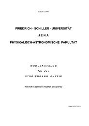
Hochaufl<br />
Hochauflösende<br />
Hochaufl<br />
Hochaufl<br />
Hochaufl sende sende<br />
sende<br />
sende<br />
Fluorezenzmikroskopie basierend basierend<br />
basierend<br />
basierend auf<br />
auf<br />
auf<br />
auf<br />
computergest<br />
computergestützter<br />
computergest<br />
computergest<br />
computergest tzter tzter<br />
tzter<br />
Datenauswertung<br />
Datenauswertung<br />
Datenauswertung<br />
Datenauswertung<br />
Datenauswertung<br />
<strong>Rainer</strong> <strong>Heintzmann</strong>,<br />
<strong>Heintzmann</strong><br />
<strong>Rainer</strong>.<strong>Heintzmann</strong>@kcl.ac.uk<br />
Randall Division, King‘s King College London<br />
RANDALL<br />
division of cell and<br />
molecular biophysics<br />
Jena, November 2007
Overview: Direct Imaging<br />
�� 4Pi Microscopy, Scanning Wavefield + I 5M �� STED Microscopy
3<br />
Bigger NA:<br />
4Pi Microscopy<br />
<strong>Rainer</strong> <strong>Heintzmann</strong>, 2007
4<br />
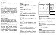
Aperture increase: 4 Pi Microscope (Type C)<br />
Dichromatic<br />
Beamsplitter<br />
Sample between<br />
Coverslips<br />
Detector<br />
Pinhole<br />
Fluorescence Emisssion<br />
2 Photon Effect<br />
z<br />
Laser<br />
Stefan W. Hell,<br />
Max Planck Inst. bpc,<br />
Goettingen, Germany<br />
High<br />
Sidelobes<br />
Fluorescence<br />
Intensity<br />
z<br />
<strong>Rainer</strong> <strong>Heintzmann</strong>, 2007
5<br />
APD1<br />
Leica 4Pi<br />
APD2<br />
http://www.leica-microsystems.com<br />
<strong>Rainer</strong> <strong>Heintzmann</strong>, 2007
6<br />
Leica 4Pi<br />
http://www.leica-microsystems.com<br />
<strong>Rainer</strong> <strong>Heintzmann</strong>, 2007
7<br />
4Pi images<br />
Deviding Escherichia Coli<br />
From: Bahlmann, K., S. Jakobs<br />
and S. W. Hell<br />
(2001). Ultramicr. 87: 155-164.<br />
<strong>Rainer</strong> <strong>Heintzmann</strong>, 2007
8<br />
4Pi images<br />
Confocal (2-Photon ) 4Pi (2-Photon)<br />
Thanks to: Elisabeth Ehler, Reiner Rygiel, Martin Fiala, Tanjef Szellas<br />
<strong>Rainer</strong> <strong>Heintzmann</strong>, 2007
9<br />
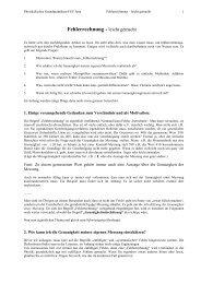
Aperture increase (3): ( ):<br />
I5 Microscope<br />
icroscope<br />
incoherent<br />
ncoherent illumination<br />
llumination interference<br />
nterference<br />
imag mage interference<br />
nterference<br />
Sample<br />
CCD<br />
Camera<br />
z<br />
Dichroic Mirror<br />
White Light<br />
Source<br />
Offset!!<br />
Fluorescence Emisssion<br />
• No XY scanning required<br />
(full field illum., full field det.)<br />
• No laser required<br />
• Higher background<br />
no pinhole ⇒ more signal<br />
Fluorescence<br />
Intensity<br />
z<br />
<strong>Rainer</strong> <strong>Heintzmann</strong>, 2007
10<br />
I5M M images and OTF<br />
Mats Gustafsson,<br />
UCSF, San Francisco<br />
http://www.msg.ucsf.edu/sedat/mats/<br />
k x,y<br />
k z<br />
<strong>Rainer</strong> <strong>Heintzmann</strong>, 2007
11<br />
Stimulated<br />
Stimulated<br />
Emission mission<br />
Depletion epletion<br />
<strong>Rainer</strong> <strong>Heintzmann</strong>, 2007
12<br />
STED Microscopy<br />
Klar, T. A., S. Jakobs, M. Dyba, A. Egner and S. W. Hell<br />
(2000). Proc.Nat. Acad. Sc. U.S.A. 97(15): 8206-8210.<br />
P s1<br />
~P s0 σI ex<br />
P s0<br />
Singlet<br />
ps<br />
ps<br />
Stimulated<br />
Emission<br />
Depletion<br />
<strong>Rainer</strong> <strong>Heintzmann</strong>, 2007
Saturation of the stimulated emission<br />
… provides the nonlinearity<br />
Klar, T. A., S. Jakobs, M. Dyba, A. Egner and S. W. Hell<br />
(2000). Proc.Nat. Acad. Sc. U.S.A. 97(15): 8206-8210.<br />
13<br />
<strong>Rainer</strong> <strong>Heintzmann</strong>, 2007
STED, the details<br />
14<br />
Intensity<br />
First<br />
(excitation)<br />
Dispersion Element<br />
Or Pulsed Laser Diode!<br />
STED, the details<br />
15<br />
Intensity<br />
Excitation<br />
Emission<br />
wavelength<br />
554nm, 250fs<br />
Detection Range<br />
STED<br />
745-760nm, 13ps<br />
Low efficiency<br />
⇒ Very strong light<br />
<strong>Rainer</strong> <strong>Heintzmann</strong>, 2007
STED, selecting a dye<br />
16<br />
S 2<br />
Singlet<br />
S 1<br />
~P s0σI ex<br />
S 0<br />
ps<br />
ps<br />
~µs<br />
∼P s1 τ –1 ~ I ex<br />
Triplet<br />
T 1<br />
T 0<br />
<strong>Rainer</strong> <strong>Heintzmann</strong>, 2007
17<br />
STED Microscopy<br />
STED beam Resulting PSF<br />
1 µm<br />
NA 1.3, 10nm pixelsize, no background<br />
<strong>Rainer</strong> <strong>Heintzmann</strong>, 2007
18<br />
STED Microscopy<br />
STED beam Resulting PSF<br />
1 µm<br />
NA 1.3, 10nm pixelsize, 10% background,<br />
STED PSF gets quite dim<br />
<strong>Rainer</strong> <strong>Heintzmann</strong>, 2007
19<br />
4Pi-STED 4Pi STED images<br />
Fluorescent plane:<br />
<strong>Rainer</strong> <strong>Heintzmann</strong>, 2007
20<br />
STED Images Confocal STED<br />
Klar, T. A., S. Jakobs, M. Dyba, A. Egner and S. W. Hell<br />
(2000). Proc.Nat. Acad. Sc. U.S.A. 97(15): 8206-8210.<br />
<strong>Rainer</strong> <strong>Heintzmann</strong>, 2007
21<br />
STED Images<br />
Confocal STED<br />
human embryonic kidney<br />
labeled with a red-emitting<br />
dye (MR 121SE)<br />
Microtubules<br />
Immunofluorescence<br />
Current Opinion in Biotechnology 2005, 16:3–12<br />
From micro to nano: recent advances in high-resolution microscopy; Yuval Garini, Bart J Vermolen and Ian T Young<br />
<strong>Rainer</strong> <strong>Heintzmann</strong>, 2007
22<br />
STED Images<br />
Willig, K. I., J. Keller, M. Bossi, S. W. Hell (2006):<br />
STED microscopy resolves nanoparticle assemblies.<br />
New J. Phys. 8: 106.<br />
<strong>Rainer</strong> <strong>Heintzmann</strong>, 2007
23<br />
Computational spectacles<br />
Methods requiring computation<br />
<strong>Rainer</strong> <strong>Heintzmann</strong>, 2007
Paradigm: Optimize for direct visibility<br />
+ →<br />
Optics<br />
Object Image<br />
E.g.: Widefield, Confocal, STED<br />
Does not necessarily optimize information content!<br />
24<br />
<strong>Rainer</strong> <strong>Heintzmann</strong>, 2007
Paradigm: Optimize for information content<br />
Object<br />
25<br />
+ →<br />
Optics<br />
Data<br />
Data Computation<br />
Image<br />
+ →<br />
<strong>Rainer</strong> <strong>Heintzmann</strong>, 2007
26<br />
Examples in Medical Imaging<br />
MRI<br />
http://www.cis.rit.edu/htbooks/mri/images/head.gif<br />
PET<br />
http://www.cerebromente.org.br/n01/pet/petdep.gif<br />
CT<br />
http://www.vetmed.lsu.edu/vth&c/Orthopedics/Images/<br />
Computed%20Tomography%20(CT)%20Scanner.RV.jpg<br />
SPECT<br />
http://www.physics.ubc.ca/research/images/spect.gif<br />
fMRI<br />
http://www.fmri.wfubmc.edu/other%20pics/<br />
lab_brain_logo.JPG<br />
<strong>Rainer</strong> <strong>Heintzmann</strong>, 2007
27<br />
Overview<br />
�� Pointilism: Localisation not resolution<br />
�� Axial Tomography<br />
�� Structured Illumination Microscopy<br />
�� Circumventing the limit<br />
… using Non-linear Non linear Effects<br />
<strong>Rainer</strong> <strong>Heintzmann</strong>, 2007
28<br />
Overview<br />
�� Pointilism: Localisation not resolution<br />
�� Axial Tomography<br />
�� Structured Illumination Microscopy<br />
�� Circumventing the limit<br />
… using Non-linear Non linear Effects<br />
<strong>Rainer</strong> <strong>Heintzmann</strong>
29<br />
Localization not resolution<br />
Localization not resolution<br />
If positions are know you can<br />
paint a picture!<br />
If particles can be separated,<br />
their relative positions can be<br />
measured accurately<br />
<strong>Rainer</strong> <strong>Heintzmann</strong>, Seurat: 2007 Tiger
30<br />
How to separate particles?<br />
Spectral precision distance microscopy<br />
Problems: Chromatic Aberrations, few dyes<br />
P. Edelmann, A. Esa, H. Bornfleth, R..<strong>Heintzmann</strong>, M. Hausmann, and C. Cremer. Proc. of SPIE , 3568:89-95, 1999<br />
Using fluorescence lifetime for separation (FLIM)<br />
Problems: Lifetime depends on microenvironm.<br />
M. Heilemann, D.P. Herten, R.<strong>Heintzmann</strong>, C. Cremer, C. Müller, P. Tinnefeld, K.D. Weston,<br />
J. Wolfrum and M. Sauer. Anal. Chem., 74, 3511-3517, 2002.<br />
Can we use the blinking characteristics?<br />
<strong>Rainer</strong> <strong>Heintzmann</strong>, 2007
31<br />
Can we separate Quantum Dots by their individual blinking?<br />
�� Localization >10 times<br />
better than resolution<br />
�� Quantum dots blink<br />
slow enough for imaging<br />
�� QDs blink independent<br />
�� Can we separate,<br />
even with strong overlap? 655 nm quantum dots<br />
on a cover slip<br />
K.A. Lidke, B. Rieger, T.M. Jovin, R. <strong>Heintzmann</strong> Optics Express 13, 7052-7062, 2005.<br />
<strong>Rainer</strong> <strong>Heintzmann</strong>, 2007
Large Separation<br />
10 frames of the times series.<br />
Sum of time series<br />
ICA analysis finds 2 blinking sources<br />
Separation 535 nm<br />
ICA Results<br />
32 K.A. Lidke, B. Rieger, T.M. Jovin, R. <strong>Heintzmann</strong> Optics Express 13, 7052-7062, <strong>Rainer</strong> 2005.<br />
<strong>Heintzmann</strong>, 2007
33<br />
Close Separation<br />
10 frames of the times series.<br />
Sum of time series ICA Results<br />
ICA analysis identifies 2 closely spaced blinking sources<br />
Can resolve 2 spots at 23 nm. (current max: 5 spots)<br />
<strong>Rainer</strong> <strong>Heintzmann</strong>, 2007
34<br />
Single Dot<br />
10 frames of the times series.<br />
Sum of time series ICA Results<br />
ICA analysis finds only 1 source<br />
<strong>Rainer</strong> <strong>Heintzmann</strong>, 2007
Outlook<br />
•Process entire images (many particles)<br />
Resolution by in Microscopy<br />
•Use of specific QD-blinking statistical distribution<br />
•Use of spatial information<br />
•Use temporal information<br />
K.A. Lidke, B. Rieger, T.M. Jovin, R. <strong>Heintzmann</strong> Optics Express 13, 7052-7062, 2005.<br />
35<br />
<strong>Rainer</strong> <strong>Heintzmann</strong>, 2007
36<br />
Photo Activated Localization Microscopy<br />
See also<br />
STORM, FPALM<br />
E. Betzig et al.,<br />
Imaging Intracellular Fluorescent<br />
Proteins at Nanometer Resolution<br />
Science 313,<br />
1642 - 1645<br />
<strong>Rainer</strong> <strong>Heintzmann</strong>, 2007
37<br />
Photo Activated Localization Microscopy<br />
WF PALM<br />
EM<br />
E. Betzig et al., Science 313, 1642 – 1645 (2006)<br />
Mitochondria<br />
COS-7 cells<br />
cryo sections Cytochrom C oxidase import sequence - dEosFP<br />
<strong>Rainer</strong> <strong>Heintzmann</strong>, 2007
38<br />
Overview<br />
�� Pointilism: Localisation not resolution<br />
�� Axial Tomography<br />
�� Structured Illumination Microscopy<br />
�� Circumventing the limit<br />
… using Non-linear Non linear Effects<br />
<strong>Rainer</strong> <strong>Heintzmann</strong>
39<br />
Problem: Limited Numerical<br />
umerical Aperture perture<br />
z<br />
x,y<br />
Cell<br />
Objective Lense<br />
Immersion Medium<br />
Cover Slip<br />
Slide<br />
α<br />
Immersion<br />
Medium<br />
NA = n sin α<br />
<strong>Rainer</strong> <strong>Heintzmann</strong>
40<br />
Solution: Axial Tomography<br />
z<br />
Cell<br />
x,y<br />
Objective Lense<br />
Immersion Medium<br />
Cover Slip<br />
Glass Fiber<br />
Immersion<br />
Medium<br />
Aperture increase<br />
by rotation of<br />
the specimen<br />
Shaw et al.,<br />
Cogswell et al.,<br />
Kawata et al.,<br />
<strong>Heintzmann</strong> et al.<br />
<strong>Rainer</strong> <strong>Heintzmann</strong>
41<br />
OTF in Axial Tomography<br />
Aperture increase<br />
by rotation of<br />
the specimen<br />
<strong>Rainer</strong> <strong>Heintzmann</strong>
42<br />
Axial Tomography Experiments<br />
Confocal image (1 view) of quartz beads<br />
∅ 416 nm<br />
RITC<br />
Quartz<br />
∅ 200 nm<br />
R. <strong>Heintzmann</strong> and C. Cremer., J. Microsc., 206 (1), 7-23, 2002<br />
R. <strong>Heintzmann</strong>, G. Kreth, and C. Cremer, Analytical Cellular Pathology, 20(1):7-15, 2000<br />
<strong>Rainer</strong> <strong>Heintzmann</strong>
43<br />
Reconstructed Image<br />
Cluster of quartz beads<br />
Positions of the quartz beads<br />
∅ 416 nm<br />
RITC<br />
Quartz<br />
∅ 200 nm<br />
One bead as PSF,<br />
Segmentation of Result<br />
R. <strong>Heintzmann</strong> and C. Cremer., J. Microsc., 206 (1), 7-23, 2002<br />
R. <strong>Heintzmann</strong>, G. Kreth, and C. Cremer, Analytical Cellular Pathology, 20(1):7-15, 2000<br />
<strong>Rainer</strong> <strong>Heintzmann</strong>
44<br />
Combined ML-Deconvolution<br />
ML Deconvolution<br />
View 1 View 2<br />
Measured<br />
Register<br />
View 1 View 2<br />
M i<br />
Reconstructed Estimate<br />
Apply<br />
,<br />
Back<br />
Convolution<br />
Convolution<br />
Compare<br />
Simulated<br />
,<br />
C NC<br />
E i<br />
<strong>Rainer</strong> <strong>Heintzmann</strong>
45<br />
Biological Specimen<br />
Polytrichum Commune<br />
R. <strong>Heintzmann</strong> and C. Cremer., J. Microsc., 206 (1), 7-23, 2002<br />
Moss Spore<br />
Fully automatically<br />
registered<br />
<strong>Rainer</strong> <strong>Heintzmann</strong>
46<br />
Biological Specimen<br />
Polytrichum Commune<br />
Moss Spore<br />
Fully automatically<br />
registered<br />
<strong>Rainer</strong> <strong>Heintzmann</strong>
Selective elective Plane lane Illumination<br />
llumination Microscopy<br />
icroscopy<br />
47<br />
Plane of focus<br />
Illumination<br />
Unnessesary<br />
Bleaching<br />
<strong>Rainer</strong> <strong>Heintzmann</strong>
Selective elective Plane lane Illumination<br />
llumination Microscopy<br />
icroscopy<br />
48<br />
Illumination<br />
Cylinder Lens<br />
Light Sheet<br />
<strong>Rainer</strong> <strong>Heintzmann</strong>
Selective elective Plane lane Illumination<br />
llumination Microscopy<br />
icroscopy<br />
49<br />
Illumination<br />
Detection<br />
<strong>Rainer</strong> <strong>Heintzmann</strong>
50<br />
Results (SPIM)<br />
View 1 Deconv. Combined Deconv.<br />
c. elegans embryo, i-lineage cells expressing RAP11-GFP (GTPase)<br />
as endocytotic marker<br />
Data: MPI Dresden, Sebastian Höpfner, Marino Zerial<br />
(collaboration with Zeiss)<br />
<strong>Rainer</strong> <strong>Heintzmann</strong>
Electrodes<br />
51<br />
Results (Cell Suspensions)<br />
Cell in Suspension<br />
View 1, Deconvolved:<br />
View 2, Deconvolved:<br />
Combined Deconvolution:<br />
Data: Olivier Renaud, Spencer Shorte (Inst. Pasteur),<br />
SW13/20, lamin-GFP (green), mito-tracker (red),<br />
spinning disc system (Andor)<br />
5 µm<br />
O. Renaud, R. <strong>Heintzmann</strong>, A. Sáez-Cirión, T. Schnelle, T. Muller and S. Shorte, Proc. SPIE,<br />
6441, 10.1117/12.699000, 2007<br />
<strong>Rainer</strong> <strong>Heintzmann</strong>
52<br />
Overview<br />
�� Pointilism: Localisation not resolution<br />
�� Axial Tomography<br />
�� Structured Illumination Microscopy<br />
�� Circumventing the limit<br />
… using Non-linear Non linear Effects<br />
<strong>Rainer</strong> <strong>Heintzmann</strong>, 2007
53<br />
Linear<br />
Structured Illumination<br />
<strong>Rainer</strong> <strong>Heintzmann</strong>, 2007
54<br />
Moiré Demo<br />
<strong>Rainer</strong> <strong>Heintzmann</strong>
55<br />
Patterned Excitation 2D Setup<br />
lightsource<br />
<strong>Rainer</strong> <strong>Heintzmann</strong>, 2007
56<br />
I em (x) = Obj(x) · I ex (x)<br />
Multiplication in real space<br />
↕<br />
Convolution in Fourier space<br />
~ ~ ~<br />
Iem (k) = Obj(k) ⊗ Iex (k)<br />
<strong>Rainer</strong> <strong>Heintzmann</strong>, 2007
57<br />
Convolution<br />
<strong>Rainer</strong> <strong>Heintzmann</strong>, 2007
58<br />
Convolution<br />
<strong>Rainer</strong> <strong>Heintzmann</strong>, 2007
59<br />
Convolution<br />
<strong>Rainer</strong> <strong>Heintzmann</strong>, 2007
60<br />
Influence of optical imaging<br />
…reconstructing the data?<br />
<strong>Rainer</strong> <strong>Heintzmann</strong>, 2007
61<br />
Separating the orders…<br />
<strong>Rainer</strong> <strong>Heintzmann</strong>, 2007
62<br />
Separating the orders…<br />
Shifting the orders…<br />
<strong>Rainer</strong> <strong>Heintzmann</strong>, 2007
63<br />
Separating the orders…<br />
Shifting the orders…<br />
Recombining the orders…<br />
<strong>Rainer</strong> <strong>Heintzmann</strong>, 2007
64<br />
Separating the orders…<br />
Shifting the orders…<br />
Recombining the orders… using the correct weights.<br />
<strong>Rainer</strong> <strong>Heintzmann</strong>, 2007
65<br />
Sample Widefield Image Reconstructed Image<br />
<strong>Rainer</strong> <strong>Heintzmann</strong>, 2007
66<br />
Experimental Setup<br />
<strong>Rainer</strong> <strong>Heintzmann</strong>, 2007
Structured Illumination experiments<br />
67<br />
<strong>Rainer</strong> <strong>Heintzmann</strong>, 2007
Structured Illumination experiments<br />
68<br />
<strong>Rainer</strong> <strong>Heintzmann</strong>, 2007
Structured Illumination experiments<br />
69<br />
Grating<br />
Resolution Enhanced ~2×<br />
<strong>Rainer</strong> <strong>Heintzmann</strong>, 2007
4 Directions: A Puzzle<br />
70<br />
<strong>Rainer</strong> <strong>Heintzmann</strong>, 2007
Structured Illumination experiments<br />
71<br />
2 µm<br />
<strong>Rainer</strong> <strong>Heintzmann</strong>, 2007
Structured Illumination experiments<br />
72<br />
Sample: Darren Williams, Guy Tear<br />
Drosophila<br />
Polyteen Squash<br />
Sitox Green<br />
<strong>Rainer</strong> <strong>Heintzmann</strong>, 2007
Structured Illumination experiments<br />
73<br />
Sample: Darren Williams, Guy Tear<br />
Drosophila<br />
Polytene Squash<br />
Sitox Green<br />
<strong>Rainer</strong> <strong>Heintzmann</strong>, 2007
74<br />
Application in Ophtalmology?<br />
Ophtalmology<br />
Structured illumination,<br />
a method for higher resolution and<br />
better optical sectioning in retinal<br />
imaging ?<br />
Local radiation density can be kept low.<br />
<strong>Rainer</strong> <strong>Heintzmann</strong>, 2007
75<br />
Overview<br />
�� Pointilism: Localisation not resolution<br />
�� Axial Tomography<br />
�� Structured Illumination Microscopy<br />
�� Circumventing the limit<br />
… using Non-linear Non linear Effects<br />
<strong>Rainer</strong> <strong>Heintzmann</strong>, 2007
Circumventing<br />
1<br />
0<br />
relative<br />
transfer<br />
|kx,y| x,y|<br />
[1/m]<br />
the Abbe Resolution Limit<br />
requires nonlinearity<br />
76<br />
<strong>Rainer</strong> <strong>Heintzmann</strong>, 2007
77<br />
Non-linear<br />
Non linear<br />
Structured Illumination<br />
<strong>Rainer</strong> <strong>Heintzmann</strong>, 2007
78<br />
Two Spaces<br />
Linear Excitation (low intensity)<br />
magnitude<br />
Border of<br />
detection OTF<br />
-ϕ0 ϕ ~<br />
0 Obj +1 (k)<br />
-K 0 0 K0 Non-Linear Excitation (high<br />
intensity)<br />
magnitude<br />
Border of<br />
detection OTF<br />
-3ϕ 0<br />
-3K 0<br />
-2ϕ 0<br />
-2K 0<br />
-ϕ0<br />
-K 0<br />
0<br />
ϕ 0<br />
K 0<br />
spatial<br />
frequency<br />
2ϕ0~ Obj +2 (k)<br />
spatial<br />
frequency<br />
R. <strong>Heintzmann</strong>, T.M. Jovin, and C. Cremer., J. Opt. Soc. Am. A,19 (8), 1599-1609, 2002<br />
<strong>Rainer</strong> <strong>Heintzmann</strong>, 2007
R. <strong>Heintzmann</strong>, T.M. Jovin, and C. Cremer.,<br />
J. Opt. Soc. Am. A,19 (8), 1599-1609, 2002<br />
79<br />
PSF comparison<br />
SPEM (7x7)<br />
70 nm<br />
λ em = 520 nm<br />
SPEM (9x9)<br />
PEM<br />
118 nm Widefield<br />
57 nm 215 nm<br />
<strong>Rainer</strong> <strong>Heintzmann</strong>, 2007
80<br />
1 µm<br />
2D Patterned SPEM Approach<br />
Conventional<br />
microscopy<br />
Mats Gustafsson, U.C. San Francisco:<br />
1 µm<br />
50 nm microspheres<br />
Linear<br />
structured<br />
illumination<br />
M.G.L. Gustafsson (2005), PNAS, 37, 13081-13086<br />
1 µm<br />
Saturated<br />
structured<br />
illumination<br />
(3 new harmonics,<br />
53 J/m 2 , triangle apodized)<br />
<strong>Rainer</strong> <strong>Heintzmann</strong>, 2007
81<br />
2D Patterned SPEM Approach<br />
Line profile through bead<br />
FWHM<br />
Mats Gustafsson, U.C. San Francisco:<br />
– including the bead diameter of ~51 nm.<br />
Distribution of FWHM<br />
50 60 70 (nm)<br />
Mean FWHM = 58.6 nm<br />
Estimate FWHM of PSF: 59 2 - (51/ 2 ) 2 = 46 nm<br />
M.G.L. Gustafsson (2005), PNAS, 37, 13081-13086<br />
<strong>Rainer</strong> <strong>Heintzmann</strong>, 2007
The Abbe limit has been<br />
circumvented,<br />
the new limit is a matter of<br />
signal and noise<br />
82<br />
<strong>Rainer</strong> <strong>Heintzmann</strong>, 2007
Saturation<br />
of<br />
Photoswitching<br />
as non-linear non linear effect<br />
83<br />
<strong>Rainer</strong> <strong>Heintzmann</strong>, 2007
84<br />
Photoswitchable Proteins<br />
DRONPA<br />
(photoswitchable protein)<br />
for resolution improvement<br />
- Transfectable, living cells<br />
- Low power lighsources<br />
Dron: to hide<br />
Pa: to appear<br />
See also:<br />
Anemonia Sulcata<br />
purple fluorescent protein<br />
asFP595,<br />
Diheteroarylethenes<br />
<strong>Rainer</strong> <strong>Heintzmann</strong>, 2007
85<br />
Intensity<br />
Photoswitchable Proteins<br />
Object: 2 Pairs of lines<br />
dark<br />
state<br />
Low intensity activation<br />
(~20% activated)<br />
position<br />
bright<br />
state<br />
depletion light<br />
(= read light)<br />
<strong>Rainer</strong> <strong>Heintzmann</strong>, 2007
86<br />
Intensity<br />
Moving the pattern<br />
Object: 2 Pairs of lines<br />
dark<br />
state<br />
position<br />
bright<br />
state<br />
Expected Resolution: < 50 nm<br />
depletion light<br />
(= read light)<br />
Related to<br />
SPEM + STED<br />
<strong>Rainer</strong> <strong>Heintzmann</strong>, 2007
87<br />
Conclusion:<br />
Computational spectacles<br />
can help to see the nature<br />
in greater detail<br />
<strong>Rainer</strong> <strong>Heintzmann</strong>, 2007
88<br />
Acknowledgement<br />
�� Liisa Hirvonen, Kai Wicker,<br />
Ondrej Mandula,<br />
Yannick Colpin,<br />
Mats Gustafsson,<br />
Christoph Cremer (PEM, Axial)<br />
Elisabeth Ehler, Darren Williams (PEM)<br />
Gustafsson, (PEM, SPEM)<br />
�� Thomas Jovin, Quentin Hanley,<br />
Keith Lidke, Bernd Rieger (PAM, Pointillism)<br />
<strong>Rainer</strong> <strong>Heintzmann</strong>, 2007