Images - Department of Radiology & Biomedical Imaging ...
Images - Department of Radiology & Biomedical Imaging ...
Images - Department of Radiology & Biomedical Imaging ...

You also want an ePaper? Increase the reach of your titles
YUMPU automatically turns print PDFs into web optimized ePapers that Google loves.
8<br />
clinical and research news<br />
MR-Guided Focused Ultrasound (MRg-FUS) Comes to UCSF<br />
Fergus V. Coakley, MD; Christian Diederich, PhD; Vanessa Jacoby, MD; Thomas M. Link, MD, PhD<br />
What is MR-Guided Focused Ultrasound?<br />
MR-guided focused ultrasound (MRg-FUS), also known as<br />
high-intensity focused ultrasound, refers to the use <strong>of</strong> tightly<br />
focused high-energy ultrasound waves to heat and ultimately<br />
kill tissue. Targeted and sustained energy deposition<br />
with focused ultrasound waves heat the tissue at the focal<br />
zone to a threshold temperature <strong>of</strong> 65 to 85°C, resulting<br />
in coagulative necrosis. The use <strong>of</strong> focused ultrasound for<br />
medical therapy is not new. Frontal lobotomy using focused<br />
ultrasound through burr holes in animals was first described<br />
in 1954. The FDA approved the use <strong>of</strong> extracorporeal shockwave<br />
lithotripsy (ESWL), also a form <strong>of</strong> focused ultrasound,<br />
in 1984. The use <strong>of</strong> focused ultrasound combined with MRI<br />
for guidance and monitoring reemerged in the 1990s due to<br />
advances in imaging, ultrasound technology, and focal therapy.<br />
MRI guidance provides three critical advantages during<br />
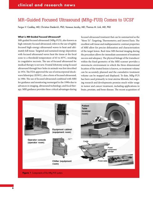
Figure 1 Components <strong>of</strong> the MRg-FUS system.<br />
focused ultrasound treatment that can be summarized as the<br />
“three Ts”: Targeting, Thermometry, and (stereo)Taxis. The<br />
excellent s<strong>of</strong>t tissue and multiparametric contrast properties<br />
<strong>of</strong> MRI allow for precise delineation and characterization<br />
<strong>of</strong> the target lesion. Real-time MR thermal imaging during<br />
the procedure allows for immediate assessment <strong>of</strong> treatment<br />
success and adequacy. The physical linkage <strong>of</strong> the transducer<br />
within the fixed geometry <strong>of</strong> the MRI scanner provides a<br />
stereotactic environment in which the three-dimensional<br />
location <strong>of</strong> the treated lesion is known, so treatment volume<br />
can be accurately planned and the cumulative treatment<br />
volume can be mapped and displayed. To date, MRg-FUS<br />
has been used primarily to treat uterine fibroids, but ongoing<br />
research and developments promise much wider usage<br />
in tumor and cancer treatment, including applications in<br />
brain, prostate, and bone disease. The recent acquisition <strong>of</strong>