maenas (intertidal zone) and Segonzacia mesatlantica - Station ...
maenas (intertidal zone) and Segonzacia mesatlantica - Station ...
maenas (intertidal zone) and Segonzacia mesatlantica - Station ...

You also want an ePaper? Increase the reach of your titles
YUMPU automatically turns print PDFs into web optimized ePapers that Google loves.
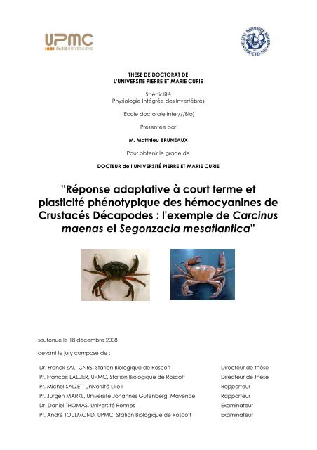
THESE DE DOCTORAT DE<br />
L’UNIVERSITE PIERRE ET MARIE CURIE<br />
Spécialité<br />
Physiologie Intégrée des Invertébrés<br />
(Ecole doctorale Inter///Bio)<br />
Présentée par<br />
M. Matthieu BRUNEAUX<br />
Pour obtenir le grade de<br />
DOCTEUR de l’UNIVERSITÉ PIERRE ET MARIE CURIE<br />
''Réponse adaptative à court terme et<br />
plasticité phénotypique des hémocyanines de<br />
Crustacés Décapodes : l'exemple de Carcinus<br />
<strong>maenas</strong> et <strong>Segonzacia</strong> <strong>mesatlantica</strong>''<br />
soutenue le 18 décembre 2008<br />
devant le jury composé de :<br />
Dr. Franck ZAL, CNRS, <strong>Station</strong> Biologique de Roscoff<br />
Pr. François LALLIER, UPMC, <strong>Station</strong> Biologique de Roscoff<br />
Pr. Michel SALZET, Université Lille I<br />
Pr. Jürgen MARKL, Université Johannes Gutenberg, Mayence<br />
Dr. Daniel THOMAS, Université Rennes I<br />
Pr. André TOULMOND, UPMC, <strong>Station</strong> Biologique de Roscoff<br />
Directeur de thèse<br />
Directeur de thèse<br />
Rapporteur<br />
Rapporteur<br />
Examinateur<br />
Examinateur
Photos de couverture : Carcinus <strong>maenas</strong> (David Busti, ENS Lyon) et <strong>Segonzacia</strong> <strong>mesatlantica</strong> (Patrick Bri<strong>and</strong>, Ifremer).
i<br />
Résumé / Abstract<br />
Réponse adaptative à court terme et plasticité phénotypique des hémocyanines de Crustacés<br />
Décapodes : l’exemple de Carcinus <strong>maenas</strong> et <strong>Segonzacia</strong> <strong>mesatlantica</strong><br />
Les hémocyanines (Hcs) des Crustacés Décapodes sont des pigments respiratoires formés de 6 ou 12 sousunités<br />
de 75 kDa. L’existence de différents types de sous-unités et d’effecteurs allostériques permet une gr<strong>and</strong>e<br />
plasticité structurale et fonctionnelle des Hcs face aux changements de conditions du milieu. L’objectif de cette<br />
thèse était de caractériser les adaptations respiratoires à court terme au niveau de l’Hc chez deux crabes vivant<br />
dans des milieux hypervariables, Carcinus <strong>maenas</strong> (en <strong>zone</strong> <strong>intertidal</strong>e) et <strong>Segonzacia</strong> <strong>mesatlantica</strong> (près des<br />
sources hydrothermales profondes).<br />
L’interaction de l’Hc de C. <strong>maenas</strong> avec certains effecteurs physiologiques (L-lactate, cations divalents) a<br />
été caractérisée par spectrométrie de masse supramoléculaire. Des sous-unités spécifiques interagissent avec le<br />
L-lactate et toutes les sous-unités ne jouent pas le même rôle dans l’assemblage du complexe d’Hc.<br />
Chez C. <strong>maenas</strong>, la plasticité phénotypique de l’Hc n’est pas impliquée dans les adaptations à un changement<br />
de salinité ou à l’hypoxie à court terme. En revanche, les sous-unités interagissant avec le L-lactate sont<br />
plus abondantes après une hypoxie longue (quelques jours).<br />
Chez S. <strong>mesatlantica</strong>, l’Hc est intrinsèquement très affine pour l’oxygène et présente un fort effet Bohr,<br />
mais le L-lactate et l’urate ne modulent que faiblement l’affinité de l’Hc. La plasticité phénotypique n’est pas<br />
impliquée dans la réponse à nos conditions d’acclimatation.<br />
Les résultats obtenus suggèrent que les adaptations respiratoires à court terme ne sont pas les mêmes dans<br />
les deux milieux hypervariables étudiés : l’affinité de l’Hc est modulée par des effecteurs hémolymphatiques<br />
chez C. <strong>maenas</strong> alors qu’elle est constitutivement très forte et peu modulée chez S. <strong>mesatlantica</strong>.<br />
Mots-clés : Carcinus <strong>maenas</strong>, hémocyanine, milieu hydrothermal, physiologie respiratoire, pigment respiratoire,<br />
plasticité phénotypique, <strong>Segonzacia</strong> <strong>mesatlantica</strong>, <strong>zone</strong> <strong>intertidal</strong>e<br />
Short-term adaptive response <strong>and</strong> phenotypic plasticity of decapod crustacean hemocyanins : the<br />
example of Carcinus <strong>maenas</strong> <strong>and</strong> <strong>Segonzacia</strong> <strong>mesatlantica</strong><br />
Decapod crustacean hemocyanins (Hcs) are respiratory pigments made of 6 or 12 subunits of 75 kDa<br />
each. Several subunit types <strong>and</strong> allosteric effectors exist, thus permitting a very high structural <strong>and</strong> functional<br />
plasticity in order to cope with changes in environmental conditions. Our aim was to characterize short-term<br />
respiratory adaptations at the Hc level in two crab species living in hypervariable environment : Carcinus<br />
<strong>maenas</strong> (<strong>intertidal</strong> <strong>zone</strong>) <strong>and</strong> <strong>Segonzacia</strong> <strong>mesatlantica</strong> (deep-sea hydrothermal vents).<br />
The interaction between C. <strong>maenas</strong> Hc <strong>and</strong> some of its physiological effectors (L-lactate <strong>and</strong> divalents<br />
cations) was studied by supramolecular ESI-MS. Specific subunits interact with L-lactate <strong>and</strong> subunits have<br />
different roles in the complex association.<br />
For C. <strong>maenas</strong>, phenotypic plasticity of Hc is not involved in the response to changes in salinity or to shortterm<br />
hypoxia. However, L-lactate sensitive subunits were more abundant after several days under hypoxia.<br />
For S. <strong>mesatlantica</strong>, Hc affinity for oxygen is very high with a strong Bohr effect but only a low modulation<br />
by L-lactate <strong>and</strong> urate. Phenotypic plasticity response was not observed under our acclimation conditions.<br />
These results suggest that short-term adaptations are different in the two studied hypervariable environments<br />
: Hc affinity is modulated by hemolymphatic effectors for C. <strong>maenas</strong> whereas it is constitutively very<br />
high <strong>and</strong> only slightly modulated for S. <strong>mesatlantica</strong>.<br />
Keywords : Carcinus <strong>maenas</strong>, deep-sea hydrothermal vents, hemocyanin, <strong>intertidal</strong> <strong>zone</strong>, <strong>Segonzacia</strong> <strong>mesatlantica</strong>,<br />
respiratory physiology, respiratory pigment, phenotypic plasticity
Sommaire<br />
Résumé<br />
Sommaire<br />
i<br />
ii<br />
1 Introduction 1<br />
1.1 Milieux et modèles . . . . . . . . . . . . . . . . . . . . . . . . . . . . . . . . . . . 4<br />
1.2 Physiologie respiratoire . . . . . . . . . . . . . . . . . . . . . . . . . . . . . . . . . 23<br />
1.3 Pigments respiratoires - présentation générale . . . . . . . . . . . . . . . . . . . . . 29<br />
1.4 L’hémocyanine des Crustacés . . . . . . . . . . . . . . . . . . . . . . . . . . . . . . 44<br />
1.5 Questions biologiques et objectifs de la thèse . . . . . . . . . . . . . . . . . . . . . 54<br />
2 La spectrométrie de masse et la diffusion de lumière appliquées à l’étude des pigments<br />
respiratoires : article de revue 55<br />
2.1 L’ESI-MS et les pigments respiratoires . . . . . . . . . . . . . . . . . . . . . . . . . 56<br />
2.2 Le MALLS et les pigments respiratoires . . . . . . . . . . . . . . . . . . . . . . . . 64<br />
2.3 Article de revue ESI-MS et MALLS . . . . . . . . . . . . . . . . . . . . . . . . . . 70<br />
2.4 Conclusion : intérêt de la spectrométrie de masse et de la diffusion de lumière pour<br />
l’étude de l’Hc . . . . . . . . . . . . . . . . . . . . . . . . . . . . . . . . . . . . . 102<br />
3 Interaction de l’hémocyanine de Carcinus <strong>maenas</strong> avec le L-lactate et les cations divalents<br />
: étude par spectrométrie de masse 103<br />
3.1 Problématique et objectifs de l’étude . . . . . . . . . . . . . . . . . . . . . . . . . . 104<br />
3.2 Manuscrit : Structural study of Carcinus <strong>maenas</strong> hemocyanin by supramolecular ESI-<br />
MS : interaction with L-lactate <strong>and</strong> divalent cations . . . . . . . . . . . . . . . . . . 108<br />
3.3 Bilan et perspectives . . . . . . . . . . . . . . . . . . . . . . . . . . . . . . . . . . 131<br />
4 Etude de la plasticité phénotypique de l’Hc de Carcinus <strong>maenas</strong> 133<br />
4.1 Introduction : la plasticité phénotypique des Hc de Crustacés . . . . . . . . . . . . . 134<br />
iii
iv<br />
SOMMAIRE<br />
4.2 Objectifs de l’étude . . . . . . . . . . . . . . . . . . . . . . . . . . . . . . . . . . . 137<br />
4.3 Matériels et méthodes communs . . . . . . . . . . . . . . . . . . . . . . . . . . . . 138<br />
4.4 Etude n°1 : Expérience préliminaire d’acclimatation à différentes salinités . . . . . . 140<br />
4.5 Etude n°2 : Acclimatation à différentes salinités et oxygénation en laboratoire . . . . 145<br />
4.6 Etude n°3 : Plasticité phénotypique d’une population naturelle dans un estuaire (rivière<br />
Penzé) . . . . . . . . . . . . . . . . . . . . . . . . . . . . . . . . . . . . . . . 159<br />
4.7 Bilan général et perspectives . . . . . . . . . . . . . . . . . . . . . . . . . . . . . . 168<br />
5 Adaptations respiratoires du crabe hydrothermal <strong>Segonzacia</strong> <strong>mesatlantica</strong> 171<br />
5.1 <strong>Segonzacia</strong> <strong>mesatlantica</strong> et le contexte hydrothermal . . . . . . . . . . . . . . . . . 172<br />
5.2 Matériels et méthodes utilisés pour l’étude d’une espèce hydrothermale . . . . . . . 176<br />
5.3 Problématique et objectifs de l’étude . . . . . . . . . . . . . . . . . . . . . . . . . . 179<br />
5.4 Manuscrit : Respiratory adaptations of the deep-sea hydrothermal vent crab <strong>Segonzacia</strong><br />
<strong>mesatlantica</strong> in response to hypoxia <strong>and</strong> temperature changes . . . . . . . . . 179<br />
5.5 Bilan et perspectives . . . . . . . . . . . . . . . . . . . . . . . . . . . . . . . . . . 213<br />
6 Conclusion et perspectives 215<br />
6.1 Conclusion générale . . . . . . . . . . . . . . . . . . . . . . . . . . . . . . . . . . . 216<br />
6.2 Perspectives . . . . . . . . . . . . . . . . . . . . . . . . . . . . . . . . . . . . . . . 221<br />
6.3 Vue intégrée des adaptations respiratoires . . . . . . . . . . . . . . . . . . . . . . . 222<br />
Annexes 225<br />
A Détermination des masses de deux complexes éluant simultanément par SEC-MALLS 227<br />
B Détermination des distributions de masse des hexamères d’Hc de Carcinus <strong>maenas</strong> 231<br />
Bibliographie 237<br />
Liste des figures 252<br />
Liste des tableaux 256<br />
Table des matières 258
Chapitre 1<br />
Introduction<br />
1
2 CHAPITRE 1. INTRODUCTION<br />
Les écosystèmes marins présentent une gr<strong>and</strong>e diversité de conditions physico-chimiques. Les<br />
océans et les plaines abyssales présentent généralement des conditions stables et bien tamponnées<br />
localement, alors que la <strong>zone</strong> côtière est un environnement plus variable à l’échelle de l’année, et<br />
la <strong>zone</strong> de balancement des marées un environnement hypervariable à l’échelle de la journée. Jusqu’à<br />
une période relativement récente, le milieu côtier peu profond à forte biomasse était opposé aux<br />
plaines abyssales peu densément peuplées mais présentant une plus forte diversité spécifique (Hessler<br />
et S<strong>and</strong>ers, 1967). Un gradient similaire était observé entre la faune arctique et la faune tropicale, et<br />
l’une des hypothèses explicatives avancées était l’existence d’un lien entre la stabilité d’un milieu et<br />
sa diversité spécifique : un milieu stable favoriserait la mise en place de relations et d’interactions<br />
entre les espèces pouvant déboucher sur de nouveaux événements de spéciation, alors qu’un milieu<br />
fluctuant favoriserait la flexibilité génétique de chaque espèce et limiterait la fréquence de ces événements<br />
(S<strong>and</strong>ers, 1968).<br />
La découverte des sites hydrothermaux profonds des Galapagos en 1977 et des efforts d’échantillonnage<br />
plus importants en <strong>zone</strong> côtière ont remis en cause ce paradigme. La faune profonde peut<br />
présenter localement des biomasses très élevées autour des sources hydrothermales ainsi qu’une forte<br />
endémicité des espèces présentes (Lonsdale, 1977) et des études présentant un effort d’échantillonnage<br />
suffisant ont montré que la diversité côtière était au moins aussi importante que celle des gr<strong>and</strong>s<br />
fonds (Gray, 2001).<br />
Les milieux hypervariables sont donc susceptibles d’héberger de nombreuses espèces (milieu côtier)<br />
et de fortes biomasses (milieu côtier et milieu hydrothermal profond) malgré leur caractère extrême.<br />
Les organismes vivant dans ces environnements présentent des adaptations spécifiques leur<br />
permettant de supporter ces conditions. Ce sont également d’excellents modèles pour comprendre<br />
les adaptations spécifiques à certains facteurs qui peuvent être exacerbées dans ces conditions. La<br />
compréhension du fonctionnement de ces écosystèmes ainsi que de l’histoire évolutive des groupes<br />
qui y sont présents nécessite de connaître les adaptations physiologiques particulières aux différents<br />
milieux hypervariables.<br />
Dans ce contexte, l’étude de la <strong>zone</strong> <strong>intertidal</strong>e et des sources hydrothermales profondes permet de<br />
comparer deux milieux hypervariables contrastés. L’étude de deux espèces d’un même groupe mais<br />
vivant dans chacun des milieux permet de mettre en évidence les adaptations partagées dans le groupe<br />
et, par une approche de physiologie comparée des organismes peuplant ces biotopes, d’identifier des<br />
mécanismes adaptatifs spécifiques. De ce point de vue, les Crustacés Décapodes représentent un taxon
3<br />
de choix car ils ont colonisé des biotopes très variés (océans, abysses, boues anoxiques, eau douce,<br />
milieu aérien) et de nombreuses données sont disponibles dans la littérature. Il est ainsi aisé de trouver<br />
des espèces de Décapodes Brachyoures afin de réaliser une approche comparative de l’adaptation<br />
physiologique aux milieux extrêmes à travers des études d’écophysiologie comparée.<br />
L’objectif de cette thèse est d’étudier ces adaptations respiratoires au niveau du pigment respiratoire<br />
lui-même, l’hémocyanine, sous l’aspect structural et fonctionnel. Une première partie des<br />
travaux aborde la question de l’interaction du pigment avec certaines molécules modulatrices pour<br />
son affinité, ainsi que la formation des complexes d’hémocyanines. La deuxième partie aborde la réponse<br />
physiologique aux variations environnementales à court terme, en particulier au niveau de la<br />
plasticité phénotypique du pigment. Quelles sont les modalités de l’interaction d’un pigment respiratoire<br />
de Crustacés (hémocyanine) avec ces effecteurs ? Quelles sont les réponses physiologiques et<br />
phénotypiques (au niveau de leur hémocyanine) de Crustacés vivant dans des milieux hypervariables<br />
contrastés ? Quels sont les points communs et les différences entre les adaptations à la <strong>zone</strong> <strong>intertidal</strong>e<br />
et au milieu hydrothermal profond ?<br />
Dans un premier temps, l’introduction présente les deux milieux, les modèles biologiques étudiés<br />
et les contraintes propres à chaque milieu. La physiologie respiratoire est ensuite présentée de manière<br />
générale, puis les pigments respiratoires dans leur globalité. Enfin, la structure des hémocyanines de<br />
Crustacés est présentée plus en détail, ainsi que leur fonction.
4 CHAPITRE 1. INTRODUCTION<br />
1.1 Milieux et modèles<br />
1.1.1 La <strong>zone</strong> <strong>intertidal</strong>e<br />
La <strong>zone</strong> <strong>intertidal</strong>e ou <strong>zone</strong> de balancement des marées correspond à la frange côtière périodiquement<br />
découverte à marée basse. La force à l’origine des marées est la résultante de la force centrifuge<br />
due au mouvement de rotation de la Terre et de la Lune l’une par rapport à l’autre et de la force<br />
d’attraction de la Lune sur la Terre. Ces deux forces se compensent exactement au niveau du centre<br />
de gravité de la Terre, mais leur résultante est non-nulle ailleurs. Toute la matière présente dans le<br />
système "Terre" est donc soumise à des forces de marées, y compris la croûte terrestre. Cependant,<br />
seul l’effet sur les masses d’eau, qui peuvent être mises en mouvement, est facilement perceptible<br />
sous la forme d’une onde de marée qui parcourt les océans. La période quotidienne des marées est<br />
due à la rotation de la Terre qui place un point du globe à la même position par rapport à la Lune<br />
en un peu plus de 24h (temps de rotation de la Terre sur elle-même plus le temps pour "rattraper" la<br />
Lune qui a tourné autour de la Terre pendant la journée). Il existe une autre période de l’ordre de 14<br />
jours correspondant au demi-cycle lunaire : l’attraction du Soleil produit également un effet de marée<br />
qui se conjugue avec celui de la Lune lorsque les 3 astres Terre-Lune-Soleil sont alignés (marées de<br />
vive-eau à la pleine lune et à la nouvelle lune) et s’y oppose au premier quartier et au dernier quartier<br />
(marées de morte-eau) (figure 1.1(a)).<br />
Selon la localisation géographique et la configuration des côtes et des bassins océaniques, le<br />
cycle d’émersion peut être diurne, semi-diurne ou mixte. A Roscoff (Finistère), les marées sont semidiurnes<br />
: l’émersion a lieu deux fois par jour. Des peuplements à forte densité existent dans la <strong>zone</strong><br />
de balancement des marées. Les organismes qui les composent sont exposés alternativement à des périodes<br />
d’émersion et d’immersion dont la durée dépend de leur position sur l’estran et peut atteindre<br />
plusieurs jours lors des marées de mortes-eaux pour les espèces colonisant le haut de l’estran (figure<br />
1.1(b)).<br />
Contraintes environnementales liées à la marée<br />
Les conditions environnementales rencontrées durant les périodes d’émersion et d’immersion sont<br />
très contrastées et imposent aux organismes vivant sur l’estran des adaptations particulières. Ces<br />
organismes peuvent être fixés ou mobiles et subir la marée basse à l’air libre, immergés dans des<br />
cuvettes, réfugiés sous les algues ou enfoncés dans le sédiment.
1.1. MILIEUX ET MODÈLES 5<br />
(a)<br />
(b)<br />
FIG. 1.1 – Cycle des marées et émersion des côtes. Pris dans Turquier et Loir (1981). (a) maréegramme<br />
présentant la période courte (environ 12h) due au mouvement de rotation de la Terre sur<br />
elle-même et la période longue (environ 14 jours) due à la rotation de la Lune autour de la Terre. (b)<br />
étagements des <strong>zone</strong>s d’émersion sur le littoral. Selon la hauteur de la <strong>zone</strong> par rapport à la hauteur<br />
moyenne de l’eau, l’immersion et l’émersion peuvent être quotidiennes ou espacées de plusieurs jours<br />
pour les <strong>zone</strong>s extrêmes de l’étagement.
6 CHAPITRE 1. INTRODUCTION<br />
Les principales contraintes liées à l’exposition à l’air libre sont la dessication et les changements<br />
de température. Les individus peuvent aussi subir un stress osmotique lorsqu’ils sont en présence<br />
d’eau de pluie. Les animaux s’isolant dans une coquille fermée pour limiter les pertes en eau (bivalves,<br />
patelles) peuvent subir une hypoxie se mettant en place au cours de la marée basse (Truchot,<br />
1987). Les cuvettes <strong>intertidal</strong>es constituent une protection face à la dessication mais leur isolement<br />
et leur volume limité amplifient l’effet des conditions climatiques (température de l’air, vent, ensoleillement,<br />
pluie) et biotiques (hypoxie, hyperoxie, hypercapnie ou alcalose liées à la photosynthèse<br />
et à la respiration) (Truchot et Duhamel-Jouve, 1980, Morris et Taylor, 1983). Le sédiment constitue<br />
un milieu plus stable que les deux précédents car les variations thermiques sont très amorties par une<br />
épaisseur même faible de sédiment et le milieu reste humide. Dans ce cas, la contrainte principale<br />
devient plutôt l’hypoxie due à l’absence de renouvellement d’eau pendant la marée basse.<br />
Les habitats de l’estran<br />
L’estran présente une gr<strong>and</strong>e variété d’habitats différents qui sont déterminés par la nature du<br />
substrat, par la hauteur par rapport au niveau moyen de l’eau, par le mode (battu ou abrité) et par<br />
les courants, et par des facteurs biologiques (e.g. couverture d’algues, massifs d’Hermelles). Chaque<br />
type d’habitat présente des peuplements différents.<br />
Le substrat peut être meuble ou rocheux. Alors qu’un substrat rocheux permet l’établissement<br />
d’algues ou d’animaux fixés et celui de brouteurs et de prédateurs associés, les substrats meubles sont<br />
plutôt peuplés d’organismes fouisseurs psammivores ou filtreurs et de leurs prédateurs. Les conditions<br />
hydrodynamiques du site déterminent si un substrat rocheux sera plutôt colonisé par des algues (mode<br />
abrité) ou par des animaux fixés (Balanes et Moules en mode battu). Les courants participent aussi<br />
à l’établissement de l’habitat en rendant possible ou non la sédimentation de petites particules, de<br />
graviers ou de galets (figure 1.2). La présence de végétaux (herbiers de Zostères) peut également<br />
modifier les conditions de courant localement et favoriser la sédimentation de petites particules.<br />
Dans les <strong>zone</strong>s d’estuaire, l’effet de marée peut se faire sentir jusqu’à plusieurs kilomètres à<br />
l’intérieur des terres. L’estuaire présente une eau dont la salinité décroît en remontant le cours d’eau.<br />
Des habitats typiques (pré salé ou schorre et estran vaseux ou slikke) existent dans ces <strong>zone</strong>s. Certaines<br />
espèces marines supportant bien la dessalure colonisent les <strong>zone</strong>s estuariennes. En particulier, des<br />
individus juvéniles d’espèces marines peuvent gr<strong>and</strong>ir dans ces <strong>zone</strong>s en étant à l’abri de prédateurs<br />
qui ne peuvent pas supporter l’eau hyposaline.<br />
Chaque type d’habitat présente un ensemble de conditions physiques et chimiques propres, le plus<br />
souvent variables selon différentes périodes (journée, saison, apériodique pour les précipitations et les<br />
tempêtes) et qui déterminent quelles sont les espèces qui peuvent potentiellement coloniser l’habitat.
1.1. MILIEUX ET MODÈLES 7<br />
FIG. 1.2 – Diagramme de Hjulström, pris dans Dercourt et Paquet (1978). Le diagramme présente les<br />
vitesses de courant pour lesquelles les particules sont entraînées (transport), sédimentent, ou peuvent<br />
être remises en suspension depuis le fond (érosion et transport). La présence d’un couvert herbacé<br />
ralentit le courant et permet la sédimentation de particules plus fines.<br />
L’exemple des cuvettes rocheuses émergées à marée basse et dans lesquelles d’importants peuplements<br />
animaux et végétaux peuvent s’établir illustre à quel point ces conditions peuvent présenter des<br />
gammes de variations importantes.<br />
Un exemple détaillé : les cuvettes rocheuses en milieu tempéré<br />
Le tableau 1.1 présente les valeurs extrêmes observées dans des cuvettes <strong>intertidal</strong>es à Roscoff et<br />
sur l’île de Cumbrae (Ecosse, embouchure de la Clyde) comparées à celles de l’eau libre qui recouvre<br />
les cuvettes à marée haute. Deux échelles de temps sont présentées : d’une part les amplitudes observées<br />
pendant des journées d’été et d’autre part l’amplitude des variations saisonnières (pour la cuvette<br />
écossaise) (tableau 1.2). Les amplitudes observées pour la température, la teneur en O 2 , en CO 2 et le<br />
pH sont beaucoup plus importantes dans l’eau des cuvettes que dans l’eau libre.<br />
A l’échelle de la journée (figures 1.3(a) et 1.3(b)), les paramètres évoluent de manière cyclique<br />
avec de brusques changements lors de la remontée du flot. Le sens et l’amplitude des variations<br />
sont différents selon la période d’émersion (journée/nuit, hiver/été) et le peuplement des cuvettes<br />
(présence d’algues, d’animaux). Il existe ainsi deux échelles temporelles de variations : l’échelle<br />
quotidienne avec deux émersions par jour et l’échelle annuelle avec l’alternance des conditions saisonnières.<br />
Quelle que soit l’échelle de temps considérée, l’amplitude des variations est toujours plus<br />
importante dans les cuvettes que dans l’eau libre : le faible volume des cuvettes exacerbe les variations<br />
des paramètres physiques et chimiques. A ces variations régulières et prévisibles (cycle jour-nuit
8 CHAPITRE 1. INTRODUCTION<br />
TAB. 1.1 – Paramètres physiques et chimiques de cuvettes <strong>intertidal</strong>es et de l’eau de mer libre ; pris<br />
dans Truchot et Duhamel-Jouve (1980), Morris et Taylor (1983). Données extrêmes pour Roscoff<br />
relevées au cours d’un mois d’été (juin) ; données extrêmes pour Cumbrae relevées au mois de juillet.<br />
Roscoff<br />
Firth of Clyde (Ecosse)<br />
cuvettes eau libre cuvettes<br />
Température (°C) 12 25,5 12,5 16 11,4 25,1<br />
Salinité () 34,1 36,7 34,5 35,2 - -<br />
P O2 (Torr) 1,6 577 134,9 194,7 40 435<br />
C O2 (mmol.L −1 ) 0,0027 0,836 0,225 0,31 - -<br />
pH 7,29 10,16 8,1 8,64 8,23 8,97<br />
Alcalinité totale (meq.L −1 ) 2,492 1,722 2,42 2,36 2,06 1,87<br />
P CO2 (Torr) 2,72 9,5.10 −5 3,6.10 −1 7,7.10 −2 0,25 1.10 −2<br />
C CO2 (mmol.L −1 ) 2,58 0,348 2,223 1,842 1,75 1,22<br />
TAB. 1.2 – Paramètres physiques et chimiques de cuvettes <strong>intertidal</strong>es et de l’eau de mer libre à<br />
Cumbrae ; données extrêmes à l’échelle de l’année prises dans Morris et Taylor (1983).<br />
Firth of Clyde (Ecosse)<br />
cuvettes<br />
eau libre<br />
Température (°C) 0,5 25 5 17<br />
Salinité () 23 36,5 28,5 35<br />
P O2 (Torr) 18 482 65 230<br />
C O2 (mmol.L −1 ) - - - -<br />
pH 6,4 9,62 7,6 9,1<br />
Alcalinité totale (meq.L −1 ) 2,65 1,69 2,53 1,87<br />
P CO2 (Torr) 1,42 4,8.10 −3 0,55 2,2.10 −2<br />
C CO2 (mmol.L −1 ) 2,63 0,97 2,32 1,63
(a) Evolution des paramètres de l’eau d’une cuvette rocheuse sur l’estran,<br />
pendant l’été à Roscoff - pris dans Truchot et Duhamel-Jouve<br />
(1980)<br />
(b) Evolution des paramètres de l’eau d’une cuvette (gauche) et de l’eau<br />
libre (droite) sur l’île de Cumbrae (Ecosse) pendant une journée d’été -<br />
pris dans Morris et Taylor (1983)<br />
FIG. 1.3 – Evolution des paramètres physiques et chimiques dans des cuvettes <strong>intertidal</strong>es<br />
1.1. MILIEUX ET MODÈLES 9
10 CHAPITRE 1. INTRODUCTION<br />
et cycle des marées) s’ajoutent des variations aléatoires mais importantes dues à l’imprévisibilité des<br />
conditions climatiques (e. g. tempête, journée très ensoleillée, hiver froid). Ces variations superposent<br />
un signal irrégulier aux variations périodiques attendues et participent à l’hypervariabilité de l’environnement<br />
<strong>intertidal</strong>.<br />
Ces variations influencent notamment la disponibilité de l’oxygène pour la fonction respiratoire<br />
des animaux. La pression partielle d’oxygène du milieu (P O2 ) dépend à la fois de la concentration<br />
en oxygène, de la température et de la salinité : une augmentation de la température ou de la salinité<br />
diminue la solubilité de l’oxygène. La teneur en CO 2 et le pH du milieu peuvent induire des modifications<br />
de l’équilibre acido-basique chez les organismes concernés, tout comme d’éventuelles réponses<br />
d’hypo- ou d’hyperventilation à l’oxygénation du milieu. Les organismes doivent maintenir un approvisionnement<br />
en oxygène adéquat à leurs tissus pendant la marée basse, ou réduire leur métabolisme<br />
voire passer en métabolisme anaérobie si l’approvisionnement ne peut être assuré.<br />
Influence des conditions de l’estran sur les peuplements<br />
Face à ces contraintes environnementales, les organismes présentent des réponses physiologiques<br />
et comportementales particulières à la dessication, à la température et à l’hypoxie. Certains utilisent<br />
l’oxygène atmosphérique en communiquant périodiquement avec l’air extérieur, de manière limitée<br />
pour éviter la dessication (Balanes, Bivalves, Gastéropodes) (Truchot, 1987). L’évaporation qui a<br />
lieu pendant ces périodes d’ouverture peut également permettre de diminuer la température et donc<br />
d’améliorer la résistance au stress thermique. Chez les crabes émergés comme Carcinus <strong>maenas</strong>,<br />
la chambre branchiale se remplit d’air mais des adaptations particulières (canal marginal renforcé)<br />
permettent de limiter l’accolement des lamelles branchiales et de préserver l’extraction d’oxygène.<br />
Les organismes aptes à respirer dans l’air maintiennent ainsi une consommation en oxygène élevée<br />
même à l’émersion. Pour d’autres espèces moins efficaces pour exploiter l’oxygène atmosphérique,<br />
une dette métabolique en oxygène (liée à l’accumulation des produits du métabolisme anaérobie) peut<br />
se constituer au fur et à mesure de l’émersion.<br />
Les cuvettes constituent un refuge face à la dessication, mais les conditions d’hypoxie voire<br />
d’anoxie qui peuvent s’y mettre en place lors de la marée basse peuvent conduire les organismes<br />
mobiles à modifier leur comportement (bullage dans la chambre branchiale de Carcinus <strong>maenas</strong>) ou<br />
à quitter la cuvette pour passer à un mode de respiration aérien.<br />
La rapidité des changements des paramètres de l’environnement (de l’ordre de quelques heures à<br />
quelques jours pour les stations extrêmes sur l’estran) et leur forte amplitude imposent aux organismes<br />
des réponses physiologiques rapides pour résister à ces contraintes. Ceci est également vrai lors de<br />
la remontée du flot et de l’envahissement d’une cuvette par la première vague d’eau libre (figure
1.1. MILIEUX ET MODÈLES 11<br />
FIG. 1.4 – Influence du mode sur l’étagement en milieu rocheux. Un mode battu tend à déplacer vers<br />
le haut les limites des ceintures de peuplement. Pris dans Turquier et Loir (1981).<br />
1.3(a)). A l’échelle de l’estran, la capacité de chaque espèce à résister à des périodes d’émersion<br />
plus ou moins longues conditionne leur répartition le long de ceintures parallèles, plus ou moins<br />
hautes sur la côte. Le littoral présente une zonation en étages successifs, caractéristiques du régime<br />
d’émersion et du mode battu ou abrité du site (figure 1.4). Localement, des conditions particulières<br />
(crevasses ombragées, couverture d’algues, cuvettes) permettent à des espèces de ceintures inférieures<br />
de remonter sur l’estran (Bournérias et al., 1995).<br />
A ces facteurs physiologiques viennent s’ajouter des facteurs biotiques comme la compétition<br />
entre espèces, qui modifie la répartition d’espèces dont les <strong>zone</strong>s de présence sont chevauchantes. La<br />
physiologie des organismes détermine donc leur aire de répartition potentielle sur l’estran, et leurs<br />
performances par rapport à celles des autres espèces leur répartition effective.<br />
1.1.2 Les sources hydrothermales profondes<br />
La découverte des sources et la remise en cause du paradigme des fonds océaniques<br />
Jusque dans les années 1970, les peuplements des habitats océaniques profonds passaient pour<br />
présenter de faibles densités et une forte diversité spécifique (S<strong>and</strong>ers, 1968, Hessler et Jumars, 1974,<br />
Hessler et al., 1988), dépendant entièrement pour leur apport en carbone organique de la matière organique<br />
produite en surface par les producteurs primaires planctoniques et sédimentant à travers la<br />
colonne d’eau (Kiørboe, 2001). La dégradation progressive de cette matière organique par les décomposeurs<br />
présents dans la colonne d’eau limite fortement la quantité de matière arrivant sur le fond.
FIG. 1.5 – Localisation des principaux sites hydrothermaux profonds actuellement connus -pris dans Desbruyères et al. (2006)<br />
12 CHAPITRE 1. INTRODUCTION
1.1. MILIEUX ET MODÈLES 13<br />
Le flux de matière arrivant aux organismes benthiques profonds représente donc une faible biomasse<br />
dépendant à la fois de la productivité primaire de surface et des producteurs secondaires la consommant<br />
avant son arrivée au fond, donc de la hauteur de la colonne d’eau (Smith et al., 2008). Les<br />
communautés d’organismes profonds peuvent proliférer ponctuellement lorsqu’un bloom en surface<br />
augmente transitoirement la quantité de matière sédimentant jusqu’à eux ou qu’un cadavre de gr<strong>and</strong><br />
animal coule au fond (requin, baleine) (Smith et Baco, 2003).<br />
Lors d’une exploration géologique de la ride des Galapagos en 1977, une communauté animale<br />
très dense fut découverte à proximité d’un site hydrothermal profond (Lonsdale, 1977, Corliss et al.,<br />
1979). Par la suite, de nombreux autres sites furent découverts au cours de l’exploration des dorsales<br />
océaniques et des bassins arrière-arc. Actuellement, l’existence de sites hydrothermaux est connue<br />
le long des dorsales Est-Pacifique et Ouest-Pacifique, Atlantique et en certains points des dorsales<br />
de l’Océan Indien et de l’Océan Arctique. Des sites actifs présentant des peuplements denses ont été<br />
observés le long des bassins arrière-arc comme dans le Pacifique ouest et sud-ouest (figure 1.5). Des<br />
<strong>zone</strong>s importantes des dorsales océaniques restent encore à explorer, notamment le sud de la dorsale<br />
Atlantique et la dorsale Antarctique et il est probable que de nouvelles sources seront découvertes dans<br />
ces <strong>zone</strong>s (Desbruyères et al., 2006). D’autres communautés profondes basées sur des écosystèmes<br />
chimiosynthétiques furent également découvertes dans des <strong>zone</strong>s de suintement froid, au niveau de<br />
marges actives présentant des prismes d’accrétion ou de marges passives comme l’embouchure des<br />
gr<strong>and</strong>s fleuves au niveau du Golfe du Mexique ou du Golfe de Guinée (Sibuet et Olu, 1998, Cordes<br />
et al., 2007).<br />
Contexte géologique et fonctionnement d’un système hydrothermal<br />
L’hydrothermalisme sous-marin profond est lié à l’existence de <strong>zone</strong>s géologiquement actives :<br />
limites de plaques tectoniques (dorsales océaniques et bassins d’arrière-arc) et points chauds (monts<br />
sous-marins dans l’alignement des archipels volcaniques intra-plaques). Les dorsales océaniques sont<br />
des <strong>zone</strong>s d’expansion océanique liées à l’écartement de deux plaques tectoniques, à la remontée de<br />
matériel chaud à l’aplomb de cette <strong>zone</strong> et à la formation de croûte océanique. Ces <strong>zone</strong>s du plancher<br />
océanique présentent un réseau de fissures dans lequel l’eau de mer peut pénétrer (figure 1.6). Cette<br />
eau s’infiltre plus ou moins profondément dans la croûte, dans des <strong>zone</strong>s plus ou moins proches du<br />
matériel chaud. Sous l’effet de la dilatation due à son échauffement, l’eau infiltrée remonte alors sous<br />
pression vers la surface pour donner naissance au fluide hydrothermal. Ce mécanisme de dissipation<br />
de la chaleur de la croûte avait été suggéré au début des années 1970, avant la découverte des sources<br />
hydrothermales, en raison d’un déficit du bilan du transfert de chaleur entre la croûte et l’eau de mer<br />
qui suggérait l’existence d’une telle circulation hydrothermale (Lister, 1972).
(a)<br />
FIG. 1.6 – Fonctionnement d’un site hydrothermal, pris sur le site de la Woods Hole Oceanographic Institution. (a) Formation du fluide hydrothermal.<br />
1 - l’eau de mer froide s’infiltre à travers les fissures de la croûte. 2 - O 2 et K + sont éliminés de l’eau de mer. 3 - Ca 2+ , SO 2−<br />
4<br />
et Mg 2+ sont<br />
éliminés du fluide. 4 - Na + , Ca 2+ et K + sont incorporés dans le fluide à partir de la croûte environnante. 5 - le fluide atteint sa température maximale<br />
et Cu, Zn, Fe et H 2 S de la croûte se dissolvent dans le fuilde. 6 - le fluide chaud contenant les métaux dissous remonte à travers la croûte. 7 - le fluide<br />
hydrothermal ses mélange à l’eau de mer froide et oxygénée, les métaux et les sulfures précipitent et forment des minéraux noirs. (b) Différents types<br />
d’émissions existant au niveau d’une source hydrothermale. 1 - l’eau de mer froide s’infiltre à travers les fissures de la croûte. 2 - l’eau de mer subit<br />
des transformations chimiques et est chauffée à haute température. L’importance et la nature des transformations subies dépend de la profondeur<br />
d’infiltration de l’eau de mer et de la nature de la roche. 3 - le fluide hydrothermal chaud et moins dense remonte vers la surface par un trajet canalisé<br />
(cheminée) ou en subissant un mélange avec l’eau de mer de surface à travers le réseau de fissures. Des suintements de fluide refroidi peuvent se<br />
former lorsque le mélange avec l’eau de mer sous le plancher océanique est important (diffuseurs). 4 - les précipités de sulfures métalliques noirs<br />
se forment à la sortie des cheminées ou sous le plancher océanique si un mélange avec l’eau de mer infiltrée a lieu, conduisant à la formation de fumeurs<br />
blancs dans ce dernier cas (précipités de silicates et d’anhydrite). Schémas pris sur http ://www.divediscover.whoi.edu/vents/chemistry.html<br />
et http ://www.divediscover.whoi.edu/vents/basics.html<br />
(b)<br />
14 CHAPITRE 1. INTRODUCTION
1.1. MILIEUX ET MODÈLES 15<br />
La composition de l’eau de mer est modifiée durant ce trajet (figure 1.6(a)). Au cours de l’infiltration,<br />
l’eau réagit avec les roches de la croûte, réductrices, et le K + , le Ca 2+ , l’O 2 sont consommés.<br />
Lors du rapprochement de la <strong>zone</strong> chaude, l’eau s’enrichit en d’autres composés (ions métalliques<br />
lourds, CO 2 , H 2 S) et devient acide. Sous l’effet de la pression due au chauffage, le fluide hydrothermal<br />
percole vers la surface. La géométrie des voies de sortie détermine la présence d’une émission<br />
localisée de fluide très chaud, sous pression, ou bien celle de diffuseurs où le fluide est déjà dilué par<br />
son passage dans les fissures sous-jacentes du plancher océanique (figure 1.6(b)). Le mélange entre<br />
le fluide hydrothermal chargé d’ions métalliques et l’eau de mer froide provoque une précipitation<br />
et la formation de cheminées hydrothermales pouvant atteindre plusieurs dizaines de mètres de haut.<br />
Les panaches correspondant à la sortie du fluide et à la précipitation des composés minéraux sont à<br />
l’origine de l’appellation usuelle de fumeurs pour désigner ces structures. Les fumeurs noirs correspondent<br />
à la formation de précipités de sulfures métalliques et les fumeurs blancs à celle de silicates<br />
et d’anhydrites. La composition du fluide de sortie peut être modifiée par la nature exacte de la croûte<br />
encaissante, par la dilution avant la sortie au niveau du plancher océanique et par la pénétration plus<br />
ou moins importante de l’eau de mer dans la croûte.<br />
Le tableau 1.3 présente les caractéristiques chimiques et physiques de différents fluides hydrothermaux<br />
(Atlantique, Pacifique) et celle de l’eau de mer du fond adjacente. Les fluides hydrothermaux<br />
sont caractérisés par leur haute température, un pH acide, la présence d’H 2 S, de CH 4 , de métaux<br />
dissous et l’absence d’oxygène. Suite aux échanges avec la croûte, la composition en sels majeurs<br />
de l’eau de mer est également modifiée (variations des teneurs en Na + , K + , et Ca 2+ , élimination<br />
du Mg 2+ ). La composition du fluide hydrothermal varie d’un site à l’autre ; ces variations sont plus<br />
importantes entre les sites Atlantique et Pacifique du tableau 1.3 qu’entre les deux sites Atlantique.<br />
La stabilité temporelle des <strong>zone</strong>s d’émission hydrothermales dépend du type de dorsales : elle est<br />
plus gr<strong>and</strong>e pour les dorsales lentes et plus faible pour les dorsales rapides. A l’échelle locale, les sites<br />
hydrothermaux se caractérisent globalement par une durée de vie de l’ordre de la dizaine (dorsale<br />
Sud-Est Pacifique) au millier d’années (dorsale Atlantique), certains sites pouvant être réactivés si<br />
l’activité géologique reprend.<br />
La <strong>zone</strong> de mélange fluide hydrothermale/eau de mer du fond<br />
Lors des premières explorations des sites hydrothermaux des Galapagos, les communautés vivantes<br />
qui ont été décrites étaient groupées autour des <strong>zone</strong>s d’émission du fluide (Lonsdale, 1977,<br />
Corliss et al., 1979). Au fur et à mesure de l’étude des environnements hydrothermaux et de la faune<br />
associée, il est apparu que les organismes vivants se répartissaient dans une <strong>zone</strong> de mélange entre le<br />
fluide hydrothermal et l’eau de mer du fond et que la source de carbone organique était une production
16 CHAPITRE 1. INTRODUCTION<br />
TAB. 1.3 – Composition du fluide hydrothermal de différents sites et de l’eau de mer du fond st<strong>and</strong>ard,<br />
pris dans Charlou et al. (2000), Desbruyères et al. (2000), Chausson (2001), Hourdez et Lallier (2007),<br />
Bris et al. (2003). EPR 13°N, site de la dorsale Est Pacifique situé à 13°N ; MAR Lucky Strike et<br />
Menez Gwen, sites de la dorsale Médio-Atlantique situés au niveau du point triple des Açores.<br />
EPR, 13°N MAR, Lucky Strike MAR, Menez Gwen eau de mer st<strong>and</strong>ard<br />
Température (°C) 317-380 172-324 275-284 2<br />
pH 3,2 3,65 4,2 7,8<br />
CO 2 (mM) - 24,2 21,7 2,3<br />
O 2 (µM) 0 0 0 250<br />
SO 4 (mM) 0 - - 27,9<br />
H 2 S (mM) 2,9-8,2 1,4-3,3 1,5-2 0<br />
H 2 (µM) - 3,3-727 24-48 4.10 −4<br />
CH 4 (mM) - 0,5-0,97 1,35-2,63 4.10 −7<br />
Alcalinité (-0,74)-(-0,4) - - 2,3<br />
Li (µM) 688 307 263 26<br />
Na (mM) 560 390 315 468<br />
Mg (mM) 0 0 0 52,9<br />
Si (mM) 22 14,4 10 0,1<br />
Cl (mM) 740 465 374 546<br />
K (mM) 29,6 23,4 22,9 10,2<br />
Ca (mM) 55 35,6 31,6 10,3<br />
Mn (µM) 0,8-1,2 249 65,6 0,002<br />
Fe (µM) 1,05-1,85 308 26,1 0,003<br />
Cu (µM) - 10,6 1,9 0,004<br />
Zn (µM) - 29 3,9 0,01<br />
Br (µM) - 840 686 838<br />
Rb (µM) 14,1 28,3 25,2 1,4<br />
Sr (µM) 175 88 105 87<br />
Cs (nM) - 196 285 2<br />
Ba (µM) - 37 27 0,14
1.1. MILIEUX ET MODÈLES 17<br />
<br />
<br />
<br />
<br />
<br />
<br />
<br />
<br />
<br />
<br />
<br />
<br />
<br />
<br />
<br />
<br />
<br />
<br />
FIG. 1.7 – Formation de la <strong>zone</strong> de mélange entre le fluide hydrothermal et l’eau de mer du fond sur<br />
le site Lucky Strike, d’après Hourdez et Lallier (2007) et les données du tableau 1.3.<br />
primaire locale par chimiosynthèse bactérienne.<br />
Le mélange entre le fluide pur et l’eau de mer du fond crée une <strong>zone</strong> à très forts gradients présentant<br />
une diminution de la température, de la teneur en H 2 S et en métaux lourds depuis la source<br />
vers l’extérieur, et une augmentation de la teneur en oxygène et du pH (figure 1.7). Ces gradients<br />
présentent globalement une géométrie concentrique autour du site d’émission et sont à l’origine de<br />
variations spatiales des conditions physiques et chimiques à petite échelle (figure 1.8).<br />
Une caractéristique importante de l’émission du fluide est sa variabilité temporelle : l’intensité du<br />
flux varie fortement et provoque un décalage spatial des gradients au cours du temps. La variabilité<br />
temporelle existe à petite échelle (figure 1.8) mais aussi sur des périodes plus longues (cycle de<br />
marées, cycle d’activité des fumeurs) (Chevaldonné et al., 1991). Il existe aussi une variabilité spatiale<br />
à gr<strong>and</strong>e échelle dans la mesure où les sources ont une durée de vie limitée : des sources s’éteignent<br />
et d’autres apparaissent le long de la dorsale au fur et à mesure de l’expansion océanique.<br />
La <strong>zone</strong> dans laquelle vivent les communautés hydrothermales est donc caractérisée par de très<br />
forts gradients des conditions physiques et chimiques et par une gr<strong>and</strong>e variabilité spatio-temporelle<br />
des émissions.<br />
Les producteurs primaires des écosystèmes hydrothermaux profonds<br />
Dès la première description des sites hydrothermaux, la concentration locale de nombreux organismes<br />
formant une biomasse importante et contrastant avec la faible biomasse abyssale a interpellé<br />
les scientifiques ; les premières hypothèses émises pour expliquer l’origine de cette forte biomasse<br />
étaient une concentration locale de matière organique détritique par les courants de convection dûs<br />
aux fumeurs (les premiers organismes observés étaient des filtreurs) ou une production primaire chimiosynthétique<br />
bactérienne (Lonsdale, 1977). Il s’est finalement avéré que la production primaire<br />
était effectivement assurée par des organismes chimiosynthétiques.
18 CHAPITRE 1. INTRODUCTION<br />
FIG. 1.8 – Fluctuations des conditions physiques et chimiques près d’un diffuseur hydrothermal, pris<br />
dans Desbruyères et al. (2001). La température et les concentrations en sulfures et en nitrites sont<br />
mesurées au niveau de différents organismes situés près du diffuseur (petites moules, gr<strong>and</strong>es moules,<br />
crevettes) ainsi que directement dans le fluide émis par le diffuseur.<br />
La synthèse de matière organique par des producteurs primaires nécessite une source d’énergie<br />
(couple oxydo-réducteur) et une source de carbone inorganique. A la surface, l’énergie nécessaire à<br />
la fixation du carbone inorganique provient en général de la photolyse de l’eau (photosynthèse). Au<br />
niveau des sources hydrothermales, elle provient de l’oxydation de composés réduits présents dans le<br />
fluide par des bactéries (chimiosynthèse). Les composés réduits peuvent être le sulfure de dihydrogène<br />
H 2 S, le méthane CH 4 , le dihydrogène H 2 , l’ammonium NH + 4<br />
ou le fer Fe(II). Les équations-bilans<br />
suivantes permettent de comparer les deux processus :<br />
Photosynthèse :<br />
Chimiosynthèse (exemple) :<br />
H 2 O+NADP + + ADP+P i<br />
hν<br />
−→ 1 2 O 2 + NADPH + H + + AT P<br />
HS − + 2O 2 + ADP+P i → SO 2−<br />
4<br />
+ H + + AT P<br />
Dans le cas de la photosynthèse, l’énergie provient du rayonnement lumineux ; dans le cas de<br />
la chimiosynthèse, elle provient de la différence de potentiel d’oxydo-réduction entre le donneur et<br />
l’accepteur d’électrons qui rend l’échange d’électrons possible.<br />
L’énergie produite permet la fixation de carbone inorganique dans des molécules organiques. La<br />
source de carbone inorganique est le CO 2 atmosphérique dans le cas de la photosynhtèse et le CO 2 ou<br />
le CH 4 contenus dans le fluide dans le cas de la chimiosynthèse hydrothermale (on utilise l’appellation<br />
"carbone inorganique" pour désigner le carbone du méthane par abus de langage, pour l’opposer au<br />
carbone incorporé dans des sucres).
1.1. MILIEUX ET MODÈLES 19<br />
FIG. 1.9 – Différentes voies de fixation du carbone chez les chimioautotrophes des sources hydrothermales<br />
profondes (pris dans Nakagawa et Takai (2008). (a) cycle de Calvin-Benson, (b) cycle rTCA,<br />
(c) cycle 3-HP et variante 3-hydroxypropionate/4-hydroybutyrate (3-HP/4-HB) en bleu, (d) voie réductrice<br />
de l’acétyl-CoA. Les enzymes clés de chaque voie sont indiquées en rouge.
20 CHAPITRE 1. INTRODUCTION<br />
Différentes voies biochimiques permettant cette fixation du carbone inorganique ont été mises en<br />
évidence dans les écosystèmes hydrothermaux (figure 1.9) : le cycle de Calvin-Benson (également<br />
utilisé par les organismes photosynthétiques) utilisé par les ε-protéobactéries et plus récemment le<br />
cycle inverse des acides tricarboxyliques (reductive TriCarboxylic Acid cycle, rTCA cycle) utilisé<br />
par les γ-protéobactéries, le cycle du 3-hydroxypropionate (3-HP) et la voie réductrice de l’acétyl<br />
coenzyme A (acetyl-CoA). Ces voies présentent des rendements énergétiques différents (3 molécules<br />
d’ATP par CO 2 fixé avec le cycle de Calvin-Benson, 1 molécule d’ATP par CO 2 fixé avec le cycle<br />
rTCA) et certaines de leurs enzymes présentent des sensibilités différentes à l’oxygène. Le cycle<br />
rTCA est ainsi utilisé par les bactéries présentes dans les <strong>zone</strong>s anoxique et hypoxique des gradients<br />
de mélange fluide/eau de mer du fond et le cycle de Calvin-Benson dans les <strong>zone</strong>s hypoxique et<br />
normoxique (Nakagawa et Takai, 2008).<br />
Les bactéries sont présentes libres dans l’eau, à la surface des édifices hydrothermaux, ou au<br />
sein de symbioses avec des animaux. Ces symbioses sont un des aspects originaux des écosystèmes<br />
hydrothermaux et sont présentes dans différents phylums (e.g. Annélide Siboglinidé Riftia pachyptila<br />
dont l’endosymbionte possèdent à la fois des enzymes caractéristiques des cycles de Calvin-Benson<br />
et rTCA, Annélide Térébellidé Alvinella pompejana, Mollusques Lamellibranches Bathymodiolus sp<br />
et Calyptogena magnifica, Crustacé Décapode Rimicaris exoculata). Les consommateurs secondaires<br />
peuvent être des brouteurs de bactéries ou des prédateurs (e.g. Gastéropode Alviniconcha hesslerii,<br />
Crustacé Décapode <strong>Segonzacia</strong> <strong>mesatlantica</strong>).<br />
Les communautés hydrothermales, relation avec le milieu, contraintes du milieu pour les organismes<br />
Les gradients physiques et chimiques du milieu définissent des ceintures concentriques de répartition<br />
des espèces autour du site d’émission, en fonction de leur résistance à la température, à l’hypoxie<br />
et aux composés toxiques, de leur besoins métaboliques (approvisionnement des symbiontes) et de la<br />
compétition inter-spécifique. On observe ainsi comme sur l’estran une zonation avec des assemblages<br />
faunistiques caractéristiques depuis le pôle chaud jusqu’au pôle froid. Des espèces opportunistes bathyales<br />
mais non endémiques des sources hydrothermales sont observées à proximité des sites et<br />
viennent s’alimenter sur la biomasse locale (e.g. le crabe bathyal Chaceon affinis).<br />
Les communautés de la dorsale Médio-Atlantique sont dominées par les crevettes Rimicaris exoculata<br />
et les moules Bathymodiolus sp.. Au sein d’un même site, ces espèces se répartissent selon un<br />
gradient depuis la <strong>zone</strong> d’émission et le haut des cheminées, où Rimicaris exoculata est l’espèce la<br />
plus abondante (T° max 25°C), jusqu’aux <strong>zone</strong>s plus diluées colonisées par les moulières de Bathymodiolus<br />
sp. (T° 6-10°C). Au gradient de température décroissant se superpose un gradient décroissant
1.1. MILIEUX ET MODÈLES 21<br />
(a)<br />
(b)<br />
FIG. 1.10 – Spécimens de Carcinus <strong>maenas</strong>. (a) vue dorsale d’un crabe capturé à Roscoff (photo<br />
David Busti, ENS Lyon). (b) Juvénile de Carcinus <strong>maenas</strong> sur un sédiment grossier, à marée basse<br />
(île Callot)<br />
de sulfures, de CO 2 et de CH 4 et un gradient croissant de pH et d’oxygénation. Les crevettes Mirocaris<br />
fortunata, Chorocaris chacei et le crabe <strong>Segonzacia</strong> <strong>mesatlantica</strong> sont plus opportunistes et<br />
se déplacent sur l’ensemble du gradient. Il existe également une différence de répartition selon la<br />
profondeur des sites, les moules étant les plus abondantes sur les sites les moins profonds (Menez<br />
Gwen, Lucky Strike) et les crevettes R. exoculata sur les sites les plus profonds (Snake Pit, TAG)<br />
(Desbruyères et al., 2001).<br />
1.1.3 Modèles biologiques : les Crustacés Décapodes Brachyoures<br />
Modèle littoral : le crabe vert Carcinus <strong>maenas</strong><br />
Carcinus <strong>maenas</strong> (crabe vert ou crabe enragé) est un Crustacé Décapode Brachyoure de la famille<br />
des Portunidés (figure 1.10). Il s’agit d’une espèce commune originaire des côtes européennes de<br />
l’océan Atlantique. Carcinus <strong>maenas</strong> vit dans la <strong>zone</strong> <strong>intertidal</strong>e et dans la <strong>zone</strong> circalittorale. Il est<br />
euryhalin, hyperosmorégulateur et supporte très bien l’eau dessalée des estuaires (Péqueux, 1995).<br />
Carcinus aestuarii (ou Carcinus mediterraneus) est une espèce proche vivant en Méditerranée. Des<br />
études génétiques ont montré qu’il s’agissait bien d’une espèce différente (Geller et al., 1997) ; les<br />
individus des deux espèces peuvent être distingués par leur morphologie (Yamada et Hauck, 2001).<br />
La taille adulte de Carcinus <strong>maenas</strong> est d’environ 70-90 mm (largeur du céphalothorax). On peut<br />
le trouver à marée basse dans les mares et sous les algues entourant les rochers. Il vit aussi bien sur<br />
substrat rocheux que sableux ou vaseux. Sa résistance lui a permis de coloniser de nombreux sites en<br />
dehors de son aire de répartition naturelle et il est à présent considéré comme une espèce invasive sur<br />
les côte nord-est et nord-ouest des Etats-Unis, en Californie et en Australie (Carlton et Cohen, 2003)
22 CHAPITRE 1. INTRODUCTION<br />
FIG. 1.11 – Répartition de Carcinus <strong>maenas</strong> pris dans Carlton et Cohen (2003) ; en noir et points<br />
noirs, distribution actuelle et observations ponctuelles, en gris, aire de répartition potentielle.<br />
(figure 1.11).<br />
Carcinus <strong>maenas</strong> est un modèle classique de physiologie pour les Crustacés Décapodes et une<br />
littérature abondante est disponible.<br />
Modèle hydrothermal : le crabe <strong>Segonzacia</strong> <strong>mesatlantica</strong><br />
<strong>Segonzacia</strong> <strong>mesatlantica</strong> (Williams 1988) est un Crustacé Décapode Brachyoure de la famille<br />
des Bythograeidés. Tous les crabes endémiques des sources hydrothermales profondes actuellement<br />
connus appartiennent à cette famille (Desbruyères et al., 2006). <strong>Segonzacia</strong> <strong>mesatlantica</strong> a été observé<br />
uniquement au niveau de la dorsale Médio-Atlantique, et représente actuellement la seule espèce de<br />
crabe hydrothermal connue sur cette dorsale.<br />
<strong>Segonzacia</strong> <strong>mesatlantica</strong> est opportuniste et vit dans la ceinture tiède où il se nourrit essentiellement<br />
de crevettes (Colaço et al., 2002). Contrairement aux crevettes Rimicaris exoculata, il ne<br />
développe pas de symbiose avec des bactéries. Il peut atteindre une taille adulte de 40 mm (largeur<br />
du céphalothorax). Les individus collectés ont une carapace de couleur blanche qui peut prendre une<br />
apparence orangée ou marron lorsqu’elle est couverte d’oxydes métalliques (figure 1.12). Certains<br />
crabes peuvent être recouverts de bactéries filamenteuses sur l’extérieur de leur carapace.<br />
Lorsqu’il est observé sur les sites hydrothermaux, il est très actif et se déplace sur les moulières<br />
et au sein des essaims de crevettes. Des individus ont été observés sur le sédiment meuble, hors de la<br />
<strong>zone</strong> d’effet du fluide hydrothermal, ainsi qu’à proximité immédiate des <strong>zone</strong>s d’émission du fluide.<br />
Les densités observées sont généralement faibles comparées à celle des crevettes : de 5 à 10 individus
1.2. PHYSIOLOGIE RESPIRATOIRE 23<br />
(a) (b) (c)<br />
FIG. 1.12 – Spécimens de <strong>Segonzacia</strong> <strong>mesatlantica</strong> capturés pendant la campagne Momareto 2006<br />
sur la dorsale Médio-Atlantique, au niveau du point triple des Açores. Largeur approximative du<br />
céphalothorax (adulte) : 4-5 cm. (a) Spécimen couvert d’oxydes - vue de dos. (b) Spécimen sans<br />
oxydes - vue de face. (c) Larve de <strong>Segonzacia</strong> <strong>mesatlantica</strong>. Photos Patrick Bri<strong>and</strong>, Ifremer.<br />
par m 2 , localement de 40 à 50 petits individus par m 2 au sein des moulières (Chausson, 2001). D’après<br />
des données isotopiques, les crabes se nourrissent plutôt de crevettes et non de moules (Chausson,<br />
2001). Peu de travaux ont été réalisés sur la physiologie de ce crabe à part ceux de Chausson (Chausson,<br />
2001, Chausson et al., 2004). Des données physiologiques sont disponibles pour d’autres crabes<br />
hydrothermaux de la même famille provenant de la dorsale Est Pacifique (Bythograea thermydron et<br />
Cyanagraea praedator) (Chausson et al., 2001, Lallier et al., 1998, Martinez et al., 2001, Gorodezky<br />
et Childress, 1994, S<strong>and</strong>ers et Childress, 1992).<br />
1.2 Physiologie respiratoire<br />
1.2.1 Généralités sur la respiration cellulaire<br />
Les organismes vivants doivent produire de l’énergie à partir de molécules chimiques pour pouvoir<br />
maintenir l’intégrité de leurs structures cellulaires et rester hors équilibre thermodynamique avec<br />
TAB. 1.4 – Comparaison de différentes voies d’oxydation du carbone organique (ici le glucose) (Voet<br />
et Voet, 1995)<br />
Processus Equation bilan Energie libérée /mol(glucose)<br />
respiration aérobie C 6 H 12 O 6 + 6O 2 → 6CO 2 + 6H 2 O ∆G o ′ = −2823kJ.mol −1<br />
fermentation lactique C 6 H 12 O 6 → 2C 3 H 5 O − 3 + 2H+ ∆G o ′ = −196kJ.mol −1<br />
fermentation alcoolique C 6 H 12 O 6 → 2CO 2 + 2C 2 H 6 O ∆G o ′ = −235kJ.mol −1
24 CHAPITRE 1. INTRODUCTION<br />
le milieu. Chez les métazoaires, il existe plusieurs voies permettant la production d’énergie à partir de<br />
l’oxydation du carbone organique réduit : la respiration aérobie au niveau des mitochondries et la fermentation<br />
(tableau 1.4). Dans le cas de la respiration aérobie, le carbone organique est complétement<br />
oxydé et le produit final est le dioxyde de carbone. La quantité d’énergie ainsi libérée pour chaque<br />
molécule de glucose et stockée sous forme d’ATP est maximale par rapport aux autres voies d’oxydation.<br />
Cette réaction consomme de l’oxygène et rejette du dioxyde de carbone ; un approvisionnement<br />
convenable des cellules en oxygène doit donc être assuré et le dioxyde de carbone doit être éliminé<br />
afin de maintenir l’équilibre acido-basique des cellules (Dejours, 1988). L’étude de la physiologie<br />
respiratoire d’un organisme considère l’ensemble des mécanismes intervenant dans le transport des<br />
gaz respiratoires (O 2 et CO 2 ) entre le milieu extérieur et les cellules qui constituent l’organisme. La<br />
fermentation, ou métabolisme anaérobie, consiste en l’oxydation partielle de composés organiques.<br />
Bien qu’énergétiquement moins efficace, elle est utilisée par de nombreux tissus chez les métazoaires<br />
lorsque l’apport en oxygène est insuffisant. Chez les Crustacés Décapodes, le produit final de la fermentation<br />
du glucose est essentiellement le L-lactate. Cette molécule intervient dans la régulation de<br />
la respiration à travers son action sur l’hémocyanine (voir partie 1.4.2).<br />
1.2.2 Transport des gaz respiratoires<br />
Les étapes du transport<br />
La respiration au sens physiologique du terme est un transport de gaz respiratoires entre le milieu<br />
extérieur et les cellules de l’organisme. Pour les organismes unicellulaires ou pluricellulaires de<br />
très petite taille, l’approvisionnement des cellules en oxygène et l’élimination du dioxyde de carbone<br />
peuvent se faire par simple diffusion à travers la membrane ou les cellules environnantes, à partir<br />
ou vers le milieu. Chez les métazoaires de plus gr<strong>and</strong>e taille, la diffusion n’est plus suffisante et des<br />
systèmes de transport depuis une surface d’échange avec le milieu (tégument, branchies, poumons)<br />
et les organes consommateurs d’oxygène ont été sélectionnés au cours de l’évolution. D’un point de<br />
vue physique, cela revient à une succession d’échanges gazeux par diffusion ou par convection entre<br />
différents compartiments depuis les organes au contact du milieu extérieur jusqu’aux tissus dem<strong>and</strong>eurs<br />
(figure 1.13(a)). Pour un gaz, le potentiel chimique intervenant dans les équilibres réactionnels<br />
est sa pression partielle. Le transport d’un gaz a donc lieu selon un gradient de pressions partielles<br />
décroissantes (figure 1.13(b)).
1.2. PHYSIOLOGIE RESPIRATOIRE 25<br />
<br />
<br />
<br />
<br />
<br />
<br />
<br />
<br />
<br />
<br />
<br />
<br />
<br />
<br />
(a)<br />
<br />
<br />
<br />
<br />
<br />
<br />
<br />
<br />
<br />
<br />
<br />
(b)<br />
FIG. 1.13 – Schéma de principe de la chaîne de transport des gaz respiratoires chez un organisme<br />
marin et évolution des pressions partielles entre les compartiments. (a) Schéma des différents compartiments<br />
impliqués dans le transport des gaz respiratoires chez un organisme aquatique possédant<br />
un système vasculaire clos ; d’après Hourdez et Lallier (2007). (b) Disposition relative des différents<br />
compartiments dans le diagramme des pressions partielles P O2 ,P CO2 . I, E, a, v, tis : compartiments eau<br />
inspirée, eau expirée, sang artériel, sang veineux, tissus
26 CHAPITRE 1. INTRODUCTION<br />
Capacitance d’un gaz dans un milieu<br />
Afin de comprendre les mécanismes de transport des gaz entre différents compartiments, il est<br />
nécessaire de bien différencier la solubilité physique et la capacitance d’un gaz dans un milieu donné.<br />
La solubilité purement physique peut être définie comme étant la quantité de gaz dissoute dans le<br />
milieu pour une pression partielle donnée : C gaz = α gaz .P gaz , où C gaz est la concentration en gaz, P gaz<br />
la pression partielle du gaz et α gaz est son coefficient de solubilité (dimension : mol.l −1 .Torr −1 ).<br />
La capacitance est définie comme étant la variation de concentration du gaz dans le milieu pour une<br />
variation de pression partielle donnée : β gaz = dC gaz<br />
dP gaz<br />
.<br />
β gaz a la même dimension que α gaz , mais la signification des deux quantités est différente : alors<br />
que α gaz correspond uniquement à la solubilité physique, β gaz englobe la fraction physiquement dissoute<br />
dans le milieu et la fraction chimiquement liée, à des protéines de transport par exemple (Dejours,<br />
1988). A titre d’exemple, la capacitance du CO 2 est plus élevée que sa simple solubilité physique<br />
dans les milieux tamponnés en raison de son implication dans l’équilibre des carbonates. La<br />
solubilité et la capacitance des gaz sont donc équivalentes dans des milieux simples (air, eau pure)<br />
mais pas dans les milieux biologiques (sang, hémolymphe). Plus la capacitance est importante, plus<br />
la quantité de gaz échangée pour un changement de pression partielle donné est gr<strong>and</strong>e.<br />
Propriétés physico-chimiques de l’air et de l’eau pour les gaz<br />
La nature du milieu extérieur avec lequel ont lieu les échanges gazeux et ses propriétés physicochimiques<br />
conditionnent le fonctionnement du système de transport de l’organisme. Les milieux aériens<br />
et aquatiques présentent des propriétés très différentes (tableau 1.5). En particulier, la capacitance<br />
de l’O 2 est beaucoup plus élevée dans l’air que dans l’eau, alors que celle du CO 2 est pratiquement<br />
identique entre les deux milieux. Cette caractéristique a des conséquences importantes sur<br />
les respirations aérienne et aquatique. Comme noté précédemment, la capacitance du CO 2 dans l’eau<br />
de mer et dans les milieux biologiques est souvent supérieure à la valeur présentée dans le tableau<br />
1.5 car ces solutions sont tamponnées et une partie du dioxyde de carbone entre dans l’équilibre des<br />
carbonates.<br />
La gr<strong>and</strong>e capacitance du CO 2 et la faible capacitance de l’O 2 dans l’eau de mer résultent en<br />
général chez les organismes marins en une P CO2 légèrement plus élevée et en une P O2 beaucoup plus<br />
basse que celles du milieu pour maintenir des échanges gazeux corrects étant donné les propriétés<br />
du milieu extérieur. De plus, une augmentation de température ou de salinité diminue la capacitance<br />
des gaz respiratoires dans l’eau de mer tout en agissant sur le métabolisme de l’animal (augmentation<br />
avec la température, régulation de l’équilibre ionique). De tels changements nécessitent une réponse<br />
physiologique de l’organisme pour maintenir des échanges respiratoires corrects.
1.2. PHYSIOLOGIE RESPIRATOIRE 27<br />
TAB. 1.5 – Propriétés physico-chimiques de l’air et de l’eau pour le transport des gaz respiratoires.<br />
Données à 15°C et à une pression de 1 atm ; pris dans (Dejours, 1988)<br />
Air Eau Air/eau<br />
Coefficient de diffusion (cm 2 .s −1 ) D O2 0,198 2,5.10 −5 8000<br />
D CO2 0,155 1,8.10 −5 9000<br />
Capacitance (µmol.l −1 .Torr −1 ) β O2 54,7 1,82 30<br />
β CO2 54,7 51,4 1<br />
Flux des gaz respiratoires entre les différents compartiments<br />
Le moteur du transport des gaz respiratoires est la différence de pression partielle existant pour ces<br />
gaz entre deux compartiments adjacents. La pression partielle des gaz, qui équivaut à leur potentiel<br />
chimique, doit donc présenter un gradient décroissant continu depuis l’extérieur jusqu’aux tissus pour<br />
l’O 2 , et un gradient inverse pour le CO 2 (figure 1.13(b)). La quantité de gaz transportée à chaque étape<br />
est déterminée par l’équation de transport :<br />
Ṁ gaz = G.∆P gaz<br />
où Ṁ gaz est le flux de gaz en mol.s −1 , G est la conductance en mol.s −1 .Torr −1 et ∆P gaz est la différence<br />
de pression partielle en Torr pour ce gaz.<br />
Cette équation de transport est valable pour tous les transferts de gaz de la chaîne respiratoire<br />
d’un organisme. L’expression de la conductance dépend de la nature de la barrière existant entre les<br />
compartiments et de la présence éventuelle de fluides en convection. Le tableau 1.6 présente l’expression<br />
de cette conductance aux différents niveaux. Lorsque les flux de gaz sont à l’équilibre, toutes les<br />
valeurs de Ṁ sont identiques et on peut en déduire une conductance globale de la chaîne, depuis le<br />
milieu extérieur jusqu’aux tissus.<br />
Pour que les besoins métaboliques de l’organisme soient satisfaits sans recourir à des mécanismes<br />
anaérobies, l’offre en oxygène doit être maintenue suffisante. La stabilité de cet équilibre entre offre<br />
et dem<strong>and</strong>e métabolique est assurée par l’ensemble des processus de transport, depuis la convection<br />
ventilatoire jusqu’à la diffusion à travers les membranes mitochondriales. L’équilibre peut être rompu<br />
lorsque la dem<strong>and</strong>e métabolique augmente (exercice) ou lorsque la quantité d’oxygène disponible<br />
dans le milieu diminue (hypoxie environnementale). Dans ce cas, différents mécanismes physiologiques<br />
permettent de modifier les flux d’oxygène à différents niveaux de la chaîne de transport pour<br />
assurer un approvisionnement correct en O 2 , comme illustré dans le tableau 1.6. On peut observer<br />
une régulation au niveau ventilatoire, une régulation de la surface et de l’épaisseur des épithéliums
28 CHAPITRE 1. INTRODUCTION<br />
TAB. 1.6 – Equations décrivant le transfert des gaz aux différents niveaux de la chaîne respiratoire, du<br />
milieu vers les tissus, dans le cas d’un organisme marin possédant un système vasculaire. Composé à<br />
partir de Dejours (1988).<br />
Compartiment Flux Conductance Adaptations potentielles à l’hypoxie<br />
eau branchiale Ṁ = G edm .∆P I/E G edm = ˙V edm .β edm augmentation du débit ventilatoire<br />
branchies Ṁ = G br .∆P E/a G br = D br . S br<br />
e br<br />
.β br augmentation de la surface branchiale<br />
diminution de l’épaisseur de l’épithélium branchial<br />
sang Ṁ = G sg .∆P a/v G sg = ˙V sg .β sg augmentation du débit circulatoire<br />
augmentation de la capacitance du sang<br />
tissus Ṁ = G tis .∆P v/tis G tis = D tis . S tis<br />
e br<br />
.β br diminution de l’épaisseur tissulaire à traverser<br />
Global Ṁ = G tot .∆P I/tis G tot =<br />
1<br />
1<br />
G + 1<br />
edm G + 1<br />
br Gsg + G 1<br />
tis<br />
(à l’équilibre, tous les Ṁ sont égaux et l’expression<br />
de G tot peut en être déduite)<br />
Quantité Unité Signification<br />
Ṁ mol.s −1 flux du gaz entre deux compartiments<br />
G edm , G br , G sg , G tis , G tot mol.s −1 .Torr −1 conductance de l’eau de mer, des branchies, du sang, du<br />
tissu et globale respectivement<br />
∆P x/y Torr différence de pression partielle du gaz entre les deux<br />
compartiments x et y<br />
˙V edm , ˙V sg m 3 .s −1 débit ventilatoire et circulatoire respectivement<br />
D br ,D tis m 2 .s −1 constante de diffusion à travers l’épithélium branchial et<br />
les tissus respectivement<br />
S br ,S tis m 2 surface de l’épithélium branchial et des tissus impliqués<br />
dans l’échange<br />
e br ,e tis m épaisseur de l’épithélium branchial et des tissus à franchir<br />
β edm ,β br ,β sg ,β tis mol.m −3 .Torr −1 capacitance de l’eau de mer, des branchies, du sang et des<br />
tissus<br />
NB : les unités utilisées ici sont celles du système international, sauf pour la pression exprimée en Torr au<br />
lieu de Pa ; pour convertir les volumes on utilise la relation l → dm 3<br />
Notation<br />
I,E<br />
a,v<br />
tis<br />
Compartiment<br />
eau inspirée, eau expirée<br />
sang artériel, sang veineux (au sens du chargement en oxygène)<br />
tissus consommateurs d’oxygène
1.3. PIGMENTS RESPIRATOIRES - PRÉSENTATION GÉNÉRALE 29<br />
d’échange séparant le milieu extérieur du milieu intérieur, la régulation circulatoire d’un éventuel milieu<br />
circulant et faisant le lien entre les organes d’échange et les tissus, la diffusion facilitée depuis ce<br />
milieu vers les mitochondries (e.g. présence éventuelle de myoglobine tissulaire).<br />
Effet de la présence d’un pigment respiratoire circulant<br />
La présence de pigments respiratoires dans les milieux intérieurs (liquide vasculaire ou coelomique)<br />
permet d’augmenter leur capacité oxyphorique par rapport à une dissolution purement physique<br />
de l’O 2 dans le fluide, la solubilité de l’O 2 dans ces milieux étant peu élevée (figure 1.14(a)).<br />
L’augmentation de concentration en oxygène qui en résulte est très importante, en particulier pour des<br />
P O2 faibles et des pigments à forte affinité. La présence de pigments coopératifs permet également<br />
d’augmenter la capacitance de l’hémolymphe autour de la P 50 du pigment (figure 1.14(b)). La quantité<br />
d’oxygène transporté par volume d’hémolymphe peut alors être calculé en connaissant P a,O2 et<br />
P v,O2 :<br />
∆C O2a,v =<br />
∫ Pa,O2<br />
P v,O2<br />
β totale dP O2<br />
Cette quantité correspond à l’aire sous la courbe de capacitance comprise entre les valeurs de P a,O2<br />
et P v,O2 . Lorsque les valeurs encadrent la P 50 , la quantité d’oxygène transportée est augmentée car la<br />
saturation du pigment varie beaucoup pour un faible changement de P O2 en raison de la coopérativité<br />
du pigment.<br />
Selon la dem<strong>and</strong>e métabolique, la consommation d’O 2 au niveau cellulaire varie. Le métabolisme<br />
peut être influencé par l’état de jeûne, l’exercice musculaire, la température, la salinité (osmorégulation).<br />
Une hypoxie environnementale peut également provoquer une diminution de la quantité d’O 2<br />
transportée dans l’hémolymphe. La modulation des propriétés de fixation de l’O 2 des pigments respiratoires<br />
par des facteurs intrinsèques (e.g. plasticité phénotypique) ou extrinsèques (e.g. effet du<br />
pH, du lactate) permet une réponse physiologique face à de telles variations (Bridges, 2001, Giomi et<br />
Beltramini, 2007).<br />
1.3 Pigments respiratoires - présentation générale<br />
1.3.1 Propriétés générales des pigments respiratoires<br />
Historique<br />
Les pigments respiratoires ont d’abord été décrits comme des substances colorées présentes dans<br />
le sang ou les fluides corporels de différents groupes d’animaux, d’où le terme de pigment. Les dif-
30 CHAPITRE 1. INTRODUCTION<br />
700<br />
600<br />
O 2 total<br />
CO2 (µmol.l −1 )<br />
(µmol.l −1 .Torr −1 )<br />
β = dC O2<br />
dPO2<br />
500<br />
400<br />
300<br />
200<br />
100<br />
0<br />
60<br />
50<br />
40<br />
30<br />
20<br />
10<br />
O 2 lié à l’Hc<br />
O 2 dissous<br />
0 20 40 60 80 100<br />
P O2 (Torr)<br />
(a)<br />
capacitance totale<br />
capacitance Hc<br />
capacitance O 2 dissous<br />
0<br />
0 20 40 60 80 100<br />
P O2 (Torr)<br />
(b)<br />
FIG. 1.14 – Effet de la présence d’un pigment respiratoire sur la capacité oxyphorique et la capacitance<br />
de l’hémolymphe de Carcinus <strong>maenas</strong>. (a) Les deux courbes pointillées représentent la<br />
concentration d’O 2 dissous dans l’hémolymphe et lié à l’hémocyanine en fonction de la P O2 de l’hémolymphe.<br />
La courbe continue est la capacité oxyphorique totale résultant de la somme des deux<br />
précédentes. (b) Capacitances dues à la solubilité physique de l’O 2 , à la liaison au pigment respiratoire<br />
et capacitance totale. La capacitance physique est constante et équivaut à la solubilité α O2 . La<br />
capacitance due au pigment est maximale autour de la P50 et tend vers 0 pour les faibles et les fortes<br />
P O2 à cause du caractère coopératif du pigment. Courbes calculées pour une solubilité α O2 = 1,5<br />
µmol.l −1 dans l’hémolymphe à 15°C et une concentration de 40 µg/µl de pigment, une P 50 de 10,92<br />
Torr et une n 50 de 4,5 (crabe en normoxie, pris dans Lallier et Truchot (1989)).
TAB. 1.7 – Caractéristiques des différents types de pigments respiratoires circulants connus chez les métazoaires. Pris dans Riggs (1998), Royer<br />
(1992), Mangum (1992), Hourdez (2000), Seamonds et al. (1971), Terwilliger (1992), Toulmond (1992), Zal et al. (1996), Weber et Vinogradov<br />
(2001), Hourdez et al. (1999), Chabasse (2005), Markl et Decker (1992), van Holde et al. (1992), D. M. Kurtz (1992), Weber et Fänge (1980),<br />
Zhang et D. M. Kurtz (1991), Terwilliger (1998)<br />
Pigment Localisation Structure quaternaire Masse native Masse sous-unité Domaine Taxons<br />
Hémoglobine intracellulaire mono, dimère 16, 32 kDa 16 kDa mono Phoronidiens<br />
(Fe 2+ et hème) vasculaire tétramère 64 kDa 16 kDa mono Vertébrés<br />
tétramère 64 kDa 16 kDa mono Bivalves (Vésicomydés, Calyptogena sp.)<br />
di, tétramère 32, 64 kDa 16 kDa mono Bivalves (Arcidés)<br />
tétramère 64 kDa 16 kDa mono Bivalves (Arcidés, Barbatia reeveana)<br />
multimère 430 kDa 33 kDa di Bivalves (Arcidés, Barbatia reeveana)<br />
coelomique monomère 16 kDa 14-16 kDa mono Polychètes (Térébéllidés, Alvinellidés)<br />
dimère 32 kDa 16 kDa mono Polychètes (Ophélidés, Capitellidés)<br />
di, tétramère 32, 64 kDa 16 kDa mono Echiuriens<br />
désoxy di, oxy tétramère 32, 64 kDa 16 kDa mono Echinodermes<br />
mono à octamère jusqu’à 128 kDa 16 kDa mono Polychètes (Glycera sp.)<br />
tétramère 64 kDa 16 kDa mono Polychètes (Glycera robusta)<br />
extracellulaire Ring-Hb 400 kDa 16 kDa mono Polychètes (Siboglinidés)<br />
vasculaire bicouche hexagonale 3-4 MDa 14-17 kDa mono Annélides<br />
14-mère 208 kDa 14-15 kDa mono Crustacés (Copépodes)<br />
? > 1 MDa 17-19 kDa mono Crustacés (Cirripèdes)<br />
bicouche penta/octagonale 250-800 kDa 30-40 kDa / 130 kDa 2 / 9 Crustacés (Branchiopodes)<br />
chevron 1,8 MDa 175 kDa deca Crustacés (Amphipodes)<br />
bicouche pentagonale 1,7-2,3 MDa 170-220 kDa multi ? Gastéropodes (Planorbidés)<br />
bâtonnet 8-12 MDa 240-320 kDa 14-24 Bivalves (Astartidés, Carditidés)<br />
coelomique di, trimère 124, 153 kDa 58 kDa tetra Polychètes (Polynoïdés)<br />
Ring-Hb 400 kDa 15-17 kDa mono Polychètes (Siboglinidés)<br />
bicouche hexagonale 3-4 MDa 15-17 kDa mono Polychètes (Nephtys sp.)<br />
Chlorocruorine extracellulaire bicouche hexagonale 3-4 MDa 15-17 kDa mono Polychètes (Sabellidés)<br />
(Fe 2+ et hème) vasculaire<br />
Hémocyanine extracellulaire hexa, dodec, 24, 48-mère 450 kDa - 3,6 MDa 75 kDa mono Arthropodes<br />
(Cu + ) vasculaire déca, didécamère 4-9 MDa 450 kDa 7-8 Mollusques<br />
Hémérythrine intracellulaire mono à octamère jusqu’à 108 kDa 13,5 kDa mono Sipunculiens<br />
(Fe 2+ ) coelomique octamère 108 kDa 13,5 kDa mono Brachiopodes<br />
1.3. PIGMENTS RESPIRATOIRES - PRÉSENTATION GÉNÉRALE 31<br />
vasculaire multimère 693 kDa 14,7 kDa mono Polychètes (Magelona sp.)
32 CHAPITRE 1. INTRODUCTION<br />
férents types de pigments actuellement connus ont été décrits et nommés dès la seconde moitié du<br />
19ème siècle. Le mot hémoglobine fut introduit par Hoppe-Seyler en 1864 à partir de l’observation du<br />
sang des vertébrés, puis les termes érythrocruorine et chlorocruorine furent proposés par Lankester en<br />
1868 pour désigner respectivement la substance rouge présente en solution chez certains Annélides<br />
et le pigment vert présent chez la Sabelle. L’appellation hémérythrine fut ensuite donnée en 1881 par<br />
Krukenberg au pigment rose observé chez le Siponcle par Lankester en 1872. Enfin, le pigment bleu<br />
présent chez la Seiche fut observé par Paul Bert en 1867 et fut nommé hémocyanine chez la Pieuvre<br />
en 1878 par Frédéricq (historique d’après Toulmond et Truchot (1993)). Au début du 20ème siècle, la<br />
présence de protéines héminiques pouvant fixer l’O 2 fut même observée dans les nodules de plantes<br />
fixatrices d’azote par Kubo en 1939 ; ce sont les léghémoglobines, rouges (Fuchsman, 1992).<br />
En 1933, Svedberg publie les résultats de l’application de la centrifugation analytique aux pigments<br />
respiratoires de nombreuses espèces, notamment des invertébrés marins. Ses travaux mettent<br />
en évidence une gr<strong>and</strong>e diversité des coefficients de sédimentation selon les groupes et il effectue une<br />
première estimation de la masse de ces composés (2,8 MDa pour l’hémoglobine d’Arenicola marina<br />
et de Lumbricus terrestris) (Svedberg, 1933, Svedberg et Eriksson, 1933a,b).<br />
Définition des pigments respiratoires<br />
Les pigments respiratoires sont des métalloprotéines fixant réversiblement une ou plusieurs molécules<br />
de dioxygène. Cette réaction de fixation peut par exemple s’écrire dans le cas de la myoglobine<br />
(Mb) :<br />
Mb+O 2 ⇄ MbO 2<br />
Cette capacité à fixer réversiblement l’oxygène leur permet d’agir comme des protéines transporteurs<br />
d’O 2 si elles sont mises en mouvement entre différents compartiments dans un liquide coelomique<br />
ou vasculaire. Elles peuvent avoir un rôle de stockage de l’O 2 (réservoirs) ou encore faciliter<br />
la diffusion de l’O 2 entre deux compartiments.<br />
Différents types de pigments ont été sélectionnés au cours de l’évolution. Ils différent par la nature<br />
des atomes métalliques impliqués dans la fixation de l’oxygène (Fe 2+ ou Cu + ), par leurs structures<br />
primaire, tertiaire et quaternaire (monomérique, multimérique), par leur localisation (intra ou extracellulaire,<br />
coelomique ou vasculaire) et par leurs propriétés fonctionnelles. L’histoire évolutive des<br />
pigments respiratoires est complexe et fait actuellement l’objet d’un effort de recherche important.<br />
Elle est liée à l’histoire évolutive des organismes et à celles de gr<strong>and</strong>es familles de métalloprotéines<br />
possédant un hème ou des atomes de cuivre. L’ensemble des fonctions physiologiques des pigments<br />
respiratoires est encore mal connu et de nombreux rôles, autre que le transport de l’oxygène, peuvent
1.3. PIGMENTS RESPIRATOIRES - PRÉSENTATION GÉNÉRALE 33<br />
être remplis par ces molécules : production de peptides antimicrobiens (hémocyanine, hémérythrine),<br />
pouvoir tampon dans les fluides circulants, protéines de réserve, propriétés antioxydantes entre autres.<br />
1.3.2 Les différents types de pigments respiratoires<br />
Les hémoglobines (Hb)<br />
Les globines sont des protéines homologues de 14 à 17 kDa, capables de fixer une molécule de<br />
dioxygène par l’intermédiaire d’un ion Fe 2+ contenu dans un hème. L’hème est un groupement prosthétique<br />
formé par l’association d’un noyau aromatique (protoporphyrine IX) et de l’ion ferreux Fe 2+ .<br />
Le terme d’hèmoglobine désigne les protéines présentant un repliement de type globine et possédant<br />
un hème, c’est à dire les globines au sens large. Le terme d’hémoglobine ne désigne rigoureusement<br />
que les globines circulant dans le sang, et non celles contenues dans le coelome par exemple. Par abus<br />
de langage, le terme hémoglobine est également souvent employé dans une acception générale pour<br />
désigner les pigments de type globines au sens large.<br />
La myoglobine de cachalot a été la première protéine dont la structure 3D a pu être déterminée<br />
par diffaction des rayons X par Kendrew en 1958 (Kendrew et al., 1958). Elle possède un repliement<br />
caratéristique, dit de type globine (globin-fold), très conservé et qui se caractérise par une succession<br />
de 6 à 8 hélices α. La cavité contenant l’hème est formé par 3 hélices faisant face à 3 autres (Weber<br />
et Vinogradov, 2001).<br />
Cette famille de pigments respiratoires présente une très gr<strong>and</strong>e diversité de structures (globines<br />
mono ou multidomaines, mono ou multimériques), le repliement de type globine étant toujours très<br />
bien conservé. Les globines peuvent être intra ou extracellulaires, vasculaires ou coelomiques. Elles<br />
sont présentes dans de nombreux groupes taxonomiques (Vertébrés, Annélides, Mollusques, Arthropodes).<br />
Elles sont rouges sombres en l’absence d’oxygène et rouge vif lorsqu’elles sont chargées<br />
en oxygène. Les plus gros édifices protéiques formés par des globines, associées à des chaînes de<br />
structure ne fixant pas l’oxygène (linkers), sont les hémoglobines extracellulaires en double-couche<br />
hexagonale (Hexagonal Bi-Layer, HBL) d’Annélides : d’après les modèles actuels, elles comportent<br />
144 chaînes de globines et de 36 à 42 chaînes de linkers, pour une masse comprise entre 3 et 4 MDa<br />
(Bruneaux et al., 2008).<br />
Des globines ont également été mises en évidence chez les plantes, les bactéries, les protistes, les<br />
algues, les nématodes et la levure (Weber et Vinogradov, 2001). Des gènes de globines ont également<br />
découverts chez la Cione (Ciona intestinalis) (Ebner et al., 2003) et ont permis de raciner l’arbre<br />
phylogénétique des globines de Vertébrés. De nouvelles classes de globines ont également été découvertes<br />
(neuroglobines, cytoglobines). La fonction de ces protéines chez ces différents taxa n’est pas<br />
toujours clairement connue.
34 CHAPITRE 1. INTRODUCTION<br />
(a)<br />
(b)<br />
FIG. 1.15 – Structure de la myoglobine de Cachalot (cristallographie) et de l’hémoglobine de Lombric<br />
(cryomicroscopie électronique). (a) Myoglobine de Cachalot (Physeter catodon) (1JP6.pdb)<br />
(Urayama et al., 2002). L’hème contient l’atome de fer. Les histidines proximale et distale sont<br />
figurées. (b) Hémoglobine en double couche hexagonale de Lumbricus terrestris (EMD1078.map)<br />
(Mouche et al., 2001). A gauche, vue apicale, à droite, vue latérale. Le diamètre de cette hémoglobine<br />
est de 24 à 27 nm environ.
1.3. PIGMENTS RESPIRATOIRES - PRÉSENTATION GÉNÉRALE 35<br />
Les globines ont un rôle de stockage de l’oxygène (myoglobine) ou dans son transport (globines<br />
circulantes). Chez les Vertébrés, les globines ont aussi un rôle dans le métabolisme de l’oxyde nitrique<br />
(NO). Chez les Invertébrés, de nombreuses fonctions sont suspectées : facilitation de la diffusion de<br />
l’O 2 , transport des sulfures, maintenance de l’équilibre acido-basique, détection ou séquestration de<br />
l’O 2 et activités oxydase ou péroxydase (Weber et Vinogradov, 2001). De par leur caractère ancestral<br />
et les nombreuses fonctions qu’elles peuvent assurer, les globines représentent une famille de protéine<br />
centrale pour la physiologie des êtres vivants (Vinogradov et al., 2006, 2007, Vinogradov et Moens,<br />
2008).<br />
Les chlorocruorines<br />
Les chlorocruorines sont des pigments respiratoires de couleur verte lorsqu’ils sont oxygénés et<br />
dont la structure est identique à celle des hémoglobines extracellulaires géantes à double couche hexagonale<br />
des Annélides. La seule différence réside dans une chaîne latérale du groupement prosthétique,<br />
qui confère la couleur verte caractéristique de ce pigment : un groupement vinyl est remplacé par un<br />
groupement formyl. Les chlorocruorines sont présentes chez des Annélides : les Sabellidés, les Serpulidés,<br />
les Ampharétidés et les Chlorohémidés (Terwilliger, 1992).<br />
Les hémérythrines<br />
Les hémérythrines sont composées de sous-unités de 13 à 14 kDa environ, pouvant s’associer en<br />
dimère, trimère, tétramère ou octamère. Chaque monomère est composé de quatre hélices α presque<br />
parallèles entre lesquelles s’insèrent deux ions ferreux Fe 2+ permettant la fixation d’une molécule<br />
de dioxygène (figure 1.16(a)). Les deux ions ferreux sont complexés par 5 histidines, un aspartate et<br />
un glutamate de la chaîne polypeptidique. Les hémérythrines sont incolores en l’absence d’oxygène<br />
et de couleur rose ou violette lorsqu’elles sont oxygénées. Dans le cas d’une structure octamèrique<br />
(Phascolopsis gouldii), le complexe est formé par l’association de deux anneaux tétramèriques se<br />
faisant face (figure 1.16(b)) (D. M. Kurtz, 1992).<br />
Les hémérythrines ont été observées chez les Sipunculiens, les Priapuliens, les Brachiopodes et<br />
chez certaines familles d’Annélides (Magelonidés, Néréididés et Glossiphoniidés (Terwilliger, 1998,<br />
Vanin et al., 2006)) (certains travaux récents suggèrent que les Sipunculiens et les Priapuliens sont<br />
eux-mêmes des Annélides (Struck et al., 2007)). Elles sont contenues dans des cellules du coelome<br />
ou du sang et dans le cas des Magelonidés, un complexe de 690 kDa probablement formé de sousunités<br />
de 14,7 kDa a été observé. Des myohémérythrines, non-circulantes, sont présentes dans les<br />
muscles de Sipunculiens et d’Annélides (Nereis diversicolor, Riftia pachyptila (Sanchez et al., 2007)).<br />
Une neuro-hémérythrine a aussi été décrite dans le système nerveux central de la sangsue Hirudo
36 CHAPITRE 1. INTRODUCTION<br />
(a)<br />
(b)<br />
FIG. 1.16 – Monomère et octamère d’hémérythrine chez le Siponcle Phascolopsis gouldii (1I4Y.pdb)<br />
(Farmer et al., 2001). La forme présentée ici est la méthémérythrine. (a) structure d’un monomère.<br />
Les deux atomes de fer sont complexés par 5 histidines, un aspartate et un glutamate. (b) structure<br />
d’un octamère d’hémérythrine. Les quatre chaînes de la première couche sont colorées et celles de la<br />
couche inférieure sont en gris. Les chaînes des deux tétramères se font face au sein de l’octamère.
1.3. PIGMENTS RESPIRATOIRES - PRÉSENTATION GÉNÉRALE 37<br />
medicinalis (Vergote et al., 2004). Des protéines proches des hémérythrines ou des myohémérythrines<br />
ont également été mises en évidence chez des bactéries, du corail et une espèce d’amibe pathogène<br />
(French et al., 2008). Un rôle d’une hémérythrine dans la défense anti-bactérienne a été montré chez<br />
l’Annélide Nereis diversicolor (Deloffre et al., 2003).<br />
Les hémocyanines<br />
Les hémocyanines sont des pigments liant l’oxygène par deux ions Cu + . On distingue deux gr<strong>and</strong>s<br />
types d’hémocyanines, pour lesquels la structure de la protéine est différente. Ces deux gr<strong>and</strong>s types<br />
correspondent aux hémocyanines de Mollusque et aux hémocyanines d’Arthropode. Chez toutes les<br />
espèces connues possédant une hémocyanine, celle-ci est extracellulaire et librement dissoute dans<br />
l’hémolymphe.<br />
Chez les Mollusques, l’hémocyanine a été observée chez les Polyplacophores, les Gastéropodes,<br />
les Céphalopodes et quelques bivalves Protobranches (van Holde et al., 1992). Chaque monomère de<br />
300 à 450 kDa est composé de 7 à 8 domaines globulaires d’une masse de 50 kDa environ, chaque<br />
domaine possédant un site actif avec deux ions Cu + (figure 1.17(a)). Les monomères s’associent<br />
en décamères de 3-4 MDa ayant une forme de tonnelet (Céphalopodes et Polyplacophores) ou en<br />
didécamères de 8-10 MDa résultant de l’association de deux dodécamères en tonnelet (Gastéropodes)<br />
(figure 1.17(b)). Une recherche de séquences d’ADN codant des hémocyanines a récemment montré<br />
l’absence de séquences de type hémocyanine chez les Scaphopodes et les Solénogastres, mais leur<br />
présence chez les Caudofovéates (Lieb et Todt, 2008).<br />
Les hémocyanines d’Arthropodes seront présentées en détail dans la partie 1.4. Brièvement, ces<br />
hémocyanines sont formées par l’assemblage de sous-unités monodomaines de 75 kDa environ. Les<br />
complexes formés sont des hexamères ou des multiples d’hexamères. On observe principalement des<br />
hexamères et des dodécamères chez les Crustacés, et des 24-mères dans un groupe (Thalassinidés).<br />
Les complexes observés chez les Chélicérates sont des 24-mères et des 48-mères. Récemment, des<br />
hémocyanines ont été mises en évidence chez des Insectes (Hagner-Holler et al., 2004, Pick et al.,<br />
2008).<br />
1.3.3 Le transport de l’oxygène par les pigments respiratoires<br />
Coopérativité et liaison de l’oxygène<br />
Les pigments respiratoires circulants sont généralement des enzymes multimériques qui présentent<br />
la plupart du temps un comportement allostérique. Les pigments allostériques fixent de manière<br />
coopérative l’oxygène sur leurs sites : la fixation d’une molécule d’oxygène sur un site facilite
38 CHAPITRE 1. INTRODUCTION<br />
(a)<br />
(b)<br />
FIG. 1.17 – Structure de l’hémocyanine de Mollusque. (a) Unité fonctionnelle de l’hémocyanine<br />
d’Octopus dofleini (1JS8.pdb). Les deux ions Cu + ainsi que les chaînes latérales des acides aminés<br />
qui les entourent sont visibles. Un monomère complet est formé par l’enchaînement de 7 unités<br />
fonctionnelles homologues. (b) Structure du décamère d’hémocyanine de Nautilus pompilius (Gatsogiannis<br />
et al., 2007). Chaque monomère est formé de 7 unités fontionnelles représentées de couleurs<br />
différentes. Ici, les unités fonctionnelles de deux monomères apparaissent colorées.
1.3. PIGMENTS RESPIRATOIRES - PRÉSENTATION GÉNÉRALE 39<br />
la fixation des molécules suivantes en modifiant la structure et l’affinité des sites pour l’oxygène. Ce<br />
comportement se traduit par l’allure sigmoïde des courbes de dissociation de l’oxygène S = f(P O2 ),<br />
nombre de sites occupés<br />
où S est la saturation en oxygène du pigment et est définie par S = .<br />
nombre total de sites<br />
On caractérise les propriétés de fixation de l’oxygène d’un pigment par la pression partielle de<br />
demi-saturation P 50 = P O2(demi−sat) et par le coefficient de Hill n 50 qui estime la coopérativité de la<br />
liaison pour une P O2 proche de la P 50 . La figure 1.18 montre l’effet de différentes valeurs de la n 50 et<br />
de la P 50 sur l’allure de la courbe de dissociation de l’oxygène.<br />
L’oxygène est un effecteur homotropique dans la mesure où sa fixation sur le site actif modifie<br />
l’affinité des autres sites actifs. Il existe également des effecteurs hétérotropiques (e.g. protons, 2,3<br />
DPG, lactate) qui se fixent sur des sites différents des sites actifs et qui modifient l’affinité pour<br />
l’oxygène. Dans le cas d’enzymes allostériques, les constantes d’association du premier et du dernier<br />
lig<strong>and</strong> ne sont pas les mêmes et une énergie libre d’interaction correspondant à la modification de la<br />
structure du pigment peut en être déduite.<br />
La quantité d’O 2 transportée par le pigment peut être calculée à partir de la courbe de dissociation<br />
du pigment dans les conditions de l’hémolymphe ainsi que la P a,O2 et la P v,O2 . On peut ainsi<br />
déterminer la saturation dans les compartiments artériels et veineux et par différence la quantité d’O 2<br />
transportée par volume d’hémolymphe. Un changement de la forme de la courbe modifiera, pour les<br />
mêmes P a,O2 et P v,O2 , la quantité d’O 2 transportée. La forme sigmoïde de la courbe de liaison permet<br />
d’avoir un transport efficace lorsque les pressions partielles artérielle et veineuse encadrent la P 50 ,<br />
même si la différence entre les deux pressions partielles est faible.<br />
Représentation de Hill<br />
La fixation coopérative de n molécules d’oxygène simultanément sur un pigment respiratoire, par<br />
exemple une hémoglobine, avec une constante d’association K ass peut s’écrire :<br />
Hb+nO 2 ⇄ Hb(O 2 ) n avec K ass = [Hb(O 2) n ]<br />
[Hb](P O2 ) n<br />
On a :<br />
S<br />
K ass =<br />
(1−S)(P O2 ) n car S = [Hb(O 2) n ]<br />
D’où :<br />
S<br />
1−S = (P O2) n<br />
K ass<br />
[Hb]+[Hb(O 2 ) n ] .<br />
La représentation de Hill (figure 1.19) est une représentation logarithmique de cette dernière relation<br />
qui permet d’estimer les paramètres (P 50 ,n 50 ) de la protéine à partir de valeurs expérimentales<br />
de P O2 et de S :<br />
log( S<br />
1−S ) = nlog(P O2) − log(K ass ).<br />
Pour S = 0,5, on a log( S<br />
1−S<br />
) = 0. La représentation de Hill coupe donc l’axe des abscisses à la<br />
valeur log(P 50 ). La pente de la représentation de Hill dans cette <strong>zone</strong> correspond à la n 50 .
40 CHAPITRE 1. INTRODUCTION<br />
1<br />
P 50 =5<br />
P 50 =15<br />
P 50 =30<br />
S<br />
0.5<br />
0<br />
0 20 40 60<br />
P O2 (Torr)<br />
(a)<br />
1<br />
n 50 =12 n 50 =4,5 n 50 =1<br />
S<br />
0.5<br />
0<br />
0 20 40 60<br />
P O2 (Torr)<br />
(b)<br />
FIG. 1.18 – Effet de l’affinité (P 50 ) et de la coopérativité (n 50 ) sur le chargement de l’oxygène par un<br />
pigment respiratoire multimétique. (a) Effet d’un changement de P 50 pour une coopérativité constante<br />
(n 50 =4,5). Le changement d’affinité déplace la courbe de saturation vers la gauche ou la droite. (b) Effet<br />
d’un changement de n 50 pour une affinité constante (P 50 =15 Torr). Le changement de coopérativité<br />
donne un caractère plus ou moins marqué à la courbe sigmoïde autour de la P 50
1.3. PIGMENTS RESPIRATOIRES - PRÉSENTATION GÉNÉRALE 41<br />
0.4<br />
0.2<br />
n 50 =5,62<br />
log(<br />
1−S S )<br />
0<br />
-0.2<br />
log(P 50 ) = 1,13<br />
-0.4<br />
1.08 1.12 1.16<br />
log(P O2 )<br />
FIG. 1.19 – Représentation de Hill permettant de calculer les valeurs de P 50 et n 50 à partir des données<br />
expérimentales. La courbe log<br />
1−S S = f(log(P O2)) est linéaire pour des valeurs de saturation comprises<br />
entre 30 et 70 % environ. Elle coupe l’axe des abscisses à log(P 50 ) et la pente vaut n 50 .<br />
De plus, on peut montrer que K ass = 1<br />
(P 50 ) n en prenant S = 0,5 dans l’expression de K ass en fonction<br />
de S. La connaissance de ces paramètres (P 50 , n 50 ) permet de générer la courbe théorique de saturation<br />
du pigment selon l’expression simple :<br />
S =<br />
1<br />
1+( P 50<br />
P O2<br />
) n 50<br />
En théorie, la myoglobine a une coopérativité de 1 car elle fixe l’oxygène sous forme de monomères<br />
indépendants ; cela correspond bien aux résultats observés. Par contre, une hémoglobine tétramèrique<br />
ou une hémocyanine hexamérique pourraient lier simultanément 4 et 6 molécules d’oxygène<br />
respectivement en supposant une coopérativité parfaite, c’est à dire un chargement ou un déchargement<br />
toujours simultanés de toutes les molécules d’oxygène portées par une protéine. Dans la<br />
pratique, un tel comportement n’est jamais observé et les valeurs de n 50 mesurées sont toujours inférieures<br />
au nombre de sites de fixation des complexes.<br />
Une représentation de Hill à partir de données expérimentales n’est linéaire que dans une gamme<br />
restreinte autour de la P 50 , de 30 à 70 % de saturation environ. En deçà et au delà, la courbe tend vers<br />
deux asymptotes de pente n = 1 (figure 1.20). D’un point de vue biologique, cela correspond à un<br />
chargement indépendant (non-coopératif) des premières et des dernières molécules d’O 2 . Qu<strong>and</strong> la<br />
P O2 est faible, le chargement va statistiquement se faire sur des protéines différentes qui sont encore<br />
vides ; les sites chargés sont donc statistiquement indépendants. De même, lorsque le pigment est
42 CHAPITRE 1. INTRODUCTION<br />
presque saturé, les sites vides restant sont statistiquement distribués sur différentes molécules et leur<br />
chargement se fait donc de manière indépendante.<br />
Le modèle Monod-Wyman-Changeux<br />
Différents modèles ont été proposés pour tenter d’expliquer le mécanisme de l’allostérie et pour<br />
interpréter les courbes de Hill ou les données expérimentales. Une première méthode pour modéliser<br />
l’allostérie est de considérer n équilibres chimiques successifs correspondant chacun à l’addition<br />
d’une molécule d’oxygène sur un site actif du pigment. Dans ce modèle proposé par Adair (Adair,<br />
1925, Brouwer, 1992) pour l’hémoglobine tétramérique, il existe n constantes d’association K i pour<br />
chacune de ces réactions. Un modèle proposé en 1965 par Monod, Wyman et Changeux (Monod et<br />
al., 1965) (MWC) postule l’existence de deux états conformationnels de la protéine, un état tendu T<br />
dans lequel les sites ont une faible affinité k T pour le lig<strong>and</strong> et un état relâché R dans lequel les sites<br />
ont une forte affinité k R pour le lig<strong>and</strong>. Ce modèle suggère que le changement de conformation est<br />
concerté, c’est à dire que la symétrie de la protéine est conservée et que toutes les sous-unités sont soit<br />
dans l’état T, soit dans l’état R. En plus des constantes k T et k R , ce modèle de transition allostérique<br />
fait intervenir la constante d’équilibre entre les deux états T et R en l’absence d’oxygène : L = [T]<br />
[R] . Ce<br />
modèle et ses dérivés sont très utilisés et permettent dans de nombreux cas de bien décrire les données<br />
observées. L’expression de la saturation selon la P O2 peut être déduite des autres paramètres :<br />
S = LK T P O2 (1+K T P O2 ) q−1 + K R P O2 (1+K R P O2 ) q−1<br />
L(1+K T P O2 ) q +(1+K R P O2 ) q<br />
Dans cette expression, q est le nombre de sites de fixation de l’oxygène qui interagissent ensemble.<br />
D’après ce modèle, un effecteur allostérique qui modifierait l’affinité du pigment pour l’oxygène<br />
pourrait modifier les affinités k T ou k R ou bien la constante d’équilibre entre les deux formes L.<br />
Dans le cas de l’hémocyanine de Carcinus <strong>maenas</strong>, les valeurs de ces constantes ont été mesurées<br />
pour l’hémocyanine à pH 7,84 et à 10°C, en l’absence et en présence de lactate (Weber et al., 2008).<br />
Une représentation de Hill comprenant de très basses et très hautes saturations présente autour de la<br />
<strong>zone</strong> linéaire centrale des <strong>zone</strong>s de transition vers un chargement pour lequel n = 1 dans les basses<br />
et les hautes saturations. Ces <strong>zone</strong>s correspondent au chargement des premières molécules d’oxygène<br />
sur la forme T et des dernières sur la forme R, avec des P 50,T et P 50,R pouvant être déterminées en<br />
extrapolant ces portions de courbes jusqu’à leur intersection avec l’axe des abscisses. Les propriétés<br />
d’un pigment peuvent être modulées par un effecteur qui influence la constante d’équilibre L entre les<br />
deux formes T et R (figure 1.20(a)) mais laisse les affinités de chaque forme inchangées, ou par un<br />
effecteur influençant les affinités de chacune des formes. Dans le cas de l’hémocyanine de Carcinus<br />
<strong>maenas</strong>, l’ajout de lactate augmente l’affinité de la forme T et diminue la constante d’équilibre L sans
1.3. PIGMENTS RESPIRATOIRES - PRÉSENTATION GÉNÉRALE 43<br />
3<br />
2<br />
n 50 =1<br />
log(<br />
1−S S )<br />
1<br />
0<br />
-1<br />
P 50,R<br />
P 50,T<br />
-2<br />
-3<br />
3<br />
2<br />
n 50 =1<br />
L 1<br />
L 2 = 10.L 1<br />
-1 0 1 2 3<br />
log(P O2 )<br />
(a)<br />
n 50 =1<br />
log(<br />
1−S S )<br />
1<br />
0<br />
-1<br />
P 50,R<br />
P 50,T<br />
-2<br />
-3<br />
n 50 =1<br />
0 mM lactate<br />
50 mM lactate<br />
-1 0 1 2 3<br />
log(P O2 )<br />
(b)<br />
FIG. 1.20 – Représentation de Hill du modèle MWC pour l’hémocyanine de Carcinus <strong>maenas</strong> (Weber<br />
et al., 2008). (a) courbe de chargement obtenue à pH 7,84 à 10°C en absence de lactate (constante<br />
d’équilibre L 1 ). Une courbe théorique présente l’effet d’une augmentation de L 1 d’un facteur 10. Les<br />
droites pointillées de pente 1 correspondent au chargement des formes R et T et sont extrapolées<br />
à partir des <strong>zone</strong>s extrêmes de la courbe de dissociation observée expérimentalement. (b) courbe<br />
de chargement obtenue en présence de 50 mM de L-lactate. Dans ce cas, la constante d’affinité k T<br />
augmente, la constante d’équilibre L diminue, la constante d’affinité k R reste pratiquement inchangée.
44 CHAPITRE 1. INTRODUCTION<br />
affecter de manière marquée l’affinité de la forme R (figure 1.20(b)).<br />
Le modèle à deux états MWC n’est pas toujours suffisant pour décrire les effets des effecteurs<br />
allostériques et dans le cas de gr<strong>and</strong>s complexes faisant intervenir plusieurs niveaux structuraux (tétramère<br />
d’hexamères chez Limulus polyphemus, dodécamère de dodécamères pour les hémoglobines<br />
géantes d’Annélides). Des modèles intermédiaires ont été proposés, faisant intervenir les deux états<br />
T et R mais avec une transition par forcément simultanée pour toutes les sous-unités. En particulier,<br />
dans le cas des hémocyanines d’Arthropodes de plus de 12 sous-unités, la notion d’allostérie<br />
emboîtée propose l’existence de différents niveaux hiérarchiques des transitions allostériques, depuis<br />
l’hexamère jusqu’au 48-mère. Cette notion est particulièrement intéressante car elle relie un modèle<br />
théorique de l’allostérie à une réalité structurale des pigments.<br />
Conséquences physiologiques de la présence de pigments respiratoires allostériques<br />
Comme expliqué précédemment (partie 1.2.2), la présence de pigments respiratoires allostériques<br />
a plusieurs conséquences fonctionnelles sur la physiologie du transport de l’oxygène : elle permet<br />
d’augmenter fortement la capacité oxyphorique du fluide circulant par rapport à une dissolution purement<br />
physique (figure 1.14(a)) et elle augmente la capacitance du fluide circulant, en particulier pour<br />
des P O2 proches de la P 50 du pigment (figure 1.14(b)). Un pigment coopératif augmente les performances<br />
respiratoires en permettant un chargement plus efficace au niveau de l’organe respiratoire, un<br />
transport d’une quantité plus importante d’oxygène à débit circulatoire constant et un déchargement<br />
plus efficace aux tissus si les pressions partielles d’oxygène artérielle et veineuse encadrent la P 50 .<br />
1.4 L’hémocyanine des Crustacés<br />
Toutes les hémocyanines d’Arthropodes possédent une structure similaire basée sur l’assemblage<br />
de sous-unités de 75 kDa présentant un site actif à deux atomes de cuivre. Chacune peut fixer une molécule<br />
de dioxygène sur ce site et les sous-unités s’assemblent en hexamères ou en oligo-hexamères.<br />
Les hémocyanines sont présentes chez les Crustacés Malacostracés, les Chélicérates (Arachnides, Xiphosures),<br />
les Onychophores, les Myriapodes et chez quelques Hexapodes basaux. Les hémocyanines<br />
appartiennent à une superfamille contenant des phénoloxydases d’Arthropodes ayant un rôle dans la<br />
sclérotisation de la cuticule et la défense immunitaire, les cryptocyanines et les hexamèrines d’Insectes.<br />
Ces deux dernières familles ne fixent pas le cuivre et sont considérées comme des protéines de<br />
réserve. Les hexamèrines pourraient aussi être impliquées dans la réponse immunitaire, le transport<br />
d’hormone ou d’autres processus (Burmester, 2004).
1.4. L’HÉMOCYANINE DES CRUSTACÉS 45<br />
1.4.1 Structure des hémocyanines de Crustacés<br />
Structure primaire<br />
Chaque chaîne polypeptidique de 75 kDa possède de 620 à 660 acides aminés environ. Des séquences<br />
primaires sont connues pour certains Crustacés (Cancer magister, Penaeus vannamei, Palinurus<br />
vulgaris, Panulirus interruptus, revu dans Magnus (2006)).<br />
Les séquences issues de Crustacés et d’autres taxa d’Arthropodes sont homologues : la similarité<br />
de séquence est de 33 % entre des sous-unités de Panulirus interruptus et d’Eurypelma californicum<br />
(Markl et Decker, 1992). Les histidines impliquées dans la liaison du cuivre à la chaîne polypeptidique<br />
sont particulièrement bien conservées. Les séquences de chaque site de fixation du cuivre (CuA<br />
et CuB) sont homologues entre elles et résultent probablement d’un événement de duplication de<br />
gène. Par contre, seul le site CuB est retrouvé chez les hémocyanines de Mollusques, le deuxième<br />
site de ce groupe ayant sans doute une origine différente (van Holde et al., 1992). Chez les Arthropodes,<br />
plusieurs sous-unités homologues existent au sein de chaque espèce. Cette diversité permet de<br />
distinguer des espèces cryptiques proches ou des populations en cours de spéciation (Mangum, 1996,<br />
Reese et Mangum, 1994).<br />
Structures secondaire et tertiaire<br />
Chaque monomère d’hémocyanine est formé de 3 domaines successifs dont le repliement global<br />
forme une structure réniforme d’environ 8 nm de long sur 4,5 nm de haut. A ce jour, la structure<br />
cristallographique de l’hémocyanine de Langouste Panulirus interruptus a été obtenu à une résolution<br />
de 3.2 Å (Gaykema et al., 1986, Volbeda et Hol, 1989) (1HCY). Des structures cristallographiques<br />
sont également disponibles pour des sous-unités d’hémocyanine de Limule, sous forme oxygénée et<br />
désoxygénée (1LLA, 1NOL et 1OXY) (Hazes et al., 1993, Magnus et al., 1994).<br />
La structure obtenue pour l’hémocyanine de Panulirus interruptus est visible sur la figure 1.21.<br />
Trois domaines peuvent être délimités sur la base de la mobilité moléculaire des résidus (Volbeda et<br />
Hol, 1989). La description des différents domaines a été résumée clairement dans Markl et Decker<br />
(1992). Le premier domaine comporte 6 hélices α et l’encombrement de ses boucles devant le site<br />
actif suggère qu’il pourrait être impliqué dans la coopérativité de la molécule. Le domaine 2 (75%<br />
de similarité avec les hémocyanines de Chélicérates) est celui qui comporte le site actif. Il possède 6<br />
hélices α et chaque atome de cuivre du site actif est complexé par trois histidines (figure 1.22). Enfin,<br />
le domaine 3 comporte un tonneau formé de 7 brins β antiparallèles, constituant une structure très<br />
stable. La sous-unité présente également un résidu glycosylé et un pont disulfure intrachaîne. Une des<br />
hélices α du domaine 1 est absente des hémocyanines de Chélicérates ; son encombrement stérique<br />
pourrait empêcher chez les Crustacés la formation des 24-mères présents chez les Arachnides.
46 CHAPITRE 1. INTRODUCTION<br />
FIG. 1.21 – Structure cristallographique d’une sous-unité de l’hémocyanine de Panulirus interruptus<br />
(PDB 1HCY, (Volbeda et Hol, 1989)). En cyan, domaine 1 ; en vert, domaine 2 ; en orange, domaine<br />
3. Les deux atomes de cuivre sont représentés en violet. Les chaînes latérales des histidines coordinant<br />
les atomes de cuivre sont également représentées. Image générée avec le logiciel UCSF Chimera<br />
FIG. 1.22 – Représentation stéréoscopique du site actif de l’hémocyanine de P. interruptus (1HCY).<br />
Les numéros des résidus histidine correspondent à la numérotation du fichier pdb. Afin de distinguer<br />
les chaînes latérales impliquées dans la complexation des atomes de cuivre, l’orientation de la<br />
molécule est différente de celle de la figure 1.21. Image générée avec le logiciel UCSF Chimera.
1.4. L’HÉMOCYANINE DES CRUSTACÉS 47<br />
Structure quaternaire<br />
Les assemblages observés chez les Crustacés Malacostracés sont des oligo-hexamères : hexamères<br />
chez les Isopodes, le krill (Euphausiacées), les crevettes (Caridés et Dendrobranchiates), les<br />
langoustes (Palinuridés) et dodécamères chez les crabes Brachyoures, les homards et les langoustines<br />
(Astacidés), les pagures (Anomoures) (Markl, 1986, Markl et Decker, 1992). Chez les Thalassinidés,<br />
une structure particulière en 4×6-mères existe (Miller et al., 1977, Taylor et al., 2000). Les structures<br />
observées chez les autres Arthropodes présentent la même organisation : 6×6-mères chez les<br />
Myriapodes, 8×6-mères chez les Limules (Xiphosures), 4×6-mères chez les Arachnides (également<br />
2×6-mères et 6-mère chez certaines espèces) (Burmester, 2004) (figure 1.23).<br />
Ces assemblages de haut poids moléculaire (450kDa et 900kDa pour un hexamère et un dodécamère)<br />
sont librement dissous dans le sang. Les concentrations observées chez les organismes vont de<br />
10-20 mg/ml à 140/160 mg/ml. Des concentrations supérieures à 200 mg/ml ont également été observées<br />
ponctuellement chez l’espèce hydrothermale <strong>Segonzacia</strong> <strong>mesatlantica</strong> (observation personnelle).<br />
Des approches de microscopie et de cryomicroscopie électronique ont permis d’obtenir la structure<br />
tridimensionnelle des complexes. La structure de l’hexamère de Palinurus elephas est ainsi disponible<br />
(Meissner et al., 2003). Récemment, la structure du 48-mères de Limule a pu être obtenue<br />
et la nature des contacts entre sous-unités a été déterminée grâce à des approches de modélisation<br />
moléculaire à l’intérieur du volume obtenu en cryomicroscopie (Martin et al., 2007).<br />
Un hexamère présente une symétrie diédrique D2. Trois monomères sont disposés dans un même<br />
plan, le domaine 2 tourné vers l’intérieur et définissant ainsi une première symétrie centrale d’ordre<br />
3 (C3). Les trois autres monomères font face aux trois premiers sur un plan parallèle, de manière<br />
décalée. Au vu des interactions existant entre les monomères, il est plus juste de parler d’un trimère<br />
de dimères que d’un dimère de trimères. Les 6 sous-unités définissent un canal central dont l’ouverture<br />
est modifiée en fonction de l’oxygénation de la molécule : d’après des données de microscopie<br />
électronique, ce canal central est ouvert chez Panulirus interruptus en l’absence d’oxygène et fermé<br />
qu<strong>and</strong> la protéine est oxygénée. Les dimensions extérieures du complexe restent inchangées, ce qui<br />
suggère un mécanisme semblable à celui du diaphragme d’un appareil photo (Haas et al., 1993).<br />
Deux hexamères s’assemblent en dodécamères chez les espèces qui en possédent (figure 1.24).<br />
Dans une telle structure, les deux hexamères sont en contact par leur tranche, l’un étant tourné d’environ<br />
180° par rapport à l’autre dans le cas des Crustacés (sauf chez un Stomatopode, voir figure<br />
1.23).<br />
Selon les espèces, des homohexamères peuvent être produits à partir de sous-unités purifiées mais<br />
l’association en dodécamères fait intervenir des sous-unités particulières qui interagissent de manière<br />
non-covalente (Stöcker et al., 1988). Deux sous-unités peuvent également être liées par un pont disul-
48 CHAPITRE 1. INTRODUCTION<br />
FIG. 1.23 – Différentes structures quaternaires d’Hcs observées chez les Arthropodes (pris dans Markl<br />
et Decker (1992)). (a) dodécamère habituellement présent chez les Crustacés (Cancer pagurus), (b)<br />
dodécamère observé chez certaines Araignées (Cupiennius salei), (c) 24-mère des crevettes Thalassinidés<br />
(Callianassa californiensis), (d) 24-mère présent chez de nombreux Arachnides (Andronoctus<br />
australis, Eurypelma californicum), (e) dodécamère de Crustacé inhabituel observé chez le Stomatopode<br />
Squilla mantis, (f) 36-mère du Myriapode Centipède Scutigera coleoptrata, (g) 48-mère de<br />
Limulus polyphemus.
1.4. L’HÉMOCYANINE DES CRUSTACÉS 49<br />
FIG. 1.24 – Hémocyanine de Carcinus <strong>maenas</strong> vue en microscopie électronique. Coloration négative<br />
d’une préparation de dodécamères purifiés ; la barre noire mesure 50 nm. Insert haut : agr<strong>and</strong>issement<br />
d’un dodécamère en coloration négative. Insert bas : agr<strong>and</strong>issement d’un dodécamère en cryomicroscopie.<br />
Photos Patrick Bron.
50 CHAPITRE 1. INTRODUCTION<br />
fure comme chez l’écrevisse Cherax destructor (Jeffrey et al., 1978).<br />
Les complexes purifiés sont stables et ne tendent pas vers un équilibre des différentes formes<br />
d’oligo-hexamères. Chez une seule espèce (Paralithodes camtschaticae), l’assemblage de dodécamère<br />
à partir d’un seul type de sous-unité a été observé, ainsi qu’un équilibre dynamique entre hexamère<br />
et dodécamère (Molon et al., 2000).<br />
1.4.2 Caractéristiques et modulation des propriétés fonctionnelles des hémocyanines<br />
de Crustacés<br />
La présence de monomères interagissant au sein de complexes permet d’avoir un comportement<br />
coopératif. La figure 1.14(a) montre le chargement de l’oxygène en fonction de la P O2 sur une hémocyanine<br />
de Crustacé. Les propriétés observées pour les hémocyanines de Crustacés sont très variables<br />
selon les espèces, la P 50 pouvant varier de 93,7 Torr (Orchestia gammarellus) à 2,2 Torr (<strong>Segonzacia</strong><br />
<strong>mesatlantica</strong>) (Truchot, 1992, Chausson et al., 2004).<br />
Les propriétés fonctionnelles de ces pigments sont souvent très sensibles à l’action de divers<br />
effecteurs ou modulateurs présents dans l’hémolymphe. D’après la définition de Morris (1990), les<br />
effecteurs sont les espèces présentes en conditions normales dans l’hémolymphe (i.e. constitutives) et<br />
ayant donc une influence constante sur les propriétés fonctionnelles. Les modulateurs sont les espèces<br />
présentes en réponse à des conditions environnementales particulières ou à un stress (i.e. inductibles)<br />
et modifient les propriétés de l’hémocyanine par rapport à celles dans l’hémolymphe au repos.<br />
Effet du pH<br />
La plupart des hémocyanines de Crustacés présentent un effet Bohr normal correspondant à une<br />
augmentation de l’affinité pour l’oxygène avec le pH ( ∆P 50<br />
∆pH<br />
< 0). L’intensité de l’effet Bohr varie<br />
selon les espèces et selon la gamme de pH considérée. Il est généralement fort comparé aux valeurs<br />
observées pour les hémoglobines.<br />
D’un point de vue physiologique, le pH de l’hémolymphe peut être modifié en particulier en<br />
condition d’hypoxie environnementale ou d’activité physique. L’hypoxie tissulaire résultant de telles<br />
conditions induit un métabolisme anaérobie dont les produits acidifient le milieu intérieur. D’autre<br />
part, l’hyperventilation provoquée par une hypoxie environnementale conduit à une baisse de la P CO2<br />
de l’hémolymphe et à une alcalose respiratoire. Une telle réaction permet d’augmenter l’affinité d’un<br />
pigment présentant un effet Bohr normal et d’améliorer le chargement du pigment aux branchies.<br />
Un effet facilitateur de l’acidification liée au métabolisme des tissus actifs pour le déchargement de<br />
l’oxygène peut exister si les pH artériels et veineux sont suffisamment différents ; cependant cette<br />
différence est généralement faible chez les Crustacés.
1.4. L’HÉMOCYANINE DES CRUSTACÉS 51<br />
Un changement de pH pouvant être dû à un changement de P CO2 , l’effet spécifique de la molécule<br />
de CO 2 à pH constant a été étudié chez plusieurs espèces. La plupart des espèces de Crustacés ne présente<br />
pas de sensibilité spécifique au CO 2 , mais chez un petit nombre de Décapodes une augmentation<br />
de la P CO2 à pH constant augmente l’affinité du pigment (Truchot, 1992).<br />
Effet des ions inorganiques<br />
L’hémolymphe des Crustacés marins est souvent proche de l’équilibre osmotique avec l’eau de<br />
mer environnante ; selon les espèces, les capacités d’osmorégulation sont variables et la concentration<br />
en ions inorganiques est donc un paramètre à prendre en compte dans la physiologie de ces animaux.<br />
Les ions inorganiques peuvent influencer de deux façons l’affinité de l’hémocyanine pour l’oxygène<br />
: d’une part, en modifiant la force ionique globale de l’hémolymphe (effet aspécifique des ions),<br />
et d’autre part en interagissant spécifiquement avec l’hémocyanine (effet spécifique de certains ions).<br />
Une augmentation de la concentration totale en ions de l’hémolymphe augmente l’affinité de l’hémocyanine<br />
chez les Crustacés. Les cations divalent Ca 2+ et Mg 2+ sont responsables spécifiquement<br />
d’une gr<strong>and</strong>e part de l’effet des ions inorganiques sur l’affinité de l’hémocyanine. Ils augmentent<br />
l’affinité du pigment en se interagissant de manière allostérique avec la protéine. Les ions Mg 2+<br />
augmentent également la valeur de l’effet Bohr (Truchot, 1975).<br />
Les ions Cl − ont également un effet sur l’affinité de l’hémoyanine ; le sens de la variation induite<br />
dépend des espèces.<br />
Le thiosulfate S 2 O 2−<br />
3<br />
a un effet sur l’affinité de l’hémocyanine de certaines espèces. Il est produit<br />
par oxydation du sulfure d’hydrogène (H 2 S) qui est ainsi détoxifié. Les espèces hydrothermales<br />
profondes et celles vivant dans des sédiments réduits sont particulèrement exposées à ce composé.<br />
Un effet du S 2 O 2−<br />
3<br />
a été observé chez le crabe hydrothermal Bythograea thermydron, chez l’isopode<br />
Saduria entomon et chez la crevette Thalassinidé Calocaris mac<strong>and</strong>reae, mais pas chez la crevette<br />
Crangon crangon, le crabe Carcinus <strong>maenas</strong> ou la langoustine Nephrops norvegicus (S<strong>and</strong>ers et Childress,<br />
1992, Hagerman et Vismann, 1999, Taylor et al., 1999). Il semblerait que cette effet thiosulfate<br />
ne soit présent que chez les espèces soumises de manière soutenue au sulfure d’hydrogène (sources<br />
hydrothermales et sédiments vaseux) et pouvant oxyder le sulfure en thiosulfate (Bridges, 2001).<br />
Effet des ions organiques<br />
Le L-lactate et l’urate sont deux effecteurs importants de l’affinité de l’hémocyanine chez les<br />
Crustacés. Le L-lactate est produit par le métabolisme anaérobie des cellules, dû à une hypoxie environnementale<br />
ou à un exercice physique (Truchot, 1980). Chez la plupart des Crustacés, une augmentation<br />
de la concentration en L-lactate provoque une augmentation de l’affinité du pigment, ce qui
52 CHAPITRE 1. INTRODUCTION<br />
améliore le chargement aux branchies. Cette fixation se fait de manière stéréospécifique, et les analogues<br />
du L-lactate (et même le D-lactate) n’ont pas un effet aussi important (Johnson et al., 1984).<br />
Chez certaines espèces, la sensibilité au lactate est faible ou nulle : c’est le cas des crevettes Thalassinidés<br />
(Taylor et al., 2000) et de la cigale de mer Scyllarides latus (Sanna et al., 2004). En général<br />
l’effet lactate diminue avec le passage à un mode de vie terrestre et la ventilation aérienne (Bridges,<br />
2001). Un effet inverse a été observé chez le crabe terrestre Gecarcoidea natalis (Adamczewska et<br />
Morris, 1998).<br />
L’urate et de manière plus générale les dérivés de purines augmentent également l’affinité de l’hémocyanine<br />
de Crustacés. Toutes les espèces ne présentent pas cet effet. Comme le L-lactate, l’urate ne<br />
modifie pas la valeur de l’effet Bohr. L’urate est produit par l’activité de la xanthine déshydrogénase et<br />
dégradé par l’uricase. En conditions normoxiques, l’activité de l’uricase est 8 fois plus importante que<br />
celle de la xanthine déshydrogénase ; en hypoxie, l’activité de l’uricase est inhibée par l’absence d’O 2<br />
et l’urate s’accumule (Dykens, 1991). La concentration d’urate dans l’hémolymphe de Carcinus <strong>maenas</strong><br />
est effectivement corrélée à l’oxygénation du milieu (Lallier et al., 1987). Par contre, l’exercice<br />
physique n’induit pas forcément une production d’urate (Lallier et Walsh, 1990). L’augmentation de<br />
concentration d’autres dérivés puriques (hypoxanthine et inosine) a été observée chez le crabe d’eau<br />
douce Potamon warreni au cours de l’exercice (Bridges, 2001).<br />
Effet de la température<br />
La température peut également être considérée comme un effecteur (ou modulateur) de l’affinité<br />
de l’hémocyanine. Elle peut agir de deux manières : d’une part par un effet sur le pH de l’hémolymphe,<br />
résultant en une modification de l’affinité due à l’effet Bohr, et d’autre part par un effet thermodynamique<br />
sur la réaction de liaison de l’oxygène qui est généralement exothermique. Lorsque<br />
la température augmente, le pH de l’hémolymphe diminue, induisant une diminution de l’affinité<br />
lorsque l’effet Bohr est normal. Cet effet s’ajoute à la diminution d’affinité causée par l’augmentation<br />
de température dans le cas d’une liaison exothermique.<br />
Chez certaines espèces, la chaleur d’oxygénation ∆H peut être nulle ou positive, résultant en<br />
une absence d’effet température ou en un effet température inverse (augmentation de l’affinité du<br />
pigment avec la température). D’un point de vue physiologique, un tel effet est adaptatif pour des<br />
espèces vivant dans des environnements où les conditions thermiques sont variables ou dans lesquels<br />
une augmentation de température est corrélée avec une diminution de l’oxygénation, comme c’est<br />
le cas dans l’environnement hydrothermal. Les espèces hydrothermales présentent souvent un effet<br />
température faible ou inverse, de même que certaines espèces littorales exposées à des températures<br />
variables (Hourdez et Lallier, 2007).
1.4. L’HÉMOCYANINE DES CRUSTACÉS 53<br />
Autres effecteurs<br />
La différence d’affinité existant entre un échantillon d’hémolymphe natif et un échantillon dialysé<br />
ne peut pas toujours être entièrement expliquée par l’ensemble des effecteurs cités plus hauts. Cela<br />
a conduit à proposer l’existence d’autres effecteurs, de faible masse moléculaire, dont la nature reste<br />
à déterminer. Les neurohormones de type monoamines, relargués par le péricarde pendant un stress,<br />
peuvent augmenter l’affinité de l’hémocyanine mais possède une durée de vie limitée dans l’hémolymphe<br />
(Bridges, 2001). Chez Rimicaris exoculata et Cyanagraea predator, un facteur inconnu diminue<br />
l’affinité de l’hémocyanine (Lallier et Truchot, 1997, Lallier et al., 1998). Un facteur diminuant<br />
l’affinité de l’hémocyanine est également présent chez les crabes terrestres et semi-terrestres Bigrus<br />
latro et Ocypode sp. (Bridges, 2001).<br />
Plasticité phénotypique<br />
Comme décrit dans la partie 1.4.1, les hémocyanines de Crustacés présentent une gr<strong>and</strong>e plasticité<br />
phénotypique potentielle, due à l’existence de différents types de sous-unités homologues et<br />
d’assemblages non-covalent de plusieurs types (dodécamères, hexamères). Cette diversité permet en<br />
théorie une gr<strong>and</strong>e plasticité des combinaisons possibles. Si les sous-unités ou les assemblages ont<br />
des propriétés différentes, ces combinaisons peuvent avoir un rôle physiologique.<br />
Il a par exemple été montré chez l’écrevisse Astacus leptodactylus qu’une acclimation à différentes<br />
températures induisait un changement de la proportion des complexes d’hémocyanine : des<br />
températures croissantes favorisent la formation d’hexamères. De plus, les hexamères de cette espèce<br />
ont une affinité plus élevée pour l’oxygène (Decker et Foll, 2000). Dans ce cas, la plasticité<br />
phénotypique s’exprime au niveau des complexes et semble avoir un effet physiologique.<br />
La plasticité au niveau des sous-unités peut également jouer un rôle. Elle a été observé au cours<br />
du développement chez Cancer magister et Cancer productus pour lesquels des types différents de<br />
sous-unités sont exprimés entre les différents stades, de la larve à l’adulte (Terwilliger et Terwilliger,<br />
1982, Wache et al., 1988, Terwilliger et Dumler, 2001). Des changements d’expression des sousunités<br />
chez l’adulte en fonction des conditions d’acclimatation ont été observés chez le crabe bleu<br />
Callinectes sapidus et chez la langouste Panulirus elephas (Bellelli et al., 1988, Mangum, 1994). Ces<br />
changements de composition ont un effet sur les propriétés fonctionnelles du pigment au moins chez<br />
C. sapidus, P. elephas et P. mauritanicus (Mangum et Rainer, 1988, Bellelli et al., 1988, Condó et al.,<br />
1991).
54 CHAPITRE 1. INTRODUCTION<br />
1.5 Questions biologiques et objectifs de la thèse<br />
Plusieurs questions concernant les adaptations respiratoires des Crustacés Décapodes ont été abordées<br />
pendant cette thèse. Ces questions sont centrées sur l’aspect moléculaire de la fonction respiratoire<br />
et sur le transport de l’oxygène par l’hémocyanine dans des environnements marins hypervariables.<br />
Une des propriétés des hémocyanines permettant aux Crustacés de supporter de tels environnements<br />
est sa plasticité fonctionnelle et sa capacité à répondre à la présence d’effecteurs hémolymphatiques<br />
dans des conditions de stress environnemental. La première moitié de la thèse s’intéresse aux<br />
relations moléculaires entre l’hémocyanine et certains de ces effecteurs chez Carcinus <strong>maenas</strong>.<br />
Dans le premier chapitre, les techniques utilisées pour l’étude de la structure des hémocyanines<br />
dans le reste de la thèse sont présentées à travers un article de revue réalisé pendant la thèse et incorporant<br />
des données personnelles. Cet article fait le bilan des données structurales obtenues sur les<br />
hémoglobines géantes d’Annélides et les hémocyanines de Crustacés par spectrométrie de masse et<br />
par diffusion de lumière multi-angulaire.<br />
Dans le deuxième chapitre, les interactions entre l’hémocyanine et le L-lactate et les cations divalents<br />
ont été étudiées par spectrométrie de masse en conditions non-covalentes (MS supramoléculaire).<br />
Cette technique d’utilisation relativement récente en biologie est de plus en plus utilisée pour<br />
l’étude des interactions protéine/protéine et protéine/lig<strong>and</strong>.<br />
La deuxième moitié de la thèse s’est intéressée à l’intégration de la plasticité de l’hémocyanine<br />
dans la réponse aux variations des conditions environnementales chez Carcinus <strong>maenas</strong> et <strong>Segonzacia</strong><br />
<strong>mesatlantica</strong>, en se focalisant particulièrement sur la plasticité phénotypique de l’Hc.<br />
Le troisième chapitre présente ainsi les études réalisées chez des crabes Carcinus <strong>maenas</strong> acclimatés<br />
en laboratoire à différentes conditions expérimentales et dont la structure de l’Hc a été analysée<br />
par la suite. Une étude sur une population naturelle estuarienne est également présentée.<br />
Le quatrième et dernier chapitre s’intéresse enfin à la plasticité phénotypique et à la réponse<br />
hémolymphatique à différentes conditions environnementales chez le crabe hydrothermal <strong>Segonzacia</strong><br />
<strong>mesatlantica</strong>. Ces travaux ont été réalisés sur des échantillons collectés au cours de la campagne<br />
MoMARETO 2006 au sud des Açores.<br />
Les travaux réalisés sur ces deux modèles permettent de comparer les mécanismes d’adaptation<br />
respiratoire mis en jeu au niveau moléculaire dans des milieux hypervariables différents.
Chapitre 2<br />
La spectrométrie de masse et la diffusion de<br />
lumière appliquées à l’étude des pigments<br />
respiratoires : article de revue<br />
55
56 CHAPITRE 2. ESI-MS ET MALLS APPLIQUÉS AUX PIGMENTS RESPIRATOIRES<br />
Les pigments respiratoires présentent une gr<strong>and</strong>e diversité de structures quaternaires. En particulier,<br />
les pigments respiratoires d’invertébrés marins comme les hémoglobines géantes (HBL-Hbs)<br />
d’Annélides ou les hémocyanines (Hcs) de Crustacés Décapodes sont formés par l’association noncovalente<br />
d’un gr<strong>and</strong> nombre de sous-unités de différents types. La détermination des structures de<br />
ces pigments nécessite des techniques adaptées à de tels édifices.<br />
Ce chapitre présente à travers un article de revue réalisé pendant la thèse et intégrant des données<br />
personnelles l’utilisation couplée de la spectrométrie de masse et de la diffusion de lumière à l’étude<br />
de la structure de ces pigments respiratoires. Les principes des techniques sont brièvement rappelés<br />
avant l’article.<br />
2.1 L’ESI-MS et les pigments respiratoires<br />
2.1.1 Principe et historique de la spectrométrie de masse<br />
Un spectromètre de masse est un appareil permettant de déterminer très précisément la masse de<br />
composés ionisées en les séparant en fonction de leur rapport masse/charge (m/z). Une analyse en<br />
spectrométrie de masse (MS) comporte trois étapes successives : l’ionisation et le passage en phase<br />
gazeuse des composés préparés en phase solide ou liquide, la séparation des ions produits selon leur<br />
rapport m/z et enfin la détection des ions séparés. Différentes méthodes d’ionisation et de séparation<br />
existent et déterminent le type de spectromètre de masse (figure 2.1).<br />
Historiquement, la première mesure d’un rapport m/z fut réalisée par J.J. Thomson en 1897 sur<br />
l’électron (Griffiths, 1997). J.J. Thomson sépara également un faisceau contenant des cations selon<br />
les différentes trajectoires paraboliques caractéristiques des rapports m/z des composés (Vinh, 1999).<br />
En 1920, A.J. Dempster crée une source à impact électronique dans laquelle un faisceau d’électrons<br />
est produit par le chauffage d’un filament et en 1933 F.W. Aaston mesure les masses atomiques et les<br />
distributions isotopiques de presque tous les éléments chimiques (Vinh, 1999).<br />
L’application de la spectrométrie de masse à la biologie nécessite une source permettant d’ioniser<br />
des molécules de masse moléculaire importante. Dans les années 1970, plusieurs types de sources<br />
permettent de mesurer les masses de peptides (PDMS, Plasma Desorption MS, SIMS, Secondary Ion<br />
MS, FAB, Fast Atom Bombardment, LISMS, Liquid Secondary Ion MS) mais les plus hautes masses<br />
atteintes sont de 25 kDa environ et la sensibilité limitée des appareils exige des dizaines de pmol<br />
d’échantillon. De plus, les mélanges protéiques perturbent le signal et les spectres obtenus sont trop
2.1. L’ESI-MS ET LES PIGMENTS RESPIRATOIRES 57<br />
<br />
<br />
<br />
<br />
<br />
<br />
<br />
<br />
<br />
<br />
<br />
<br />
<br />
<br />
FIG. 2.1 – Principe d’un spectromètre de masse. Les molécules contenues dans l’échantillon sont<br />
ionisées par la source et passent en phase gazeuse, puis sont séparées selon leur rapport m/z par<br />
l’analyseur et enfin les ions séparés sont comptés par le détecteur. L’ESI et le MALDI sont les sources<br />
les plus utilisées en biologie, classiquement associées à des analyseurs quadripôle (Q) ou temps de<br />
vol (TOF) ou à une géométrie en t<strong>and</strong>em (Q-TOF par exemple).<br />
sélectifs (Vinh, 1999). En 1988, deux techniques d’ionisation nouvelles sont mises au point : l’ESI<br />
(ElectroSpray Ionization) par John Fenn et le MALDI (Matrix Assisted Laser Desorption Ionization)<br />
par Franz Hillenkamp. Ces deux techniques d’ionisation sont les principales utilisées en biologie<br />
aujourd’hui et ont ouvert la voie à de nouvelles performances en terme de masse et de résolution.<br />
2.1.2 L’ESI-MS en biologie<br />
La spectrométrie de masse (MS) est une technique à présent couramment employée en biologie.<br />
Elle permet de déterminer de manière beaucoup plus précise et exacte la masse moléculaire des<br />
composés analysés que d’autres techniques comme l’électrophorèse sur gel ou l’ultracentrifugation<br />
analytique. C’est une technique au développement relativement récent qui fut d’abord employée pour<br />
déterminer la masse de petits composés (fortuitine de 1,4 kDa, insuline de 5,8 kDa (Vinh, 1999))<br />
puis d’édifices plus importants avec le développement des sources ESI et MALDI (hémocyanine de<br />
2,2 MDa, capside du batériophage MS2 de 2,5 MDa (Sanglier et al., 2003, Tito et al., 2000)). Elle a<br />
de nombreuses applications en biologie (séquençages, phénotypage, interactions protéine/protéine ou<br />
protéine/substrat, association et dissociation de complexes, cinétiques).<br />
Les sources et les analyseurs utilisés en biologie<br />
Le MALDI et l’ESI sont les deux principales méthodes d’ionisation actuellement utilisées. Le<br />
MALDI consiste à déposer un mélange solide contenant l’échantillon et une matrice de dépôt sur<br />
une plaque métallique, la cible, puis à l’exposer à une décharge laser permettant d’ioniser le mélange<br />
matrice/échantillon et de produire les ions envoyés dans le détecteur (Karas et Hillenkamp, 1988,<br />
Tanaka et al., 1988). L’ESI est une méthode plus douce consistant à produire un spray (nébulisation)<br />
à partir de l’échantillon en phase liquide, au niveau d’un cône possédant un fort potentiel électrique
58 CHAPITRE 2. ESI-MS ET MALLS APPLIQUÉS AUX PIGMENTS RESPIRATOIRES<br />
<br />
<br />
<br />
<br />
<br />
<br />
<br />
<br />
<br />
<br />
<br />
<br />
<br />
<br />
<br />
<br />
<br />
<br />
<br />
<br />
<br />
<br />
<br />
<br />
<br />
<br />
FIG. 2.2 – Principe de l’ESI (pris dans Vinh (1999)). L’échantillon en solution est expulsé par une<br />
aiguille portée à un fort potentiel. Les gouttelettes formées sont chargées et le solvant s’évapore lors du<br />
passage vers l’entrée de l’échantillonneur grâce au flux de gaz sec (N 2 ), provoquant la concentration<br />
des charges surfaciques et l’explosion coulombienne des gouttelettes jusqu’à ce que les molécules<br />
soient désolvatées plus ou moins complétement.
2.1. L’ESI-MS ET LES PIGMENTS RESPIRATOIRES 59<br />
permettant de charger la surface des gouttelettes. Le spray traverse ensuite un flux de gaz sec et chaud<br />
provoquant l’évaporation du solvant et la concentration des charges ; les gouttelettes explosent en<br />
gouttelettes plus petites lorsque la répulsion électrostatique est plus forte que les forces de tension<br />
superficielles, jusqu’à concentrer les charges sur les composés contenus dans l’échantillon (figure<br />
2.2) (Fenn et al., 1989). Contrairement au MALDI qui produit généralement des ions monochargés<br />
(M+H + ) + , la source ESI produit des ions multichargés (M+nH + ) n+ . Ceci permet de mesurer des rapports<br />
m/z pour des macromolécules de haute masse dans la gamme de mesure m/z des analyseurs<br />
actuels. L’ESI est également utilisée pour l’analyse de complexes non-covalents (ESI supramoléculaire)<br />
car elle permet de conserver intacts ces complexes. Des particules virales de virus de plantes<br />
collectées après une ionisation par ESI sont toujours capables d’infecter des hôtes, démontrant ainsi<br />
que la conformation native et fonctionnelle des complexes est conservée lors du processus (Siuzdak et<br />
al., 1996). La mise au point de sources miniaturisées pour l’ESI (nanospray, picospray) a également<br />
permis d’augmenter la sensibilité de l’analyse et de diminuer les quantités d’échantillons requises,<br />
certaines expériences permettant de mesurer la masse de composés à partir d’une seule cellule (Vinh,<br />
1999).<br />
La deuxième étape de l’analyse MS, après l’ionisation des composés, est la séparation des ions<br />
dans l’analyseur et la mesure de leur rapport m/z. Les deux principales méthodes de séparation actuellement<br />
utilisées sont le quadripôle (Q) et le temps de vol (time of flight, TOF).<br />
Un quadripôle est constitué de quatre électrodes allongées disposées le long de la trajectoire des<br />
ions. Les électrodes opposées deux à deux sont portées au même potentiel électrique, les deux couples<br />
d’électrodes étant soumis à des potentiels opposés. Le potentiel imposé est la somme d’une composante<br />
sinusoïdale et d’une composante continue. Lors de leur passage dans le quadripôle, les ions<br />
suivent une trajectoire oscillante sous l’effet de la composante alternative. Les ions ayant un rapport<br />
m/z inférieur à une valeur seuil dépendant de l’amplitude et de la fréquence de la composante alternative<br />
ont une amplitude d’oscillation trop élevée et sont arrêtés par les barres du quadripôle. De même,<br />
les ions dérivent sous l’effet de la composante continue et ceux ayant un rapport m/z supérieur à une<br />
valeur seuil dépendant de la valeur de la composante continue ont trop dévié à la sortie du quadripôle<br />
et ne sont pas détecté par la cellule du détecteur (figure 2.3). Le quadripôle agit donc comme un filtre<br />
passe-b<strong>and</strong>e et permet l’acquisition d’un spectre en balayant la gamme m/z transmise au cours d’une<br />
analyse.<br />
Le principe du TOF est différent : les ions sont stockés pendant un bref instant en amont du tube<br />
de TOF, puis sont soumis à une accélération dans un champ électrique. Leur accélération dépend de<br />
leur rapport m/z. Ils passent ensuite dans le tube de vol libre de champ électrique dans lequel ils ne<br />
sont plus accélérés et se déplacent à la vitesse acquise pendant l’accélération. La détection se fait au<br />
bout du tube selon le temps d’arrivée des ions, les ions les plus accélérés (rapport m/z faible) arrivant
60 CHAPITRE 2. ESI-MS ET MALLS APPLIQUÉS AUX PIGMENTS RESPIRATOIRES<br />
<br />
<br />
<br />
<br />
<br />
<br />
<br />
<br />
(a)<br />
<br />
<br />
<br />
<br />
<br />
<br />
<br />
<br />
<br />
<br />
<br />
<br />
<br />
<br />
<br />
(b)<br />
<br />
<br />
<br />
<br />
<br />
<br />
<br />
<br />
<br />
<br />
<br />
<br />
<br />
<br />
<br />
<br />
<br />
<br />
(c)<br />
FIG. 2.3 – Principe des analyseurs quadripôle (Q) et temps de vol (TOF) (pris dans Vinh (1999)).<br />
(a) dans un quadripôle, seuls les ions résonnants présentant un rapport m/z adéquat (dépendant des<br />
valeurs U, V et ω) sont transmis par l’appareil et détectés. (b) TOF linéaire (ici associé à une source<br />
MALDI) dans lequel les ions sont accélérés, puis traversent une <strong>zone</strong> sans champ électrique avant<br />
d’être détectés. (c) TOF avec réflectron dans lequel les ions effectuent un demi-tour dans le réflectron<br />
puis effectuent un nouveau trajet sans accélération avant d’être détectés. Le réflectron permet d’augmenter<br />
la distance parcourue par les ions sans augmenter la longueur du tube de vol et refocalise les<br />
ions de même rapport m/z ce qui permet de corriger la dispersion cinétique.
2.1. L’ESI-MS ET LES PIGMENTS RESPIRATOIRES 61<br />
en premiers (figure 2.3). La présence d’un rélectron en bout de tube permet de refocaliser les ions de<br />
même rapport m/z et de corriger la dispersion cinétique des ions, tout en augmentant la distance de<br />
vol sans augmenter la taille de l’appareil. Le TOF ne présente pas de limite théorique au rapport m/z<br />
détectable puisqu’il s’agit d’un chronométrage du temps de vol des ions (Verentchikov et al., 1994,<br />
Tang et al., 1994).<br />
Des systèmes en t<strong>and</strong>em existent afin de caractériser des peptides précis présents dans un mélange.<br />
Une première analyse en Q peut par exemple révéler un ion que l’on désire caractériser ; il est<br />
alors sélectionné et transmis par le premier analyseur, puis traverse une chambre de collision à faible<br />
pression de gaz dans laquelle l’ion est fragmenté. Les produits de collision sont alors séparés par le<br />
deuxième analyseur. Ce type d’analyse permet par exemple d’obtenir des microséquences protéiques.<br />
On caractérise un analyseur de ce type par l’association des deux analyseurs : Q-TOF, TOF-TOF. Un<br />
analyseur en t<strong>and</strong>em de type Q-TOF peut aussi être utilisé avec une composante continue nulle (mode<br />
RF, Radio Frequence) sur le quadripôle afin d’agir uniquement comme un focaliseur avant l’analyse<br />
en TOF.<br />
Spectres obtenus à en ESI-MS<br />
Le spectre brut obtenu en MS présente des données en terme de rapport m/z. La masse m d’un<br />
composé peut être déterminée si la charge z est connue. Dans le cas d’une source MALDI, les ions<br />
sont en général monochargés : z = 1, ce qui permet de calculer m. Dans le cas d’une source ESI, une<br />
distribution d’ions multichargés (M+nH + ) n+ est en général obtenue pour chaque composé. Le spectre<br />
présente alors une série de pics différant chacun d’une charge. La masse peut être calculée en utilisant<br />
deux pics successifs de rapport m/z p 1 et p 2 et de charge n et n+1pour lesquels on a :<br />
p 1 = m+n.m H +<br />
n<br />
La masse d’un ion H + étant de 1 g.mol −1 , on déduit :<br />
et p 2 = m+(n+1)m H +<br />
n+1<br />
n = p 1 − 1<br />
p 2 − p 1<br />
et m = (p 1 − 1)(p 2 − 1)<br />
p 2 − p 1<br />
Dans la pratique, un spectre ESI-MS obtenu à partir d’un mélange peut rapidement devenir très<br />
complexe en raison du chevauchement des distributions. Des programmes de déconvolution sont alors<br />
utilisés pour convertir le spectre en m/z en un spectre dit zéro de charge, en m uniquement (programme<br />
MaxEnt pour Maximum Entropy).<br />
D’un point de vue quantitatif, les spectres de masse ne permettent pas de quantifier des abondances<br />
de composés de manière absolue, car l’intensité d’un pic dépend également de la facilité avec laquelle
62 CHAPITRE 2. ESI-MS ET MALLS APPLIQUÉS AUX PIGMENTS RESPIRATOIRES<br />
une molécule peut être ionisée et non pas uniquement de son abondance. Une approche quantitative<br />
en spectrométrie de masse doit être menée avec précaution et en contrôlant la méthode pour chaque<br />
molécule. Cependant, entre échantillons contenant des protéines similaires, une comparaison relative<br />
d’échantillon à échantillon pour chaque protéine est possible.<br />
L’ESI-MS en conditions dénaturantes<br />
Selon le type d’analyse désiré, on peut préparer les échantillons et régler les paramètres de la<br />
source de manière à séparer les constituants des complexes non-covalents et à dénaturer les molécules<br />
pour mesurer les masses des chaînes polypeptidiques, ou au contraire conserver les interactions noncovalentes<br />
et mesurer les masses des complexes associés à leur substrat.<br />
Pour réaliser une analyse en conditions dénaturantes, l’échantillon est dilué dans un mélange<br />
eau/solvant organique tel que l’acétonitrile ou le méthanol avec une faible proportion d’acide organique.<br />
Un mélange typique est par exemple eau/acétonitrile/acide formique dans les proportions volumiques<br />
50 :50 :1. Le pH final de la préparation est acide ( pH 3) et les interactions non-covalentes sont<br />
détruites. Les molécules sont protonées et multi-chargées. Dans ce cas, on peut mesurer la masse des<br />
sous-unités constituant un complexe, et celle de groupements prosthétiques éventuellement présents.<br />
Dans le cas des hémoglobines géantes d’Annélides, on peut mesurer la masse des chaînes de globines<br />
et des chaînes de structure (linkers). Un pic à 616 Da correspond à l’hème dissocié des chaînes de<br />
globines. En réalisant des modifications chimiques sur l’échantillon avant analyse (e.g. réduction des<br />
ponts disulfures, coupure des liaisons glycosidiques, carbamidométhylation), on peut obtenir d’autres<br />
informations structurales comme le nombre de cystéines libres et impliquées dans les ponts disulfures<br />
ou la présence de chaînes glycosylées. De plus, en réalisant des analyses sur des échantillons du même<br />
type mais provenant d’individus différents par exemple, une comparaison relative entre échantillons<br />
pour les mêmes ions peut être réalisée.<br />
L’ESI-MS supramoléculaire<br />
Comparée au MALDI, l’ESI-MS est une source douce réalisant une ionisation à pression atmosphérique<br />
et sans impact sur l’échantillon, permettant de conserver les interactions non-covalentes<br />
entre les molécules si le solvant de départ est adapté et si les paramètres de la source sont correctement<br />
ajustés (pression du gaz de nébulisation à 1 bar, augmentation du potentiel de sortie de capillaire<br />
à 400 V par exemple). L’ESI a ainsi ouvert la voie à l’analyse de complexes non-covalents par MS<br />
(Fenn et al., 1989). Le tampon utilisé peut être de l’eau ou plus généralement un tampon aqueux et<br />
volatile à pH compris entre 6 et 8. Ces conditions sont plus proches des conditions physiologiques et<br />
permettent a priori de se rapprocher de la conformation native des molécules. Le tampon doit contenir
2.1. L’ESI-MS ET LES PIGMENTS RESPIRATOIRES 63<br />
peu de sels afin de limiter le bruit et la formation d’adduits lors de l’analyse. Les tampons les plus<br />
souvent utilisés sont l’acétate d’ammonium NH 4 CH 3 CO 2 et le carbonate d’ammonium NH 4 HCO 3 en<br />
raison de leur volatilité, dans des concentrations de 1 à 100 mM.<br />
Des interactions non-covalentes entre protéines et protéines et entre protéines et cofacteurs, lig<strong>and</strong>s<br />
ou groupement prosthétiques ont été observées par cette méthode (Katta et Chait, 1991, van<br />
Duijn et al., 2006, Potier et al., 1997, Ganem et al., 1991a,b, Robinson et al., 1998, McCammon et<br />
al., 2004, Oldham et al., 2003, Robinson et al., 2007, Benesch et Robinson, 2006). L’utilisation du<br />
TOF permet en outre de détecter de hauts rapports m/z sans limite supérieure théorique, ce qui est<br />
nécessaire lorsque des complexes de haute masse sont étudiés. La technique du nanospray permet<br />
d’introduite de petites quantités d’échantillon (Wilm et Mann, 1994). Des complexes de très haute<br />
masse moléculaire ont ainsi pu être analysés : GroEL, la capside du virus MS2, le ribosome (Rostom<br />
et Robinson, 1999, Tito et al., 2000, Rostom et al., 2000). En faisant varier les conditions expérimentales,<br />
on peut accéder à des constantes d’association protéine-lig<strong>and</strong> ou à des constantes cinétiques.<br />
L’analyse de la structure tertiaire est également possible par cette méthode (Robinson et Radford,<br />
1995, Ashcroft et al., 2002, Konermann et al., 2001, Ganem et al., 1991a).<br />
Lorsque des complexes non-covalents sont analysés par ESI-MS supramoléculaire, une question<br />
importante est de savoir si les interactions observées en phase gazeuse correspondent effectivement<br />
aux interactions existant en solution, c’est à dire si les interactions en solution sont conservées durant<br />
l’ionisation et l’analyse en MS. Etant donné que pour une espèce en solution, un changement de pH<br />
ou de force ionique peuvent être suffisants pour induire une modification des interactions existantes,<br />
il faut examiner avec précaution l’effet d’un changement de phase. Les mécanismes précis ayant lieu<br />
lors de l’ESI ne sont pas encore connus exactement (Nguyen et Fenn, 2007) mais il est probable que<br />
les différents types d’interactions pouvant exister sont modifiés de manière différente. Les interactions<br />
hydrophobes disparaîtraient et les interactions hydrophiles et de Van der Waals seraient renforcées<br />
(Robinson et al., 1998). Des études par simulation informatique de l’évaporation du solvant suggèrent<br />
que la conformation est influencée par le processus mais que les éléments structuraux principaux<br />
(structures secondaires) sont conservés (Patriksson et al., 2007). L’utilisation de composés basiques<br />
à la place de l’acétate d’ammonium pour la préparation de l’échantillon pourrait également aider à<br />
conserver une conformation plus proche de celle en solution en limitant la charge de la protéine en<br />
phase gazeuse (Catalina et al., 2005). La possibilité de collecter des virus viables après un passage en<br />
ESI-MS et à travers un quadripôle suggère que la conformation native est retenue durant l’ensemble<br />
du processus (Siuzdak et al., 1996). Une limite maximale pour la taille des particules analysables par<br />
ESI-MS seraient la taille de la gouttelette dans le spray (C. J. Hogan et al., 2006).
64 CHAPITRE 2. ESI-MS ET MALLS APPLIQUÉS AUX PIGMENTS RESPIRATOIRES<br />
<br />
<br />
<br />
FIG. 2.4 – ESI-MS supramoléculaire et dénaturante appliquées à l’Hc de Crustacé. A partir d’un<br />
échantillon natif, les conditions d’analyse peuvent permettre de conserver les interactions noncovalentes<br />
et donc d’ioniser des complexes intacts qui sont séparés par l’analyseur (en haut). Dans des<br />
conditions dénaturantes, les complexes sont dissociés en sous-unités qui sont ionisées puis séparées<br />
(en bas).<br />
2.1.3 Application de l’ESI-MS à l’étude des pigments respiratoires<br />
Les capacités de l’ESI-MS en font un outil de choix pour l’étude des pigments respiratoires multimériques<br />
de haute masse moléculaire. Son utilisation en conditions dénaturantes permet d’accéder au<br />
nombre de sous-unités différentes, à leur taille, au nombre d’hèmes liés dans le cas des hémoglobines,<br />
au nombre de cystéines libres et liées et à des informations sur la présence de chaînes glycosylées.<br />
L’ESI-MS supramoléculaire permet d’avoir la masse des complexes natifs (hémocyanines de Crustacés)<br />
ou des sous-ensembles non-covalents (dodécamères de globines des HBL-Hb d’Annélides)<br />
(figure 2.4). La forme native d’une HBL-Hb d’Annélide (3-4 MDa) n’a pas encore été observé en<br />
raison d’un manque de sensibilité dans cette gamme de masse et de l’élargissement des pics de cette<br />
espèce moléculaire dû à la présence d’adduits. La connaissance des masses natives des complexes<br />
(par ESI-MS supramoléculaire ou par diffusion de lumière) et celle des masses des sous-unités (par<br />
ESI-MS en conditions dénaturantes) permet de proposer des modèles structuraux pour ces protéines.<br />
2.2 Le MALLS et les pigments respiratoires<br />
2.2.1 Principe de la diffusion de lumière<br />
Lorsqu’une molécule polarisable est excitée par une onde électromagnétique (e.g. laser), un dipôle<br />
oscillant est induit au sein de la molécule qui réémet à son tour une onde électromagnétique (figure<br />
2.5). La quantité de lumière réémise dépend la polarisabilité des molécules qui peut être estimée par
2.2. LE MALLS ET LES PIGMENTS RESPIRATOIRES 65<br />
leur coefficient dn/dc (variation d’indice de réfraction d’une solution en fonction des variations de<br />
concentration des solutés). L’intensité diffusée dépend aussi de la quantité de molécules et double pour<br />
une quantité double de molécules. Lorsque deux molécules forment un dimère, leurs mouvements<br />
browniens se synchronisent et les ondes diffusées par chacune sont alors en phase. L’intensité diffusée<br />
est alors le double de la somme des intensités diffusées individuellement (figure 2.6). En connaissant<br />
la concentration des solutés, la masse molaire peut donc être déduite par la mesure des intensités<br />
diffusées.<br />
La diffusion de lumière par une particule se fait dans toutes les directions de l’espace. La diffusion<br />
peut être isotrope (identique dans toutes les directions) : c’est le cas des petites particules globulaires<br />
(e.g. BSA). Selon la forme et la taille des particules, elle peut également être anisotrope (l’intensité<br />
mesurée dépend de l’angle d’observation).<br />
Les premiers appareils construits pour exploiter le principe de la diffusion de lumière ne pouvaient<br />
mesurer la lumière diffusée qu’à un seul angle. Pour pouvoir déduire la masse molaire d’un échantillon<br />
avec une seule mesure, il faut en principe réaliser la mesure à 0° par rapport au rayon incident :<br />
l’intensité dépend alors uniquement de la masse molaire et non de la forme. Dans la pratique, cet<br />
angle correspond également à celui du rayon transmis par la cellule et la mesure était réalisée à un petit<br />
angle proche de 0° pour ne pas mesurer l’intensité du rayon simplement transmis par l’échantillon.<br />
Dans les années 1970, P.J. Wyatt développa un appareil permettant de mesurer l’intensité diffusée<br />
à plusieurs angles. Ce type de mesure permet d’extrapoler la valeur de la mesure à 0° et de déduire<br />
avec précision la masse du composé analysé, t<strong>and</strong>is que la dépendance angulaire de l’intensité diffusée<br />
permet de déduire la taille de la particule (rayon de gyration R g correspondant au rayon moyen de la<br />
particule pondéré par la distribution de masse autour de son centre de gravité). Ce type de méthode<br />
est appelé MALLS pour Multi-Angle Laser-Light Scattering (diffusion de lumière laser à plusieurs<br />
angles).<br />
Le MALLS est une méthode d’emploi relativement récent en biologie, permettant une détermination<br />
de la masse moléculaire de composés en solution sans établir de calibration a priori comme c’est<br />
le cas par exemple lors d’une calibration de système SEC avec des molécules de masses connues.<br />
La détermination du rayon de gyration permet également de déduire la forme générale des particules<br />
lorsque les populations sont légèrement polydisperses en représentant log(R h ) en fonction de<br />
log(MM) : la pente de la courbe indique si les particules ont plutôt une forme de bâtonnet, de sphère<br />
ou de pelote statistique.
66 CHAPITRE 2. ESI-MS ET MALLS APPLIQUÉS AUX PIGMENTS RESPIRATOIRES<br />
FIG. 2.5 – Induction d’un dipôle oscillant sous l’effet d’un laser. L’onde électromagnétique excitatrice<br />
induit l’oscillation de charges au sein de la particule polarisable qui émet à son tour une onde. Les<br />
illustrations proviennent du site de la société Wyatt Technology hhtp ://www.wyatt.com/theory.<br />
(a)<br />
(b)<br />
FIG. 2.6 – Diffusion de lumière d’un dimère. (a) deux particules isolées diffusent la lumière avec<br />
un déphasage aléatoire dû à leurs mouvements browniens indépendants ; l’intensité diffusée est la<br />
somme simple des deux intensités. (b) deux particules liées subissent le même mouvement brownien<br />
et la lumière est diffusée en phase par les deux particules ; l’intensité diffusée est le double de<br />
celle de deux monomères isolés. Les illustrations proviennent du site de la société Wyatt Technology<br />
hhtp ://www.wyatt.com/theory.
2.2. LE MALLS ET LES PIGMENTS RESPIRATOIRES 67<br />
<br />
<br />
<br />
<br />
<br />
<br />
<br />
<br />
<br />
<br />
<br />
<br />
<br />
<br />
<br />
FIG. 2.7 – Schéma d’un système SEC-MALLS. Les signaux mesurés sont les intensités I θ diffusées<br />
aux angles θ par la cellule de mesure, illuminée par l’intensité incidente I.<br />
2.2.2 Utilisation du MALLS pour l’étude de complexes non-covalents<br />
Le principe d’une analyse en diffusion de lumière (light scattering) est de mesurer l’intensité<br />
lumineuse diffusée par une cellule de mesure transparente contenant l’échantillon et illuminée par une<br />
source avec une intensité incidente connue. L’intensité diffusée dépend de l’intensité incidente, de la<br />
masse molaire et de la concentration du composé, de sa forme et de l’angle d’observation. L’intensité<br />
incidente et les angles de mesure étant connus, il est nécessaire de déterminer la concentration dans<br />
la cellule pour obtenir l’information de masse et de forme (rayon de gyration).<br />
Le système MALLS est en général couplé à un système de séparation afin de réaliser les mesures<br />
sur des composés séparés purs (e.g. SEC) (figure 2.7). Il est associé à une mesure de la concentration<br />
de l’échantillon passant dans la cellule, en général par mesure du changement d’indice de réfraction<br />
(refractive index, RI) ou par mesure de l’absorbance à 280 nm pour les protéines.<br />
Les profils d’indice de réfraction ou d’absorbance obtenus peuvent être déconvolués en pics gaussiens<br />
à l’aide du logiciel Origin afin de déterminer le nombre d’espèces différentes présentes dans<br />
l’échantillon, leurs abondances relatives et les <strong>zone</strong>s de recouvrement. La figure 2.8 présente le profil<br />
classique obtenu pour de l’hémolymphe native de Carcinus <strong>maenas</strong>. Deux espèces protéiques majoritaires<br />
sont présentes (absorbance à 280 nm) ; il s’agit d’Hc fonctionnelle (vérifié par l’absorbance<br />
caractéristique à 337 nm, coefficient d’extinction plus faible qu’à 280 nm). L’analyse par MALLS<br />
donne des masses de 922 kDa et 486 kDa, correspondant respectivement au dodécamère et à l’hexamère<br />
d’Hc. Une déconvolution en pics gaussiens montre la présence d’une troisième espèce en faible<br />
quantité qui élue avant le dodécamère et qui correspond à un 18-mère. L’erreur due aux calculs lors
68 CHAPITRE 2. ESI-MS ET MALLS APPLIQUÉS AUX PIGMENTS RESPIRATOIRES<br />
<br />
<br />
<br />
<br />
<br />
<br />
<br />
<br />
<br />
<br />
<br />
<br />
<br />
(a)<br />
0.8<br />
0.7<br />
réel<br />
gaussien<br />
reconstitué<br />
1e+007<br />
Abs. 280nm (UA)<br />
0.6<br />
0.5<br />
0.4<br />
0.3<br />
0.2<br />
1e+006<br />
Masse molaire (g/mol)<br />
0.1<br />
0<br />
9 10 11 12 13 14 15 16<br />
100000<br />
volume (ml)<br />
(b)<br />
FIG. 2.8 – Profil SEC-MALLS typique de l’hémolymphe de Carcinus <strong>maenas</strong>. (a) Profil FPLC (SEC).<br />
Le dodécamère est majoritaire devant l’hexamère (en général l’hexamère représente de 5 à 20 %<br />
de l’hémocyanine totale). Une faible fraction de 18-mère est présente. L’hémocyanine représente la<br />
quasi-totalité des protéines présentes dans l’hémolymphe après centrifugation et culottage des protéines<br />
coagulantes. (b) Profil SEC-MALLS des complexes. Les masses estimées correspondent au<br />
18-mère, au dodécamère et à l’hexamère (1 314 kDa, 923 kDa, 486 kDa). 3 pics gaussiens sont suffisants<br />
pour reconstituer le profil.
2.2. LE MALLS ET LES PIGMENTS RESPIRATOIRES 69<br />
d’une estimation de masse par MALLS est faible et est en général inférieure à 1 %. Par contre, l’erreur<br />
réelle possible est de l’ordre de 10 % en raison de l’incertitude introduite lors de la détermination des<br />
lignes de base et du coefficient dn/dc qui varie suivant la glycosylation des protéines.<br />
Pour les acquisitions MALLS sur des échantillons d’Hc de Crustacés, les expériences ont eu lieu<br />
avec un débit de 0,25 ou 0,5 ml/min. Le coefficient dn/dc utilisé est 0,190 g/ml, typique des protéines<br />
non-glycosylées. Une normalisation des détecteurs a été réalisée en utilisant de la BSA (Sigma). La<br />
méthode de Zimm a été utilisée pour calculer les masses à partir des intensités diffusées avec le<br />
logiciel Astra 4.90.08 (Wyatt Technology Corp., Santa Barbara).<br />
2.2.3 Autres possibilités du MALLS<br />
Les méthodes présentées précédemment mesurent la diffusion statique de la lumière, c’est à dire la<br />
diffusion due à des particules dont les mouvements browniens sont moyennés sur le temps d’acquisition<br />
des détecteurs. Ce type de mesure donne accès à la masse et au rayon de gyration des molécules.<br />
Il existe une méthode d’acquisition utilisant des compteurs de photons rapides qui permettent de<br />
mesurer les fluctuations de l’intensité diffusée au cours du temps ; il s’agit alors de diffusion de lumière<br />
dynamique ou QELS pour Quasi-Elastic Light Scattering (diffusion de lumière quasi-élastique).<br />
Ces fluctuations sont dues aux mouvements aléatoires des particules diffusant la lumière, qui provoquent<br />
des interférences alternativement constructives et destructives des ondes émises par chacune<br />
d’entre elles. La corrélation entre les intensités diffusées sur un petit intervalle de temps dépend de la<br />
constante de diffusion des particules dans le solvant. Il est donc possible de déduire la constante de<br />
diffusion à partir d’une mesure en QELS, puis de calculer le rayon hydrodynamique R h des particules.<br />
Le rapport R g /R h donne une indication sur la forme de la particule.<br />
Le MALLS permet également de mesurer le second coefficient de viriel A 2 , qui estime l’interaction<br />
entre les molécules analysées et le solvant.<br />
2.2.4 Application du MALLS à l’étude des pigments respiratoires<br />
Le MALLS est une méthode permettant de déterminer la masse des pigments respiratoires de<br />
haute masse moléculaire en solution de manière relativement précise et rapide (typiquement 1 à 2 h<br />
pour une acquisition en SEC-MALLS). Cette technique permet de tester l’effet de différents tampons,<br />
physiologiques ou non, sur la structure des complexes et ainsi de suivre des processus de dissociation<br />
ou de réassociation (Rousselot et al., 2006). Elle a été utilisée notamment pour déterminer la masse<br />
des hémoglobines géantes d’Annélides (non encore accessible en ESI-MS) et celle de l’Hc de certains<br />
Crustacés Décapodes. Les données de masses natives acquises en MALLS peuvent être utilisées avec
70 CHAPITRE 2. ESI-MS ET MALLS APPLIQUÉS AUX PIGMENTS RESPIRATOIRES<br />
les masses des sous-unités mesurées en ESI-MS dénaturante pour établir des modèles structuraux. De<br />
plus, le MALLS peut donner des indications sur la forme des particules étudiées (R g et R h ).<br />
2.3 Article de revue : The Structural Analysis of Large Noncovalent Oxygen<br />
Binding Proteins by MALLS <strong>and</strong> ESI-MS : A Review on Annelid Hexagonal<br />
Bilayer Hemoglobin <strong>and</strong> Crustacean Hemocyanin<br />
Les travaux réalisés sur les hémoglobines géantes d’Annélides et sur les hémocyanines de Crustacés<br />
en utilisant ces deux techniques ont été revus dans un article rédigé pendant la thèse ; des données<br />
personnelles concernant les Hcs figurent également dans l’article. Cet article est reproduit ici tel qu’il<br />
est paru ; sa bibliographie n’a pas été reprise dans la bibliographie générale de la thèse.
150 Current Protein <strong>and</strong> Peptide Science, 2008, 9, 150-180<br />
The Structural Analysis of Large Noncovalent Oxygen Binding Proteins by<br />
MALLS <strong>and</strong> ESI-MS: A Review on Annelid Hexagonal Bilayer Hemoglobin<br />
<strong>and</strong> Crustacean Hemocyanin<br />
Matthieu Bruneaux 1,2 , Morgane Rousselot 1,2 , Emmanuelle Leize 3 , François H. Lallier 1,2 <strong>and</strong><br />
Franck Zal 1,2,*<br />
1 UPMC Univ. Paris 06, UMR 7144, Equipe Ecophysiologie: Adaptation et Evolution Moléculaires, <strong>Station</strong> Biologique<br />
de Roscoff, 29682, France; 2 CNRS, UMR 7144, <strong>Station</strong> Biologique de Roscoff, 29682, France; 3 CNRS-ULP, UMR 7177,<br />
Laboratoire de Dynamique et Structure Moléculaire par Spectrométrie de Masse, Institut de Chimie, ISIS, 67083 Strasbourg,<br />
France<br />
Abstract: Underst<strong>and</strong>ing the function of macromolecular complexes is related to a precise knowledge of their structure.<br />
These large complexes are often fragile high molecular mass noncovalent multimeric proteins. Classical biochemical<br />
methods for determination of their native mass <strong>and</strong> subunit composition were used to resolve their quaternary structure,<br />
sometimes leading to different models. Recently, the development of mass spectrometry <strong>and</strong> multi-angle laser light scattering<br />
(MALLS) has enabled absolute determination of native masses <strong>and</strong> subunit masses. Electrospray ionization mass<br />
spectrometry (ESI-MS) was used in denaturing <strong>and</strong> native conditions to probe subunit composition <strong>and</strong> noncovalent assemblies<br />
masses up to 2.25 MDa. In a complementary way, MALLS provides mass <strong>and</strong> size estimation in various aqueous<br />
solvents. ESI-MS method can also give insights into post-translational modifications (glycosylation, disulfide bridges …).<br />
By combining native mass <strong>and</strong> subunit composition data, structural models can be proposed for large edifices such as annelid<br />
extracellular hexagonal bilayer hemoglobins (HBL-Hb) <strong>and</strong> crustacean hemocyanins (Hc). Association/dissociation<br />
mechanisms, protein-protein interactions, structural diversity among species <strong>and</strong> environmental adaptations can also be<br />
addressed with these methods. With their absolute mass determination, the very high precision of spectrometry <strong>and</strong> the<br />
versatile nature of light scattering, ESI-MS <strong>and</strong> MALLS have provided a wealth of data helping to resolve parts of controversies<br />
for HBL-Hb models <strong>and</strong> opening access to new fields of investigation in structural diversity <strong>and</strong> molecular adaptation.<br />
In this review we will focus on annelid HBL-Hb <strong>and</strong> on crustacean Hc <strong>and</strong> on the original contributions of ESI-<br />
MS <strong>and</strong> MALLS in this field.<br />
Keywords: Hemoglobin, hemocyanin, mass spectrometry, light scattering, noncovalent macromolecular complexes, structural<br />
model, quaternary structure.<br />
INTRODUCTION<br />
Many biological functions necessary for cellular life are<br />
performed by multimeric protein complexes. Their constituents<br />
can interact noncovalently through electrostatic forces,<br />
hydrogen bonds, van der Waals forces or hydrophobic forces<br />
or be covalently linked by disulfide bonds. These complexes<br />
are either homo- or hetero-polymers. Each subunit can exhibit<br />
an independent activity without any particular properties<br />
emerging from the association step; on the contrary,<br />
some subunits may depend on the interactions within the<br />
complex to gain full activity. To underst<strong>and</strong> the biochemical<br />
mechanisms undertaken by multimeric proteins, the need to<br />
elucidate the subunits identity <strong>and</strong> arrangement (quaternary<br />
structure) as well as their biochemical properties arises. Once<br />
able to determine structures <strong>and</strong> resulting functions, <strong>and</strong> by<br />
relating them to in vivo physico-chemical parameters <strong>and</strong><br />
individual life history <strong>and</strong> environment, one can expect to<br />
lead an integrated physiological approach <strong>and</strong> to gain insight<br />
into physiological adaptations at the molecular level.<br />
*Address correspondence to this author at the UPMC Univ. Paris 06, UMR<br />
7144, Equipe Ecophysiologie: Adaptation et Evolution Moléculaires, <strong>Station</strong><br />
Biologique de Roscoff, 29682, France; Tel: 0033 (0)298292309; Fax:<br />
0033 (0)298292324; E-mail: zal@sb-roscoff.fr<br />
The study of respiratory adaptations in animals is a pertinent<br />
field for leading such an integrated physiological approach.<br />
Respiration is essential for animal life <strong>and</strong> many<br />
animal phyla have evolved specialized transport molecules in<br />
their circulating fluid permitting enhanced oxygen delivery<br />
to the dem<strong>and</strong>ing tissues. These oxygen binding proteins or<br />
respiratory pigments, so-called because of their various colors<br />
when oxygenated, exhibit high structural diversity among<br />
the different groups [1-4].<br />
Biochemical study of the multimeric respiratory pigments<br />
calls for methods able to resolve quaternary structure<br />
of the molecules <strong>and</strong> identification of the constituting<br />
subunits, as well as characterization of their interactions.<br />
When investigating these complexes, the ideal analytical<br />
technique must be able to preserve these often fragile interactions.<br />
Methods widely used for determination of the native<br />
mass are sedimentation velocity (SV), sedimentation equilibrium<br />
(SE), scanning transmission electron microscopy<br />
(STEM) <strong>and</strong> size exclusion chromatography (SEC). More<br />
recently, supramolecular mass spectrometry (MS) <strong>and</strong> multiangle<br />
laser light scattering (MALLS) were also used to ob-<br />
1389-2037/08 $55.00+.00 © 2008 Bentham Science Publishers Ltd.<br />
71
The Structural Analysis of Large Noncovalent Oxygen Binding Proteins Current Protein <strong>and</strong> Peptide Science, 2008, Vol. 9, No. 2 151<br />
tain precise <strong>and</strong> accurate masses of native molecules [5, 6].<br />
To obtain the masses of each constituent, gel electrophoresis<br />
has been widely used but the current method of choice is<br />
mass spectrometry under denaturing conditions. By associating<br />
data concerning both the native protein <strong>and</strong> its subunits,<br />
new insights into the quaternary structure <strong>and</strong> the fine tuning<br />
of its physiological functions can be achieved.<br />
1. INVERTEBRATE OXYGEN BINDING PROTEINS:<br />
THE EXAMPLE OF GIANT HEXAGONAL BILAYER<br />
HEMOGLOBIN AND HEMOCYANIN<br />
Oxygen binding proteins occur in all phyla but reach a<br />
high diversity in metazoans [2-4]. They exhibit a high diversity<br />
of localization <strong>and</strong> structure. They are found in tissues<br />
<strong>and</strong> in coelomic or vascular fluids. They can be stored in<br />
cells (intracellular pigments) or freely dissolved in the internal<br />
fluids (extracellular pigments). They are metalloproteins<br />
in which either iron (hemoglobin, hemerythrin) or copper<br />
(hemocyanin) reversibly bind O 2 .<br />
In hemoglobins, one iron atom is bound to the tetrapyrrole<br />
ring to form the heme group. The heme is linked to a<br />
polypeptide which presents a typical globin-fold [4]. They<br />
have been notably observed in annelids, arthropods <strong>and</strong> molluscs.<br />
They range from single chains (e.g. myoglobin, cytoglobin,<br />
leghemoglobin) found in bacteria, algae, protozoa<br />
<strong>and</strong> plants to large, multisubunit, multidomain complexes<br />
found in nematodes, molluscs <strong>and</strong> crustaceans, <strong>and</strong> the giant<br />
annelid hemoglobins comprised of globin <strong>and</strong> nonglobin<br />
subunits [4]. In hemocyanins, two copper atoms are bound to<br />
histidine residues of the polypeptide chain; the resulting<br />
“blue” proteins are present in molluscs <strong>and</strong> arthropods.<br />
In this review, we will focus on the analytical structure of<br />
two classes of multimeric respiratory pigments: the annelid<br />
extracellular hexagonal bilayer hemoglobins (HBL-Hb) <strong>and</strong><br />
crustacean hemocyanins (Hc). These pigments exhibit high<br />
complexation levels <strong>and</strong> the wide range of living environments<br />
of the concerned animal phyla allows for different<br />
functional properties among species. In addition, the recently<br />
proposed use of Arenicola marina Hb (AmHb) as a model<br />
for a potential blood substitute [7, 8] <strong>and</strong> the use of arthropod<br />
Hc as a model for nested allostery further support the<br />
interest of structural investigation of these complexes [9-12].<br />
1.1. The Hexagonal Bilayer Hemoglobin (HBL-Hb) of<br />
Annelids<br />
The annelid phylum comprises over 8000 species subdivided<br />
into terrestrial oligochaetes, aquatic leeches, <strong>and</strong> marine<br />
polychaetes. Annelids are present in a wide variety of<br />
biotopes, from aerated soil (Lumbricus terrestris) to challenging<br />
hypoxic or toxic environments (<strong>intertidal</strong> muds for<br />
Arenicola marina; deep-sea hydrothermal vents for Alvinella<br />
pompejana). Most of them have extracellular or intracellular<br />
hemoglobins (Hbs) <strong>and</strong> sometimes both [13, 14]. However,<br />
in many annelids, oxygen transport relies on giant extracellular<br />
respiratory proteins (~3.6 x 10 6 Da), known as either<br />
erythrocruorins or hexagonal bilayer hemoglobins (HBL-<br />
Hbs). Such large complexes offer a number of important<br />
advantages as oxygen transport vehicles: HBL-Hbs are readily<br />
retained in the vascular system as freely dissolved entities,<br />
each complex possesses large oxygen binding capacity<br />
<strong>and</strong> subunits can be arranged to permit cooperative oxygen<br />
binding <strong>and</strong> additional regulatory features enhancing oxygen<br />
transport. The HBL-Hbs are heteromultimeric complexes<br />
consisting of globins (14-18 kDa) <strong>and</strong> nonglobin, linker<br />
chains (23-28 kDa) in an approximate 2:1 ratio, <strong>and</strong> represent<br />
a summit of complexity for heme proteins binding O 2<br />
reversibly. Chlorocruorins (Chl) are respiratory pigments<br />
structurally similar to HBL-Hb except for a different side<br />
group in the tetrapyrrole ring. Their absorption spectrum is<br />
thus modified so that the oxygenated form is green rather<br />
than red. They are found in some annelid families (Sabellidae,<br />
Serpulidae, Chlorohaemidae, Amphateridae [4]).<br />
The HBL-Hbs of the polychaete A. marina <strong>and</strong> the oligochaete<br />
L. terrestris were among the first proteins investigated<br />
by ultracentrifugation [15] <strong>and</strong> electron microscopy<br />
[16]. They exhibit a highly symmetric hexagonal bilayer<br />
appearance (HBL), with dimensions of about 20 nm x 30 nm<br />
[17]. The first biochemical studies by Svedberg <strong>and</strong> collaborators<br />
yielded sedimentation constant for numerous erythrocruorins,<br />
ranging from 54.1 S to 61.5 S, <strong>and</strong> also produced a<br />
first estimation of the mass for A. marina <strong>and</strong> L. terrestris<br />
hemoglobins of about 2.8 MDa [15, 18].<br />
HBL-Hbs exhibit some original <strong>and</strong> remarkable properties,<br />
such as an acidic isoelectric point, a high resistance to<br />
auto-oxidation <strong>and</strong> self-assembling properties, which could<br />
be explained by the selection of robust structures for oxygen<br />
transport as a freely dissolved molecule in the blood. The<br />
high aggregation degree also permits a broad range of cooperativity<br />
(from n 50 = 0.91 in Amphitrite ornata [19] to n 50 =<br />
6.5 in Megascolides australis [20]). A large affinity range is<br />
also observed between species (P 50 = 0.2 Torr for Alvinella<br />
pompejana Hb at 20°C; P 50 = 80 Torr for Spirographis spallanzanii<br />
Chl at 20°C [21]).<br />
As the global structure detected by transmission electron<br />
microscopy (TEM) seems quite uniform in annelids from<br />
different biotopes, the underst<strong>and</strong>ing of the inner structural<br />
organization of these respiratory complexes is of crucial interest<br />
to point out potential specific molecular adaptations to<br />
the environmental conditions.<br />
The determination of the precise composition <strong>and</strong> structure<br />
of these complexes has been a major issue in invertebrate<br />
Hb biochemistry for a long time. Determination of a<br />
model called for knowledge of the complex mass, the<br />
subunit masses <strong>and</strong> stoichiometry <strong>and</strong> the spatial arrangement<br />
of the chains. Several models were proposed to account<br />
for the hexagonal bilayer structure observed for all extracellular<br />
giant hemoglobins <strong>and</strong> the relationship between globin<br />
<strong>and</strong> linker subunits. Determinations of native masses sometimes<br />
led to different results as is the case for L. terrestris<br />
[22, 23]. Availability of high resolution methods for determination<br />
of native <strong>and</strong> denatured masses was a keystone in<br />
the resolution of parts of the controversies in this field.<br />
1.2. Arthropod Hemocyanin (Hc)<br />
Hemocyanin is the respiratory pigment responsible for<br />
dioxygen transport in many arthropods <strong>and</strong> molluscs. It is a<br />
polypeptide complex of high molecular mass, ranging from<br />
450 kDa (crustaceans) to about 10 MDa (gastropods). They<br />
share a seemingly similar active site involving two Cu atoms<br />
72
152 Current Protein <strong>and</strong> Peptide Science, 2008, Vol. 9, No. 2 Bruneaux et al.<br />
linked to histidine residues to bind one O 2 molecule [3].<br />
However, apart from the fact that Cu is the metallic atom<br />
responsible for O 2 binding in both phyla, arthropod <strong>and</strong> molluscan<br />
Hc have very different structures.<br />
Arthropod hemocyanin basic functional subunit is a<br />
~75 kDa polypeptide chain with only one active site. Hexamer<br />
is the basic aggregation state met in the hemolymph but<br />
dodecamers <strong>and</strong> complexes of higher molecular mass can be<br />
formed from hexamer association, depending on the species.<br />
The largest known assembly st<strong>and</strong>s for Limulus polyphemus<br />
with a 48-mer (~3.6 MDa) resulting from the association of 8<br />
hexamers. The spatial arrangement of the hexamers in the<br />
whole assembly can vary from one group to another. In crustacean,<br />
Hc is usually found as hexamers <strong>and</strong> dodecamers.<br />
Many species can produce different subunits <strong>and</strong> thus different<br />
complexes can be formed. The combination of high aggregation<br />
states <strong>and</strong> basic subunit diversity leads to a very<br />
high potential structural plasticity.<br />
Such hierarchical levels of assembly enable modulation<br />
of the oxygen binding properties of the pigments. Affinity<br />
<strong>and</strong> cooperativity can be affected by various factors such as<br />
H + , Ca 2+ , Mg 2+ <strong>and</strong> other inorganic ions, organic cofactors or<br />
the aggregation state itself [24, 25]. These mechanisms allow<br />
individuals to rapidly modulate their pigment properties<br />
when facing a change in environmental conditions. The role<br />
of structural plasticity in the response to environmental challenge<br />
has also been demonstrated in some species [26-29].<br />
Considering the aggregation properties of hemocyanins<br />
<strong>and</strong> the subunit diversity in each species, it is crucial to know<br />
which complexes are present <strong>and</strong> their precise composition<br />
in order to be able to compare between species of the same<br />
phyla <strong>and</strong> between different conditions for a unique species.<br />
2. METHODS USUALLY EMPLOYED TO INVESTI-<br />
GATE MACROMOLECULAR COMPLEX MASS AND<br />
STRUCTURE<br />
Determination of protein mass <strong>and</strong> structure has been a<br />
central field of investigation since the beginning of biochemistry.<br />
Numerous methods have been developed along the<br />
decades to obtain information about mass, size, composition,<br />
subunit arrangement <strong>and</strong> physical or biochemical constants.<br />
Commonly used methods in this field are sedimentation velocity<br />
(SV), sedimentation equilibrium (SE), gel electrophoresis,<br />
size-exclusion chromatography (SEC), scanning electron<br />
transmission microscopy (STEM), cryoelectron microscopy<br />
(cryo-EM), crystallography <strong>and</strong> primary sequence determination.<br />
SV <strong>and</strong> SE are powerful methods for investigation of<br />
native mass, diffusion coefficient, hydrodynamic shape, protein<br />
interactions <strong>and</strong> stoichiometry of assemblies [30, 31].<br />
However, an inaccuracy on the specific volume of the molecule<br />
can exist <strong>and</strong> results in an up to 10 % error [32]. SEC<br />
also allows determination of native masses but is very inaccurate<br />
because of the influence of the protein shape <strong>and</strong> potential<br />
interaction with the matrix [33, 34]. Both SEC <strong>and</strong><br />
ultracentrifugation techniques can use various solvents to<br />
test their effect on protein stability. The STEM method permits<br />
determination of native mass with a 3-10 % precision<br />
while providing images of the macromolecules for visual<br />
control <strong>and</strong> local mass mapping [35]. Unfortunately very few<br />
laboratories have access to the needed instrumentation. On<br />
the contrary, gel electrophoresis methods can be relatively<br />
easily developed in a laboratory <strong>and</strong> provides rapid analysis<br />
of samples. The resolution <strong>and</strong> accuracy are low but close<br />
proteins can sometimes be separated depending on their<br />
isoelectric point in 2D gel electrophoresis. Low quantification<br />
reproducibility can also be a limitation [36]. Nonetheless<br />
the rapidity <strong>and</strong> ease of use of classical SDS-PAGE<br />
make it very convenient for rapid monitoring of protein purification<br />
for example. Protein spots can also be excised <strong>and</strong><br />
analyzed by mass spectrometry.<br />
Crystallography or 3D reconstruction by cryoelectron<br />
microscopy [37] are useful for determining subunits spatial<br />
arrangement <strong>and</strong> interactions between them. Crystallography<br />
can yield almost all structural information (active-site structure,<br />
interaction) if the structure is well resolved but it is<br />
really difficult to obtain a crystal structure of a large biopolymer<br />
such as hexagonal-bilayer hemoglobin or hemocyanin.<br />
Polypeptide chain mass can also be calculated from<br />
cDNA sequence. However, these methods cannot reveal heterogeneous<br />
post-translational modifications. Major difficulties<br />
of crystallography are the production of pure protein <strong>and</strong><br />
the determination of crystallization conditions. Cryo-EM can<br />
be easier to achieve but also calls for highly specialized material<br />
<strong>and</strong> yields a lower resolution. However, hybrid approaches<br />
combining cryo-EM, sequence analysis <strong>and</strong> X-ray<br />
can yield molecular model of entire complexes.<br />
All the cited techniques are of major interest for investigation<br />
of protein mass <strong>and</strong> structure: they can either provide<br />
high quality information (SE, SV, STEM, crystallography<br />
<strong>and</strong> cryo-EM) or provide rapid <strong>and</strong> convenient analysis (gel<br />
electrophoresis, SEC). Major limitations are either accuracy<br />
or precision: the resulting error for sedimentation techniques<br />
<strong>and</strong> gel electrophoresis can be as high as 10% for invertebrate<br />
respiratory pigments [32]. Another limitation is the<br />
need for a very specific instrumentation (e.g. crystallography,<br />
STEM) or the delay from the beginning of the process<br />
until achievement (e.g. crystallography, cDNA sequencing).<br />
In this context <strong>and</strong> to study high molecular mass noncovalent<br />
complexes such as invertebrate respiratory pigments,<br />
one would expect to use methods enabling relatively<br />
rapid analysis of samples, in native <strong>and</strong> denaturing conditions<br />
to obtain information about quaternary structure, with a<br />
high precision to be able to detect slight variations in close<br />
molecular species. Multi-angle laser light scattering<br />
(MALLS) <strong>and</strong> mass spectrometry are two complementary<br />
approaches permitting to satisfy most of these requirements.<br />
3. TWO COMPLEMENTARY TECHNIQUES YIEL-<br />
DING PROTEIN MASS: LIGHT SCATTERING AND<br />
MASS SPECTROMETRY<br />
3.1. Multi-Angle Laser Light Scattering (MALLS)<br />
Recent improvements in instrumentation have made the<br />
light scattering (LS) technique a powerful method to determine<br />
the absolute molecular mass of proteins. On-line connection<br />
of advanced laser LS devices with high-performance<br />
liquid chromatography (HPLC) equipment can provide quick<br />
<strong>and</strong> accurate molecular mass determinations for proteins <strong>and</strong><br />
help to characterize protein-protein interactions in solution.<br />
73
The Structural Analysis of Large Noncovalent Oxygen Binding Proteins Current Protein <strong>and</strong> Peptide Science, 2008, Vol. 9, No. 2 153<br />
Light scattering is a phenomenon directly related to the<br />
mass of protein molecules in solution. As an electromagnetic<br />
wave passes through the particle, the oscillating electromagnetic<br />
field induces an oscillating dipole in the particle itself<br />
which in return reemits an electromagnetic wave: a particle<br />
excited by a laser beam will remit (scatter) light in all directions.<br />
Scattered intensity increases with increasing mass <strong>and</strong><br />
concentration of the particles. Scattered intensity is also direction<br />
dependent. Small particles will scatter light equally<br />
in all directions while bigger particles will scatter light in an<br />
anisotropic way, depending on their size <strong>and</strong> shape. This<br />
information can be used either statically (static light scattering<br />
- SLS) by averaging the light intensity for estimating the<br />
mass of the scattering particle or dynamically (dynamic light<br />
scattering - DLS), by analyzing the fluctuation of the scattered<br />
light, for computing the hydrodynamic radius of the<br />
scattering particle (under Brownian motion hypothesis).<br />
The principle of Multi-Angle Laser Light Scattering<br />
(MALLS) is to illuminate a sample cell with a laser beam<br />
<strong>and</strong> to detect the intensities scattered by the particles at several<br />
angles around the cell. A relationship exists between<br />
scattered intensity at a given angle, molar mass, concentration,<br />
size <strong>and</strong> shape of the particle: when measuring the concentration<br />
by the mean of a refractive index (RI) detector, the<br />
other parameters can be deduced from the light-scattering<br />
pattern. The amount of scattered light is directly proportional<br />
to the product of the weight-average molar mass <strong>and</strong> the solute<br />
concentration: LS~Mw.c. This relationship is based on<br />
Zimm’s formalism of the Rayleigh Debye-Gans light scattering<br />
model for dilute polymer solutions [38-43]. While lowangle<br />
light scattering only provides mass determination,<br />
multi-angle detection also allows determination of size <strong>and</strong><br />
shape. Since scattering intensity is strongly dependent on<br />
particle radius, a small amount of large materials in the sample<br />
would give a large response with the light scattering detector,<br />
although their concentration, as measured by UV or<br />
RI response, is very small. The MALLS detector is much<br />
less sensitive to small molecules [34].<br />
To obtain accurate measurements, LS experiments must<br />
be carried out with a particle sorting system connected before<br />
the actual LS device. If no separation is performed before<br />
LS analysis, then average values of molar mass <strong>and</strong> root<br />
mean square (rms) radius are determined for all particles in<br />
the sample. The necessity for a stable scattering baseline<br />
when performing acquisition forbids use of solvent gradient<br />
as in affinity or reverse-phase chromatography. Commonly<br />
used systems are size-exclusion chromatography (SEC) or<br />
field flow fractionation (FFF) in which the eluting solvent<br />
has a constant composition. The quality of LS measurements<br />
depends on the separation performed before analysis: if two<br />
molecular species co-elute or overlap during elution, the<br />
molar mass values will be averaged <strong>and</strong> no precise determination<br />
can be made. Heterogeneity in a seemingly pure elution<br />
peak can be assessed by calculating the polydispersity<br />
over the peak.<br />
To obtain reliable values for masses <strong>and</strong> rms radius with<br />
MALLS the choice of the fitting method is very important.<br />
Three methods are usually employed (Zimm, Debye <strong>and</strong><br />
Berry methods). They rely on plotting a function of scattered<br />
intensities versus sin 2 (/2) where is the scattering angle<br />
<strong>and</strong> on extrapolating the curves towards 0° angle value. Mass<br />
is calculated from the intercept <strong>and</strong> rms radius from the slope<br />
at 0°. If the molecules are small (
154 Current Protein <strong>and</strong> Peptide Science, 2008, Vol. 9, No. 2 Bruneaux et al.<br />
tion mass spectrometry (ESI-MS) is well suited for the detection<br />
of noncovalent protein complexes <strong>and</strong> opened a new<br />
era in protein characterization due to the high accuracy of<br />
molecular mass determination [50, 51].<br />
Compared to MALDI, ESI is a gentle method of ionization-desorption<br />
of the sample at atmospheric pressure. In the<br />
last decades, it was successfully applied to biomolecules<br />
when it was made possible to transfer macromolecules into a<br />
gas phase. The inherently mild ionization has allowed the<br />
successful examination of noncovalent interactions of proteins<br />
with lig<strong>and</strong>s, cofactors <strong>and</strong> prosthetic groups, including<br />
peptides, with other proteins <strong>and</strong> of the quaternary structure<br />
of proteins [5, 50-54]. Until ten years ago, observation of<br />
large noncovalent multi-protein complexes in aqueous solutions<br />
at neutral pH has been limited by the upper m/z range<br />
of about 4000 available on most commercial mass spectrometers.<br />
The development of orthogonal time-of-flight<br />
(ToF) mass analyzers [55, 56] <strong>and</strong> their commercial exploitation<br />
has dramatically extended the experimentally available<br />
m/z range to at least 25000. At the present time, the most<br />
appropriate mass spectrometers for studying proteins <strong>and</strong><br />
protein complexes under native conditions have a time-offlight<br />
analyzer [56], with a theoretically unlimited m/z range<br />
in addition to parallel detection ensuring high sensitivity<br />
[55]. Hybrid instruments of the quadrupole-time-of-flight<br />
geometry are also well-suited for the analysis of native proteins<br />
<strong>and</strong> protein complexes [57]. In addition, nanospray<br />
development [58] has allowed the introduction of small<br />
amounts of sample <strong>and</strong> enabled the determination of the<br />
masses of very large protein complexes such as GroEL [59],<br />
the intact MS2 virus capsid [60] <strong>and</strong> ribosome [61].<br />
Under st<strong>and</strong>ard denaturing conditions, the protein is dissolved<br />
in a mixture of water <strong>and</strong> organic solvent such as acetonitrile<br />
or methanol, with a trace of an added organic acid<br />
(e.g. formic acid). The resulting solution has a pH~3.0. Under<br />
these conditions noncovalent bonds are broken, the protein<br />
sample is protonated <strong>and</strong> generates an m/z spectrum that<br />
has a wide distribution of multiply charged ions from which<br />
the mass of the protein can be calculated according to the<br />
formula m /z = M + nH +<br />
, where M is the molecular mass of<br />
n<br />
the protein <strong>and</strong> n is the number of protons associated with it.<br />
The mass accuracy of the ESI-MS analysis under these denaturing<br />
conditions is typically 0.01% of the protein mass using<br />
a modern mass spectrometer. In addition, as ESI-MS<br />
analyses proteins from a liquid phase, the solvent conditions<br />
can be adapted so that the protein can be studied under more<br />
physiological conditions, for example in water alone or more<br />
typically in an aqueous, volatile buffer at pH 6-8. The use of<br />
a buffered solution at neutral pH is a typical method permitting<br />
preservation of the native state of the protein in solution,<br />
which is required for successful analysis of noncovalent<br />
macromolecular complexes. The solvent must not contain<br />
too much inorganic salts which would result in low signal/noise<br />
ratio, but must be of enough ionic strength to maintain<br />
the assembly. Besides, the use of a buffer can add a variety<br />
of metallic or other ions or small molecules to the analyte<br />
that may complicate the spectra due to formation of adduct<br />
with the protein. The choice of buffer <strong>and</strong> data interpretation<br />
are thus of paramount importance [62]. The two most<br />
commonly used buffers are ammonium acetate <strong>and</strong> ammonium<br />
hydrogen carbonate, due to their pH range <strong>and</strong> volatility,<br />
at concentrations of 1-100mM. In such cases, as the protein<br />
remains in its native, folded state, the generated m/z<br />
spectrum exhibits a narrower distribution of charge states. In<br />
general the protein is less charged because protonation is less<br />
efficient than in its denatured state.<br />
Noncovalent analysis requires adjusted parameters of the<br />
ESI source such as increased pressure in the nebulization<br />
chamber, decreased declustering potential <strong>and</strong> extended m/z<br />
detection range. For respiratory pigments, samples are usually<br />
dissolved in aqueous ammonium acetate 10 mM, pH 6.8<br />
[62]. It has been shown that the experimental masses decreased<br />
slightly with increased declustering potential (60 to<br />
160 V) <strong>and</strong> were generally 0.1 to 0.2 % higher than the calculated<br />
masses, due probably to complexation with cations<br />
<strong>and</strong> water molecules [63].<br />
Such data not only allow the mass of the protein to be<br />
calculated, but also enable an assessment of the protein’s<br />
conformation to be made due to the preservation of its tertiary<br />
structure during the analysis [64]. Co-populated protein<br />
conformers can often be identified if each population gives<br />
rise to a unique charge state distribution [5, 65, 66]. The<br />
evaluation of quaternary structures such as noncovalently<br />
bound macromolecular protein complexes <strong>and</strong> the interaction<br />
of proteins with DNA, RNA <strong>and</strong> other lig<strong>and</strong>s are also<br />
within reach. The first examples of non-covalently bound<br />
protein complexes monitored by ESI-MS were reported in<br />
1991 [5, 52, 67], just two years after ESI-MS development.<br />
Since then, there has been significant progress in this field:<br />
many instrumental developments have been made <strong>and</strong> a<br />
wealth of examples of quaternary structures increasing in<br />
size <strong>and</strong> complexity has accumulated.<br />
The high precision of the method also allows determination<br />
of post-translational modifications such as glycosylation<br />
[68, 69]. By using reducing conditions, occurrence of intra<strong>and</strong><br />
inter-chain disulfide bridges can be investigated <strong>and</strong><br />
monomers from covalent complexes identified. Carbamidomethylation<br />
of free cysteine (Cys) can provide the number<br />
of free Cys residues. Characteristic differences in successive<br />
species masses can be linked with modifications such as<br />
methylation, phosphorylation <strong>and</strong> others. As the intensity of<br />
the detected signal depends on the ionization behavior of the<br />
molecule, quantification from deconvoluted spectrum should<br />
be approached with extreme care. No absolute quantification<br />
can be made but abundance of identical species in different<br />
samples can be compared. The technique is of great interest<br />
when studying macromolecular complexes as it gives insight<br />
into the subunit composition <strong>and</strong> their structural relation (disulfide<br />
bridges) [70]. From the subunit masses models can be<br />
constructed provided the macromolecular complex mass is<br />
known. Noncovalent ESI-MS was recently successfully applied<br />
to invertebrate hemoglobins [63, 71, 72] <strong>and</strong> to crustacean<br />
hemocyanins [62, 73, 74].<br />
An important issue in noncovalent ESI-MS is whether<br />
the detected species are relevant to the species present in<br />
biological conditions. Since different buffer conditions (e.g.<br />
pH, ionic strength) are sufficient to induce dissociation or<br />
aggregation in solution, the question of the effect of a trans-<br />
75
The Structural Analysis of Large Noncovalent Oxygen Binding Proteins Current Protein <strong>and</strong> Peptide Science, 2008, Vol. 9, No. 2 155<br />
fer into a gas phase is pertinent. To obtain reliable masses,<br />
the interactions between subunits must be maintained<br />
throughout the ionization <strong>and</strong> analysis process. The mechanism<br />
of ESI is not fully understood yet [75]. During ionization<br />
the different kinds of interaction are probably modified<br />
in different ways: hydrophobic interactions are thought to be<br />
broken while hydrogen bonds <strong>and</strong> electrostatic <strong>and</strong> van der<br />
Waals interactions could be favored [53]. Successful recovery<br />
of viable viruses after ESI <strong>and</strong> quadrupole filtering<br />
strongly suggests that native conformation is retained<br />
throughout the ionization process [76]. However, a limit for<br />
the size of analyzed macromolecular assemblies could be the<br />
droplet size in the spray [77]. Recent studies using computer<br />
simulation of solvent evaporation suggested that the conformation<br />
is influenced but the major structural features such as<br />
secondary structures are conserved [78]. It was also suggested<br />
that the use of basic compounds rather than ammonium<br />
acetate in sample preparation could help to reproduce<br />
more closely the solution-phase conformation in the gasphase<br />
since the charge of the protein would be reduced <strong>and</strong><br />
would be less likely to influence the structure [79].<br />
MALLS <strong>and</strong> ESI-MS have proved to be useful techniques<br />
for monitoring interactions between proteins <strong>and</strong><br />
lig<strong>and</strong>s, association <strong>and</strong> dissociation kinetics, protein folding<br />
<strong>and</strong> enzymatic mechanisms. The complementarity of<br />
MALLS solvent versatility <strong>and</strong> ESI-MS high mass accuracy<br />
allows new <strong>and</strong> original approaches for investigation of large<br />
noncovalent protein structures. We have recently been using<br />
this kind of approach in the study of annelid HBL-Hbs <strong>and</strong><br />
crustacean Hc.<br />
4. REBUILDING RESPIRATORY PIGMENT<br />
STRUCTURE FROM EXPERIMENTAL DATA: THE<br />
EXAMPLE OF ANNELID HBL-HB AND CRUS-<br />
TACEAN HC<br />
By using a broad spectrum of biophysical <strong>and</strong> biochemical<br />
techniques <strong>and</strong> particularly ESI-MS <strong>and</strong> MALLS, we<br />
were able to provide precise <strong>and</strong> coherent structural information<br />
on the subunit types <strong>and</strong> masses, on the assemblies of<br />
subunits, as well as on the mass, subunit composition, <strong>and</strong><br />
isoform multiplicity of the native respiratory pigment.<br />
4.1. Building Models for Lumbricus terrestris <strong>and</strong> Arenicola<br />
marina HBL-Hb from the Masses<br />
The hexagonal bilayer hemoglobins are ~3.6 MDa complexes<br />
of globin chains <strong>and</strong> non-globin linker chains found<br />
in annelids <strong>and</strong> related species. To comprehend the quaternary<br />
structure <strong>and</strong> to conceive models of these giant Hb has<br />
been a challenge over several years, aiming to underst<strong>and</strong> the<br />
structure-function relationships of these hetero-multimeric<br />
complexes. Even though a total agreement has not yet been<br />
attained, a general consensus emerges from these studies. All<br />
HBL-Hb examined so far are built from 12 subassemblies<br />
composed of globin chains (14-17kDa) <strong>and</strong> linked together<br />
by a complex central structure comprised of linker chains<br />
(24-32 kDa). Major milestones in this field were the recent<br />
reconstructions of LtHb <strong>and</strong> AmHb from crystallographic<br />
data [80, 81] (Fig. 1).<br />
The knowledge of the accurate molecular mass of the<br />
native HBL-Hb is crucial for the calculation of the number<br />
of polypeptide chains involved in the hetero-multimeric<br />
complex. However, there is appreciable scattering in the<br />
published values for the mass of HBL-Hb obtained with different<br />
techniques <strong>and</strong> preparation conditions [32]. In most of<br />
the studies related to structural modeling, MALLS has been<br />
used as a quasi absolute method for determining the molecular<br />
mass of macromolecules since it does not require any<br />
“universal” calibration or other a priori hypothesis.<br />
Since 1996, significant efforts have been devoted by several<br />
laboratories to elucidate in greater details the arrangements<br />
between the subunits of HBL-Hb from the structural<br />
point of view. Ten years ago, Brian N. Green was one of the<br />
first to be able to use maximum entropy deconvolution<br />
(MaxEnt) of the raw ESI-MS spectra of several native <strong>and</strong><br />
chemically modified annelid [23, 82-84], vestimentiferan<br />
[68, 85] <strong>and</strong> pogonophoran [86] Hb in order to obtain complete<br />
zero charge spectra of all their constituent subunits <strong>and</strong><br />
polypeptide chains <strong>and</strong> to determine their masses with an<br />
undreamed accuracy of ± 1-3 Da (for subunits ranging from<br />
16 x 10 3 to 50 x 10 3 Da). MaxEnt analysis of ESI-MS spectra<br />
produces semiquantitative relative intensity data: an estimation<br />
of the number of copies of each chain in the whole<br />
molecule can be derived assuming that each chain behave<br />
similarly, i.e., that the ratio K=(peak intensity)/(number of<br />
copies) is the same for each component [63, 71, 87, 88].<br />
In summary, knowing the mass of the HBL-Hb complexes<br />
as obtained by MALLS (Fig. 1c) <strong>and</strong> the exact masses<br />
of each chain as determined by ESI-MS [22, 23, 83, 89] (Fig.<br />
1e), <strong>and</strong> further assuming that the molecule possesses a D 6<br />
point-group symmetry, as evidenced by TEM (Fig. 1a), 3D<br />
reconstruction by cryoelectron microscopy [90, 91] (Fig. 1b)<br />
<strong>and</strong> crystallography in some cases [80, 81] (Fig. 1g), structural<br />
models can be proposed for the HBL-Hbs (Fig. 1f).<br />
The entire HBL-Hb complexes of ~3 600 kDa have not<br />
yet been observed by mass spectrometry even if an unresolved<br />
signal at ~22500 m/z could be attributed to AmHb<br />
HBL-Hb as observed in Fig. (7a). Several reasons can explain<br />
this fact. Firstly, divalent cations are needed to maintain<br />
the quaternary structure but provoke adduct formation<br />
<strong>and</strong> peak enlargement. The existence of a structural polymorphism<br />
which has been evidenced recently for AmHb (see<br />
below –[92]) in combination with a much lower resolution at<br />
this m/z scale makes the detection harder. However, the noncovalent<br />
subassembly comprising 12 globin chains (204 to<br />
214 kDa) was observed directly by electrospray ionization<br />
time-of-flight mass spectrometry in the native hexagonal<br />
bilayer hemoglobins from several annelids [63]. All the native<br />
HBL-Hb complexes analyzed so far exhibited peaks<br />
with charge distributions from 32+ to 38+ <strong>and</strong> masses from<br />
204 to 214 kDa (Fig. 1d). These were attributed to dodecameric<br />
globin subassemblies on the basis of correspondence<br />
between the experimental <strong>and</strong> calculated molecular masses<br />
(Table 1,2).<br />
Several HBL-Hb complexes from representative species<br />
of the three groups of annelids (achaetes, oligochaetes <strong>and</strong><br />
polychaetes) were investigated using the complementarity of<br />
MALLS <strong>and</strong> ESI-MS under denaturing <strong>and</strong> non-denaturing<br />
conditions (see Table (1)). In this review, we will focus on<br />
the proposed structural model of the most extensively stud-<br />
76
156 Current Protein <strong>and</strong> Peptide Science, 2008, Vol. 9, No. 2 Bruneaux et al.<br />
77
The Structural Analysis of Large Noncovalent Oxygen Binding Proteins Current Protein <strong>and</strong> Peptide Science, 2008, Vol. 9, No. 2 157<br />
Fig. (1). Experimental data <strong>and</strong> models for AmHb <strong>and</strong> LtHb.<br />
(a) The bracelet model of HBL-Hb. Left: Electron micrographs of HBL-Hb, negatively stained. The arrows indicate the two frequent orientations;<br />
top view (t) <strong>and</strong> side view (s). Right: The bracelet model where 12 dodecamers decorate a framework of linkers. (b) Surface representation<br />
at a threshold displaying the expected molecular volume (EMV) of the native AmHb (left) <strong>and</strong> LtHb (right).The molecules observed<br />
along their 6-fold axis are displayed in such a way that the upper hexagonal layers have exactly the same orientations with the broken line<br />
joining opposite vertices vertically oriented. The vertices of the lower layer are 1° clockwise <strong>and</strong> 16° anticlockwise rotated relatively to those<br />
of the upper layer in AmHb <strong>and</strong> LtHb respectively. Another consequence of this rotation is the different shape of the holes (bordered by a<br />
black line). Because of the eclipsed disposition of the two layers, the hole is large in AmHb while the 16° rotation strongly reduces its size in<br />
LtHb (reprinted from [91], with permission from Elsevier). (c) The molecular weight of AmHb <strong>and</strong> LtHb determined by SEC-MALLS. Left:<br />
MALLS analysis of AmHb give an average molecular mass of 3 648 ± 24 kDa with a slight polydispersity as indicated by the slope of the<br />
individual estimates. Right: The calculated apparent molecular weight of the CO-hemoglobin of L. terrestris across the chromatographic peak<br />
(reprinted from [96], with permission from ASBMB). The calculated weight-average molecular mass for the entire peak is 4.10 ± 0.1 MDa<br />
78
158 Current Protein <strong>and</strong> Peptide Science, 2008, Vol. 9, No. 2 Bruneaux et al.<br />
(Legend Fig. 1) contd….<br />
on the basis of the measured dn/dc value of 0.185 ml/g. (d) ESI m/z spectrum of the ~200 kDa subassembly from AmHb (left) <strong>and</strong> LtHb<br />
(right). Inset, result of deconvoluting the ESI spectrum over the m/z range 5700-7500 by MaxEnt. M, T <strong>and</strong> D set for the monomer chain, the<br />
trimer <strong>and</strong> dodecamers subassembly respectively. The number after the colon indicates the number of charges on the ion. (reprinted from<br />
[71], with permission from ASBMB). (e) MaxEnt-processed spectra of native AmHb (left) <strong>and</strong> LtHb (right) analyzed under denaturing conditions.<br />
a1-2, b, c, d1-3 design the monomeric chains, T1-4; the trimer subunits <strong>and</strong> L1-4; the linkers monomer <strong>and</strong> D1-2; the dimeric linkers<br />
(reprinted from [83] <strong>and</strong> [23], with permission from Elsevier for [23]). (f) Structural model proposed for AmHb (left) <strong>and</strong> LtHb (right). Drawing<br />
of typical HBL-Hb on top view displaying the subunits for half a Hb molecule. Left: Each AmHb one-twelfth is constituted of one trimer<br />
(T3, T4 or T5), with nine monomeric chains; M 9 T. The one-twelfth is detailed showing the possible arrangement of polypeptide chains [83].<br />
Right: Each LtHb one-twelfth is constituted of three trimers T, with three monomeric chains; M 3 T 3 . (g) 3D reconstruction from crystallographic<br />
data. Left: The 6.2 Å electron density of AmHb. Density corresponding to the globin chains is depicted dark violet, the linker chains<br />
associated with the top of the molecule are shown in orange, while those associated with the bottom linker subunits are shown in blue (reprinted<br />
from [81], with permission from Elsevier). Right: The 5.5 Å electron density of LtHb. Density corresponding to the globin chains is<br />
depicted magenta, the linker chains associated with the top of the molecule are shown in yellow, while those associated with the bottom linker<br />
subunits are shown in blue (reprinted from [80], copyright PNAS).<br />
ied HBL-Hbs of the oligochaete Lumbricus terrestris (LtHb)<br />
<strong>and</strong> the polychaete Arenicola marina (AmHb).<br />
Prior to the use of ESI-MS <strong>and</strong> MALLS, the bracelet<br />
model was the first model proposed for HBL-Hb based on<br />
observation of dissociation products of LtHb. It consists of<br />
12 dodecamers decorating a bracelet or framework of 30-40<br />
linkers [93, 94] (Fig. 1a). On the basis of the mass provided<br />
by STEM analysis (3.55 x 10 6 Da) <strong>and</strong> subunit masses estimated<br />
by SDS-PAGE, the authors were able to propose a<br />
structural model where the 12 dodecamers (2.13 x 10 5 x 12 =<br />
2.56 x 10 6 Da) account for 72 % of the total mass, in excellent<br />
agreement with the iron <strong>and</strong> heme content data [37].<br />
Another proposed model suggested that LtHb was comprised<br />
of 192 globin subunits <strong>and</strong> 24 linkers. This model was based<br />
on native mass from light scattering measurement (4.1 x 10 6<br />
Da), subunit masses from mass spectrometry <strong>and</strong> linker proportion<br />
from capillary electrophoresis <strong>and</strong> HPLC experiments<br />
[22, 95, 96].<br />
Concurrent ESI-MS investigations of LtHb [23] <strong>and</strong><br />
AmHb [83] were led to obtain the molecular masses of their<br />
constituent chains <strong>and</strong> subunits as well as their relative proportions.<br />
Fig. (1d) shows the raw ESI-MS spectra of AmHb<br />
<strong>and</strong> LtHb respectively obtained under non-denaturing conditions<br />
<strong>and</strong> Fig. (1e) shows the resulting deconvolution on a<br />
zero charge mass scale, using the MaxEnt analysis of the<br />
ESI-MS spectra under denaturing conditions. The mass spectra<br />
under denaturing conditions of the native HBL-Hb in<br />
combination with those of reduced, reduced <strong>and</strong> carbamidomethylated<br />
<strong>and</strong> unreduced carbamidomethylated forms<br />
permit the determination of the total number of Cys residues<br />
as well as the number of free <strong>and</strong> disulfide bound Cys residues.<br />
ESI-MS has also provided unambiguous determination<br />
of the nature of glycosylation, either of globin or of linker<br />
subunits [69]. This detailed mass information coupled with<br />
the MALLS determination of the molecular mass of the<br />
whole HBL-Hb complexes has allowed to propose suitable<br />
models for the quaternary structures with much greater confidence<br />
than was possible heretofore [23, 82-85] (Fig. 1f).<br />
Table 2 sums up representative masses <strong>and</strong> their SD of<br />
polypeptide chains <strong>and</strong> the globin <strong>and</strong> linker subassemblies<br />
observed in the native HBL-Hb of A. marina <strong>and</strong> L. terrestris<br />
<strong>and</strong> the closest matching masses calculated for a dodecamers<br />
globin subassembly <strong>and</strong> other possible combinations<br />
of globin subunits. Intensity-weighted mean masses for<br />
the globin subunits were derived from these data <strong>and</strong> used to<br />
generate the calculated subassembly masses in Table 2.<br />
When allowance was made for variations in the relative intensity<br />
measurements, the estimated errors in the calculated<br />
subassembly masses shown in Table 2 were < 0.05 %. It<br />
should be noted that among the oligochaete <strong>and</strong> polychaete<br />
Hb that have been investigated, there are two different patterns<br />
of globin subunits [97, 98] as well as linker subunits<br />
(see Table 1 <strong>and</strong> below). They are composed of one or more<br />
~17 kDa monomer globin chains (M) <strong>and</strong> one or more disulfide-bonded<br />
~50 kDa trimer subunits (T) [83, 84, 88, 99] <strong>and</strong><br />
the linker chains are either present as monomers or disulfidebound<br />
dimers. This diversity in the small globin subunits can<br />
obviously lead to more than one combination of these<br />
subunits to give a dodecamer subassembly.<br />
LtHb contains four globin chains (chains a-d) ranging in<br />
mass from 15962 Da to 19390 Da, with the monomer<br />
subunit (chain d) existing as three isoforms (d1-d3), <strong>and</strong><br />
chain a occurring as four glycosylated isoforms (a1-a4); the<br />
latter forms four different disulfide-bound trimer subunits<br />
with chains b <strong>and</strong> c [23]. Four monomeric linkers (L1-L4)<br />
are observed (Table 2). AmHb contains eight different globin<br />
chains (a1,a2,b1,b2,b3,c,d1 <strong>and</strong> d2) ranging in mass from<br />
15 922 to 17 032 Da, with the b, c <strong>and</strong> d chains forming five<br />
of the six possible disulfide-bound trimers (T1-T5)<br />
(c+b1+d1, c+b1+d2, c+b1+d2, c+b2+d1, c+b2+d2, <strong>and</strong><br />
c+b3+d2) [83]. The two linkers (L1-L2) are disulfide-bound<br />
to form hetero- or homo-dimers as detailed in Table 2.<br />
The complete ESI-MS determination of the masses of the<br />
constituent globin <strong>and</strong> linker chains <strong>and</strong> the disulfide-bound<br />
trimer subunit of LtHb provided a calculated mass of<br />
213.434 kDa for the assembly [M] 3 [T] 3 [23] (Fig. 1d, Table<br />
2). Furthermore, the calculated masses for the Hb comprised<br />
of 12 subassemblies <strong>and</strong> 36 or 42 linker chains are 3.523 <strong>and</strong><br />
3.687 MDa, respectively, in good agreement with the masses<br />
determined by scanning transmission electron microscopy<br />
<strong>and</strong> sedimentation equilibrium (3.56 ± 0.13 <strong>and</strong> 3.41 ± 0.39<br />
MDa, respectively) [23]. However, the structural model of<br />
the AmHb dodecamers proposed by Zal <strong>and</strong> collaborators<br />
[83] (Fig. 1f) is slightly different than the model proposed by<br />
Green <strong>and</strong> collaborators: D=M 3 T 3 . A model of the quarternary<br />
structure of Arenicola marina HBL-Hb has been proposed<br />
by Zal <strong>and</strong> collaborators based on ESI-MS analysis<br />
<strong>and</strong> MALLS measurements (Fig. 1f). Three <strong>and</strong> six copies of<br />
79
The Structural Analysis of Large Noncovalent Oxygen Binding Proteins Current Protein <strong>and</strong> Peptide Science, 2008, Vol. 9, No. 2 159<br />
Table 1.<br />
Experimental Subunit Masses, Native Masses, Calculated Masses <strong>and</strong> Architectural Features of Annelid HBL-Hbs<br />
Species<br />
Clitellata<br />
Hirudinida<br />
Erpobdellidae<br />
Nephelopsis<br />
oscura<br />
Number<br />
<strong>and</strong> type of<br />
subunits a<br />
Subunit Composition Dodecamer Mass Native Mass<br />
Expl mass<br />
(Da) ±s.d.<br />
Subunits from<br />
covalent<br />
complexes b<br />
Expl mass<br />
(Da) ±s.d.<br />
Expl mass<br />
(Da) ±s.d.<br />
[reference]<br />
Cltd mass<br />
(model)<br />
Expl mass<br />
(kDa) ±s.d.<br />
Method c<br />
[reference]<br />
Calculated<br />
Mass (kDa)<br />
144G + 36L<br />
model<br />
[reference]<br />
3D Structure<br />
type I/II p <strong>and</strong><br />
central piece<br />
[reference]<br />
3 MG 16534.5 ±1 208670 ±40 208330 nd 3398 nd<br />
17170.5 ±1 6M + 3D [144]<br />
(Hb) 17314.7 ±1 [144] ESI-MS data<br />
2 DG 32949.5 ±3 3 G < DG 16321.8 ±1<br />
33125.4 ±3 16498.6 ±1<br />
16632.4 ±1<br />
5 ML 24512.3 ±2<br />
24585.5 ±2<br />
24978.9 ±2<br />
25005.8 ±2<br />
25565.8 ±2<br />
[144]<br />
Haemopidae<br />
Haemopis gr<strong>and</strong>is 4 MG 16634,1 ±0,5 207550 ±191 207437 3710 d SE 3361 nd<br />
17013,4 ±0,5 6M + 3D [146] [32]<br />
(Hb) 17592,9 ±0,5 [63] ESI-MS data<br />
17573,3 ±0,8<br />
2 DG 32501,1 ±0,7 3 G < DG 16100,4 ±0,5<br />
32629,6 ±0,8 16405,8 ±0,4<br />
16705,2 ±0,5<br />
3 ML 24004,2 ±0,4<br />
24449,2 ±1,1<br />
24548,9 ±0,6<br />
[145]<br />
Hirudinidae<br />
Macrobdella<br />
decora<br />
2 MG 16770,1 ±0,2 206528 ±16 206371 3560 ±160 STEM 3357 e type I<br />
16841,9 ±0,5 6M + 3D [147] toroid<br />
(Hb) 3 DG 32586 ±1,1 4 G < DG 16052,2 ±0,5 [63] 3804 ±70 MALLS [149]<br />
32714,5 ±0,8 16537,3 ±0,6 [148]<br />
32842,9 ±1,3 16666,7 ±1<br />
16792,9 ±0,6<br />
3 ML 24340,1 ±0,7<br />
24398,6 ±0,3<br />
24420 ±0,4<br />
[82]<br />
Oligochaeta<br />
Glossoscolecidae<br />
Glossoscolex<br />
paulistus<br />
4 MG 16372 ±18 212833,5 e 3100 SV 3500 e<br />
16447 ±19 3M + 3T [151]<br />
(Hb) 16613 ±7 3200 SAXS<br />
16750 ±70 [152]<br />
1 DG 32520 f<br />
1 TG 51933 3 G < TG 16492 ±24<br />
17363 ±17<br />
18258 ±30<br />
3 ML 25817 ±50<br />
26761 ±16<br />
34130 g<br />
[150]<br />
Lumbricidae<br />
Lumbricus<br />
terrestris<br />
3 MG 15992,4 ±1 213593 ±203 213436 3560 ±130 STEM 3531 type I<br />
15978 ±1 3M + 3T [23] [23] toroid<br />
(Hb) 15962,1 ±1 [63] 3410-3660 SE ESI-MS data [153]<br />
4 TG 52922,6 6 G < TG 16254,4 ±1 [23] 3516 ±16<br />
52760 17289,2 ±1 3755 ±80 MALLS [32]<br />
52598,5 19389,9 ±1 h [148] statistical model<br />
3 ML 27607 i<br />
52435,4 19227,4 ±1 h 4100 ±100 MALLS<br />
32104,3 ±5<br />
24912,9 ±2<br />
24090 j<br />
[23]<br />
19065,3 ±1 h [22]<br />
19065,3 ±2 h<br />
80
160 Current Protein <strong>and</strong> Peptide Science, 2008, Vol. 9, No. 2 Bruneaux et al.<br />
(Table 1) contd….<br />
Species<br />
Number<br />
<strong>and</strong> type of<br />
subunits a<br />
Subunit Composition Dodecamer Mass Native Mass<br />
Expl mass<br />
(Da) ±s.d.<br />
Subunits from<br />
covalent<br />
complexes b<br />
Expl mass<br />
(Da) ±s.d.<br />
Expl mass<br />
(Da) ±s.d.<br />
Cltd mass<br />
(model)<br />
Expl mass<br />
(kDa) ±s.d.<br />
Method c<br />
[reference]<br />
Calculated<br />
Mass (kDa)<br />
144G + 36L<br />
model<br />
[reference]<br />
Tubificidae<br />
Tubifex tubifex ? MG 15810,7 205817 ±64 204734 3010 SE 3368<br />
? TG 49968,1 3M + 3T [154] [32]<br />
(Hb) [63] [63] 3630 LS ESI-MS data<br />
[155]<br />
3090 ±150 SAXS<br />
[156]<br />
3D Structure<br />
type I/II p <strong>and</strong><br />
central piece<br />
[reference]<br />
Polychaeta<br />
Palpata<br />
Alvinellidae<br />
Alvinella caudata 3 MG 16163,2 ±1 3517 ±14 MALLS 3401 k<br />
16203,2 ±1 [87]<br />
(Hb) 16531,8 ±1<br />
TG 51133,9 ±4 3 G < TG 16148 ±1<br />
16929,8 ±1<br />
18064,7 ±1<br />
3 ML 23025,2 ±2<br />
24368,2 ±2<br />
26362,2 ±2<br />
[87]<br />
Alvinella<br />
pompejana<br />
4 MG 16348,9 ±1 211739 ±227 211733 3833 ±14 MALLS 3428 k type II<br />
16419,6 ±1 3M + 3T [88] [157] toroid<br />
(Hb) 16532,4 ±1 [63] [158]<br />
16633,4 ±1<br />
TG 51431,9 ±4 3 G < TG 16477,5 ±1<br />
16916,1 ±1<br />
18048,8 ±1<br />
4 ML 22887,1 ±2<br />
24230,5 ±2<br />
26233,6 ±2<br />
25974,4 ±2 l<br />
[88]<br />
Paralvinella 4 MG 16616,8 ±1 3750 ±150 MALLS 3482 k<br />
palmiformis 16660,5 ±1 [87]<br />
(Hb) 16745,9 ±1<br />
16790,1 ±1<br />
TG 50983,2 ±4 3 G < TG 16247,6 ±1<br />
16723,9 ±1<br />
18020,2 ±1<br />
1 ML 26598,8 ±2<br />
[87]<br />
Paralvinella 2 MG 16685,1 ±1 3822 ±28 MALLS 3426 k<br />
grasslei 16814,1 ±1 [87]<br />
(Hb) TG 51059,2 ±4 3 G < TG 16247,8 ±1<br />
16754,5 ±1<br />
18064,5 ±1<br />
4 ML 24303,7 ±2<br />
24334,8 ±2<br />
24349,8 ±2<br />
26565,7 ±2<br />
[87]<br />
Eunicidae<br />
Marphysa sp. 3609 ±60 MALLS 3370<br />
(Hb) [148] [32]<br />
ESI-MS data<br />
Nereididae<br />
Tylorrhynchus 1 MG 15575,4 ±1 203953 ±209 204329 3370 SE 3422<br />
heterochaetus 1 TG 50068,4 ±4 3 G < TG 16601,9 ±1 3M + 3T [159] [32]<br />
(Hb) 16680,4 ±1 [63] ESI-MS data<br />
16794 ±1<br />
3 ML 23233,8 ±2<br />
24835,4 ±3<br />
81
The Structural Analysis of Large Noncovalent Oxygen Binding Proteins Current Protein <strong>and</strong> Peptide Science, 2008, Vol. 9, No. 2 161<br />
(Table 1) contd….<br />
Species<br />
Number<br />
<strong>and</strong> type of<br />
subunits a<br />
Subunit Composition Dodecamer Mass Native Mass<br />
Expl mass<br />
(Da) ±s.d.<br />
Subunits from<br />
covalent<br />
complexes b<br />
Expl mass<br />
(Da) ±s.d.<br />
Expl mass<br />
(Da) ±s.d.<br />
Cltd mass<br />
(model)<br />
Expl mass<br />
(kDa) ±s.d.<br />
Method c<br />
[reference]<br />
Calculated<br />
Mass (kDa)<br />
144G + 36L<br />
model<br />
[reference]<br />
3D Structure<br />
type I/II p <strong>and</strong><br />
central piece<br />
[reference]<br />
25326,9 ±2<br />
2 DL 52609,4 ±4 2 L < DL 26317,2 ±2<br />
nd 28202,2 ±2<br />
[84]<br />
Sabellidae<br />
Eudistylia<br />
vancouverii<br />
3 DG 33207,2 ±0,3 6 G < DG,QG 16051,5 ±0,1 205863 3261 ±80 MALLS 3539 type I<br />
33373,8 ±0,6 16172,4 ±0,4 3Q [148]<br />
12Q + 48D +<br />
42L<br />
ellipsoid<br />
(Chlr) 33149,7 ±1 16853,5 ±0,3 207043,3 3480 ±225 STEM 3288 [161]<br />
1 QG 66154,8 ±2,5 17088,9 ±0,6 3D [160]<br />
12Q + 48D +<br />
36L<br />
17161,2 ±0,7 [70] [70]<br />
17103,6 ±0,8 m 208000<br />
10 ML 25000,4 ±2,5 ±23000<br />
25017,9 ±2,5 [160]<br />
25039,6 ±2,5<br />
25057 ±2,5<br />
25074,4 ±2,5<br />
25096,8 ±2,5<br />
25402,7 ±2,5<br />
25446 ±2,5<br />
25461,6 ±2,5<br />
25478,3 ±2,5<br />
[70]<br />
Myxicola<br />
? QG<br />
infundibulum<br />
67841,2 n 211304 ±52 210946 nd<br />
[63] 3Q<br />
(Chlr) [63]<br />
Sabella<br />
spallanzanii<br />
2800 SD 3360 type I<br />
[21] [32] ellipsoid<br />
Chlr) Sequence data [162]<br />
Siboglinidae<br />
Riftia pachyptila 2 MG 16133,3 ±1,5 199488 ±1173 3396 ±540 STEM 3284 ±10 type I<br />
16805,6 ±1,5 statistical model [85] [32] toroid<br />
(Hb) 1 DG 31720,8 ±3 2 G < DG 15578,5 ±1,5 [32] 3503 ±13 MALLS statistical model [163]<br />
16150,2 ±1,5 [85]<br />
4 ML 23505,2 ±3<br />
23851,4 ±3<br />
26342,4 ±3<br />
27425,8 ±3<br />
[68]<br />
Tevnia<br />
jerichonana<br />
3 MG 16045,2 ±1,5 200638,8 o 3336 ±9 MALLS 3325 e<br />
16059,1 ±1,5 [148]<br />
(Hb) 16710,7 ±1,5<br />
1 DG 31695,5 ±3 2 G < DG 15578 ±1,5<br />
16124 ±1,5<br />
3 ML 23742,6 ±2,5<br />
26274,2 ±2,5<br />
26435,7 ±2,5<br />
[148]<br />
Terebellidae<br />
Amphitrite ornata ? MG 16744,8 n 206712 ±15 206490 nd<br />
? TG 49619,3 3M + 3T<br />
(Hb) [63] [63]<br />
Scolecida<br />
Arenicolidae<br />
Arenicola marina 2 MG 15952,5 ±1 204481 ±37 204340 3648 ±24 MALLS 3385 type II<br />
15974,8 ±1 3M + 3T [83] [32] ellipsoid<br />
(Hb) 5 TG 49560,4 ±4 6 G < TG 15921,8 ±1 [63] 3510 ±108 MALLS ESI-MS data [91]<br />
49613,9 ±4 16019,6 ±1 [89]<br />
49658,6 ±4 16036 ±1<br />
49706,8 ±4 16664,4 ±1<br />
49724,5 ±4 16981,5 ±1<br />
17032,4 ±1<br />
3 ML 23122 ±1<br />
82
162 Current Protein <strong>and</strong> Peptide Science, 2008, Vol. 9, No. 2 Bruneaux et al.<br />
(Table 1) contd….<br />
Species<br />
Number<br />
<strong>and</strong> type of<br />
subunits a<br />
Subunit Composition Dodecamer Mass Native Mass<br />
Expl mass<br />
(Da) ±s.d.<br />
Subunits from<br />
covalent<br />
complexes b<br />
Expl mass<br />
(Da) ±s.d.<br />
24065 ±1<br />
24219 ±1<br />
2 DL 50323,1 ±4 2 L < DL 25170 ±2<br />
51981,5 ±4 26827,3 ±2<br />
[83,89]<br />
Expl mass<br />
(Da) ±s.d.<br />
Cltd mass<br />
(model)<br />
Expl mass<br />
(kDa) ±s.d.<br />
Method c<br />
[reference]<br />
Calculated<br />
Mass (kDa)<br />
144G + 36L<br />
model<br />
[reference]<br />
3D Structure<br />
type I/II p <strong>and</strong><br />
central piece<br />
[reference]<br />
Orbiniidae<br />
Methanoaricia 4 MG 15824,8 ±1,5 210000 191515,2 o 3500 SEC 3265 e<br />
dendrobranchiata 15835,1 ±1,5 SEC [164]<br />
(Hb) 15864,5 ±1,5 [164]<br />
16255,4 ±1,5<br />
DG 31972,6 ±3 2 G < DG 15375,7 ±1,5<br />
16602,1 ±1,5<br />
4 DL 48715,5 ±3 3 L < DL 24366,2 ±3<br />
48755,1 ±3 24390 ±3<br />
48777,4 ±3 24413,1 ±3<br />
48804,9 ±3<br />
[164]<br />
aAbbreviations: MG, globin monomer; DG, globin dimer; TG, globin trimer; QG, globin tetramer; ML, linker monomer; DL, linker dimer.<br />
bLegend: '
The Structural Analysis of Large Noncovalent Oxygen Binding Proteins Current Protein <strong>and</strong> Peptide Science, 2008, Vol. 9, No. 2 163<br />
Table 2.<br />
ESI-MS-determined Masses of AmHb <strong>and</strong> LtHb Polypeptide Chains <strong>and</strong> Subunits<br />
A .marina<br />
L. terrestris<br />
Globin Chains/Subunits<br />
Chain/<br />
Subunit<br />
Mass Mass +<br />
(Da) a heme b<br />
(Da)<br />
References<br />
Chain/<br />
Subunit<br />
Mass Mass +<br />
(Da) a heme b<br />
(Da)<br />
References<br />
Monomer (M)<br />
Monomer (M)<br />
a1 15954.4 16569.0 [83] d1 15992.4 16608.9 [23]<br />
a2 15976.8 16591.3 d2 15978.0 16594.5<br />
b1 15920.9 16538.3 d3 15962.1 16578.6<br />
b2 16020.1 16636.1 b 16254.4 16870.9<br />
b3 16036.2 16652.5 c 17289.2 17905.7<br />
c 16664.8 17280.9 a 17524.0 18140.5<br />
d1 16983.2 17598.0 a1 19389.9 20006.4<br />
d2 17033.1 17648.9 a2 19227.4 19843.9<br />
a3 19065.3 19681.8<br />
a4 18902.9 19519.4<br />
M c 15966.7 16583.2 M c 15983.4 16599.9<br />
Trimer (T)<br />
Trimer (T)<br />
T1 49560.4 51409.9 b1+c+d1 [83] T1 52922.6 54772.1 b+c+a1 [23]<br />
T2 49613.9 51463.4 b1+c+d2 T2 52760.0 54609.5 b+c+a2<br />
T3 49658.6 51508.1 b2+c+d1 T3 52598.5 54448.0 b+c+a3<br />
T4 49706.8 51556.3 b2+c+d2 T4 52435.4 54284.9 b+c+a4<br />
T5 49724.5 51574.0 b3+c+d2<br />
T c 49680.7 b+c+d T c 52695.2 54544.7 b+c+a<br />
Dodecamer (D)<br />
Dodecamer (D)<br />
D 204600 g [71] D 214200 g [71]<br />
200779 e M9T= [(3a1)3 (3a2)9T] 203943 e M9T<br />
202560 e M6T2 208689 e M6T2<br />
204340 e M3T3 213436 e M3T3= [d3(bac)3]<br />
284578 e M4T4 284578 e M4T4f<br />
Non-globin Chains/Subunits (Linkers)<br />
Monomer (L)<br />
Monomer (L)<br />
L1 25174.1 [83]<br />
L1a 27702.4<br />
[23]<br />
L2 26829.7<br />
L1b 27540.8<br />
L1 25837.5<br />
Dimer<br />
L2 32104.3<br />
L3 24912.9<br />
84
164 Current Protein <strong>and</strong> Peptide Science, 2008, Vol. 9, No. 2 Bruneaux et al.<br />
(Table 2) contd….<br />
Chain/<br />
Subunit<br />
Mass Mass +<br />
(Da) a heme b<br />
(Da)<br />
References<br />
Chain/<br />
Subunit<br />
Mass Mass +<br />
(Da) a heme b<br />
(Da)<br />
References<br />
D1 50323.1 L1+L1 [83]<br />
L4a 24169.9<br />
D2 51981.5 L1+L2<br />
L4b 24102.3<br />
L4c 24019.0<br />
a-Estimated error of measurement ± 1.0Da for M <strong>and</strong> ± 4.0Da for T<br />
b-Mass(heme)= 616.5 Da<br />
c-Weighted mean based on relative intensities [23]<br />
d-Estimated error of measurement ± 5.0 Da<br />
e-Calculated mass using average, intensity weighted masses of the apo-monomer (M) <strong>and</strong> apo-trimer (T) subunits: 15 983.2 <strong>and</strong> 52 696.1 Da for LtHb <strong>and</strong> 15 966.7 <strong>and</strong> 49 680.7 Da<br />
for AmHb respectively. At each calculated mass, 12 hemes are added.<br />
f-Subassembly proposed by Riggs <strong>and</strong> collaborators for LtHb [95, 96]<br />
g-Representative measured mass of the dodecamers obtained by ESI-MS under non-denaturing conditions<br />
were determined by denaturing ESI-MS. Given these<br />
masses, the structure the most likely to explain dodecamer<br />
masses was found to be M 3 T 3 (3 monomers <strong>and</strong> 3 trimers).<br />
From this structure <strong>and</strong> using relative proportions from ESI-<br />
MS data, a r<strong>and</strong>om assembly model of the dodecamers can<br />
be performed. For M <strong>and</strong> T, chains are selected from the<br />
existing pool for each species with probabilities depending<br />
on their proportion in the ESI-MS spectrum, <strong>and</strong> incorporated<br />
independently of each other in the dodecamer. Calculated<br />
expected masses are 213 436 ± 319 Da <strong>and</strong> 204 342 ±<br />
80 Da for Lumbricus <strong>and</strong> Arenicola, respectively. These are<br />
in good agreement with measured masses of 214 200 Da <strong>and</strong><br />
204 600 Da. The calculated expected range <strong>and</strong> the experimental<br />
mass range are both larger for Lumbricus than for<br />
Arenicola. This supports the M 3 T 3 model for these species<br />
<strong>and</strong> suggests than the broader experimental mass range for<br />
Lumbricus is due to a higher number of possible combinations<br />
for dodecamer assemblies (200 versus 140 for Arenicola).<br />
It is worthwhile to note that masses obtained by the<br />
statistical model <strong>and</strong> by simple intensity-average mass calculation<br />
are the same since the model incorporates probabilities<br />
based on intensity from ESI-MS. However the statistical<br />
approach also gives the expected range <strong>and</strong> the st<strong>and</strong>ard deviation<br />
which are in good agreement with experimental data<br />
<strong>and</strong> thus supports the model.<br />
Another work investigated the theoretical mass distribution<br />
of the whole assembly for Lumbricus terrestris <strong>and</strong><br />
Riftia pachyptila HBL-Hb [32]. Considering subunit masses<br />
<strong>and</strong> proportions from ESI-MS data, mass distributions were<br />
calculated for dodecamers, for 12 dodecamers, for linkers<br />
<strong>and</strong> finally for the whole HBL-Hb structure. For the dodecamer<br />
distribution, the M 3 T 3 model was used for Lumbricus<br />
while Riftia dodecamers were built from a r<strong>and</strong>om number<br />
of monomers <strong>and</strong> dimers totalizing 12 globin chains. At each<br />
step additions of different subunits were considered independent<br />
from each other. Expected masses for the whole<br />
assemblies were 3 516 ± 16 kDa <strong>and</strong> 3 284 ± 10 kDa for<br />
Lumbricus <strong>and</strong> Riftia HBL-Hb, respectively. These masses<br />
were shown to be in very good agreement with the mean of<br />
known masses for Lumbricus (3 524 ± 481 kDa) <strong>and</strong> the<br />
mean of all the known polychaete HBL-Hb masses for Riftia<br />
(3 209 ± 389 kDa). MALLS values of 3 755 ± 80 kDa <strong>and</strong><br />
3 503 ± 13 kDa were obtained for Lumbricus <strong>and</strong> Riftia, respectively<br />
[32, 85]. The corresponding differences represent<br />
6.8 % <strong>and</strong> 6.7 % variations. The higher heterogeneity of<br />
Lumbricus linker subunit masses compared to Riftia results<br />
in calculated variation values (ratio range/expected mass) of<br />
30.2 % for Lumbricus <strong>and</strong> 15.9 % for Riftia, for the linker<br />
subassembly.<br />
Probabilistic approaches are thus a convenient way to<br />
reproduce theoretical assembly from subunit masses <strong>and</strong><br />
proportions. Expected masses <strong>and</strong> variations can support a<br />
model when compared to experimental measurements <strong>and</strong><br />
ought to be closer to biological reality since they deal with<br />
mass distribution rather than with a unique determined mass.<br />
5. REBUILDING RESPIRATORY PIGMENT STRUC-<br />
TURE FROM EXPERIMENTAL DATA: THE<br />
EXAMPLE OF CRUSTACEAN HC<br />
In his thorough study of respiratory pigments by sedimentation<br />
velocity <strong>and</strong> sedimentation equilibrium, Svedberg<br />
examined sedimentation constant <strong>and</strong> molecular mass for<br />
various decapod crustacean Hc [104]. Two values were determined<br />
for sedimentation constants: 16.9 S <strong>and</strong> 23.4 S. The<br />
forms present in the hemolymph depended on the species.<br />
Remarkably, Svedberg had observed the two main Hc forms<br />
found in crustacean hemolymph <strong>and</strong> had accurately determined<br />
their sedimentation constant values. However, the<br />
determined masses (360 kDa <strong>and</strong> 640 kDa instead of 450<br />
kDa <strong>and</strong> 900 kDa, respectively) were underestimated, as was<br />
the case for annelid hemoglobins.<br />
Nowadays MALLS is a rapid <strong>and</strong> convenient way to determine<br />
the mass of an unknown Hc complex <strong>and</strong> to assess<br />
the polydispersity of the different fractions. Fig. (2) presents<br />
a typical MALLS analysis displaying the calculated molecular<br />
mass versus elution volume during a SEC experiment on<br />
Rimicaris exoculata hemolymph [105]. Crustacean samples<br />
analyzed by SEC-MALLS usually exhibit one or two Hc<br />
peaks with molecular masses around 900 kDa <strong>and</strong> 450 kDa.<br />
Since the known mass of a single subunit is about 75 kDa,<br />
these masses fit unambiguously with dodecameric <strong>and</strong> hexameric<br />
forms, respectively. This is confirmed by TEM observation<br />
of hemolymph samples in which globular particles<br />
are visible either isolated (hexamers) or associated by two<br />
(dodecamers). When analyzing Hc from different decapod<br />
species, dominant forms can be observed within each group<br />
<strong>and</strong> permit to outline some evolutionary patterns [106] (see<br />
85
The Structural Analysis of Large Noncovalent Oxygen Binding Proteins Current Protein <strong>and</strong> Peptide Science, 2008, Vol. 9, No. 2 165<br />
below). Crossed immunoelectrophoresis suggested that a<br />
particular type of subunit, subunit beta, is necessary for dodecamer<br />
assembly in most species, but that a few groups<br />
have independently evolved the ability to produce dodecamers<br />
from the other subunit types [106].<br />
The quality of the MALLS analysis depends on the efficiency<br />
of the separation process: two badly-resolved molecular<br />
species will yield an average molecular mass with high<br />
polydispersity. In addition, a limitation of the MALLS<br />
method is that complexes with the same number of subunits<br />
but of slightly different types will differ only very slightly in<br />
mass. They cannot be resolved by SEC <strong>and</strong> thus MALLS<br />
will only give an average mass for all complexes of the same<br />
order. ESI-MS can provide further characterization in this<br />
case.<br />
ESI-MS has also been used recently for investigation of<br />
crustacean Hc. Noncovalent ESI-MS can resolve different<br />
molecular species in hemolymph without any prior separation.<br />
However, the sample preparation is a critical step in the<br />
ESI-MS procedure <strong>and</strong> Hc samples are desalted thoroughly<br />
in 10 mM ammonium acetate through at least 7 cycles of<br />
concentration/dilution on centrifugal filters before analysis;<br />
an incomplete desalting causes low signal-to-noise ratio.<br />
Under the adjusted conditions of noncovalent ESI-MS<br />
[62, 73] (see before), the different species are clearly visible<br />
as several Gaussian peak distributions along the m/z axis as<br />
presented in Fig. (3a). These distributions are well separated<br />
between different aggregation states (hexamers versus dodecamers);<br />
for a given aggregation state several close distributions<br />
are sometimes detected, corresponding to a slight mass<br />
difference. In Fig. (3a), two distributions are observed for<br />
hexamer (a,b) <strong>and</strong> dodecamer (c,d). As previously noted,<br />
such discrimination is not possible using the average mass<br />
determined by MALLS because of the very close mass <strong>and</strong><br />
coelution of these complexes. Thus ESI-MS is a complementary<br />
method enabling thorough investigation of the complex<br />
native masses.<br />
In addition to being a technique allowing high mass accuracy<br />
measurements, ESI-MS can also detect species present<br />
in low amount or hardly or not resolved by MALLS due to<br />
coelution of different complexes. In Fig. (3a) a higher aggregate<br />
is observed with a mass of 1 374 136 Da (18-meric<br />
form). Higher masses corresponding to 24-mers <strong>and</strong> even<br />
30-mers can be also be found [62]. It is known that aggregation<br />
can occur in the ESI when sample concentration is too<br />
high but it is unlikely that the assemblies observed here<br />
could be artifacts at least for the 18-mer as it can be observed<br />
by MALLS under low flow rate (0.25 ml/min, personal observation).<br />
However, the very low detected amount of these<br />
“unusual” complexes raises the question of their biological<br />
significance ; since no study by electron microscopy was<br />
performed on them, it is still possible that they arise from<br />
r<strong>and</strong>om post-translational cross-linking.<br />
For such high masses, charge state assignation may be<br />
difficult due to peak enlargement <strong>and</strong> the consecutive proximity<br />
of st<strong>and</strong>ard deviation values calculated for subsequent<br />
charge states [62, 74]. Even the use of deconvolution programs<br />
like MaxEnt cannot resolve the problem. The precision<br />
of the determined masses decreases with increasing<br />
masses but is nonetheless sufficient to unambiguously determine<br />
the number of subunit for each distribution as the<br />
uncertainty is inferior to the subunit average mass.<br />
Fig. (2). SEC-MALLS analysis obtained for a Rimicaris exoculata hemolymph sample.<br />
Analysis was performed with a Superose 6-C column. Optical density at 280nm (full curve) <strong>and</strong> estimated molecular weight (dots) are represented<br />
as a function of the eluted volume. Insert: cumulative molecular weight curve (reprinted from [105], with permission).<br />
86
166 Current Protein <strong>and</strong> Peptide Science, 2008, Vol. 9, No. 2 Bruneaux et al.<br />
Fig. (3). ESI-MS analysis of Carcinus <strong>maenas</strong> Hc.<br />
(a) ESI-MS spectrum obtained in native conditions. Charge state of<br />
main peaks <strong>and</strong> magnification are indicated on the spectrum.<br />
Analysis was performed on whole, desalted hemolymph in 10mM<br />
ammonium acetate buffer, pH 6.8 (b) Deconvoluted mass spectrum<br />
obtained in denaturing conditions from whole, desalted hemolymph<br />
diluted in 50:50:1 water-acetonitrile-formic acid mixture, pH 2.3<br />
(reprinted from [62]).<br />
Analysis of Hc with denaturing ESI-MS reveals a high<br />
subunit diversity, from 4 to 8-9 different subunits for a given<br />
species (Table 3 <strong>and</strong> Fig. 3b). All subunits have a molecular<br />
mass close to 75 kDa. Due to its high mass accuracy, ESI-<br />
MS can detect more subunits than polyacrylamide gel electrophoresis<br />
(PAGE) [107]. ESI-MS is thus the method of<br />
choice for complete investigation of subunit composition.<br />
The structural diversity observed in the decapod species<br />
which have been studied by MS indicates a narrow range of<br />
structures (hexamer assemblies comprised of ~75 kDa<br />
chains), but high diversity within this range (see below).<br />
Whereas immunochemistry permitted the identification of<br />
subunit types differing in function, methods such as PAGE<br />
<strong>and</strong> ESI-MS can help to detect additional heterogeneities<br />
which could be functionally neutral but nevertheless useful<br />
for phylogenic studies.<br />
MALLS <strong>and</strong> ESI-MS analysis produce robust data for Hc<br />
structural modeling. Hc structure is much easier to analyze<br />
than annelid HLB-Hb structure: the lower native mass allows<br />
ESI-MS analysis of the native assembly <strong>and</strong> the excellent<br />
agreement between the model, subunit masses <strong>and</strong> native<br />
masses dismiss any ambiguity. Crystallographic <strong>and</strong> cryo-<br />
EM data of native proteins confirm these models [108, 109].<br />
ESI-MS results are very useful to investigate the heterogeneity<br />
of the Hc complexes. It has higher resolution than<br />
MALLS <strong>and</strong> is more sensitive for high order complexes in<br />
low amount. However the system is also much more expensive<br />
<strong>and</strong> requires competent <strong>and</strong> specialized users for operation.<br />
While MALLS provides masses in a physiological<br />
buffer, ESI-MS measures masses of gas-phase ions. Thus<br />
MALLS <strong>and</strong> ESI-MS are complementary methods to explore<br />
structure heterogeneity <strong>and</strong> its relevance in biological conditions.<br />
Fig. (4a) permits to compare between native masses<br />
determined by non-covalent ESI-MS <strong>and</strong> MALLS <strong>and</strong> theoretical<br />
masses calculated from subunit masses. The good<br />
agreement between the determined masses shows the reliability<br />
of the method <strong>and</strong> validates the use of ESI-MS for<br />
investigation of noncovalent assemblies representative of the<br />
native state in solution. It is worthwhile to note that MALLS<br />
gives higher masses than those predicted from subunit<br />
masses (see below). For Carcinus <strong>maenas</strong>, slightly higher<br />
differences (up to 7%) are observed when several complexes<br />
with the same aggregation state exist. A more accurate study<br />
of phenotypic plasticity shows that specific subunits can account<br />
for these differences (see below). Another possibility<br />
is the presence of adduct products such as small organic ions<br />
(lactate, urate or other) which are of functional <strong>and</strong> physiological<br />
relevance <strong>and</strong> which would be conserved in the<br />
physiological buffer but could be removed in the ESI-MS<br />
analysis conditions. In any case, these differences are small<br />
<strong>and</strong> the native state of observed products in ESI-MS is unambiguously<br />
verified by the agreement with MALLS determined<br />
masses.<br />
6. USING MASS DATA TO EXPLORE DISSOCIA-<br />
TION/REASSOCIATION MECHANISMS IN GIANT<br />
HBL-HB<br />
6.1. Interest of the Knowledge of Dissociation/ Reassociation<br />
Mechanisms<br />
Protein denaturing involves disruption of the higher order<br />
structure, such as their secondary <strong>and</strong> tertiary structure. It<br />
may be reversible or irreversible <strong>and</strong> be caused by temperature,<br />
pH, shear forces, surface area changes, surfactants,<br />
buffers, ionic strength, freeze-thaw cycles, organic solvents,<br />
<strong>and</strong> exposure to interfaces or denaturing chemicals often<br />
leading to aggregation <strong>and</strong> precipitation phenomena. Denaturation<br />
also typically involves reversible or irreversible unfolding<br />
depending on various factors. Size-exclusion chromatography<br />
(SEC) can provide information as to the levels<br />
of aggregation <strong>and</strong> fragmentation in a pharmaceutical protein,<br />
<strong>and</strong> combined with light scattering detection offers an<br />
easy, accurate <strong>and</strong> reliable alternative technique to investigate<br />
the association of macromolecules in solution. An additional<br />
benefit of this calculation lies in the molecular mass<br />
being simply that of the dissolved protein in solution. SEC-<br />
MALLS associated with ESI-MS analysis can provide useful<br />
results on the mechanism of dissociation/reassociation of<br />
HBL-Hb. We will illustrate these assumptions with the example<br />
of AmHb [6].<br />
Polymerization is needed in extracellular respiratory proteins<br />
for retention in the vascular system <strong>and</strong> for adequate<br />
oxygen capacity at a manageable osmotic pressure, but this<br />
size requirement poses issues for a spontaneous assembly.<br />
The in vivo association of such complex proteins remains<br />
unclear in polychaete annelids. The pathway of folding of<br />
87
The Structural Analysis of Large Noncovalent Oxygen Binding Proteins Current Protein <strong>and</strong> Peptide Science, 2008, Vol. 9, No. 2 167<br />
Table 3.<br />
Experimental Subunit Masses, Native Masses, Calculated Masses of Crustacean Hcys<br />
Species<br />
Ag. state<br />
Subunit Composition Native Mass Calculated Mass<br />
Expl Mass<br />
(Da) ±s.d. a<br />
Method<br />
[ref]<br />
Ag. state<br />
Expl mass<br />
(Da) ±s.d.<br />
Relative<br />
Abundance<br />
Method<br />
[ref]<br />
Cltd mass<br />
from mean<br />
subunit mass<br />
Difference %<br />
Pleocyemata<br />
Caridea<br />
Bresiliidae<br />
Rimicaris 6 74 816,2 ±1 ESI-MS 12 935 000 ±40 000 0,03 MALLS 905 060,8 3,20<br />
exoculata 6 74 865,3 ±2,3 [165] 6 469 000 ±9 000 0,89 [105] 450 283,9 3,99<br />
6 74 902,5 ±1,5 1 80 000 ±10 000 0,08<br />
6 74 939,6 ±2,6<br />
6 74 979,8 ±4,5<br />
6/12 75 018,5 ±6<br />
12 75 326,4 ±5,1<br />
12 75 539,3 ±6,2<br />
Brachyura<br />
Bythograeidae 6/12 75 008,3 ±1,2 ESI-MS 12 nd 0,45 905 212,65<br />
Cyanagraea 6/12 75 062,2 ±4,2 [107] 6 0,55 [107] 452 173,4<br />
praedator 6/12 75 110,3 ±2,2<br />
6/12 75 321,6 ±1,3<br />
6/12 75 375,9 ±6<br />
12 75 419,1 ±5,3<br />
6/12 75 533,1 ±1,1<br />
12 75 628,6 ±3,8<br />
Bythograea nd 74 454 ESI-MS 2 235 051 ±303 ESI-MS 2 242 410 0,33<br />
thermydron 74 549 [62]<br />
30 b 2 256 550 ±396 [62] 2 242 410 0,63<br />
74 647 24 1 804 368 ±284 1 793 928 0,58<br />
74 722 18 1 354 950 ±480 0,01 ESI-MSc 1 345 446 0,70<br />
74 728 12 902 570 ±110 0,13 SEC (proportions) 896 964 0,62<br />
6 450 310 ±260 0,67 [73] 448 482 0,41<br />
1 74 999 ±85 0,18<br />
<strong>Segonzacia</strong> nd 75 234 ESI-MS 18 1 364 968 ±264 ESI-MS 1 361 574 0,25<br />
<strong>mesatlantica</strong> 75 322 [62] 1 347 998 ±300 [62] 1361574 1,01<br />
75 541 12 909 628 ±157 907 716 0,21<br />
75 967 6 453 889 ±45 453 858 0,01<br />
Portunidae<br />
Carcinus nd 73 931 ESI-MS 18 1 374 136 ±280 ESI-MS 1 349 334 1,80<br />
<strong>maenas</strong> 74 049 [62] 12 975 332 ±151 [62] 899 556 7,77<br />
74 117 888 652 ±299 899 556 1,23<br />
74 966<br />
12 b 902 675 ±392 899 556 0,35<br />
75 088 6 487 337 ±35 449 778 7,71<br />
75 161 450 541 ±44 449 778 0,17<br />
75 234<br />
75 459 12 915 913 ±3 032 MALLS 899 556 1,79<br />
75 519 6 466 631 ±10 110<br />
personal<br />
observations<br />
449 778 3,61<br />
Dendrobranchiata<br />
Penaeoidea<br />
Penaeidae<br />
Penaeus nd 24d 1 800 000 MALLS nd<br />
monodon 12 950 000 [166]<br />
6 nd<br />
a<br />
When subunit is present in dodecamer <strong>and</strong> hexamer, mean of dodecamer <strong>and</strong> hexamer determinations is given; given sd is the maximum sd of the determinations<br />
b<br />
Precise charge state assignment is ambiguous <strong>and</strong> two mean masses are possible<br />
c<br />
For mass data; similar results for 18,12,6 <strong>and</strong> 1-mer were obtained by [62]<br />
d<br />
The 24-mer is a minor component; the major component is the hexamer<br />
88
168 Current Protein <strong>and</strong> Peptide Science, 2008, Vol. 9, No. 2 Bruneaux et al.<br />
weight; retention times do not vary between different hemolymph<br />
samples (b) Masses obtained for LtHb. Values for MALLS is taken<br />
from [32]. Partially native value for ESI-MS is based on the native<br />
mass obtained by [63] for the dodecamer; it is calculated for 12<br />
dodecamers <strong>and</strong> 36 linkers. The linker average mass is calculated<br />
from [23] data, with abundances from [167] (27 659.94 Da). Values<br />
for other methods are taken from [116] <strong>and</strong> include LS, STEM, EM<br />
<strong>and</strong> SE determination. The model mass was taken from [32]. (c)<br />
Masses obtained for Arenicola marina HBL-Hb. MALLS values are<br />
from [89]. Partially native ESI-MS values were calculated from<br />
native dodecamer mass from [64] <strong>and</strong> from the average linker dimer<br />
mass calculated from [83] (50 945 Da). It is calculated for 12<br />
dodecamers <strong>and</strong> 18 linker dimers. Values for other methods are<br />
taken from [32], considering only data posterior to 1950. They includes<br />
SE, SD <strong>and</strong> SAXS. The model mass was calculated from the<br />
dodecamer mass calculated by [71] from denaturing ESI-MS data<br />
<strong>and</strong> from the average linker dimer mass calculated from [83]. It<br />
should be noted that the partially native ESI-MS determination for<br />
HBL-Hb masses shares the linker data with the calculated model<br />
mass.<br />
Fig. (4). Comparison between masses estimated by MALLS,<br />
ESI-MS <strong>and</strong> other methods for Hc <strong>and</strong> HBL-Hb.<br />
Bars are mean values with s.d.; the dashed line corresponds to a<br />
model calculated from masses obtained in denaturing ESI-MS (a)<br />
Masses obtained for Carcinus <strong>maenas</strong> Hc. The same samples<br />
(n=16) were analyzed by MALLS <strong>and</strong> ESI-MS except for one<br />
which could not analyzed by ESI-MS. The model mass was calculated<br />
for a dodecamer using an intensity-weighted mean subunit<br />
mass with two copper atoms of 74 826.91 Da, obtained from all<br />
samples in denaturing conditions. The SEC mass was obtained after<br />
calibration of a Superose 6 column by markers of known molecular<br />
HBL-Hb has been reported to involve independent folding of<br />
individual domains followed by domain interaction for the<br />
oligochaete Lumbricus terrestris Hb [110]. The stability of<br />
the quaternary structure of annelid HBL-Hbs has been studied<br />
by changing the chemical composition of the medium: by<br />
varying pH, incubation in the presence of chaotropic salts, or<br />
denaturing agents. There is an increasing interest in underst<strong>and</strong>ing<br />
the dissociation <strong>and</strong> association process of this Hb<br />
because it provides useful information about subunit interactions<br />
necessary to maintain the quaternary structure. Moreover,<br />
AmHb has been proposed as useful model system for<br />
developing therapeutic extracellular blood substitute [7, 8]<br />
<strong>and</strong> required a full study of subunit interactions in order to<br />
find the correct storage <strong>and</strong> transfusion buffer composition.<br />
In order to analyze the interactions between globin <strong>and</strong> linker<br />
subunits constituting HBL-Hb, dissociation <strong>and</strong> reassociation<br />
experiments were carried out under several conditions: exposure<br />
to urea <strong>and</strong> alkaline pH <strong>and</strong> the effect was followed by<br />
SEC-MALLS <strong>and</strong> ESI-MS. Thanks to the complementarity<br />
of information obtained with ESI-MS <strong>and</strong> MALLS analysis,<br />
Rousselot <strong>and</strong> collaborators have bring to light a novel <strong>and</strong><br />
complex mechanism of dissociation for AmHb compared to<br />
other annelid extracellular Hbs studied to date [6]. Even<br />
though the chemically-induced dissociation triggers partial<br />
degradation of some subunits, spontaneous reassociation was<br />
observed to some extent. Parallel dissociation of LtHb under<br />
similar conditions shows striking differences that allow us to<br />
propose a hypothesis on the nature of the intersubunit contacts<br />
that are essential to form <strong>and</strong> to hold such a complex<br />
quaternary structure.<br />
6.2. An Application Example: The Case of Arenicola marina<br />
HBL-Hb<br />
MALLS analysis of partially dissociated AmHb at<br />
slightly alkaline pH yielded profiles shown in Fig. (5), with<br />
molecular masses Mw (Fig. 5a) <strong>and</strong> gyration radius Rg<br />
(Fig. 5b) estimated during the elution profile at 3 different<br />
incubation times. The estimated Mw (Fig. 5a) decreases during<br />
incubation time <strong>and</strong> the polydispersity estimated by the<br />
molar mass slopes assumes a downward curvature shape,<br />
89
The Structural Analysis of Large Noncovalent Oxygen Binding Proteins Current Protein <strong>and</strong> Peptide Science, 2008, Vol. 9, No. 2 169<br />
Fig. (5). Evaluation of the molecular weight <strong>and</strong> gyration radius during the dissociation process of AmHb.<br />
Dissociation of AmHb followed by MALLS during the elution from a gel exclusion column (Superose 6-C). The solid curve represents the<br />
refractive index (RI) profile overlaid with the dotted curve which represents the light scattering profil at 90° (LS), versus the retention time.<br />
The RI <strong>and</strong> LS data have been scaled to make the comparison easier. (a) Distribution of the molecular weight values at different incubation<br />
times: Mw profiles of AmHb immediately after exposure at pH 7.8 (red crosses), after 2 hours dissociation (black squares) <strong>and</strong> 24 hours dissociation<br />
(blue triangles). The RI <strong>and</strong> LS profile correspond to a dissociation time of 2 hours. (b) Distribution of the gyration radius values at<br />
different incubation times: Rw profiles of AmHb immediately after exposure at pH 7.8 (red crosses), after 2 hours dissociation (black<br />
squares) <strong>and</strong> 24 hours dissociation (blue triangles).<br />
characteristic of a less homogenous population. The polydispersity<br />
of the peak I HBL indicates that it includes intermediates<br />
of dissociation which are truncated HBL AmHb (Fig.<br />
5a). Truncated HBL-Hb (partially dissociated HBL-Hb particles<br />
lacking 1/6 to 1/2 of the HBL structure) are also observed<br />
on the TEM images of I HBL fraction purified by gel<br />
filtration (Fig. 6b). Even if the estimated gyration radius<br />
average values (Fig. 5b) are close to the angular variation<br />
detection limit of 10 nm, Rg decreases after 2 hours with an<br />
important scattering <strong>and</strong> increase after 24 hours of incubation<br />
for I 1 <strong>and</strong> I 2 . The same samples were analyzed by ESI-<br />
MS under non-denaturing conditions <strong>and</strong> provided the results<br />
shown in Fig. (7a,c). ESI-MS spectrum of partially dissociated<br />
AmHb under non-denaturing conditions (Fig. 7a)<br />
reveals the presence of a subunit of ~224 kDa which should<br />
correspond to the 1/12 th subunits (D+L). However, ESI-MS<br />
spectrum of entirely dissociated AmHb under denaturing <strong>and</strong><br />
non-denaturing conditions (Fig. 7c) revealed the presence of<br />
a subunit of ~204 kDa which correspond to the dodecamers<br />
D. Several simultaneous dissociations of a HBL-Hb structure<br />
can be envisioned as proposed for Lumbricus Hb dissociation<br />
[111]. However, the dissociation process of AmHb is<br />
more complex to interpret. The dissociation leads to the<br />
rapid formation of the 1/12 th subunits (D+L) through truncated<br />
HBL. Indeed, SEC-MALLS (Fig. 6a), TEM (Fig. 7b)<br />
<strong>and</strong> ESI-MS results (Fig. 7a,b) reveal the presence of a small<br />
amount of truncated HBL at the early stage of the dissociation<br />
process <strong>and</strong> the formation of one major peak I D (Fig. 5)<br />
interpreted as the 1/12 th subunits (D+L) because of the<br />
higher Mw of peak I D (MALLS <strong>and</strong> ESI-MS results,<br />
Fig. 7a,b). All indicate that the dodecamer is still associated<br />
with linkers at the beginning of the dissociation. Then, the<br />
linkers dissociate from the dodecamer resulting in a decrease<br />
of Mw (peak I D , Fig. 7c). The dodecamer does not dissociate<br />
into stable trimers <strong>and</strong> monomers as observed for Lumbricus<br />
Hb [111] but into higher Mw units (peaks I 1 <strong>and</strong> I 2 , Fig.<br />
5a,7a), in low abundance <strong>and</strong> transitory. The denaturation of<br />
these subunits is evident from the variation of the gyration<br />
radius Rg (Fig. 5b). Rg increases while the molecular mass<br />
decreases after 24 hours dissociation. These variations<br />
90
170 Current Protein <strong>and</strong> Peptide Science, 2008, Vol. 9, No. 2 Bruneaux et al.<br />
Fig. (6). Dissociation/reassociation properties of AmHb followed by SEC-MALLS <strong>and</strong> electron micrographs.<br />
(a) After dissociation of AmHb (red), some rearrangements are observed when the sample is returned to neutral pH (blue). This rearrangment<br />
is only observed in the presence of partially dissociated HBL-Hbs <strong>and</strong> the 1/12th subunits (D+L) <strong>and</strong> led to a recovery of the structure of the<br />
1/12th subunits at different degree of polymerisation, <strong>and</strong> up to a completely reassociated HBL-Hb. (b) Electron micrographs of AmHb, negatively<br />
stained showing self-association properties of Arenicola Hb. (Native) View of native AmHb. (Dissociated) View of truncated HBL <strong>and</strong><br />
dodecamers of AmHb isolated by gel filtration after dissociation. (Reassociated) View of reassociated AmHb isolated by gel filtration. The<br />
yellow <strong>and</strong> pink arrows indicate the top <strong>and</strong> side view of the AmHb HBL-Hb, respectively.<br />
of Rg are characteristic of an extended unfolded conformation<br />
during the dissociation process. The decrease of Rg after<br />
2 hours of dissociation is explained by the formation of<br />
smaller subunits with smaller radius. The important scatter is<br />
due to the presence of a mix of small structured subunits <strong>and</strong><br />
small destructed subunits which have a higher gyration radius.<br />
After 24 hours of dissociation most of these dissociated<br />
subunits are denaturated so the scattering is less important.<br />
The extent of reassociation of AmHb was investigated by<br />
MALLS after dissociation at alkaline pH. Since scattering<br />
intensity is strongly dependent on particle radius, a small<br />
amount of large particles in the sample would give a large<br />
response with the light scattering detector, although their<br />
amount, as measured by the RI response, is low. These interesting<br />
properties allowed us to observe a reassembly of<br />
AmHb which was not so easily observed using gel filtration<br />
only (Fig. 6). The reassociation is limited, as revealed by the<br />
RI profiles of peak I HBL after reassociation <strong>and</strong> the proportion<br />
of reassociated HBL-Hb (Fig. 8). The observation of the<br />
reassociation is characterized by the differences of the LS<br />
signals for the I HBL peak before <strong>and</strong> after the reassociation<br />
(Fig. 8). The reassociation is not observed after 1 hour of<br />
dissociation at alkaline pH (Fig. 8c) <strong>and</strong> is less important as<br />
pH increases (Fig. 8b) <strong>and</strong> coincides with the absence of<br />
truncated HBL-Hbs (retention time between 20 <strong>and</strong> 25 min)<br />
as revealed by MALLS profile (Fig. 8c). The reassociation is<br />
confirmed by the TEM images of I HBL isolated by gel filtration<br />
after reassociation process (Fig. 6b). We can distinguish<br />
truncated HBL-Hbs in a more structured conformation than<br />
before reassociation (Fig. 6a) <strong>and</strong> structured HBL-Hb similar<br />
to native Arenicola HBL-Hb (Fig. 6b).<br />
91
The Structural Analysis of Large Noncovalent Oxygen Binding Proteins Current Protein <strong>and</strong> Peptide Science, 2008, Vol. 9, No. 2 171<br />
Fig. (7). MALLS <strong>and</strong> ESI-MS analysis of dissociated AmHb.<br />
(a,b) Partially dissociated AmHb analyzed by ESI-MS under non-denaturing conditions (a) <strong>and</strong> by SEC-MALLS (b). (a) The smoothed <strong>and</strong><br />
subtracted multiply charged spectrum of partially dissociated AmHb, analyzed under non-denaturing conditions. The molecular weight of the<br />
components A, B <strong>and</strong> C were respectively 229 399.22, 70 387.7 <strong>and</strong> 449 558.7 Da calculated with MaxEnt. The number after the colon in the<br />
Inset indicates the number of charges on the ion. The components A were attributed to the 1/12th subunit (D+L), B to a transitory dissociated<br />
product <strong>and</strong> C to truncated HBL-Hb. MaxEnt does not allow the calculation of the other components because of low resolution. They could<br />
correspond to other truncated HBL-Hb. The envelop at ~22 000 m/z could correspond to the native HBL-Hb. (b) The SEC-MALLS chromatogram<br />
of partially dissociated AmHb allow the observation of native AmHb HBL-Hb at 3608 kDa, of the truncated HBL-Hb <strong>and</strong> of the<br />
1/12th subunits at 228.8 kDa. (c) The smoothed <strong>and</strong> subtracted spectrum of AmHb analysed under non-denaturing conditions. Three envelops<br />
are observed <strong>and</strong> MaxEnt allowed the calculation of the component A: 203 660 Da <strong>and</strong> B: 399 419 Da which correspond respectively to<br />
AmHb Dodecamer (D) <strong>and</strong> dimerisation of D probably due to the high concentration used for the analysis. The complementarities of these<br />
data allowed to propose a novel dissociation mechanism for AmHb, through the formation of truncated HBL-Hb, 1/12th subunits <strong>and</strong> the<br />
dissociation of linkers from the dodecamer D [6].<br />
92
172 Current Protein <strong>and</strong> Peptide Science, 2008, Vol. 9, No. 2 Bruneaux et al.<br />
(a)<br />
4000<br />
Lt<br />
y = 1,807x - 2318<br />
R 2 = 0,8485<br />
MALLS mass (kDa)<br />
3800<br />
3600<br />
Rp<br />
Md<br />
Am<br />
Ms<br />
Am<br />
Ac<br />
Ap<br />
Pg<br />
Pp<br />
Lt<br />
y = 1,0576x + 46,043<br />
R 2 = 0,9582<br />
3400<br />
Tj<br />
y = 1,1252x - 304,64<br />
R 2 = 0,9971<br />
Ev<br />
3200<br />
3200 3400 3600 3800<br />
Model mass (kDa)<br />
4000<br />
(b)<br />
16<br />
Group 3<br />
14<br />
∆(MALLS mass – model mass) %<br />
12<br />
10<br />
8<br />
6<br />
4<br />
2<br />
(12)<br />
Group 1<br />
(6)<br />
Group 2<br />
0<br />
Fig. (8). Self-association properties of AmHb.<br />
Self-association properties of AmHb followed by MALLS detector<br />
during the elution on a gel exclusion column (Superose 6-C). The<br />
solid curve represents the refractive index (RI) profile overlaid with<br />
the dotted curve which represents the light scattering profil at 90°<br />
(LS), versus the retention time. The red curves set for AmHb after<br />
dissociation <strong>and</strong> the blue curves set for AmHb after reassociation.<br />
(a) AmHb immediately after exposure in 0.1 M Tris-HCl buffer pH<br />
8.0 <strong>and</strong> immediately reassociated. (b) AmHb dissociated immediately<br />
after exposure in 0.1 M Tris-HCl buffer pH 9.0 <strong>and</strong> immediately<br />
reassociated. (c) AmHb dissociated in 0.1 M Tris-HCl buffer<br />
pH 8.0 after 1 hour <strong>and</strong> reassociated. The percentages of HBL are<br />
indicated after dissociation (red) <strong>and</strong> after reassociation (blue).<br />
They are calculated from the integration of HPLC chromatogram at<br />
414 nm.<br />
7. INVESTIGATING RESPIRATORY PIGMENT DI-<br />
VERSITY AMONG PHYLA<br />
7.1. HBL-Hb Diversity in Annelids<br />
Annelid HBL-Hbs share the same global quaternary<br />
structure, as demonstrated by the typical shape observed in<br />
electron microscopy for these molecules. However, precise<br />
structural studies <strong>and</strong> three-dimensional reconstructions of<br />
their volume in the recent years shed light on high diversity<br />
among the different groups. Table 1 presents available data<br />
- 2<br />
Ev Cm Tj Ac Am Cm Lt Rp Ms Pp Am Pg Ap Md Lt<br />
Species<br />
Fig. (9). Comparison between native mass (MALLS) <strong>and</strong> model<br />
mass (ESI-MS).<br />
Comparison between measured native mass determined by MALLS<br />
<strong>and</strong> model mass calculated from ESI-MS data. The masses are<br />
taken from in Table 1. (a) Plot of MALLS mass versus model mass<br />
for annelid HBL-Hbs. The dashed line corresponds to equality between<br />
MALLS <strong>and</strong> model masses. The three linear regression displayed<br />
were performed on each group mentioned in (b). (b) Relative<br />
difference between MALLS <strong>and</strong> model masses. The species are<br />
sorted by increasing difference value. The values for Carcinus<br />
<strong>maenas</strong> dodecamers (12) <strong>and</strong> hexamers (6) are added for comparison.<br />
Three groups were considered based on the difference values.<br />
Ac, Alvinella caudata; Am, Arenicola marina; Ap, Alvinella pompejana;<br />
Cm, Carcinus <strong>maenas</strong>; Ev, Eudistylia vancouverii; Lt,<br />
Lumbricus terrestris; Md, Macrobdella decora; Ms, Marphysa sp.;<br />
Pg, Paralvinella grasslei; Pp, Paralvinella palmiformis; Rp, Riftia<br />
pachyptila.<br />
concerning subunit composition of HBL-Hbs from different<br />
species.<br />
Denaturing ESI-MS is very useful for subunit diversity<br />
investigation. It is worthwhile to note that the MS analyses<br />
were often performed on a pool from several individuals.<br />
Hence the observed diversity is rather representative of the<br />
pool <strong>and</strong> single individuals could exhibit lesser diversity. All<br />
93
The Structural Analysis of Large Noncovalent Oxygen Binding Proteins Current Protein <strong>and</strong> Peptide Science, 2008, Vol. 9, No. 2 173<br />
groups present a high diversity of polypeptide chains <strong>and</strong><br />
subassemblies. They have from 2 to 4 monomeric globin<br />
chains, except for Eudistylia vancouverii (no monomeric<br />
chain) <strong>and</strong> Tylorrhynchus heterochaetus (one monomeric<br />
chain). Concerning linker chains, 3 or 4 different monomeric<br />
chains are usually found, except for Paralvinella palmiformis<br />
which surprisingly has only one linker chain, thus<br />
evidencing that one linker type is sufficient to build HBL-Hb<br />
structure [87]. A few polychaete species have linker dimers<br />
(Methanoaricia dendrobranchiata, Tylorrhynchus heterochaetus,<br />
Arenicola marina). The case of Eudistylia Chl is<br />
original since globin chains are arranged in divalent dimers<br />
<strong>and</strong> tetramer <strong>and</strong> 10 monomeric linker chains are present.<br />
Few data are available for other Chl; it would be of interest<br />
to analyze other Chl with the same precision in order to test<br />
whether such high variability is specific to sabellids or if<br />
Eudistylia is an isolated case [70].<br />
As previously reported, groups can be separated on the<br />
basis of their globin covalent assemblies [37]. In hirudinids<br />
(achaetes), globin chains are monomeric <strong>and</strong> dimeric <strong>and</strong><br />
globin dodecamers are made of 6 monomers <strong>and</strong> 3 dimers. In<br />
oligochaetes, globin chains are monomeric <strong>and</strong> trimeric (except<br />
for Glossoscolex paulistus with a small amount of dimer)<br />
<strong>and</strong> dodecamers are made of 3 monomers <strong>and</strong> 3 trimers.<br />
In polychaetes, globin chains are mostly monomeric <strong>and</strong><br />
trimeric except for Methanoaricia dendrobranchiata, Riftia<br />
pachyptila <strong>and</strong> Tevnia jerichonana which present dimeric<br />
chains <strong>and</strong> sabellids with dimeric <strong>and</strong> tetrameric globin<br />
chains. Interestingly, dimeric globin chains are also observed<br />
in the 400 kDa vascular Hb from Riftia pachyptila <strong>and</strong> the<br />
shallow water pogonophoran Oligobrachia mashikoi [68,<br />
86]. These observations suggest that the occurrence of a dimeric<br />
globin chain cannot be linked simply to a particular<br />
annelid group (present in Palpata <strong>and</strong> Scolecida) nor to an<br />
environmental condition (present in species living in shallow<br />
water <strong>and</strong> deep-sea communities).<br />
Thus subassembly structure does not seem to follow simple<br />
taxonomic distribution. However general trends can be<br />
drawn: hirudinids, oligochaetes <strong>and</strong> polychaetes have mainly<br />
a 6M+3D, 3M+3T <strong>and</strong> 3M+3T structure, respectively. A<br />
number of exceptions exist concerning polychaetes. It is<br />
tempting to relate the high diversity within the polychaete<br />
group either with its broader taxonomic diversity or with the<br />
high diversity of habitats these species live in. Whereas oligochaetes<br />
<strong>and</strong> achaetes are limited to fresh water <strong>and</strong> terrestrial<br />
habitats, polychaetes are found in various <strong>and</strong> contrasted<br />
marine habitats, e.g. <strong>intertidal</strong> mud, shallow water, deep-sea<br />
hydrothermal vents <strong>and</strong> cold seeps. In addition, annelid<br />
phylogeny is still under debate <strong>and</strong> could be reconsidered in<br />
the forthcoming years [112-115].<br />
The high diversity evidenced by ESI-MS analysis also<br />
exists at the spatial level, as illustrated by the occurrence of<br />
architectural type I or II <strong>and</strong> toroid or ellipsoid central piece<br />
in the different groups (Table 1). However no relation between<br />
spatial arrangement <strong>and</strong> subunit composition can be<br />
observed with the available data.<br />
ESI-MS has helped to determine precise distribution of<br />
subunit among species <strong>and</strong> to characterize some of their<br />
structural features. The global scheme which is depicted is a<br />
globally well-conserved organization of HBL-Hb with<br />
subunit assemblies that are typical of some groups, whereas<br />
some particular features such as dimeric globin chain presence,<br />
architectural type <strong>and</strong> central piece nature seem to follow<br />
no phylogenic determination.<br />
Fig. (9) presents the comparison between MALLS<br />
masses <strong>and</strong> model masses calculated from a typical LtHb<br />
model (144 globins + 36 linkers), from denaturing ESI-MS<br />
mass data. MALLS mass is almost always superior to model<br />
mass. A slight difference seems consistent with Carcinus<br />
<strong>maenas</strong> data. In C. <strong>maenas</strong> case, the comparison occurs between<br />
MALLS mass <strong>and</strong> native ESI-MS mass (personal<br />
data). The resulting ~2-4 % difference is thus occurring between<br />
two molecular species which are certainly the same<br />
(native Hc). Three main groups can be observed in Fig. (9b).<br />
The first one exhibit mass differences ~2-4 %, which is consistent<br />
with C. <strong>maenas</strong> observed differences <strong>and</strong><br />
suggests a good agreement between MALLS mass <strong>and</strong><br />
model. The second group presents mass differences of about<br />
6 to 7 % <strong>and</strong> the third one is well separated with differences<br />
over 10 % (Paralvinella grasslei, Alvinella pompejana,<br />
Macrobdella decora <strong>and</strong> one measurement for Lumbricus<br />
terrestris). Several elements can be proposed to explain<br />
these differences. As already mentioned, differences below<br />
5% can be thought of as st<strong>and</strong>ard (observed for C. <strong>maenas</strong><br />
for the same structure in MALLS <strong>and</strong> ESI-MS). Higher differences<br />
can mean that the proposed structural model 144<br />
globins + 36 linkers is not well adapted for these species [83,<br />
87, 88]. Daniel <strong>and</strong> collaborators [116] have proposed that<br />
the discrepancy observed in LtHb native masses could be<br />
due to the existence of two forms, one form of 4.4 MDa with<br />
192 globin <strong>and</strong> 36 linker chains, <strong>and</strong> the classical form of<br />
3.6 MDa with 144 globin <strong>and</strong> 36 linker chains. They proposed<br />
that the 4.4 MDa form would be the truly native form<br />
present in the hemolymph corresponding to the 144 + 36<br />
model associated with 48 globins in the central part. The 3.6<br />
MDa form would result from the dissociation of the fragile<br />
former one. They support this hypothesis with the observation<br />
of Riftia pachyptila V2 hemoglobin of ~430 kDa, composed<br />
of 24 globin chains [68, 85]. The observed differences<br />
in Fig. (9) could result from a partial degradation of a hypothetical<br />
192 + 36 form in the sample: species with differences<br />
5 % would present a mixture of<br />
complete <strong>and</strong> dissociated HBL-Hbs (192 + 36 <strong>and</strong> 144 + 36)<br />
<strong>and</strong> thus an average mass would be measured. In this case,<br />
the two groups (differences of about 6-7 % <strong>and</strong> >10 %)<br />
could differ in the fragility of their HBL-Hbs. A higher<br />
thermostability <strong>and</strong> resistance to reduction has already been<br />
observed for Alvinella pompejana Hb [88, 117].<br />
7.2. Hc Diversity in Crustaceans<br />
Two main features can be studied when investigating Hc<br />
diversity in crustaceans: the dodecamer <strong>and</strong> hexamer proportions<br />
<strong>and</strong> the subunit heterogeneity. These domains have<br />
been thoroughly investigated in past decades [106, 118] but<br />
to our knowledge MALLS <strong>and</strong> MS have been applied only a<br />
very few times <strong>and</strong> very recently to this kind of study, even<br />
if light scattering was frequently used for investigation of<br />
molluscan Hc <strong>and</strong> sporadically crustacean Hc [119-122].<br />
The determination of aggregation forms in crustacean<br />
hemolymph was performed most of the time by using sedi-<br />
94
174 Current Protein <strong>and</strong> Peptide Science, 2008, Vol. 9, No. 2 Bruneaux et al.<br />
mentation methods to separate 24S <strong>and</strong> 16S species (corresponding<br />
to dodecamer <strong>and</strong> hexamer, respectively). Hexamer<br />
is the dominant form in isopods, euphausiids (krill), carideans<br />
(shrimps), penaeids (prawns), most palinurids (spiny<br />
lobsters) <strong>and</strong> Uca species (brachyuran). Dodecamer is dominant<br />
in brachyurans (except Uca), astacids (lobsters, crayfishes),<br />
anomurans (hermit crabs) <strong>and</strong> some Scyllarus species<br />
(palinurid). Thalassinid shrimps (ghost shrimps) are<br />
original since they present a 24-mer aggregation state [106].<br />
MALLS was used to determine Hc aggregation states for<br />
the hydrothermal shrimp Rimicaris exoculata (caridean)<br />
[105]. A dominant hexameric form was observed with a<br />
mass of 469 ± 9 kDa, along with minor monomeric <strong>and</strong> dodecameric<br />
forms. This is consistent with the data previously<br />
observed in other caridean species from shallow water.<br />
MALLS determined masses of 950 000 kDa (12-mer) <strong>and</strong><br />
1 800 000 kDa (24-mer) were obtained for Penaeus monodon,<br />
with a dominant 6-mer (mass undetermined) <strong>and</strong> a minor<br />
24-mer [123]. Non-covalent ESI-MS was also used to<br />
determine the complex masses for the brachyurans Bythograea<br />
thermydron, <strong>Segonzacia</strong> <strong>mesatlantica</strong> <strong>and</strong> Carcinus<br />
<strong>maenas</strong> (Table 3). Dodecamers <strong>and</strong> hexamers were detected<br />
in agreement with previous observations, <strong>and</strong> their mass<br />
could be precisely determined. Higher aggregation states<br />
were also discovered in these crabs [62].<br />
Subunit diversity in a given species was investigated in<br />
several groups [29, 106, 118, 124-127]. These works used<br />
native PAGE <strong>and</strong> evidenced high heterogeneity among species<br />
but no clear relation was found between native PAGE<br />
pattern <strong>and</strong> taxonomic position. However, Hc subunit heterogeneity<br />
revealed as a useful tool for asserting cryptic or<br />
morphologically similar species [118, 125]. An hypothesis is<br />
that Hc could be among the first protein to diverge when a<br />
speciation event occurs [118, 126].<br />
Denaturing ESI-MS experiments allowed determination<br />
of subunit masses for caridean <strong>and</strong> brachyuran species (Table<br />
3). This method showed higher subunit diversity than<br />
could be previously detected by PAGE method. Application<br />
of these methods to comparison of Hc diversity within other<br />
decapod groups should be of interest since subunits could be<br />
identified precisely <strong>and</strong> unambiguously <strong>and</strong> precise mass of<br />
complexes could be determined. Since no relation was observed<br />
between PAGE pattern <strong>and</strong> taxonomy, it is possible<br />
that no other relation could be observed even with the high<br />
resolution of MS. Even in this case, MS would indubitably<br />
be of great interest for distinguishing between sibling species<br />
or even for populations within a unique species.<br />
8. FROM STRUCTURE TO BIOLOGICAL POPULA-<br />
TIONS: HB POLYMORPHISM IN ARENICOLA<br />
MARINA<br />
Structural models have been proposed for the giant extracellular<br />
hemoglobin of A. marina based on the complementarities<br />
of ESI-MS <strong>and</strong> MALLS [83], on ESI-MS under<br />
non-denaturing conditions [71] <strong>and</strong> from crystallographic<br />
data [81], all established from a pool of lugworm blood<br />
(Fig. 1). They concluded to different structural models;<br />
AmHb dodecamers subassemblies is either M 3 T 3 or M 9 T,<br />
with or without a central unit [83]. Nowadays, no such accurate<br />
method as ESI-MS exists to validate one structural<br />
model. However, they might be all correct. Indeed, very recent<br />
results obtained on AmHb analyzed individually by<br />
ESI-MS, have revealed the existence of a structural polymorphism<br />
[92].<br />
The extracellular Hb are invariably synthesized intracellularly<br />
<strong>and</strong> are then secreted. Because they lack the cellular<br />
microenvironment where effector levels maybe regulated as<br />
invertebrates, their operating conditions are more dependent<br />
on the vagaries of ambient conditions than the Hbs enclosed<br />
in ion regulating cells. Invertebrates abundantly display Hbs<br />
heterogeneity that involves both multiplicity (different<br />
isoHbs occurring in the same individual organisms) <strong>and</strong><br />
polymorphism (different Hbs components, Hbs patterns, or<br />
relative concentrations occurring in different genetic strains).<br />
This phenomenon is well known for branchiopod groups<br />
[128] but has never been evidenced in annelids so far. The<br />
preliminary observations for AmHb suggest that it is not a<br />
differential gene expression but rather a genetic polymorphism<br />
[92]. This structural polymorphism was evidenced<br />
thanks to the ESI-MS mass accuracy which allowed to evidence<br />
different globin chains <strong>and</strong> subunits composition<br />
among AmHb taken individually while the individuals were<br />
collected under exactly the same conditions.<br />
Fig. (10) shows 3 different <strong>and</strong> characteristic MaxEnt<br />
processed spectra of unreduced <strong>and</strong> reduced AmHb obtained<br />
from 20 A. marina from the same population. Table 4 summarizes<br />
the three different profiles <strong>and</strong> highlights the possible<br />
existence of a genetic polymorphism with homozygote<strong>and</strong><br />
heterozygote-like profiles as detailed in Fig. (10) <strong>and</strong><br />
Table 4. This assumption is actually under investigation<br />
however preliminary mRNA results seems to confirm this<br />
fact [92]. The resolution of ESI-MS under non-denaturing<br />
conditions <strong>and</strong> of the SEC-MALLS does not allow to evidence<br />
the existence of a structural polymorphism from the<br />
mass of the dodecamers subunits or from the entire HBL-<br />
Hbs.<br />
The results obtained here revealed that the structure of<br />
the AmHb is not unique but different possible models exist<br />
for the species. However, HBL-Hb are always composed of<br />
the association of globin monomer <strong>and</strong> globin disulfidebound<br />
trimers. Today, we can also imagine that blood of one<br />
A. marina individual is composed of several structurally different<br />
HBL-Hb.<br />
9. INVESTIGATING THE ROLE OF PHENOTYPIC<br />
PLASTICITY IN RESPONSE TO ENVIRONMENTAL<br />
CHALLENGE AT THE MACROMOLECULAR AND<br />
SUBUNIT LEVELS<br />
9.1. Monitoring Aggregation State of Crustacean Hc<br />
As crustacean Hc can exist under several aggregation<br />
states in some groups, the knowledge of their proportions<br />
<strong>and</strong> how these evolve between different environmental conditions<br />
or between different species is of interest for physiological<br />
studies [106, 129]. The aggregation states of the respiratory<br />
pigment from an individual can be rapidly determined<br />
by SEC-MALLS. A spectrophotometric monitoring at<br />
340 nm enables to discriminate between the pigment <strong>and</strong><br />
other non-respiratory proteins.<br />
A physiological buffer is used for elution to maintain the<br />
same chemical environment as in the animal as far as we can<br />
95
The Structural Analysis of Large Noncovalent Oxygen Binding Proteins Current Protein <strong>and</strong> Peptide Science, 2008, Vol. 9, No. 2 175<br />
(a) ESI-MS under denaturating conditions<br />
100<br />
15952<br />
15974<br />
49581<br />
49531<br />
49650<br />
49708<br />
49724<br />
(b) ESI-MS under denaturating <strong>and</strong> reduced conditions<br />
15975<br />
100 15952<br />
16662<br />
AB<br />
%<br />
49781.0<br />
%<br />
15891<br />
16018<br />
16034<br />
16981 17031<br />
0<br />
∫∫<br />
0<br />
B<br />
100<br />
%<br />
15974<br />
49581<br />
49620<br />
49724<br />
100<br />
%<br />
15930<br />
15891<br />
15975<br />
16035<br />
16662<br />
17031<br />
0<br />
15900 16000<br />
∫∫<br />
49500 50000<br />
0<br />
49670<br />
16662<br />
16000 16500 17000<br />
100<br />
15950<br />
49655<br />
100<br />
A<br />
%<br />
49528 49696<br />
%<br />
15952<br />
16018<br />
16981<br />
0<br />
15900 16000<br />
∫∫<br />
49500 50000 50500<br />
0<br />
15891<br />
16000 16500 17000<br />
Mass (Da)<br />
Mass (Da)<br />
Fig. (10). MaxEnt-processed spectra of three AmHb structural profiles.<br />
MaxEnt-processed spectra of native (a) <strong>and</strong> reduced (b) AmHb characterizing three different structural profiles (A, B <strong>and</strong> AB). AB is the<br />
combination of A <strong>and</strong> B. The Mw of each globin subunit <strong>and</strong> chains characterizing each profile are summarized Table 4.<br />
Table 4.<br />
Tree Different Structural Profils of AmHb Obtained from ESI-MS Analysis<br />
Profils<br />
Mass (Da) of Globin Subunits<br />
A AB B<br />
Monomers (± 2Da)<br />
M1–15921 X X<br />
M2–15975 X X<br />
T1–49528 X X<br />
T2–49581 X X<br />
T3–49620 X X<br />
Trimers (± 4Da)<br />
T4–49655 X X<br />
T5–49670 X<br />
T6–49708 X<br />
T7–49724 X X<br />
Mass (Da) of globin chains<br />
Monomers (± 2Da)<br />
M1–15921 X X<br />
M2–15975 X X<br />
96
176 Current Protein <strong>and</strong> Peptide Science, 2008, Vol. 9, No. 2 Bruneaux et al.<br />
(Table 4) contd….<br />
Profils<br />
A AB B<br />
Mass (Da) of Globin Subunits<br />
M3–15891 T1 T1 T2<br />
M4–15930 T3 T3<br />
Monomers forming the disulfide<br />
bounded trimers (±2Da)<br />
M5 – 16 018 T4 T4,6<br />
M6 – 16 035 T5,7 T7<br />
M7 – 16 664 T1,4 T1,3,4,6,7 T2,3,7<br />
M8 – 16 981 T1,4 T1,4,5<br />
M9 – 17 032 T2,3,6,7 T2,3,7<br />
reproduce it. This condition is essential to obtain biologically<br />
relevant data <strong>and</strong> to avoid any experimental degradation or<br />
aggregation due to inadequate solvent. It also permits to test<br />
the effect of changes in buffer composition on the aggregation<br />
state, for example to test the influence of divalent<br />
cations on complex stability <strong>and</strong> to follow dissociation <strong>and</strong><br />
association mechanisms. Light scattering at one angle is very<br />
widely used to follow dissociation <strong>and</strong> reassociation of multimeric<br />
proteins; it was an important method used by<br />
Herskovits to investigate the dissociation of gastropods<br />
hemocyanins [122, 130].<br />
MS can also be used to investigate differences between<br />
experimental conditions. Aggregation states in hydrothermal<br />
vent crabs exposed to hypoxia <strong>and</strong> hyperoxia were monitored<br />
by noncovalent ESI-MS [74]. <strong>Segonzacia</strong> <strong>mesatlantica</strong><br />
were held at deep-sea pressure under hypoxia or hyperoxia<br />
alternatively for 6h periods. ESI-MS analysis detected hexamers,<br />
dodecamers <strong>and</strong> 18-mers in crabs from both conditions.<br />
Two overlapping distributions were detected for the<br />
dodecamer <strong>and</strong> 18-mer ion series, revealing the presence of<br />
two different forms for each aggregation state. Most interestingly,<br />
a difference in the proportion of each dodecamer species<br />
was detected between the two conditions (ratio 21:79<br />
after hypoxia <strong>and</strong> 27:73 after hyperoxia). These experiments<br />
were made on hemolymph pooled from 4 crabs <strong>and</strong> it is still<br />
possible that r<strong>and</strong>om interindividual variability might exist<br />
within the pools, but these results suggest an environmental<br />
effect upon dodecamer proportions <strong>and</strong> in any case demonstrate<br />
the possibility to detect precise adjustment in proportion<br />
of very close complexes in different experimental conditions.<br />
9.2. Investigating the Role of Phenotypic Plasticity in Response<br />
to Environmental Challenge at the Subunit Level<br />
As mentioned earlier, crustacean Hc present a very high<br />
potential phenotypic plasticity due to (a) the simultaneous<br />
existence of several aggregation states made of numerous<br />
subunits (mainly hexamer <strong>and</strong> dodecamer) <strong>and</strong> (b) the diversity<br />
of the subunits types. These two elements allow many<br />
potential combinations to be produced at the molecular level,<br />
with modulation of oxygen binding properties. Crustaceans<br />
can live in a wide variety of habitats: some are terrestrial<br />
species (e.g. l<strong>and</strong> crab Birgus latro) <strong>and</strong> most are aquatic<br />
species. They are found from fresh water to sea water, from<br />
the <strong>intertidal</strong> <strong>zone</strong> to deep-sea hydrothermal vents. The comparative<br />
study of physiological mechanisms occurring in the<br />
very large range of conditions experienced by the different<br />
groups is of great interest. It is also very appealing to study<br />
some species of which individuals can support changing<br />
conditions. Contrary to the crab Cancer pagurus, which can<br />
only survive in non-diluted sea water, Carcinus <strong>maenas</strong> <strong>and</strong><br />
Callinectes sapidus can support very diluted sea water in<br />
estuary areas <strong>and</strong> large pO2 range. Deep-sea hydrothermal<br />
vent species living in the mixing <strong>zone</strong> between sea water <strong>and</strong><br />
hydrothermal fluid are exposed to rapid <strong>and</strong> unpredictable<br />
changes [131].<br />
The diversity of experienced environment suggests the<br />
presence of original physiological adaptations. Many<br />
mechanisms are at work: ventilatory <strong>and</strong> circulatory rates<br />
compensation, metabolism adjustment [132]. Hemocyanin<br />
oxygen affinity can be modified by hemolymph modulators<br />
(inorganic <strong>and</strong> organic ions) [25]. Phenotypic plasticity has<br />
been observed in several situations. It is related to ontogeny<br />
when different subunits are expressed during successive developmental<br />
stages as in the Dungeness crab Cancer magister<br />
[133-135], as is observed for some vertebrates which<br />
switch from fetal hemoglobin to adult hemoglobin. Environmental<br />
changes induce phenotypic changes in the blue<br />
crab Callinectes sapidus or the spiny lobster Panulirus elephas:<br />
individuals caught from different conditions exhibit<br />
different subunits [27, 29]. Genetic differences cannot explain<br />
this observation since the subunit expression pattern<br />
changes when individuals are transferred to different conditions.<br />
It has been proved that these composition changes are<br />
related with functional property changes in the case of<br />
C. sapidus, P. elephas <strong>and</strong> P. mauritanicus [26-28]. An environmental<br />
influence has also been evidenced in the case of<br />
the Hb from the microcrustacean Daphnia magna [136].<br />
Two approaches to comparative phenotypic plasticity are<br />
possible. Comparison between different species living in<br />
different environments should help to outline long-term adaptations.<br />
On the other h<strong>and</strong>, comparison between individuals<br />
of the same species exposed to different conditions<br />
should help to detect short-term phenotypic adaptations.<br />
Subunits are to be identified precisely to investigate phenotypic<br />
plasticity involved in short-term adaptation. As mass<br />
spectrometry is a very precise <strong>and</strong> accurate method for de-<br />
97
The Structural Analysis of Large Noncovalent Oxygen Binding Proteins Current Protein <strong>and</strong> Peptide Science, 2008, Vol. 9, No. 2 177<br />
termining molecular masses, it can be used with great profit<br />
for unambiguous determination of subunit identity by their<br />
masses.<br />
In the previously mentioned study of Hc from a hydrothermal<br />
vent crab, subunit composition was also determined<br />
[74]. In both experienced conditions (hypoxia <strong>and</strong> hyperoxia),<br />
the same 4 polypeptide chains were detected. However,<br />
when comparing MaxEnt deconvoluted spectra from<br />
the two samples, a change in the abundance of one subunit<br />
can be observed: the 75 541 Da subunit is more abundant<br />
after hyperoxia. Since it has often been demonstrated that an<br />
oligo-hexameric state of arthropod Hc can depend on the<br />
presence or absence of certain subunit types, the observed<br />
change in the 75 541 Da subunit abundance could be suggested<br />
as a cause for the change in aggregation states within<br />
such a short period observed in the same experiment. Once<br />
again, even if interindividual variations could still have interfered<br />
with condition-induced variations, this results shows<br />
that a small change in subunit abundance can be detected by<br />
ESI-MS <strong>and</strong> encourage to perform such analysis on individual<br />
samples. This is made possible by the high sensitivity of<br />
the method <strong>and</strong> would permit to distinguish between interindividual<br />
variability <strong>and</strong> reproducible trends induced by experimental<br />
conditions. ESI-MS is a powerful method to explore<br />
subunit plasticity for these complexes, considering the<br />
similarities existing between subunits <strong>and</strong> the need to identify<br />
them precisely.<br />
CONCLUSION: WHICH QUESTIONS DO MALLS<br />
AND ESI-MS HELP TO SOLVE ?<br />
All the results presented here clearly reveal the complementarities<br />
of ESI-MS <strong>and</strong> MALLS analysis to investigate<br />
the structure of noncovalent multimeric complexes such as<br />
HBL-Hb <strong>and</strong> Hc.<br />
Indeed, the molecular masses of native proteins determined<br />
by light scattering systems agree well with known<br />
molecular masses based on gel permeation chromatography<br />
or analytical centrifugation, <strong>and</strong> LS technologies can be alternatives<br />
to these conventional methods. In theory, SEC-<br />
MALLS provides the capability of determining the “absolute”<br />
molecular masses of proteins <strong>and</strong> their protein-protein<br />
complexes as it is in solution. Molecular masses determined<br />
by SEC-MALLS depend only on the readings obtained from<br />
the downstream MALLS <strong>and</strong> refractive index (RI) detectors<br />
<strong>and</strong> not on the SEC elution position [38-41]. As for analytical<br />
centrifugation, the resulting average mass is independent<br />
of any external calibration. Other advantages of<br />
SEC-MALLS are that it is a “non-invasive” technique without<br />
incorporation of a radioactive or fluorescent tag <strong>and</strong> it is<br />
non-destructive. The sample may thus be recovered for use<br />
in subsequent studies. Compared with techniques such as<br />
analytical centrifugation, SEC-MALLS is more rapid <strong>and</strong><br />
samples may be analyzed easily at various pH values, ionic<br />
strengths, <strong>and</strong> temperature <strong>and</strong> in the presence or absence of<br />
lig<strong>and</strong>s. SEC-MALLS is a particularly useful tool for studying<br />
homoassociations <strong>and</strong> heteroassociations of proteins <strong>and</strong><br />
other biological macromolecules [38-41, 137] as illustrated<br />
with AmHb dissociation/reassociation mechanism. In conclusion,<br />
SEC-MALLS offers a powerful tool for characterization<br />
of the biophysical properties of such proteins <strong>and</strong> their<br />
biologically relevant complexes in solution. MALLS <strong>and</strong><br />
analytical centrifugation are the techniques the most likely to<br />
keep molecules in their native state, in particular for fragile<br />
assemblies such as annelid HBL-Hb in which the central<br />
piece may be lost by other techniques.<br />
Concerning protein conformation, the MALLS system<br />
can measure the rms gyration radius (Rg) of globular proteins<br />
but values are generally below the angular variation<br />
detection limit of 10 nm, making the determination unreliable.<br />
Conformational changes in the protein molecules during<br />
aggregation/denaturation can be predicted from these<br />
measurements. More accurate measurements of the conformation<br />
of these molecules would require the determination<br />
of additional parameters such as hydrodynamic radius (Rh)<br />
<strong>and</strong> are limited by difficulties in accurately measuring Rg of<br />
these globular proteins by MALLS.<br />
Even if MALLS gave plausible information on the mass<br />
of the native pigment in solution, it lacks the resolution capability<br />
to test for the existence of similar-sized isoforms.<br />
This can be achieved with a variety of mass spectrometric<br />
(MS) approaches. Indeed, MS can be used to determine molecular<br />
masses more accurately than by SEC-MALLS, but<br />
the ESI process may break fragile noncovalent interactions<br />
<strong>and</strong> the reliability of the method must be tested first. The<br />
good agreement between MALLS <strong>and</strong> ESI-MS results suggests<br />
that the interactions maintaining the complex are conserved<br />
during ionization. Thus the species observed by ESI-<br />
MS are likely to be representative of the species in solution<br />
<strong>and</strong> the high mass accuracy of the method can be used to<br />
distinguish between very close isoforms.<br />
The utility of MS for detection <strong>and</strong> quantification of noncovalent<br />
protein-protein interactions under native, equilibrium<br />
conditions in solution can be limited by several factors.<br />
Although the m/z range of the time-of-flight analyzer is<br />
theoretically unlimited, in practice it is often difficult to detect<br />
larger proteins <strong>and</strong> noncovalently bound macromolecular<br />
complexes in excess of 100kDa as their flight through<br />
technique used for improving the detection of these large<br />
structures is collisional cooling [138-143]. However, this<br />
seems to be protein dependent as revealed by the analysis of<br />
hemocyanin complexes presented in this review. In this case,<br />
ESI-MS in native mode enables to measure absolute mass of<br />
native complexes similar to the species in solution. MALLS<br />
has the advantage that it can be performed on-line with separative<br />
techniques such as FPLC using a physiological buffer<br />
as eluent, thus minimizing sample manipulation <strong>and</strong> possible<br />
deterioration, but does not permit to distinguish isoforms.<br />
When native masses can be determined by both techniques,<br />
small differences (2 to 4 % for Carcinus <strong>maenas</strong>) are<br />
often observed between MALLS <strong>and</strong> ESI-MS, with MALLS<br />
masses often being superior to MS masses. Two hypotheses<br />
can be proposed to explain this. First, as the two techniques<br />
are based on different physical principles, the differences<br />
could be inherent to the devices themselves <strong>and</strong> reflect no<br />
real difference. On the other h<strong>and</strong>, one can consider that during<br />
the ionization process in ESI-MS, labile solvent molecules<br />
<strong>and</strong> ions could be separated from the protein complex<br />
during vaporization in the nebulization chamber, while the<br />
MALLS measurement could take into account divalent<br />
cations or other physiological adducts <strong>and</strong> solvating layer<br />
98
178 Current Protein <strong>and</strong> Peptide Science, 2008, Vol. 9, No. 2 Bruneaux et al.<br />
involved in the pigment structure in the physiological buffer,<br />
hopefully the same as in the hemolymph. This is supported<br />
by the progressive decrease in mass observed with increasing<br />
denaturation/desolvatation: native MALLS mass is superior<br />
to native ESI-MS mass, which is superior to model mass<br />
from denatured subunits (Fig. 7). It is thus possible that what<br />
is really measured is not always exactly the same from one<br />
method to another. However, the good agreement between<br />
these two methods using different principles suggest that<br />
both of them provides accurate results <strong>and</strong> support further<br />
use of them in a complementary approach to study macromolecular<br />
complexes <strong>and</strong> protein-protein interactions.<br />
ACKNOWLEDGEMENTS<br />
The authors would like to thank their academic structures<br />
(CNRS, UPMC, ULP) for supporting their work. M.B. was<br />
funded by a MRT grant, n°18213-2005.<br />
ABBREVIATIONS<br />
Hb = Hemoglobin<br />
HBL-Hb = Hexagonal bilayer hemoglobin<br />
Chl = Chlorocruorin<br />
Hc = Hemocyanin<br />
TEM = Transmission electron microscopy<br />
STEM = Scanning transmission electron microscopy<br />
ESI-MS = Electrospray ionization mass spectrometry<br />
FFF = Field flow fractionation<br />
MALLS = Multi-angle laser light scattering<br />
n 50 = Hill-coefficient at half-saturation<br />
P 50 = Oxygen partial pressure at half-saturation<br />
LS = Light scattering<br />
HPLC = High performance liquid chromatography<br />
SDS-PAGE =<br />
Sodium dodecyl sulfate polyacrylamide gel<br />
electrophoresis<br />
rms = Root mean square<br />
Rg = Gyration radius<br />
Rh = Hydrodynamic radius<br />
cryo-EM = Cryoelectron microscopy<br />
AmHb = Arenicola marina hemoglobin<br />
LtHb = Lumbricus terrestris hemoglobin<br />
SAXS = Small-angle x-ray scattering.<br />
REFERENCES<br />
[1] Toulmond, A. <strong>and</strong> Truchot, J.P. (1993) La Recherche, 254, 562-<br />
570.<br />
[2] Kurtz, D.M., Jr. (1992) In Blood <strong>and</strong> tissue oxygen carriers (Mangum,<br />
C.P., Ed.), pp. 151-171, Springer-Verlag, Berlin.<br />
[3] van Holde, K.E., Miller, K.I. <strong>and</strong> Decker, H. (2001) J. Biol. Chem.,<br />
276, 15563-15566.<br />
[4] Weber, R.E. <strong>and</strong> Vinogradov, S.N. (2001) Physiol. Rev., 81, 569-<br />
628.<br />
[5] Katta, V. <strong>and</strong> Chait, B.T. (1991) J. Am. Chem. Soc., 113, 8534-<br />
8535.<br />
[6] Rousselot, M., Le Guen, D. <strong>and</strong> Zal, F. (2006) FEBS J., 273, 1582-<br />
1596.<br />
[7] Zal, F., Lallier, F.H. <strong>and</strong> Toulmond, A. (2000) International patent,<br />
PCT/FR01/01505<br />
[8] Rousselot, M., Delpy, E., Drieu La Rochelle, C., Lagente, V., Pirow,<br />
R., Rees, J.F., Hagege, A., Le Guen, D., Hourdez, S. <strong>and</strong> Zal,<br />
F. (2006) Biotechnol. J., 1, 333-345.<br />
[9] Martin, A.G., Depoix, F., Stohr, M., Meissner, U., Hagner-Holler,<br />
S., Hammouti, K., Burmester, T., Heyd, J., Wriggers, W. <strong>and</strong><br />
Markl, J. (2007) J. Mol. Biol., 366, 1332-1350.<br />
[10] Menze, M.A., Hellmann, N., Decker, H. <strong>and</strong> Grieshaber, M.K.<br />
(2005) Biochemistry, 44, 10328-10338.<br />
[11] van Holde, K.E., Miller, K.I. <strong>and</strong> van Olden, E. (2000) Biophys.<br />
Chem., 86, 165-172.<br />
[12] Decker, H. <strong>and</strong> Sterner, R. (1990) J. Mol. Biol., 211, 281-293.<br />
[13] Mangum, C.P. (1976) In Adaptation to environment: physiology of<br />
marine animals. (Newell, P.C., Ed.), pp. 191, Butterworth's: London,<br />
London.<br />
[14] Weber, R.E. (1978) in Physiology of annelids (Mill, P., Ed.), pp.<br />
393-446, Academic Press, New York.<br />
[15] Svedberg, T. <strong>and</strong> Eriksson, I.B. (1933) J. Am. Chem. Soc., 55,<br />
2834-2841.<br />
[16] Roche, J. (1965) in Studies in comparative biochemistry. (Munday,<br />
D.A., Ed.), Pergamon Press, Oxford.<br />
[17] Terwilliger, N.B. (1992) In Blood <strong>and</strong> tissue oxygen carriers<br />
(Mangum, C.P., Ed.), pp. 193-229, Springer-Verlag, Berlin.<br />
[18] Svedberg, T. <strong>and</strong> Eriksson, I.B. (1933) J. Am. Chem. Soc., 56,<br />
1700-1705.<br />
[19] Mangum, C.P., Woodin, B.R., Bonaventura, C., Sullivan, B. <strong>and</strong><br />
Bonaventura, J. (1975) Comp. Biochem. Physiol., 51A, 281-294.<br />
[20] Weber, R.E. <strong>and</strong> Baldwin, J. (1985) Mol. Physiol., 7, 93-106.<br />
[21] Antonini, E., Rossi-Fanelli, A. <strong>and</strong> Caputo, A. (1962) Arch. Biochem.<br />
Biophys., 97, 343-350.<br />
[22] Zhu, H., Ownby, D.W., Riggs, C.K., Nolasco, N.J., Stoops, J.K.<br />
<strong>and</strong> Riggs, A.F. (1996) J. Biol. Chem., 271, 30007-30021.<br />
[23] Martin, P.D., Kuchumov, A.R., Green, B.N., Oliver, R.W.,<br />
Braswell, E.H., Wall, J.S. <strong>and</strong> Vinogradov, S.N. (1996) J. Mol.<br />
Biol., 255, 154-169.<br />
[24] Truchot, J.P. (1992) In Blood <strong>and</strong> tissue oxygen carriers. (Mangum,<br />
C.P., Ed.), pp. 377-410, Springer-Verlag, Berlin.<br />
[25] Bridges, C.R. (2001) J. Exp. Biol., 204, 1021-1032.<br />
[26] Mangum, C.P. <strong>and</strong> Rainer, J.S. (1988) Bio. Bull., 174, 77-82.<br />
[27] Bellelli, A., Giardina, B., Corda, M., Pellegrini, M.G., Cau, A.,<br />
Condó, S.G. <strong>and</strong> Brunori, M. (1988) Comp. Biochem. Physiol.,<br />
91A, 445-449.<br />
[28] Condó, S.G., Pellegrini, M.G., Corda, M., Sanna, M.T., Cau, A.<br />
<strong>and</strong> Giardina, B. (1991) Biochem. J., 277, 419-421.<br />
[29] Mangum, C.P. (1994) Comp. Biochem. Physiol. Biochem. Mol.<br />
Biol., 108, 537-541.<br />
[30] Howlett, G.J., Minton, A.P. <strong>and</strong> Rivas, G. (2006) Curr. Opin.<br />
Chem. Biol., 10, 430-436.<br />
[31] Lebowitz, J., Lewis, M.S. <strong>and</strong> Schuck, P. (2002) Protein Sci., 11,<br />
2067-2079.<br />
[32] Hanin, L., Green, B., Zal, F. <strong>and</strong> Vinogradov, S. (2003) J. Biosci.,<br />
28, 557-568.<br />
[33] Lee, S.C. <strong>and</strong> Whitaker, J.R. (2004) J. Agric. Food Chem., 52,<br />
4948-4952.<br />
[34] Philo, J.S. (2006) AAPS J., 8, E564-571.<br />
[35] Müller, S.A. <strong>and</strong> Engel, A. (2001) Micron, 32, 21-31.<br />
[36] Schlags, W., Walther, M., Masree, M., Kratzel, M., Noe, C.R. <strong>and</strong><br />
Lachmann, B. (2005) Electrophoresis, 26, 2461-2469.<br />
[37] Lamy, J.N., Green, B.N., Toulmond, A., Wall, J.S., Weber, R.E.<br />
<strong>and</strong> Vinogradov, S.N. (1996) Chem. Rev., 96, 3113-3124.<br />
[38] Harding, S.E. <strong>and</strong> Jumel, K. (1998) Curr. Protocols Protein Sci.,<br />
7.8.1-7.8.14.<br />
[39] Wyatt, P.J. (1993) Anal. Chim. Acta, 272, 1-40.<br />
[40] Takagi, T. (1990) J. Chromatogr., 506, 409-416.<br />
[41] Wen, J., Arakawa, T. <strong>and</strong> Philo, J.S. (1996) Anal. Biochem., 240,<br />
155-166.<br />
[42] Doty, P.M., Zimm, B.H. <strong>and</strong> Mark, H. (1944) J. Chem. Phys., 12,<br />
144-145.<br />
[43] Zimm, B.H. (1948) J. Chem. Phys., 16, 1093-1116.<br />
[44] Andersson, M., Wittgren, B. <strong>and</strong> Wahlund, K.G. (2003) Anal.<br />
Chem., 75, 4279-4291.<br />
[45] Silveira, J.R., Raymond, G.J., Hughson, A.G., Race, R.E., Sim,<br />
V.L., Hayes, S.F. <strong>and</strong> Caughey, B. (2005) Nature, 437, 257-261.<br />
99
The Structural Analysis of Large Noncovalent Oxygen Binding Proteins Current Protein <strong>and</strong> Peptide Science, 2008, Vol. 9, No. 2 179<br />
[46] Williams, S.K. <strong>and</strong> Lee, D. (2006) J. Sep. Sci., 29, 1720-1732.<br />
[47] Fenn, J.B., Mann, M., Meng, C.K., Wong, S.F. <strong>and</strong> Whitehouse,<br />
C.M. (1989) Science, 246, 64-71.<br />
[48] Karas, M. <strong>and</strong> Hillenkamp, F. (1988) Anal. Chem., 60, 2299-2301.<br />
[49] Tanaka, K., Wki, H., Ido, Y., Akita, S., Yoshida, Y. <strong>and</strong> Yoshida,<br />
T. (1988) Rapid Commun. Mass Spectrom., 2, 151-153.<br />
[50] van Duijn, E., Simmons, D.A., van den Heuvel, R.H., Bakkes, P.J.,<br />
van Heerikhuizen, H., Heeren, R.M., Robinson, C.V., van der Vies,<br />
S.M. <strong>and</strong> Heck, A.J. (2006) J. Am. Chem. Soc., 128, 4694-4702.<br />
[51] Potier, N., Barth, P., Tritsch, D., Biellmann, J.F. <strong>and</strong> Van Dorsselaer,<br />
A. (1997) Eur. J. Biochem., 243, 274-282.<br />
[52] Ganem, B., Li, Y.T. <strong>and</strong> Henion, J.D. (1991) J. Am. Chem. Soc.,<br />
113, 7818-7819.<br />
[53] Robinson, C.V., Gross, M. <strong>and</strong> Radford, S.E. (1998) Methods<br />
Enzymol., 290, 296-313.<br />
[54] McCammon, M.G., Hern<strong>and</strong>ez, H., Sobott, F. <strong>and</strong> Robinson, C.V.<br />
(2004) J. Am. Chem. Soc., 126, 5950-5951.<br />
[55] Verentchikov, A.N., Ens, W. <strong>and</strong> St<strong>and</strong>ing, K.G. (1994) Anal.<br />
Chem., 66, 126-133.<br />
[56] Tang, X.J., Brewer, C.F., Saha, S., Chernushevich, I.V., Ens, W.<br />
<strong>and</strong> St<strong>and</strong>ing, K.G. (1994) Rapid Commun. Mass Spectrom., 8,<br />
750-754.<br />
[57] Morris, H.R., Paxton, T., Dell, A., Langhorne, J., Berg, M., Bordoli,<br />
R.S., Hoyes, J. <strong>and</strong> Bateman, R.H. (1996) Rapid Commun.<br />
Mass Spectrom., 10, 889-896.<br />
[58] Wilm, M. <strong>and</strong> Mann, M. (1994) Int. J. Mass Spectrom. Ion Proc.,<br />
136, 167-180.<br />
[59] Rostom, A.A. <strong>and</strong> Robinson, C.V. (1999) J. Am. Chem. Soc., 121,<br />
4718-4719.<br />
[60] Tito, M.A., Tars, K., Valegard, K., Hajdu, J. <strong>and</strong> Robinson, C.V.<br />
(2000) J. Am. Chem. Soc., 122, 3550-3551.<br />
[61] Rostom, A., Fucini, P., Benjamin, D., Juenemann, R., Nierhaus, K.,<br />
Hartl, F., Dobson, C. <strong>and</strong> Robinson, C. (2000) Proc. Natl Acad.<br />
Sci. USA., 97, 5185-5190.<br />
[62] Sanglier, S., Leize, E., Van Dorsselaer, A. <strong>and</strong> Zal, F. (2003) J. Am.<br />
Soc. Mass Spectrom., 14, 419-429.<br />
[63] Green, B.N., Gotoh, T., Suzuki, T., Zal, F., Lallier, F.H., Toulmond,<br />
A. <strong>and</strong> Vinogradov, S.N. (2001) J. Mol. Biol., 309, 553-560.<br />
[64] Robinson, C.V. <strong>and</strong> Radford, S.E. (1995) Structure, 3, 861-865.<br />
[65] Ashcroft, A.E., Brinker, A., Coyle, J.E., Weber, F., Kaiser, M.,<br />
Moroder, L., Parsons, M.R., Jaeger, J., Hartl, U.F., Hayer-Hartl, M.<br />
<strong>and</strong> Radford, S.E. (2002) J. Biol. Chem., 277, 33115-33126.<br />
[66] Konermann, L., Silva, E.A. <strong>and</strong> Sogbein, O.F. (2001) Anal. Chem.,<br />
73, 4836-4844.<br />
[67] Ganem, B., Li, Y.T. <strong>and</strong> Henion, J.D. (1991) J. Am. Chem. Soc.,<br />
113, 6294-6296.<br />
[68] Zal, F., Lallier, F.H., Green, B.N., Vinogradov, S.N. <strong>and</strong> Toulmond,<br />
A. (1996) J. Biol. Chem., 271, 8875-8881.<br />
[69] Zal, F., Kuster, B., Green, B.N., Harvey, D.J. <strong>and</strong> Lallier, F.H.<br />
(1998) Glycobiology, 8, 663-673.<br />
[70] Green, B.N., Kuchumov, A.R., Walz, D.A., Moens, L. <strong>and</strong> Vinogradov,<br />
S.N. (1998) Biochemistry, 37, 6598-6605.<br />
[71] Green, B.N., Bordoli, R.S., Hanin, L.G., Lallier, F.H., Toulmond,<br />
A. <strong>and</strong> Vinogradov, S.N. (1999) J. Biol. Chem., 274, 28206-28212.<br />
[72] Lamkemeyer, T., Zeis, B., Decker, H., Jaenicke, E., Waschbusch,<br />
D., Gebauer, W., Markl, J., Meissner, U., Rousselot, M., Zal, F.,<br />
Nicholson, G.J. <strong>and</strong> Paul, R.J. (2006) FEBS J., 273, 3393-3410.<br />
[73] Zal, F., Chausson, F., Leize, E., Van, D.A., Lallier, F.H. <strong>and</strong> Green,<br />
B.N. (2002) Biomacromolecules, 3, 229-231.<br />
[74] Chausson, F., Sanglier, S., Leize, E., Hagege, A., Bridges, C.R.,<br />
Sarradin, P.M., Shillito, B., Lallier, F.H. <strong>and</strong> Zal, F. (2004) Micron,<br />
35, 31-41.<br />
[75] Nguyen, S. <strong>and</strong> Fenn, J.B. (2007) Proc. Natl. Acad. Sci. USA., 104,<br />
1111-1117.<br />
[76] Siuzdak, G., Bothner, B., Yeager, M., Brugidou, C., Fauquet, C.M.,<br />
Hoey, K. <strong>and</strong> Chang, C.M. (1996) Chem. Biol., 3, 45-48.<br />
[77] Hogan, C.J., Jr, Kettleson, E.M., Ramaswami, B., Chen, D.R. <strong>and</strong><br />
Biswas, P. (2006) Anal. Chem., 78, 844-852.<br />
[78] Patriksson, A., Marklund, E. <strong>and</strong> van der Spoel, D. (2007) Biochem.,<br />
46, 933-945.<br />
[79] Catalina, M.I., van den Heuvel, R.H., van Duijn, E. <strong>and</strong> Heck, A.J.<br />
(2005) Chem. Eur. J., 11, 960-968.<br />
[80] Royer, W.E., Jr., Str<strong>and</strong>, K., van Heel, M. <strong>and</strong> Hendrickson, W.A.<br />
(2000) Proc. Natl. Acad. Sci. USA, 97, 7107-7111.<br />
[81] Royer, W.E., Jr., Omartian, M.N. <strong>and</strong> Knapp, J.E. (2007) J. Mol.<br />
Biol., 365, 226-236.<br />
[82] Weber, R.E., Malte, H., Braswell, E.H., Oliver, R.W., Green, B.N.,<br />
Sharma, P.K., Kuchumov, A. <strong>and</strong> Vinogradov, S.N. (1995) J. Mol.<br />
Biol., 251, 703-720.<br />
[83] Zal, F., Green, B.N., Lallier, F.H., Vinogradov, S.N. <strong>and</strong> Toulmond,<br />
A. (1997) Eur. J. Biochem., 243, 85-92.<br />
[84] Green, B.N., Suzuki, T., Gotoh, T., Kuchumov, A.R. <strong>and</strong> Vinogradov,<br />
S.N. (1995) J. Biol. Chem., 270, 18209-18211.<br />
[85] Zal, F., Lallier, F.H., Wall, J.S., Vinogradov, S.N. <strong>and</strong> Toulmond,<br />
A. (1996) J. Biol. Chem., 271, 8869-8874.<br />
[86] Yuasa, H.J., Green, B.N., Takagi, T., Suzuki, N., Vinogradov, S.N.<br />
<strong>and</strong> Suzuki, T. (1996) Biochim. Biophys. Acta, 1296, 235-244.<br />
[87] Zal, F., Green, B.N., Martineu, P., Lallier, F.H., Toulmond, A.,<br />
Vinogradov, S.N. <strong>and</strong> Childress, J.J. (2000) Eur. J. Biochem., 267,<br />
5227-5236.<br />
[88] Zal, F., Green, B.N., Lallier, F.H. <strong>and</strong> Toulmond, A. (1997) Biochemistry,<br />
36, 11777-11786.<br />
[89] Rousselot, M. (2006) Ph.D. Thesis, Université Pierre-et-Marie-<br />
Curie, Paris VI<br />
[90] Taveau, J.C., Boisset, N., Vinogradov, S.N. <strong>and</strong> Lamy, J.N. (1999)<br />
J. Mol. Biol., 289, 1343-1359.<br />
[91] Jouan, L., Taveau, J.C., Marco, S., Lallier, F.H. <strong>and</strong> Lamy, J.N.<br />
(2001) J. Mol. Biol., 305, 757-771.<br />
[92] Rousselot, M., Harnois, T., Leize, E., Craig, S., Olive, P., LeGuen,<br />
D., Zal, F. (2008) in prep.<br />
[93] Vinogradov, S.N., Lugo, S.D., Mainwaring, M.G., Kapp, O.H. <strong>and</strong><br />
Crewe, A.V. (1986) Proc. Natl. Acad. Sci. USA, 83, 8034-8038.<br />
[94] Vinogradov, S.N., Sharma, P.K., Qabar, A.N., Wall, J.S., Westrick,<br />
J.A., Simmons, J.H. <strong>and</strong> Gill, S.J. (1991) J. Biol. Chem., 266,<br />
13091-13096.<br />
[95] Ownby, D.W., Zhu, H., Schneider, K., Beavis, R.C., Chait, B.T.<br />
<strong>and</strong> Riggs, A.F. (1993) J. Biol. Chem., 268, 13539-13547.<br />
[96] Zhu, H., Hargrove, M., Xie, Q., Nozaki, Y., Linse, K., Smith, S.S.,<br />
Olson, J.S. <strong>and</strong> Riggs, A.F. (1996) J. Biol. Chem., 271, 29999-<br />
30006.<br />
[97] Vinogradov, S.N. (1985) Comp. Biochem. Physiol., 82B, 1-15.<br />
[98] Vinogradov, S.N. (1985) In Respiratory pigments in animals.<br />
(Lamy, J., Truchot, J.P. <strong>and</strong> Gilles, R., Eds.), pp. 9-20, Springer-<br />
Verlag, Berlin.<br />
[99] Gotoh, T. <strong>and</strong> Suzuki, T. (1990) Zool. Sci., 7, 1-16.<br />
[100] Pionetti, J.M. <strong>and</strong> Pouyet, J. (1980) Eur. J. Biochem., 105, 131-<br />
138.<br />
[101] Ohtsuki, M. <strong>and</strong> Crewe, A.V. (1983) J. Ultrastruct. Res., 83, 312-<br />
318.<br />
[102] Messerschmidt, U., Wilhelm, P., Pilz, I., Kapp, O.H. <strong>and</strong> Vinogradov,<br />
S. (1983) Biochim. Biophys. Acta, 742, 366-373.<br />
[103] Krebs, A., Lamy, J., Vinogradov, S.N. <strong>and</strong> Zipper, P. (1998) Biopolymers,<br />
45, 289-298.<br />
[104] Svedberg, T. (1933) J. Biol. Chem., 103, 311-325.<br />
[105] Lallier, F.H. <strong>and</strong> Truchot, J.P. (1997) J. Exp. Zool., 277, 357-364.<br />
[106] Markl, J. (1986) Bio. Bull., 171, 90-115.<br />
[107] Chausson, F., Bridges, C.R., Sarradin, P.M., Green, B.N., Riso, R.,<br />
Caprais, J.C. <strong>and</strong> Lallier, F.H. (2001) Proteins, 45, 351-359.<br />
[108] Volbeda, A. <strong>and</strong> Hol, W.G. (1989) J. Mol. Biol., 209, 249-279.<br />
[109] Meissner, U., Stohr, M., Kusche, K., Burmester, T., Stark, H.,<br />
Harris, J. R., Orlova, E. V. <strong>and</strong> Markl, J. (2003) J. Mol. Biol., 325,<br />
99-109.<br />
[110] Kuchumov, A.R., Taveau, J.C., Lamy, J.N., Wall, J.S., Weber, R.E.<br />
<strong>and</strong> Vinogradov, S.N. (1999) J. Mol. Biol., 289, 1361-1374.<br />
[111] Sharma, P.K., Kuchumov, A.R., Chottard, G., Martin, P.D., Wall,<br />
J.S. <strong>and</strong> Vinogradov, S.N. (1996) J. Biol. Chem., 271, 8754-8762.<br />
[112] Struck, T.H., Schult, N., Kusen, T., Hickman, E., Bleidorn, C.,<br />
McHugh, D. <strong>and</strong> Halanych, K.M. (2007) BMC Evol. Biol., 7, 57.<br />
[113] Rouse, G.W. (2001) Zoolog. J. Linn. Soc., 132, 55-80.<br />
[114] McHugh, D. (1997) Proc. Natl. Acad. Sci. USA, 94, 8006-8009.<br />
[115] Rouse, G.W. <strong>and</strong> Fauchald, K. (1998) Am. Zool., 38, 953-964.<br />
[116] Daniel, E., Lustig, A., David, M.M. <strong>and</strong> Tsfadia, Y. (2003) Biochim.<br />
Biophys. Acta, 1649, 1-15.<br />
[117] Terwilliger, N.B. <strong>and</strong> Terwilliger, R.C. (1984) Mar. Biol. Lett., 5,<br />
191-201.<br />
[118] Mangum, C.P. (1996) Physiol. Zool., 69, 568-585.<br />
[119] Herskovits, T.T., Erhunmwunsee, L.J., San George, R.C. <strong>and</strong> Herp,<br />
A. (1981) Biochim. Biophys. Acta, 667, 44-58.<br />
[120] Herskovits, T.T., San George, R.C. <strong>and</strong> Erhunmwunsee, L.J. (1981)<br />
Biochemistry, 20, 2580-2587.<br />
[121] Herskovits, T.T. <strong>and</strong> Hamilton, M.G. (1994) Comp. Biochem.<br />
Physiol. Biochem. Mol. Biol., 107, 433-441.<br />
100
180 Current Protein <strong>and</strong> Peptide Science, 2008, Vol. 9, No. 2 Bruneaux et al.<br />
[122] Herskovits, T.T., Edwards, M.D. <strong>and</strong> Hamilton, M.G. (1995)<br />
Comp. Biochem. Physiol. B Biochem. Mol. Biol., 110, 515-521.<br />
[123] Beltramini, M., Colangelo, N., Giomi, F., Bubacco, L., Di Muro,<br />
P., Hellmann, N., Jaenicke, E. <strong>and</strong> Decker, H. (2005) FEBS J., 272,<br />
2060-2075.<br />
[124] Hodgson, E. <strong>and</strong> Spicer, J.I. (2001) Comp. Biochem. Physiol.,<br />
128A, 873-888.<br />
[125] Reese, J.E. <strong>and</strong> Mangum, C.P. (1994) Biol. Bull., 187, 385-397.<br />
[126] Mangum, C.P. <strong>and</strong> McKenney, A.L. (1996) Biol. Bull., 191.<br />
[127] Giomi, F., Raicevich, S., Di Muro, P., Pranovi, F. <strong>and</strong> Beltramini,<br />
M. (2004) Micron, 35, 51-52.<br />
[128] Guadagnoli, J.A., Braun, A.M., Roberts, S.P. <strong>and</strong> Reiber, C.L.<br />
(2005) J. Exp. Biol., 208, 3543-3551.<br />
[129] Decker, H. <strong>and</strong> Foll, R. (2000) Comp. Biochem. Physiol. A Mol.<br />
Integr. Physiol., 127, 147-154.<br />
[130] Herskovits, T.T. <strong>and</strong> Russell, M.W. (1984) Biochemistry, 23, 2812-<br />
2819.<br />
[131] Hourdez, S. <strong>and</strong> Lallier, F.H. (2007) Rev. Environ. Sci. Biotechnol.,<br />
6, 143-159.<br />
[132] McMahon, B.R. (2001) Respir. Physiol., 128, 349-364.<br />
[133] Terwilliger, N.B. <strong>and</strong> Terwilliger, R.C. (1982) J. Exp. Zool., 221,<br />
181-191.<br />
[134] Wache, S., Terwilliger, N.B. <strong>and</strong> Terwilliger, R.C. (1988) J. Exp.<br />
Zool., 247, 23-32.<br />
[135] Terwilliger, N.B. <strong>and</strong> Dumler, K. (2001) J. Exp. Biol., 204, 1013-<br />
1020.<br />
[136] Zeis, B., Lamkemeyer, T. <strong>and</strong> Paul, R.J. (2004) Micron, 35, 47-49.<br />
[137] Wen, J., Arakawa, T., Talvenheimo, J., Welcher, A.A., Horan, T.P.,<br />
Kita, Y.A., Tseng, J., Nicolson, M. <strong>and</strong> Philo, J.S. (1996) In Techniques<br />
in Protein Chemistry, pp. 23-31, Academic Press, San Diego.<br />
[138] Sobott, F., Hernández, H., McCammon, M.G., Tito, M.A. <strong>and</strong><br />
Robinson, C.V. (2002) Anal. Chem., 74, 1402-1407.<br />
[139] Sobott, F., Benesch, J.L., Vierling, E. <strong>and</strong> Robinson, C.V. (2002) J.<br />
Biol. Chem., 277, 38921-38929.<br />
[140] Krutchinsky, A.N., Chernushevich, I.V., Spicer, V.L., Ens, W. <strong>and</strong><br />
St<strong>and</strong>ing, K.G. (1998) J. Am. Soc. Mass Spectrom., 9, 569-579.<br />
[141] Chernushevich, I.V. <strong>and</strong> Thomson, B.A. (2004) Anal. Chem., 76,<br />
1754-1760.<br />
[142] van Berkel, W.J., van den Heuvel, R.H., Versluis, C. <strong>and</strong> Heck,<br />
A.J. (2000) Protein Sci., 9, 435-439.<br />
[143] Tahallah, N., Pinkse, M., Maier, C.S. <strong>and</strong> Heck, A.J. (2001) Rapid<br />
Commun. Mass Spectrom., 15, 596-601.<br />
[144] Green, B.N. (2004) J. Am. Soc. Mass. Spectrom., 15(1), 22-7.<br />
[145] Green, B.N., Kuchumov, A.R., Klemm, D.J. <strong>and</strong> Vinogradov, S.N.<br />
(1999) Int. J. Mass Spectrom., 188, 115-120.<br />
[146] Wood, E.J., Mosby, L. <strong>and</strong> Robinson, M. (1976) Biochem. J., 153,<br />
589-596.<br />
[147] Kapp, O.H., Qabar, A.N., Bonner, M.C., Stern, M.S., Walz, D.A.,<br />
Schmuck, M., Pilz, I., Wall, J.S. <strong>and</strong> Vinogradov, S.N. (1990) J.<br />
Mol. Biol., 213, 141-158.<br />
[148] Zal, F. (1996) Ph.D. Thesis, Université Pierre-et-Marie-Curie, Paris<br />
VI<br />
[149] de Haas, F., Boisset, N., Taveau, J.C., Lambert, O., Vinogradov,<br />
S.N. <strong>and</strong> Lamy, J.N. (1996) Biophys. J., 70, 1973-1984.<br />
[150] Oliveira, M.S., Moreira, L.M. <strong>and</strong> Tabak, M. (2007) Int. J. Biol.<br />
Macromol., 40, 429-436.<br />
[151] Costa, M.C., Bonafe, C.F., Meirelles, N.C. <strong>and</strong> Galembeck, F.<br />
(1988) Braz. J. Med. Biol. Res., 21, 115-118.<br />
[152] El Idrissi Slitine, F., Torriani, I. <strong>and</strong> Vachette, P. (1990) In Invertebrate<br />
oxygen carriers (Préaux, G. <strong>and</strong> Lontie, R., Eds.), pp. 225-<br />
228, Leuven University Press, Leuven.<br />
[153] Taveau, J.C., Boisset, N., Vinogradov, S.N. <strong>and</strong> Lamy, J.N. (1999)<br />
J. Mol. Biol., 289, 1343-1359.<br />
[154] Scheler, W. <strong>and</strong> Schneiderat, L. (1959) Acta Biol. Med. Germ., 3,<br />
588-597.<br />
[155] Russell, J. <strong>and</strong> Osborn, J. (1968) Nature, 220, 1125-1127.<br />
[156] Theuer, M., Pilz, I., Schwarz, E., Wilhelm, P., Mainwaring, M.G.<br />
<strong>and</strong> Vinogradov, S.N. (1985) Int. J. Biol. Macromol., 7, 25-29.<br />
[157] Jouan, L., Marco, S. <strong>and</strong> Taveau, J.C. (2003) J. Struct. Biol., 143,<br />
33-44.<br />
[158] de Haas, F., Zal, F., You, V., Lallier, F., Toulmond, A. <strong>and</strong> Lamy,<br />
J.N. (1996) J. Mol. Biol., 264, 111-120.<br />
[159] Suzuki, T. <strong>and</strong> Gotoh, T. (1986) J. Biol. Chem., 261, 9257-9267.<br />
[160] Qabar, A.N., Stern, M.S., Walz, D.A., Chiu, J.T., Timkovich, R.,<br />
Wall, J.S., Kapp, O.H. <strong>and</strong> Vinogradov, S.N. (1991) J. Mol. Biol.,<br />
222, 1109-1129.<br />
[161] de Haas, F., Taveau, J.C., Boisset, N., Lambert, O., Vinogradov,<br />
S.N. <strong>and</strong> Lamy, J.N. (1996) J. Mol. Biol., 255, 140-153.<br />
[162] Lanzavecchia, S., Wade, R.H., Ghiretti Magaldi, A., Tognon, G.<br />
<strong>and</strong> Bellon, P. L. (1999) J. Struct. Biol., 127, 53-63.<br />
[163] de Haas, F., Zal, F., Lallier, F.H., Toulmond, A. <strong>and</strong> Lamy, J.N.<br />
(1996) Proteins, 26, 241-256.<br />
[164] Hourdez, S., Weber, R.E., Green, B.N., Kenney, J.M. <strong>and</strong> Fisher,<br />
C.R. (2002) J. Exp. Biol., 205, 1669-1681.<br />
[165] Chausson, F. (2001) Ph.D. Thesis, Université Pierre-et-Marie-<br />
Curie, Paris VI<br />
[166] Beltramini, M., Colangelo, N., Giomi, F., Bubacco, L., Di Muro,<br />
P., Hellmann, N., Jaenicke, E. <strong>and</strong> Decker, H. (2005) FEBS J., 272,<br />
2060-2075.<br />
[167] Vinogradov, S.N. (2004) Micron, 35, 127-129.<br />
Received: April 14, 2007 Revised: October 02, 2007 Accepted: November 27, 2007<br />
101
102 CHAPITRE 2. ESI-MS ET MALLS APPLIQUÉS AUX PIGMENTS RESPIRATOIRES<br />
2.4 Conclusion : intérêt de la spectrométrie de masse et de la diffusion de lumière<br />
pour l’étude de l’Hc<br />
L’étude de la structure des complexes non-covalents servant de pigments respiratoires aux invertébrés<br />
marins repose sur deux aspects complémentaires : d’une part la détermination des masses natives<br />
des complexes, dans des conditions aussi proches que possible de la réalité physiologique, et d’autre<br />
part l’identification et la détermination des masses des sous-unités qui composent ces complexes. Le<br />
MALLS permet d’obtenir les masses dans des conditions de tampons physiologiques, alors que l’ESI-<br />
MS permet de déterminer les masses des sous-unités de manière très précise (et donc de déterminer<br />
le phénotype d’un individu) et également celles des complexes, au moins pour les Hcs de Crustacés.<br />
Ces deux aspects sont mis à profit dans les chapitres suivants de cette thèse.<br />
Enfin, l’ESI-MS supramoléculaire permet également d’étudier les interactions protéine/lig<strong>and</strong>.<br />
Cette technique est utilisée dans le chapitre suivant pour l’étude des interactions entre l’Hc et deux de<br />
ses effecteurs physiologiques : le L-lactate et les cations divalents.
Chapitre 3<br />
Interaction de l’hémocyanine de Carcinus<br />
<strong>maenas</strong> avec le L-lactate et les cations<br />
divalents : étude par spectrométrie de masse<br />
103
104 CHAPITRE 3. STRUCTURE DE L’HC DE C. MAENAS PAR ESI-MS<br />
Chez les Crustacés Décapodes, la régulation de la fonction respiratoire d’un individu fait intervenir<br />
une modulation des propriétés de fixation de l’oxygène par l’hémocyanine à l’échelle moléculaire.<br />
Afin de comprendre les mécanismes mis en jeu, il est important de connaître les interactions entre le<br />
pigment respiratoire et ses modulateurs physiologiques.<br />
Ces interactions sont non-covalentes et réversibles et sont donc plus fragiles que des liaisons<br />
chimiques covalentes ; elles doivent être étudiées par des techniques permettant de les conserver lors<br />
de l’analyse. La spectrométrie de masse supramoléculaire, de développement relativement récent,<br />
permet de conserver ce type d’interaction et d’observer la masse de complexes non-covalents ; c’est<br />
un outil performant et puissant pour l’étude des interactions entre molécules biologiques de type<br />
protéine/protéine et protéine/lig<strong>and</strong> (Invernizzi et al., 2007).<br />
L’objectif du travail exposé dans ce chapitre est de caractériser par cette méthode les interactions<br />
entre l’hémocyanine de Carcinus <strong>maenas</strong> et deux modulateurs importants : le L-lactate et les cations<br />
divalents. Le principe de la spectrométrie de masse par nébulisation électrique (ElectroSpray Ionization<br />
Mass Spectrometry, ESI-MS) est d’abord présenté ; cette méthode est utilisée en particulier dans<br />
ce chapitre mais aussi dans les études portant sur la plasticité phénotypique de Carcinus <strong>maenas</strong> et<br />
<strong>Segonzacia</strong> <strong>mesatlantica</strong>. Le contexte biologique de l’étude est ensuite présenté, puis les travaux et<br />
les résultats obtenus sont détaillés dans le manuscrit d’un article issu de cette étude et reproduit ici<br />
(actuellement en cours de soumission).<br />
L’ensemble des travaux présentés dans ce chapitre ont été réalisés en collaboration avec Peran Terrier<br />
de l’équipe LDSM2 de l’UMR 7177 (CNRS - Université Louis Pasteur) dirigée par Emmanuelle<br />
Leize-Wagner.<br />
3.1 Problématique et objectifs de l’étude<br />
3.1.1 Liaison et effet du L-lactate<br />
Chez les Crustacés, le L-lactate est un effecteur organique important de l’Hc (voir partie 1.4.2<br />
dans l’introduction, page 51) (figure 3.1) (Truchot, 1980). Comme expliqué précédemment, il est<br />
produit par le métabolisme anaérobie de l’animal lors d’une hypoxie environnementale ou métabolique.<br />
Le L-lactate augmente en général l’affinité de l’hémocyanine pour l’oxygène (Truchot, 1980,<br />
1992, Bridges, 2001) mais chez certaines espèces le pigment est peu ou pas sensible au L-lactate (par<br />
exemple chez des crevettes Thalassinidés vivant dans la vase (Taylor et al., 2000) ou chez Scyllarides
3.1. PROBLÉMATIQUE ET OBJECTIFS DE L’ÉTUDE 105<br />
<br />
<br />
<br />
FIG. 3.1 – Structure du L-lactate. Le L-lactate est asymétrique au niveau du carbone central (*). Sa<br />
masse molaire est de 90 g.mol −1 et son pKa à 25°C, 100 kPa est de 3,85. Il est donc présent sous<br />
forme d’ion lactate à pH physiologique.<br />
latus (Sanna et al., 2004)). Un effet lactate inverse a même été observé chez une espèce terrestre, Gecarcoidea<br />
natalis (Adamczewska et Morris, 1998). La diminution de la sensibilité au lactate semble<br />
corrélée avec le passage à un mode de vie plus terrestre et à la ventilation aérienne (Morris et Bridges,<br />
1994, Bridges, 2001).<br />
Des expériences avec des analogues du L-lactate et l’absence d’effet du D-lactate suggèrent que<br />
le L-lactate se lie de manière stéréospécifique à un site de l’hémocyanine (Johnson et al., 1988) et<br />
que la liaison se fait par l’intermédiaire des 4 positions autour du carbone chiral (Graham, 1985). Des<br />
expériences de calorimétrie (ITC, Isothermal Titration Calorimetry) réalisées sur des hémocyanines<br />
sensibles et insensibles au L-lactate ont montré que les hémocyanines insensibles ne liaient pas le<br />
L-lactate, contrairement aux hémocyanines sensibles (Taylor et al., 2000).<br />
Pour l’hémocyanine du crabe bleu Callinectes sapidus, des titrations du lactate donnent une valeur<br />
de constante de dissociation du lactate de 1,8 mM pour l’état oxygéné et 2,2 sites de fixation<br />
par hexamère, et des mesures par ultrafiltration donnent une valeur de 3,2 mM pour la constante de<br />
dissociation et 2,8 sites par hexamère (Johnson et al., 1984). Pour l’hémocyanine de la langouste Panulirus<br />
interruptus, une purification des sous-unités et la formation d’homohexamères sensibles ou<br />
insensibles a montré que la sensibilité au lactate dépend des sous-unités présentes (Johnson et al.,<br />
1987). L’analyse des courbes de dissociation de l’oxygène montre chez cette espèce la présence d’environ<br />
un site de fixation seulement par homohexamère et non six, ce qui suggère que le site n’est pas<br />
localisé sur une seule sous-unité mais émergerait de l’association des sous-unité en structure quaternaire<br />
(figure 3.2(c)) (Johnson et al., 1987). Pour l’hémocyanine du homard Homarus vulgaris, des<br />
expériences d’ultrafiltration et de dialyse à l’équilibre en utilisant des lig<strong>and</strong>s marqués ont montré la<br />
présence d’environ 2 sites de fixation du lactate par dodécamère, ce qui est en accord avec les données<br />
obtenues pour Panulirus interruptus. L’urate aurait autant de site de fixation mais les sites de fixation<br />
du lactate et de l’urate seraient distincts et indépendants (Nies et al., 1992). Le lactate ne se lie pas de
106 CHAPITRE 3. STRUCTURE DE L’HC DE C. MAENAS PAR ESI-MS<br />
(a) (b) (c)<br />
FIG. 3.2 – Différentes modalités hypothétiques de fixation du L-lactate à l’Hc. Représentation schématique<br />
d’un dodécamère d’Hc de Crustacé formé de sous-unités différentes (bleu clair, moyen,<br />
foncé) et fixant le L-lactate (rouge). (a) Fixation aspécifique du L-lactate sur chaque type de sousunité,<br />
(b) Fixation spéficique du L-lactate sur un type de sous-unité seulement, (c) Fixation spécifique<br />
du L-lactate dans un site formé par la structure quaternaire de l’Hc.<br />
manière coopérative à l’hémocyanine, ce qui suggère que les changements conformationnels induits<br />
par la fixation d’une molécule de lactate sur un site n’influencent pas l’autre site (Nies et al., 1992).<br />
Une autre étude chez Homarus americanus par SAXS (diffusion des rayons X aux petits angles) ainsi<br />
que par microscopie électronique suggère que l’addition de 10 mM de L-lactate provoque le rapprochement<br />
des 2 hexamères constituant le dodécamère d’environ 1,1 nm (Hartmann et al., 2001). Cela<br />
suggère que des changements locaux au niveau de sites de fixation peuvent entraîner une modification<br />
de la structure quaternaire globale de la protéine.<br />
Une étude récente réalisée chez Carcinus <strong>maenas</strong> a mesuré la liaison de 0,35 ion lactate par unité<br />
fonctionnelle, soit 4 sites par dodécamère (analyse de courbes de dissociation de l’oxygène) (Weber<br />
et al., 2008). Ces données diffèrent de celles obtenues pour Homarus vulgaris et Panulirus interruptus<br />
mais concordent avec celles du crabe brachyoure Callinectes sapidus.<br />
Ces études fournissent des informations sur la stoechiométrie et la spécificité de la liaison du L-<br />
lactate à l’hémocyanine. Le site de liaison ainsi que les acides aminés impliqués dans cette liaison<br />
restent à mettre en évidence.<br />
3.1.2 Rôle structural et effet des cations divalents<br />
Le rôle des cations divalents et du pH dans le maintien de la structure des complexes d’Hc a été<br />
mis en évidence par de nombreuses études (Herskovits, 1988). En particulier, le rôle du pH dans la
3.1. PROBLÉMATIQUE ET OBJECTIFS DE L’ÉTUDE 107<br />
stabilité des hémocyanines était connu dès 1936 avec les études d’Eriksson et Svedberg en centrifugation<br />
analytique (Eriksson-Quensel et Svedberg, 1936). A pH alcalin et en présence d’EDTA, les<br />
complexes d’hémocyanine se dissocient en sous-unités et ne peuvent plus lier l’oxygène (Molon et al.,<br />
2000, Olianas et al., 2006). Cette dissociation est réversible dans certaines limites en présence d’ions<br />
Ca 2+ et à pH physiologique. Les cations divalents et le pH ont également un effet sur les propriétés<br />
fonctionnelles de l’hémocyanine (voir partie 1.4.2).<br />
Les travaux étudiant la stoechiométrie de la fixation des cations divalents ainsi que les constantes<br />
d’association impliquées montrent l’existence de deux types de sites de fixation : quelques sites à<br />
haute affinité pour les cations divalents (de 1 à 3 par hexamère) et de nombreux sites avec une affinité<br />
de 10 à 100 fois moins forte (jusqu’à 42 sites par hexamère). Une étude sur l’hémocyanine<br />
de Panulirus interruptus a mesuré une affinité similaire des sites de liaison pour les ions Ca 2+ et<br />
Mg 2+ (Andersson et al., 1982). En conditions physiologiques, les sites à haute affinité sont saturés en<br />
permanence et ont probablement un rôle structural alors que ceux à faible affinité ont une saturation<br />
dépendant de la concentration de Ca 2+ dans des gammes physiologiques et ont probablement un rôle<br />
dans la modulation de l’affinité du pigment par les cations divalents.<br />
3.1.3 Problématique et approche expérimentale<br />
L’objectif initial de l’étude de l’hémocyanine de Carcinus <strong>maenas</strong> par ESI-MS supramoléculaire<br />
est la caractérisation de l’interaction du L-lactate avec les sous-unités et le complexe d’hémocyanine<br />
(figure 3.2). Le L-lactate interagit-il avec un type particulier de sous-unité ? La structure quaternaire<br />
est-elle effectivement nécessaire pour avoir une interaction comme le suggèrent les études précédentes<br />
?<br />
Dans ce cadre, la démarche expérimentale suivie consiste à tester l’interaction du lactate avec des<br />
sous-unités séparées tout en restant dans des conditions d’analyse ESI-MS permettant de conserver<br />
les interactions non-covalentes. Le principe est de dissocier les complexes en tampon aqueux à pH<br />
alcalin, puis d’ajouter du lactate tout en revenant à un pH physiologique afin de tester s’il est possible<br />
de détecter au cours des analyses en ESI-MS supramoléculaire des sous-unités isolées dont la masse<br />
augmente de la masse du lactate (90,4 g.mol −1 ). D’autre part, si la réassociation des sous-unités peut<br />
effectivement être suivie par ESI-MS il est intéressant de tester l’effet de la chélation des cations<br />
divalents sur la réassociation des complexes ainsi que de comparer les mécanismes de réassociation<br />
entre hexamères et dodécamères purifiés.<br />
La démarche expérimentale suit donc le plan suivant :<br />
– réaliser une dissociation à pH alcalin des complexes en sous-unités<br />
– comparer la réassociation par acidification en présence et en absence de L-lactate
108 CHAPITRE 3. STRUCTURE DE L’HC DE C. MAENAS PAR ESI-MS<br />
– comparer les mécanismes de réassociation entre les dodécamères et les hexamères<br />
– tester l’effet d’une chélation des cations divalents sur la réassociation<br />
L’hémocyanine de Carcinus <strong>maenas</strong> est choisie pour cette étude en raison des données abondantes<br />
existant pour cette espèce et de la possibilité d’obtenir des échantillons en quantité importante.<br />
3.2 Manuscrit : Structural study of Carcinus <strong>maenas</strong> hemocyanin by supramolecular<br />
ESI-MS : interaction with L-lactate <strong>and</strong> divalent cations<br />
Les travaux de ce chapitre ont fait l’objet d’un manuscrit actuellement en cours de soumission.<br />
Ce manuscrit est reproduit ici ; les références bibliographiques citées dans le texte ont été insérées<br />
dans les références globales de la thèse.
3.2. MANUSCRIT : STRUCTURAL STUDY OF C. MAENAS HC BY ESI-MS 109<br />
Structural study of Carcinus <strong>maenas</strong> hemocyanin by supramolecular<br />
ESI-MS : interaction with L-lactate <strong>and</strong> divalent cations<br />
Matthieu Bruneaux 1,2 , Peran Terrier 3,4 , Emmanuelle Leize 3 , Jean Mary 1,2 , François H. Lallier 1,2 ,<br />
Franck Zal 1,2 *<br />
1 UPMC Univ. Paris 06, UMR 7144, Equipe Ecophysiologie : Adaptation et Evolution Moléculaires,<br />
<strong>Station</strong> Biologique de Roscoff, 29682 Roscoff, France<br />
2 CNRS, UMR 7144, <strong>Station</strong> Biologique de Roscoff, 29682 Roscoff, France<br />
3 CNRS-ULP, UMR 7177, Laboratoire de Dynamique et Structure Moléculaire par Spectrométrie<br />
de Masse, Institut de Chimie, ISIS, 67083 Strasbourg, France<br />
4 CNRS-UEVE, UMR 8587, Laboratoire Analyse et Modélisation pour la Biologie et l’Environnement,<br />
Université d’Evry-Val d’Essonne, 91025 Evry, France<br />
M.B. was funded by a MRT grant, n°18213-2005. P.T. was funded by an ANR grant, n°ANR-05-<br />
MIIM-030-03 <strong>and</strong> an ATER grant from UEVE.<br />
*Corresponding Author : F. Zal, <strong>Station</strong> Biologique de Roscoff, Place Georges Teissier, BP74,<br />
29682 Roscoff cedex, FRANCE Phone : 0033 (0)298292309 Fax : 0033 (0)298292324 E-mail :<br />
zal@sb-roscoff.fr<br />
Running title : Structural study of C. <strong>maenas</strong> hemocyanin by supramolecular ESI-MS<br />
Abbreviations : AcNH4, ammonium acetate ; DPG, D-2,3-diphosphoglycerate ; ESI-MS, electrospray<br />
ionization mass spectrometry ; FA, formic acid ; MWC model, Monod-Wyman-Changeux<br />
model ; SEC, size-exclusion chromatography ; TEA, triethanolamine
110 CHAPITRE 3. STRUCTURE DE L’HC DE C. MAENAS PAR ESI-MS<br />
Abstract<br />
The interaction of L-lactate <strong>and</strong> divalent cations with Carcinus <strong>maenas</strong> hemocyanin has been<br />
probed by electrospray ionization mass spectrometry in non-covalent conditions (supramolecular ESI-<br />
MS). Carcinus <strong>maenas</strong> native hemocyanin in the hemolymph occurs mainly as dodecamers <strong>and</strong> to a<br />
lesser extent as hexamers. A progressive acidification with formic acid after alkaline dissociation<br />
resulted in the preferential recruitment of the two lightest subunits into light dodecamers, a molecular<br />
complex absent from native hemolymph, in addition to regular dodecamers <strong>and</strong> hexamers. Addition<br />
of L-lactic acid also induced the recruitment of these subunits, even at alkaline pH. A dodecamerspecific<br />
subunit is needed to enable aggregation over the hexameric state. Experiments with EDTA<br />
suggested the existence of different binding sites <strong>and</strong> association constants for divalent cations within<br />
hexameric structures <strong>and</strong> at the interface between two hexamers. L-lactic acid specific interaction<br />
with the lightest subunits was not inhibited by removal of the divalent cations.
3.2. MANUSCRIT : STRUCTURAL STUDY OF C. MAENAS HC BY ESI-MS 111<br />
3.2.1 Introduction<br />
Hemocyanin, the blue respiratory pigment responsible for oxygen transport in decapod crustaceans,<br />
is a macromolecular complex comprised of 75 kDa subunits. These subunits can each reversibly<br />
bind one dioxygen molecule between the two copper ions of their active site <strong>and</strong> they associate<br />
into non-covalent complexes which can be hexamers (e.g. in isopods, krill, shrimps, prawns, spiny<br />
lobsters), dodecamers (e.g. in brachyurans, lobsters, crayfishes, hermit crabs) <strong>and</strong> in some species<br />
24-mers (e.g. in thalassinid shrimps) (Markl, 1986, Markl et Decker, 1992, Terwilliger, 1998). Hemocyanin<br />
circulates as a freely dissolved protein in the hemolymph of the animals. Its functional<br />
properties can be modulated by various effectors such as H+, divalent cations <strong>and</strong> organic ions (Truchot,<br />
1992, Bridges, 2001). Among them, the L stereoisomer of lactate is an important allosteric<br />
effector produced by the anaerobic metabolism of the animal, which can be driven by either environmental<br />
or metabolic hypoxia (Truchot, 1980, Truchot et Lallier, 1992). Divalent cations <strong>and</strong> pH have<br />
an effect both on the structure <strong>and</strong> on the properties of the complexes since most hemocyanins can be<br />
dissociated under alkaline pH <strong>and</strong> by dialysis against EDTA (Herskovits, 1988).<br />
Arthropod hemocyanins are useful models for studying allosteric regulation mechanisms. The<br />
existence of several types of allosteric effectors <strong>and</strong> the occurrence of huge hierarchical quaternary<br />
structures in some groups (e.g. 24-mers in arachnids, 48-mers in the horseshoe crab Limulus polyphemus<br />
(Markl et Decker, 1992)) allow for investigation of allosteric mechanisms with different<br />
molecular actors <strong>and</strong> using original models such as nested allostery (Decker, 1990, Menze et al.,<br />
2005). In most decapods, an increase in lactate concentration increases the oxygen affinity of the<br />
pigment (Truchot, 1992, Bridges, 2001, Truchot, 1980). However, some decapods exhibit low or no<br />
sensitivity to lactate (e.g. thalassinid mud-shrimps (Taylor et al., 2000), the slipper lobster Scyllarides<br />
latus (Sanna et al., 2004)), notably when the lifestyle becomes more terrestrial (Bridges, 2001, Morris<br />
et Bridges, 1994). For one terrestrial species, Gecarcoidea natalis, a reverse lactate effect has been observed<br />
(Adamczewska et Morris, 1998). The use of chemical analogs <strong>and</strong> the fact that D-lactate does<br />
not affect O2 affinity suggested that lactate binds stereospecifically to a specific site of hemocyanin<br />
(Johnson et al., 1984) <strong>and</strong> that the binding occurs at all four positions around the chiral carbon (Graham,<br />
1985). For Callinectes sapidus hemocyanin, lactate titrations gave values of lactate dissociation<br />
constant of 1.8 mM for the oxy-state <strong>and</strong> 2.2 binding sites per hexamer, <strong>and</strong> ultrafiltration techniques
112 CHAPITRE 3. STRUCTURE DE L’HC DE C. MAENAS PAR ESI-MS<br />
gave values of 3.2 mM for the dissociation constant <strong>and</strong> 2.8 sites per hexamer (Johnson et al., 1984).<br />
For the spiny lobster Panulirus interruptus, purification of each subunit type <strong>and</strong> reassembly into homohexamers<br />
showed that the sensitivity to lactate was subunit-dependent : some homohexamers were<br />
sensitive whereas others were not (Johnson et al., 1987). However, in native hexamers as well as in<br />
sensitive reassociated homohexamers, analysis of oxygen equilibrium curves showed that there was<br />
approximately only one binding site per hexamer, evidencing that the specific site was not located<br />
on a single subunit but was formed within the quaternary structure of the protein (Johnson et al.,<br />
1987). For Homarus vulgaris, ultrafiltration <strong>and</strong> equilibrium dialysis using labelled lig<strong>and</strong>s showed<br />
that there were approximately 2 molecules bound per dodecamer, which is consistent with the finding<br />
of one site per hexamer for Panulirus interruptus ; the same result was observed for urate but urate<br />
<strong>and</strong> lactate binding sites were different <strong>and</strong> independent (Nies et al., 1992). Moreover, lactate does<br />
not bind to hemocyanin in a cooperative way, suggesting that the conformational changes induced<br />
by fixation on one site does not influence the other site (Nies et al., 1992). However, a small angle<br />
X-ray scattering study (SAXS) of Homarus americanus hemocyanin along with electron microscopy<br />
showed that upon addition of 10 mM lactate the two hexamers constituting the dodecamer got closer<br />
by about 0.5 nm (Hartmann et al., 2001), thus implying that even local changes at the binding sites<br />
may have a global effect on the quaternary structure of the protein. Isothermal titration calorimetry<br />
performed on sensible <strong>and</strong> insensible hemocyanins revealed that insensible hemocyanins did not bind<br />
lactate, whereas the sensible ones did (Taylor et al., 2000). A recent study of O2-dissociation curves<br />
of Carcinus <strong>maenas</strong> hemocyanin reported the binding of 0.35 lactate ion per functional subunit, that<br />
is to say 4 sites per dodecamer or 2 sites per hexamer (Weber et al., 2008). This is different from the<br />
data from Homarus vulgaris <strong>and</strong> Panulirus interruptus but close to the results for Callinectes sapidus, a<br />
brachyuran crab like Carcinus. In this context, the precise mechanism of lactate binding, the involved<br />
amino-acids <strong>and</strong> the induced conformational changes remained to be determined.<br />
Many studies have also demonstrated the crucial role of calcium <strong>and</strong> pH for hemocyanin structure<br />
<strong>and</strong> function. Upon dialysis against EDTA to remove calcium <strong>and</strong> pH increase, complexes are dissociated<br />
<strong>and</strong> O2-binding capacity is lost (Herskovits, 1988, Molon et al., 2000, Brenowitz et al., 1983,<br />
Olianas et al., 2006). Some species show unusual stability against dissociation (Beltramini et al.,<br />
2005). Usually, reassociation occurs upon return to physiological pH values <strong>and</strong> addition of calcium.<br />
Divalent cations <strong>and</strong> pH are also known to modify hemocyanin oxygen-binding properties (Truchot,<br />
1992, Bridges, 2001, Olianas et al., 2006, deFur et al., 1990, Andersson et al., 1982, Morimoto et<br />
Kegeles, 1971).<br />
In recent years, mass spectrometry (MS) has emerged as a new <strong>and</strong> potent tool for structural<br />
analysis of protein complexes <strong>and</strong> protein interactions with various lig<strong>and</strong>s (Chausson et al., 2004,<br />
Sanglier et al., 2003, Zal et al., 2002, Bruneaux et al., 2008, Oldham et al., 2003, Robinson et al.,
3.2. MANUSCRIT : STRUCTURAL STUDY OF C. MAENAS HC BY ESI-MS 113<br />
2007). The possibility of preserving the fragile non-covalent interactions through the electrospray ionization<br />
(ESI) process has allowed probing quaternary structures of various assemblies <strong>and</strong> following<br />
association <strong>and</strong> dissociation of non-covalent complexes <strong>and</strong> incorporation of small organic molecules<br />
or inorganic ions (Potier et al., 1997, van Duijn et al., 2006, Tahallah et al., 2002, van den Heuvel<br />
et al., 2004, Rogniaux et al., 2001, Levy et al., 2008). All these works are examples illustrating the<br />
structural studies made possible by ESI-MS ; numerous works have successfully used the ESI source<br />
since its development.<br />
In this context, it was interesting to attempt using an ESI-MS approach to study the interaction<br />
between L-lactate <strong>and</strong> Carcinus <strong>maenas</strong> hemocyanin <strong>and</strong> the effect of divalent cations on its structure.<br />
C. <strong>maenas</strong> hemocyanin is the major protein found in the hemolymph of the crab <strong>and</strong> occurs mainly as<br />
dodecamers <strong>and</strong> to a lesser extent as hexamers. pH, lactate <strong>and</strong> divalent cations are known to have an<br />
effect on its properties (Truchot, 1980, 1975). Its constituting subunits were characterized by ESI-MS<br />
in denaturing conditions <strong>and</strong> the complexes by ESI-MS in native conditions, as described in the study<br />
by Sanglier <strong>and</strong> collaborators (Sanglier et al., 2003). In this paper, we expose an attempt to dissociate<br />
the subunits at alkaline pH <strong>and</strong> to probe their potential association with L-lactate in non-covalent<br />
conditions. An investigation of the influence of divalent cations on the reassociation of subunits was<br />
also performed using EDTA.<br />
3.2.2 Experimental procedures<br />
Animal collection <strong>and</strong> hemocyanin sampling <strong>and</strong> purification<br />
Carcinus <strong>maenas</strong> individuals were caught by baited traps in the <strong>intertidal</strong> <strong>zone</strong> in Roscoff, France,<br />
<strong>and</strong> kept in running sea water at ambient temperature (salinity 35-35.4 , temperature 15-17.5°C).<br />
Hemolymph was withdrawn with a syringe through the articular membrane of a walking leg <strong>and</strong> immediately<br />
put on ice. Samples were centrifugated for 10 min at 10000 g to pellet cells <strong>and</strong> coagulated<br />
proteins. For whole hemolymph study, the supernatant was recovered <strong>and</strong> frozen at -20°C. For purified<br />
complexes study, dodecamers <strong>and</strong> hexamers were separated by size-exclusion chromatography (SEC)<br />
using a Superose-6 10/300 GL column (Amersham Bioscience) at an elution rate of 0.25 ml/min with<br />
a crustacean physiological saline buffer (500 mM NaCl, 10 mM KCl, 30 mM MgSO4, 20 mM CaCl2,<br />
50 mM Tris, pH 7.8, modified from (Chausson et al., 2004)). Collected fractions purity was assessed<br />
with the same system. Purified samples were frozen at -20°C.<br />
For preliminary analyses of hemocyanin in denaturing <strong>and</strong> non-covalent conditions, samples were<br />
withdrawn from several individuals kept in running sea water under several salinity <strong>and</strong> oxygenation<br />
conditions. For all dissociation <strong>and</strong> association experiments using triethanolamine (TEA), formic acid<br />
(FA), L-lactic acid <strong>and</strong> EDTA, the whole-hemolymph samples were issued from the same individual
114 CHAPITRE 3. STRUCTURE DE L’HC DE C. MAENAS PAR ESI-MS<br />
<strong>and</strong> the purified complexes were issued from another single individual ; those samples were withdrawn<br />
one day after catching the crabs.<br />
Sample preparation for ESI-MS<br />
All samples used for ESI-MS were desalted through concentration-dilution cycles using 10 kDa<br />
Microcon (Millipore) prior to analysis. Typically, 30 µl of native hemolymph diluted in 470 µl 10 mM<br />
ammonium acetate (AcNH4), pH 6.8 or 250 µl of purified sample (protein concentration about 1-<br />
3 µg/µl) were first concentrated to almost dryness <strong>and</strong> then resuspended in 400 µl 10 mM AcNH4.<br />
At least 10 successive similar concentration-dilution steps were performed in order to ensure that the<br />
samples were correctly desalted prior to ESI-MS analysis. The samples were stored at 4°C in 10 mM<br />
AcNH4 until analysis. The desalting was realised just prior to MS analysis <strong>and</strong> desalted samples for<br />
non-covalent analysis were not kept more than 2 days at 4°C because of dissociation occurring upon<br />
longer storage.<br />
Non-covalent <strong>and</strong> denaturing ESI-MS<br />
Mass spectrometry experiences were performed on a MicroTOF instrument (BRUKER Daltonics,<br />
Bremen, Germany) equipped with an ESI source.<br />
For non-covalent analysis, desalted samples were diluted in 10 mM AcNH4 at a final concentration<br />
of approximately 1.5 µM (for 900 kDa dodecamers). The injection rate for the samples was<br />
4 µl/min. The nebulization voltage was set to 5 kV, nebulization gas (N2) pressure was 1 bar, drying<br />
gas flow was 4 l/min, source temperature was 200°C <strong>and</strong> the capillary exit voltage was set to 400 V.<br />
The calibration was made using 1 mg/ml CsI in water/isopropanol (50 :50 volume). The acquisition<br />
range was 500 to 20000 m/z. Spectra were smoothed using a Savitzky-Golay method <strong>and</strong> baseline<br />
subtracted. Complexes masses were estimated using an in-built ruler (BRUKER DataAnalysis v3.2<br />
software).<br />
For denaturing analysis, desalted samples were diluted in a water/acetonitrile/formic acid mix<br />
(H2O/ACN/FA 50 :50 :1 volume) at a final concentration of approximately 4 µM (for 75 kDa subunits).<br />
The nebulization gas (N2) pressure was 0.3 bar, drying gas flow was 3 l/min, <strong>and</strong> the capillary<br />
exit voltage was set to 160 V. Calibration was performed using myoglobin (SIGMA-ALDRICH).<br />
Mass spectra were analysed using maximum entropy deconvolution (BRUKER DataAnalysis v3.2<br />
software).<br />
In the following, denaturing analysis refers to ESI-MS analysis in H2O/ACN/FA with classical<br />
instrumental parameters. In these conditions, all non-covalent interactions are broken. Non-covalent<br />
analysis refers to ESI-MS analysis in AcNH4 with "gentle" instrumental parameters. In this case, non-
3.2. MANUSCRIT : STRUCTURAL STUDY OF C. MAENAS HC BY ESI-MS 115<br />
covalent interactions are preserved during the analysis, even if biochemical treatments can partially<br />
dissociate the complexes before analysis.<br />
Dissociation in TEA<br />
For alkaline denaturation of hemocyanin, desalted samples were diluted in 10 mM AcNH4 with<br />
various TEA concentrations (from 0.005 % to 0.05 % TEA in volume, pH 7.52 to 9.02 respectively)<br />
<strong>and</strong> incubated for 15 min at ambient temperature before non-covalent ESI-MS analysis was performed.<br />
Reassociation with formic acid<br />
For acidic reassociation, desalted hemocyanin was first dissociated in 10 mM AcNH4, 0.03 %<br />
TEA (pH 8.6) for 15 min at ambient temperature <strong>and</strong> then formic acid was added to a final concentration<br />
of 0.001 % to 0.04 % (pH 8.48 to 4.22, respectively) ; the preparation was incubated for another<br />
15 min before performing non-covalent ESI-MS analysis to allow for reassociation.<br />
Effect of L-lactic acid<br />
For investigation of L-lactic acid effect, desalted hemocyanin was first dissociated in 10 mM<br />
AcNH4, 0.03 % TEA (pH 8.6) for 15 min at ambient temperature <strong>and</strong> then lactic acid was added to<br />
a final concentration of 2 mM (L(+)-lactic acid, SIGMA-ALDRICH), along with TEA to counterbalance<br />
the acidifying effect of lactic acid. After addition, the final TEA concentration ranged from<br />
the original 0.03 % to 0.07 %, <strong>and</strong> pH values ranged from 5.89 to 8.58. The preparation was left to<br />
incubate for 15 min at ambient temperature before non-covalent ESI-MS analysis.<br />
Effect of chelation of divalent cations by EDTA<br />
Divalent cations were removed using EDTA in order to investigate their structural role. Two<br />
10 mM Na+-EDTA, 10 mM AcNH4 mixes were prepared with different pH, one with 0.11 % TEA<br />
(approximative pH 7.5) <strong>and</strong> the other with 0.15 % TEA (approximative pH 9). A desalted total hemolymph<br />
sample was diluted 20-fold in each of these mixes, concentrated on a 10 kDa Microcon filter,<br />
diluted in the same EDTA mix again <strong>and</strong> then washed by 6 concentration-dilution steps in 10 mM<br />
AcNH4, 0.005 % TEA <strong>and</strong> 0.05 % TEA respectively in order to remove the Na+ <strong>and</strong> EDTA salts<br />
along with the chelated cations. Non-covalent analysis was performed at this step. Then, L-lactic acid<br />
was added up to 2 mM final concentration with TEA to adjust pH either at 7.5 or 9 approximately <strong>and</strong><br />
the preparation was left to incubate for 15 min at ambient temperature before analysis.
116 CHAPITRE 3. STRUCTURE DE L’HC DE C. MAENAS PAR ESI-MS<br />
TAB. 3.1 – Subunit masses for Carcinus <strong>maenas</strong> hemocyanin obtained by ESI-MS<br />
Subunit<br />
name<br />
m/z value for<br />
the (17 H + )<br />
peak<br />
Mass obtained<br />
by in denaturing<br />
Average mass<br />
Sanglier et al. ESI-MS (Da<br />
(2003) (Da) a ±s.d.) b<br />
Mass difference<br />
Examples of<br />
ESI-MS (Da) c denaturing ESI-MS<br />
masses estimated between noncovalent<br />
by non-covalent<br />
<strong>and</strong><br />
(Da) d<br />
n°1 n°2 n°1 n°2<br />
Cm1 73931 73922 ±1.3 4360 74116 74090 194 168<br />
Cm2 74049 74043 ±1.1 4368 74218 74207 175 164<br />
Cm3 75088 75073 ±3.2 4418 75106 75089 33 16<br />
Cm4 75161 75187 ±5.8 4426 75253 75214 66 27<br />
Cm5 75234 75224 ±1.8 4435 75344 75355 120 131<br />
Cm6* 75459 75449 ±0.9 4450 75684 75629 235 180<br />
*Dodecamer specific subunit<br />
a Nine masses were determined by Sanglier <strong>and</strong> collaborators ; the masses that were the closest to those we determined<br />
are reported here<br />
b Average masses <strong>and</strong> st<strong>and</strong>ard deviations are calculated from data for 14 different individuals ; the number of values for<br />
each subunit varies from 9 to 35 depending on the occurrence of each subunit in different individuals <strong>and</strong> in purified<br />
fractions for each individual<br />
c These masses were calculated from the monomers charge-state distributions from two different experiments<br />
d These differences were calculated using the average masses determined in denaturing conditions in this study ;<br />
molecular masses of potential adducts in non-covalent conditions are for example 127.1Da (the two Cu of the active<br />
site), 23Da (Na), 24.3Da (Mg), 40.1Da (Ca) <strong>and</strong> combinations thereof<br />
3.2.3 Results<br />
Preliminary analysis of Carcinus <strong>maenas</strong> hemocyanin<br />
28 different hemolymph samples from 14 different male individuals issued from control or acclimation<br />
experiments were analysed by ESI-MS in denaturing conditions. Six main subunits were<br />
most frequently observed, of which one was only observed in the dodecameric fraction when analyses<br />
were performed on SEC-separated, purified complexes. No covalently bound dimer was ever observed.<br />
Table 3.1 presents the masses obtained in our study for these subunits <strong>and</strong> the nearest masses<br />
obtained in a previous study by Sanglier <strong>and</strong> collaborators (Sanglier et al., 2003). In this previous<br />
study, a total of nine subunits were identified by the authors with four of them exhibiting low intensity<br />
peaks, the other five being identical to the subunits identified in our study.<br />
Hemocyanin analysis by ESI-MS in non-covalent conditions produced two main charge-state distributions<br />
corresponding to dodecameric <strong>and</strong> hexameric species (figure 3.3). The observed dodecamer<br />
to hexamer ratio is similar to the one determined by SEC ; this confirms that the desalting steps do<br />
not markedly alter the oligomerization equilibrium provided that the analysis is made just after desalting<br />
(storage shorter than 2 days at 4°C). Some high-mass distributions can also be observed with a<br />
lower intensity, corresponding to 18-meric <strong>and</strong> 24-meric complexes (up to 13 <strong>and</strong> 7 %, respectively).
3.2. MANUSCRIT : STRUCTURAL STUDY OF C. MAENAS HC BY ESI-MS 117<br />
<br />
<br />
<br />
<br />
<br />
<br />
<br />
<br />
<br />
<br />
<br />
<br />
<br />
<br />
<br />
<br />
<br />
<br />
<br />
<br />
FIG. 3.3 – Mass spectrum of Carcinus <strong>maenas</strong> native hemolymph analysed in non-covalent conditions.<br />
Spectra were acquired using individual whole hemolymph samples. Aggregation state, estimated<br />
mass <strong>and</strong> m/z value for the main peak of each distribution are indicated. Insert : focus on the<br />
10000 m/z area of a spectrum showing two overlapping distributions for the hexameric state. The<br />
higher mass hexamer was observed only in one individual <strong>and</strong> is likely to be made of six 81 kDa<br />
non-hemocyanin subunits (observed in denaturing conditions) (see the text for details).<br />
SEC experiments coupled with light-scattering mass determination (not presented here - personal observation)<br />
also revealed the presence of a slight amount of these high molecular-mass complexes. It<br />
is worth noting that for one crab, two different distributions were observed for the hexamer (figure<br />
3.3, insert) ; this crab also harbored an original subunit with a molecular mass of 80962 Da (data not<br />
shown). The mass of the second hexameric species for this crab fits well with a complex made of<br />
six such subunits. This original hexamer could be a non-hemocyanin protein such as cryptocyanin<br />
which has a similar hexameric quaternary structure <strong>and</strong> is closely related to hemocyanin but cannot<br />
bind dioxygen (Terwilliger et al., 2005). Further analysis is required to unambiguously identify this<br />
protein.
118 CHAPITRE 3. STRUCTURE DE L’HC DE C. MAENAS PAR ESI-MS<br />
<br />
<br />
<br />
<br />
<br />
<br />
<br />
<br />
<br />
<br />
<br />
<br />
<br />
<br />
<br />
<br />
<br />
<br />
<br />
<br />
<br />
<br />
<br />
<br />
<br />
<br />
FIG. 3.4 – Alkaline dissociation of Carcinus <strong>maenas</strong> hemocyanin by TEA. Left part, mass spectra<br />
of a whole hemolymph sample desalted in 10 mM AcNH4 with increasing quantities of TEA (0.005,<br />
0.03 <strong>and</strong> 0.05 % TEA for pH 7.55, 8.55 <strong>and</strong> 9, respectively). m, monomers peaks, h, hexamer peaks<br />
(450 kDa), d, dodecamer peaks (900 kDa), dl, light dodecamer peaks (885 kDa). Right part, successive<br />
focuses on the monomers peak distributions. Six overlapping distributions can be observed. Masses<br />
obtained in non-covalent <strong>and</strong> denaturing conditions for each subunit are compared in table 1.
3.2. MANUSCRIT : STRUCTURAL STUDY OF C. MAENAS HC BY ESI-MS 119<br />
Alkaline dissociation of complexes by TEA<br />
A native hemolymph sample was progressively dissociated in its constituting subunits by adding<br />
increasing quantities of TEA (figure 3.4). Before addition of TEA, one main species is observed<br />
<strong>and</strong> corresponds to a 900 kDa mass (dodecamer). A less intense signal is visible corresponding to a<br />
hexamer mass around 450 kDa <strong>and</strong> a slight monomer signal is also visible. It can be noted that the<br />
particular native sample shown here contains almost only dodecamers <strong>and</strong> only very few hexamers.<br />
Experiments on SEC-purified dodecamers <strong>and</strong> hexamers are detailed later. Upon TEA addition, the<br />
intensity of the dodecamer distribution decreases while the monomers peaks get more intense. A second<br />
distribution with higher m/z values is observed ; precise charge-state assignment is difficult for<br />
this due to the high mass of the complex. This distribution can correspond either to an 885 kDa or to<br />
an 864 kDa mass. Given that no subunit of 864/12 = 72 kDa is observed in denaturing conditions, this<br />
distribution can be identified unambiguously with the 885 kDa mass. This mass fits well with a dodecamer<br />
reassociated from the lightest subunits (below 74 kDa) produced by alkaline dissociation. The<br />
occurrence of this species before acidic reassociation suggests that a dynamic equilibrium between it<br />
<strong>and</strong> the free subunits exists as soon as dissociation of the dodecamers begins. Relative abundance of<br />
residual dodecamer after alkaline dissociation could vary slightly from one experiment to the other,<br />
but a massive dissociation was always observed.<br />
The monomer charge-state distributions can be examined to determine the different subunits<br />
which are present during the course of the dissociation (figure 3.4). Six main charge-state distributions<br />
are visible, corresponding to six subunits. The estimated masses are depicted in table 3.1. They<br />
could vary slightly from one experiment to another, but were almost always superior to the masses<br />
observed in denaturing conditions.<br />
Acidic reassociation of subunits by formic acid<br />
After alkaline dissociation of the complexes into their subunits, the preparation is acidified by progressive<br />
addition of formic acid (figure 3.5). The presence of dissociated subunits at various stages<br />
of the acidification process can be monitored by examining the distribution around m/z 4400, corresponding<br />
to the monomers with a 17 H+ charge. When pH decreases from 8.55 to 7.5, a progressive<br />
<strong>and</strong> partial reassociation into complexes is observed, mainly into light dodecamer (885 kDa) <strong>and</strong> dodecamer<br />
(900 kDa) complexes. In the meantime, a progressive disappearance of the Cm1 <strong>and</strong> Cm2<br />
peaks corresponding to the lightest monomers suggests that these subunits are preferentially recruited<br />
for reassociation into complexes. When pH becomes strongly acidic (below the isoelectric point),<br />
complexes are dissociated anew <strong>and</strong> all subunits peaks are visible again.
120 CHAPITRE 3. STRUCTURE DE L’HC DE C. MAENAS PAR ESI-MS<br />
<br />
<br />
<br />
<br />
<br />
<br />
<br />
<br />
<br />
<br />
<br />
<br />
<br />
<br />
<br />
<br />
<br />
<br />
<br />
<br />
<br />
<br />
<br />
<br />
<br />
<br />
FIG. 3.5 – Acidic reassociation of Carcinus <strong>maenas</strong> hemocyanin by formic acid. Left part, mass<br />
spectra of a whole hemolymph sample dissociated in 10 mM AcNH4, 0.03 % TEA <strong>and</strong> acidified<br />
with increasing quantities of formic acid (0, 0.004, 0.0045 <strong>and</strong> 0.04 % formic acid for pH 8.55, 7.94,<br />
7.7 <strong>and</strong> 4.22, respectively). For pH 4.22, the isoelectric point is passed since a precipitate forms for<br />
slightly higher pH values. m, monomers peaks, h, hexamer peaks (450 kDa), d, dodecamer peaks<br />
(900 kDa), dl, light dodecamer peaks (885 kDa). Right part, focus for each mass spectrum on the<br />
4300-4600 m/z range, in which the 17 H+ charged peak of each monomer is expected. The presence<br />
or absence of each dissociated subunit can be determined by examining the peaks visible in this m/z<br />
range.
3.2. MANUSCRIT : STRUCTURAL STUDY OF C. MAENAS HC BY ESI-MS 121<br />
Reassociation by L-lactic acid<br />
L-lactic acid also induced a partial reassociation of the subunits into complexes, as observed with<br />
formic acid (figure 3.6). The main complex observed is the light dodecamer, with a relative intensity<br />
higher that in the case of formic acid reassociation. A preferential mobilization of the lightest subunits<br />
is observed again, which is consistent with the appearance of light dodecamer. While this preferential<br />
recruitment is also observed with formic acid, there is an important difference since L-lactic acid<br />
induces an immediate disappearance of the two light subunits (Cm1 <strong>and</strong> Cm2) as soon as it is added,<br />
even if the pH is maintained over 8.4. This specific <strong>and</strong> immediate effect contrasts with the progressive<br />
effect observed with formic acid. Cm1 <strong>and</strong> Cm2 remain undetectable at pH 5.9, over the isoelectric<br />
point.<br />
Experiments on purified complexes (dodecamers <strong>and</strong> hexamers)<br />
Dissociation/reassociation experiments were performed on purified complexes, using formic acid<br />
as the acidifying agent. Figure 3.7 presents the results obtained for purified dodecamers <strong>and</strong> hexamers.<br />
In both cases, dissociation is induced at alkaline pH. When pH is returned to 7.5, dissociated<br />
dodecamer subunits reassociate partially into dodecamers <strong>and</strong> light dodecamers (a very small signal<br />
for hexamers is also visible) whereas dissociated hexamer subunits only reassociate up to hexamers<br />
<strong>and</strong> no dodecamers is formed. Hexamers were not fully dissociated upon alkalinization ; however for<br />
dodecamers also the efficiency of the dissociation could vary from one experiment to another.<br />
Chelation of divalent cations by EDTA<br />
When divalent cations are removed by EDTA washing after alkaline dissociation of whole hemolymph<br />
preparation, returning to pH 7.52 by washing with a 10 mM AcNH4, 0.005 % TEA solution<br />
induces a partial reassociation mainly into hexamers <strong>and</strong> to a lesser extent into dodecamers (figure<br />
3.8). Interestingly, all dissociated monomers peaks disappear upon reassociation except for the 4450<br />
peak corresponding to the subunit specific of the dodecameric assembly. Addition of L-lactic acid to<br />
the sample after removal of EDTA still resulted in the disappearance of the lightest subunits peaks<br />
(figure 3.8). No major dissociation into hexamers or monomers was observed upon addition of EDTA<br />
at neutral pH (data not shown).
122 CHAPITRE 3. STRUCTURE DE L’HC DE C. MAENAS PAR ESI-MS<br />
<br />
<br />
<br />
<br />
<br />
<br />
<br />
<br />
<br />
<br />
<br />
<br />
<br />
<br />
<br />
<br />
<br />
<br />
<br />
<br />
<br />
<br />
<br />
FIG. 3.6 – Acidic reassociation of Carcinus <strong>maenas</strong> hemocyanin by lactic acid. Left part, first spectrum,<br />
mass spectrum of a whole hemolymph sample dissociated in 10 mM AcNH4, 0.03 % TEA.<br />
Second <strong>and</strong> third mass spectra, mass spectra of the dissociated hemocyanin treated with 2mM lactic<br />
acid <strong>and</strong> with or without addition of a further 0.04 % TEA (pH 8.58 <strong>and</strong> 5.89, respectively). m, monomers<br />
peaks, h, hexamer peaks (450 kDa), d, dodecamer peaks (900 kDa), dl, light dodecamer peaks<br />
(885 kDa). Right part, focus for each mass spectrum on the monomer peaks m/z range.
3.2. MANUSCRIT : STRUCTURAL STUDY OF C. MAENAS HC BY ESI-MS 123<br />
<br />
<br />
<br />
<br />
<br />
<br />
<br />
<br />
<br />
<br />
<br />
<br />
<br />
<br />
<br />
<br />
<br />
<br />
<br />
<br />
<br />
<br />
<br />
<br />
<br />
<br />
<br />
FIG. 3.7 – Alkaline dissociation <strong>and</strong> formic acid reassociation of purified Carcinus <strong>maenas</strong> hemocyanin dodecamers<br />
<strong>and</strong> hexamers. Left part, mass spectra for purified dodecamers. Right part, mass spectra for purified hexamers. First row,<br />
purified complexes in 10 mM AcNH4, pH 6.66. Second row, complexes dissociated by addition of 0.03 % TEA (pH 8.55).<br />
Third row, complexes reassociated by addition of 0.0045 % formic acid (pH 7.7). Fourth row, complexes dissociated anew<br />
by the addition of formic acid to a final concentration of 0.065 % (pH
124 CHAPITRE 3. STRUCTURE DE L’HC DE C. MAENAS PAR ESI-MS<br />
<br />
<br />
<br />
<br />
<br />
<br />
<br />
<br />
<br />
<br />
<br />
<br />
<br />
<br />
<br />
<br />
<br />
<br />
<br />
<br />
<br />
<br />
<br />
<br />
<br />
<br />
<br />
<br />
<br />
<br />
<br />
FIG. 3.8 – Alkaline dissociation <strong>and</strong> acidic reassociation of Carcinus <strong>maenas</strong> hemocyanin coupled with chelation of<br />
divalent cations by EDTA. Whole hemolymph sample was washed by an EDTA mix (pH 9) as explained in the materials<br />
<strong>and</strong> methods section <strong>and</strong> salts were then removed by washing with 10 mM AcNH4 containing TEA at various concentrations.<br />
Left part, whole mass spectra, right part, focus in the monomer peaks m/z range. Spectra were acquired as follow :<br />
first spectrum, washing with 0.05 % TEA (pH 9.02) ; second spectrum, washing with 0.005 % TEA (pH 7.52) ; third<br />
spectrum, washing with 0.05 % TEA <strong>and</strong> addition of 2 mM lactic acid (pH 8.09) ; fourth spectrum, washing with 0.005 %<br />
TEA <strong>and</strong> addition of 2 mM lactic acid (pH 7.35). m, monomers peaks, h, hexamer peaks (450 kDa), d, dodecamer peaks<br />
(900 kDa), dl, light dodecamer peaks (885 kDa). On the third spectrum (pH 8.09, 2mM lactate), the hexamer peaks could<br />
correspond to a classical hexamer <strong>and</strong> to a "light" hexamer.
3.2. MANUSCRIT : STRUCTURAL STUDY OF C. MAENAS HC BY ESI-MS 125<br />
3.2.4 Discussion<br />
Subunits masses in denaturing <strong>and</strong> non-covalent conditions<br />
The masses observed in denaturing conditions are in good agreement with those previously published<br />
by Sanglier <strong>and</strong> collaborators, also in denaturing conditions (Sanglier et al., 2003). The five<br />
most abundant subunits observed in Sanglier’s study are part of the six subunits observed here.<br />
In our study, a slight <strong>and</strong> variable difference is observed between masses estimated from denaturing<br />
<strong>and</strong> from non-covalent conditions. The higher masses estimated in non-covalent conditions can<br />
be accounted for by the occurrence of various adducts to the dissociated subunits in these conditions,<br />
such as alkaline ions, divalent cations or unremoved copper ions in the active site. These adducts are<br />
permitted by the unusual conditions used here, namely an alkaline dissociation observed in ESI-MS<br />
conditions preserving the remaining non-covalent interactions during the analysis. Desolvation conditions<br />
also differ between ESI-MS in denaturing <strong>and</strong> in non-covalent conditions, hence the amount of<br />
the remaining solvent is likely to be different.<br />
Complexes masses <strong>and</strong> abundances in non-covalent conditions<br />
The use of ESI-MS in non-covalent conditions enables to acquire signal for high-molecular mass<br />
non-covalent complexes in their native state (dodecamers <strong>and</strong> hexamers). The good agreement between<br />
dodecamer to hexamer ratios from ESI-MS <strong>and</strong> SEC confirms that our supramolecular ESI-MS<br />
data are relevant for studying complexes occurring in solution. For occurrence of 18-mer <strong>and</strong> 24-mer,<br />
aggregation is known to be possible during the ESI process but a slight fraction of 18-mer <strong>and</strong> 24-<br />
mer is also visible by SEC coupled with a light-scattering system (personal observation for Carcinus<br />
<strong>maenas</strong> hemocyanin ; Beltramini et al. (2005) for Penaeus monodon hemocyanin). However, 24-mers<br />
can be detected from purified 12-mers in figure 5. This is unlikely to come from a contamination<br />
during the purification step since no 18-mer is visible, but must be due to aggregation of the sample<br />
during preparation or during the ESI-MS process. Whatever the extent of aggregation process is for<br />
these high-mass complexes (18-mer <strong>and</strong> 24-mer), their concentration is always very low compared to<br />
dodecamer <strong>and</strong> hexamer. Even if they may really exist in the hemolymph of the animal <strong>and</strong> not be<br />
formed during the sampling <strong>and</strong> storage, they are probably of very little physiological significance<br />
due to this low concentration.<br />
In Sanglier <strong>and</strong> collaborators study, the hexamer peak intensity was higher than the dodecamer<br />
peak intensity, which is in contradiction with our results (figure 3.3). We found a lower signal for<br />
hexamers ; this is in accordance with the relative proportions found by SEC, with a typical hexamer<br />
abundance of 5 to 20 % of whole hemocyanin in native hemolymph. The difference with Sanglier’s<br />
study must be due to storage in liquid nitrogen whereas our samples were kept at -20°C, since partial
126 CHAPITRE 3. STRUCTURE DE L’HC DE C. MAENAS PAR ESI-MS<br />
dissociation of the higher complexes is known to occur at low temperatures (39).<br />
Alkaline dissociation <strong>and</strong> reassociation (native <strong>and</strong> purified)<br />
Crustacean hemocyanin dissociation is known to be induced by high or low pH (Herskovits, 1988),<br />
with varying stability ranges depending on the species : for example, contrary to other crustacean<br />
hemocyanins, Penaeus monodon hemocyanin has been shown to have a high stability against dissociation<br />
at high pH <strong>and</strong> in the presence of EDTA (Beltramini et al., 2005). For other crustaceans,<br />
various experiments have shown that alkaline dissociation was reversible <strong>and</strong> that complexes could be<br />
reassociated from whole subunit sets or from selected purified subunits (Johnson et al., 1987, Dainese<br />
et al., 1998). In our study the dissociation is induced by alkaline pH <strong>and</strong> yields separated subunits<br />
from the complexes. The dissociation efficiency could vary from one experiment to another, <strong>and</strong> part<br />
of the complex could resist the dissociation ; however most of the time the dissociation was almost<br />
complete, showing that ESI-MS can be used to monitor a process taking place in solution <strong>and</strong> that the<br />
different aggregation states present in the aqueous phase are likely to be conserved during the ionization<br />
<strong>and</strong> analysis processes. Previous studies (Dainese et al., 1998, Chantler et al., 1973) also showed<br />
the dissociating effect of EDTA <strong>and</strong> alkaline pH on a close species (Carcinus aestuarii = Carcinus<br />
mediterraneus).<br />
The reassociation by returning to a more neutral pH could also be monitored by ESI-MS. Complete<br />
reassociation was never observed <strong>and</strong> the fact that the lightest subunits are reassociated first, as<br />
evidenced by the monomers peaks <strong>and</strong> by the occurrence of the light dodecamer, shows that the reassociation<br />
process after dissociation is not exactly identical to the one occurring naturally. A dynamic<br />
equilibrium between subunits <strong>and</strong> light dodecamer is also observed (figure 3.4). In several species,<br />
homohexamers could be reassociated from purified subunits but reassociation into dodecamers necessitated<br />
the occurrence of specific subunit types (Stöcker et al., 1988). However, it was suggested in a<br />
study of Paralithodes camtschaticae hemocyanin that homododecamers could be formed for this species<br />
but this case is original because dodecamers <strong>and</strong> hexamers seem to exist in a chemical equilibrium<br />
(Molon et al., 2000). Here we cannot distinguish if two light homododecamers are produced since the<br />
distributions would overlay, but the fact that the purified hexamers cannot reassociate into dodecamers<br />
evidences that the dodecamer specific subunit is compulsory to form dodecamers. Hence, we should<br />
have a simultaneous association of the light subunits <strong>and</strong> of the dodecamer-specific subunit when<br />
reassociating dodecamers from alkaline dissociation. This implies different association constants <strong>and</strong><br />
different interactions between subunit types.
3.2. MANUSCRIT : STRUCTURAL STUDY OF C. MAENAS HC BY ESI-MS 127<br />
Specific effect of L-lactate<br />
L-lactic acid addition has the same effect that acidification by formic acid since the same light<br />
subunits are mobilized first <strong>and</strong> light dodecamers are formed first. However, the fact that lactic acid<br />
has an effect even at a high pH where formic acid has no effect yet shows a specific effect of the<br />
molecule. Moreover, it triggers the formation of light dodecamers much more than classic dodecamers,<br />
enabling a specific stabilization/association of this form. Based on the previously published data<br />
suggesting the occurrence of a site emerging from the association of several subunits <strong>and</strong> the specificity<br />
of some subunits for lactate sensitivity in Panulirus interruptus (Johnson et al., 1987), it can<br />
be hypothesized that these two lactate-sensitive subunits interact together to form a lactate-binding<br />
site within the quaternary structure of the dodecamer. Another possibility is that each of the lactatesensitive<br />
subunit is self-sufficient for the formation of a lactate binding site, <strong>and</strong> that two types of<br />
sites involving one or the other light subunit exist within the quaternary structure of the dodecamer.<br />
The interaction of L-lactate with the binding site could stabilize the subunits association <strong>and</strong> hence<br />
explain the complexes formation even at high pH. For Carcinus <strong>maenas</strong>, 4 such lactate binding sites<br />
would exist per dodecamer according to Weber <strong>and</strong> collaborators (Weber et Fänge, 1980). However<br />
our data are not of a type helping to resolve the number of binding sites per dodecamer. As noted by<br />
Graham <strong>and</strong> collaborators (Graham, 1985), the existence of a binding site arising from the quaternary<br />
structure of the complex <strong>and</strong> not from the tertiary structure of a single subunit would be similar to the<br />
organophosphate binding site of vertebrate tetrameric hemoglobin.<br />
Effect of chelation of divalent cations<br />
Divalent cations are known to have both functional <strong>and</strong> structural roles in hemocyanins (Bridges,<br />
2001, Truchot, 1975). Here, the reassociation of subunits into complexes is still possible after alkaline<br />
dissociation <strong>and</strong> chelation of the divalent cations by EDTA but the reassociation tends to stop at the<br />
hexamer level, <strong>and</strong> at least part of the dodecamer-specific subunit remains unassociated. The incorporation<br />
of the specific subunit must involve lower association constants or need more cations that the<br />
rest of the assembly process since Ca2+ <strong>and</strong> Mg2+ are removed or at least severely depleted upon<br />
chelation by EDTA. The dodecamer formation process can be limited either by lower incorporation<br />
of the specific subunit in hexameric assemblies or by a lower rate of hexamers association into dodecamers<br />
even with the specific subunit incorporated within the hexameric structure. As underlined by<br />
Stöcker <strong>and</strong> collaborators, the inter-hexamer link varies between species : it can be an ionic bond with<br />
divalent cations as for Homarus americanus or a disulfide linked dimer as for Astacus leptodactylus<br />
<strong>and</strong> Cherax destructor (Markl et Decker, 1992, Stöcker et al., 1988, Marlborough et al., 1981). The<br />
same group also suggested that subunits could substitute during the hexamer formation due to homo-
128 CHAPITRE 3. STRUCTURE DE L’HC DE C. MAENAS PAR ESI-MS<br />
TAB. 3.2 – Association constants <strong>and</strong> number of sites for calcium binding with hemocyanin<br />
High-affinity sites<br />
Species K Ca (M −1 ) number<br />
of sites<br />
per<br />
hexamer<br />
Low-affinity sites<br />
K Ca (M −1 )<br />
number<br />
of sites<br />
per<br />
hexamer<br />
pH<br />
references<br />
Panulirus interruptus 1×10 4 50 7.6 Kuiper et al. (1979)<br />
Panulirus interruptus 3×10 4 1 1-5×10 3 3-17 7.0 Andersson et al. (1982)<br />
Callinectes sapidus 4-9.1×10 4 2.4-3 2-10×10 2 14-42 Brouwer et al. (1983)<br />
Callinectes sapidus 7.1×10 2 1.6-2 a 7.01 Johnson et al. (1988)<br />
Callinectes sapidus 3.3 ×10 2 3.5-5.4 a 7.55 Johnson et al. (1988)<br />
Scyllarides latus 5.6×10 4 1.3 7.0 Sanna et al. (2004)<br />
a The number of sites calculated in this study corresponds only to oxygen-linked binding sites <strong>and</strong> not to the total number<br />
of binding sites ; the difference with results from Brouwer <strong>and</strong> collaborators led the authors to the conclusion that many<br />
of the calcium binding sites did not affect oxygen binding<br />
logous interaction sites between them whereas some specific interactions between precise subunits<br />
could be needed to form the dodecamer, hence explaining the reassociation limited to hexamers. In<br />
Carcinus <strong>maenas</strong> hemocyanin, the interactions between hexamers must be ionic since no covalentlybound<br />
dimer can be found. The study of the hemocyanin of Paralithodes camtschaticae by Molon <strong>and</strong><br />
collaborators (Molon et al., 2000) also showed that after dialysis against EDTA, dodecamers were dissociated<br />
into hexamers. However, the hemocyanin of this anomuran exists as a chemical equilibrium<br />
between the two forms (dodecamers <strong>and</strong> hexamers) in native conditions <strong>and</strong> homododecamers <strong>and</strong><br />
homohexamers can be formed from one purified subunit type. In this case, one type of monomer is<br />
able to form all the interactions needed to form both forms. The fact that dissociated hexamers cannot<br />
reassociate into dodecamers in our study implies that for Carcinus only some specific monomers are<br />
able to establish the interactions needed for the dodecamer assembly.<br />
Typically, two different types of calcium-binding sites with high <strong>and</strong> low affinities are observed for<br />
crustacean hemocyanins (table 3.2) (Sanna et al., 2004, Molon et al., 2000, Andersson et al., 1982) <strong>and</strong><br />
in the case of Andersson’s study Ca2+ <strong>and</strong> Mg2+ were shown to have similar affinities for the binding<br />
sites. There are usually a few sites with high affinity (1 to 3 sites per hexamer) <strong>and</strong> numerous with a<br />
10 to 100-fold lower affinity (1.6 to 42 sites per hexamer). Given the physiological range of calcium<br />
concentration, the sites with the highest affinity are always saturated <strong>and</strong> would rather play a structural<br />
role while the sites with the lowest affinity would have a modulating effect on oxygen affinity since<br />
their saturation can vary depending on the calcium concentration in the hemolymph (Andersson et al.,<br />
1982, Johnson et al., 1988). It is likely that the structural calcium <strong>and</strong> magnesium ions bound to the<br />
high-affinity sites are retained throughout the desalting process while the others divalent cations <strong>and</strong>
3.2. MANUSCRIT : STRUCTURAL STUDY OF C. MAENAS HC BY ESI-MS 129<br />
Na+ ions are removed (consistent with a 100-fold lower affinity of hemocyanin for Na+ (Andersson<br />
et al., 1982)). Since no EDTA effect is observed here without prior alkaline dissociation, these sites<br />
must be located at the interface between different subunits (Hazes et al., 1993) <strong>and</strong> be accessible to<br />
chelation by EDTA only after separation of the subunits. The reassociation into hexamers rather than<br />
dodecamers after removal of the structural cations can indicate that less cationic bridges are needed<br />
between subunits within a single hexamer than between two hexamers implicated in a dodecamer.<br />
Another possibility is the existence of some intra-hexamer sites which would retain divalent cations<br />
with a very high affinity. The fact that the specific effect of L-lactate is not inhibited by EDTA shows<br />
that no low-affinity bound divalent cations are necessary for the direct interaction between lactate <strong>and</strong><br />
hemocyanin.<br />
3.2.5 Conclusion<br />
Crustacean hemocyanin is a very complete model for the study of structural <strong>and</strong> functional properties<br />
of respiratory pigments <strong>and</strong> more generally allosteric proteins. The multimeric structure made<br />
of functionally different yet similar subunits <strong>and</strong> the diversity of effectors <strong>and</strong> of their effect allow<br />
for numerous biochemical issues to be addressed. Here, we used non-covalent ESI-MS to probe the<br />
structural effects of L-lactate <strong>and</strong> divalent cations on Carcinus <strong>maenas</strong> hemocyanin. The specific interaction<br />
of L-lactate with 2 subunits <strong>and</strong> its stabilizing effect have been evidenced, as well as the role<br />
of divalent cations for multimeric assembly. The question of the precise structure of the binding site<br />
of L-lactate remains to be solved. The specificity of interaction with some subunits, the symmetry of<br />
the quaternary structure <strong>and</strong> the L-lactate asymmetry are to be considered (Johnson et al., 1984). The<br />
fact that sensitive homohexamers only harbor one site <strong>and</strong> that the number of observed sites per hexamer<br />
is about two for brachyuran crabs suggests that the sites are located on the three-fold axis of the<br />
hexamer, <strong>and</strong> hence possess the same symmetry. How does the binding between such a symmetric site<br />
<strong>and</strong> a chiral lig<strong>and</strong> occur ? The site may present three potential positions for lactate binding <strong>and</strong> the<br />
binding of one molecule on one site would prevent the binding at the other sites by steric hindrance.<br />
Another possibility is that the binding site is stabilized in an asymmetric conformation when L-lactate<br />
binds to it. Arnone showed for human hemoglobin that the crystallographic map of the protein with<br />
the asymmetric lig<strong>and</strong> D-2,3-diphosphoglycerate (DPG) showed the same non-crystallographic dyad<br />
axis as the map for the protein alone, <strong>and</strong> deduced that the binding of DPG occurred in two symmetric<br />
orientations related by a 180° rotation (Arnone, 1972). More recent studies with lower-salt crystals or<br />
using 31P nuclear magnetic resonance in solution showed that the binding site of DPG was actually<br />
asymmetric in the presence of the lig<strong>and</strong> (Richard et al., 1993, Pomponi et al., 2000). Molecular dynamics<br />
simulations also suggested that a dynamic heterogeneity existed in the hemoglobin tetramer
130 CHAPITRE 3. STRUCTURE DE L’HC DE C. MAENAS PAR ESI-MS<br />
<strong>and</strong> that DPG influences the tertiary states explored by the protein (Laberge et Yonetani, 2008). A similar<br />
mechanism can be postulated for hemocyanin, with the occurrence of a dynamic heterogeneity<br />
influenced by the presence of effectors such as oxygen <strong>and</strong> lactate, resulting in a stabilized asymmetric<br />
binding site when L-lactate is effectively bound. Performing studies similar to those made on<br />
hemoglobin <strong>and</strong> DPG with hemocyanin <strong>and</strong> L-lactate would help to test this hypothesis.<br />
It has been showed that the oxygenated Hc hexamer of Panulirus interruptus has a reduced channel<br />
along the three-fold axis compared to the deoxygenated form (Haas et al., 1993). Using the simple<br />
Monod-Wyman-Chnageux (MWC) model for Carcinus <strong>maenas</strong> Hc dodecamer, Weber <strong>and</strong> collaborators<br />
showed that L-lactate increases O2 affinity in part by shifting the allosteric equilibrium towards<br />
the high-affinity R state (Weber et al., 2008). From these data it can be suggested that the potential<br />
binding of lactate in the reduced central channel could stabilize the oxygenated R conformation. Such<br />
a hypothesis must be considered with care <strong>and</strong> at the hexamer scale, since SAXS studies of the Hc dodecamer<br />
of Homarus americanus have showed that the oxygenated dodecamer exists in two different<br />
forms, the one without lactate <strong>and</strong> the one in the presence of lactate with the two hexamers closer<br />
by 0.5 nm. In this case the simple MWC model is not longer relevant <strong>and</strong> nested allostery must be<br />
considered (Hartmann et al., 2001).<br />
It would also be of interest to perform studies similar to those conducted here using hemocyanin<br />
from terrestrial crabs or mud shrimps with little or no lactate effect, <strong>and</strong> to test the effect or other<br />
effectors such as urate or D-lactate.<br />
Acknowledgments :<br />
The authors would like to thank their academic structures (CNRS, UPMC, ULP) for supporting<br />
their work. We would also like to thank the people from the Service Mer et Observation (<strong>Station</strong><br />
Biologique de Roscoff) for supplying the Carcinus <strong>maenas</strong> specimens.
3.3. BILAN ET PERSPECTIVES 131<br />
3.3 Bilan et perspectives<br />
Les études présentées dans ce chapitre ont permis de mettre en évidence l’interaction spécifique<br />
du L-lactate avec les deux sous-unités les plus légères de l’Hc de Carcinus <strong>maenas</strong>. La liaison du L-<br />
lactate sur chacune de ces sous-unités isolées n’a pas été observée ; le site de fixation est plutôt formé<br />
par la structure quaternaire de l’Hc, au niveau de l’axe de symétrie d’ordre 3 des hexamères. Ainsi<br />
la fixation du L-lactate à l’Hc de Carcinus <strong>maenas</strong> fait intervenir à la fois des sous-unités spécifiques<br />
avec lesquelles il interagit et la structure quaternaire globale du complexe.<br />
Le rôle central des cations divalents dans l’assemblage des dodécamères a également été mis en<br />
évidence : l’incorporation de la sous-unité spécifique du dodécamère est inhibée par la chélation des<br />
ions divalents et l’assemblage est essentiellement limité à la forme hexamèrique. Les interactions<br />
entre les sous-unités et les cations divalents impliquées dans la formation des hexamères sont différentes<br />
de celles impliquées dans la formation des dodécamères.<br />
Ces résultats fournissent des informations sur les modalités de l’interaction entre le L-lactate et<br />
l’hémocyanine mais ne permettent pas de déterminer les acides aminés formant le site actif ni les<br />
changements structuraux provoqués par la fixation du L-lactate. Ces données seraient intéressantes<br />
pour comprendre de quelle manière la liaison du L-lactate augmente l’affinité du pigment pour l’oxygène.<br />
La cristallisation d’un complexe d’Hc en présence de L-lactate pourrait donner des informations<br />
sur la localisation précise du site actif mais peut se révéler difficile à mettre en oeuvre. Une approche<br />
en cryomicroscopie électronique pourrait permettre de reconstruire le volume du complexe en l’absence<br />
et en présence de L-lactate et d’observer les changements résultant de la fixation du L-lactate.<br />
Le chapitre suivant présente des travaux réalisés selon cette méthode.
132 CHAPITRE 3. STRUCTURE DE L’HC DE C. MAENAS PAR ESI-MS
Chapitre 4<br />
Etude de la plasticité phénotypique de l’Hc<br />
de Carcinus <strong>maenas</strong><br />
133
134 CHAPITRE 4. PLASTICITÉ PHÉNOTYPIQUE DE L’HC DE C. MAENAS<br />
L’hémocyanine des Crustacés présente une gr<strong>and</strong>e plasticité fonctionnelle grâce à l’existence<br />
de nombreux effecteurs physiologiques pouvant moduler les propriétés de liaison à l’oxygène des<br />
pigments. Ces mécanismes représentent une plasticité liée à des facteurs extrinsèques au pigment.<br />
L’existence de multimères non-covalent formés de différents types de sous-unités rend possible une<br />
gr<strong>and</strong>e plasticité combinatoire qui peut être associée à des changements d’expression des sous-unités.<br />
Ces mécanismes peuvent également modifier les propriétés fonctionnelles des Hcs et constituent une<br />
plasticité phénotypique intrinsèque du pigment. Dans quelle mesure ces mécanismes peuvent-ils être<br />
impliqués dans l’adaptation à court terme chez Carcinus <strong>maenas</strong> ?<br />
4.1 Introduction : la plasticité phénotypique des Hc de Crustacés<br />
Les Hc de Crustacés présentent une très gr<strong>and</strong>e diversité inter et intra-spéficique de sous-unités,<br />
ainsi que des variations des abondances des complexes. Ces variations peuvent avoir des origines<br />
évolutives (entre les espèces, les populations) ou adaptatives (dans une même espèce, entre individus<br />
exposés à différentes conditions environnementales). Le rôle de l’hétérogénéité moléculaire de l’Hc<br />
dans la plasticité adaptative des Crustacés a été récemment revue par Giomi et Beltramini (Giomi et<br />
Beltramini, 2007).<br />
4.1.1 La plasticité interspécifique<br />
Du point de vue des complexes, il existe deux gr<strong>and</strong>s groupes d’espèces : celles présentant à la<br />
fois des dodécamères et des hexamères d’Hc, les dodécamères étant généralement majoritaires (Brachyoures,<br />
Homards, Ecrevisses, Paguridés), et celles présentant presque uniquement des hexamères<br />
(Isopodes, Krill, Crevettes, Langouste) (Markl, 1986, Markl et Decker, 1992, Terwilliger, 1998). La<br />
répartition taxonomique de ces assemblages peut être corrélé au moins partiellement avec les 3 gr<strong>and</strong>s<br />
types immunologiques de sous-unités α, β et γ et avec l’histoire évolutive des taxons (Markl, 1986).<br />
De nombreuses études ont également montré que la réassociation de dodécamères à partir de monomères<br />
purifiés nécessitait en général la présence de certaines sous-unités indispensables (Stöcker et<br />
al., 1988).<br />
La diversité des sous-unités varie gr<strong>and</strong>ement d’une espèce à l’autre. De 3 à 7 sous-unités différentes<br />
sont souvent observées. Des études ont montré qu’il n’existait pas de relation simple entre<br />
la diversité des sous-unités au sein d’une espèce et sa position phylogénétique (Hodgson et Spicer,
4.1. INTRODUCTION : LA PLASTICITÉ PHÉNOTYPIQUE DES HC DE CRUSTACÉS 135<br />
2001). Par contre, des espèces très proches peuvent présenter des profil similaires (Giomi et al., 2007).<br />
A l’inverse, des espèces cryptiques ou en cours de spéciation peuvent être différenciées par leur profil<br />
d’Hc en gel natif (Mangum, 1996, Mangum et McKenney, 1996).<br />
4.1.2 La plasticité intraspécifique<br />
Au sein d’une même espèce, des profils de sous-unités différents ou des proportions variables des<br />
complexes peuvent être observées. Ces variations peuvent être liées au stade de développement de<br />
l’individu, à son sexe, à son état physiologique (mue), aux conditions environnementales (Terwilliger,<br />
1998, Giomi et Beltramini, 2007). Des études récentes d’expression de gènes permettent une étude<br />
fine de la régulation des gènes de la famille des Hcs (Brouwer et al., 2004, Brown-Peterson et al.,<br />
2008, Terwilliger et al., 2006, Brouwer et al., 2007).<br />
Plasticité intrinsèque : développement, sexe et mue<br />
Il a été montré chez Cancer magister que différents types de sous-unités étaient exprimées au<br />
cours du développement : 4 types sont présents chez la larve mégalope et 6 chez l’adulte. Ces Hcs<br />
ont des affinités intrinsèques différentes ; il a été proposé que ces différences d’affinité permettaient<br />
de maintenir une affinité identique dans l’animal sous l’effet des différentes concentrations de Mg 2+ ,<br />
qui varient entre la larve et l’adulte (Terwilliger et Brown, 1993, Terwilliger et Terwilliger, 1982,<br />
Brown et Terwilliger, 1998, Durstewitz et Terwilliger, 1997, Terwilliger et Dumler, 2001) (chez Cancer<br />
productus Wache et al. (1988)). Des études similaires portant sur la production séquentielle des<br />
différents profils de sous-unités chez Homarus americanus ont également été réalisées (Giomi et<br />
Beltramini, 2007). Une expression régulée au cours du développement est également observée pour<br />
l’hémoglobine des Vertébrés (passage de l’hémoglobine foetale à l’hémoglobine adulte après la naissance).<br />
Des différences liées au sexe de l’animal peuvent aussi exister. Cela a notamment été montré chez<br />
la Langouste (Bellelli et al., 1988).<br />
Des changements de composition ont également été observés en relation avec le cycle de mue,<br />
notamment chez C. <strong>maenas</strong> et C. magister : une protéine proche de l’Hc, la cryptocyanine, est produite<br />
avant la mue puis mobilisée lors de la mue (Truchot, 1978, Terwilliger et al., 2005). Des changements<br />
ont également été observés chez Astacus leptodactylus (Giomi et Beltramini, 2007).<br />
Plasticité extrinsèque ou adaptative : environnement<br />
La plasticité des Hcs de Crustacés peut également être mise en jeu dans des adaptations à l’environnement.<br />
Le crabe Callinectes sapidus a été beaucoup étudié ; la composition de son Hc varie
136 CHAPITRE 4. PLASTICITÉ PHÉNOTYPIQUE DE L’HC DE C. MAENAS<br />
suivant les conditions d’oxygénation et de salinité du milieu et conduit à des proportions différentes<br />
de complexes, eux-mêmes présentant des affinités différentes pour l’oxygène. Des individus transférés<br />
d’une condition à une autre modifient leur profil d’Hc, démontrant la nature phénotypique de<br />
ces adaptations (Mangum, 1994, Mangum et Rainer, 1988, deFur et al., 1990, Mangum et al., 1991,<br />
Rainer et al., 1985). Des variations saisonnières ainsi que liées à la température ont été observées chez<br />
la Langouste (Bellelli et al., 1988, Condó et al., 1991). Chez l’amphipode estuarien Chaetogammarus<br />
marinus, une exposition à une eau hyposaline induit en 2 jours un changement de l’abondance<br />
d’une sous-unité et de l’affinité pour l’oxygène (Spicer et Hodgson, 2003). Des réponses à une hypoxie<br />
environnementale pendant le développement au niveau de la composition en sous-unités des<br />
hémoglobines des Branchiopodes Artemia, Daphnia magna et Triops longicaudatus ont également<br />
été observées (Terwilliger et Ryan, 2001, Zeis et al., 2004, Guadagnoli et al., 2005). Ces réponses<br />
sont réversibles chez D. magna et Artemia mais pas chez T. longicaudatus.<br />
Les proportions des complexes pouvant dépendre de la composition en sous-unité, il a été montré<br />
chez certaines espèces que les variations des profils de sous-unités induisait une modification des proportions<br />
des complexes. Des modifications des proportions de complexes en réponse à une acclimatation<br />
à différentes températures ont été mises en évidence sans être corrélées avec des changements<br />
de composition en sous-unités chez Astacus leptodactylus ; la proportion d’hexamères augmente avec<br />
la température et les hexamères sont plus affins pour l’oxygène que les dodécamères (Decker et Foll,<br />
2000).<br />
Différentes sous-unités au sein d’une même espèce peuvent avoir des propriétés différentes. En<br />
plus des travaux montrant un changement de la composition en sous-unité et un changement d’affinité<br />
sur l’Hc native, des expériences de réassociation de complexes à partir de sous-unités purifiées ont<br />
montré que les sous-unités avaient des affinités pour l’oxygène différentes ainsi que des sensibilités<br />
aux effecteurs différentes (exemple des sous-unités sensibles ou non à l’effet du L-lactate, Johnson et<br />
al. (1987)).<br />
Les changements de concentration ou de composition de l’Hc peuvent avoir lieu à différentes<br />
échelles de temps. Ils sont saisonniers chez la Langouste et journaliers chez le Krill chez lequel l’Hc<br />
est consommée pendant la période à plus forte profondeur, plus pauvre en nourriture (Bellelli et al.,<br />
1988, Spicer et Strömberg, 2002). De manière générale, la réponse à un changement de conditions<br />
environnementales semble activée rapidement (de 1 à 4 jours après le début du stimulus) (Giomi et<br />
Beltramini, 2007).<br />
Plus récemment l’expression de gènes ou de famille de gènes dans des situations de stress physiologiques<br />
a été suivie par hybridation soustractive (SSH), par PCR quantitative, par macroarrays et<br />
microarrays (Brouwer et al., 2004, Brown-Peterson et al., 2008, Terwilliger et al., 2006, Brouwer et<br />
al., 2007). Des travaux ont permis de montrer la diminution de la transcription de l’Hc après 5 jours
4.2. OBJECTIFS DE L’ÉTUDE 137<br />
en hypoxie chez Callinectes sapidus alors que la concentration d’Hc augmente dans l’hépatopancréas<br />
(montré par Western Blot), ce qui pourrait être dû à une augmentation rapide de l’expression d’Hc<br />
puis à une diminution de sa transcription lors du passage à un métabolisme anaérobie alors que les<br />
protéines ne sont pas encore dégradées (Brouwer et al., 2004). Chez Palaemonetes pugio, l’hypoxie<br />
induit une augmentation de l’expression d’Hc après 3 jours avant de revenir à une expression normale<br />
(Brouwer et al., 2007, Brown-Peterson et al., 2008).<br />
4.1.3 Bilan<br />
La plasticité phénotypique des Hcs de Crustacés joue un gr<strong>and</strong> rôle dans les capacités d’adaptations<br />
de ce groupe et dans la colonisation de milieux très différents. Une gr<strong>and</strong>e variabilité de fonction<br />
et de structure permet d’une part à une espèce de s’adapter à différentes conditions environnementales<br />
(variations saisonnières par exemple), et d’autre part de coloniser de nouvelles niches, ce qui<br />
peut conduire à des événements de spéciation.<br />
Qu<strong>and</strong> la plasticité phénotypique des Hcs est considérée d’un point de vue physiologique, deux<br />
échelles existent : l’adaptation à long terme qui correspond à la sélection de certaines propriétés structurales<br />
et fonctionnelles de l’Hc d’une espèce au cours de l’évolution, en relation avec ses conditions<br />
de vie, ou l’adaptation à court terme à l’échelle d’un individu confronté à différents environnements et<br />
présentant des réponses adaptatives rapides. Les deux niveaux peuvent être liés dans la mesure où une<br />
forte plasticité à court terme peut permettre, en bordure d’aire de répartition ou de niche, de coloniser<br />
de nouveaux milieux à long terme.<br />
4.2 Objectifs de l’étude<br />
Les adaptations respiratoires à court terme mises en jeu chez Carcinus <strong>maenas</strong> lors d’un stress<br />
environnemental sont relativement bien connues au niveau anatomique et fonctionnel (ventilation,<br />
circulation), hémolymphatique (pH, concentrations des effecteurs) et métabolique (consommation<br />
d’oxygène). Des travaux réalisés par Burnett ont déjà montré une différence d’affinité intrinsèque de<br />
l’Hc de C. <strong>maenas</strong> entre des individus du Maine (USA) et d’Arcachon (France) (Burnett et Mangum,<br />
1991). Cette différence est-elle due à une réponse des individus à des conditions différentes et seraitelle<br />
réversible en conditions contrôlées ou bien s’agit-il d’une divergence génétique au niveau de l’Hc<br />
entre les deux populations ?<br />
L’objectif des travaux de ce chapitre est de déterminer si la plasticité phénotypique à court terme<br />
est un mécanisme mis en jeu dans l’adaptation de C. <strong>maenas</strong> au milieu hypervariable de l’estran.<br />
La démarche choisie consiste à acclimater des individus à différentes conditions expérimentales afin
138 CHAPITRE 4. PLASTICITÉ PHÉNOTYPIQUE DE L’HC DE C. MAENAS<br />
de déterminer si des changements de structure de leur hémocyanine sont induits par les différentes<br />
conditions.<br />
Les premiers travaux présentés sont des expériences préliminaires réalisées sur des échantillons<br />
d’hémolymphe de crabes acclimatés à différentes conditions de salinité, les échantillons ayant subi<br />
une congélation à -80°C. La seconde partie consiste en l’acclimatation de crabes à différentes conditions<br />
de salinité et d’oxygénation et à l’étude des variations individuelles grâce à des prélévements<br />
appariés sur les même crabes avant et après acclimatation. Enfin, la troisième partie présente des<br />
analyses réalisées sur des individus prélevés dans un estuaire et exposés naturellement à un habitat<br />
hyposalin afin de déterminer si des effets de l’environnement existent dans le milieu naturel.<br />
4.3 Matériels et méthodes communs<br />
Les méthodes d’analyse utilisées pour l’ensemble de ce chapitre sont présentées ici. Certaines<br />
méthodes sont décrites dans d’autres parties de la thèse ; dans ce cas il est fait référence à la partie en<br />
question. Les démarches utilisées pour chaque étude du chapitre sont présentées au début de chaque<br />
étude.<br />
4.3.1 Prélévement d’hémolymphe sur les crabes<br />
L’hémolymphe est prélevée à l’aide d’une seringue hypodermique au niveau de la membrane<br />
articulaire des pattes locomotrices, à leur jonction avec le céphalothorax, sur la face dorsale. Un sinus<br />
branchial situé à cet endroit permet de prélever facilement une quantité importante d’hémolymphe.<br />
Les échantillons sont conservés sur glace pendant le prélévement puis immédiatement centrifugés<br />
pendant 10 min à 10000 trs/min, à 4°C. Le surnageant est ensuite récupéré et conservé à 4°C ou<br />
congelé pour utilisation ultérieure.<br />
4.3.2 Dosage de protéine<br />
Les protéines totales ont été dosées en utilisant le réactif de Bradford (Biorad) et en réalisant une<br />
gamme étalon à partir de BSA (Sigma).<br />
4.3.3 Chromatographie et MALLS<br />
La chromatographie liquide d’exclusion de taille (SEC) a été utilisée pour caractériser les composants<br />
présents dans l’hémolymphe et pour purifier les différents complexes d’Hc (dodécamère,<br />
hexamère). Les protocoles utilisés sont expliqués dans les parties 3.2.2 (page 113) et 5.4.2 (page
4.3. MATÉRIELS ET MÉTHODES COMMUNS 139<br />
186). Briévement, les chromatographies sont réalisées dans un tampon 500 mM NaCl, 10 mM KCl,<br />
30 mM MgSO4, 20 mM CaCl2, 50 mM Tris, pH 7,8 sur une colonne Superose 6 10/300 (Amersham)<br />
à un débit de 0,25 à 0,5 ml/min. 50 µl d’échantillon natif dilué au 1/40 sont injectés pour les analyses<br />
et environ 3 à 5 mg de protéine sont injectés pour les purifications.<br />
La diffusion de lumière multi-angulaire (Multi-Angular Laser Light Scattering, MALLS) a été<br />
utilisée pour déterminer la masse des complexes analysés à l’aide d’un appareil Dawn EOS (Wyatt<br />
Technology Corp.,Santa Barbara) et du logiciel Astra 4.90.08.<br />
4.3.4 Electrophorèse sur gel d’acrylamide<br />
SDS-PAGE<br />
Les électrophorèses en conditions dénaturantes sont réalisées sur gel SDS-PAGE (sodium dodecylsulfate<br />
polyacrylamide gel electrophoresis) (système mini-protean, Biorad). Un gel de compression<br />
(stacking ou upper) à 4 % d’acrylamide et un gel de séparation (resolving ou lower) à 7 % sont utilisés,<br />
contenant 0,1 % de SDS. Les échantillons sont dilués dans un tampon Tris HCl pH 6,8 125 mM,<br />
4 % SDS, 20 % glycerol et 10 % β-mercapto-éthanol puis incubés à 95°C pendant 90 s avant dépôt.<br />
La coloration après migration est réalisée au bleu de Coomassie.<br />
PAGE natif<br />
Les électrophorèses en conditions natives sont réalisées avec un gel de compression à 4 % et<br />
un gel de séparation à 7 %. Les échantillons sont dilués (dilution au moins 1/2) dans un tampon de<br />
dissociation EDTA 50 mM, Tris HCl pH 9,1 50 mM, 30 % glycerol pendant 10 min avant dépôt. Pour<br />
suivre la migration, 10 µl d’une solution de bleu de bromophénol peuvent être ajoutés dans chaque<br />
échantillon. La coloration après migration est réalisée au bleu de Coomassie.<br />
4.3.5 Spectrométrie de masse<br />
Les analyses en spectrométrie de masse sont réalisées par ESI-MS en conditions dénaturantes<br />
et non-covalentes (ESI-MS supramoléculaire). Elles ont été réalisées avec Peran Terrier et Emmanuelle<br />
Leize dans l’équipe LDSM2 à Strasbourg (UMR 7177, CNRS-ULP). Le protocole utilisé est<br />
détaillé dans les parties 3.2.2 (page 114) et 5.4.2 (page 186). L’utilisation de l’ESI-MS en biologie est<br />
présentée dans la partie 2.1 (page 56).
140 CHAPITRE 4. PLASTICITÉ PHÉNOTYPIQUE DE L’HC DE C. MAENAS<br />
4.3.6 Analyses statistiques<br />
Les analyses statistiques ont été réalisées en utilisant principalement les fonctions rdaTest et<br />
graph.rdaTest écrites par Pierre Legendre et Sébastien Dur<strong>and</strong> en langage R. La fonction rdaTest<br />
permet de réaliser des analyses canoniques de redondance sous forme de MANOVA, en réalisant des<br />
tests de signification par permutation ce qui la rend utilisable pour des échantillons de petite taille et<br />
ne suivant pas une distribution normale. Elle est présentée dans la partie 5.4.2 et son utilisation est<br />
détaillée pour l’analyse des données concernant <strong>Segonzacia</strong> <strong>mesatlantica</strong> dans la partie 5.4.3 (page<br />
191). Le cas échéant, deux groupes d’échantillons ont pu être comparés entre eux en utilisant le test<br />
non-paramètrique de Wilcoxon-Mann-Whitney.<br />
4.4 Etude n°1 : Expérience préliminaire d’acclimatation à différentes salinités<br />
4.4.1 Démarche expérimentale<br />
Des crabes adultes Carcinus <strong>maenas</strong> capturés sur l’estran à Roscoff ont été acclimatés pendant 3<br />
semaines à différentes conditions de salinité (hyposalinité 1/2 et 3/4, hypersalinité 5/4). Les crabes<br />
étaient régulièrement nourris et l’eau était oxygénée par bullage. L’hémolymphe des crabes a été<br />
prélevée après acclimatation, centrifugée puis le surnageant a été stocké pendant 1 mois et demi à<br />
-80°C.<br />
Les différents échantillons ont été décongelés sur glace puis analysés par SEC-MALLS et par gels<br />
d’électrophorèse natifs afin de détecter un éventuel changement de structure après acclimatation.<br />
4.4.2 Résultats<br />
Profils SEC-MALLS<br />
Le profil FPLC de tous les échantillons décongelés, quelle que soit la condition d’acclimatation,<br />
est très différent de celui observé sur de l’hémolymphe fraîche : des agrégats ont été formés et une<br />
dégradation a eu lieu ; d’autre part un pic supplémentaire S apparaît à la suite du pic d’hexamère<br />
(figure 4.1). L’absorbance à 337 nm montre pour les espèces Agr., D, H et S qu’elles sont bien formées<br />
d’Hc encore fonctionnelle.<br />
Afin de vérifier les masses et l’identité des pics et notamment du pic S, une analyse en MALLS a<br />
été réalisée (figure 4.2). Pour l’échantillon présenté, les masses des pics de dodécamère et d’hexamère<br />
correspondent bien à l’ordre de gr<strong>and</strong>eur des masses attendues. De manière surprenante, la masse<br />
estimée pour le pic S ne correspond pas à des sous-unités isolées mais est très proche de celle estimée<br />
pour l’hexamère : ce pic est donc composé d’une fraction hexamérique. De plus, la déconvolution
4.4. ETUDE 1 : EXPÉRIENCE PRÉLIMINAIRE (ACCL. SALINITÉ) 141<br />
en pics gaussiens révèlent un autre composé probablement hexamérique : une déconvolution à 4 pics<br />
ne permet pas de reconstituer correctement le signal alors qu’une déconvolution à 5 pics est correcte<br />
(figure 4.2a,b). Le pic d’hexamère est alors visiblement constituté de 2 pics proches.<br />
Les profils obtenus varient selon les conditions d’acclimatation des crabes : les agrégats et les<br />
produits de dissociation sont plus abondants dans les échantillons hypersalins. Une analyse RDA à un<br />
facteur (salinité) montre que l’effet de l’acclimatation est statistiquement significatif sur les aires des<br />
différents pics à 337 nm (R 2 =0,46 ; p-value=0,009) (figure 4.3). L’abondance des dodécamères et des<br />
hexamères classiques est plus importante dans les échantillons hyposalins et le pic supplémentaire et<br />
les agrégats sont plus abondants dans les échantillons hypersalins.<br />
Composition en sous-unités par PAGE<br />
Afin de vérifier si les différences observées au niveau de l’abondance des différents complexes est<br />
corrélée avec des composition en sous-unités différentes, les échantillons ont été analysés en PAGE<br />
natif (figure 4.4). Trois b<strong>and</strong>es sont présentes chez tous les individus ; une b<strong>and</strong>e est présente chez<br />
un individu de chaque condition. A part cette b<strong>and</strong>e, aucune différence marquée de profil PAGE n’est<br />
observée entre les deux conditions d’acclimatation.<br />
4.4.3 Discussion<br />
L’hémolymphe décongelée comporte une fraction dodécamèrique et 3 fractions hexamériques de<br />
masse proche. Les hexamères présents doivent différer dans leur conformation puisqu’ils éluent à des<br />
temps différents, ce qui peut être dû à des rayons hydrodynamiques différents ou des interactions<br />
différentes avec la matrice.<br />
La salinité d’acclimatation a un effet sur les proportions des complexes. Cet effet peut être direct<br />
(changement de la composition en sous-unités chez le crabe) ou indirect (conservation différente<br />
d’une même structure pendant la congélation). D’après les profils PAGE, la composition en sousunités<br />
ne différe pas d’une condition à l’autre. Il est donc probable que la différence de salinité qui<br />
résulte en une osmolarité différente de l’hémolymphe agit sur les proportions des complexes par une<br />
conservation différente à -80°C selon la force ionique de l’hémolymphe des crabes.<br />
Cette expérience préliminaire ne montre donc pas de changement du profil de sous-unités après<br />
3 semaines d’acclimatation à différentes salinités. Cependant, il est également possible que des sousunités<br />
migrent au même endroit sur le gel et que certaines soient ainsi "masquées". L’usage de la<br />
spectrométrie de masse dans l’étude suivante permet de résoudre ce problème.
142 CHAPITRE 4. PLASTICITÉ PHÉNOTYPIQUE DE L’HC DE C. MAENAS<br />
<br />
<br />
<br />
<br />
<br />
<br />
<br />
<br />
<br />
<br />
<br />
<br />
<br />
<br />
<br />
<br />
<br />
<br />
(a)<br />
<br />
<br />
<br />
<br />
<br />
<br />
<br />
<br />
<br />
<br />
<br />
<br />
<br />
<br />
(b)<br />
FIG. 4.1 – Spectres FPLC d’hémolymphe totale de crabes acclimatés à une hypo ou une hypersalinité.<br />
(a) Spectre FPLC d’un crabe maintenu à une salinité de 1/2, (b) crabe maintenu à une salinité de 5/4.<br />
Les échantillons ont subi une congélation à -80°C. Une forte aggrégation, une forte dégradation et un<br />
pic supplémentaire sont présents. Ag., agrégats, D, dodécamère, H, hexamère, S, pic supplémentaire,<br />
Dgr., produits de dégradation.
4.4. ETUDE 1 : EXPÉRIENCE PRÉLIMINAIRE (ACCL. SALINITÉ) 143<br />
1<br />
0.9<br />
réel<br />
gaussien<br />
reconstitué<br />
H<br />
1e+007<br />
0.8<br />
Abs. 280nm (UA)<br />
0.7<br />
0.6<br />
0.5<br />
0.4<br />
0.3<br />
D<br />
1e+006<br />
Masse molaire (g/mol)<br />
0.2<br />
S<br />
0.1<br />
0<br />
8 10 12 14 16 18<br />
100000<br />
volume (ml)<br />
(a)<br />
1<br />
1e+007<br />
0.9<br />
0.8<br />
Abs. 280nm (UA)<br />
0.7<br />
0.6<br />
0.5<br />
0.4<br />
0.3<br />
1e+006<br />
Masse molaire (g/mol)<br />
0.2<br />
0.1<br />
0<br />
8 10 12 14 16 18<br />
100000<br />
volume (ml)<br />
(b)<br />
FIG. 4.2 – Détermination des pics gaussiens d’un échantillon hyposalin. (a) 5 pics gaussiens permettent<br />
de reconstituer le profil expérimental. (b) 4 pics gaussiens ne permettent pas de reconstituer<br />
correctement le profil expérimental entre 16-18 ml.
144 CHAPITRE 4. PLASTICITÉ PHÉNOTYPIQUE DE L’HC DE C. MAENAS<br />
<br />
<br />
<br />
<br />
<br />
<br />
<br />
<br />
<br />
<br />
<br />
<br />
FIG. 4.3 – Biplot de l’effet de la salinité sur l’abondances des différents complexes. L’analyse a<br />
été réalisée avec un seul facteur explicatif (salinité) ; le deuxième axe correspond aux résidus. Les<br />
variables descriptives sont en rouge. Les points ouverts sont les crabes acclimatés à l’hyposalinité, les<br />
points fermés sont ceux acclimatés en hypersalinité.<br />
<br />
<br />
FIG. 4.4 – PAGE natif sur hémolymphe de crabes acclimatés à différentes salinités. Trois b<strong>and</strong>es sont<br />
toujours présentes (en bas) et une n’est présente que chez 2 individus (b<strong>and</strong>e notée *). Les accolades<br />
désignent les b<strong>and</strong>es attribuées aux complexes non-dissociés.
4.4. ETUDE 2 : EFFET DE LA SALINITÉ ET DE L’OXYGÉNATION 145<br />
4.5 Etude n°2 : Acclimatation à différentes salinités et oxygénation en laboratoire<br />
4.5.1 Démarche expérimentale<br />
Collecte et acclimatation des crabes à différentes conditions<br />
Des crabes mâles adultes Carcinus <strong>maenas</strong> ont été capturés par des nasses appâtées disposées à<br />
marée basse sur l’estran en face de la <strong>Station</strong> Biologique de Roscoff et récupérées le lendemain. Ces<br />
crabes ont été gardés une journée en aquarium d’eau de mer courante avant d’être transférés dans les<br />
aquariums expérimentaux d’acclimatation.<br />
Trois expériences ont été réalisées (les valeus de n correspondent au nombre de crabes survivants<br />
à la fin de l’acclimatation) :<br />
a) une expérience à trois salinités différentes, en normoxie, pendant 20 jours (n=21) ;<br />
b) une expérience en normoxie et en oxygénation variable, à salinité constante, pendant 10 jours<br />
(n=12) ;<br />
c) une expérience associant différentes salinités et une hypoxie progressive, pendant 13 jours<br />
(n=10).<br />
Le suivi des conditions d’acclimatation est présenté dans la figure 4.5.<br />
Les échantillons d’hémolymphe ont été prélevés avant et après acclimatation de manière appariée<br />
pour chaque crabe. Pendant toute la durée des expériences, les crabes étaient nourris régulièrement<br />
au chinchard ou à l’arénicole.<br />
Analyse des échantillons<br />
Les échantillons ont été analysés par SEC-MALLS, par gel SDS-PAGE et PAGE natif et par<br />
ESI-MS. Les protéines ont également été dosées. Les proportions de dodécamères et d’hexamères<br />
ont été mesurées sur les profils à 280 nm lorsqu’ils étaient identiques à ceux à 337 nm en raison du<br />
meilleur rapport signal/bruit. Pour certains échantillons présentant des hexamères non-fonctionnels<br />
(cryptocyanine, voir partie suivante), les proportions ont été mesurées sur le profil à 337 nm afin de<br />
toujours obtenir les proportions de complexes fonctionnels.
146 CHAPITRE 4. PLASTICITÉ PHÉNOTYPIQUE DE L’HC DE C. MAENAS<br />
180<br />
50<br />
160<br />
140<br />
40<br />
PO2 (Torr)<br />
120<br />
100<br />
80<br />
60<br />
hyposalin<br />
normosalin<br />
hypersalin<br />
sal ()<br />
30<br />
20<br />
a)<br />
40<br />
20<br />
0<br />
0 5 10 15 20<br />
temps (jours)<br />
10<br />
0<br />
hyposalin<br />
normosalin<br />
hypersalin<br />
0 5 10 15 20<br />
temps (jours)<br />
180<br />
50<br />
160<br />
140<br />
40<br />
PO2 (Torr)<br />
120<br />
100<br />
80<br />
60<br />
40<br />
20<br />
normoxie<br />
variable<br />
sal ()<br />
30<br />
20<br />
10<br />
normoxie<br />
variable<br />
b)<br />
0<br />
0 3 6 9 12<br />
temps (jours)<br />
0<br />
0 3 6 9 12<br />
temps (jours)<br />
180<br />
160<br />
140<br />
hyposalin<br />
normosalin<br />
hypersalin<br />
50<br />
40<br />
PO2 (Torr)<br />
120<br />
100<br />
80<br />
60<br />
40<br />
20<br />
sal ()<br />
30<br />
20<br />
10<br />
hyposalin<br />
normosalin<br />
hypersalin<br />
c)<br />
0<br />
0 3 6 9 12<br />
temps (jours)<br />
0<br />
0 3 6 9 12<br />
temps (jours)<br />
FIG. 4.5 – Conditions d’acclimatation en laboratoire pour Carcinus <strong>maenas</strong>. a) trois salinités testées,<br />
en normoxie. b) deux conditions d’oxygénation testées (normoxie et oxygénation variable), en<br />
normosalinité. c) trois salinités testées couplées à une hypoxie progressive.
4.5. ETUDE 2 : EFFET DE LA SALINITÉ ET DE L’OXYGÉNATION 147<br />
4.5.2 Résultats expérience a : effet de la salinité<br />
Proportions des complexes<br />
L’analyse des échantillons congelés lors de l’étude précédente avait montré des différences au<br />
niveau des proportions des complexes et la présence de plusieurs formes d’hexamères (profils SEC-<br />
MALLS), mais cependant sans différence au niveau de la composition en sous-unités. Les analyses<br />
présentées ici sont réalisées sur des échantillons frais et permettent de vérifier quelles sont les parts<br />
respectives de l’acclimatation et de la congélation dans les résultats préliminaires exposés précédemment.<br />
Après 20 jours d’acclimatation à différentes salinités, la plupart des crabes présente une diminution<br />
des concentrations en protéines dans l’hémolymphe (tableau 4.1). La salinité n’a pas d’effet<br />
sur la valeur absolue des concentrations en protéines après acclimatation (R 2 =0,03 ; p-value=0,48)<br />
mais elle a un effet faible mais presque significatif sur la variation relative de concentration en protéines<br />
dans l’hémolymphe (R 2 =0,17 ; p-value=0,06), la diminution de concentration étant plus forte<br />
en hypersalinité.<br />
La salinité n’a pas d’effet sur la proportion en dodécamère après acclimatation (R 2 =0,05 ; p-<br />
value=0,18). Les crabes présentent une proportion de dodécamère classique aux environs de 90 %.<br />
Une seule fraction hexamérique est observée pour tous les échantillons ; un crabe présentait des hexamères<br />
non-fonctionnels et une b<strong>and</strong>e correspondant à la b<strong>and</strong>e * (figure 4.4) en gel natif. Cette particularité<br />
était observée avant et après acclimatation pour ce crabe. La nature de ce complexe nonfonctionnel<br />
est discutée plus loin. L’absence d’effet de la salinité sur les proportions des complexes<br />
d’échantillons frais contredit l’hypothèse selon laquelle les différentes proportions observées dans les<br />
résultats préliminaires sont une réponse à l’acclimatation à différentes salinités.<br />
TAB. 4.1 – Données obtenues à différentes salinités chez Carcinus <strong>maenas</strong>. t0, avant acclimatation,<br />
t1, après acclimatation. Les proportions des complexes à t0 n’ont pas pu être déterminées.<br />
Hyposalin (n=8) Normosalin (n=6) Hypersalin (n=7)<br />
Protéines t0 (mg/ml) moy ± d.s. 40,3 ± 29,4 41,4 ± 21,7 40,5 ± 25,7<br />
Protéines t1 (mg/ml) moy ± d.s. 30,2 ± 23,3 27,2 ± 18,6 23,2 ± 15,3<br />
∆([prot]) moy ± d.s. -28 % ± 18 -36 % ± 19 -43 % ± 3<br />
% dodécamère t1 ± d.s. 89,2 ± 2,7 88,8 ± 3,5 91,3 ± 1,9
148 CHAPITRE 4. PLASTICITÉ PHÉNOTYPIQUE DE L’HC DE C. MAENAS<br />
<br />
Après stabulation<br />
Avant stabulation<br />
<br />
FIG. 4.6 – PAGE natif des échantillons provenant de crabes acclimatés à différentes salinités. Trois<br />
échantillons représentatifs de chaque condition sont présentés. Les accolades désignent les complexes<br />
non-dissociés et les 3 flèches du bas les sous-unités séparées en gel natif.<br />
Composition en sous-unités<br />
Une analyse de la composition en sous-unités par PAGE natif ne révèle pas non-plus de variations<br />
entre les échantillons prélevés avant et après l’acclimatation de 20 jours, quelle que soit la condition<br />
considérée. Ces observations sont en accord avec celles réalisées sur les échantillons congelés.<br />
Aucun effet de la salinité sur la structure de l’Hc n’a donc été observé dans les conditions expérimentales<br />
utilisées.<br />
4.5.3 Résultats expérience b : effet de l’oxygénation<br />
L’analyse par gel d’acrylamide ne permet pas de séparer avec une résolution suffisamment bonne<br />
les différentes sous-unités. La quantification sur gel est également peu précise et peut être gênée par<br />
des migrations différentes d’un gel à l’autre suivant les conditions de polymérisation de l’acrylamide.<br />
Afin de réaliser une caractérisation plus précise de la composition en sous-unité et de pouvoir réaliser<br />
une quantification relative des sous-unités présentes, des échantillons de dodécamère et d’hexamère<br />
purifiés ont été analysés en ESI-MS en conditions dénaturantes. Des échantillons d’hémolymphe<br />
totale d’individus acclimatés à différentes conditions ont également été analysés en ESI-MS supramoléculaire<br />
afin de caractériser les complexes présents.
4.5. ETUDE 2 : EFFET DE LA SALINITÉ ET DE L’OXYGÉNATION 149<br />
TAB. 4.2 – Données obtenues à différentes oxygénations chez Carcinus <strong>maenas</strong>. t0, avant acclimatation,<br />
t1, après acclimatation.<br />
Normoxie (n=8)<br />
Ox. Variable (n=4)<br />
Protéines t0 (mg/ml) moy ± d.s. 67,1 ± 21,7 79,3 ± 23,4<br />
Protéines t1 (mg/ml) moy ± d.s. 53,3 ± 18,4 75,6 ± 28,3<br />
∆([prot]) moy ± d.s. -17 % ± 22 -6 % ± 10<br />
% dodécamère t0 ± d.s. 87 ± 3,8 87,5 ± 2,8<br />
% dodécamère t1 ± d.s. 88,7 ± 3,2 88,4 ± 2,7<br />
∆(prop.dodec) moy ± d.s. 2 % ± 3 1 % ± 4<br />
Proportions des complexes<br />
Après 10 jours d’acclimatation à une oxygénation variable (hypoxie avec retours brusques à la normoxie)<br />
ou en normoxie, à une salinité constante (normosalinité), la plupart des crabes présentent une<br />
diminution de la concentration de protéines dans l’hémolymphe. Les conditions d’oxygénation n’ont<br />
d’effet ni sur la concentration absolue en protéines après acclimatation (R 2 =0,14 ; p-value=0,10), ni<br />
sur les variations relatives de concentrations en protéines (R 2 =0,08 ; p-value=0,49).<br />
Les conditions d’acclimatation n’ont pas d’effet sur les proportions des complexes observées,que<br />
ce soit en valeurs absolues après acclimatation (R 2 =0 ; p-value=0,87) ou en variations relatives (R 2 =0,02 ;<br />
p-value=0,70). Les crabes présentent environ 88 % de dodécamère avant et après acclimatation avec<br />
des variations relatives très faibles pour chacun des crabes.<br />
Dans cette expérience, certains crabes présentent encore les complexes hexamériques non-fonctionnels ;<br />
ces crabes possèdent ce complexe avant et après acclimatation, et l’acclimatation ne semble pas avoir<br />
d’effet sur la présence de ce complexe.<br />
Composition en sous-unités<br />
L’hémolymphe native ainsi que les complexes purifiés de 4 crabes ont été analysés en ESI-MS,<br />
avant et après acclimatation. Les hauteurs des pics ont été normalisées de manière à ce que leur<br />
somme soit égale à 1 pour chaque spectre ; elles permettent de quantifier les abondances relatives<br />
des sous-unités et de les comparer d’un spectre à l’autre (figure 4.11). Il est à noter que cette quantification<br />
permet une comparaison relative d’un échantillon à l’autre et est donc utilisable dans les<br />
analyses statistiques, mais qu’elle ne reflète pas forcément les abondances relatives réelles présentes<br />
dans l’échantillon car les sous-unités peuvent avoir un comportement différent lors de l’ionisation et<br />
être ionisées plus ou moins facilement.<br />
Les analyses statistiques sur les compositions en sous-unités sont détaillées dans la partie concernant<br />
<strong>Segonzacia</strong>. Pour tester l’effet de l’acclimatation, on peut utiliser uniquement les échantillons
150 CHAPITRE 4. PLASTICITÉ PHÉNOTYPIQUE DE L’HC DE C. MAENAS<br />
<br />
<br />
<br />
<br />
<br />
<br />
<br />
<br />
<br />
<br />
<br />
FIG. 4.7 – Spectre ESI-MS des complexes natifs d’Hc de Carcinus <strong>maenas</strong>. Cet échantillon provient<br />
d’un crabe acclimaté à une oxygénation variable. Les distributions de pics correspondant à l’hexamère<br />
(450 000 kDa), au dodécamère (900 000 kda) et à des complexes de plus haute masse moléculaire (18-<br />
mère et 24-mère) sont visibles. Pour l’hexamère, les pics sont légèrement dédoublés à leur sommet,<br />
ce qui permet de calculer deux masses différentes.
4.5. ETUDE 2 : EFFET DE LA SALINITÉ ET DE L’OXYGÉNATION 151<br />
<br />
<br />
<br />
<br />
<br />
<br />
<br />
<br />
<br />
<br />
<br />
<br />
FIG. 4.8 – Spectre ESI-MS des complexes natifs d’Hc présentant un hexamère particulier (observé<br />
chez deux crabes seulement, sans lien avec les conditions d’acclimatation). Cet échantillon provient<br />
d’un crabe avant acclimatation. L’acclimatation ne modifie pas la présence de l’hexamère supplémentaire.<br />
En conditions dénaturantes, une sous-unités de 80962,2 Da est également observée ; l’hexamère<br />
observée ici est formé de 6 exemplaires de cette sous-unités. Il est tentant d’assimiler cette hexamère à<br />
l’hexamère de cryptocyanine observé chez certains crabes ; cependant il est à noter qu’une corrélation<br />
entre la présence de cette hexamère en ESI-MS et celle de la cryptocyanine en SEC-MALLS n’a pas<br />
encore été observée.
152 CHAPITRE 4. PLASTICITÉ PHÉNOTYPIQUE DE L’HC DE C. MAENAS<br />
<br />
<br />
<br />
<br />
<br />
<br />
FIG. 4.9 – Spectre ESI-MS en conditions dénaturantes d’hexamère de Carcinus <strong>maenas</strong>. Cet échantillon<br />
provient d’un crabe acclimaté à une oxygénation variable. 4 sous-unités majoritaires sont observées.
4.5. ETUDE 2 : EFFET DE LA SALINITÉ ET DE L’OXYGÉNATION 153<br />
<br />
<br />
<br />
<br />
<br />
<br />
<br />
FIG. 4.10 – Spectre ESI-MS en conditions dénaturantes de dodécamère de Carcinus <strong>maenas</strong>. Cet<br />
échantillon provient d’un crabe acclimaté à une oxygénation variable. 5 sous-unités majoritaires sont<br />
observées. La sous-unité de 75450 Da n’est observée que dans les fractions dodécamèriques, jamais<br />
dans les fractions hexamèriques.
154 CHAPITRE 4. PLASTICITÉ PHÉNOTYPIQUE DE L’HC DE C. MAENAS<br />
<br />
<br />
<br />
<br />
<br />
<br />
(a)<br />
<br />
<br />
<br />
<br />
(b)<br />
FIG. 4.11 – Abondance des sous-unités en normoxie et en oxygénation variable. (a), abondances<br />
relatives des sous-unités dodécamériques après acclimatation. (b) différences en abondances relatives<br />
entre les échantillons avant et après acclimatation. La sous-unité spécifique du dodécamère (75450<br />
Da) est celle présentant le moins de variations.
4.5. ETUDE 2 : EFFET DE LA SALINITÉ ET DE L’OXYGÉNATION 155<br />
TAB. 4.3 – Données obtenues à différentes oxygénations et salinités chez Carcinus <strong>maenas</strong>. t0, avant<br />
acclimatation, t1, après acclimatation.<br />
Hyposalin (n=3) Normosalin (n=4) Hypersalin (n=3)<br />
Protéines t0 (mg/ml) moy ± d.s. 58 ± 53,7 37,3 ± 37,5 37,3 ± 29,4<br />
Protéines t1 (mg/ml) moy ± d.s. 41 ± 27,5 30,9 ± 18,1 27,8 ± 18,5<br />
∆([prot]) moy ± d.s. -15 % ± 38 7 % ± 47 -21 % ± 10<br />
% dodécamère t0 ± d.s. 93,1 ± 2 91,2 ± 2,4 91,2 ± 4,5<br />
% dodécamère t1 ± d.s. 93,7 ± 2 91,9 ± 2,4 91,1 ± 4,8<br />
∆(prop.dodec) moy ± d.s. 1 % ± 1 1 % ± 1 0 % ± 1<br />
issus de normoxie et comparer la composition des dodécamères entre t0 et t1. Dans ce cas, l’effet<br />
n’est pas significatif (p-value=0,39). L’effet de l’oxygénation est testé en comparant les abondances<br />
relatives des sous-unités entre les deux groupes de crabes à t1. L’oxygénation n’a d’effet ni sur la<br />
composition des dodécamères (p-value=0,91) ni sur la composition des hexamères (p-value=0,82).<br />
Comme l’oxygénation n’a pas d’effet, on peut tester l’effet global de l’acclimatation en regroupant<br />
tous les individus et en comparant les compositions en sous-unités des dodécamères entre t0<br />
et t1. L’acclimatation n’a pas d’effet non-plus avec ces données sur la composition en sous-unités<br />
(p-value=0,60).<br />
Les prélévements sur les crabes ont été réalisés de manière appariée ; il est donc possible de<br />
tester l’effet de l’oxygénation non sur les abondances mesurées à t1 mais sur les variations relatives<br />
observées pour chaque sous-unité et pour chaque crabe, ce qui permet de s’affranchir des variations<br />
interindividuelles. En considérant les dodécamères pour lesquels les données sont disponibles à t0 et<br />
t1, le test de l’effet de l’oxygénation n’est pas significatif (p-value=0,95).<br />
Les résultats obtenus montrent que l’acclimatation en aquarium n’a pas d’effet sur la composition<br />
en sous-unités. L’oxygénation variable (alternance hypoxie / normoxie) n’a pas d’effet non plus sur<br />
la composition en sous-unités. En revanche, une sous-unité est observée uniquement dans la fraction<br />
dodécamérique pour tous les échantillons (sous-unité à 75449 Da) (figure 4.9, 4.10).<br />
4.5.4 Résultats expérience c : couplage salinité/hypoxie<br />
Proportions des complexes<br />
Après 2 épisodes d’acclimatation en hypoxie progressive pendant 6 jours à des salinités différentes,<br />
les données observées ne montrent pas d’effet des différentes salinités sur les concentrations<br />
en protéines à t1 (R 2 =0,07 ; p-value=0,47) ou sur les variations relatives de concentrations entre t0 et<br />
t1 (R 2 =0 ; p-value=0,85). La salinité n’a pas d’effet non-plus sur les proportions de dodécamère à t1
156 CHAPITRE 4. PLASTICITÉ PHÉNOTYPIQUE DE L’HC DE C. MAENAS<br />
(R 2 =0,02 ; p-value=0,3) ou sur les variations des proportions de dodécamère entre t0 et t1 (R 2 =0,08 ;<br />
p-value=0,44). Les proportions de dodécamère restent à peu près constantes entre t0 et t1, les crabes<br />
présentant environ 91 % de dodécamère.<br />
Les spectres observées en ESI-MS sont semblables à ceux observés précédemment.<br />
Composition en sous-unités<br />
Pour cette expérience, des échantillons totaux d’hémolymphe ont été analysés en conditions dénaturantes.<br />
Les abondances relatives des sous-unités sur lesquelles les tests statistiques sont réalisés<br />
correspondent aux abondances dans l’hémolymphe totale (figure 4.12).<br />
L’effet de la taille des individus sur la composition en sous-unité n’est pas significatif, en se<br />
basant sur les données avant acclimatation (R 2 =0,25 ; p-value=0,29). En comparant les compositions<br />
en sous-unités à t1, l’effet de la salinité n’est pas significatif (p-value=0,46). Comme la salinité n’a pas<br />
d’influence sur la composition en sous-unités, tous les échantillons peuvent être utilisés pour tester<br />
l’effet de l’acclimatation à l’hypoxie progressive (comparaison entre t0 et t1). Un effet très significatif<br />
de l’acclimatation est observé (R 2 =0,45 ; p-value=0,004) (figure 4.13). L’effet le plus important est<br />
une augmentation de l’abondance de la sous-unité de 73946 Da, et une légère diminution de la sousunité<br />
de 75459 Da (visible sur la figure 4.12).<br />
4.5.5 Discussion<br />
Les expériences réalisées ont montré qu’il n’y avait pas de changement des proportions des complexes<br />
de l’Hc de Carcinus <strong>maenas</strong> lors d’une acclimatation en hypo ou hypersalinité pendant 20<br />
jours. Les proportions des complexes ne sont pas modifiées non plus par une oxygénation variable<br />
(alternance hypoxie et normoxie) ou par une hypoxie progressive de plusieurs jours. La salinité n’a<br />
pas non plus d’effet sur la composition en sous-unités. Si une oxygénation variable n’a pas d’effet<br />
sur la composition en sous-unité, une hypoxie progressive de plusieurs jours a en revanche un effet<br />
et induit une augmentation de l’abondance de la sous-unité de 73946 Da et une diminution de la<br />
sous-unité spécifique des dodécamères (75459 Da). Cette diminution de la sous-unité spécifique des<br />
dodécamères ne résulte pas en une diminution des proportions des dodécamères.<br />
Il est intéressant de noter que la sous-unité de 73946 Da correspond à une des sous-unités légères<br />
sensibles au lactate mises en évidence dans le chapitre 3. De même, la deuxième sous-unité<br />
légère sensible au lactate présente également une tendance à l’augmentation, même si elle n’est pas<br />
significative (figure 4.12).<br />
D’après nos résultats, la plasticité phénotypique n’est pas impliquée dans l’acclimatation à court<br />
terme à l’hypoxie ou à long terme à des changements de salinité. En revanche, un changement de
4.5. ETUDE 2 : EFFET DE LA SALINITÉ ET DE L’OXYGÉNATION 157<br />
<br />
<br />
<br />
<br />
<br />
<br />
(a)<br />
<br />
<br />
<br />
<br />
(b)<br />
FIG. 4.12 – Abondances relatives des sous-unités en acclimatation couplée à l’hypoxie et à différentes<br />
salinités. (a) abondances des sous-unités avant et après acclimatation. (b), différences des abondances<br />
relatives avant et après acclimatation.
158 CHAPITRE 4. PLASTICITÉ PHÉNOTYPIQUE DE L’HC DE C. MAENAS<br />
<br />
<br />
<br />
<br />
<br />
<br />
<br />
<br />
<br />
<br />
<br />
<br />
<br />
<br />
<br />
FIG. 4.13 – Effet de l’acclimatation en hypoxie sur la composition en sous-unités.<br />
la composition en sous-unités est observé pour une hypoxie à long terme (6 jours d’hypoxie). Ces<br />
changements peuvent avoir des conséquences fonctionnelles dans la mesure où les sous-unités interagissant<br />
spécifiquement avec le L-lactate sont plus abondantes après hypoxie. Des études détaillées<br />
des propriétés fonctionnelles de ces pigments (effet Bohr, effet lactate) à l’échelle de l’individu seront<br />
nécessaires pour vérifier cette hypothèse.<br />
D’un point de vue structural, deux masses d’hexamères sont souvent observées, 448 641 Da et<br />
449 632 Da (tableau 4.4). Ces masses suggèrent que l’association des sous-unités constituant les<br />
hexamères ne s’associent pas de manière aléatoire : un calcul de l’ensemble des masses possibles à<br />
TAB. 4.4 – Bilan des masses des complexes d’Hc obtenues en ESI-MS supramoléculaire.<br />
Complexe Masse moyenne ± d.s. Fréquence observée<br />
6-mère 448 641 ± 85 Da fréquent<br />
6-mère 449 632 ± 210 Da habituel<br />
6-mère 486 572 Da rare<br />
12-mère 900 531 ± 328 Da habituel<br />
18-mère 1 350 320 ± 662 Da habituel<br />
18-mère 1 387 052 Da rare<br />
24-mère 1 800 918 ± 686 Da habituel
4.5. ETUDE 3 : ÉTUDE D’UNE POPULATION NATURELLE ESTUARIENNE 159<br />
partir des 4 masses des sous-unités formant les hexamères donne une distribution avec 5 valeurs de<br />
masses plus probables que les autres (voir annexe B).<br />
Carcinus <strong>maenas</strong> est un crabe présentant une gr<strong>and</strong>e capacité à supporter les eaux dessalés des<br />
estuaires. Afin de vérifier si les populations naturelles ne présentent pas une plasticité phénotypique<br />
qui n’aurait pas été observée dans les conditions du laboratoire, il est intéressant de procéder à l’étude<br />
d’une population estuarienne.<br />
4.6 Etude n°3 : Plasticité phénotypique d’une population naturelle dans un<br />
estuaire (rivière Penzé)<br />
4.6.1 Démarche expérimentale<br />
Afin de déterminer si des changements de composition en sous-unités sont détectables dans des<br />
populations naturellement exposées à une eau plus douce, des échantillonnanges ont été réalisés sur<br />
des populations de crabes vivant dans l’estuaire de la Penzé.<br />
Deux sites ont été échantillonnés, à différents niveaux dans l’estuaire (figure 4.14). Le site le plus<br />
en amont est celui du Pont Eon. Les crabes ont été capturés à marée basse dans la vase du lit de la<br />
rivière. L’eau courante est douce mais des entéromorphes sont présentes dans le lit immergé et des<br />
Fucus sur des cailloux sur les côtés du lit. Des crabes sont présents dans la vase, sous les cailloux. Les<br />
individus sont petits (taille ∼ 3 cm). L’eau interstitielle dans la vase a une salinité de 24 environ.<br />
Les autres animaux présents sont de petites anguilles et des annélides. Le deuxième site est celui du<br />
Pont de la Corde. Il est plus en aval de la rivière et l’eau y présente une salinité de 28 , les berges<br />
sont rocheuses avec une couverture d’Ascophyllum et de Fucus, le sédiment est un peu vaseux en bas<br />
de berge. Des éponges et des hydraires sont présents sur les rochers. Les crabes sont trouvés sous les<br />
algues. Les individus sont un peu plus gros qu’au site du Pont Eon.<br />
Les crabes sont transportés dans des bacs secs jusqu’à la station puis placés en stabulation dans<br />
de l’eau de mer courante. Certains crabes sont mous, ce qui indique une mue récente. L’hémolymphe<br />
de ces crabes n’est pas prélevée. Les crabes sont nourris une fois par semaine pendant 4 semaines<br />
(chinchard).<br />
Un premier prélévement est réalisé immédiatement à l’arrivée à la station sur 8 crabes du Pont<br />
Eon (largeur du céphalothorax moyenne 31,5 ± 1,7 mm) et 5 crabes du Pont de la Corde (taille 34,6<br />
± 6,2 mm), soit environ deux heures après la capture sur site. La petite taille des individus rend les<br />
prélévements difficiles. Un deuxième prélévement a lieu après 4 semaines de stabulation dans l’eau<br />
de mer courante sur 6 crabes du Pont Eon et 4 crabes du Pont de la Corde.
160 CHAPITRE 4. PLASTICITÉ PHÉNOTYPIQUE DE L’HC DE C. MAENAS<br />
<br />
<br />
<br />
<br />
<br />
<br />
<br />
<br />
<br />
FIG. 4.14 – Sites de prélévement des populations estuariennes. Les populations habituelles sont prélevées<br />
près de la <strong>Station</strong> Biologique, en eau de mer. Les sites du Pont de la Corde et du Pont Eon sont<br />
situés dans l’estuaire de la rivière Penzé.
4.6. ETUDE 3 : ÉTUDE D’UNE POPULATION NATURELLE ESTUARIENNE 161<br />
4.6.2 Résultats<br />
Maintien des individus en eau de mer courante<br />
Une forte mortalité et un nombre important de mues ont été observées dans les aquariums de<br />
stabulation. La mortalité peut être attribuée au prélévement d’un volume important d’hémolymphe<br />
sur des individus de petite taille qui n’ont sans doute pas pu récupérer ; le nombre important de mues<br />
est lié au fait que les crabes sont jeunes et en période de croissance rapide. Un certain nombre de<br />
crabes trouvés morts venaient juste de muer.<br />
Profil SEC-MALLS avant et après stabulation<br />
Les profils FPLC obtenus avant stabulation sont de deux types. On observe des profils présentant<br />
une forte quantité d’hexamère (absorbance à 280 nm) dont seulement une faible proportion est<br />
fonctionnelle (absorbance à 337 nm) (figure 4.15). Si on considère uniquement le profil à 337 nm,<br />
il présente les proportions classiques d’hexamères (de 0 à 20 %). Une fraction excédentaire d’hexamères<br />
non-fonctionnels est donc présente. Le deuxième type de profil observé correspond aux profil<br />
classique obtenu pour Carcinus <strong>maenas</strong>, avec une bonne correspondance des profils à 280 nm et à<br />
337 nm (figure 4.16).<br />
Avant stabulation, les crabes du Pont Eon présentent un profil avec des hexamères non-fonctionnels<br />
et ceux du Pont de la Corde un profil classique (la différence entre les deux groupes est significative<br />
avec une p-value de 0,004 pour un test de Wilcoxon-Mann-Whitney sur les abondances d’hexamère<br />
non-fonctionnel). Après stabulation, les crabes du Pont Eon présentent les deux types de profils et<br />
certains crabes du Pont de la Corde présentent des hexamères non-fonctionnels (la différence pour les<br />
rapports des abondances d’hexamères entre les deux sites n’est plus significative, p-value de 0,19).<br />
En revanche, si on considère uniquement les profils à 337 nm (Hc fonctionnelle), les deux sites ne<br />
différent ni avant stabulation (p-value de 0,72) ni après (p-value de 0,90). Entre avant et après stabulation,<br />
les crabes du Pont Eon présentent une diminution significative de l’abondance de l’hexamère<br />
non-fonctionnel (p-value=0,006) mais ceux du Pont de la Corde ne présentent pas de différence.<br />
Les masses estimées en MALLS différent légèrement au niveau de l’hexamère selon les échantillons<br />
; elles ont tendance à augmenter avec la proportion d’hexamère non-fonctionnel. Cela peut être<br />
dû à la coélution simultanée de deux hexamères (le fonctionnel f et le non-fonctionnel n f ) de masses<br />
différentes. La masse estimée par MALLS dépend de la proportion des deux espèces f et n f dans le<br />
pic d’élution.<br />
Afin de vérifier si ce modèle est valide et de déterminer les masses des composés, une représentation<br />
graphique des données des différents crabes est utilisée (voir annexe A page 228). Les masses<br />
obtenues sont de 444 kDa pour l’hexamère fonctionnel et 518,2 kDa pour l’hexamère non-fonctionnel.
162 CHAPITRE 4. PLASTICITÉ PHÉNOTYPIQUE DE L’HC DE C. MAENAS<br />
<br />
<br />
<br />
<br />
<br />
<br />
<br />
<br />
<br />
<br />
<br />
<br />
<br />
<br />
<br />
<br />
(a)<br />
abs. 280nm<br />
abs. 337nm<br />
Abs. 280nm (UA)<br />
0.1<br />
1e+006<br />
Masse molaire (g/mol)<br />
0<br />
8 10 12 14 16 18<br />
volume (ml)<br />
(b)<br />
100000<br />
FIG. 4.15 – Profil SEC-MALLS de l’hémolymphe d’un crabe du Pont Eon avant stabulation. Masse<br />
estimée pour la fraction correspondant au dodécamère : 908,2 kDa, à l’hexamère : 494,6 kDa.
4.6. ETUDE 3 : ÉTUDE D’UNE POPULATION NATURELLE ESTUARIENNE 163<br />
<br />
<br />
<br />
<br />
<br />
<br />
<br />
<br />
<br />
<br />
<br />
<br />
<br />
<br />
<br />
(a)<br />
abs. 280nm<br />
abs. 337nm<br />
Abs. 280nm (UA)<br />
0.05<br />
1e+006<br />
Masse molaire (g/mol)<br />
0<br />
8 10 12 14 16 18<br />
volume (ml)<br />
(b)<br />
100000<br />
FIG. 4.16 – Profil SEC-MALLS de l’hémolymphe d’un crabe du Pont de la Corde avant stabulation.<br />
Masse estimée pour la fraction correspondant au dodécamère : 924,5 kDa, à l’hexamère : 460 kDa.
164 CHAPITRE 4. PLASTICITÉ PHÉNOTYPIQUE DE L’HC DE C. MAENAS<br />
TAB. 4.5 – Changements protéiques avant et après stabulation. Lorsque la proportion d’hexamère<br />
à 337 nm est supérieure à celle à 280 nm (chez quelques crabes), la proportion d’hexamère nonfonctionnelle<br />
est considérée comme nulle.<br />
Pont Eon<br />
Pont de la Corde<br />
Avant stab. Après stab. Avant stab. Après stab.<br />
protéines ± d.s. (mg/ml) 89,6 ± 17,3 39,9 ± 21,9 36 ± 37,8 23,3 ± 4,9<br />
proportions d’hexamère n f ± d.s. 22,4 ± 8,2 13,4 ± 11,7 0,2 ± 0,4 2,3 ± 3,5<br />
Nb crabes avec b<strong>and</strong>e * / Nb total 7/8 5/6 1/5 4/4<br />
Profils PAGE<br />
Les protéines totales des échantillons d’hémolymphe prélevés ont été dosées : avant stabulation,<br />
les individus capturés en amont ont une concentration en protéines supérieure à celle des individus<br />
pris en aval. Après acclimatation, les teneurs en protéine sont faibles dans les deux groupes.<br />
La composition en sous-unités des crabes a été analysée en PAGE dénaturant et natif (figure 4.17).<br />
Les profils des crabes provenant des deux sites sont similaires en gel dénaturant avec 3 b<strong>and</strong>es bien<br />
visibles d’une masse d’environ 75 kDa. En gel natif, trois b<strong>and</strong>es migrant rapidement sont observées<br />
dans tous les échantillons et une b<strong>and</strong>e est observée chez 7 individus sur 8 de Pont Eon avant stabulation<br />
et 1 sur 5 du Pont de la Corde. Après stabulation, cette b<strong>and</strong>e est observée chez les 4 individus<br />
restant du Pont de la Corde. La présence de cette b<strong>and</strong>e est corrélée avec la présence d’hexamère<br />
non-fonctionnel et constitue probablement ce complexe. En gel dénaturant après stabulation, la b<strong>and</strong>e<br />
la plus lente est plus importante chez les individus présentant une forte b<strong>and</strong>e * en gel natif et doit<br />
correspondre à cette sous-unité.<br />
Profils PAGE des complexes purifiés<br />
Afin de vérifier si la b<strong>and</strong>e * est bien reliée à la fraction hexamérique, les complexes des échantillons<br />
prélevés après stabulation ont été purifiés par SEC et les fractions récupérées analysées en<br />
PAGE (figure 4.18). La b<strong>and</strong>e * est présente dans la fraction hexamèrique uniquement ; l’intensité de<br />
la b<strong>and</strong>e * est corrélée à l’abondance d’hexamère non-fonctionnel dans l’hémolymphe (gels natifs).<br />
Cette b<strong>and</strong>e * est également visible sur les gels dénaturants au niveau de la b<strong>and</strong>e la plus lente. En<br />
dehors de cette b<strong>and</strong>e, les profils observés après stabulation pour les différents complexes sont très<br />
similaires pour les crabes des deux sites.
Avant stabulation<br />
Après stabulation<br />
<br />
<br />
<br />
<br />
Dénaturant<br />
Natif<br />
<br />
<br />
<br />
<br />
<br />
<br />
<br />
<br />
<br />
FIG. 4.17 – Profils PAGE dénaturant et natif de crabes estuariens<br />
<br />
<br />
4.6. ETUDE 3 : ÉTUDE D’UNE POPULATION NATURELLE ESTUARIENNE 165
Dénaturant<br />
Natif<br />
<br />
<br />
<br />
<br />
<br />
<br />
<br />
<br />
<br />
<br />
FIG. 4.18 – Profils PAGE dénaturant et natif de complexes purifiés après stabulation. D et H, fractions dodécamérique et hexamérique. Les pourcentages<br />
indiqués correspondent à la proportion d’hexamère non-fonctionnel présente dans l’hémolympe (en tenant compte des dodécamères et des<br />
hexamères fonctionnels).
4.6. ETUDE 3 : ÉTUDE D’UNE POPULATION NATURELLE ESTUARIENNE 167<br />
4.6.3 Discussion<br />
Les deux populations échantillonnées à différentes salinités présentent avant stabulation des profils<br />
SEC-MALLS qui différent par la présence d’un complexe n’absorbant pas à 337 nm chez les individus<br />
du site le plus hyposalin (Pont Eon, en amont). Ce complexe élue en même temps que les hexamères<br />
fonctionnels d’Hc et il est constitué de monomères séparables de l’Hc en gel natif ; sa masse native<br />
est environ de 518,2 kDa, soit 86 kDa par monomère. Cette valeur est cohérente avec la migration<br />
de cette sous-unité spécifique avec les sous-unités d’Hc les plus lentes en gel dénaturant. Cette sousunité<br />
est absente des échantillons possédant uniquement de l’Hc fonctionnelle. Il ne s’agit donc pas<br />
d’apo-Hc car la migration très différente en gel natif indique une structure primaire différente des<br />
autres sous-unités fonctionnelles. Ce complexe peut être proposé comme étant de la cryptocyanine.<br />
Ce complexe appartient à la famille de l’Hc et est un hexamère de structure semblable à l’Hc. Il est<br />
vraisemblablement en relation avec les cycles de mue (Terwilliger et al., 2005).<br />
Du point de vue de l’Hc fonctionnelle, les deux populations ne différent ni par l’abondance des<br />
complexes ni par la composition en sous-unités telle qu’elle est observée en gel PAGE, que ce soit<br />
avant ou après stabulation. Ces résultats suggèrent l’absence d’expression spécifique de certaines<br />
sous-unités d’Hc selon les conditions de salinité du milieu dans cette population estuarienne.<br />
En revanche, l’abondance de la cryptocyanine varie au cours de l’expérience. Ces variations se<br />
répercutent sur les concentrations en protéines totales, qui est élevée chez les crabes du Pont Eon avant<br />
stabulation. Avant stabulation, ces crabes présentent clairement de la cryptocyanine alors qu’elle est<br />
peu présente chez les crabes du site en aval. Après trois semaines en stabulation, les crabes du site<br />
amont présentent toujours la cryptocyanine mais elle est également observée, avec des abondances<br />
plus faibles, chez les crabes du site en aval.<br />
Les crabes collectés étaient de petite taille et de nombreuses mues ont été constatées dans les<br />
aquariums, ce qui indique que ces individus subissent des cycles de mues rapprochés. La cryptocyanine<br />
est impliquée dans les mécanismes ayant lieu à la mue et sa présence peut être le reflet de cet<br />
état physiologique. Les individus prélevés au Pont de la Corde étaient un peu plus gros, ce qui pourrait<br />
expliquer qu’un seul crabe sur 4 présentait de la cryptocyanine avant stabulation. Le fait que les<br />
niveaux de cryptocyanine augmentent après stabulation est peut-être le signe d’un déclenchement du<br />
cycle de mue par la mise en eau normosaline.<br />
Les masses des hexamères déterminées à partir des données de MALLS correspondent relativement<br />
bien aux masses obtenues en spectrométrie de masse supramoléculaire pour un échantillon<br />
présentant deux distributions de pics donnant des masses de 449 809 Da et 486 572 Da. La cryptocyanine<br />
pourrait donc être un hexamère de 486 kDa formé de sous-unités de 81 kDa environ.
168 CHAPITRE 4. PLASTICITÉ PHÉNOTYPIQUE DE L’HC DE C. MAENAS<br />
4.6.4 Conclusion et perspective<br />
Aucun effet de la salinité plus faible de l’estuaire n’a pu être observé sur la structure de l’Hc.<br />
Les populations présentes étaient des populations jeunes, que l’on trouve préférentiellement dans ces<br />
milieux en raison de la pression de prédation moindre. Les cycles de mue rapides que subissent ces<br />
jeunes individus rajoutent un changement dans la présence de la cryptocyanine, de structure proche<br />
de l’Hc mais sans fonction de transport de l’oxygène, et qui complique l’interprétation des données<br />
obtenues.<br />
Il serait intéressant de pouvoir réitérer ces travaux en se basant sur une population de crabes<br />
adultes vivant dans un milieu estuarien. Il existe des populations de C. <strong>maenas</strong> adultes vivant dans<br />
l’estuaire du Guillec, à l’ouest de Roscoff, dans le sédiment meuble des berges. Des suivis de cette<br />
population et une acclimatation à l’eau de mer sont envisageables.<br />
Du point de vue développemental, il pourrait également être intéressant de travailler sur les premiers<br />
stades de développement de C. <strong>maenas</strong>. Il a été montré chez Cancer magister que la composition<br />
de l’Hc varie au cours du développement de l’animal entre la larve mégalope, les mues juvéniles<br />
et l’adulte et que les différents complexes ont des propriétés différentes (Terwilliger et Brown, 1993).<br />
Une expérience sur une cohorte de crabes élevées au laboratoire permettrait de vérifier si cela est aussi<br />
le cas chez C. <strong>maenas</strong><br />
Enfin, les échantillons de ces expériences n’ont pas été analysés en spectrométrie de masse. Un<br />
examen attentif des gels natifs avant et après stabulation suggère qu’il pourrait exister une légère différence<br />
de stoichiométrie entre les trois sous-unités rapides, celle migrant le moins rapidement semblant<br />
être un peu moins abondante par rapport aux deux autres après stabulation. Une quantification relative<br />
en ESI-MS permettrait de vérifier si cette différence est réelle ou non.<br />
4.7 Bilan général et perspectives<br />
Carcinus <strong>maenas</strong> est un crabe supportant des modifications rapides et importantes des conditions<br />
de son environnement (estran) ainsi qu’une vie prolongée dans les estuaires (juvéniles notamment).<br />
Ces capacités lui ont permis de se rép<strong>and</strong>re dans de nombreuses <strong>zone</strong>s côtières en dehors de son aire<br />
de répartition naturelle et font de lui une espèce invasive dans beaucoup de <strong>zone</strong>s littorales.<br />
D’un point de vue physiologique, la plasticité phénotypique ne semble pas impliquée dans l’adaptation<br />
à court terme à l’hypoxie ou dans l’adaptation à l’hyposalinité. En revanche, elle semble impliquée<br />
dans des réponses à plus long terme (6 jours dans nos expériences) qui sortent du cadre du<br />
milieu <strong>intertidal</strong>. La réponse phénotypique observée en hypoxie longue correspond à une abondance<br />
plus élevée d’une sous-unité sensible au L-lactate et pourrait donc avoir des conséquences fonction-
4.7. BILAN GÉNÉRAL ET PERSPECTIVES 169<br />
nelles sur le transport de l’oxygène. Les hypoxies rencontrées dans le milieu naturel sont plus courtes<br />
et correspondent davantage aux expériences d’oxygénation variable qui n’ont pas induit de changement<br />
phénotypique. Il peut être suggéré que le coût métabolique d’un ajustement du pH ou de la<br />
concentration en effecteurs dans l’hémolymphe est moindre pour une réponse à court terme par rapport<br />
à un changement de production de protéines. De plus, ces réponses hémolymphatiques peuvent<br />
être mises en route très rapidement (quelques heures). Le basculement vers une réponse phénotypique<br />
peut refléter l’avantage à long terme de posséder un pigment intrinséquement plus adapté aux<br />
conditions et permettant de relâcher la pression physiologique sur la régulation de la composition de<br />
l’hémolymphe.<br />
Pour étayer ces hypothèses, une étude détaillée des propriétés fonctionnelles de ces pigments est<br />
nécessaire. Une expérience d’acclimatation longue à une hypoxie sévère, en réalisant des prélèvements<br />
quotidiens pour suivre les concentrations en effecteurs et le pH hémolymphatique ainsi que la<br />
composition et les propriétés des pigments constituerait la prochaine étape de l’étude de la plasticité<br />
phénotypique de l’Hc.<br />
En ce qui concerne les populations naturelles, le suivi dans la rivière Penzé n’a pas révélé de<br />
changements de composition en sous-unité en relation avec la salinité du milieu. L’ESI-MS pourrait<br />
permettre de confirmer ces résultats, de préférence en choississant une population de crabes adultes<br />
présentant moins d’interférence avec les protéines hémolymphatiques impliquées dans la mue. Un<br />
suivi saisonnier d’une population marine naturelle serait également intéressant afin de vérifier si la<br />
plasticité phénotypique peut être impliquée dans des adaptations à long terme dans le milieu naturel<br />
(cycle de température annuel).<br />
D’un point de vue méthodologique, ces études nous ont permis de vérifier la pertinence de l’emploi<br />
de la spectrométrie de masse pour le phénotypage de nos individus. Même si l’équipement nécessaire<br />
est moins facilement accessible et si moins d’analyses peuvent être réalisées que par des analyses<br />
de routine type PAGE, la qualité des données recueillies, la possibilité de quantifier les abondances de<br />
manière relative et d’appliquer des méthodes statistiques aux résultats en font une méthode de choix<br />
pour étudier les variations de faible amplitude dans l’expression de protéines proches en masse.
170 CHAPITRE 4. PLASTICITÉ PHÉNOTYPIQUE DE L’HC DE C. MAENAS
Chapitre 5<br />
Adaptations respiratoires du crabe<br />
hydrothermal <strong>Segonzacia</strong> <strong>mesatlantica</strong><br />
171
172 CHAPITRE 5. ADAPTATIONS RESPIRATOIRES DE S. MESATLANTICA<br />
En comparaison avec le milieu côtier, relativement peu d’espèces de crabes hydrothermaux ont<br />
été décrites jusqu’à présent (56 espèces de Crustacés Décapodes observés sur les sites hydrothermaux<br />
d’après Desbruyères et al. (2006), dont seulement 13 Brachyoures réellement inféodés au milieu hydrothermal,<br />
tous de la famille des Bythograeidae). Quelques études de physiologie respiratoire déjà<br />
réalisées sur certaines d’entre elles ont permis de mettre en évidence des adaptations particulières<br />
des propriétés de l’Hc de ces crabes au milieu hydrothermal ; cependant les études sur les réponses<br />
de l’animal à différentes conditions d’acclimatation sont encore peu nombreuses. <strong>Segonzacia</strong> <strong>mesatlantica</strong><br />
est l’unique crabe hydrothermal présent sur la dorsale Médio-Atlantique et des expériences<br />
d’acclimatation à l’hypoxie et à l’hyperoxie ont déjà été réalisées sur cette espèce : la caractérisation<br />
de pools d’Hc issus de ces individus a suggéré que la plasticité phénotypique pourrait être impliqué<br />
dans la réponse aux différentes conditions (Chausson et al., 2004).<br />
Afin d’établir plus précisément si la plasticité phénotypique est mise en oeuvre lors de la réponse<br />
à l’hypoxie ainsi que les réponses hémolymphatiques aux stress environnementaux, il est nécessaire<br />
de pouvoir travailler sur des échantillons séparés pour chaque individu. L’opportunité d’une campagne<br />
sur la dorsale Médio-Atlantique et la possibilité d’utiliser des enceintes pressurisées couplées<br />
à un système de régulation de la composition de l’eau de mer a permis de réaliser des expériences<br />
d’acclimatation sur S. <strong>mesatlantica</strong> et d’analyser les échantillons individuellement afin de caractériser<br />
la réponse physiologique de cette espèce aux variations de son environnement, à la fois en terme<br />
de composition de l’hémolymphe et de structure de l’Hc. Les propriétés de l’Hc ont également été<br />
caractérisées.<br />
5.1 <strong>Segonzacia</strong> <strong>mesatlantica</strong> et le contexte hydrothermal<br />
5.1.1 Micro-environnement de <strong>Segonzacia</strong> <strong>mesatlantica</strong><br />
L’espèce modèle <strong>Segonzacia</strong> <strong>mesatlantica</strong> a été décrite dans la partie 1.1.3 (page 22). <strong>Segonzacia</strong><br />
<strong>mesatlantica</strong> est la seule espèce de crabe hydrothermal endémique de la MAR. Il n’a pas été observé<br />
sur les autres dorsales et les autres espèces de crabes observées à proximité des sites Atlantique sont<br />
des espèces bathyales opportunistes (Chaceon affinis).<br />
S. <strong>mesatlantica</strong> est très actif sur les édifices hydrothermaux et il a été observé depuis la <strong>zone</strong><br />
meuble entourant les édifices hors de l’influence du fluide hydrothermal jusqu’à proximité immédiate<br />
des <strong>zone</strong>s d’émission (crabes couverts de bactéries filamenteuses) mais non dans le fluide (réflexe de
5.1. SEGONZACIA MESATLANTICA ET LE CONTEXTE HYDROTHERMAL 173<br />
TAB. 5.1 – Caractéristiques des micro-environnements rencontrés par S. <strong>mesatlantica</strong> (pris dans<br />
Chausson (2001)). Les valeurs présentées ici sont celles qui ont pu être mesurées. La présence de<br />
S. <strong>mesatlantica</strong> près des <strong>zone</strong>s d’émission et celle de bactéries filamenteuses sur sa carapace indique<br />
qu’il doit subir des températures et des teneurs en CO 2 et en H 2 S plus élevées, et un pH et une teneur<br />
en O 2 plus bas que ceux présentés ici.<br />
Vers le fluide<br />
vers l’eau de mer<br />
Température (°C) 27 4<br />
pH 6,3 8<br />
H 2 S (µM) 75 0,4<br />
CO 2 (µM) 3100 1700<br />
O 2 (µM) 0-50 250<br />
Cu (µM) 12,5 0<br />
Pb (µM) 0,53 0<br />
Cd (µM) 0,12 0<br />
retrait des pattes au contact du fluide) (Chausson, 2001). Il a été observé sur tous les sites hydrothermaux<br />
de la dorsale Médio-Atlantique entre l’équateur et les Açores, depuis Menez Gwen (850 m)<br />
jusqu’aux sites les plus profonds (TAG, 3650 m). Il est donc exposé à des pressions comprises entre<br />
85 et 365 bars.<br />
Les données chimiques des micro-environnements rencontrés par S. <strong>mesatlantica</strong> ont été compilées<br />
par Fabienne Chausson (tableau 5.1) (Chausson, 2001). S. <strong>mesatlantica</strong> peut supporter une<br />
très large gamme de conditions physiques et chimiques ; il est mobile et peut changer rapidement de<br />
localisation et donc d’environnement (figure 5.1).<br />
5.1.2 Adaptations respiratoires dans les environnements hydrothermaux profonds<br />
Adaptations fonctionnelles et hémolymphatiques<br />
Des études physiologiques ont déjà été réalisées chez des Crustacés hydrothermaux, notamment<br />
les crabes Bythograea thermydron, Cyanagraea praedator, et les crevettes Rimicaris exoculata, Chorocaris<br />
chacei et Mirocaris fortunata.<br />
Des études sur la régulation du rythme cardiaque chez B. thermydron ont montré que le rythme<br />
cardiaque dépendait de la température et de l’oxygénation et que lorsque la pression hydrostatique<br />
diminue, des troubles cardiaques apparaissent. Ces troubles sont sans doute liés à des modifications<br />
de la fluidité et de la perméabilité des membranes des cellules nerveuses.<br />
Des études portant sur l’hémolymphe des Crustacés hydrothermaux ont montré que les concen-
174 CHAPITRE 5. ADAPTATIONS RESPIRATOIRES DE S. MESATLANTICA<br />
FIG. 5.1 – <strong>Segonzacia</strong> <strong>mesatlantica</strong> dans l’environnement hydrothermal. Les crabes sont présents<br />
au niveau des moules, des cheminées, près des diffuseurs. Ils sont parfois difficiles à remarquer en<br />
raison de la couleur ocre des oxydes qui les recouvrent. Lorsque les moules sont cassées par la pince<br />
de prélévement, les crabes mangent les tissus accessibles. Photographies prises durant la campagne<br />
MoMARETO 2006 aux Açores (Ifremer).
5.1. SEGONZACIA MESATLANTICA ET LE CONTEXTE HYDROTHERMAL 175<br />
trations ioniques (Na + , K + , Ca 2+ , Mg 2+ , Cl − ) sont proches de celle observées chez des espèces<br />
littorales. Les espèces hydrothermales sont isotoniques pour le Na + et légèrement hypotoniques pour<br />
le Cl − . Elles peuvent être exposées à des variations de salinité, en particulier pour le Mg 2+ et le Ca 2+ ,<br />
lorsqu’elles se déplacent le long du gradient de mélange (fluide hydrothermal pauvre en magnésium<br />
et plus riche en calcium). Ces ions divalents peuvent avoir un effet sur l’affinité de l’Hc pour l’oxygène.<br />
B. thermydron est sténohalin et osmoconformeur, mais s’adapte plus rapidement (15-24 h) à un<br />
changement de salinité que les espèces littorales (plusieurs jours) (Martinez et al., 2001). En revanche,<br />
les espèces hydrothermales présentent une forte accumulation de cuivre libre dans leur hémolymphe<br />
(entre 730 et 1240 µM) alors que les doses létales chez Carcinus <strong>maenas</strong> sont de l’ordre de 150-220<br />
µM (Chausson, 2001).<br />
Les espèces hydrothermales présentent aussi de fortes concentrations en composés soufrés (thiosulfate<br />
S 2 O 2−<br />
3<br />
, sulfate SO2−<br />
4<br />
et sulfure S 2− ) (Chausson, 2001). Ces composés proviennent de la détoxification<br />
des sulfures du milieu par oxydation. Le thiosulfate peut avoir un effet sur l’affinité de<br />
l’Hc pour l’oxygène (S<strong>and</strong>ers et Childress, 1992). Il a été montré chez B. thermydron qu’une exposition<br />
à des sulfures augmentait le métabolisme des individus (Gorodezky et Childress, 1994, Vetter<br />
et al., 1987) et que des crabes capturés récemment avaient un pH hémolymphatique plus acide et des<br />
concentrations en L-lactate plus élevées que des crabes acclimatés dans des aquariums sans sulfures<br />
(S<strong>and</strong>ers et Childress, 1992).<br />
Les espèces hydrothermales ont également de fortes concentrations des modulateurs organiques<br />
de l’affinité de l’Hc (L-lactate et urate). Chez B. thermydron, la teneur en lactate augmente en hypoxie<br />
sous pression atmosphérique ou sous pression du fond (Chausson, 2001). Ce crabe peut supporter une<br />
anoxie de 12 h (Mickel et Childress, 1982b).<br />
Les espèces hydrothermales ont aussi de fortes concentrations moyennes en protéines (de l’ordre<br />
de 100 mg/ml), plus élevées que celles observées chez les espèces littorales.<br />
Adaptations moléculaires de l’Hc<br />
D’un point de vue structural, les Hc hydrothermales sont similaires à celles des espèces littorales :<br />
sous-unités de 75 kDa de différents types, assemblages en dodécamères et en hexamères. Les espèces<br />
de Brachyoures hydrothermales présentent cependant une plus forte proportion en hexamères que les<br />
espèces littorales.<br />
Les Hc des Crustacés hydrothermaux présentent toutes une très forte affinité pour l’oxygène et<br />
un fort effet Bohr. Ces éléments permettent d’une part un chargement efficace aux branchies même<br />
en milieu hypoxique (forte affinité) et un déchargement facilité au niveau des tissus actifs (fort effet<br />
Bohr).
176 CHAPITRE 5. ADAPTATIONS RESPIRATOIRES DE S. MESATLANTICA<br />
FIG. 5.2 – Carte des sites hydrothermaux de la MAR connus situés entre l’équateur et les Açores. Pris<br />
sur le site MOMARNET (http ://www.ipgp.jussieu.fr/rech/lgm/MOMARNET.<br />
L’effet température est souvent nul ou inverse, ce qui est adaptatif dans la mesure où l’augmentation<br />
de température est corrélée avec l’hypoxie environnementale dans le milieu hydrothermal :<br />
l’absence d’effet température normal permet d’éviter une baisse de l’affinité en hypoxie.<br />
Les effecteurs organiques ont des effets variables : le L-lactate a pas ou peu d’effet sur les Hc<br />
hydrothermales, le thiosulfate augmente l’affinité de l’Hc de B. thermydron et il existe également un<br />
facteur dialysable de naure inconnue au moins chez R. exoculata qui diminue l’affinité de l’Hc pour<br />
l’oxygène (Lallier et Truchot, 1997).<br />
Ces adaptations révèlent une sélection des caractères avantageux dans des situations d’hypoxie<br />
environnementale. Ces adaptations sont présentes chez d’autres espèces colonisant des milieux similaires<br />
comme les sédiments anoxiques (e.g. l’isopode Saduria entomon, Hagerman et Vismann (1999,<br />
2001)) ou le milieu terrestre (facteurs X présents diminuant l’affinité de l’Hc observés chez deux<br />
espèces semi-terrestres, Ocypode saratan et Ocypode ryderi, Bridges et Taylor (1992)).<br />
5.2 Matériels et méthodes utilisés pour l’étude d’une espèce hydrothermale<br />
5.2.1 Collecte des individus<br />
La collecte de spécimens en provenance des environnements hydrothermaux profonds nécessite<br />
l’utilisation de moyens lourds : navire océanographique hauturier et engin submersible habité ou comm<strong>and</strong>é<br />
à distance (figure 5.3). Les individus utilisés dans cette étude ont été collectés lors de la cam-
5.2. M&M POUR L’ÉTUDE D’UNE ESPÈCE HYDROTHERMALE 177<br />
FIG. 5.3 – Equipements de la campagne MoMARETO 2006 : le navire océanographique Pourquoi<br />
Pas ? avec le portique arrière de mise à l’eau visible (port de Horta, Açores), le ROV Victor 6000 à<br />
la récupération et l’aspirateur à faune utilisé sur une cheminée hydrothermale. Photos Jean-Baptiste<br />
Burnet (Pourquoi Pas ?), Michel Gouillou (Victor 6000), Ifremer.
178 CHAPITRE 5. ADAPTATIONS RESPIRATOIRES DE S. MESATLANTICA<br />
FIG. 5.4 – Systèmes DESEARES et SYRENE utilisés pour l’acclimatation sous pression. A gauche,<br />
la colonne de mélange est visible ainsi que les systèmes de contrôles des mélanges gazeux et de<br />
régulation du pH. L’eau de la colonne est envoyée vers la pompe haute-pression alimentant les 3<br />
chambres pressurisées (à droite).<br />
pagne MoMARETO 2006 dans la <strong>zone</strong> du point triple des Açores (http ://www.ifremer.fr/momareto/)<br />
(figure 5.2). Le navire océanographique utilisé était le N/O Pourquoi Pas ?. Il s’agissait de la deuxième<br />
campagne océanographique opérationnelle du navire après la campagne Vicking au niveau des suintements<br />
froids au large des côtes de Norvège. Le submersible embarqué était le sous-marin comm<strong>and</strong>é<br />
à distance (ROV, Remotely Operated Vehicle) Victor 6000. La campagne s’insérait dans le cadre du<br />
programme européen Exocet/D.<br />
Les échantillons animaux peuvent être capturés par aspiration (crabes, crevettes, faune vagile de<br />
petite taille), par pince (moules) ou par nasses appâtées. Les individus de <strong>Segonzacia</strong> <strong>mesatlantica</strong><br />
ont été capturés à l’aspirateur à faune ; ils n’ont pas été trouvés dans les nasses appâtées.<br />
5.2.2 Acclimatation à bord<br />
Les invertébrés profonds supportent plus ou moins bien la remontée à la surface à pression et à<br />
température ambiante. Les Crustacés présentent en général des troubles du comportement (mouvements<br />
désordonnées, pas de réflexe de retournement qu<strong>and</strong> mis sur le dos) à la remontée. Ces troubles<br />
disparaissent après acclimatation à la pression atmosphérique ou repressurisation immédiate.<br />
Afin de réaliser des expériences de physiologie sur les animaux vivants et de se rapprocher des<br />
conditions réelles du fond, un dispositif de chambres pressurisées alimentées par un mélange d’eau<br />
de mer contrôlé a été mis en place (figure 5.4). Les aquariums DESEARES forment un ensemble de 3<br />
chambres d’un litre alimentées par une pompe haute-pression ; la pression dans les chambres peut être<br />
régulées de 1 à 350 bars. Un système de double-enveloppe permet la circulation d’un fluide calopor-
5.3. PROBLÉMATIQUE ET OBJECTIFS DE L’ÉTUDE 179<br />
teur afin de réguler la température des enceintes. Le mélange délivré par la pompe haute-pression est<br />
contrôlé par le système SYRENE composé d’une colonne de mélange de 20 l dans laquelle la composition<br />
en gaz dissous ainsi que le pH sont régulés. Une voie supplémentaire permet éventuellement<br />
d’introduire d’autres composés (e.g. métaux dissous).<br />
Lors de la campagne, les crabes recoltés étaient immédiatement récupérés à la remontée du robot.<br />
Les individus utilisés provenaient principalement de Lucky Strike (1600 m) et aussi de Rainbow (2200<br />
m). Après une éventuelle stabulation à 4°C dans des aquariums d’eau de mer de surface en fonction de<br />
la disponibilité des enceintes pressurisées, des individus étaient placés en acclimatation sous pression<br />
pendant 16 h alors qu’un lot témoin était disséqué lors de la mise sous pression des aquariums afin de<br />
connaître l’état physiologique avant mise sous pression.<br />
5.3 Problématique et objectifs de l’étude<br />
Les mécanismes physiologiques mis en jeu dans l’adaptation à des changements de conditions<br />
environnementales peuvent être étudiés en acclimatant les animaux à différentes conditions. Dans<br />
le cas des espèces hydrothermales, peu d’études de ce type sont disponibles. L’étude d’organismes<br />
hydrothermaux a conduit au développement d’enceintes pressurisées permettant de reproduire la pression<br />
hydrostatique du fond. L’objectif de notre étude est d’examiner les réponses physiologiques d’un<br />
crabe hydrothermal, <strong>Segonzacia</strong> <strong>mesatlantica</strong>, à des conditions d’acclimatation différentes (température,<br />
oxygénation) tout en reproduisant la pression de son milieu afin de pouvoir déterminer quels<br />
sont les mécanismes physiologiques lui permettant de supporter les variations qu’il rencontre dans<br />
son milieu naturel.<br />
Afin de répondre à cette question, deux conditions de température et d’oxygénation sont testées<br />
de manière croisée. Les échantillons d’hémolymphe prélevés ont été analysés au laboratoire pour déterminer<br />
la composition en ions majeurs, la quantité d’Hc présente, la concentration des modulateurs<br />
organiques (L-lactate et urate) et la structure de l’Hc (proportion des complexes et composition). Les<br />
propriétés fonctionnelles de l’Hc ont également été examinées. La comparaison de ces données avec<br />
les autres espèces hydrothermales et les espèces littorales permet de mettre en évidence les adaptations<br />
communes et particulières aux différents environnements.<br />
5.4 Manuscrit : Respiratory adaptations of the deep-sea hydrothermal vent crab<br />
<strong>Segonzacia</strong> <strong>mesatlantica</strong> in response to hypoxia <strong>and</strong> temperature changes<br />
Les travaux de ce chapitre ont fait l’objet d’un manuscrit actuellement en cours de soumission.
180 CHAPITRE 5. ADAPTATIONS RESPIRATOIRES DE S. MESATLANTICA<br />
Ce manuscrit est reproduit ici ; les références bibliographiques citées dans le texte ont été insérées<br />
dans les références globales de la thèse.
5.4. MANUSCRIT : RESPIRATORY ADAPTATIONS OF S. MESATLANTICA 181<br />
Respiratory adaptations of the deep-sea hydrothermal vent crab<br />
<strong>Segonzacia</strong> <strong>mesatlantica</strong> in response to hypoxia <strong>and</strong> temperature<br />
changes<br />
Matthieu Bruneaux 1,2 , Jérémy Pasquier 1,2 , Peran Terrier 3 , Pierre Legendre 4 , Emmanuelle Leize 3 ,<br />
François H. Lallier 1,2 , Franck Zal 1,2 *<br />
1 UPMC Univ. Paris 06, UMR 7144, Equipe Ecophysiologie : Adaptation et Evolution Moléculaires,<br />
<strong>Station</strong> Biologique de Roscoff, 29682 Roscoff, France<br />
2 CNRS, UMR 7144, <strong>Station</strong> Biologique de Roscoff, 29682 Roscoff, France<br />
3 CNRS-ULP, UMR 7177, Laboratoire de Dynamique et Structure Moléculaire par Spectrométrie<br />
de Masse, Institut de Chimie, ISIS, 67083 Strasbourg, France<br />
4 Département de sciences biologiques, Université de Montréal, C.P. 6128, succursale Centre-ville,<br />
Montréal, Québec, Canada H3C 3J7<br />
*Corresponding Author : F. Zal, <strong>Station</strong> Biologique de Roscoff, Place Georges Teissier, BP74,<br />
29682 Roscoff cedex, FRANCE Phone : 0033 (0)298292309 Fax : 0033 (0)298292324 E-mail :<br />
zal@sb-roscoff.fr<br />
Abbreviations : AcNH 4 , ammonium acetate ; ESI-MS, electrospray ionization mass spectrometry ;<br />
FA, formic acid ; Hc, hemocyanin ; MALLS, multi-angle laser-light scattering ; MANOVA, multivariate<br />
analysis of variance ; RDA, canonical redundancy analysis<br />
Keywords : canonical redundancy analysis, crustacean, deep-sea hydrothermal vents, ESI-MS,<br />
hemocyanin, hypoxia, lactate, <strong>Segonzacia</strong> <strong>mesatlantica</strong>, urate
182 CHAPITRE 5. ADAPTATIONS RESPIRATOIRES DE S. MESATLANTICA<br />
Abstract<br />
<strong>Segonzacia</strong> <strong>mesatlantica</strong> individuals were collected at deep-sea hydrothermal vents near the Azores<br />
Triple Junction <strong>and</strong> exposed to temperature <strong>and</strong> oxygenation changes in pressurized chambers. Both<br />
factors had a significant effect on hemolymph composition. At high temperature (20°C), magnesium<br />
levels <strong>and</strong> the hemocyanin hexamer proportion were higher than at low temperature (10°C), but hemocyanin<br />
content was higher at 10°C. Hemocyanin <strong>and</strong> calcium contents were also higher under high<br />
oxygenation. In all acclimated crabs, urate concentrations were higher compared to control. No lactate<br />
response was observed with an average basal level at 4 mM. Hemocyanin subunit composition was<br />
determined by ESI-MS under each experimental condition ; no evidence for phenotypic plasticity as a<br />
short-term adaptation was found under any of the experimental conditions used. Purified hemocyanin<br />
had a high affinity for O 2 , a strong Bohr effect, a strong L-lactate effect, but was insensitive to urate.<br />
The two former characteristics are typical of hydrothermal vent crustaceans, whereas the latter two<br />
are different from other hydrothermal crabs.
5.4. MANUSCRIT : RESPIRATORY ADAPTATIONS OF S. MESATLANTICA 183<br />
5.4.1 Introduction<br />
Deep-sea hydrothermal vents are hypervariable environments in which the physical <strong>and</strong> chemical<br />
properties of the water exhibit rapid <strong>and</strong> large changes. These vents are located on geologically active<br />
<strong>zone</strong>s such as oceanic spreading centers <strong>and</strong> subduction <strong>zone</strong>s. Dense faunal assemblages live around<br />
hydrothermal vents, in contrast to the usually low biomass of the deep-sea environment. Such ecosystems<br />
rely on chemosynthetic bacteria for their primary production of organic carbon. Hydrothermal<br />
organisms live in the mixing <strong>zone</strong> between the hot, anoxic, acidic <strong>and</strong> toxic hydrothermal fluid <strong>and</strong><br />
the cold oxygenated surrounding deep-sea water. The chaotic emission of hydrothermal fluid causes<br />
fluctuations of the environmental variables both spatially <strong>and</strong> temporally, thereby modulating oxygen<br />
availability. Since metabolic levels seem to be comparable between species from this environment <strong>and</strong><br />
shallow water species (Childress, 1995), hydrothermal vent animals must cope with these conditions<br />
to maintain an adequate oxygenation of their tissues. Original respiratory adaptations are thus likely<br />
to exist in such environments (Hourdez et Lallier, 2007).<br />
Crustaceans are useful models for comparative physiology since they have colonized a wide range<br />
of environments (marine, freshwater, terrestrial), including deep-sea hydrothermal vents. <strong>Segonzacia</strong><br />
<strong>mesatlantica</strong> is the only known endemic crab living on hydrothermal vents on the Mid-Atlantic Ridge.<br />
Like all other endemic deep-sea hydrothermal brachyuran species described up to now, it belongs to<br />
the Bythograeidae family. In decapod crustaceans, the respiratory pigment responsible for oxygen<br />
transport from the gills to the tissues is the blue copper-protein hemocyanin (Hc). Hc is made of<br />
non-covalent hexamers of 75 kDa subunits <strong>and</strong> can bind dioxygen reversibly on the two copper<br />
atoms of the active site of each subunit (Markl et Decker, 1992). Different subunit types exist for<br />
each species thus permitting potential combinatory diversity of the complexes. Hc is freely dissolved<br />
in the hemolymph <strong>and</strong> its functional properties can be modulated by a wide range of effectors (pH,<br />
temperature, divalent cations, L-lactate, urate, thiosulfate) (Bridges, 2001). The phenotypic plasticity<br />
due to the different subunit types can also result in changes in the intrinsic affinity of the pigment for<br />
O 2 (Giomi et Beltramini, 2007). All these regulatory mechanisms of Hc affinity enable crustaceans<br />
to adjust the functional properties of their pigment to a variety of stressful conditions (hypoxia, high<br />
temperature, exercise, terrestrial way of life).<br />
Crustacean Hc affinity usually decreases with decreasing pH (normal Bohr effect) <strong>and</strong> with in-
184 CHAPITRE 5. ADAPTATIONS RESPIRATOIRES DE S. MESATLANTICA<br />
creasing temperature (normal temperature effect). L-lactate, urate <strong>and</strong> thiosulfate are also well-known<br />
positive modulators of Hc affinity (triggered by anaerobic metabolism, stress, or sulfide detoxification<br />
for thiosulfate). Many crab species harbor Hc with original properties resulting from specific molecular<br />
adaptations to the environmental conditions they live in. For example, one terrestrial crab Hc<br />
has a reverse L-lactate effect (Adamczewska et Morris, 1998) ; Hc from the hydrothermal vent crab<br />
Bythograea thermydron is sensitive to thiosulfate (S<strong>and</strong>ers et Childress, 1992) whereas shore crab Hc<br />
is insensitive to it (Taylor et al., 1999).<br />
Some specific functional adaptations of Hc are known for hydrothermal crustaceans. The intrinsic<br />
affinity of their Hc is very high (2 to 5 Torr), as observed in others groups (annelids, molluscs) (Hourdez<br />
et Lallier, 2007). The temperature effect is usually negligible or slightly inverse, which is adaptive<br />
near hydrothermal vents since an increase in temperature is usually correlated with a decrease of the<br />
oxygen content of the mixed water due to a higher fraction of anoxic fluid. Such a specific temperature<br />
effect avoids a decrease of Hc affinity when animals are in the mixing <strong>zone</strong> which would result<br />
in an impaired oxygen loading at the gills in the depleted water. The Bohr effect is also often very<br />
strong for hydrothermal species, enabling an improved unloading of Hc near the active tissues. For the<br />
L-lactate, urate <strong>and</strong> thiosulfate effects, such general trends cannot be observed <strong>and</strong> the Hc sensitivity<br />
to each of them is rather species-specific.<br />
A previous study of <strong>Segonzacia</strong> <strong>mesatlantica</strong> showed that the affinity of its Hc was high, with a<br />
strong Bohr effect <strong>and</strong> a slightly reverse temperature effect (Chausson et al., 2004). Its hemolymph<br />
had a large buffer capacity <strong>and</strong> Hc accounted for approximatively 31 % of the total protein content.<br />
However, no other major protein was observed during Hc purification. Interestingly, structural studies<br />
made on hemolymph pools from crabs exposed to hypoxia <strong>and</strong> hyperoxia suggested that Hc composition<br />
could vary slightly between conditions (Chausson et al., 2004). This called for further study on<br />
hemolymph samples from single crabs in order to confirm this hypothesis.<br />
In this work, we used pressurized chambers coupled with a chemical regulation system in order<br />
to acclimate <strong>Segonzacia</strong> <strong>mesatlantica</strong> individuals to different conditions of temperature <strong>and</strong> oxygenation.<br />
Hemolymph composition <strong>and</strong> Hc structure <strong>and</strong> composition were determined for each condition.<br />
Mass spectrometry was used to determine subunit composition of the Hc complex. Multivariate analyses<br />
of variance were performed to test whether composition reacted to the experimental conditions<br />
experienced by the crabs. The effect of allosteric effectors (L-lactate, urate, Mg 2+ ) on Hc affinity was<br />
also determined.
5.4. MANUSCRIT : RESPIRATORY ADAPTATIONS OF S. MESATLANTICA 185<br />
5.4.2 Materials <strong>and</strong> methods<br />
Animal collection, acclimation under pressure <strong>and</strong> hemolymph sampling<br />
<strong>Segonzacia</strong> <strong>mesatlantica</strong> individuals were collected during the Momareto 2006 oceanographic<br />
cruise on the Mid-Atlantic Ridge. Animals were captured with a slurp-gun manipulated by the remotely<br />
operated vehicle (ROV) Victor 6000. Collection sites were Lucky Strike (37°13.52’ N, 32°26.18’ W,<br />
1600 m deep) for 58 animals <strong>and</strong> Rainbow (36°08.44’ N, 34°00.02’ W, 2200 m deep) for 5 animals.<br />
Animals were either used for experiments on the day they were caught or kept at sea-level pressure at<br />
4°C for up to 3 days depending on the availability of the pressurized chambers. In each experiment,<br />
hemolymph from a control set of individuals was sampled at the beginning of acclimation for the<br />
experimental set. Acclimation lasted for approximately 16 h <strong>and</strong> took place in three 1-liter pressurized<br />
chambers DESEARES (Sarrazin et Sarradin, 2006) for which pressure can be set between 1<br />
<strong>and</strong> 300 atm. These chambers were provided with surface seawater by the SYRENE system (described<br />
in Chausson et al. (2004)) enabling regulation of the gas content <strong>and</strong> pH. The chambers were<br />
thermostated by a water-jacket system <strong>and</strong> the pressure was set to 160-170 atm, corresponding to<br />
the Lucky Strike bottom pressure. The same pressure was used for all crabs to keep the acclimation<br />
results comparable between sites. Two temperatures <strong>and</strong> two oxygen levels were tested, leading to<br />
four experimental conditions : cool normoxia, hot normoxia, cool hypoxia, hot hypoxia. Temperature<br />
was comprised between 9 <strong>and</strong> 13°C in the cool conditions <strong>and</strong> 18 <strong>and</strong> 22°C in the hot conditions.<br />
These temperature levels are referred to as the 10°C <strong>and</strong> the 20°C conditions, respectively. O2 content<br />
was approximately 170 µM (range 140-200 µM) for normoxia <strong>and</strong> 60 µM (range 40-70 µM) for hypoxia.<br />
For control <strong>and</strong> experimental individuals, hemolymph was withdrawn from the arthropodial<br />
membrane of a walking leg with a syringe <strong>and</strong> immediately put on ice, then frozen at -80°C. Before<br />
analysis in the lab, samples were thawed on ice <strong>and</strong> centrifuged for 15 min at 10000 rpm to pellet<br />
coagulated proteins <strong>and</strong> cells ; the supernatant was kept on ice until analysis. Some samples were<br />
frozen <strong>and</strong> thawed twice since they were used in another study (oxidative stress).<br />
Hemolymph composition<br />
Protein content - Total protein content was determined using a Biuret assay with BSA st<strong>and</strong>ards<br />
(Sigma-Aldrich).<br />
Hemocyanin content - Hc concentration was determined with the method described by S<strong>and</strong>ers<br />
<strong>and</strong> Childress (S<strong>and</strong>ers et Childress, 1992). Briefly, hemolymph is diluted 1 :100 in a 50 mM Tris,<br />
0.05 mM EDTA, pH 8.9 buffer <strong>and</strong> absorbance is read at 347 nm. The extinction coefficient used for<br />
Hc is 0.269 l.g-1.cm-1.
186 CHAPITRE 5. ADAPTATIONS RESPIRATOIRES DE S. MESATLANTICA<br />
Sodium <strong>and</strong> potassium - Sodium <strong>and</strong> potassium concentrations are determined using a flame photometer<br />
(Radiometer FLM3, Copenhagen, Denmark).<br />
Calcium <strong>and</strong> magnesium - calcium <strong>and</strong> magnesium concentrations were determined using the<br />
colorimetric kits Ca-kit 61041 <strong>and</strong> Mg-kit 61411 from Biomérieux, France.<br />
L-lactate <strong>and</strong> urate - L-lactate <strong>and</strong> urate concentrations were determined using the L-lactic acid<br />
Enzytec fluid kit from Scil Diagnostics GmbH, Germany <strong>and</strong> the AU PAP 150 kit from Biomérieux,<br />
France.<br />
Hemocyanin purification<br />
Native hemolymph was analyzed by size-exclusion chromatography (SEC) using a Superose-<br />
6 10/300 GL column (Amersham Bioscience) at an elution rate of 0.5 ml/min with a crustacean<br />
physiological saline buffer (500 mM NaCl, 10 mM KCl, 30 mM MgSO4, 20 mM CaCl2, 50 mM<br />
Tris, pH 7.8, modified from Chausson et al. (2004)). Absorbance was recorded at 280 nm <strong>and</strong> 340 nm<br />
for protein <strong>and</strong> Hc detection, respectively. Complex proportions were determined using the area under<br />
each Hc elution peak.<br />
For purification, native hemolymph was injected <strong>and</strong> complexes were separated using the same<br />
method but at an elution rate of 0.25 ml/min. Dodecameric <strong>and</strong> hexameric fractions were collected<br />
<strong>and</strong> concentrated on centrifugal filters (10 kDa Microcon - Millipore).<br />
Multi-angle laser-light scattering (MALLS)<br />
Native mass of the Hc complexes was determined using multi-angle laser-light scattering. A<br />
MALLS device (Dawn EOS, Wyatt Technology, USA) was connected at the end of the SEC system<br />
previously described, along with a refractive index (RI) detector (Waters 2414). The dn/dc value<br />
taken for Hc is 0.190 ml/g, typical of non-glycosylated proteins. Masses are determined using the Astra<br />
4.90.08 software (Wyatt Technology, USA) <strong>and</strong> employing the Zimm fit method. Normalization<br />
of the signals from the MALLS detectors was performed using BSA (Sigma).<br />
Electrospray ionization mass spectrometry (ESI-MS)<br />
Hemocyanin samples were analyzed by ESI-MS in non-covalent (supramolecular ESI-MS) <strong>and</strong><br />
in denaturing conditions in order to determine the masses of the complexes <strong>and</strong> of their constituting<br />
subunits, respectively. Analyses were performed on 25 individuals, for each of which whole hemolymph<br />
was analyzed in non-covalent <strong>and</strong> denaturing conditions <strong>and</strong> purified dodecamers <strong>and</strong> hexamers<br />
were analyzed in denaturing conditions. All samples used for ESI-MS were desalted through<br />
concentration-dilution cycles using 10 kDa Microcon (Millipore) prior to analysis. Typically, 30 µl of
5.4. MANUSCRIT : RESPIRATORY ADAPTATIONS OF S. MESATLANTICA 187<br />
native hemolymph diluted in 470 µl 10 mM ammonium acetate (AcNH 4 ), pH 6.8 or 250 µl of purified<br />
sample (protein concentration about 1-3 µg/µl) were first concentrated to almost dryness <strong>and</strong> then resuspended<br />
in 400 µl 10 mM AcNH 4 . At least 10 successive similar concentration-dilution steps were<br />
performed in order to ensure that the samples were correctly desalted prior to ESI-MS analysis. The<br />
samples were stored at 4°C in 10 mM AcNH 4 until analysis. The desalting was realized just prior to<br />
MS analysis <strong>and</strong> desalted samples for non-covalent analysis were not kept more than 2 days at 4°C<br />
because of dissociation occurring upon longer storage.<br />
Mass spectrometry experiments were performed on a MicroTOF instrument (Bruker Daltonics,<br />
Bremen, Germany) equipped with an ESI source.<br />
For non-covalent analysis, desalted samples were diluted in 10 mM AcNH 4 at a final concentration<br />
of approximately 1.5 µM (for 900 kDa dodecamers). The injection rate for the samples was 4 µl/min.<br />
The nebulization voltage was set to 5 kV, nebulization gas (N 2 ) pressure was 1 bar, drying gas flow was<br />
4 l/min, source temperature was 200°C <strong>and</strong> the capillary exit voltage was set to 400 V. The calibration<br />
was made using 1 mg/ml CsI in water/isopropanol (50 :50 volume). The acquisition range was 500 to<br />
20000 m/z. Spectra were smoothed using a Savitzky-Golay method <strong>and</strong> baseline subtracted. Complex<br />
masses were estimated using an in-built ruler (Bruker DataAnalysis v3.2 software).<br />
For denaturing analysis, desalted samples were diluted in a water/acetonitrile/formic acid mix<br />
(H 2 O/ACN/FA 50 :50 :1 volume) at a final concentration of approximately 4 µM (for 75 kDa subunits).<br />
The nebulization gas (N 2 ) pressure was 0.3 bar, drying gas flow was 3 l/min, <strong>and</strong> the capillary<br />
exit voltage was set to 160 V. Calibration was performed using myoglobin (Sigma-Aldrich). Mass<br />
spectra were analyzed using maximum entropy deconvolution (Bruker DataAnalysis v3.2 software).<br />
Determination of O 2 -binding properties of hemocyanin<br />
Oxygen equilibrium curves were recorded with a modified diffusion chamber (Sick et Gersonde,<br />
1969) using the step by step method (Bridges et al., 1979, Lallier et Truchot, 1989)). A 3-5 µl sample<br />
was used for saturation measurements ; gas mixtures (N 2 <strong>and</strong> O 2 ) were prepared using mass flow<br />
controllers (MKS Instruments) connected to a multi gas controller unit (647B) <strong>and</strong> the absorbance<br />
of the sample was monitored at 340 nm. Samples were washed in the crustacean physiological saline<br />
buffer described above by at least 7 concentration-dilution steps using centrifugal filters (10 kDa Amicon,<br />
Millipore). For determination of the magnesium effect, samples were washed in an Mg 2+ -free<br />
saline buffer <strong>and</strong> mixed with physiological saline buffer containing various concentrations of Mg 2+ .<br />
The Mg 2+ experiments were simultaneously led on Carcinus <strong>maenas</strong> hemocyanin. For determination<br />
of the Bohr effect <strong>and</strong> of the L-lactate <strong>and</strong> urate effects, the Hc samples washed in the saline buffer<br />
were mixed with identical buffer containing various concentrations of L-lactate (Sigma) <strong>and</strong> urate
188 CHAPITRE 5. ADAPTATIONS RESPIRATOIRES DE S. MESATLANTICA<br />
(Sigma), or buffered at a different pH with HCl or NaOH solutions. All experiments were performed<br />
at 15°C ; the pH of an identical sample was simultaneously determined using a microelectrode<br />
(Orion, Thermo Corporation). Saturation of the sample for each PO2 step was determined from the<br />
absorbance measurement at 340 nm <strong>and</strong> the P 50 (P O2 for half-saturation) <strong>and</strong> n 50 (Hill coefficient)<br />
were determined using the Hill plot log(<br />
S<br />
(1−S) ) = f(log(P O2)) where S is the fractional saturation of<br />
the pigment.<br />
Statistical analyses<br />
Multivariate analysis methods must be used to carry out analyses of the whole data sets of hemolymph<br />
variables <strong>and</strong> of subunit compositions. Parametric multivariate analysis of variance (MA-<br />
NOVA) could not be used since the number of individuals for each experimental condition or control<br />
set was low (from 3 to 20 individuals). Canonical redundancy analysis (RDA) was used as a form of<br />
MANOVA, as proposed by Legendre <strong>and</strong> Anderson (Legendre et Anderson, 1999). RDA uses permutation<br />
tests of significance, which remain valid for small samples <strong>and</strong> non-normally distributed data.<br />
We used RDA to test the significance of the effect of the experimental conditions <strong>and</strong> obtain biplot<br />
graphs which present interesting representations of the relationships between the response variables<br />
<strong>and</strong> the experimental conditions. The functions rdaTest <strong>and</strong> graph.rdaTest, written in the R language<br />
by P. Legendre <strong>and</strong> S. Dur<strong>and</strong>, were used for our analyses. See :<br />
http ://www.bio.umontreal.ca/legendre/indexEn.html<br />
5.4.3 Results<br />
Hemolymph composition<br />
<strong>Segonzacia</strong> <strong>mesatlantica</strong> hemolymph has a high protein content with an average value of 117 µg/µl<br />
± 44 % for all crabs (st<strong>and</strong>ard deviation calculated for all crabs). A high variability exists between<br />
crabs <strong>and</strong> values are comprised between 23 µg/µl <strong>and</strong> 200 <strong>and</strong> 282 µg/µl for the crabs with the highest<br />
protein content. Hc is always the main hemolymph protein in all crabs <strong>and</strong> accounts for approximately<br />
80 % of all proteins in the hemolymph (Figure 5.5a). This result is confirmed when samples are<br />
analyzed by FPLC.<br />
For all measured inorganic ions, concentrations are in the range of those observed for the shallow<br />
water species Carcinus <strong>maenas</strong> (Robertson, 1960). For monovalent cations, sodium content is quite<br />
stable with an average value of 490 mM ± 7 %. Potassium levels remain low but show a higher<br />
variability than sodium with an average value of 10.7 mM ± 15 % (Figure 5.5b). These low potassium<br />
levels confirm that the cellular integrity of individuals is conserved at the end of the experiments <strong>and</strong>
5.4. MANUSCRIT : RESPIRATORY ADAPTATIONS OF S. MESATLANTICA 189<br />
<br />
<br />
<br />
<br />
<br />
<br />
<br />
<br />
<br />
<br />
<br />
<br />
<br />
<br />
<br />
<br />
<br />
<br />
<br />
<br />
<br />
<br />
<br />
<br />
<br />
<br />
<br />
<br />
<br />
<br />
<br />
<br />
<br />
<br />
<br />
<br />
<br />
<br />
<br />
<br />
<br />
<br />
<br />
<br />
<br />
<br />
<br />
<br />
<br />
<br />
<br />
<br />
<br />
<br />
<br />
<br />
<br />
<br />
<br />
<br />
<br />
<br />
<br />
<br />
<br />
<br />
<br />
<br />
<br />
<br />
<br />
<br />
<br />
<br />
<br />
<br />
<br />
<br />
<br />
<br />
<br />
<br />
<br />
<br />
<br />
<br />
<br />
<br />
<br />
<br />
<br />
<br />
<br />
<br />
<br />
<br />
<br />
<br />
<br />
<br />
<br />
<br />
<br />
<br />
<br />
<br />
<br />
<br />
<br />
<br />
<br />
<br />
<br />
<br />
<br />
<br />
<br />
<br />
<br />
<br />
<br />
<br />
<br />
<br />
<br />
<br />
<br />
<br />
<br />
<br />
<br />
<br />
<br />
<br />
<br />
<br />
<br />
<br />
<br />
<br />
<br />
<br />
<br />
<br />
<br />
<br />
<br />
<br />
<br />
<br />
<br />
<br />
<br />
<br />
<br />
<br />
<br />
<br />
<br />
<br />
<br />
<br />
<br />
<br />
<br />
<br />
<br />
<br />
<br />
<br />
<br />
<br />
<br />
<br />
<br />
<br />
<br />
<br />
<br />
<br />
<br />
<br />
<br />
<br />
<br />
<br />
<br />
<br />
<br />
<br />
<br />
<br />
<br />
<br />
<br />
<br />
<br />
<br />
<br />
<br />
<br />
<br />
<br />
<br />
<br />
<br />
<br />
<br />
<br />
<br />
<br />
<br />
<br />
<br />
<br />
<br />
<br />
<br />
<br />
<br />
<br />
<br />
<br />
<br />
<br />
<br />
<br />
<br />
<br />
<br />
<br />
<br />
<br />
<br />
<br />
<br />
<br />
<br />
<br />
<br />
<br />
<br />
<br />
<br />
FIG. 5.5 – Hemolymph composition for control <strong>and</strong> acclimated crabs for each experiment. (a) total<br />
protein content <strong>and</strong> Hc/protein ratio ; (b) monovalent cations concentration ; (c) divalent cations<br />
concentration ; (d) organic ions concentration. Bars are average values ± st<strong>and</strong>ard deviations. c,<br />
control crabs, exp, experimental crabs, hypo, hypoxia, normo, normoxia. When present, the horizontal<br />
dotted line represents the average value for Carcinus <strong>maenas</strong> hemolymph.
190 CHAPITRE 5. ADAPTATIONS RESPIRATOIRES DE S. MESATLANTICA<br />
<br />
<br />
<br />
<br />
<br />
<br />
<br />
<br />
<br />
<br />
<br />
<br />
<br />
<br />
<br />
<br />
<br />
<br />
<br />
<br />
<br />
<br />
<br />
<br />
<br />
<br />
<br />
<br />
<br />
<br />
<br />
<br />
<br />
<br />
<br />
<br />
<br />
<br />
<br />
<br />
<br />
<br />
<br />
<br />
<br />
<br />
<br />
<br />
<br />
<br />
<br />
<br />
<br />
<br />
<br />
<br />
<br />
<br />
<br />
<br />
<br />
<br />
<br />
<br />
<br />
<br />
<br />
<br />
<br />
<br />
<br />
<br />
FIG. 5.6 – Hemocyanin complex proportion in <strong>Segonzacia</strong> <strong>mesatlantica</strong> hemolymph. (a) example of a<br />
SEC-MALLS profile obtained for a "high-dodecamer" sample ; (b) example of a SEC-MALLS profile<br />
obtained for a "high-hexamer" sample. Full line is the refractive index profile, dots are the calculated<br />
masses from the MALLS data. 18, 12, 6, octadecamer, dodecamer <strong>and</strong> hexamer peaks, respectively.<br />
(c) dodecamer <strong>and</strong> hexamer proportions under the different experimental conditions. Bars are average<br />
values ± st<strong>and</strong>ard deviations. c, control crabs, exp, experimental crabs, hypo, hypoxia, normo,<br />
normoxia.<br />
that the crabs are globally in good health (Tentori et Lockwood, 1990). For divalent cations, average<br />
calcium level is 13 mM ± 8 % <strong>and</strong> average magnesium level is 23 mM ± 18 % for all measured<br />
crabs (Figure 5.5c). These levels are thus relatively stable with a higher inter-individual variability for<br />
magnesium. Divalent cations are known to be potential modulators for Hc (Truchot, 1975).<br />
Lactate <strong>and</strong> urate are crucial Hc modulators in numerous crustacean species <strong>and</strong> are typical of<br />
an environmental <strong>and</strong>/or metabolic stress (Bridges, 2001). Lactate levels are high in all groups for<br />
<strong>Segonzacia</strong> <strong>mesatlantica</strong>, with values ranging from 1.85 to 11.9 mM <strong>and</strong> an average value of 4.21 mM<br />
± 33 %. The variability between individuals is very high for two groups (normoxia 10°C control <strong>and</strong><br />
hypoxia 20°C experimental groups) <strong>and</strong> relatively low in the other groups (Figure 5.5d). Urate levels<br />
exhibit a very strong interindividual variability, with values ranging from 34.5 to 601 µM <strong>and</strong> an<br />
average value of 191 µM ± 61 %. This variability is particularly high in hypoxia experimental groups<br />
(Figure 5.5d). In general, urate levels tend to be higher in the experimental groups (see later in the text<br />
for statistical analysis). The highest values observed for <strong>Segonzacia</strong> <strong>mesatlantica</strong> urate levels are very<br />
high compared to Carcinus <strong>maenas</strong> (maximum value for C. <strong>maenas</strong> 221 µM, observed in temperature<br />
<strong>and</strong> oxygenation stress experiments (Lallier, 1988)).<br />
Dodecamer <strong>and</strong> hexamer abundances were also determined by FPLC. A higher proportion of
5.4. MANUSCRIT : RESPIRATORY ADAPTATIONS OF S. MESATLANTICA 191<br />
hexamer is visible in all samples compared to shallow water brachyurans (average hexamer proportion<br />
of 54 % for S. <strong>mesatlantica</strong>). This proportion varies from one crab to another, with values ranging from<br />
33 to 66 % of Hc content. For Carcinus <strong>maenas</strong> fresh hemolymph, hexamer content is typically 5 to<br />
20 % (personal observation). Figure 5.6 shows an example of a "high-hexamer" <strong>and</strong> a "low-hexamer"<br />
sample analyzed by FPLC.<br />
Canonical redundancy analysis of experimental effects<br />
The effect of experimental conditions was tested using RDA, a non-parametric method enabling<br />
to test if the multivariate response data from different experimental groups are significantly different,<br />
without a priori assumptions about normality. It also permits to produce graphical representations of<br />
the relationships between the response variables (in our case, 11 hemolymph variables) <strong>and</strong> the explanatory<br />
variables representing the experimental conditions (Legendre et Legendre, 1998, Legendre<br />
et Anderson, 1999).<br />
The analysis was performed on the table of 11 measured response variables for 44 crabs for which<br />
information without missing values was available (total protein, Hc, sodium, potassium, calcium, magnesium,<br />
L-lactate <strong>and</strong> urate concentrations, dodecamer, hexamer <strong>and</strong> 18-mer proportions). For each<br />
crab, the explanatory variables are the collection site, sex, size, number of thawings (some hemolymph<br />
samples were thawed once before analysis for another study on oxidative stress), experimental<br />
groups (one group for each of the 4 experiments ; in each group, the control crabs were dissected prior<br />
to acclimating the others), <strong>and</strong> experimental conditions (control or acclimated crab, temperature <strong>and</strong><br />
oxygen levels if the crab was acclimated).<br />
The interaction between oxygen <strong>and</strong> temperature effects must be tested first. If it is significant,<br />
the main factors cannot be tested in a 2-way anova ; factor 1 has to be tested separately in each of<br />
the classes of factor 2 <strong>and</strong> conversely. If the interaction is not significant, we do not have to take it<br />
into account as a covariable in the tests involving the environmental variables. Coding variables were<br />
created to represent the oxygen <strong>and</strong> temperature treatments <strong>and</strong> control/acclimated status of each<br />
crab. A variable Control with mean 0 separated the 18 control crabs (coding value = 1/18) from the<br />
26 acclimated animals (coding value = -1/26) ; a variable Oxygenation with mean 0 represented the<br />
17 crabs submitted to hypoxia (coding value = -1/17), the 18 control crabs (coding value = 0), <strong>and</strong><br />
the 9 animals subjected to normoxia (coding value = 1/9) ; <strong>and</strong> a variable Temperature with mean 0<br />
represented the 10 crabs subjected to 10°C (coding value = -1/10), the 18 control crabs (coding value<br />
= 0), <strong>and</strong> the 16 animals subjected to 20°C (coding value = 1/16). An Interaction variable was created<br />
by multiplying the corresponding values of the oxygen <strong>and</strong> temperature treatment variables. Control<br />
was orthogonal (correlation r = 0) to the two variables representing the main factors, but because of
192 CHAPITRE 5. ADAPTATIONS RESPIRATOIRES DE S. MESATLANTICA<br />
TAB. 5.2 – Summary of the forward selection results for the choice of covariables. Exp4 , Exp2 <strong>and</strong><br />
Exp3 are the experimental groups ; Site is the collection site. Since the power of the forward selection<br />
is reduced for the small number of control crabs (18), variables with a p-value up to 0.10 were chosen<br />
as covariables.<br />
Variables R 2 Cumulative R 2 Cumulative adjusted R 2 p-value<br />
Exp4 0.1625 0.1625 0.1102 0.0048<br />
Size 0.1447 0.3073 0.2149 .0114<br />
Site 0.0909 0.3982 0.2693 0.0674<br />
Exp2 0.0457 0.4440 0.2729 0.3638<br />
Exp3 0.0333 0.4773 0.2595 0.5637<br />
Sex 0.0369 0.5142 0.2493 0.4845<br />
the imbalance of the experimental design, it was not quite orthogonal to Interaction (correlation r =<br />
0.06). For the same reason, Oxygenation was not quite orthogonal to Temperature (r = -0.09), <strong>and</strong> the<br />
Interaction variable was not quite orthogonal to the main factors (r(Oxygenation, Interaction) = -0.06,<br />
r(Temperature, Interaction) = 0.04). Lack of orthogonality means that the test of significance of the<br />
Interaction term will be based upon type III sums-of-squares, which will result in slightly reduced<br />
power for the test, due to a small interference from the two main factors whose sums-of-squares will<br />
be taken into account in the analysis before computing that of the interaction.<br />
The following variables represent conditions that may affect the physiological response of the<br />
crabs to the experimental conditions : hydrothermal field of origin of the animals (site, binary variable<br />
: Lucky Strike or Rainbow), sex (binary variable : male or female), width of the cephalothorax<br />
(quantitative variable : size in mm), <strong>and</strong> whether or not the hemolymph had been unfrozen prior to<br />
our measurement of the response variables for study on oxydative stress (binary variable : unfrozen).<br />
The four experimental groups (exp1 to exp4) may also explain part of the variation in the response<br />
variables. Forward selection of these variables against the 11 hemolymph response variables showed<br />
that the variables site, size, <strong>and</strong> exp4 explained significant amounts of the variation in the table of<br />
response variables (Table 5.2). These three variables were used as covariables (matrix W) during the<br />
tests of the interaction <strong>and</strong> main factors.<br />
The interaction was tested by performing an RDA on the 11 hemolymph response variables (Y),<br />
with the interaction term as the explanatory variable (X) <strong>and</strong> temperature <strong>and</strong> oxygen added to the<br />
other covariables (W). The interaction was found to be significant (R2=0.04, p-value=0.027) ; so, the<br />
effect of each factor had to be tested separately in each of the classes of the other factor.<br />
Both factors had very significant effects in both classes of the other factor. Oxygenation had an<br />
effect at low (R2=0.12, p-value=0.028) <strong>and</strong> high (R2=0.12, p-value=0.005) temperature, <strong>and</strong> temperature<br />
had an effect at low (R2=0.12, p-value=0.003) <strong>and</strong> high (R2=0.16, p-value=0.008) oxygenation
5.4. MANUSCRIT : RESPIRATORY ADAPTATIONS OF S. MESATLANTICA 193<br />
<br />
<br />
<br />
<br />
<br />
<br />
<br />
<br />
<br />
<br />
<br />
<br />
<br />
<br />
<br />
<br />
<br />
<br />
<br />
<br />
<br />
<br />
<br />
<br />
<br />
<br />
<br />
<br />
<br />
<br />
<br />
<br />
<br />
<br />
<br />
<br />
<br />
<br />
<br />
<br />
<br />
<br />
<br />
<br />
<br />
<br />
<br />
<br />
<br />
<br />
<br />
<br />
<br />
<br />
<br />
<br />
<br />
<br />
<br />
<br />
<br />
<br />
<br />
<br />
<br />
<br />
<br />
<br />
<br />
<br />
<br />
<br />
<br />
<br />
<br />
<br />
<br />
<br />
<br />
FIG. 5.7 – Biplot graphs of the canonical redundancy analyses (RDA) performed between hemolymph<br />
composition (descriptive variables Y) <strong>and</strong> acclimation conditions (explanatory variables X) along the<br />
two first canonical axis. See the text for details on statistical analyses. Graphs were produced from an<br />
RDA performed without covariable in order to avoid distortion of the explanatory variables by the covariables.<br />
Dotted arrows, explanatory variables ; full lines, response variables. The analysis was made<br />
for each factor separately in each class of the other factor since the interaction term was significant. (a)<br />
Oxygenation effect under low temperature, (b) oxygenation effect under high temperature, (c) temperature<br />
effect under low oxygenation, (d) temperature effect under high oxygenation, (e) example<br />
graph. See the text for an interpretation of the example graph.
194 CHAPITRE 5. ADAPTATIONS RESPIRATOIRES DE S. MESATLANTICA<br />
levels. The observed R2 values for the main factors are small but a large part of the variance was explained<br />
by the covariables used (cumulative R2 for the three covariables : 0.40) ; so, the R2 values<br />
of 0.12 to 0.16 for the main factors represent 20 to 26 % of the remaining unexplained variance.<br />
The four resulting biplot graphs are depicted in Figure 5.7 ; for these representations, analyses were<br />
rerun without the covariables in order to produce clear graphs, without distortion of the explanatory<br />
variables by the covariables. Each graph has four explanatory arrows : the control or acclimated status<br />
of the crabs, <strong>and</strong> the two states of the tested factor for acclimated crabs. The relationships between the<br />
response variables <strong>and</strong> the explanatory factors can be described considering three main orientations,<br />
as shown in Figure 5.7e : the α variable increases in acclimated crabs in condition "a", the β variable<br />
increases in acclimated crabs in condition "b" <strong>and</strong> the γ variable decreases in all acclimated crabs.<br />
The opposite directions correspond to a decrease in condition "a", in condition "b", <strong>and</strong> an increase in<br />
acclimated crabs in both conditions, respectively.<br />
Some general trends can be observed in all graphs : urate <strong>and</strong> sodium contents tend to increase<br />
in all acclimated crabs regardless of the experimental conditions, <strong>and</strong> lactate content only slightly<br />
correlates with the explanatory factors. It is also clear on the graphs that the dodecamer proportion<br />
increases at 10°C <strong>and</strong> decreases at 20°C, independently of oxygenation (Figure 5.7a,b). Sodium is<br />
more correlated with high temperature. Magnesium content decreases at low temperature <strong>and</strong> increases<br />
at high temperature ; under high temperature it is more correlated with hypoxia. Calcium<br />
content increases under high oxygenation, whatever the temperature is. Potassium content increases<br />
under simultaneous high oxygenation <strong>and</strong> high temperature. Hemocyanin content increases under low<br />
temperature <strong>and</strong> under high oxygenation.<br />
Hemocyanin structure<br />
<strong>Segonzacia</strong> <strong>mesatlantica</strong> Hc presents the classical dodecameric <strong>and</strong> hexameric assemblies, along<br />
with a small amount of 18-meric complexes (Figure 5.6a,b). As already pointed out above, hexamer<br />
proportion is high for a brachyuran crab, with a mean value of 54 % compared to typical values of<br />
5-20 % for the shore crab Carcinus <strong>maenas</strong> (personal observation). Statistical analysis of the effect of<br />
experimental conditions on complex proportions is presented above. The MALLS estimated masses<br />
fit well with the expected masses of 1350 kDa, 900 kDa <strong>and</strong> 450 kDa for the 18-meric, dodecameric<br />
<strong>and</strong> hexameric fractions, respectively. Native hemolymph samples were analyzed in supramolecular<br />
ESI-MS (non-covalent conditions) (Figure 5.8). Hexamer <strong>and</strong> dodecamer are also the main complexes<br />
detected by this method ; a lower amount of 18-mer <strong>and</strong> monomer are detected. Masses obtained by<br />
supramolecular ESI-MS also fit unambiguously with the estimated masses of the complexes.<br />
ESI-MS analysis in denaturing conditions gives access to the subunit composition of the Hc com-
5.4. MANUSCRIT : RESPIRATORY ADAPTATIONS OF S. MESATLANTICA 195<br />
<br />
<br />
<br />
<br />
<br />
<br />
<br />
<br />
<br />
<br />
<br />
<br />
<br />
<br />
FIG. 5.8 – Examples of supramolecular ESI-MS spectra of <strong>Segonzacia</strong> <strong>mesatlantica</strong> native Hc. Whole<br />
hemolymph was desalted in 10 mM AcNH 4 <strong>and</strong> analyzed in non-covalent conditions. (a) example of<br />
a dodecamer-rich sample (9 % 18-mer, 48 % dodecamer, 43 % hexamer, proportions in mass as<br />
measured by SEC), (b) example of an hexamer-rich sample.
196 CHAPITRE 5. ADAPTATIONS RESPIRATOIRES DE S. MESATLANTICA<br />
<br />
<br />
<br />
<br />
<br />
<br />
<br />
<br />
<br />
<br />
<br />
<br />
<br />
<br />
<br />
<br />
FIG. 5.9 – Examples of ESI-MS deconvoluted mass spectra in denaturing conditions for purified<br />
dodecamers (top) <strong>and</strong> purified hexamers (bottom) from the same individual. Desalted samples were<br />
analyzed in a H 2 O/ACN/FA (50 :50 :1 volume) mix. Spectra were deconvoluted using maximum entropy<br />
(Bruker DataAnalysis v3.2 software). Asterisks indicate the dodecamer-specific subunits which<br />
were never detected in any hexamer sample. The masses for the 8 more abundant subunits on average<br />
are indicated.
5.4. MANUSCRIT : RESPIRATORY ADAPTATIONS OF S. MESATLANTICA 197<br />
<br />
<br />
<br />
<br />
<br />
<br />
<br />
<br />
<br />
<br />
<br />
<br />
<br />
<br />
<br />
<br />
<br />
<br />
<br />
<br />
<br />
<br />
<br />
<br />
<br />
FIG. 5.10 – Relative abundances of the 16 subunits in <strong>Segonzacia</strong> <strong>mesatlantica</strong> whole hemolymph<br />
samples <strong>and</strong> purified complexes. Bars are average values ± st<strong>and</strong>ard deviation. Asterisks mark the<br />
dodecamer specific subunits.<br />
plexes. Purified dodecamers <strong>and</strong> hexamers from 24 crabs were analyzed <strong>and</strong> for 23 of these crabs<br />
whole hemolymph samples were also analyzed. A very high subunit diversity exists for <strong>Segonzacia</strong><br />
<strong>mesatlantica</strong>, with 16 subunits identified (Figure 5.9). This diversity is higher than previously described<br />
(Chausson et al., 2004). Two subunits were detected in dodecamer samples but never in hexamer<br />
samples <strong>and</strong> are thus dodecamer-specific (75 749 Da <strong>and</strong> 75 870 Da, Figure 5.9). It was not determined<br />
whether each detected subunit corresponds to a different polypeptide chain or if some subunits<br />
are identical polypeptide chains with different post-translational modifications.<br />
Relative abundances of the subunits were determined by measuring peak heights in the deconvoluted<br />
spectra <strong>and</strong> by normalizing them with respect to the highest peak height. These values do not<br />
reflect with certainty the abundances in solution since different subunits can be ionized with different<br />
efficiencies in the ESI source. However, these values can be used for relative comparisons between<br />
samples ; their use for statistical analysis is thus relevant. There was a very high inter-individual variability<br />
in subunit abundances (Figure 5.10).<br />
The 23 observations on whole hemolymph were used to test the effect of oxygenation <strong>and</strong> temperature<br />
on subunit composition. The canonical analyses (RDA) were conducted as described above for<br />
the effect of acclimation conditions on hemolymph composition. The 16 different subunits were used<br />
for the analysis as response variables (Y). A forward selection was performed to determine which<br />
explanatory variables among size, sex, site, <strong>and</strong> experimental groups were to be used as covariables.
198 CHAPITRE 5. ADAPTATIONS RESPIRATOIRES DE S. MESATLANTICA<br />
Since the number of thawings could have had an effect on complex proportions or lactate <strong>and</strong> urate<br />
contents but not on the subunit composition, this variable was not used for the forward selection. Only<br />
sex <strong>and</strong> exp3 explained significant amounts of variance of the response variables (p-values lower than<br />
0.1 ; 0.09 for both) <strong>and</strong> were kept as covariables in the following analyses. Experimental conditions<br />
were described with the same method as above : a variable Control with mean 0 separated the 13<br />
control crabs (coding value = 1/13) from the 10 acclimated animals (coding value = -1/10) ; a variable<br />
Oxygenation with mean 0 represented the 6 crabs submitted to hypoxia (coding value = -1/6), the<br />
13 control crabs (coding value = 0), <strong>and</strong> the 4 animals subjected to normoxia (coding value = 1/4) ;<br />
<strong>and</strong> a variable Temperature with mean 0 represented the 3 crabs subjected to 10°C (coding value =<br />
-1/3), the 13 control crabs (coding value = 0), <strong>and</strong> the 7 animals subjected to 20°C (coding value =<br />
1/7). The Interaction variable was created by multiplying the corresponding values of the oxygen <strong>and</strong><br />
temperature treatment variables. There was also a small lack of orthogonality because of the imbalance<br />
of the experimental design (r(Oxygen, Interaction) = 0.04, r(Temperature, Interaction) = -0.08,<br />
r(Oxygenation, Temperature) = 0.09).<br />
The interaction was tested by performing an RDA with the 16 subunit abundances as response<br />
variables (Y), the Interaction term as the explanatory variable (X) <strong>and</strong> sex, exp3, Oxygenation <strong>and</strong><br />
Temperature as covariables (W). No significant interaction was found (R2=0.03, p-value=0.53). The<br />
oxygenation <strong>and</strong> temperature effects were tested separately, with Oxygenation <strong>and</strong> Control or Temperature<br />
<strong>and</strong> Control as explanatory variables <strong>and</strong> sex <strong>and</strong> exp3 as covariables. No significant effect<br />
of either factor on subunit abundances in the native samples was found (R2=0.11, p-value=0.19 for<br />
oxygenation ; R2=0.12, p-value=0.09 for temperature). Results were not significant either when only<br />
the 8 more abundant subunits were considered.<br />
The next step was to determine whether the subunit composition was different between hexamers<br />
<strong>and</strong> dodecamers, in addition to the two dodecamer-specific subunits. For this, the 24 experiments on<br />
purified complexes were associated into a single data set with 48 observations (24 rows for dodecamer<br />
<strong>and</strong> 24 rows for hexamer composition) <strong>and</strong> a dummy binary variable accounting for the type of complex<br />
analyzed was created (Dodecamer = 1 for dodecamer or 0 for hexamer). Using the 16 subunits in<br />
a univariate permutation test against the complex type, without covariables, gave a significant p-value<br />
of 0.003. For clarity, the analysis was rerun with only the 8 more abundant subunits <strong>and</strong> gave a p-value<br />
5.4. MANUSCRIT : RESPIRATORY ADAPTATIONS OF S. MESATLANTICA 199<br />
<br />
<br />
<br />
<br />
<br />
<br />
<br />
<br />
<br />
<br />
FIG. 5.11 – Biplot graph of the canonical redundancy analysis (RDA) of the repartition of main subunits<br />
between Hc dodecamers <strong>and</strong> hexamers. Dotted arrows, explanatory variables ; full lines, response<br />
variables. Since only one canonical axis is present (the two explanatory variables "dodecamer" <strong>and</strong><br />
"hexamer" are colinear), the second axis used for the graphical representation is the first PCA axis of<br />
the residuals.<br />
proportions <strong>and</strong> the explanatory variables (X) were the subunit relative abundances. The test was not<br />
significant (R2=0.57, p-value=0.959) <strong>and</strong> no change was observed when only the 8 more abundant<br />
subunits were taken into account (R2=0.28, p-value=0.811).<br />
Finally, a last analysis was led to determine whether the other hemolymph variables had a significant<br />
effect on complexes proportions. The canonical analysis (RDA) was performed with the 3 complexes<br />
proportions as response variables (Y) <strong>and</strong> the 8 hemolymph variables (protein, Hc, L-lactate,<br />
urate, <strong>and</strong> inorganic ions contents) as the explanatory variables (X). No covariable was considered<br />
since we examined the relationships between hemolymph composition <strong>and</strong> complex proportions in<br />
all conditions. The effect of hemolymph composition on complex proportions is significant (R2=0.27,<br />
p-value=0.001). The biplot graph shows that high dodecamer proportion <strong>and</strong> low hexamer proportion<br />
are correlated with high protein (Hc) content <strong>and</strong> lower sodium concentrations, while 18-mer proportion<br />
is positively correlated with calcium content <strong>and</strong> negatively with lactate, urate, magnesium <strong>and</strong><br />
potassium contents (Figure 5.12).<br />
Hemocyanin properties<br />
Hemocyanin washed in crustacean physiological saline buffer was used to study the effect of pH,<br />
L-lactate, urate <strong>and</strong> Mg 2+ on its oxygen affinity. <strong>Segonzacia</strong> <strong>mesatlantica</strong> Hc has a very strong affinity<br />
for oxygen (P 50 =1.1-1.4 Torr at 15°C, pH 7.61). The Bohr effect is very pronounced (-1.4 to -2.7)<br />
compared to Carcinus <strong>maenas</strong> (-0.66 at pH 7.6 (Lallier, 1988)). S. <strong>mesatlantica</strong> Hc is also sensitive
200 CHAPITRE 5. ADAPTATIONS RESPIRATOIRES DE S. MESATLANTICA<br />
<br />
<br />
<br />
<br />
<br />
<br />
<br />
<br />
<br />
<br />
<br />
FIG. 5.12 – Biplot graph of the canonical redundancy analysis (RDA) of the hemolymph composition<br />
effect on complex proportions along the two first canonical axis. Dotted arrows, explanatory<br />
variables ; full lines, response variables.<br />
to L-lactate. Lactate curves were calculated with the assumption that lactate did not modify the Bohr<br />
effect, even if lactate can alter the Bohr effect in some species. A second order polynomial was used<br />
to fit P 50 values vs pH without lactate (log(P 50 ) = 0.9088pH 2 − 15.781pH + 67.625, r2=0.9847) <strong>and</strong><br />
the second <strong>and</strong> first order coefficients were kept for fitting with the data points with lactate, resulting<br />
in a change in the third constant only. From these curves a L-lactate effect was calculated ( ∆log(P 50)<br />
∆log[lactate] )<br />
<strong>and</strong> had a value of -0.276 <strong>and</strong> -0.174 in another experiment (average = 0.225), compared to -0.095<br />
for C. <strong>maenas</strong> (Figure 5.13). The cooperativity of S. <strong>mesatlantica</strong> Hc is quite low (1.28-2.58), which<br />
could be due to the freezing <strong>and</strong> storage at -80°C (Morris, 1988).<br />
When the urate effect was studied, no effect could be observed up to 333 µM urate (Figure 5.14).<br />
The Bohr effect was not affected either. This is in contrast with the data for C. <strong>maenas</strong> ( ∆log(P 50)<br />
∆log[urate] =-<br />
0.22, (Lallier et Truchot, 1989)).<br />
The magnesium effect was tested simultaneously on S. <strong>mesatlantica</strong> <strong>and</strong> C. <strong>maenas</strong> Hc. For both<br />
species, affinity increased with increasing concentrations of Mg 2+ ; however the magnesium effect<br />
was more pronounced for S. <strong>mesatlantica</strong> than for C. <strong>maenas</strong> ( ∆log(P 50)<br />
=-0.24 vs -0.14, respectively)<br />
(Figure 5.15). It was also clear during these experiments that C. <strong>maenas</strong> Hc had a much<br />
∆log[Mg 2+ ]<br />
higher<br />
cooperativity than S. <strong>mesatlantica</strong> Hc (4.74-5.54 vs 1.83-2.06, respectively).
5.4. MANUSCRIT : RESPIRATORY ADAPTATIONS OF S. MESATLANTICA 201<br />
<br />
<br />
<br />
<br />
<br />
<br />
<br />
<br />
<br />
<br />
<br />
<br />
<br />
<br />
<br />
<br />
<br />
<br />
<br />
<br />
<br />
<br />
<br />
<br />
<br />
<br />
<br />
<br />
<br />
<br />
<br />
<br />
<br />
<br />
FIG. 5.13 – L-lactate <strong>and</strong> pH effect on <strong>Segonzacia</strong> <strong>mesatlantica</strong> Hc affinity. Functional parameters<br />
P 50 <strong>and</strong> n 50 were measured at different pH <strong>and</strong> with several L-lactate concentrations. Experiments<br />
were performed at 15°C on physiological saline buffer washed hemocyanin from a single individual.<br />
For comparison, data for Hc from the shore crab Carcinus <strong>maenas</strong> is also presented on the graph<br />
(Truchot, 1980). For S. <strong>mesatlantica</strong> Hc, a second order polynomial was fitted to the P 50 values with<br />
0 mM L-lactate. For fitting to the values in presence of L-lactate, the same equation was used <strong>and</strong> the<br />
value of the constant term was adjusted to minimize the difference with the experimental points. This<br />
is valid under the hypothesis that the Bohr effect is not affected by L-lactate, which is false for some<br />
species. The value for the L-lactate effect is then calculated from the P 50 values at different L-lactate<br />
concentrations interpolated at the same pH.
202 CHAPITRE 5. ADAPTATIONS RESPIRATOIRES DE S. MESATLANTICA<br />
<br />
<br />
<br />
<br />
<br />
<br />
<br />
<br />
<br />
<br />
<br />
<br />
<br />
<br />
<br />
<br />
<br />
<br />
<br />
<br />
<br />
<br />
<br />
<br />
<br />
<br />
<br />
<br />
<br />
<br />
<br />
<br />
<br />
<br />
<br />
<br />
<br />
<br />
FIG. 5.14 – Urate effect on <strong>Segonzacia</strong> <strong>mesatlantica</strong> Hc affinity. Functional parameters P 50 <strong>and</strong> n 50<br />
were measured with several urate concentrations. Experiments were performed at 15°C on physiological<br />
saline buffer washed hemocyanin from a single individual. Given the smaller pH range <strong>and</strong><br />
number of points compared to the L-lactate experiment, simple linear regression was used to fit the<br />
data. For comparison, data for Hc from the shore crab Carcinus <strong>maenas</strong> is also presented on the graph<br />
(Lallier et Truchot, 1989).
5.4. MANUSCRIT : RESPIRATORY ADAPTATIONS OF S. MESATLANTICA 203<br />
<br />
<br />
<br />
<br />
<br />
<br />
<br />
<br />
<br />
<br />
<br />
<br />
<br />
<br />
<br />
<br />
<br />
<br />
<br />
<br />
<br />
<br />
<br />
<br />
<br />
<br />
<br />
<br />
<br />
<br />
<br />
FIG. 5.15 – Magnesium effect on <strong>Segonzacia</strong> <strong>mesatlantica</strong> <strong>and</strong> Carcinus <strong>maenas</strong> Hc affinity. Functional<br />
parameters P 50 <strong>and</strong> n 50 were measured with several magnesium concentrations for each species.<br />
Experiments were performed at 15°C on physiological buffer washed hemocyanin from a single individual.<br />
Data were fitted using simple linear regression. The magnesium effect is quantified by the<br />
slope ∆log(P 50)<br />
∆log[Mg 2+ ] .
204 CHAPITRE 5. ADAPTATIONS RESPIRATOIRES DE S. MESATLANTICA<br />
TAB. 5.3 – Comparison of composition of sea water, hydrothermal fluid <strong>and</strong> hemolymph from various<br />
decapod species. Values are mean ± s.d.<br />
Species Group Location Na+ (mM) K+ (mM) Ca 2+ (mM) Mg 2+ (mM)<br />
S. <strong>mesatlantica</strong> a Brachyura Hydroth. vent 491 ± 33 10.7 ± 1.6 13 ± 1.1 23 ± 4<br />
C. praedator a Brachyura Hydroth. vent 479.6 ± 43.7 8.7 ± 2.2 14.9 ± 6.2 21.6 ± 9.3<br />
B. thermydron a Brachyura Hydroth. vent 431.6 ± 73.4 6.9 ± 1.7 9.3 ± 4.5 19 ± 9.6<br />
R. exoculata a Caridea Hydroth. vent 403.4 ± 37.4 5.4 ± 1.1 4.6 ± 1.5 14.2 ± 6.4<br />
C. chacei a Caridea Hydroth. vent 451.8 ± 1.2 9.6 ± 0.8 4.8 ± 1 6.2 ± 0.7<br />
M. fortunata a Caridea Hydroth. vent 518 10.8 17.9 11<br />
C. affinis a Brachyura Deep sea 504.8 ± 19.2 8.8 ± 0.7 14.8 ± 4.6 19.5 ± 2.1<br />
C. <strong>maenas</strong> Brachyura Shallow water 506.8 ± 16.9 b 9.07 ±0.83 b 26 c 16.2-21.5 d -40 c<br />
Hydrothermal fluid (Lucky Strike) e 390 23.4 35.6 0<br />
St<strong>and</strong>ard deep-sea water e 468 10.2 10.3 52.9<br />
Mix (10 % fluid for a 27°C mix) f 460.2 11.52 12.83 47.61<br />
a Chausson (2001)<br />
b personal data<br />
c Truchot (1975)<br />
d Tentori et Lockwood (1990)<br />
e Charlou et al. (2000)<br />
f Chausson et al. (2004)<br />
5.4.4 Discussion<br />
Hemolymph composition<br />
The measured inorganic ions concentrations show that S. <strong>mesatlantica</strong> hemolymph is almost isotonic<br />
to the surrounding sea water, except for Mg 2+ for which it is hypotonic. No particular difference<br />
is observed compared to other hydrothermal <strong>and</strong> deep-sea species <strong>and</strong> to shallow water crab species,<br />
except for the magnesium content which is generally lower in hydrothermal brachyurans compared<br />
to shallow water ones (Table 5.3). For Bythograea thermydron, a strong hyporegulation of Mg 2+ was<br />
observed over a large range of salinities (Martinez et al., 2001). Mg 2+ is an inhibitor of the neuromuscular<br />
activity in crustaceans (Katz, 1936) <strong>and</strong> a negative correlation between general level of activity<br />
<strong>and</strong> hemolymph Mg 2+ concentration has been observed in brachyuran crustaceans (Robertson, 1953).<br />
The lower Mg 2+ level in hydrothermal vent crustaceans is close to the level in the more active crabs<br />
such as C. <strong>maenas</strong> <strong>and</strong> P. marmoratus (Martinez et al., 2001) <strong>and</strong> is interpretable as a way to perform<br />
faster <strong>and</strong> more important locomotive activity in an environment in which rapid fluctuations can occur<br />
<strong>and</strong> force animals to move rapidly in response to a sudden stress (Chausson, 2001, Thiberge, 2006).<br />
Thus, the proximity to the vent source does not induce marked modification of hemolymph inorganic<br />
ion composition <strong>and</strong> hypo-regulation of Mg 2+ can be associated with the necessity for rapid locomotory<br />
responses. The animals experience rapid fluctuations of salinities when foraging in the mixing
5.4. MANUSCRIT : RESPIRATORY ADAPTATIONS OF S. MESATLANTICA 205<br />
<strong>zone</strong> <strong>and</strong> the average experienced ionic concentrations must be quite close to those of deep-sea water<br />
on the whole.<br />
Measured organic ions levels are high compared to shallow water species. The observed range for<br />
L-lactate concentration is similar to those for other decapods, but a high basal level is evidenced by the<br />
average value of 4 mM even in normoxia acclimations. This indicates that a basal level of anaerobic<br />
metabolism may be triggered at all times in S. <strong>mesatlantica</strong>. Values of 5.57 ± 1.07 mM lactate were<br />
recorded for freshly caught B. thermydron <strong>and</strong> 3.47 ± 0.5 mM in 24-48h maintained individuals (S<strong>and</strong>ers<br />
et Childress, 1992), showing that high basal levels of lactate are found also for this hydrothermal<br />
vent crab. The maximum observed value of 12 mM for S. <strong>mesatlantica</strong> is inferior to the 31.1 mM<br />
found in hypoxic B. thermydron (Chausson, 2001), but this could be due to a hypoxia not severe enough<br />
in our study. Higher levels could possibly be detected in tougher conditions. For urate levels, a<br />
very high variability was observed between individuals. Urate levels measured in hydrothermal vent<br />
crustaceans are generally high. The highest values measured here are well above those observed for<br />
shallow water species <strong>and</strong> were always found in acclimated crabs, whatever the condition was. Urate<br />
is a typical modulator present under stress in decapods (Bridges, 2001) ; the urate response to acclimation<br />
indicates that the repressurization process in itself can be a stress, maybe due to the rapid rise<br />
of pressure. This calls for control experiments with progressive increase of the pressure.<br />
The hemolymph protein content is high for S. <strong>mesatlantica</strong>, as observed for other hydrothermal<br />
crustacean species. This results in a higher oxyphoric capacity of the blood since hemocyanin is the<br />
main protein present, as in other brachyurans/decapods. For an average value of 117 µg/µl Hc for S.<br />
<strong>mesatlantica</strong>, the oxyphoric capacity is 1.56 mM O 2 compared to 0.97 mM O 2 for Carcinus <strong>maenas</strong><br />
(average value of 73 µg/µl Hc for 32 individuals, personal observation). A very high variabwility was<br />
observed between individuals as in other species. The hemocyanin content is in contradiction with<br />
previous results from Chausson (Chausson et al., 2004). In this study, a high protein concentration was<br />
also observed but only 30 % of protein was identified as hemocyanin. Our results are in accordance<br />
with the protein content but not on the hemocyanin content. Since the authors could not detect other<br />
protein by FPLC, we hypothesize that the difference comes from the determination of hemocyanin<br />
content in the previous study.<br />
Hemocyanin structure<br />
As previously described by Chausson <strong>and</strong> Sanglier (Sanglier et al., 2003, Chausson et al., 2004),<br />
S. <strong>mesatlantica</strong> presents a typical decapod hemocyanin structure comprised mainly of dodecamers<br />
<strong>and</strong> hexamers of ˜75 kDa subunits. The subunit diversity observed here is higher than previously described<br />
with 8 major subunits <strong>and</strong> 8 minor subunits detected by denaturing ESI-MS. Only 4 subunits
206 CHAPITRE 5. ADAPTATIONS RESPIRATOIRES DE S. MESATLANTICA<br />
were found previously, all of which are also observed in our study. Since the crabs came from different<br />
sites (Menez Gwen for the previous study, Lucky Strike <strong>and</strong> Rainbow here), these differences<br />
could be due to the existence of two separated population or to long term adaptations to different<br />
conditions of pressure or of hydrothermal fluid composition. ESI-MS is the method of choice for<br />
identifying such a high diversity among proteins so close in molecular mass. A high diversity was<br />
also observed for Cyanagraea praedator with at least 8 different subunits (Chausson et al., 2001).<br />
The observed hexamer proportion was always very high in S. <strong>mesatlantica</strong>, which is different from<br />
what is generally observed for shallow water brachyuran species. Dodecamer is usually the main complex<br />
in brachyurans, lobsters, crayfishes, hermit crabs species, <strong>and</strong> hexamer in isopods, krill, shrimps,<br />
prawns, spiny lobsters (Markl, 1986, Markl et Decker, 1992, Terwilliger, 1998). This hexamer abundance<br />
is also generally found in the other hydrothermal vent species, such as Bythograea thermydron<br />
(26.8 % dodecamer) <strong>and</strong> Cyanagraea praedator (46.5 % dodecamer) (Lallier et al., 1998, Chausson,<br />
2001). It is also the case for the terrestrial crab Ocypode quadrata (44 % dodecamer) (Johnson, 1987)<br />
<strong>and</strong> two crabs from Amazon River (Dilocarcinus pagei cristatus, mainly hexamer, <strong>and</strong> Sylviocarcinus<br />
pardalinus, 30 % dodecamer) (Bonaventura et al., 1979). For <strong>Segonzacia</strong> <strong>mesatlantica</strong> Hc complexes<br />
are formed by non-covalent interactions since no covalently bound dimer of 75 kDa subunits was<br />
ever detected. Two subunits were found to be dodecamer-specific <strong>and</strong> were never found in hexamer.<br />
For some individuals (7/24), only one of the two subunit was detected from purified dodecamer, <strong>and</strong><br />
for one individual amongst 24 none of them was detected, suggesting that these subunits promotes<br />
dodecamer assembly but the two of them are not compulsory. The canonical redundancy analysis<br />
showed a consistent strong correlation between the occurrence of these subunits <strong>and</strong> the type of complex<br />
analyzed, whereas the other subunits were non-specific of the complex type. Two subunits were<br />
also dodecamer-specific in Cyanagraea praedator (Chausson et al., 2001). In numerous crustacean<br />
species, dodecamer assembly is dependent on one or several specific subunits (Stöcker et al., 1988).<br />
Response to acclimation to different conditions<br />
In the hydrothermal <strong>zone</strong>, temperature <strong>and</strong> low oxygenation are correlated since a rise in temperature<br />
indicates a higher fraction of the anoxic hydrothermal fluid in the mix. Hence, it is of interest<br />
to know if crabs have a physiological response to high temperature, low oxygen, or both, if the same<br />
response is triggered for high temperature when associated with normoxia rather than hypoxia, <strong>and</strong> reciprocally,<br />
<strong>and</strong> what are the specific effects of these factors. The use of canonical redundancy analysis<br />
provided a global view of how the response variables changed over the 4 experimental conditions.<br />
The first observation is that both temperature <strong>and</strong> oxygenation had a significant effect, <strong>and</strong> that<br />
their effect was different depending on the state of the other factor (significant interaction). One could
5.4. MANUSCRIT : RESPIRATORY ADAPTATIONS OF S. MESATLANTICA 207<br />
have hypothesized that in an environment in which two stresses (high temperature <strong>and</strong> low oxygen)<br />
are correlated, the physiological responses to them could have evolved to be triggered by only one of<br />
the factors. Here both temperature <strong>and</strong> oxygenation had a separate, uncorrelated effect.<br />
Mg 2+ levels increased at high temperature, particularly under low oxygenation, <strong>and</strong> decreased at<br />
low temperature (average values are 25.1 mM at high temp, 19.2 mM at low temp <strong>and</strong> 22.8 mM in<br />
control crabs). Mg 2+ is known to act as an anesthesiant that inhibits neuromuscular activity (Katz,<br />
1936) <strong>and</strong> is often correlated with lower activity in Reptantia (Robertson, 1953). High Mg 2+ causes<br />
relaxation in crustacean (Frederich et al., 2000) <strong>and</strong> is correlated with a lower heart activity (Walters et<br />
Uglow, 1981). The same pattern was observed for the oxygenation effect at high temperature (average<br />
values 25.75 mM for low O 2 , 23.2 mM at high O 2 ). Thus Mg 2+ increase was general in respiratory<br />
stressful conditions. It has been shown that lower Mg 2+ level correlates with a higher heart rate <strong>and</strong><br />
a lower O2 consumption in polar species (Frederich et al., 2001). In S. <strong>mesatlantica</strong> it may act at<br />
high temperature or low oxygenation by decreasing the metabolism of the crab <strong>and</strong> thus decreasing<br />
its O 2 consumption. It has been showed for B. thermydron that high temperature induces an increase<br />
of heart rate (Mickel et Childress, 1982a). Mg 2+ could limit the increase in heart rate due to the high<br />
temperature in S. <strong>mesatlantica</strong>.<br />
Both low temperature <strong>and</strong> high oxygenation conditions induced a strong production of hemocyanin.<br />
This results in an improved oxyphoric capacity that would rather be expected <strong>and</strong> adaptive at<br />
high temperature or at low oxygenation. A possible explanation for this is that whereas hemocyanin<br />
biosynthesis was reactivated under pressure at low temperature or high oxygenation, it was not at high<br />
temperature or low oxygenation because of the excessive metabolic cost.<br />
No clear lactate response to acclimation was observed. On the contrary, urate levels increased in all<br />
experimental crabs. This shows that a non-specific urate response to repressurization exists <strong>and</strong> thus<br />
repressurization itself could be considered as a stress. This has also been observed for Paralvinella<br />
grasslei for which higher levels of DNA damage were observed after 18 h in worms reacclimated<br />
under pressure compared to worms kept at sea surface pressure (Dixon et al., 2002) ; in this precise<br />
case oxidative stress can have been the cause since worms were acclimated with surface water with<br />
higher O 2 contents than their natural environment. Note that the experimental hypoxia was moderate<br />
<strong>and</strong> that crabs can undergo more severe hypoxia in their natural environment. The Pacific hydrothermal<br />
vent crab Bythograea thermydron was found to be able to survive up to 12 h without O 2 (Mickel<br />
et Childress, 1982b). Thus, it is possible that more severe hypoxia could have induced a lactate response,<br />
but this was not tested since specimens were rare <strong>and</strong> precious <strong>and</strong> those available were not<br />
exposed to conditions that could have been lethal to them <strong>and</strong> would have prevented using them for<br />
physiological studies.<br />
Na + levels were higher in acclimated crabs compared to the controls. The average values were
208 CHAPITRE 5. ADAPTATIONS RESPIRATOIRES DE S. MESATLANTICA<br />
471.6 mM for control crabs <strong>and</strong> 503.8 mM for acclimated crabs. This reflects a change in ionic<br />
regulation with Na+ being hyper-regulated after repressurization compared to the sea water salinity.<br />
The cause <strong>and</strong> the role of this change are unclear. High calcium <strong>and</strong> potassium levels were correlated<br />
with normoxia but no clear physiological interpretation can be made.<br />
In most crustaceans, hypoxia, high temperature or stress usually induce a lactate <strong>and</strong>/or urate<br />
response (Bridges, 2001). For hydrothermal vent crustaceans, a strong lactate response was observed<br />
in Bythograea thermydron under severe hypoxia (31.1 mM lactate in 5 % O 2 ) <strong>and</strong> up to 20.4 mM<br />
lactate was measured in Cyanagraea praedator after a 4h30 stabulation at atmospheric pressure.<br />
Thus the absence of lactate response in S. <strong>mesatlantica</strong> is original. An absence of lactate response<br />
was also observed in the buried isopod Saduria entomon which has only about 3 mM lactate after<br />
144 h exposure to severe hypoxia (Hagerman et Szaniawska, 1988).<br />
Response in term of phenotypic plasticity / Hc structure<br />
Phenotypic plasticity has been observed in several species during development (Cancer magister)<br />
(Terwilliger et Terwilliger, 1982, Terwilliger et Dumler, 2001) or in response to environmental<br />
changes (Callinectes sapidus, Panulirus elephas, Panulirus mauritanicus) (Bellelli et al., 1988, Condó<br />
et al., 1991, Mangum, 1994). In our study, no effect of acclimation condition was found on the subunit<br />
composition for S. <strong>mesatlantica</strong>. Thus phenotypic plasticity at the subunit level is not involved<br />
in the physiological response to respiratory stresses under the conditions tested here. The short term<br />
adaptation in our crabs must be rather hemolymphatic than phenotypic. It can be interpreted in term<br />
of metabolic cost : the cost of protein synthesis for short time adaptation (16 h) must be higher than<br />
a hemolymphatic response such as urate production or a local acidification near metabolizing tissues<br />
coupled with a strong Bohr effect.<br />
A strong response in complex proportions was observed, with a higher proportion of hexamer in<br />
high temperature acclimated crab <strong>and</strong> a higher proportion of dodecamer in low temperature acclimated<br />
crabs. A similar difference in complex proportions under temperature adaptation was observed<br />
for Astacus leptodactylus <strong>and</strong> was associated with a higher affinity of the hexamer (Decker et Foll,<br />
2000). In our case, functional properties of purified complexes from a single individual could not<br />
be investigated due to the low quantity of available material for each crab. The potential physiological<br />
advantages for oxygen transport of this proportion adjustment are unclear. Previous studies have<br />
shown similar properties for both complexes (Ocypode quadrata (Johnson, 1987)), a higher affinity<br />
for the hexamer (Astacus leptodactylus, Callinectes sapidus (Mangum et al., 1991), Carcinus aestuarii<br />
(Dainese et al., 1998)) or the dodecamer (Penaeus monodon (Beltramini et al., 2005), Litopenaeus<br />
vannamei (y. Pan et al., 2008), Calappa granulata at high pH (Olianas et al., 2006)) depending on
5.4. MANUSCRIT : RESPIRATORY ADAPTATIONS OF S. MESATLANTICA 209<br />
the species. Different thermal stability is not involved since it was shown that no dissociation occurs<br />
upon incubation at 35°C (Chausson, 2001). In S. <strong>mesatlantica</strong> we observed a correlation between high<br />
dodecamer proportion <strong>and</strong> high protein content : this enables to decrease blood viscosity <strong>and</strong> osmotic<br />
pressure at the very high protein contents observed in some crabs (up to 200 µg/µl <strong>and</strong> 282 µg/µl),<br />
compared to what it would be with the same protein quantity under the hexameric form. It must also<br />
be noted that freezing can cause partial dissociation of dodecamers, as observed for several decapod<br />
species Morris (1988). The correlation between sodium <strong>and</strong> protein concentration <strong>and</strong> the dodecamer<br />
proportion can possibly reflect stabilizing effect of one complex or the other during freezing. The ideal<br />
control experiment would be to perform SEC experiments on board on freshly sampled hemolymph.<br />
An interesting alternative explanation is that hydrostatic pressure could have an effect on oligomerization.<br />
Elevated hydrostatic pressure can modify chemical equilibria <strong>and</strong> rate constants depending<br />
on the specific volumes <strong>and</strong> activation volumes of reactions <strong>and</strong> has an effect on ionic interactions,<br />
hydrogen bonds <strong>and</strong> hydrophobic cores. Pressure can stabilize or destabilize protein conformation<br />
depending on the type of interaction involved in the tertiary structure. Dissociation of oligomeric<br />
proteins is usually promoted by high pressures (Gross et Jaenicke, 1994, Boonyaratanakornkit et al.,<br />
2002). Glossoscolex paulistus giant hemoglobin is dissociated at pressures above 1 kbar (equivalent<br />
to 10 000 m depth) (Silva et al., 1989). Lactate dehydrogenase apoenzyme is progressively dissociated/inactivated<br />
at pressures comprised between 0 <strong>and</strong> 80 MPa (0 <strong>and</strong> 800 bar, equivalent to 8 000 m<br />
depth) (Gross et Jaenicke, 1994). To our knowledge, all deep-sea crabs studied to date have higher<br />
hexamer proportions than shallow-water crabs (including for a bathyal species non-endemic from<br />
hydrothermal vents, Chaceon affinis (Chausson, 2001)). These proportions could reflect a specific<br />
molecular adaptation of the oligomeric complexes to deep-sea pressure.<br />
Functional properties<br />
S. <strong>mesatlantica</strong> Hc had a very high affinity for O 2 <strong>and</strong> exhibited a strong Bohr effect. Along with<br />
no or inverse temperature effect on affinity, this is a common feature of hydrothermal-vent crustacean<br />
Hc <strong>and</strong> is adaptive in hypoxic environments for efficient loading at the gills <strong>and</strong> unloading near the<br />
metabolizing tissues (Hourdez et Lallier, 2007).<br />
The strong lactate effect observed <strong>and</strong> the absence of urate effect for S. <strong>mesatlantica</strong> is not typical<br />
of hydrothermal species. Cyanagraea praedator Hc is insensitive to lactate (Chausson, 2001) whereas<br />
lactate sensitivity is small for Bythograea thermydron Hc ( ∆log(P 50)<br />
∆log[lactate]<br />
=-0.06 (S<strong>and</strong>ers et Childress,<br />
1992)), moderate for Rimicaris exoculata Hc (-0.12 (Lallier et Truchot, 1997)) <strong>and</strong> Chorocaris chacei<br />
(-0.16 (Lallier et al., 1998)). From our data, L-lactate is not a physiological modulator of Hc for S.<br />
<strong>mesatlantica</strong> given the occurrence of a strong lactate effect, a high basal lactate level <strong>and</strong> no lactate
210 CHAPITRE 5. ADAPTATIONS RESPIRATOIRES DE S. MESATLANTICA<br />
response during acclimation. Interestingly, urate does not have a physiological effect on Hc affinity<br />
either since Hc was insensitive to urate even if a strong urate response to acclimation was observed.<br />
These two organic ions which are classic modulators in decapod crustaceans do not have a marked<br />
role in the respiratory response in S. <strong>mesatlantica</strong> according to our data. This is in accordance with<br />
some results from Cyanagraea praedator <strong>and</strong> Bythograea thermydron for which a lactate response<br />
occurs but no or a very small lactate effect on Hc affinity is observed (Chausson, 2001).<br />
A stronger effect of Mg 2+ on Hc affinity was observed for S. <strong>mesatlantica</strong> than for C. <strong>maenas</strong>.<br />
Mg 2+ content increased in stressful conditions but the calculated effect for a change from 20 mM<br />
to 25 mM Mg 2+ is -0.023 for log(P 50 ), while an increase from the average value of 4.2 mM to<br />
5.6 mM lactate (1.4 mM being the st<strong>and</strong>ard deviation in our measures) results in a change of -0.027<br />
for log(P 50 ) <strong>and</strong> of -0.1 for a 11.9 mM final lactate concentration (the maximum value observed). Thus<br />
the intensity of the Mg 2+ is not negligible compared to the lactate effect for example, <strong>and</strong> provides<br />
another interpretation for the increase in magnesium content observed at high temperature <strong>and</strong> in<br />
hypoxia which would increase Hc affinity for oxygen <strong>and</strong> improve loading at the gills in the stressful<br />
conditions.<br />
Acclimation to our conditions did not result in a significant modulation of Hc affinity by the usual<br />
organic modulators : Hc affinity is always high. Another effector is the hemolymph pH which could<br />
not be measured on board during the cruise ; an acid-base response of the hemolymph of <strong>Segonzacia</strong><br />
<strong>mesatlantica</strong> during environmental stresses is not known yet but is likely to occur during hyperventilation<br />
or exercise (avoidance of hydrothermal fluid). The high affinity without organic modulation is<br />
adaptive for hypoxic media. Such a high affinity results in a 95 % saturation of the pigment even at an<br />
oxygen arterial partial pressure of 3.9 Torr (without lactate at 15°C, pH 7.8) or 2.6 Torr (with 4mM<br />
lactate at 15°C, pH 7.8), which in a very efficient pigment for O 2 extraction even in severe hypoxia.<br />
Thus the main issue when exposed to stressful conditions may not be to improve extraction but to be<br />
able to unload the bound O 2 near the active tissues, which can be facilitated by a strong Bohr effect.<br />
The potential effect of pressure also has to be considered ; pressure can modify chemical equilibria<br />
depending on the reaction volumes (Gross et Jaenicke, 1994). Structural studies of oxygenated <strong>and</strong><br />
deoxygenated Hc subunits from Limulus polyphemus have suggested the rotation of one of its three<br />
domain by 8° upon oxygenation (Magnus et al., 1994). Work on complexes have led to "turning<br />
wheel" <strong>and</strong> camera iris aperture models for describing the movement of the two trimer layer in Hc<br />
hexamer upon oxygenation (Haas et al., 1993, Decker et al., 1996) ; Hc from the spider Eurypelma<br />
californicum is less compact in the oxygenated form (Decker et al., 1996) <strong>and</strong> the distance between<br />
the two oxygenated hexamers in a dodecamer of Homarus americanus Hc is shortened in the presence<br />
of L-lactate (Hartmann et al., 2001). Based on these data, pressure could result in a decrease of Hc<br />
affinity (the deoxygenated form being the more compact for Eurypelma californicum) <strong>and</strong> an increase
5.4. MANUSCRIT : RESPIRATORY ADAPTATIONS OF S. MESATLANTICA 211<br />
of the lactate sensitivity (the lactate-bearing form being more compact for Homarus americanus)<br />
compared to data collected at sea-surface pressure.<br />
5.4.5 Conclusion<br />
<strong>Segonzacia</strong> <strong>mesatlantica</strong> Hc has some common features present in hydrothermal crustacean Hc :<br />
an intrinsic high affinity for O 2 , a strong Bohr effect <strong>and</strong> a high hemolymphatic concentration resulting<br />
in a high capacitance. No phenotypic plasticity at the subunit level was involved in the respiratory<br />
response to the conditions tested here but complex proportions varied according to temperature, with<br />
an increased hexamer proportion at high temperature. The specific properties of each complex remain<br />
to be determined to physiologically interpret this observation. Hemolymph composition responses<br />
included a dramatic increase in urate levels in repressurized crabs, regardless of the experienced<br />
conditions, a higher Hc level at low temperature <strong>and</strong> at high oxygenation, <strong>and</strong> higher magnesium<br />
levels at high temperature <strong>and</strong> in moderate hypoxia. No lactate response was observed. Magnesium<br />
can act at the metabolic level by decreasing the activity of the crab but also increases Hc affinity for<br />
O 2 . Interestingly, S. <strong>mesatlantica</strong> Hc was sensitive to lactate but not to urate. This results in a physiological<br />
uncoupling between the usual organic effectors of Hc <strong>and</strong> Hc properties since no effector<br />
exhibits simultaneously a concentration response to stress <strong>and</strong> an effect on Hc affinity. This is also<br />
the case in other hydrothermal crustaceans for lactate at least. The common adaptive strategies for<br />
hydrothermal crustaceans can be summarized as 1) a constant high affinity of Hc for O 2 , but not or<br />
weakly modulated by organic cofactors <strong>and</strong> temperature, for efficient loading at the gills in the whole<br />
range of conditions they experience <strong>and</strong> 2) a strong Bohr effect to facilitate the unloading near the<br />
metabolizing tissues. The absence of an excessively strong increase of affinity by L-lactate or urate in<br />
hypoxia is adaptive since it permits to keep unloading O 2 at the tissues. Changes in complex proportions<br />
<strong>and</strong> thiosulfate levels are also potential functional modulators for which further investigations<br />
are needed. To reach an integrated underst<strong>and</strong>ing of the respiratory response in S. <strong>mesatlantica</strong> <strong>and</strong><br />
in other hydrothermal crustaceans, it would be of interest to measure physiological variables such as<br />
ventilation, cardiac frequency <strong>and</strong> output, arterial <strong>and</strong> venous pH <strong>and</strong> oxygen partial pressure, <strong>and</strong><br />
oxygen consumption under various conditions including severe hypoxia <strong>and</strong> in the presence of sulphides.<br />
These experiments should be made under pressure <strong>and</strong> the physiological rest status of the<br />
individuals should be checked before applying the stress, since the urate data in our study suggest that<br />
repressurization itself can act as a stressor.<br />
Acknowledgments :<br />
P. Legendre got involved in this research as recipient of a CNRS Associate Research Director-
212 CHAPITRE 5. ADAPTATIONS RESPIRATOIRES DE S. MESATLANTICA<br />
ship in Unité Mixte de Recherche #7144, <strong>Station</strong> Biologique de Roscoff, April to August 2008.<br />
M.Bruneaux was funded by a MRT grant, n°18213-2005. The authors would like to thank the crews<br />
of the N/O Pourquoi Pas ? <strong>and</strong> the ROV Victor 6000 <strong>and</strong> all the participants to the MoMARETO 2006<br />
cruise. The MoMARETO 2006 cruise was part of the Exocet/D project.
5.5. BILAN ET PERSPECTIVES 213<br />
5.5 Bilan et perspectives<br />
La réponse de <strong>Segonzacia</strong> <strong>mesatlantica</strong> aux conditions testées indique que les adaptations pour<br />
le transport par l’Hc ont lieu principalement au niveau de la composition de l’hémolymphe et que la<br />
plasticité phénotypique n’est pas mise en jeu. Cependant, il existe une gr<strong>and</strong>e variabilité des profils<br />
de composition en sous-unités, à l’image de ce qui est observé chez Carcinus <strong>maenas</strong>.<br />
Il semble exister un découplage fonctionnel entre la réponse aux variations du milieu et l’effet<br />
des modulateurs organiques sur les propriétés du pigment. Cette caractéristique est différente de ce<br />
qui est observé en milieu <strong>intertidal</strong> et pourrait permettre d’éviter une trop forte affinité du pigment<br />
en conditions d’hypoxie environnementale, qui empêcherait un relargage correct aux tissus. Le principal<br />
effecteur hémolymphatique serait donc plutôt le pH grâce au fort effet Bohr caractéristique des<br />
espèces hydrothermales.<br />
Afin d’obtenir une vue globale et intégrée des réponses respiratoires de S. <strong>mesatlantica</strong>, il serait intéressant<br />
de pouvoir disposer de l’ensemble des variables respiratoires dans les différents conditions :<br />
consommation d’oxygène, fréquences et débits ventilatoires et cardiaques, pH artériel et veineux. Il<br />
est également à noter que, même en reproduisant la pression du fond, les conditions imposées différent<br />
toujours des conditions réelles et représentent la meilleure approximation possible à cet instant de<br />
ces conditions. Le développement d’appareils à mettre en oeuvre au fond serait un outil appréciable<br />
(cage de respirométrie par exemple). Chez certaines espèces possédant des cuticules transparentes, il<br />
est possible de visualiser les battements des scaphognathites sur les vidéos prises au fond et de les<br />
corréler avec l’emplacement des individus au moment de la prise de vue (près d’une <strong>zone</strong> de diffusion<br />
ou plus éloigné) (crevettes Alvinocaris sp., Decelle (2007)).
214 CHAPITRE 5. ADAPTATIONS RESPIRATOIRES DE S. MESATLANTICA
Chapitre 6<br />
Conclusion et perspectives<br />
215
216 CHAPITRE 6. CONCLUSION ET PERSPECTIVES<br />
6.1 Conclusion générale<br />
Les travaux réalisés pendant cette thèse avaient pour objectif de répondre à plusieurs questions<br />
concernant les adaptations respiratoires à court terme et au niveau moléculaire des Crustacés Décapodes,<br />
à travers l’étude de l’hémocyanine. La première question concernait l’aspect structural et<br />
biochimique : quelles sont les modalités de l’interaction entre l’Hc et ses effecteurs allostériques ? La<br />
deuxième question concernait l’aspect physiologique : quelles sont les réponses adaptatives à court<br />
terme chez Carcinus <strong>maenas</strong> et <strong>Segonzacia</strong> <strong>mesatlantica</strong> face à des conditions environnementales<br />
difficiles ? En particulier, la plasticité phénotypique des Hc est-elle impliquée dans l’adaptation aux<br />
milieux hypervariables ?<br />
6.1.1 Interactions entre l’Hc et ses effecteurs<br />
A travers les études réalisées en ESI-MS chez Carcinus <strong>maenas</strong>, plusieurs caractéristiques structurales<br />
de l’Hc ont été déterminées. Les résultats obtenus mettent en évidence des rôles différents de<br />
chacune des sous-unités impliquées dans la formation des complexes d’Hc.<br />
Une sous-unité spécifique est impliquée dans la formation du dodécamère chez C. <strong>maenas</strong>. Cette<br />
sous-unité est nécessaire pour former les dodécamères et n’est pas présente dans les hexamères. De<br />
même, deux sous-unités spécifiques du dodécamère ont été observées chez <strong>Segonzacia</strong> <strong>mesatlantica</strong>.<br />
Deux autres sous-unités légères interagissent spécifiquement avec le L-lactate chezC. <strong>maenas</strong>. Le site<br />
de fixation du L-lactate est formé par l’assemblage des sous-unités en complexe : le L-lactate ne peut<br />
pas se fixer sur une sous-unité isolée. L’analyse des masses des hexamères révèle que les sous-unités<br />
lourdes sont préférentiellement incorporées dans l’hexamère chez C. <strong>maenas</strong>.<br />
Les cations divalents jouent un rôle important dans l’assemblage des sous-unités en complexes.<br />
La formation de dodécamère est plus dépendante de ces ions que celle d’hexamère, soit à cause<br />
d’une affinité moindre des sites de fixation de ces cations entre deux hexamères, soit à cause d’une<br />
incorporation diminuée de la sous-unité spécifique du dodécamère dans les structures hexamériques<br />
pour de faibles concentrations de cations divalents. L’interaction entre le L-lactate et les sous-unités<br />
légères est peu dépendante de la concentration en cations divalents.<br />
La diversité des sous-unités existantes dans les Hcs semble donc liée à l’existence de propriétés<br />
différentes d’une sous-unité à l’autre. Les données disponibles chez d’autres Crustacés ont également<br />
montré l’existence d’une spécificité pour la sensibilité au lactate, l’incorporation dans le dodécamère
6.1. CONCLUSION GÉNÉRALE 217<br />
ou des affinités différentes pour l’oxygène. Le maintien de cette diversité de sous-unité au cours de<br />
l’évolution peut être dû à la sélection d’un système souple et plastique dont les propriétés globales<br />
(celles du complexe) peuvent être modifiées par ajustement de la composition en sous-unités, comme<br />
cela a été observé chez Callinectes sapidus ou chez Cancer magister pendant le développement par<br />
exemple.<br />
Les études structurales nous ont permis de mettre en évidence la diversité de fonction des sousunités<br />
chez Carcinus <strong>maenas</strong>. Pour comprendre la physiologie respiratoire de cette espèce, ces résultats<br />
biochimiques doivent être intégrés au niveau de la réponse globale lors d’un stress environnemental<br />
afin de faire ressortir leur signification adaptative.<br />
6.1.2 Adaptations respiratoires à court terme et plasticité phénotypique chez<br />
Carcinus <strong>maenas</strong><br />
Les expériences réalisées montrent que la proportion des complexes d’Hc chez C. <strong>maenas</strong> n’est<br />
pas modifiée à court ou à plus long terme lors d’une réponse à des changements de salinité ou à une<br />
hypoxie. La plasticité phénotypique au niveau des sous-unités n’est pas non-plus impliquée dans la<br />
réponse à des variations de salinité ou à une oxygénation variable. En revanche, la plasticité phénotypique<br />
intervient lors d’une acclimatation à plus long terme à l’hypoxie (quelques jours) et se traduit<br />
par une augmentation de l’abondance relative d’une sous-unité légère, les proportions des complexes<br />
restant inchangées. Les données biochimiques acquises dans la première partie indiquent que cette<br />
sous-unité interagit avec le L-lactate. Ce changement de composition peut ainsi avoir un effet physiologique<br />
à l’échelle du transport de l’oxygène dans l’organisme en rendant l’Hc plus sensible au<br />
L-lactate en hypoxie longue. L’interprétation complète et fine au niveau physiologique nécessitera de<br />
déterminer les propriétés de fixation de l’oxygène pour ces complexes enrichis en sous-unités légères.<br />
Le schéma global qui se dégage pour Carcinus <strong>maenas</strong> est donc plutôt une réponse aux stress<br />
respiratoires à court terme par un ajustement ventilatoire, circulatoire, métabolique et au niveau du<br />
pigment par une modulation par des effecteurs hémolymphatiques. La plasticité phénotypique ne<br />
serait mise en route qu’à partir d’une certaine durée du stress, de l’ordre de quelques jours.<br />
Les mécanismes observés pour Carcinus <strong>maenas</strong> correspondent à des adaptations sélectionnées<br />
dans un environnement hypervariable côtier. L’approche comparée avec <strong>Segonzacia</strong> <strong>mesatlantica</strong> permet<br />
de déterminer si ces mécanismes sont aussi mis en jeu dans un environnement hypervariable<br />
hydrothermal.
218 CHAPITRE 6. CONCLUSION ET PERSPECTIVES<br />
6.1.3 Adaptation respiratoire à court terme et plasticité phénotypique chez<br />
<strong>Segonzacia</strong> <strong>mesatlantica</strong><br />
Les expériences réalisées en acclimatation avec <strong>Segonzacia</strong> <strong>mesatlantica</strong> ont permis de mettre en<br />
évidence des caractéristiques originales de son Hc et de ses réponses aux stress respiratoires. Certaines<br />
sont communes aux espèces hydrothermales : forte affinité du pigment pour l’oxygène et fort effet<br />
Bohr par exemple, ainsi qu’une forte proportion d’hexamère d’Hc chez les Crustacés Décapodes<br />
hydrothermaux. D’autres sont plus spécifiques : <strong>Segonzacia</strong> <strong>mesatlantica</strong> présente ainsi une forte<br />
réponse en urate à l’acclimatation alors que son Hc y est insensible, et une absence de réponse en L-<br />
lactate avec un niveau basal déjà élevé alors que son Hc y est très sensible. Ces résultats, associés à des<br />
observations chez d’autres espèces de crabes hydrothermaux, suggèrent l’existence d’un découplage<br />
fonctionnel entre les modulateurs organiques et les propriétés de l’Hc dû à l’absence de réponse en<br />
concentration de modulateur ou à l’absence de sensibilité de l’Hc au modulateur. Ce phénomène<br />
est différent de ce qui est observé chez de nombreuses espèces littorales où les stress respiratoires<br />
induisent une réponse en concentration des modulateurs organiques qui ont eux-mêmes un effet sur<br />
l’affinité du pigment.<br />
Dans les conditions testées (16 h), aucune plasticité phénotypique au niveau des sous-unités n’a<br />
pu être mise en évidence chez <strong>Segonzacia</strong> <strong>mesatlantica</strong>. Ces résultats rejoignent ceux concernant<br />
Carcinus <strong>maenas</strong> pour la réponse à court terme. En revanche, il semblerait que les proportions des<br />
complexes soient affectées par les conditions d’acclimatation. Afin de vérifier la pertinence physiologique<br />
de cette observation, il serait nécessaire de purifier suffisamment de complexes à partir d’un<br />
individu pour pouvoir caractériser leurs propriétés de liaison à l’oxygène.<br />
Les mécanismes mis en jeu chez <strong>Segonzacia</strong> <strong>mesatlantica</strong> sont donc pratiquement indépendants<br />
des effecteurs organiques et la plasticité au niveau des sous-unités n’est pas impliquée. Son Hc présente<br />
constitutivement une affinité forte et principalement modifiée par le pH et la température, ainsi<br />
que par un effet éventuel des proportions des complexes.<br />
6.1.4 Comparaison entre les milieux hypervariables : le côtier face à l’hydrothermal<br />
Les deux environnements étudiés sont tous deux hypervariables ; les conditions physiques et chimiques<br />
déterminant la disponibilité de l’oxygène pour les animaux qui y vivent varient dans une<br />
gamme très large et rapidement. Les mécanismes d’adaptation à court terme dans les deux espèces<br />
modèles étudiées ne font pas intervenir la plasticité phénotypique au niveau des sous-unités. Chez<br />
C. <strong>maenas</strong>, la réponse à court terme est donc essentiellement hémolymphatique. Chez <strong>Segonzacia</strong> me-
6.1. CONCLUSION GÉNÉRALE 219<br />
satlantica, de manière étonnante, le découplage fonctionnel observé entre les effecteurs organiques<br />
et l’Hc suggèrent que la réponse hémolymphatique joue un rôle faible. Les deux espèces modèles<br />
différent beaucoup par cet aspect. Quelle peut être la raison de cette différence ?<br />
Les deux milieux étudiés sont hypervariables mais présentent des caractéristiques différentes :<br />
en milieu <strong>intertidal</strong>, les périodes d’immersion à marée haute correspondent à des périodes de repos<br />
respiratoire dans la mesure où l’eau est tamponnée en température, en salinité et en oxygénation.<br />
C. <strong>maenas</strong> rencontre donc des périodes sans stress respiratoire lié à son environnement. De plus, si<br />
les périodes d’émersion peuvent présenter des variations apériodiques et imprévisibles des conditions<br />
rencontrées (pluie, vent, soleil), les périodes régulières d’immersion permettent un retour à des conditions<br />
stables. Dans le milieu hydrothermal, il n’existe pas de période récurrente similaire de trêve<br />
respiratoire pour les individus ; ils peuvent éventuellement s’éloigner des <strong>zone</strong>s d’émission pour aller<br />
vers l’eau de mer du fond mais leurs activités de nourrissage nécessitent une présence fréquente dans<br />
la <strong>zone</strong> de mélange. Les observations au fond révèlent également que les crabes sont présents le plus<br />
souvent au niveau de cette <strong>zone</strong>. Le temps effectif passé dans des conditions de stress respiratoire doit<br />
donc être plus important pour S. <strong>mesatlantica</strong>. L’affinité intrinsèque plus forte de l’Hc et le niveau<br />
basal élevé de L-lactate observé confortent cette hypothèse.<br />
C. <strong>maenas</strong> présente donc plutôt une stratégie lui permettant de s’adapter périodiquement, de manière<br />
rapide, à des épisodes de stress respiratoire. Les propriétés de l’Hc peuvent être modulées sur<br />
une large gamme par différents effecteurs et une gr<strong>and</strong>e plasticité fonctionnelle existe. S. <strong>mesatlantica</strong><br />
présenterait plutôt une stratégie de réponse permanente à un stress respiratoire de l’environnement.<br />
L’Hc est très affine mais la plasticité fonctionnelle est moindre ; dans ce cas, il semble y avoir désactivation<br />
de certains mécanismes augmentant l’affinité de l’Hc en conditions difficiles (réponse lactate,<br />
effet urate) ce qui permettrait d’éviter d’empêcher le relargage au niveau des tissus. Le fort effet Bohr<br />
est alors important pour favoriser le chargement aux branchies et le déchargement aux tissus.<br />
On peut schématiser ces adaptations en distinguant trois niveaux successifs dans les contraintes<br />
imposées par le milieu : le premier niveau est un milieu parfaitement tamponné et oxygéné, dans<br />
lequel l’affinité du pigment reste relativement faible en raison de l’absence d’épisode hypoxique.<br />
C’est le cas des espèces vivant dans la <strong>zone</strong> infralittorale par exemple. Le deuxième niveau est un<br />
milieu hypervariable avec des périodes régulières de "sauvetage" par l’eau libre, dans lequel l’affinité<br />
du pigment est régulée sur une large gamme de P 50 par des mécanismes rapides. C’est le cas de C.<br />
<strong>maenas</strong> et des espèces <strong>intertidal</strong>es en général. Le troisième niveau est un milieu hypervariable sans<br />
période régulière de "sauvetage" et présentant des gammes de conditions physiques et chimiques<br />
très contraignantes, dans lequel l’affinité du pigment est constitutivement haute et serait peu régulée<br />
face aux variations du milieu sauf à l’échelle des organes grâce à un fort effet Bohr. C’est le cas de<br />
<strong>Segonzacia</strong> <strong>mesatlantica</strong>.
220 CHAPITRE 6. CONCLUSION ET PERSPECTIVES<br />
D’après nos données, la plasticité phénotypique au niveau de la composition en sous-unités n’est<br />
donc pas impliquée dans les réponses à court terme que ce soit chez C. <strong>maenas</strong> ou S. <strong>mesatlantica</strong>.<br />
Cependant un stimulus plus long (saison) pourrait avoir un effet chez C. <strong>maenas</strong> dans l’environnement<br />
naturel.<br />
6.1.5 Evolution de la fonction de transport de l’oxygène chez les pigments respiratoires<br />
Les Hcs d’Arthropode présentent, à partir de structures de base identiques (l’hexamère), une<br />
gr<strong>and</strong>e variété de structures quaternaires et de propriétés fonctionnelles. Les différences entre les<br />
structures observées chez les Chélicérates et chez les Crustacés illustrent la souplesse et la plasticité<br />
du système. A travers l’étude des Hcs des Crustacés de différents milieux, la diversification des propriétés<br />
de la forme dodécamérique montre une adaptation des propriétés aux contraintes de chaque<br />
milieu. L’Hc d’Arthropode est une protéine dont les propriétés possèdent un large spectre potentiel<br />
de P 50 et de sensibilité à différents effecteurs allostériques. Ce spectre peut être maintenu large pour<br />
certaines espèces comme chez C. <strong>maenas</strong>, ou décalé et centré sur une gamme précise (haute affinité<br />
par exemple) comme c’est le cas chez S. <strong>mesatlantica</strong>. Les Hcs des différentes espèces occupent en<br />
quelque sorte des niches fonctionnelles différentes liées aux conditions du milieu de vie des espèces.<br />
Les autres types de pigments respiratoires présentent également des gammes de P 50 et des propriétés<br />
très différentes. Par exemple, l’affinité de la chlorocruorine est de l’ordre de plusieurs dizaines<br />
de Torr chez Eudistylia vancouverii et celles de l’HBL-Hb est de l’ordre de quelques dixièmes de<br />
Torr chez les Annélides hydrothermaux. D’un point de vue structural, le repliement de type globine<br />
a pu évoluer en des pigments respiratoires très diversifiés (hémoglobines mono et multidomaines,<br />
monomères et complexes).<br />
La diversité actuelle des pigments respiratoires montre ainsi qu’à partir d’éléments de départ différents<br />
(protéine héminique, site CuA de liaison du cuivre) des histoires évolutives indépendantes<br />
peuvent avoir lieu en parallèle et présenter des convergences (haute affinité dans les milieux hydrothermaux).<br />
La présence de groupes possédant des types de pigments différents dans les écosystèmes<br />
extrêmes (Annélides, Crustacés, Mollusques) illustre qu’un même problème physiologique peut avoir<br />
plusieurs solutions aussi performantes les unes que les autres et que la contingence joue un gr<strong>and</strong> rôle<br />
dans l’établissement de tels systèmes biologiques et le "choix" des solutions.<br />
Il est également important de noter que les protéines désignées sous l’appellation de pigments<br />
respiratoires peuvent avoir d’autres fonctions (e.g. détoxification des métaux ou des sulfures, réactions<br />
de défense immunitaire) qui dépassent le cadre de la physiologie respiratoire et qui peuvent elles-aussi<br />
être soumises à sélection. Il est donc important de ne pas perdre de vue dans les études sur les pigments
6.2. PERSPECTIVES 221<br />
respiratoires que d’autres fonctions physiologiques peuvent aussi entrer en jeu et être régulées par ces<br />
protéines.<br />
6.2 Perspectives<br />
6.2.1 Mécanismes moléculaires de la modulation de l’affinité<br />
Afin de caractériser plus précisément les mécanismes moléculaires modifiant l’affinité de l’Hc, des<br />
expériences de reconstruction tridimensionnelle en cryomicroscopie (cryoEM) du dodécamère d’Hc<br />
dans différentes conditions (pH différents, avec et sans lactate) ont été entreprises en collaboration<br />
avec Patrick Bron et l’équipe SDM dirigée par Daniel Thomas (UMR6026, CNRS Rennes I). Un<br />
premier objectif est de déterminer s’il est techniquement possible de reconstituer un volume à partir<br />
de préparations très salines (tampons physiologiques) au lieu des tampons peu salins habituellement<br />
utilisés en cryoEM. Les premières analyses ont montré qu’il était possible de réaliser des images,<br />
d’extraire des particules et de corriger correctement le signal malgré le bruit de fond dû aux sels. Des<br />
reconstructions sont actuellement en cours pour répondre à la question biologique et des clichés ont<br />
été réalisés pour 3 conditions : pH 7,8 sans lactate, pH 7,8 avec lactate, pH 8,2 sans lactate à 15°C.<br />
6.2.2 Pourquoi une telle diversité de sous-unités ?<br />
La plasticité phénotypique ne semble pas avoir de rôle à court terme dans les milieux hypervariables<br />
; pourtant une gr<strong>and</strong>e diversité des sous-unités existe. Pour quelle raison cette diversité est-elle<br />
maintenue au cours de l’évolution ?<br />
Les études structurales de la première partie et celles réalisées chez d’autres espèces ont montré<br />
que les sous-unités ne sont pas interchangeables et ont des propriétés différentes. La diversité permet<br />
donc de maintenir un ensemble de propriétés qui ne sont pas présentes chez une seule sous-unité.<br />
Cette diversité permet également des réponses par plasticité phénotypique sur le long terme (saison).<br />
Le maintien de cette diversité permet a posteriori aux espèces de coloniser d’autres milieux et de<br />
présenter une plus large gamme de sous-unités à la sélection. Pour tester l’hypothèse d’une sélection<br />
d’un système plastique par la nécessité d’ajuster les propriétés du pigment, il serait intéressant de<br />
comparer la diversité des sous-unités entre espèces vivant dans des milieux très tamponnés (espèces<br />
abyssales) et celles vivant dans des milieux variables. Il n’existe pas de corrélation entre le profil de<br />
sous-unités et les taxa des espèces ; peut-être existe-il une corrélation entre la complexité des profils<br />
et la variabilité des environnements ?<br />
Les expériences de type dissociation / purification des sous-unités et réassociation en complexes<br />
homogènes permettent également de caractériser fonctionnellement les sous-unités et de préciser les
222 CHAPITRE 6. CONCLUSION ET PERSPECTIVES<br />
avantages adaptatifs lors qu’un changement de composition du complexe en réponse à une acclimatation<br />
à de nouvelles conditions.<br />
6.2.3 Le passage du court terme au long terme<br />
Les adaptations à court terme sont plutôt de type hémolymphatiques alors que celles sur le long<br />
terme peuvent faire intervenir la plasticité phénotypique. D’un point de vue métabolique, ce changement<br />
de stratégie peut correspondre à un équilibre entre un investissement (le changement de composition<br />
en sous-unités) et les bénéfices (une affinité plus adaptée sans modification de la composition<br />
de l’hémolymphe). Il serait intéressant de faire des expériences d’acclimatation à long terme avec<br />
des prélévements quotidiens afin de déterminer précisément le rapport entre les réponses hémolymphatiques<br />
et protéiques au cours du temps. Pour ce genre d’approche, les analyses en ESI-MS pour<br />
caractériser les sous-unités ainsi que des analyses multivariées intégrant les données chimiques (composition<br />
de l’hémolymphe), structurales et fonctionnelles permettrait de faire ressortir les tendances<br />
fortes.<br />
Pour confirmer l’implication de la plasticité phénotypique dans les adaptations à long terme chez<br />
C. <strong>maenas</strong>, un suivi saisonnier d’une population naturelle de la <strong>zone</strong> <strong>intertidal</strong>e serait pertinent. De<br />
même, le suivi des profils d’expression des sous-unités au cours du développement (de la larve mégalope<br />
à l’adulte) permettrait de déterminer s’il existe une régulation liée à l’ontogenèse comme cela<br />
est le cas chez Cancer magister.<br />
6.2.4 Le découplage entre effecteurs organiques et propriétés de l’Hc<br />
Les données actuellement disponibles et qui suggèrent un découplage fonctionnel entre les effecteurs<br />
organiques et les propriétés de l’Hc pour certains crabes hydrothermaux pourraient être complétées<br />
par des études d’acclimatation sur d’autres espèces de Crustacés hydrothermaux (notamment crevettes)<br />
et par des comparaison avec des espèces vivant dans d’autres milieux mais avec des contraintes<br />
proches (boues anoxiques par exemple).<br />
Il serait également nécessaire de vérifier pour <strong>Segonzacia</strong> <strong>mesatlantica</strong> si des conditions d’hypoxie<br />
plus sévères que celles testées dans notre étude peuvent induire une réponse en L-lactate.<br />
6.3 Vue intégrée des adaptations respiratoires<br />
Pour des raisons pratiques, la plupart des études physiologiques s’intéressent à une étape en particulier<br />
du transport de l’oxygène (ici, le transport par l’Hc). Afin d’obtenir une vue intégrée de la<br />
fonction respiratoire, il est nécessaire de connaître d’autres variables et de pouvoir les intégrer dans
6.3. VUE INTÉGRÉE DES ADAPTATIONS RESPIRATOIRES 223<br />
une perspectives globale : consommation d’O 2 , ventilation, circulation, pH artériel et veineux, P O2<br />
artérielle et veineuse. La physiologie respiratoire ne peut être appréhendée et comprise que si toutes<br />
les étapes du transport, du milieu aux mitochondries, ainsi que la régulation du métabolisme sont<br />
connus. Il est également nécessaire de garder à l’esprit que l’organisme vit dans un environnement<br />
donné, et de s’attacher à déterminer les réponses dans des gammes de conditions rencontrées dans la<br />
nature.<br />
Cette étude s’est basée sur une approche d’écophysiologie comparée pour essayer de mettre en<br />
évidence les adaptations spécifiques à des milieux différents chez deux espèces du même groupe. Au<br />
fur et à mesure que des données sont accumulées sur les adaptations chez différentes espèces et à<br />
différents niveaux du transport de l’oxygène, il est important de toujours revenir à ce type d’approche<br />
comparée afin de comprendre l’histoire évolutive de ces adaptations et quels sont les facteurs les<br />
plus significatifs (contingence, contrainte du milieu) pour expliquer la sélection de ces mécanismes<br />
physiologiques.
224 CHAPITRE 6. CONCLUSION ET PERSPECTIVES
Annexes<br />
225
Annexe A<br />
Détermination des masses de deux complexes<br />
éluant simultanément par SEC-MALLS<br />
227
228 ANNEXE A. MASSES DE DEUX COMPLEXES CO-ÉLUANT PAR SEC-MALLS<br />
1 Présentation des calculs<br />
Pour les crabes estuariens prélevés dans l’estuaire de la Penzé, les données disponibles en SEC-<br />
MALLS pour chaque échantillon sont les proportions en dodécamères et en hexamères déterminées<br />
à 280 nm et à 337 nm (d 280 , h 280 , d 337 et h 337 ) et les masses estimées en malls (M d et M h ). On<br />
a : d 280 + h 280 = 1 et d 337 + h 337 = 1. On note M f et M n f les masses des hexamères fonctionnel et<br />
non-fonctionnel.<br />
D’après l’hypothèse que le pic de dodécamère est constitué uniquement de dodécamère fonctionnel<br />
alors que le pic d’hexamère est constitué à la fois d’hexamère fonctionnel f et d’hexamère<br />
non-fonctionnel n f , on a : h 280 = h f + h n f et h 337 = h f où h f et h n f sont les proportions d’hexamère<br />
fonctionnel et non-fonctionnel dans l’échantillon. On a donc : d 280 +h f +h n f = 1. On note M f et M n f<br />
les masses des hexamères fonctionnel et non-fonctionnel.<br />
La masse mesurée à chaque instant pour la fraction hexamèrique est M h = h f .M f +h n f .M n f<br />
h f +h n f<br />
. On a :<br />
(h f + h n f ).M h = h f .M f + h n f .M n f<br />
h f + h n f<br />
.M h = h f<br />
.M f + M n f<br />
h n f h n f<br />
h 280<br />
h 337<br />
.M h = .M f + M n f<br />
h 280 − h 337 h 280 − h 337<br />
h 280<br />
h 337<br />
En représentant<br />
h 280 −h 337<br />
.M h = f(<br />
h 280 −h 337<br />
), on obtient une représentation linéaire dont la pente<br />
est la masse de l’hexamère fonctionnel M f et l’ordonnée à l’origine la masse de l’hexamère nonfonctionnel<br />
M n f .<br />
2 Application aux échantillons estuariens<br />
h<br />
Les données utilisées sont uniquement celles pour lesquelles 280<br />
h 280 −h 337<br />
.M h est positif. Il existe des<br />
échantillons pour lesquels h 280 < h 337 en raison de l’imprécision sur la détermination de h 280 et h 337 .<br />
Les valeurs de h 280 et h 337 dépendent des aires sélectionnées sous les profils d’absorbance et assignées<br />
au pic "dodécamère" et au pic "hexamère" ; dans le pic "dodécamère" est inclus un épaulement à<br />
gauche du pic correspondant à des agrégats qui peut varier entre les échantillons et qui influence<br />
également la valeur h. Ces points aberrants sont éliminés de la régression linéaire. Il est cependant
229<br />
10000<br />
h280<br />
.M h)<br />
h280−h337<br />
8000<br />
6000<br />
4000<br />
2000<br />
M f = 442kDa<br />
(<br />
0<br />
-2000<br />
M n f = 518,4kDa<br />
-4000<br />
-10 -5 0 5 10 15 20 25<br />
(<br />
h 337<br />
h 280 −h 337<br />
)<br />
FIG. A.1 – Représentation linéaire pour la détermination des masses des hexamères. Les cercles pleins<br />
sont utilisés pour la régression et les cercles ouverts sont exclus. L’ordonnée à l’origine correspond à<br />
M n f et la pente à M f .
230 ANNEXE A. MASSES DE DEUX COMPLEXES CO-ÉLUANT PAR SEC-MALLS<br />
inattendu de constater qu’ils correspondent bien avec la courbe de régression. Un échantillon pour<br />
lequel la masse estimée de la fraction hexamérique est anormalement basse (370 kDa) a également<br />
été exclu.<br />
Les masses obtenues sont de 444 kDa et 518,2 kDa pour l’hexamère fonctionnel et le nonfonctionnel,<br />
respectivement (R 2 =0,998) (figure A.1). Ces masses correspondent relativement bien aux<br />
masses obtenues en spectrométrie de masse supramoléculaire pour un échantillon présentant deux distributions<br />
de pics donnant des masses de 449 809 Da et 486 572 Da (cf partie xxx).<br />
3 Généralisation et amélioration de la méthode<br />
Il a été possible de déterminer la masse de deux composés éluant simultanément à partir de la<br />
masse moyenne estimée en MALLS grâce à la connaissance de leurs proportions respectives dans<br />
différents spectres. Le procédé peut être utilisé à partir d’un seul spectre présentant des <strong>zone</strong>s de<br />
mélange entre les différentes espèces en réalisant une déconvolution en pics gaussiens puis en appliquant<br />
le même type de représentation, les différents points du graphique ne correspondant plus à<br />
des échantillons différents mais à des acquisitions successives d’un même spectre pour lesquelles les<br />
proportions des espèces sont connues grâce aux pics gaussiens et la masse moyenne est estimée par<br />
le MALLS.<br />
Dans notre exemple, les deux pics coéluent exactement en même temps mais la déconvolution<br />
pourrait qu<strong>and</strong> même être utilisée pour estimer plus précisément les proportions des hexamères.
Annexe B<br />
Détermination des distributions de masse des<br />
hexamères d’Hc de Carcinus <strong>maenas</strong><br />
231
232 ANNEXE B. DISTRIBUTIONS DE MASSE DES HEXAMÈRES<br />
1 Principe<br />
La distribution des masses des hexamères d’Hc à partir des données de spectrométrie de masse<br />
peut être établie avec le modèle suivant : un hexamère est formé de l’assemblage de 6 sous-unités<br />
prises au hasard dans un ensemble de 4 types de sous-unités principales, dont les masses sont 73922,<br />
74043, 75073 et 75187 Da. On peut déterminer l’ensemble des masses possibles des complexes en<br />
établissant la somme des masses de toutes les combinaisons possibles de sous-unités ; si on considère<br />
que toutes les sous-unités ont la même probabilité d’être intégrées dans un hexamère, la probabilité<br />
d’avoir un hexamère d’une masse donnée est le rapport entre le nombre de combinaisons de sousunités<br />
donnant cette masse et le nombre total de combinaisons possibles. On peut établir des modèles<br />
différents en imposant par exemple la présence d’une ou plusieurs sous-unités dans l’hexamère (imposer<br />
la présence de sous-unités lourdes ou légères par exemple).<br />
2 Implémentation dans le langage R<br />
La distribution des masses selon les différents modèles a été déterminée en utilisant le langage R<br />
pour calculer les masses de chaque combinaison.<br />
Modèle 1 : 6 sous-unités aléatoires<br />
masses_hexa=function() { ### 6 su aléatoires<br />
liste_masses_hexa=NULL<br />
masses_su=c(73922,74043,75073,75184)<br />
for (i in 1:4) {<br />
for (j in 1:4) {<br />
for (k in 1:4) {<br />
for (r in 1:4) {<br />
for (s in 1:4) {<br />
for (q in 1:4) {<br />
liste_masses_hexa=append(liste_masses_hexa,masses_su[i]<br />
+masses_su[j]+masses_su[k]+masses_su[r]<br />
+masses_su[s]+masses_su[q])}}}}}}<br />
return(liste_masses_hexa)}<br />
masses_hexa1=sort(masses_hexa())
233<br />
Modèle 2 : 2 sous-unités légères imposées<br />
masses_hexa=function() { ### 4 su aléatoires, 2 légères imposées<br />
liste_masses_hexa=NULL<br />
masses_su=c(73922,74043,75073,75184)<br />
for (i in 1:2) {<br />
for (j in 1:2) {<br />
for (k in 1:4) {<br />
for (r in 1:4) {<br />
for (s in 1:4) {<br />
for (q in 1:4) {<br />
liste_masses_hexa=append(liste_masses_hexa,masses_su[i]<br />
+masses_su[j]+masses_su[k]+masses_su[r]<br />
+masses_su[s]+masses_su[q])}}}}}}<br />
return(liste_masses_hexa)}<br />
masses_hexa2=sort(masses_hexa())<br />
Modèle 3 : 2 sous-unités lourdes imposées<br />
masses_hexa=function() { ### 4 su aléatoires, 2 lourdes imposées<br />
liste_masses_hexa=NULL<br />
masses_su=c(73922,74043,75073,75184)<br />
for (i in 3:4) {<br />
for (j in 3:4) {<br />
for (k in 1:4) {<br />
for (r in 1:4) {<br />
for (s in 1:4) {<br />
for (q in 1:4) {<br />
liste_masses_hexa=append(liste_masses_hexa,masses_su[i]<br />
+masses_su[j]+masses_su[k]+masses_su[r]<br />
+masses_su[s]+masses_su[q])}}}}}}<br />
return(liste_masses_hexa)}<br />
masses_hexa3=sort(masses_hexa())<br />
Modèle 4 : 3 sous-unités lourdes imposées<br />
masses_hexa=function() { ### 3 su aléatoires, 3 lourdes imposées<br />
liste_masses_hexa=NULL<br />
masses_su=c(73922,74043,75073,75184)<br />
for (i in 3:4) {<br />
for (j in 3:4) {<br />
for (k in 3:4) {<br />
for (r in 1:4) {
234 ANNEXE B. DISTRIBUTIONS DE MASSE DES HEXAMÈRES<br />
for (s in 1:4) {<br />
for (q in 1:4) {<br />
liste_masses_hexa=append(liste_masses_hexa,masses_su[i]<br />
+masses_su[j]+masses_su[k]+masses_su[r]<br />
+masses_su[s]+masses_su[q])}}}}}}<br />
return(liste_masses_hexa)}<br />
masses_hexa4=sort(masses_hexa())<br />
Modèle 5 : 4 sous-unités lourdes imposées<br />
masses_hexa=function() { ### 2 su aléatoires, 4 lourdes imposées<br />
liste_masses_hexa=NULL<br />
masses_su=c(73922,74043,75073,75184)<br />
for (i in 3:4) {<br />
for (j in 3:4) {<br />
for (k in 3:4) {<br />
for (r in 3:4) {<br />
for (s in 1:4) {<br />
for (q in 1:4) {<br />
liste_masses_hexa=append(liste_masses_hexa,masses_su[i]<br />
+masses_su[j]+masses_su[k]+masses_su[r]<br />
+masses_su[s]+masses_su[q])}}}}}}<br />
return(liste_masses_hexa)}<br />
masses_hexa5=sort(masses_hexa())<br />
3 Résultats obtenus<br />
Les distributions de masses obtenues peuvent être représentées sous la forme d’une probabilité<br />
cumulée en fonction de la masse des complexes (figure B.1). Selon le modèle utilisé, les masses<br />
présentant les plus fortes probabilités sont différentes.<br />
Alors qu’avec un modèle avec 6 sous-unités aléatoires la masse la plus probable est 447334 Da<br />
et les deux masses les plus probables ensuite sont 446187 Da et 448479 Da, pour un modèle avec 3<br />
sous-unités lourdes imposées les deux masses les plus probables sont 448479 Da et 449625 Da (à peu<br />
près équiprobables) et pour un modèle à 4 sous-unités lourdes imposées la masse de 449625 Da est<br />
plus probable.<br />
En ESI-MS supramoléculaire, les masses les plus souvent observées pour les hexamères sont<br />
448641 Da ± 85 et 449632 Da ± 210.
235<br />
1<br />
0.9<br />
0.8<br />
6 s-u aléatoires<br />
2 s-u légères imposées<br />
2 s-u lourdes imposées<br />
3 s-u lourdes imposées<br />
4 s-u lourdes imposées<br />
Probabilité cumulée<br />
0.7<br />
0.6<br />
0.5<br />
0.4<br />
0.3<br />
0.2<br />
0.1<br />
0<br />
442000 444000 446000 448000 450000 452000<br />
Masses des complexes Da<br />
FIG. B.1 – Distribution de masses des hexamères d’Hc de Carcinus <strong>maenas</strong> à partir des données<br />
d’ESI-MS
236 ANNEXE B. DISTRIBUTIONS DE MASSE DES HEXAMÈRES<br />
4 Conclusion<br />
Le bon accord entre les masses théoriques les plus probables et les masses observées en ESI-<br />
MS supramoléculaire suggère que 3 sous-unités lourdes sont préférentiellement incorporées dans les<br />
hexamères d’Hc de Carcinus <strong>maenas</strong>. D’un point de vue quantitatif, cela suggère que ces sous-unités<br />
seraient plus abondantes que les autres dans les fractions hexamèriques. Il est intéressant de noter<br />
que pour les échantillons d’hexamères purifiés analysés en ESI-MS en conditions dénaturantes, les<br />
sous-unités légères ont une abondance relative moyenne de 0,196 ± 0,048 et 0,195 ± 0,104 (sousunités<br />
de 73922 Da et 74043 Da) et les sous-unités lourdes une abondance relative moyenne de 0,249<br />
± 0,072 et 0,244 ± 0,13 (sous-unités de 75073 Da et 75187 Da). Même s’il n’est pas rigoureux de<br />
comparer les abondances relatives de sous-unités différentes tant qu’il n’a pas été vérifié qu’elles<br />
étaient ionisées de la même manière, ces proportions vont dans le sens de sous-unités lourdes plus<br />
abondantes. Une séparation et un dosage des sous-unités par chromatographie d’affinité par exemple<br />
permettrait de vérifier cette hypothèse.
Bibliographie<br />
Adair, G. S. The hemoglobine system. IV. the oxygen dissociation curve of hemoglobin. J. Biol.<br />
Chem., 63 :pp 529–545, 1925.<br />
Adamczewska, A. M. et S. Morris. The functioning of the haemocyanin of the terrestrial Christmas<br />
Isl<strong>and</strong> red crab Gecarcoidea natalis <strong>and</strong> roles for organic modulators. J. Exp. Biol., 201 :pp 3233–<br />
3244, 1998.<br />
Andersson, T., E. Chiancone, et S. Forsén. Characterization of cation-binding sites on Panulirus<br />
interruptus hemocyanin by 43 Ca <strong>and</strong> 23 Na NMR. Eur. J. Biochem., 125 :pp 103–108, 1982.<br />
Arnone, A. X-ray diffraction study of binding of 2,3-dophosphoglycerate to human deoxyhaemoglobin.<br />
Nature, 237 :pp 146–149, 1972.<br />
Ashcroft, A. E., A. Brinker, J. E. Coyle, F. Weber, M. Kaiser, L. Moroder, M. R. Parsons, J. Jaeger,<br />
U. F. Hartl, M. Hayer-Hartl, et S. E. Radford. Structural plasticity <strong>and</strong> noncovalent substrate<br />
binding in the GroEL apical domain. J. Biol. Chem., 277(36) :pp 33115–33126, 2002.<br />
Bellelli, A., B. GiardIna, M. Corda, M. G. Pellegrini, A. Cau, S. G. Condo, et M. Brunori. Sexual <strong>and</strong><br />
seasonal variability of lobster hemocyanin. Comp. Biochem. Physiol., 91A(3) :pp 445–449, 1988.<br />
Beltramini, M., N. Colangelo, F. Giomi, L. Bubacco, P. D. Muro, N. Hellmann, E. Jaenicke, et H. Decker.<br />
Quaternary structure <strong>and</strong> functional properties of Penaeus monodon hemocyanin. Febs J,<br />
272(8) :pp 2060–75, 2005.<br />
Benesch, J. L. et C. V. Robinson. Mass spectrometry of macromolecular assemblies : preservation<br />
<strong>and</strong> dissociation. Curr. Opin. Struct. Biol., 16(2) :pp 245–251, 2006.<br />
Bonaventura, J., M. Brunori, M. T. Wilson, J. P. Martin, R. L. Garlick, et B. L. Davis. Properties<br />
of hemocyanins isolated from Amazon River arthropods <strong>and</strong> molluscs. Comp. Biochem. Physiol.,<br />
62A :pp 251–256, 1979.<br />
Boonyaratanakornkit, B. B., C. B. Park, et D. S. Clark. Pressure effects on intra- <strong>and</strong> intermolecular<br />
interactions within proteins. Biochim Biophys Acta, 1595(1-2) :pp 235–49, 2002.<br />
Bournérias, M., C. Pomerol, et Y. Turquier. La Bretagne du Mont-St-Michel à la Pointe du Raz.<br />
Guides naturalistes des côtes de France. Delachaux et Niestlé, Lausanne-Paris, 1995.<br />
Brenowitz, M., C. Bonaventura, et J. Bonaventura. Assembly <strong>and</strong> calcium-induced cooperativity of<br />
Limulus IV hemocyanin : a model system for analysis of structure-function relationships in the<br />
absence of subunit heterogeneity. Biochemistry, 22(20) :pp 4707–4713, 1983.<br />
Bridges, C. R. Modulation of haemocyanin oxygen affinity : properties <strong>and</strong> physiological implications<br />
in a changing world. J. Exp. Biol., 204 :pp 1021–1032, 2001.<br />
237
238 BIBLIOGRAPHIE<br />
Bridges, C. R., J. E. P. W. Bicudo, et G. Lykkeboe. Oxygen content measurements in blood containing<br />
hemocyanin. Comp. Biochem. Physiol., 62A :pp 457–462., 1979.<br />
Bridges, C. R. et A. C. Taylor. Ecophysiological adaptations of the haemocyanin of Ocypode sp. to<br />
an aquatic <strong>and</strong> terrestrial mode of life. Verh<strong>and</strong>lungen der Deutschen Zoologischen Gesellshaft,<br />
85(1) :p 10, 1992.<br />
Bris, N. L., P.-M. Sarradin, et J.-C. Caprais. Constrasted sulphide chemistries in the environment of<br />
13°N EPR vent fauna. Deep-Sea Research I, 50 :pp 737–747, 2003.<br />
Brouwer, M. Oxygen carriers as molecular models of allosteric behavior. C. Mangum, Editeur, Blood<br />
<strong>and</strong> tissue oxygen carriers, vol. 13 de Advances in comparative <strong>and</strong> environmental physiology.<br />
Springer-Verlag, Berlin, 1992.<br />
Brouwer, M., C. Bonaventura, et J. Bonaventura. Metal ion interactions with Limulus polyphemus<br />
<strong>and</strong> Callinectes sapidus hemocyanins : stoichiometry <strong>and</strong> structural <strong>and</strong> functional consequences<br />
of calcium(II), cadmium(II), zinc(II), <strong>and</strong> mercury(II) binding. Biochemistry, 22 :pp 4713–4723,<br />
1983.<br />
Brouwer, M., N. J. Brown-Peterson, P. Larkin, V. Patel, N. D. Denslow, S. Manning, et T. H. Brouwer.<br />
Molecular <strong>and</strong> whole animal responses of grass shrimp, Palaemonetes pugio, exposed to chronic<br />
hypoxia. J. Exp. Mar. Biol. Ecol., 341 :pp 16–31, 2007.<br />
Brouwer, M., P. Larkin, N. Brown-Peterson, C. King, S. Manning, et N. Denslow. Effects of hypoxia<br />
on gene <strong>and</strong> protein expression in the blue crab, Callinectes sapidus. Mar. Environ. Res., 58(2-<br />
5) :pp 787–92, 2004.<br />
Brown, A. C. et N. B. Terwilliger. Ontogeny of hemocyanin function in the dungeness crab Cancer<br />
magister : hemolymph modulation of hemocyanin oxygen-binding. J. Exp. Biol., 201 :pp 819–826,<br />
1998.<br />
Brown-Peterson, N. J., C. S. Manning, V. Patel, N. D. Denslow, et M. Brouwer. Effects of cyclic<br />
hypoxia on gene expression <strong>and</strong> reproduction in a grass shrimp, Palaemonetes pugio. Biol. Bull.,<br />
214 :pp 6–16, 2008.<br />
Bruneaux, M., M. Rousselot, E. Leize, F. H. Lallier, et F. Zal. The structural analysis of large noncovalent<br />
oxygen binding proteins by MALLS <strong>and</strong> ESI-MS : a review on annelid hexagonal bilayer<br />
hemoglobin <strong>and</strong> crustacean hemocyanin. Curr. Prot. Pept. Sci., 9 :pp 150–180, 2008.<br />
Burmester, T. Evolutionary history <strong>and</strong> diversity of arthropod hemocyanins. Micron, 35(1-2) :pp<br />
121–2, 2004.<br />
Burnett, L. E. et C. P. Mangum. Different hemocyanin oxygen affinities in Carcinus <strong>maenas</strong> from<br />
Maine <strong>and</strong> Arcachon. J. Exp. Biol., 155 :pp 675–678, 1991.<br />
C. J. Hogan, J., E. M. Kettleson, B. Ramaswami, D. R. Chen, et P. Biswas. Charge reduced electrospray<br />
size spectrometry of mega- <strong>and</strong> gigadalton complexes : whole viruses <strong>and</strong> virus fragments.<br />
Anal. Chem., 78(3) :pp 844–852, 2006.<br />
Carlton, J. T. et A. N. Cohen. Episodic global dispersal in shallow water marine organisms : the case<br />
history of the European shore crabs Carcinus <strong>maenas</strong> <strong>and</strong> C. aestuarii. J. Biogeography, 30 :pp<br />
1809–1820, 2003.
BIBLIOGRAPHIE 239<br />
Catalina, M. I., R. H. van den Heuvel, E. van Duijn, et A. J. Heck. Decharging of globular proteins<br />
<strong>and</strong> protein complexes in electrospray. Chem. Eur. J., 11 :pp 960–968, 2005.<br />
Chabasse, C. Caractérisation des familles multigéniques des globines et des linkers codant l’hémoglobine<br />
extracellulaire de Arenicola marina, dans le cadre de la mise au point d’un substitut<br />
sanguin. thèse de doctorat, Université Pierre et Marie Curie, Paris VI, 2005.<br />
Chantler, E. N., R. R. Harris, et W. H. Bannister. Oxygenation <strong>and</strong> aggregation properties of haemocyanin<br />
from Carcinus mediterraneus <strong>and</strong> Potamon edulis. Comp. Biochem. Physiol. A Physiol.,<br />
46(2) :pp 333–343, 1973.<br />
Charlou, J. L., J. P. Donval, E. Douville, P. Jean-Baptiste, J. Radford-Knoery, Y. Fouquet, A. Dapoigny,<br />
et M. Stievenard. Compared geochemical signatures <strong>and</strong> the evolution of Menez Gwen<br />
(37°50’N) <strong>and</strong> Lucky Strike (37°17’N) hydrothermal fluids, south of the Azores Triple Junction on<br />
the Mid-Atlantic Ridge. Chem. Geol., 171 :pp 49–75, 2000.<br />
Chausson, F. Adaptation au milieu hydrothermal profond : étude comparative de l’écophysiologie<br />
respiratoire des Crustacés Décapodes des dorsales Pacifique et Atlantique. thèse de doctorat,<br />
Université Pierre-et-Marie-Curie, Paris VI, 2001.<br />
Chausson, F., C. R. Bridges, P. M. Sarradin, B. N. Green, R. Riso, J. C. Caprais, et F. H. Lallier.<br />
Structural <strong>and</strong> functional properties of hemocyanin from Cyanagraea praedator, a deep-sea hydrothermal<br />
vent crab. Proteins, 45(4) :pp 351–9, 2001.<br />
Chausson, F., S. Sanglier, E. Leize, A. Hagege, C. R. Bridges, P. M. Sarradin, B. Shillito, F. H.<br />
Lallier, et F. Zal. Respiratory adaptations to the deep-sea hydrothermal vent environment : the case<br />
of <strong>Segonzacia</strong> <strong>mesatlantica</strong>, a crab from the Mid-Atlantic Ridge. Micron, 35(1-2) :pp 31–41, 2004.<br />
Chevaldonné, P., D. Desbruyères, et M. L. Haitre. Time-series of temperature from three deep-sea<br />
hydrothermal vent sites. Deep-Sea Res., 38(11) :pp 1417–1430, 1991.<br />
Childress, J. J. Are there physiological <strong>and</strong> biochemical adaptations of metabolism in deep-sea animals<br />
? Trends Ecol. Evol., 10 :pp 30–36, 1995.<br />
Colaço, A., F. Dehairs, et D. Desbruyères. Nutritional relations of deep-sea hydrothermal fields at the<br />
Mid-Atlantic Ridge : a stable isotope approach. Deep-Sea Res. I, 49 :pp 395–412, 2002.<br />
Condó, S. G., M. G. Pellegrini, M. Corda, M. T. Sanna, A. Cau, et B. Giardina. Lobster haemocyanin.<br />
Influence of acclimatization on subunit composition <strong>and</strong> functional properties. Biochem. J., 277(Pt<br />
2) :pp 419–421, 1991.<br />
Cordes, E. E., S. L. Carney, S. Hourdez, R. S. Carney, J. M. Brooks, et C. R. Fisher. Cold seeps of the<br />
deep Gulf of Mexico : community structure <strong>and</strong> biogeographic comparisons to Atlantic equatorial<br />
belt seep communities. Deep-Sea Res. I, 54(4) :pp 637–653, 2007.<br />
Corliss, J. B., J. Dymond, L. I. Gordon, J. M. Edmond, R. P. V. Herzen, R. D. Ballard, K. Green,<br />
D. Williams, A. Bainbridge, K. Crane, et T. H. van Andel. Submarine thermal springs on the<br />
Galapagos rift. Science, 203 :pp 1073–1083, 1979.<br />
D. M. Kurtz, J. Molecular structure/function relationships of hemerythrins. C. Mangum, Editeur,<br />
Blood <strong>and</strong> tissue oxygen carriers, vol. 13 de Advances in comparative <strong>and</strong> environmental physiology,<br />
pp 151–171. Springer-Verlag, Berlin, 1992.
240 BIBLIOGRAPHIE<br />
Dainese, E., P. DiMuro, M. Beltramini, B. Salvato, et H. Decker. Subunits composition <strong>and</strong> allosteric<br />
control in Carcinus aestuarii hemocyanin. Eur J Biochem, 256(2) :pp 350–358, 1998.<br />
Decelle, J. Adaptations respiratoires des crevettes du genre Alvinocaris sp. des suintements froids et<br />
des sources hydrothermales. thèse de doctorat, Université Paris VI - Pierre et Marie Curie, 2007.<br />
Decker, H. Nested allostery of arthropod hemocyanins. S. Vinogradov et O. H. Kapp, Editeurs,<br />
Structure <strong>and</strong> function of invertebrate oxygen carriers, pp 89–98. Springer-Verlag, New York, 1990.<br />
Decker, H. et R. Foll. Temperature adaptation influences the aggregation state of hemocyanin from<br />
Astacus leptodactylus. Comp Biochem Physiol A Mol Integr Physiol, 127(2) :pp 147–54, 2000.<br />
Decker, H., H. Hartmann, R. Sterner, E. Schwarz, et I. Pilz. Small-angle X-ray scattering reveals differences<br />
between the quaternary structures of oxygenated <strong>and</strong> deoxygenated tarantula hemocyanin.<br />
FEBS Lett., 393(2-3) :pp 226–230, 1996.<br />
deFur, P. L., C. P. Mangum, et J. E. Reese. Respiratory responses of the blue crab Callinectes sapidus<br />
to long-term hypoxia. Biol. Bull., 178 :pp 46–54, 1990.<br />
Dejours, P. Respiration in Water <strong>and</strong> Air. Adaptations, Regulation, Evolution. Elsevier/North Holl<strong>and</strong>,<br />
Amsterdam, 1988.<br />
Deloffre, L., B. Salzet, D. Vieau, J. C. Andries, et M. Salzet. Antibacterial properties of hemerythrin<br />
of the s<strong>and</strong> worm Nereis diversicolor. Neuro. Endocrinol. Lett., 24(1-2) :pp 39–45, 2003.<br />
Dercourt, J. et J. Paquet. Géologie objets et méthodes. Dunod Université, 1978.<br />
Desbruyères, D., A. Almeida, M. Biscoito, T. Comtet, A. Khripounoff, N. L. Bris, P.-M. Sarradin, et<br />
M. Segonzac. A review of the distribution of hydrothermal vent communities along the northern<br />
Mid-Atlantic Ridge : dispersal vs. environmental control. Hydrobiologia, 440 :pp 201–216, 2000.<br />
Desbruyères, D., M. Biscoito, J.-C. Caprais, A. Colaço, T. Comtet, P. Crassous, Y. Fouquet, A. Khripounoff,<br />
N. L. Bris, K. Olu, R. Riso, P.-M. Sarradin, M. Segonzac, et A. Vangriesheim. Variations<br />
in deep-sea hydrothermal vent communities on the Mid-Atlantic Ridge near the Azores plateau.<br />
Deep-Sea Res. I, 48 :pp 1325–1346, 2001.<br />
Desbruyères, D., M. Segonzac, et M. Bright, Editeurs. H<strong>and</strong>book of deep-sea hydrothermal vent<br />
fauna, vol. 18. Denisia, 2006.<br />
Dixon, D. R., L. R. J. Dixon, B. Shillito, et J. P. Gwynn. Background <strong>and</strong> induced levels of DNA<br />
damage in Pacific deep-sea vent polychaetes : the case for avoidance. Cah. Biol. Mar., 43 :pp<br />
333–336, 2002.<br />
Durstewitz, G. et N. B. Terwilliger. Developmental changes in hemocyanin expression in the Dungeness<br />
crab, Cancer magister. J. Biol. Chem., 272(7) :pp 4347–4350, 1997.<br />
Dykens, J. A. Purineolytic capacity <strong>and</strong> origin of hemolymph urate in Carcinus <strong>maenas</strong> during hypoxia.<br />
Comp. Biochem. Physiol., 98B :pp 579–582, 1991.<br />
Ebner, B., T. Burmester, et T. Hankeln. Globin genes are present in Ciona intestinalis. Mol. Biol.<br />
Evol., 20(9) :pp 1521–1525, 2003.<br />
Eriksson-Quensel, I. G. et T. Svedberg. The molecular weights <strong>and</strong> pH-stability regions of the hemocyanins.<br />
Biol. Bull., 71 :pp 498–547, 1936.
BIBLIOGRAPHIE 241<br />
Farmer, C. S., J. D. M. Kurtz, Z. J. Liu, B. C. Wang, J. Rose, J. Ai, et J. S<strong>and</strong>ers-Loehr. The crystal<br />
structures of Phascolopsis gouldii wild type <strong>and</strong> L98Y methemerythrins : structural <strong>and</strong> functional<br />
alterations of the O 2 binding pocket. J Biol Inorg Chem, 6(4) :pp 418–29, 2001.<br />
Fenn, J. B., M. Mann, C. K. Meng, S. F. Wong, et C. M. Whitehouse. Electrospray ionization for<br />
mass spectrometry of large biomolecules. Science, 246(4926) :pp 64–71, 1989.<br />
Frederich, M., F. J. Sartoris, W. E. Arntz, et H.-O. Pörtner. Haemolymph Mg 2+ regulation in decapod<br />
crustaceans : physiological correlates <strong>and</strong> ecological consequences in polar areas. J. Exp. Biol.,<br />
203 :pp 1383–1393, 2000.<br />
Frederich, M., F. J. Sartoris, et H.-O. Pörtner. Distribution patterns of decapod crustaceans in polar<br />
areas : a result of magnesium regulation ? Polar Biol., 24 :pp 719–723, 2001.<br />
French, C. E., J. M. Bell, et F. B. Ward. Diversity <strong>and</strong> distribution of hemerythrin-like proteins in<br />
prokaryotes. FEMS Microbiol. Lett., 279(2) :pp 131–45, 2008.<br />
Fuchsman, W. H. Plant hemoglobins. C. Mangum, Editeur, Advances in comparative <strong>and</strong> environmental<br />
physiology., vol. 13, pp 23–49. Springer-Verlag, Berlin, 1992.<br />
Ganem, B., Y. T. Li, et J. D. Henion. Detection of non-covalent receptor lig<strong>and</strong> complexes by mass<br />
spectrometry. J. Am. Chem. Soc., 113 :pp 6294–6296, 1991a.<br />
Ganem, B., L. Y.T., et J. D. Henion. Observation of noncovalent enzyme-substrate <strong>and</strong> enzymeproduct<br />
complexes by ion-spray mass spectrometry. J. Am. Chem. Soc., 113(20) :pp 7818–7819,<br />
1991b.<br />
Gatsogiannis, C., A. Moeller, F. Depoix, U. Meissner, et J. Markl. Nautilus pompilius hemocyanin :<br />
9 å cryo-EM structure <strong>and</strong> molecular model reveal the subunit pathway <strong>and</strong> the interfaces between<br />
the 70 functional units. J. Mol. Biol., 374(2) :pp 465–86, 2007.<br />
Gaykema, W. P. J., A. Volbeda, et W. G. J. Hol. Structure determination of Panulirus interruptus<br />
hemocyanin at 3.2 å resolution. Successful phase extension by sixfold density averaging. J. Mol.<br />
Biol., 187 :pp 255–275, 1986.<br />
Geller, J. B., E. D. Walton, E. D. Grosholz, et G. M. Ruiz. Cryptic invasions of the crab Carcinus<br />
detected by molecular phylogeography. Mol. Ecol., 6(10) :pp 901–6, 1997.<br />
Giomi, F. et M. Beltramini. The molecular heterogeneity of hemocyanin : its role in the adaptive<br />
plasticity of Crustacea. Gene, 398(1-2) :pp 192–201, 2007.<br />
Giomi, F., S. Raicevich, A. Ferrarese, F. Pranovi, P. D. Muro, et M. Beltramini. Structural <strong>and</strong> functional<br />
heterogeneity of hemocyanin : intra- <strong>and</strong> inter-specific comparison in four species of portunid<br />
crabs (Crustacea : Portunidae). Mar. Biol., 151 :pp 1237–1247, 2007.<br />
Gorodezky, L. A. et J. J. Childress. Effects of sulfide exposure history <strong>and</strong> hemolymph thiosulfate on<br />
oxygen consumption rates <strong>and</strong> regulation in the hydrothermal vent crab Bythograea thermydron.<br />
Mar. Biol., 120(1) :pp 123–131, 1994.<br />
Graham, R. A. A model for L-lactate binding to Cancer magister hemocyanin. Comp. Biochem.<br />
Physiol. B, 81(4) :pp 885–887, 1985.<br />
Gray, J. S. Marine diversity : the paradigms in patterns of species richness examined. Scientia Marina,<br />
65(Suppl.2) :pp 41–56, 2001.
242 BIBLIOGRAPHIE<br />
Griffiths, I. W. J. J. Thomson - the centenary of his discovery of the electron <strong>and</strong> of his invention of<br />
mass spectrometry. Rapid Commun. Mass Spectrom., 11(1) :pp 2–16, 1997.<br />
Gross, M. et R. Jaenicke. Proteins under pressure. The influence of high hydrostatic pressure on<br />
structure, function <strong>and</strong> assembly of proteins <strong>and</strong> protein complexes. Eur J Biochem, 221(2) :pp<br />
617–30, 1994.<br />
Guadagnoli, J. A., A. M. Braun, S. P. Roberts, et C. L. Reiber. Environmental hypoxia influences<br />
hemoglobin subunit composition in the branchiopod crustacean Triops longicaudatus. J. Exp. Biol.,<br />
208(Pt 18) :pp 3543–51, 2005.<br />
Haas, F. D., J. F. L. van Breemen, E. J. Boekema, W. Keegstra, et E. F. J. van Bruggen. Comparative<br />
electron microscopy <strong>and</strong> image analysis of oxy-hemocyanin <strong>and</strong> deoxy-hemocyanin from the spiny<br />
lobster Panulirus interruptus. Ultramicroscopy, 49(1-4) :pp 426–435, 1993.<br />
Hagerman, L. et A. Szaniawska. Respiration, ventilation <strong>and</strong> circulation under hypoxia in the glacial<br />
relict Saduria (Mesidotea) entomon. M.E.P.S., 47 :pp 55–63, 1988.<br />
Hagerman, L. et B. Vismann. Effects of thiosulphate on haemocyanin oxygen affinity in the isopod<br />
Saduria entomon (L.) <strong>and</strong> the brown shrimp Crangon crangon (L.). J. Comp. Physiol. B, 169(8) :pp<br />
549–554, 1999.<br />
Hagerman, L. et B. Vismann. Modulators of haemocyanin oxygen affinity in the hypoxia- <strong>and</strong><br />
sulphide-tolerant Baltic isopod Saduria entomon (L.). J. Comp. Physiol. B, 171(8) :pp 695–9,<br />
2001.<br />
Hagner-Holler, S., A. Schoen, W. Erker, J. H. Marden, R. Rupprecht, H. Decker, et T. Burmester. A<br />
respiratory hemocyanin from an insect. Proc Natl Acad Sci U S A, 101(3) :pp 871–4, 2004.<br />
Hartmann, H., B. Lohkamp, N. Hellmann, et H. Decker. The allosteric effector L-lactate induces a<br />
conformational change of 2x6-meric lobster hemocyanin in the oxy state as revealed by small angle<br />
X-ray scattering. J. Biol. Chem., 276(23) :pp 19954–19958, 2001.<br />
Hazes, B., K. A. Magnus, C. Bonaventura, J. Bonaventura, Z. Dauter, K. H. Kalk, et W. G. J. Hol.<br />
Crystal structure of deoxygenated Limulus polyphemus subunit-II hemocyanin at 2.18-Angstrom<br />
resolution - clues for a mechanism for allosteric regulation. Protein Science, 2(4) :pp 597–619,<br />
1993.<br />
Herskovits, T. T. Recent aspects of the subunit organization <strong>and</strong> dissociation of hemocyanins. Comp.<br />
Biochem Physiol. B, 91(4) :597-611, 1988.<br />
Hessler, R. R. et P. A. Jumars. Abyssal community analysis from replicate box cores in the central<br />
North Pacific. Deep-sea Res., 21 :pp 185–209, 1974.<br />
Hessler, R. R. et H. L. S<strong>and</strong>ers. Faunal diversity in the deep-sea. Deep-sea Res., 14 :pp 65–78, 1967.<br />
Hessler, R. R., W. M. Smithey, M. A. Boudrias, C. H. Keller, R. A. Lutz, et J. J. Childress. Temporal<br />
change in megafauna at the Rose Garden hydrothermal vent. Deep-Sea Res., 35(10/11) :pp 1681–<br />
1710, 1988.<br />
Hodgson, E. et J. I. Spicer. Subunit compositions of crustacean haemocyanins are species-specific :<br />
evidence from non-decapod species. Comp. Biochem. Physiol., 128A :pp 873–888, 2001.
BIBLIOGRAPHIE 243<br />
Hourdez, S. Adaptations respiratoires des annélides polychètes de milieux abyssaux hypoxiques.<br />
thèse de doctorat, Université Pierre et Marie Curie, Paris VI, 2000.<br />
Hourdez, S. et F. H. Lallier. Adaptations to hypoxia in hydrothermal-vent <strong>and</strong> cold-seep invertebrates.<br />
Rev. Environ. Sci. Biotechnol., 6 :pp 143–159, 2007.<br />
Hourdez, S., F. H. Lallier, B. N. Green, et A. Toulmond. Hemoglobins from deep-sea hydrothermal<br />
vent scaleworms of the genus Branchipolynoe : a new type of quaternary structure. Proteins,<br />
34(4) :427-34, 1999.<br />
Invernizzi, G., A. Natalello, M. Smalikova, et R. Gr<strong>and</strong>ori. Prontein-protein <strong>and</strong> protein-lig<strong>and</strong> interactions<br />
studied by electrospray-ionization mass spectrometry. Prot. Pept. Letters, 14(9) :pp 894–<br />
902, 2007.<br />
Jeffrey, P. D., D. C. Shaw, et G. B. Treacy. Hemocyanin from the Australian freshwater crayfish<br />
Cherax destructor. Characterization of a dimeric subunit <strong>and</strong> its involvement in the formation of<br />
the 25S component. Biochemistry, 17(15) :pp 3078–3084, 1978.<br />
Johnson, B. A. Structure <strong>and</strong> function of the hemocyanin from a semi-terrestrial crab, Ocypode<br />
quadrata. J. Comp. Physiol. B, 157 :pp 501–509, 1987.<br />
Johnson, B. A., C. Bonaventura, et J. Bonaventura. Allosteric modulation of Callinectes sapidus<br />
hemocyanin by binding of L-lactate. Biochemistry, 23 :pp 872–878, 1984.<br />
Johnson, B. A., C. Bonaventura, et J. Bonaventura. Allostery in Callinectes sapidus hemocyanin :<br />
cooperative oxygen binding <strong>and</strong> interactions with L-lactate, calcium <strong>and</strong> protons. Biochemistry,<br />
27 :pp 1995–2001, 1988.<br />
Johnson, B. A., J. Bonaventura, et C. Bonaventura. Determination of L-lactate binding stoichiometry<br />
<strong>and</strong> differences in allosteric interactions of structurally distinct homohexamers from Panulirus<br />
interruptus hemocyanin. Biochim. Biophys. Acta, 916 :pp 376–380, 1987.<br />
Karas, M. et F. Hillenkamp. Laser desorption ionization of proteins with molecular masses exceeding<br />
10,000 daltons. Anal. Chem., 60(20) :pp 2299–2301, 1988.<br />
Katta, V. et B. T. Chait. Observation of the heme globin complex in native myoglobin by electrosprayionization<br />
mass spectrometry. J. Am. Chem. Soc., 113 :pp 8534–8535, 1991.<br />
Katz, B. Neuromuscular transmission in crabs. J. Physiol., 87 :pp 199–220, 1936.<br />
Kendrew, J. C., G. Bodo, H. M. Dintzis, R. G. Parrish, H. Wyckoff, et D. C. Philips. A threedimensional<br />
model of the myoglobin molecule obtained by X-ray analysis. Nature, 181 :pp 662–<br />
666, 1958.<br />
Kiørboe, T. Formation <strong>and</strong> fate of marine snow : small-scale processes with large-scale implications.<br />
Sci. Mar., 65(suppl.2) :pp 57–71, 2001.<br />
Konermann, L., E. A. Silva, et O. F. Sogbein. Electrochemically induced pH changes resulting in<br />
protein unfolding in the ion source of an electrospray mass spectrometer. Anal. Chem., 73(20) :pp<br />
4836–4844, 2001.<br />
Kuiper, H. A., L. Forlani, E. Chiancone, E. Antonini, M. Brunori, et J. Wyman. Multiple linkage in<br />
Panulirus interruptus hemocyanin. Biochemistry, 18(6) :pp 5849–, 1979.
244 BIBLIOGRAPHIE<br />
Laberge, M. et T. Yonetani. Molecular dynamics simulations of hemoglobin A in different states<br />
<strong>and</strong> bound to DPG : effector-linked perturbation of tertiary conformations <strong>and</strong> HbA concerted<br />
dynamics. Biophys. J., 94 :pp 2737–2751, 2008.<br />
Lallier, F. Adaptation de la fonction de transport de l’oxygène par l’hémocyanine en milieu hypoxique<br />
: étude chez la crevette Penaeus japonicus et le crabe Carcinus <strong>maenas</strong>. thèse de doctorat,<br />
Université de Bordeaux I, 1988. Thèse.<br />
Lallier, F. H., F. Boitel, et J. P. Truchot. The effect of ambient oxygen <strong>and</strong> temperature on haemolymph<br />
L-lactate <strong>and</strong> urate concentrations in the shore crab Carcinus <strong>maenas</strong>. Comp. Biochem. Physiol.,<br />
86 A :pp 255–260., 1987.<br />
Lallier, F. H., L. Camus, F. Chausson, et J. P. Truchot. Structure <strong>and</strong> function of hydrothermal vent<br />
crustacean haemocyanin : an update. Cah Biol Mar, 39(3-4) :pp 313–316, 1998.<br />
Lallier, F. H. et J. P. Truchot. Hemolymph oxygen transport during environmental hypoxia in the<br />
shore crab, Carcinus <strong>maenas</strong>. Respir. Physiol., 77 :pp 323–336, 1989.<br />
Lallier, F. H. et J. P. Truchot. Hemocyanin oxygen-binding properties of a deep-sea hydrothermal<br />
vent shrimp : evidence for a novel cofactor. J. Exp. Zool., 277 :pp 357–364, 1997.<br />
Lallier, F. H. et P. J. Walsh. Urate does not accumulate in the haemolymph of exercised blue crabs,<br />
Callinectes sapidus. J. Exp. Biol., 154 :pp 581–585, 1990.<br />
Legendre, P. et M. J. Anderson. Distance-based redundancy analysis : testing multispecies responses<br />
in multifactorial ecological experiments. Ecol. Monogr., 69(1) :pp 1–24, 1999.<br />
Legendre, P. et L. Legendre. Numerical ecology, 2nd English edition. Elsevier, Amsterdam, 1998.<br />
Levy, E. D., E. B. Erba, C. V. Robinson, et S. A. Teichmann. Assembly reflects evolution of protein<br />
complexes. Nature, 453 :pp 1262–1265, 2008.<br />
Lieb, B. et C. Todt. Hemocyanin in mollusks-a molecular survey <strong>and</strong> new data on hemocyanin genes<br />
in Solenogastres <strong>and</strong> Caudofoveata. Mol. Phylogenet. Evol., 2008.<br />
Lister, C. R. B. On the thermal balance of a mid-ocean ridge. Geophysical Journal International,<br />
26(5) :pp 515–535, 1972.<br />
Lonsdale, P. F. Clustering of suspension-feeding macrobenthos near abyssal hydrothermal vents at<br />
oceanic spreading centers. Deep-Sea Res., 24 :pp 857–863, 1977.<br />
Magnus, K. A. Hemocyanins from arthropods <strong>and</strong> molluscs. H<strong>and</strong>book of metalloproteins, 2006.<br />
Magnus, K. A., B. Hazes, H. Tonthat, C. Bonaventura, J. Bonaventura, et W. G. J. Hol. Crystallographic<br />
analysis of oxygenated <strong>and</strong> deoxygenated states of arthropod hemocyanin shows unusual<br />
differences. Proteins - Structure Function <strong>and</strong> Genetics, 19(4) :pp 302–309, 1994.<br />
Mangum, C. P. Respiratory function of the red blood cell hemoglobin of six animal phyla. Adv. Comp.<br />
Physiol., 13 :pp 117–149, 1992.<br />
Mangum, C. P. Subunit composition of hemocyanins of Callinectes sapidus : phenotypes from naturally<br />
hypoxic waters <strong>and</strong> isolated oligomers. Comp. Biochem. Physiol. Biochem. Mol. Biol.,<br />
108(4) :pp 537–541, 1994.
BIBLIOGRAPHIE 245<br />
Mangum, C. P. Subunit composition of polymorphic hemocyanins in the decapod crustaceans : differences<br />
between sibling species. Physiol Zool, 69(3) :pp 568–585, 1996.<br />
Mangum, C. P., J. Greaves, et J. S. Rainer. Oligomer composition <strong>and</strong> oxygen binding of the hemocyanin<br />
of the blue crab Callinectes sapidus. Biol. Bull., 181 :pp 453–458, 1991.<br />
Mangum, C. P. et A. L. McKenney. Subunit composition of the crustacean hemocyanins : divergence<br />
in incipient speciation. Biol. Bull., 191(33-41), 1996.<br />
Mangum, C. P. et J. S. Rainer. The relationship between subunit composition <strong>and</strong> O 2 binding of blue<br />
crab hemocyanin. Biol. Bull., 174 :pp 77–82, 1988.<br />
Markl, J. Evolution <strong>and</strong> function of structurally diverse subunits in the respiratory protein hemocyanin<br />
from arthropods. Bio. Bull., 171 :pp 90–115, 1986.<br />
Markl, J. et H. Decker. Molecular structure of the arthropod hemocyanins. C. Mangum, Editeur, Blood<br />
<strong>and</strong> tissue oxygen carriers, Advances in comparative <strong>and</strong> environmental physiology, pp 325–376.<br />
Springer-Verlag, Berlin, 1992.<br />
Marlborough, D. I., P. D. Jeffrey, et G. B. Treacy. Aggregation patterns in Cherax destructor hemocyanin<br />
: control of oligomer distribution by incorporation of specific subunits. Biochemistry, 20 :pp<br />
4816–4821, 1981.<br />
Martin, A. G., F. Depoix, M. Stohr, U.oMeissner, S. Hagner-Holler, K. Hammouti, T. Burmester,<br />
J. Heyd, W. Wriggers, et J. Markl. Limulus polyphemus hemocyanin : 10 å cryo-EM structure,<br />
sequence analysis, molecular modelling <strong>and</strong> rigid-body fitting reveal the interfaces between the<br />
eight hexamers. J Mol Biol, 366(4) :pp 1332–50, 2007.<br />
Martinez, A. S., J. Y. Toullec, B. Shillito, M. Charmantier-Daures, et G. Charmantier. Hydromineral<br />
regulation in the hydrothermal vent crab Bythograea thermydron. Biol Bull, 201(2) :pp 167–74,<br />
2001.<br />
McCammon, M. G., H. Hern<strong>and</strong>ez, F. Sobott, et C. V. Robinson. T<strong>and</strong>em mass spectrometry defines<br />
the stoichiometry <strong>and</strong> quaternary structural arrangement of tryptophan molecules in the multiprotein<br />
complex TRAP. J. Am. Chem. Soc., 126(19) :pp 5950–5951, 2004.<br />
Meissner, U., M. Stohr, K. Kusche, T. Burmester, H. Stark, J. R. Harris, E. V. Orlova, et J. Markl.<br />
Quaternary structure of the European spiny lobster (Palinurus elephas) 1x6-mer hemocyanin from<br />
cryoEM <strong>and</strong> amino acid sequence data. J. Mol. Biol., 325(1) :pp 99–109, 2003.<br />
Menze, M. A., N. Hellmann, H. Decker, et M. K. Grieshaber. Allosteric models for multimeric<br />
proteins : oxygen-linked effector binding in hemocyanin. Biochemistry, 44(30) :pp 10328–38,<br />
2005.<br />
Mickel, T. J. et J. J. Childress. Effects of pressure <strong>and</strong> temperature on the EKG <strong>and</strong> heart rate of the<br />
hydrothermal vent crab Bythograea thermydron (Brachyura). Biol. Bull., 162 :pp 70–82, 1982a.<br />
Mickel, T. J. et J. J. Childress. Effects of temperature, pressure <strong>and</strong> oxygen concentration on the oxygen<br />
consumption rate of the hydrothermal vent crab Bythograea thermydron (Brachyura). Physiol.<br />
Zool., 55 :pp 199–207, 1982b.<br />
Miller, K. I., N. W. Eldred, F. Arisaka, et K. E. V. Holde. Structure <strong>and</strong> function of hemocyanin from<br />
thalassinid shrimp. J. Comp. Physiol., 115 :pp 115–171, 1977.
246 BIBLIOGRAPHIE<br />
Molon, A., P. D. Muro, L. Bubacco, V. Vasilyev, B. Salvato, M. Beltramini, W. Conze, N. Hellmann,<br />
et H. Decker. Molecular heterogeneity of the hemocyanin isolated from the king crab Paralithodes<br />
camtschaticae. Eur. J. Biochem., 267 :pp 7046–7057, 2000.<br />
Monod, J., J. Wyman, et J. P. Changeux. On the nature of allosteric transitions : a plausible model. J.<br />
Mol. Biol., 12 :pp 88–118, 1965.<br />
Morimoto, K. et G. Kegeles. Subunit interactions of lobster hemocyanin - I. ultracentrifuge studies.<br />
Arch. Biochem. Biophys., 142 :pp 247–257, 1971.<br />
Morris, S. Effects of freezing on the function <strong>and</strong> association state of crustacean haemocyanins. J.<br />
Exp. Biol., 138 :pp 535–539, 1988.<br />
Morris, S. et C. R. Bridges. Properties of respiratory pigments in bimodal breathing animals : air <strong>and</strong><br />
water breathing by fish <strong>and</strong> crustaceans. Amer. Zool., 34(2) :pp 216–228, 1994.<br />
Morris, S. et A. C. Taylor. Diurnal <strong>and</strong> seasonal variation in physico-chemical conditions within<br />
<strong>intertidal</strong> rock pools. Estuarine Coastal Shelf Sci., 17 :pp 339–355, 1983.<br />
Mouche, F., N. Boisset, et P. A. Penczek. Lumbricus terrestris hemoglobin–the architecture of linker<br />
chains <strong>and</strong> structural variation of the central toroid. J. Struct. Biol., 133(2-3) :pp 176–192, 2001.<br />
Nakagawa, S. et K. Takai. Deep-sea vent chemoautotrophs : diversity, biochemistry <strong>and</strong> ecological<br />
significance. FEMS Microbiol. Ecol., 65 :pp 1–14, 2008.<br />
Nguyen, S. et J. B. Fenn. Inaugural article : Gas-phase ions of solute species from charged droplets<br />
of solutions. Proc. Natl Acad. Sci. U.S.A., 104(4) :pp 1111–1117, 2007.<br />
Nies, B., B. Zeis, C. R. Bridges, et M. K. Grieshaber. Allosteric modulation of haemocyanin oxygenaffinity<br />
by L-lactate <strong>and</strong> urate in the lobster Homarus vulgaris. II. Characterization of specific<br />
effector binding sites. J. Exp. Biol., 168 :pp 111–124, 1992.<br />
Oldham, N. J., O. Lissina, M. A. Nunn, et G. O. Paesen. Non-denaturing electrospray ionisation-mass<br />
spectrometry reveals lig<strong>and</strong> selectivity in histamine-binding protein RaHBP2. Org. Biomol. Chem.,<br />
1 :pp 3645–3646, 2003.<br />
Olianas, A., M. T. Sanna, I. Messana, M. Castagnola, D. Masia, B. Manconi, A. Cau, B. Giardina, et<br />
M. Pellegrini. The hemocyanin of the shamefaced crab Calappa granulata : structural-functional<br />
characterization. J Biochem (Tokyo), 139(6) :pp 957–66, 2006.<br />
Patriksson, A., E. Marklund, et D. van der Spoel. Protein structures under electrospray conditions.<br />
Biochem., 46(4) :pp 933–945, 2007.<br />
Pick, C., S. Hagner-Holler, et T. Burmester. Molecular characterization of hemocyanin <strong>and</strong> hexamerin<br />
from the firebrat Thermobia domestica (Zygentoma). Insect Biochem. Mol. Biol., 2008.<br />
Pomponi, M., C. Bertonati, E. Fuglei, . Wiig, et A. E. Derocher. 2,3-DPG-Hb complex : a hypothesis<br />
for an asymmetric binding. Biophys. Chem., 84 :pp 253–260, 2000.<br />
Potier, N., P. Barth, D. Tritsch, J. F. Biellmann, et A. V. Dorsselaer. Study of non-covalent enzymeinhibitor<br />
complexes of aldose reductase by electrospray mass spectrometry. Eur J Biochem, 243(1-<br />
2) :pp 274–82, 1997.<br />
Péqueux, A. Osmotic regulation in crustaceans. Journal of Crustacean Biology, 15(1) :pp 1–60, 1995.
BIBLIOGRAPHIE 247<br />
Rainer, J., C. P. Mangum, et G. Godette. Subunit heterogeneity of the blue crab (Callinectes sapidus)<br />
hemocyanin along a salinity gradient. Am. Zool., 25 :p 47A, 1985.<br />
Reese, J. E. et C. P. Mangum. Subunit composition <strong>and</strong> O 2 binding of the crustacean hemocyanins :<br />
interspecific relationships. Biol. Bull., 187 :pp 385–397, 1994.<br />
Richard, V., G. G. Dodson, et Y. Mauguen. Human deoxyhaemoglobin-2,3-diphosphoglycerate complex<br />
low-salt structure at 2.5 å resolution. J. Mol. Biol., 233(2) :pp 270–274, 1993.<br />
Riggs, A. F. Self-association, cooperativity <strong>and</strong> supercooperativity of oxygen binding by hemoglobins.<br />
J Exp Biol, 201(8) :pp 1073–1084, 1998.<br />
Robertson, J. D. Further studies on ionic regulation in marine invertebrates. J. Exp. Biol., 30 :pp<br />
277–296, 1953.<br />
Robertson, J. D. Ionic regulation in the crab Carcinus <strong>maenas</strong> (L.) in relation to the moulting cycle.<br />
Comp. Biochem. Physiol., 1 :pp 183–212, 1960.<br />
Robinson, C. V., M. Gross, et S. E. Radford. Probing conformations of GroEL-bound substrate<br />
proteins by mass spectrometry. Methods Enzymol., 290 :pp 296–313, 1998.<br />
Robinson, C. V. et S. E. Radford. Weighing the evidence for structure : electrospray ionization mass<br />
spectrometry of proteins. Structure, 3(9) :pp 861–865, 1995.<br />
Robinson, C. V., A. Sali, et W. Baumeister. The molecular sociology of the cell. Nature, 450 :pp<br />
973–982, 2007.<br />
Rogniaux, H., S. Sanglier, K. Strupat, S. Azza, O. Roitel, V. Ball, D. Tritsch, G. Branlant, et A. V.<br />
Dorsselaer. Mass spectrometry as a novel approach to probe cooperativity in multimeric enzymatic<br />
systems. Anal. Biochem., 291 :pp 48–61, 2001.<br />
Rostom, A., P. Fucini, D. Benjamin, R. Juenemann, K. Nierhaus, F. Hartl, C. Dobson, et C. Robinson.<br />
Detection <strong>and</strong> selective dissociation of intact ribosomes in a mass spectrometer. Proc. Natl Acad.<br />
Sci. U.S.A., 97 :pp 5185–5190, 2000.<br />
Rostom, A. A. et C. V. Robinson. Detection of the intact GroEL chaperonin assembly by mass<br />
spectrometry. J. Am. Chem. Soc., 121 :pp 4718–4719, 1999.<br />
Rousselot, M., D. L. Guen, et F. Zal. Novel dissociation mechanism of a polychaetous annelid extracellular<br />
haemoglobin. FEBS J., 273(7) :pp 1582–1596, 2006.<br />
Royer, W. E. J. Structures of red blood cell hemoglobins. C. P. Mangum, Editeur, Oxygen Carriers in<br />
Blood <strong>and</strong> Tissues, vol. 13 de Advances in Comparative <strong>and</strong> Environmental Physiology, pp 87–116.<br />
Springer Verlag, Berlin Heidelberg, 1992.<br />
Sanchez, S., S. Hourdez, et F. H. Lallier. Identification of proteins involved in the functioning of Riftia<br />
pachyptila symbiosis by Subtractive Suppression Hybridization. BMC Genomics, 8 :p 337, 2007.<br />
S<strong>and</strong>ers, H. L. Marine benthic diversity : a comparative study. Amer. Nat., 102 :pp 243–282, 1968.<br />
S<strong>and</strong>ers, N. K. et J. J. Childress. Specific effects of thiosulphate <strong>and</strong> L-lactate on hemocyanin-O 2<br />
affinity in a brachyuran hydrothermal vent crab. Mar. Biol., 113 :pp 175–180, 1992.
248 BIBLIOGRAPHIE<br />
Sanglier, S., E. Leize, A. V. Dorsselaer, et F. Zal. Comparative ESI-MS study of approximately 2.2<br />
MDa native hemocyanins from deep-sea <strong>and</strong> shore crabs : from protein oligomeric state to biotope.<br />
J Am Soc Mass Spectrom, 14(5) :pp 419–29, 2003.<br />
Sanna, M. T., A. Olianas, M. Castagnola, L. Sollai, B. Manconi, S. Salvadori, B. Giardina, et M. Pellegrini.<br />
Oxygen-binding modulation of hemocyanin from the slipper lobster Scyllarides latus. Comp<br />
Biochem Physiol B Biochem Mol Biol, 139(2) :pp 261–8, 2004.<br />
Sarrazin, J. et P. M. Sarradin. MoMARETO : a cruise dedicated to the spatio-temporal dynamics <strong>and</strong><br />
the adaptations of hydrothermal vent fauna on the Mid-Atlantic Ridge. InterRidge News, 15 :pp<br />
24–33, 2006.<br />
Seamonds, B. R., R. E. Forster, et A. J. Gottleib. Heterogeneity of the hemoglobin from the common<br />
bloodwormoGlycera dibranchiata. J. Biol. Chem., 246 :pp 1700–1705., 1971.<br />
Sibuet, M. et K. Olu. Biogeography, biodiversity <strong>and</strong> fluid dependence of deep-sea cold-seep communities<br />
at active <strong>and</strong> passive margins. Deep-Sea Research, 45(II) :pp 517–567, 1998.<br />
Sick, H. et K. Gersonde. Method of continuous registration of O 2 binding curves of hemoproteins by<br />
means of a diffusion chamber. Anal. Biochem., 32 :pp 362–376, 1969.<br />
Silva, J. L., M. Villas-Boas, C. F. S. Bonafé, et N. C. Meireilles. Anomalous pressure dissociation of<br />
large protein aggregates. Lack of concentration dependence <strong>and</strong> irreversibility at extreme degrees<br />
of dissociation of extracellular hemoglobin. J. Biol. Chem., 264 :pp 15863–15868., 1989.<br />
Siuzdak, G., B. Bothner, M. Yeager, C. Brugidou, C. M. Fauquet, K. Hoey, et C. M. Chang. Mass<br />
spectrometry <strong>and</strong> viral analysis. Chem. Biol., 3(1) :pp 45–48, 1996.<br />
Smith, C. R. et A. R. Baco. Ecology of whale falls at the deep-sea floor. Oceanography <strong>and</strong> Marine<br />
Biology, 41 :pp 311–354, 2003.<br />
Smith, C. R., F. C. D. Leo, A. F. Bernardino, A. K. Sweetman, et P. M. Arbizu. Abyssal food limitation,<br />
ecosystem structure <strong>and</strong> climate change. Trends Ecol. Evol., 23(9) :pp 518–528, 2008.<br />
Spicer, J. I. et E. Hodgson. Structural basis for salinity-induced alteration in oxygen binding by<br />
haemocyanin from the estuarine amphipod Chaetogammarus marinus (L.). Physiol Biochem Zool,<br />
76(6) :pp 843–9, 2003.<br />
Spicer, J. I. et J.-O. Strömberg. Diel vertical migration <strong>and</strong> the haemocyanin of krill Meganyctiphanes<br />
norvegica. Mar. Ecol. Prog. Ser., 238 :pp 153–162, 2002.<br />
Stöcker, W., U. Raeder, M. M. C. Bijlholt, T. Wichertjes, E. F. J. van Bruggen, et J. Markl. The quaternary<br />
structure of four crustacean two-hexameric hemocyanins : immunocorrelation, stoichiometry,<br />
reassembly <strong>and</strong> topology of individual subunits. J. Comp. Physiol. B, 158 :pp 271–289, 1988.<br />
Struck, T. H., N. Schult, T. Kusen, E. Hickman, C. Bleidorn, D. McHugh, et K. M. Halanych. Annelid<br />
phylogeny <strong>and</strong> the status of Sipuncula <strong>and</strong> Echiura. BMC Evol Biol, 7(1) :p 57, 2007.<br />
Svedberg, T. Sedimentation constants, molecular weights <strong>and</strong> isoelectric points of the respiratory<br />
proteins. J. Biol. Chem., 103 :pp 311–325., 1933.<br />
Svedberg, T. et I. B. Eriksson. The molecular weight of erythrocruorin I. J. Am. Chem. Soc., 55 :pp<br />
2834–2841, 1933a.
BIBLIOGRAPHIE 249<br />
Svedberg, T. et I. B. Eriksson. The molecular weight of erythrocruorin II. J. Am. Chem. Soc., 56 :pp<br />
1700–1705, 1933b.<br />
Tahallah, N., R. H. H. van den Heuvel, W. A. M. van den Berg, C. S. Maier, W. J. van Berkel, et<br />
A. J. R. Heck. Cofactor-dependent assembly of the flavoenzyme vanillyl-alcohol oxidase. J. Biol.<br />
Chem., 277(39) :pp 36425–36432, 2002.<br />
Tanaka, K., H. Waki, Y. Ido, S. Akita, Y. Yoshida, et T. Yoshida. Protein <strong>and</strong> polymer analyses up to<br />
m/z 100 000 by laser ionization time-of-flight mass spectrometry. Rapid Commun. Mass Spectrom.,<br />
2(20) :pp 151–153, 1988.<br />
Tang, X. J., C. F. Brewer, S. Saha, I. V. Chernushevich, W. Ens, et K. G. St<strong>and</strong>ing. Investigation of<br />
protein-protein noncovalent interactions in soybean agglutinin by electrospray ionization time-offlight<br />
mass spectrometry. Rapid Commun. Mass Spectrom., 8 :pp 750–754, 1994.<br />
Taylor, A. C., C. M. Astall, et R. J. A. Atkinson. A comparative study of the oxygen transporting<br />
properties of the haemocyanin of five species of thalassinidean mud-shrimps. J. Exp. Mar. Biol.<br />
Ecol., 244 :pp 265–83, 2000.<br />
Taylor, A. C., A. R. Johns, R. J. A. Atkinson, et C. R. Bridges. Effects of sulfide <strong>and</strong> thiosulphate<br />
on the respiratory properties of the haemocyanin of the benthic crustaceans Calocaris mac<strong>and</strong>reae<br />
Bell, Nephrops norvegicus (L.), <strong>and</strong> Carcinus <strong>maenas</strong> (L.). Journal of Experimental Marine Biology<br />
<strong>and</strong> Ecology, 233 :pp 163–170, 1999.<br />
Tentori, E. et A. P. M. Lockwood. Haemolymph magnesium levels in some oceanic Crustacea. Comp.<br />
Biochem. Physiol. [A], 95A(4) :pp 545–548, 1990.<br />
Terwilliger, N. B. Molecular structure of the extracellular heme proteins. C. Mangum, Editeur, Blood<br />
<strong>and</strong> tissue oxygen carriers, Advances in comparative <strong>and</strong> environmental physiology, pp 193–229.<br />
Springer-Verlag, Berlin, 1992.<br />
Terwilliger, N. B. Functional adaptations of oxygen-transport proteins. J. Exp. Biol., 201(Pt 8) :pp<br />
1085–98, 1998.<br />
Terwilliger, N. B. et A. C. Brown. Ontogeny of hemocyanin function in the dungeness crab Cancer<br />
magister - the interactive effects of developmental stage <strong>and</strong> divalent cations on hemocyanin<br />
oxygenation properties. J. Exp. Biol., 183 :pp 1–13, 1993.<br />
Terwilliger, N. B. et K. Dumler. Ontogeny of decapod crustacean hemocyanin : effects of temperature<br />
<strong>and</strong> nutrition. J. Exp. Biol., 204(Pt 5) :pp 1013–1020, 2001.<br />
Terwilliger, N. B. et M. Ryan. Ontogeny of crustacean respiratory proteins. Amer. Zool., 41 :pp<br />
1057–1067, 2001.<br />
Terwilliger, N. B., M. Ryan, et M. R. Phillips. Crustacean hemocyanin gene family <strong>and</strong> microarray<br />
studies of expression change during eco-physiological stress. Int. Comp. Biol., 46(6) :pp 991–999,<br />
2006.<br />
Terwilliger, N. B., M. C. Ryan, et D. Towle. Evolution of novel functions : cryptocyanin helps build<br />
new exoskeleton in Cancer magister. J Exp Biol, 208(Pt 13) :pp 2467–74, 2005.<br />
Terwilliger, N. B. et R. C. Terwilliger. Changes in the subunit structure of Cancer magister hemocyanin<br />
during larval development. J. Exp. Zool., 221 :pp 181–191, 1982.
250 BIBLIOGRAPHIE<br />
Thiberge, A.-L. Etude comparative de la structure et de la fonction des hémocyanines de deux espèces<br />
de crustacés décapodes des sources hydrothermales du bassin arrière-arc de Lau : Austinograea<br />
alayseae et Paralomis hirtella. thèse de doctorat, 2006.<br />
Tito, M. A., K. Tars, K. Valegard, J. Hajdu, et C. V. Robinson. Electrospray time-of-flight mass<br />
spectrometry of the intact MS2 virus capsid. J. Am. Chem. Soc., 122 :pp 3550–3551, 2000.<br />
Toulmond, A. Properties <strong>and</strong> functions of extracellular heme pigments. C. P. Mangum, Editeur,<br />
Blood <strong>and</strong> Tissues Oxygen Carriers, Advances in Comparative <strong>and</strong> Environmental Physiology, pp<br />
231–256. Springer-Verlag, 1992.<br />
Toulmond, A. et J.-P. Truchot. Les transporteurs d’oxygène. La Recherche, 254 :pp 562–570, 1993.<br />
Truchot, J. P. Factors controlling the in vitro <strong>and</strong> in vivo oxygen affinity of the hemocyanin in the crab<br />
Carcinus <strong>maenas</strong> (L.). Respir. Physiol., 24 :pp 173–189, 1975.<br />
Truchot, J.-P. Variations de la concentration sanguine d’hémocyanine fonctionnelle au cours du cycle<br />
d’intermue chez le crabe Carcinus <strong>maenas</strong> (L.). Arch. Zool. exp. gén., 119 :pp 265–282, 1978.<br />
Truchot, J. P. Lactate increases the oxygen affinity of crab hemocyanin. J. Exp. Biol., 214 :pp 205–<br />
208, 1980.<br />
Truchot, J. P. How do <strong>intertidal</strong> invertebrates breathe both water <strong>and</strong> air ? P. Dejours, L. Bolis,<br />
C. R. Taylor, et E. R. Weibel, Editeurs, Comparative physiology : life in water <strong>and</strong> on l<strong>and</strong>. Fidia<br />
Research Series, IX-Liviana Press, Padova, 1987.<br />
Truchot, J.-P. Respiratory function of Arthropod hemocyanins. C. P. Mangum, Editeur, Blood <strong>and</strong><br />
Tissues Oxygen Carriers, Advances in Comparative <strong>and</strong> Environmental Physiology, pp 377–410.<br />
Springer-Verlag, Berlin Heidelberg, 1992.<br />
Truchot, J. P. et A. Duhamel-Jouve. Oxygen <strong>and</strong> carbon dioxide in the marine <strong>intertidal</strong> environment :<br />
diurnal <strong>and</strong> tidal changes in rockpools. Respir. Physiol., 39 :pp 241–254, 1980.<br />
Truchot, J. P. et F. H. Lallier. Modulation of the oxygen-carrying function of hemocyanin in the<br />
Crustaceans. News Physiol. Sci., 7 :pp 49–52, 1992.<br />
Turquier, Y. et M. Loir. Connaître et reconnaître la faune du littoral. Ouest France, Rennes, 1981.<br />
Urayama, P., J. G. N. Phillips, et S. M. Gruner. Probing substrates in sperm whale myoglobin using<br />
high-pressure crystallography. Structure, 10 :pp 51–60, 2002.<br />
van den Heuvel, R. H. H., A. H. Westphal, A. J. Heck, M. A. Walsh, S. Rovida, W. J. H. van Berkel,<br />
et A. Mattevi. Structural studies on flavin reductase PheA2 reveal binding of NAD in an unusual<br />
folded conformation <strong>and</strong> support novel mechanism of action. J. Biol. Chem., 279(13) :pp 12860–<br />
12867, 2004.<br />
van Duijn, E., D. A. Simmons, R. H. van den Heuvel, P. J. Bakkes, H. van Heerikhuizen, R. M.<br />
Heeren, C. V. Robinson, S. M. van der Vies, et A. J. Heck. T<strong>and</strong>em mass spectrometry of intact<br />
GroEL-substrate complexes reveals substrate-specific conformational changes in the trans ring. J.<br />
Am. Chem. Soc., 128(14) :pp 4694–4702, 2006.<br />
van Holde, K. E., K. I. Miller, et W. H. Lang. Molluscan hemocyanins : structure <strong>and</strong> function.<br />
C. Mangum, Editeur, Blood <strong>and</strong> tissue oxygen carriers., Advances in comparative <strong>and</strong> environmental<br />
physiology., pp 257–300. Springer-Verlag, Berlin, 1992.
BIBLIOGRAPHIE 251<br />
Vanin, S., E. Negrisolo, X. Bailly, L. Bubacco, M. Beltramini, et B. Salvato. Molecular evolution <strong>and</strong><br />
phylogeny of sipunculan hemerythrins. J Mol Evol, 62(1) :pp 32–41, 2006.<br />
Verentchikov, A. N., W. Ens, et K. G. St<strong>and</strong>ing. A reflecting time of flight mass spectrometer with an<br />
electrospray ion source <strong>and</strong> orthogonal extraction. Anal. Chem., 66 :pp 126–133, 1994.<br />
Vergote, D., P. E. Sautiere, F. V<strong>and</strong>enbulcke, D. Vieau, G. Mitta, E. R. Macagno, et M. Salzet. Upregulation<br />
of neurohemerythrin expression in the central nervous system of the medicinal leech,<br />
Hirudo medicinalis, following septic injury. J Biol Chem, 279(42) :pp 43828–37, 2004.<br />
Vetter, R. D., M. E. Wells, A. L. Kurtsman, et G. N. Somero. Sulfide detoxification by the hydrothermal<br />
vent crab Bythograea thermydron <strong>and</strong> other decapod crustaceans. Physiol. Zool., 60 :pp<br />
121–137, 1987.<br />
Vinh, J. Etude et caractérisation structurale de modifications post-traductionnelles de la tubuline par<br />
spectrométrie de masse - La polyglycylation et la polyglutamylation. thèse de doctorat, Université<br />
Pierre et Marie Curie - Paris VI, 1999.<br />
Vinogradov, S. N., D. Hoogewijs, X. Bailly, R. Arredondo-Peter, J. Gough, S. Dewilde, L. Moens, et<br />
J. R. Vanfleteren. A phylogenomic profile of globins. BMC Evol. Biol., 6(31), 2006.<br />
Vinogradov, S. N., D. Hoogewijs, X. Bailly, K. Mizuguchi, S. Dewilde, L. Moens, et J. R. Vanfleteren.<br />
A model of globin evolution. Gene, 398 :pp 132–142, 2007.<br />
Vinogradov, S. N. et L. Moens. Diversity of globin function : enzymatic, transport, storage, <strong>and</strong><br />
sensing. J. Biol. Chem., 283(14) :pp 8773–8777, 2008.<br />
Voet, D. et J. G. Voet. Biochemistry. John Wiley & Sons, Inc., New York, second edition édition,<br />
1995.<br />
Volbeda, A. et W. G. J. Hol. Crystal structure of hexameric haemocyanin from Panulirus interruptus<br />
refined at 3.2 å resolution. J. Mol. Biol., 209 :pp 249–279, 1989.<br />
Wache, S., N. B. Terwilliger, et R. C. Terwilliger. Hemocyanin structure changes during early development<br />
of the crab Cancer productus. J. Exp. Zool., 247 :pp 23–32, 1988.<br />
Walters, N. J. et R. F. Uglow. Haemolymph magnesium <strong>and</strong> relative heart activity of some species of<br />
marine decapod crustaceans. J. Exp. Mar. Biol. Ecol., 55 :pp 255–265, 1981.<br />
Weber, R. E., J. W. Behrens, H. Malte, et A. Fago. Thermodynamics of oxygenation-linked proton <strong>and</strong><br />
lactate binding govern the temperature sensitivity of O 2 binding in crustacean (Carcinus <strong>maenas</strong>)<br />
hemocyanin. J. Exp. Biol., 211 :pp 1057–1062, 2008.<br />
Weber, R. E. et R. Fänge. Oxygen equilibrium of Priapulus hemerythrin. Experientia, 36 :pp 427–<br />
428, 1980.<br />
Weber, R. E. et S. N. Vinogradov. Nonvertebrate hemoglobins : functions <strong>and</strong> molecular adaptations.<br />
Physiol Rev, 81(2) :pp 569–628, 2001.<br />
Wilm, M. et M. Mann. Electrospray <strong>and</strong> Taylor-Cone theory, Dole’s beam of macromolecules at last ?<br />
Int. J. Mass Spectrom. Ion Proc., 136 :pp 167–180, 1994.<br />
y. Pan, J., Y. l. Zhang, S. y. Wang, et X. x. Peng. Dodecamer is required for agglutination of Litopenaeus<br />
vannamei hemocyanin with bacterial cells <strong>and</strong> red blood cells. Mar. Biotechnol., 2008.
252 BIBLIOGRAPHIE<br />
Yamada, S. B. et L. Hauck. Field identification of the european green crab species : Carcinus <strong>maenas</strong><br />
<strong>and</strong> Carcinus aestuarii. Journal of Shellfish Research, 20(3) :pp 905–912, 2001.<br />
Zal, F., F. Chausson, E. Leize, A. D. Van, F. H. Lallier, et B. N. Green. Quadrupole time-of-flight<br />
mass spectrometry of the native hemocyanin of the deep-sea crab Bythograea thermydron. Biomacromolecules,<br />
3(2) :pp 229–231, 2002.<br />
Zal, F., F. H. Lallier, J. S. Wall, S. N. Vinogradov, et A. Toulmond. The multi-hemoglobin system of<br />
the hydrothermal vent tube worm Riftia pachyptila. I. Reexamination of the number <strong>and</strong> masses of<br />
its constituents. J. Biol. Chem., 271(15) :pp 8869–8874, 1996.<br />
Zeis, B., T. Lamkemeyer, et R. J. Paul. Molecular adaptation of Daphnia magna hemoglobin. Micron,<br />
35 :pp 47–49, 2004.<br />
Zhang, J.-H. et J. D. M. Kurtz. Two distinct subunits of hemerythrin from the brachiopod Lingula<br />
reevii : an apparent requirement for cooperativity in O 2 binding. Biochemistry, 30(38) :pp 9121–<br />
9125, 1991.
Liste des figures<br />
1.1 Cycle des marées et émersion des côtes . . . . . . . . . . . . . . . . . . . . . . . . 5<br />
1.2 Diagramme de Hjulström . . . . . . . . . . . . . . . . . . . . . . . . . . . . . . . . 7<br />
1.3 Evolution des paramètres physiques et chimiques dans des cuvettes <strong>intertidal</strong>es . . . 9<br />
1.4 Influence du mode sur l’étagement en milieu rocheux . . . . . . . . . . . . . . . . . 11<br />
1.5 Localisation des principaux sites hydrothermaux profonds actuellement connus . . . 12<br />
1.6 Fonctionnement d’un site hydrothermal . . . . . . . . . . . . . . . . . . . . . . . . 14<br />
1.7 Schéma de la <strong>zone</strong> de mélange hydrothermale . . . . . . . . . . . . . . . . . . . . . 17<br />
1.8 Fluctuations des conditions physiques et chimiques près d’un diffuseur hydrothermal 18<br />
1.9 Voies de fixation du carbone chez les autotrophes . . . . . . . . . . . . . . . . . . . 19<br />
1.10 Spécimens de Carcinus <strong>maenas</strong> . . . . . . . . . . . . . . . . . . . . . . . . . . . . 21<br />
1.11 Aire de répartition de Carcinus <strong>maenas</strong> . . . . . . . . . . . . . . . . . . . . . . . . 22<br />
1.12 Spécimens de <strong>Segonzacia</strong> <strong>mesatlantica</strong> . . . . . . . . . . . . . . . . . . . . . . . . . 23<br />
1.13 Schéma du transport des gaz respiratoires entre les compartiments d’un organisme . . 25<br />
1.14 Effet d’un pigment respiratoire sur la capacité oxyphorique et la capacitance . . . . . 30<br />
1.15 Structure de la myoglobine de Cachalot et de l’hémoglobine de Lombric . . . . . . . 34<br />
1.16 Monomère et octamère d’hémérythrine . . . . . . . . . . . . . . . . . . . . . . . . . 36<br />
1.17 Structure de l’hémocyanine de Mollusque . . . . . . . . . . . . . . . . . . . . . . . 38<br />
1.18 Effet de l’affinité et de la coopérativité sur le chargement de l’oxygène . . . . . . . . 40<br />
1.19 Représentation de Hill . . . . . . . . . . . . . . . . . . . . . . . . . . . . . . . . . 41<br />
1.20 Représentation de Hill du modèle Monod-Wyman-Changeux . . . . . . . . . . . . . 43<br />
1.21 Structure cristallographique de l’hémocyanine de Panulirus interruptus . . . . . . . 46<br />
1.22 Site actif de l’hémocyanine de Panulirus interruptus . . . . . . . . . . . . . . . . . 46<br />
1.23 Différentes structures quaternaires des Hcs d’Arthropodes . . . . . . . . . . . . . . 48<br />
1.24 Hémocyanine de Carcinus <strong>maenas</strong> vue en microscopie électronique . . . . . . . . . 49<br />
2.1 Principe d’un spectromètre de masse . . . . . . . . . . . . . . . . . . . . . . . . . . 57<br />
2.2 Principe de l’ESI . . . . . . . . . . . . . . . . . . . . . . . . . . . . . . . . . . . . 58<br />
253
254 LISTE DES FIGURES<br />
2.3 Principe des analyseurs quadripôle (Q) et temps de vol (TOF) . . . . . . . . . . . . . 60<br />
2.4 ESI-MS supramoléculaire et dénaturante appliquées à l’Hc de Crustacé . . . . . . . 64<br />
2.5 Induction d’un dipôle oscillant sous l’effet d’un laser . . . . . . . . . . . . . . . . . 66<br />
2.6 Diffusion de lumière d’un dimère . . . . . . . . . . . . . . . . . . . . . . . . . . . . 66<br />
2.7 Schéma d’un système SEC-MALLS . . . . . . . . . . . . . . . . . . . . . . . . . . 67<br />
2.8 Profil SEC-MALLS typique de l’hémolymphe de Carcinus <strong>maenas</strong> . . . . . . . . . . 68<br />
3.1 Structure du L-lactate . . . . . . . . . . . . . . . . . . . . . . . . . . . . . . . . . . 105<br />
3.2 Différentes modalités hypothétiques de fixation du L-lactate à l’Hc . . . . . . . . . . 106<br />
3.3 Mass spectrum of Carcinus <strong>maenas</strong> native hemolymph analysed in non-covalent conditions<br />
. . . . . . . . . . . . . . . . . . . . . . . . . . . . . . . . . . . . . . . . . . . 117<br />
3.4 Alkaline dissociation of Carcinus <strong>maenas</strong> hemocyanin by TEA . . . . . . . . . . . . 118<br />
3.5 Acidic reassociation of Carcinus <strong>maenas</strong> hemocyanin by formic acid . . . . . . . . . 120<br />
3.6 Acidic reassociation of Carcinus <strong>maenas</strong> hemocyanin by lactic acid . . . . . . . . . 122<br />
3.7 Alkaline dissociation <strong>and</strong> formic acid reassociation of purified Carcinus <strong>maenas</strong> hemocyanin<br />
dodecamers <strong>and</strong> hexamers . . . . . . . . . . . . . . . . . . . . . . . . . . 123<br />
3.8 Alkaline dissociation <strong>and</strong> acidic reassociation of Carcinus <strong>maenas</strong> hemocyanin coupled<br />
with chelation of divalent cations by EDTA . . . . . . . . . . . . . . . . . . . . 124<br />
4.1 Spectres FPLC d’hémolymphe de crabes acclimatés à une hypo ou une hypersalinité 142<br />
4.2 Détermination des pics gaussiens d’un échantillon hyposalin . . . . . . . . . . . . . 143<br />
4.3 Biplot de l’effet de la salinité sur l’abondances des différents complexes . . . . . . . 144<br />
4.4 PAGE natif sur hémolymphe de crabes acclimatés à différentes salinités . . . . . . . 144<br />
4.5 Conditions d’acclimatation en laboratoire pour C. <strong>maenas</strong> . . . . . . . . . . . . . . . 146<br />
4.6 PAGE natif des échantillons acclimatés à différentes salinités . . . . . . . . . . . . . 148<br />
4.7 Spectre ESI-MS des complexes natifs d’Hc de Carcinus <strong>maenas</strong> . . . . . . . . . . . 150<br />
4.8 Spectre ESI-MS des complexes natifs d’Hc présentant un hexamère particulier . . . . 151<br />
4.9 Spectre ESI-MS en conditions dénaturantes d’hexamère de Carcinus <strong>maenas</strong> . . . . 152<br />
4.10 Spectre ESI-MS en conditions dénaturantes de dodécamère de Carcinus <strong>maenas</strong> . . . 153<br />
4.11 Abondance des sous-unités en normoxie et en oxygénation variable . . . . . . . . . 154<br />
4.12 Abondances relatives des sous-unités en acclimatation couplée à l’hypoxie et à différentes<br />
salinités . . . . . . . . . . . . . . . . . . . . . . . . . . . . . . . . . . . . . . 157<br />
4.13 Effet de l’acclimatation en hypoxie sur la composition en sous-unités . . . . . . . . . 158<br />
4.14 Sites de prélévement des populations estuariennes . . . . . . . . . . . . . . . . . . . 160<br />
4.15 Profil SEC-MALLS de l’hémolymphe d’un crabe du Pont Eon . . . . . . . . . . . . 162<br />
4.16 Profil SEC-MALLS de l’hémolymphe d’un crabe du Pont de la Corde . . . . . . . . 163
LISTE DES FIGURES 255<br />
4.17 Profils PAGE dénaturant et natif de crabes estuariens . . . . . . . . . . . . . . . . . 165<br />
4.18 Profils PAGE dénaturant et natif de complexes purifiés après stabulation . . . . . . . 166<br />
5.1 <strong>Segonzacia</strong> <strong>mesatlantica</strong> dans l’environnement hydrothermal . . . . . . . . . . . . . 174<br />
5.2 Carte des sites hydrothermaux de la MAR situés près du point triple des Açores . . . 176<br />
5.3 Equipements de la campagne MoMARETO 2006 . . . . . . . . . . . . . . . . . . . 177<br />
5.4 Systèmes DESEARES et SYRENE . . . . . . . . . . . . . . . . . . . . . . . . . . . 178<br />
5.5 Hemolymph composition for control <strong>and</strong> acclimated crabs for each experiment . . . 189<br />
5.6 Hemocyanin complex proportion in <strong>Segonzacia</strong> <strong>mesatlantica</strong> hemolymph . . . . . . 190<br />
5.7 Biplot graphs of the RDA performed between hemolymph composition <strong>and</strong> acclimation<br />
conditions . . . . . . . . . . . . . . . . . . . . . . . . . . . . . . . . . . . . . 193<br />
5.8 Examples of supramolecular ESI-MS spectra of <strong>Segonzacia</strong> <strong>mesatlantica</strong> native Hc . 195<br />
5.9 Examples of ESI-MS deconvoluted mass spectra in denaturing conditions for purified<br />
dodecamers <strong>and</strong> hexamers . . . . . . . . . . . . . . . . . . . . . . . . . . . . . . . 196<br />
5.10 Relative abundances of the 16 subunits in <strong>Segonzacia</strong> <strong>mesatlantica</strong> whole hemolymph<br />
samples <strong>and</strong> purified complexes . . . . . . . . . . . . . . . . . . . . . . . . . . . . 197<br />
5.11 Biplot graph of the RDA of the repartition of main subunits between Hc dodecamers<br />
<strong>and</strong> hexamers . . . . . . . . . . . . . . . . . . . . . . . . . . . . . . . . . . . . . . 199<br />
5.12 Biplot graph of the RDA of the hemolymph composition effect on complex proportions200<br />
5.13 L-lactate <strong>and</strong> pH effect on <strong>Segonzacia</strong> <strong>mesatlantica</strong> Hc affinity . . . . . . . . . . . . 201<br />
5.14 Urate effect on <strong>Segonzacia</strong> <strong>mesatlantica</strong> Hc affinity . . . . . . . . . . . . . . . . . . 202<br />
5.15 Magnesium effect on <strong>Segonzacia</strong> <strong>mesatlantica</strong> <strong>and</strong> Carcinus <strong>maenas</strong> Hc affinity . . . 203<br />
A.1 Représentation linéaire pour la détermination des masses des hexamères . . . . . . . 229<br />
B.1 Distribution de masses des hexamères d’Hc . . . . . . . . . . . . . . . . . . . . . . 235
Liste des tableaux<br />
1.1 Paramètres physiques et chimiques de cuvettes <strong>intertidal</strong>es à Roscoff et à Cumbrae<br />
(journée) . . . . . . . . . . . . . . . . . . . . . . . . . . . . . . . . . . . . . . . . . 8<br />
1.2 Paramètres physiques et chimiques de cuvettes <strong>intertidal</strong>es à Cumbrae (saison) . . . . 8<br />
1.3 Composition de fluides hydrothermaux profonds (Atlantique et Pacifique) . . . . . . 16<br />
1.4 Oxydation aérobie et anaérobie du glucose . . . . . . . . . . . . . . . . . . . . . . . 23<br />
1.5 Différences physico-chimiques entre l’air et l’eau pour l’O 2 et le CO 2 . . . . . . . . 27<br />
1.6 Equations de transport des gaz respiratoires . . . . . . . . . . . . . . . . . . . . . . 28<br />
1.7 Différents types de pigments respiratoires chez les métazoaires . . . . . . . . . . . . 31<br />
3.1 Subunit masses for Carcinus <strong>maenas</strong> hemocyanin obtained by ESI-MS . . . . . . . . 116<br />
3.2 Association constants <strong>and</strong> number of sites for calcium binding with hemocyanin . . . 128<br />
4.1 Données obtenues à différentes salinités chez C. <strong>maenas</strong> . . . . . . . . . . . . . . . 147<br />
4.2 Données obtenues à différentes oxygénations chez C. <strong>maenas</strong> . . . . . . . . . . . . . 149<br />
4.3 Données obtenues à différentes oxygénations et salinités chez C. <strong>maenas</strong> . . . . . . . 155<br />
4.4 Bilan des masses des complexes d’Hc obtenues en ESI-MS supramoléculaire . . . . 158<br />
4.5 Changements protéiques avant et après stabulation . . . . . . . . . . . . . . . . . . . 164<br />
5.1 Caractéristiques des micro-environnements rencontrés par S. <strong>mesatlantica</strong> . . . . . . 173<br />
5.2 Summary of the foward selection results for the choice of covariables . . . . . . . . 192<br />
5.3 Comparison of composition of sea water, hydrothermal fluid <strong>and</strong> hemolymph from<br />
various decapod species . . . . . . . . . . . . . . . . . . . . . . . . . . . . . . . . . 204<br />
257
Table des matières<br />
Résumé<br />
Sommaire<br />
i<br />
ii<br />
1 Introduction 1<br />
1.1 Milieux et modèles . . . . . . . . . . . . . . . . . . . . . . . . . . . . . . . . . . . 4<br />
1.1.1 La <strong>zone</strong> <strong>intertidal</strong>e . . . . . . . . . . . . . . . . . . . . . . . . . . . . . . . 4<br />
1.1.2 Les sources hydrothermales profondes . . . . . . . . . . . . . . . . . . . . . 11<br />
1.1.3 Modèles biologiques : les Crustacés Décapodes Brachyoures . . . . . . . . . 21<br />
1.2 Physiologie respiratoire . . . . . . . . . . . . . . . . . . . . . . . . . . . . . . . . . 23<br />
1.2.1 Généralités sur la respiration cellulaire . . . . . . . . . . . . . . . . . . . . . 23<br />
1.2.2 Transport des gaz respiratoires . . . . . . . . . . . . . . . . . . . . . . . . . 24<br />
1.3 Pigments respiratoires - présentation générale . . . . . . . . . . . . . . . . . . . . . 29<br />
1.3.1 Propriétés générales des pigments respiratoires . . . . . . . . . . . . . . . . 29<br />
1.3.2 Les différents types de pigments respiratoires . . . . . . . . . . . . . . . . . 33<br />
1.3.3 Le transport de l’oxygène par les pigments respiratoires . . . . . . . . . . . 37<br />
1.4 L’hémocyanine des Crustacés . . . . . . . . . . . . . . . . . . . . . . . . . . . . . . 44<br />
1.4.1 Structure des hémocyanines de Crustacés . . . . . . . . . . . . . . . . . . . 45<br />
1.4.2 Caractéristiques et modulation des propriétés fonctionnelles des hémocyanines<br />
de Crustacés . . . . . . . . . . . . . . . . . . . . . . . . . . . . . . . 50<br />
1.5 Questions biologiques et objectifs de la thèse . . . . . . . . . . . . . . . . . . . . . 54<br />
2 La spectrométrie de masse et la diffusion de lumière appliquées à l’étude des pigments<br />
respiratoires : article de revue 55<br />
2.1 L’ESI-MS et les pigments respiratoires . . . . . . . . . . . . . . . . . . . . . . . . . 56<br />
2.1.1 Principe et historique de la spectrométrie de masse . . . . . . . . . . . . . . 56<br />
2.1.2 L’ESI-MS en biologie . . . . . . . . . . . . . . . . . . . . . . . . . . . . . 57<br />
2.1.3 Application de l’ESI-MS à l’étude des pigments respiratoires . . . . . . . . . 64<br />
259
260 TABLE DES MATIÈRES<br />
2.2 Le MALLS et les pigments respiratoires . . . . . . . . . . . . . . . . . . . . . . . . 64<br />
2.2.1 Principe de la diffusion de lumière . . . . . . . . . . . . . . . . . . . . . . . 64<br />
2.2.2 Utilisation du MALLS pour l’étude de complexes non-covalents . . . . . . . 67<br />
2.2.3 Autres possibilités du MALLS . . . . . . . . . . . . . . . . . . . . . . . . . 69<br />
2.2.4 Application du MALLS à l’étude des pigments respiratoires . . . . . . . . . 69<br />
2.3 Article de revue ESI-MS et MALLS . . . . . . . . . . . . . . . . . . . . . . . . . . 70<br />
2.4 Conclusion : intérêt de la spectrométrie de masse et de la diffusion de lumière pour<br />
l’étude de l’Hc . . . . . . . . . . . . . . . . . . . . . . . . . . . . . . . . . . . . . 102<br />
3 Interaction de l’hémocyanine de Carcinus <strong>maenas</strong> avec le L-lactate et les cations divalents<br />
: étude par spectrométrie de masse 103<br />
3.1 Problématique et objectifs de l’étude . . . . . . . . . . . . . . . . . . . . . . . . . . 104<br />
3.1.1 Liaison et effet du L-lactate . . . . . . . . . . . . . . . . . . . . . . . . . . 104<br />
3.1.2 Rôle structural et effet des cations divalents . . . . . . . . . . . . . . . . . . 106<br />
3.1.3 Problématique et approche expérimentale . . . . . . . . . . . . . . . . . . . 107<br />
3.2 Manuscrit : Structural study of Carcinus <strong>maenas</strong> hemocyanin by supramolecular ESI-<br />
MS : interaction with L-lactate <strong>and</strong> divalent cations . . . . . . . . . . . . . . . . . . 108<br />
3.2.1 Introduction . . . . . . . . . . . . . . . . . . . . . . . . . . . . . . . . . . . 111<br />
3.2.2 Experimental procedures . . . . . . . . . . . . . . . . . . . . . . . . . . . . 113<br />
3.2.3 Results . . . . . . . . . . . . . . . . . . . . . . . . . . . . . . . . . . . . . 116<br />
3.2.4 Discussion . . . . . . . . . . . . . . . . . . . . . . . . . . . . . . . . . . . 125<br />
3.2.5 Conclusion . . . . . . . . . . . . . . . . . . . . . . . . . . . . . . . . . . . 129<br />
3.3 Bilan et perspectives . . . . . . . . . . . . . . . . . . . . . . . . . . . . . . . . . . 131<br />
4 Etude de la plasticité phénotypique de l’Hc de Carcinus <strong>maenas</strong> 133<br />
4.1 Introduction : la plasticité phénotypique des Hc de Crustacés . . . . . . . . . . . . . 134<br />
4.1.1 La plasticité interspécifique . . . . . . . . . . . . . . . . . . . . . . . . . . 134<br />
4.1.2 La plasticité intraspécifique . . . . . . . . . . . . . . . . . . . . . . . . . . 135<br />
4.1.3 Bilan . . . . . . . . . . . . . . . . . . . . . . . . . . . . . . . . . . . . . . 137<br />
4.2 Objectifs de l’étude . . . . . . . . . . . . . . . . . . . . . . . . . . . . . . . . . . . 137<br />
4.3 Matériels et méthodes communs . . . . . . . . . . . . . . . . . . . . . . . . . . . . 138<br />
4.3.1 Prélévement d’hémolymphe sur les crabes . . . . . . . . . . . . . . . . . . . 138<br />
4.3.2 Dosage de protéine . . . . . . . . . . . . . . . . . . . . . . . . . . . . . . . 138<br />
4.3.3 Chromatographie et MALLS . . . . . . . . . . . . . . . . . . . . . . . . . . 138<br />
4.3.4 Electrophorèse sur gel d’acrylamide . . . . . . . . . . . . . . . . . . . . . . 139
TABLE DES MATIÈRES 261<br />
4.3.5 Spectrométrie de masse . . . . . . . . . . . . . . . . . . . . . . . . . . . . . 139<br />
4.3.6 Analyses statistiques . . . . . . . . . . . . . . . . . . . . . . . . . . . . . . 140<br />
4.4 Etude n°1 : Expérience préliminaire d’acclimatation à différentes salinités . . . . . . 140<br />
4.4.1 Démarche expérimentale . . . . . . . . . . . . . . . . . . . . . . . . . . . . 140<br />
4.4.2 Résultats . . . . . . . . . . . . . . . . . . . . . . . . . . . . . . . . . . . . 140<br />
4.4.3 Discussion . . . . . . . . . . . . . . . . . . . . . . . . . . . . . . . . . . . 141<br />
4.5 Etude n°2 : Acclimatation à différentes salinités et oxygénation en laboratoire . . . . 145<br />
4.5.1 Démarche expérimentale . . . . . . . . . . . . . . . . . . . . . . . . . . . . 145<br />
4.5.2 Résultats expérience a : effet de la salinité . . . . . . . . . . . . . . . . . . . 147<br />
4.5.3 Résultats expérience b : effet de l’oxygénation . . . . . . . . . . . . . . . . 148<br />
4.5.4 Résultats expérience c : couplage salinité/hypoxie . . . . . . . . . . . . . . . 155<br />
4.5.5 Discussion . . . . . . . . . . . . . . . . . . . . . . . . . . . . . . . . . . . 156<br />
4.6 Etude n°3 : Plasticité phénotypique d’une population naturelle dans un estuaire (rivière<br />
Penzé) . . . . . . . . . . . . . . . . . . . . . . . . . . . . . . . . . . . . . . . 159<br />
4.6.1 Démarche expérimentale . . . . . . . . . . . . . . . . . . . . . . . . . . . . 159<br />
4.6.2 Résultats . . . . . . . . . . . . . . . . . . . . . . . . . . . . . . . . . . . . 161<br />
4.6.3 Discussion . . . . . . . . . . . . . . . . . . . . . . . . . . . . . . . . . . . 167<br />
4.6.4 Conclusion et perspective . . . . . . . . . . . . . . . . . . . . . . . . . . . 168<br />
4.7 Bilan général et perspectives . . . . . . . . . . . . . . . . . . . . . . . . . . . . . . 168<br />
5 Adaptations respiratoires du crabe hydrothermal <strong>Segonzacia</strong> <strong>mesatlantica</strong> 171<br />
5.1 <strong>Segonzacia</strong> <strong>mesatlantica</strong> et le contexte hydrothermal . . . . . . . . . . . . . . . . . 172<br />
5.1.1 Micro-environnement de <strong>Segonzacia</strong> <strong>mesatlantica</strong> . . . . . . . . . . . . . . 172<br />
5.1.2 Adaptations respiratoires dans les environnements hydrothermaux profonds . 173<br />
5.2 Matériels et méthodes utilisés pour l’étude d’une espèce hydrothermale . . . . . . . 176<br />
5.2.1 Collecte des individus . . . . . . . . . . . . . . . . . . . . . . . . . . . . . 176<br />
5.2.2 Acclimatation à bord . . . . . . . . . . . . . . . . . . . . . . . . . . . . . . 178<br />
5.3 Problématique et objectifs de l’étude . . . . . . . . . . . . . . . . . . . . . . . . . . 179<br />
5.4 Manuscrit : Respiratory adaptations of the deep-sea hydrothermal vent crab <strong>Segonzacia</strong><br />
<strong>mesatlantica</strong> in response to hypoxia <strong>and</strong> temperature changes . . . . . . . . . 179<br />
5.4.1 Introduction . . . . . . . . . . . . . . . . . . . . . . . . . . . . . . . . . . . 183<br />
5.4.2 Materials <strong>and</strong> methods . . . . . . . . . . . . . . . . . . . . . . . . . . . . . 185<br />
5.4.3 Results . . . . . . . . . . . . . . . . . . . . . . . . . . . . . . . . . . . . . 188<br />
5.4.4 Discussion . . . . . . . . . . . . . . . . . . . . . . . . . . . . . . . . . . . 204<br />
5.4.5 Conclusion . . . . . . . . . . . . . . . . . . . . . . . . . . . . . . . . . . . 211
262 TABLE DES MATIÈRES<br />
5.5 Bilan et perspectives . . . . . . . . . . . . . . . . . . . . . . . . . . . . . . . . . . 213<br />
6 Conclusion et perspectives 215<br />
6.1 Conclusion générale . . . . . . . . . . . . . . . . . . . . . . . . . . . . . . . . . . . 216<br />
6.1.1 Interactions entre l’Hc et ses effecteurs . . . . . . . . . . . . . . . . . . . . 216<br />
6.1.2 Adaptations respiratoires à court terme et plasticité phénotypique chez Carcinus<br />
<strong>maenas</strong> . . . . . . . . . . . . . . . . . . . . . . . . . . . . . . . . . . 217<br />
6.1.3 Adaptation respiratoire à court terme et plasticité phénotypique chez <strong>Segonzacia</strong><br />
<strong>mesatlantica</strong> . . . . . . . . . . . . . . . . . . . . . . . . . . . . . . . 218<br />
6.1.4 Comparaison entre les milieux hypervariables : le côtier face à l’hydrothermal 218<br />
6.1.5 Evolution de la fonction de transport de l’oxygène chez les pigments respiratoires<br />
. . . . . . . . . . . . . . . . . . . . . . . . . . . . . . . . . . . . . . 220<br />
6.2 Perspectives . . . . . . . . . . . . . . . . . . . . . . . . . . . . . . . . . . . . . . . 221<br />
6.2.1 Mécanismes moléculaires de la modulation de l’affinité . . . . . . . . . . . . 221<br />
6.2.2 Pourquoi une telle diversité de sous-unités ? . . . . . . . . . . . . . . . . . . 221<br />
6.2.3 Le passage du court terme au long terme . . . . . . . . . . . . . . . . . . . . 222<br />
6.2.4 Le découplage entre effecteurs organiques et propriétés de l’Hc . . . . . . . 222<br />
6.3 Vue intégrée des adaptations respiratoires . . . . . . . . . . . . . . . . . . . . . . . 222<br />
Annexes 225<br />
A Détermination des masses de deux complexes éluant simultanément par SEC-MALLS 227<br />
1 Présentation des calculs . . . . . . . . . . . . . . . . . . . . . . . . . . . . 228<br />
2 Application aux échantillons estuariens . . . . . . . . . . . . . . . . . . . . 228<br />
3 Généralisation et amélioration de la méthode . . . . . . . . . . . . . . . . . 230<br />
B Détermination des distributions de masse des hexamères d’Hc de Carcinus <strong>maenas</strong> 231<br />
1 Principe . . . . . . . . . . . . . . . . . . . . . . . . . . . . . . . . . . . . . 232<br />
2 Implémentation dans le langage R . . . . . . . . . . . . . . . . . . . . . . . 232<br />
3 Résultats obtenus . . . . . . . . . . . . . . . . . . . . . . . . . . . . . . . . 234<br />
4 Conclusion . . . . . . . . . . . . . . . . . . . . . . . . . . . . . . . . . . . 236<br />
Bibliographie 237<br />
Liste des figures 252<br />
Liste des tableaux 256
TABLE DES MATIÈRES 263<br />
Table des matières 258
Résumé / Abstract<br />
Réponse adaptative à court terme et plasticité phénotypique des hémocyanines de Crustacés<br />
Décapodes : l’exemple de Carcinus <strong>maenas</strong> et <strong>Segonzacia</strong> <strong>mesatlantica</strong><br />
Les hémocyanines (Hcs) des Crustacés Décapodes sont des pigments respiratoires formés de 6 ou 12 sousunités<br />
de 75 kDa. L’existence de différents types de sous-unités et d’effecteurs allostériques permet une gr<strong>and</strong>e<br />
plasticité structurale et fonctionnelle des Hcs face aux changements de conditions du milieu. L’objectif de cette<br />
thèse était de caractériser les adaptations respiratoires à court terme au niveau de l’Hc chez deux crabes vivant<br />
dans des milieux hypervariables, Carcinus <strong>maenas</strong> (en <strong>zone</strong> <strong>intertidal</strong>e) et <strong>Segonzacia</strong> <strong>mesatlantica</strong> (près des<br />
sources hydrothermales profondes).<br />
L’interaction de l’Hc de C. <strong>maenas</strong> avec certains effecteurs physiologiques (L-lactate, cations divalents) a<br />
été caractérisée par spectrométrie de masse supramoléculaire. Des sous-unités spécifiques interagissent avec le<br />
L-lactate et toutes les sous-unités ne jouent pas le même rôle dans l’assemblage du complexe d’Hc.<br />
Chez C. <strong>maenas</strong>, la plasticité phénotypique de l’Hc n’est pas impliquée dans les adaptations à un changement<br />
de salinité ou à l’hypoxie à court terme. En revanche, les sous-unités interagissant avec le L-lactate sont<br />
plus abondantes après une hypoxie longue (quelques jours).<br />
Chez S. <strong>mesatlantica</strong>, l’Hc est intrinsèquement très affine pour l’oxygène et présente un fort effet Bohr,<br />
mais le L-lactate et l’urate ne modulent que faiblement l’affinité de l’Hc. La plasticité phénotypique n’est pas<br />
impliquée dans la réponse à nos conditions d’acclimatation.<br />
Les résultats obtenus suggèrent que les adaptations respiratoires à court terme ne sont pas les mêmes dans<br />
les deux milieux hypervariables étudiés : l’affinité de l’Hc est modulée par des effecteurs hémolymphatiques<br />
chez C. <strong>maenas</strong> alors qu’elle est constitutivement très forte et peu modulée chez S. <strong>mesatlantica</strong>.<br />
Mots-clés : Carcinus <strong>maenas</strong>, hémocyanine, milieu hydrothermal, physiologie respiratoire, pigment respiratoire,<br />
plasticité phénotypique, <strong>Segonzacia</strong> <strong>mesatlantica</strong>, <strong>zone</strong> <strong>intertidal</strong>e<br />
Short-term adaptive response <strong>and</strong> phenotypic plasticity of decapod crustacean hemocyanins : the<br />
example of Carcinus <strong>maenas</strong> <strong>and</strong> <strong>Segonzacia</strong> <strong>mesatlantica</strong><br />
Decapod crustacean hemocyanins (Hcs) are respiratory pigments made of 6 or 12 subunits of 75 kDa<br />
each. Several subunit types <strong>and</strong> allosteric effectors exist, thus permitting a very high structural <strong>and</strong> functional<br />
plasticity in order to cope with changes in environmental conditions. Our aim was to characterize short-term<br />
respiratory adaptations at the Hc level in two crab species living in hypervariable environment : Carcinus<br />
<strong>maenas</strong> (<strong>intertidal</strong> <strong>zone</strong>) <strong>and</strong> <strong>Segonzacia</strong> <strong>mesatlantica</strong> (deep-sea hydrothermal vents).<br />
The interaction between C. <strong>maenas</strong> Hc <strong>and</strong> some of its physiological effectors (L-lactate <strong>and</strong> divalents<br />
cations) was studied by supramolecular ESI-MS. Specific subunits interact with L-lactate <strong>and</strong> subunits have<br />
different roles in the complex association.<br />
For C. <strong>maenas</strong>, phenotypic plasticity of Hc is not involved in the response to changes in salinity or to shortterm<br />
hypoxia. However, L-lactate sensitive subunits were more abundant after several days under hypoxia.<br />
For S. <strong>mesatlantica</strong>, Hc affinity for oxygen is very high with a strong Bohr effect but only a low modulation<br />
by L-lactate <strong>and</strong> urate. Phenotypic plasticity response was not observed under our acclimation conditions.<br />
These results suggest that short-term adaptations are different in the two studied hypervariable environments<br />
: Hc affinity is modulated by hemolymphatic effectors for C. <strong>maenas</strong> whereas it is constitutively very<br />
high <strong>and</strong> only slightly modulated for S. <strong>mesatlantica</strong>.<br />
Keywords : Carcinus <strong>maenas</strong>, deep-sea hydrothermal vents, hemocyanin, <strong>intertidal</strong> <strong>zone</strong>, <strong>Segonzacia</strong> <strong>mesatlantica</strong>,<br />
respiratory physiology, respiratory pigment, phenotypic plasticity