De waarde van MRI bij DCIS - NKI-AVL
De waarde van MRI bij DCIS - NKI-AVL
De waarde van MRI bij DCIS - NKI-AVL

You also want an ePaper? Increase the reach of your titles
YUMPU automatically turns print PDFs into web optimized ePapers that Google loves.
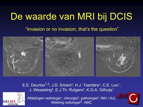
<strong>De</strong> <strong>waarde</strong> <strong>van</strong> <strong>MRI</strong> <strong>bij</strong> <strong>DCIS</strong><br />
“Invasion or no invasion, that’s the question”<br />
E.E. <strong>De</strong>urloo 1,2 , J.D. Sriram 3 , H.J. Teertstra 1 , C.E. Loo 1 ,<br />
J. Wesseling 4 , E.J.Th. Rutgers 3 , K.G.A. Gilhuijs 1<br />
Afdelingen radiologie 1 , chirurgie 3 , pathologie 4 , <strong>NKI</strong> / AvL<br />
Afdeling radiologie 2 , AMC
Inleiding<br />
Materiaal en methode<br />
Resultaten<br />
Conclusie
Inleiding<br />
• <strong>DCIS</strong>: premaligne, geen metastasering<br />
• Geen sampling <strong>van</strong> de oksel nodig<br />
• Diagnose <strong>DCIS</strong>: core biopsie of vacuüm biopsie<br />
• Onderschatting invasie core biopsie >20%, vacuüm<br />
biopsie tot 17%
Inleiding<br />
• Als er onderschatting is <strong>van</strong> invasie: tweede chirurgische<br />
procedure om axilla te stageren<br />
• Diverse aanbevelingen en richtlijnen voor sentinel node<br />
biopsie <strong>bij</strong> <strong>DCIS</strong>, gericht op klinische / mammografische /<br />
echografische / histologische kenmerken<br />
• <strong>MRI</strong> hoge sensitiviteit voor invasief carcinoom
Vraagstelling<br />
Kan <strong>MRI</strong> gebruikt worden <strong>bij</strong> patiënten met <strong>DCIS</strong> om<br />
onderscheid te maken tussen hoog risico op invasie en<br />
laag risico (naast klinische, mammografische,<br />
echografische en histologische kenmerken)?
Inleiding<br />
Materiaal en methode<br />
Resultaten<br />
Conclusie
Patiënten<br />
• Prospectieve studie, januari 2000 - december 2007<br />
• Inclusie: alle patiënten met <strong>DCIS</strong> op core biopt<br />
• Exclusie: invasie op core biopt, ipsilateraal invasief<br />
carcinoom, <strong>MRI</strong> na chirurgische excisie <strong>DCIS</strong><br />
• Beeldvorming: mammografie, evt. echografie, <strong>MRI</strong> na<br />
core biopt
Variabelen en analyses<br />
Klinisch:<br />
Mammografie:<br />
Echografie:<br />
Leeftijd<br />
Palpabele afwijking<br />
Dichtheid klierweefsel<br />
Aanwezigheid afwijking<br />
Type laesie<br />
Type calcificaties<br />
Aantal calcificaties<br />
Verdeling calcificaties<br />
Grootste diameter<br />
Aanwezigheid afwijking<br />
Type afwijking<br />
Histologie(core): <strong>DCIS</strong> graad<br />
Verdenking op invasie<br />
<strong>MRI</strong>:<br />
Type laesie<br />
Vroege aankleuring<br />
Late aankleuring (curve)<br />
Grootste diameter<br />
Univariate en multivariate<br />
logistische regressie analyse<br />
ROC analyse
Inleiding<br />
Materiaal en methode<br />
Resultaten<br />
Conclusie
Resultaten<br />
• 134 patiënten, 137 laesies (3x bilateraal)<br />
• Gemiddelde leeftijd 52.6 jaar (27 – 84 jaar)<br />
• Reden voor verwijzing:<br />
BOB 58.2% (78/134)<br />
“Symptomatisch” 23.9% (32/134)<br />
Follow up mammografie 9.7% (13/134)<br />
Hoog-risico screening 8.2% (11/134)<br />
• Invasie (definitieve histologie): 16.8% (23/137)
Resultaten mammografie<br />
• 95.6% microcalcificaties<br />
• Type laesie:<br />
Alleen microcalcificaties 88.1% (119/135)<br />
Massa (+/- microcalc) 10.4% (14/135)<br />
Architectuurverstoring 1.5% (2/135)
Diameter <strong>DCIS</strong> mammografie<br />
Aantal<br />
Diameter afwijking op mammografie (mm)<br />
Mediaan 35 mm<br />
SD 31 mm<br />
Range 3-130 mm<br />
N = 134
Resultaten <strong>MRI</strong><br />
• 59.1% aankleuring op <strong>MRI</strong> (81/137):<br />
Graad 1 35.7% (5/14)<br />
Graad 2 61.5% (32/52)<br />
Graad 3 63.2% (43/68)<br />
(Graad onbekend 1)<br />
• Geen significant verschil aangetoond tussen <strong>DCIS</strong><br />
gradering en aanwezigheid <strong>van</strong> aankleuring op <strong>MRI</strong><br />
(p= 0.13)
Type aankleuring<br />
Massa:<br />
32.1% (26/81)<br />
Non-mass:<br />
67.9% (55/81)
Late aankleuring: curves<br />
Computer-supported image interpretation<br />
Contrast opname<br />
Doorstijgend<br />
Plateau<br />
Uitwas<br />
t 1 t 2 t 3<br />
Time
Late aankleuring<br />
Type 1 curve: doorstijgende aankleuring 15% (12/81)<br />
Precontrast<br />
Postcontrast<br />
Inwas<br />
Late aankleuring
Late aankleuring<br />
Type 2 curve: plateau 28% (22/81)<br />
Precontrast<br />
Postcontrast<br />
Postcontrast<br />
Inwas<br />
Late aankleuring
Late aankleuring<br />
Type 3 curve: uitwas 58% (47/81)<br />
Precontrast<br />
Postcontrast<br />
Inwas<br />
Late aankleuring
Resultaten analyses<br />
<strong>MRI</strong> late aankleuring<br />
<strong>MRI</strong> grootste diameter<br />
<strong>MRI</strong> type afwijking<br />
15<br />
14<br />
13<br />
12<br />
11<br />
A Z = 0.80<br />
p=0.00002<br />
137 tumoren<br />
23 invasie (16.8%)<br />
<strong>MRI</strong> vroege aankleuring<br />
10<br />
XM aantal calcificaties<br />
9<br />
XM distr calcificaties<br />
US afwijking zichtbaar<br />
8<br />
7<br />
XM grootste diameter<br />
6<br />
XM densiteit<br />
5<br />
US type afwijking<br />
4<br />
PA core histologie<br />
3<br />
PA core graad<br />
Leeftijd<br />
2<br />
1<br />
0<br />
Opp. Area onder under ROC ROC curve curve= A Z<br />
0.0 0.1 0.2 0.3 0.4 0.5 0.6 0.7 0.8 0.9 1.0
Late aankleuring<br />
<strong>De</strong>finitieve histologie<br />
<strong>MRI</strong> Geen invasie Invasie Totaal<br />
Geen aankleuring of<br />
type 1 curve<br />
67 1 68<br />
Type 2 of 3 curve 47 22 69<br />
Totaal 114 23 137<br />
Negatief-voorspellende <strong>waarde</strong> (NPV): 67 / 68 = 98.5%<br />
Positief-voorspellende <strong>waarde</strong> (PPV): 22 / 69 = 31.9%
NPV en PPV<br />
NPV (%) PPV (%)<br />
Late aankleuring <strong>MRI</strong> 98.5 31.9<br />
<strong>DCIS</strong> gr 2/3, < 55 jr, >2.5 cm* 87.0 25.0<br />
Palpabele afwijking 85.6 28.0<br />
Mammografie ≥ 50 mm 85.6 22.0<br />
Graad 3 <strong>DCIS</strong> op core* 83.4 18.5<br />
Massa op het mammogram* 82.4 12.5<br />
* Aanbevelingen CBO richtlijn mammacarcinoom 2008
53 jaar, via BOB<br />
Ø 80 mm, echo g.a.; core biopt <strong>DCIS</strong> graad 3
Precontrast<br />
Postcontrast<br />
<strong>DCIS</strong> graad 3, geen invasie<br />
Inwas: aankleuring 25 mm<br />
Late aankleuring: type 1 curve
50 jaar, follow up na <strong>DCIS</strong> rechts<br />
Ø 20 mm, echo g.a.; core biopt <strong>DCIS</strong> graad 3
Precontrast<br />
Postcontrast<br />
<strong>DCIS</strong> graad 2, 26 mm, invasief focus 6 mm<br />
Inwas: aankleuring 23 mm<br />
Late aankleuring: type 2 curve
Inleiding<br />
Materiaal en methode<br />
Resultaten<br />
Conclusie
Conclusie<br />
• <strong>MRI</strong> kenmerken leiden tot beter onderscheid tussen<br />
hoog en laag risico op invasie dan klinische,<br />
mammografische, echografische of histologische<br />
kenmerken<br />
• ‘Geen aankleuring’ of type 1 curve is het meest<br />
nauwkeurige kenmerk om afwezigheid <strong>van</strong> invasie te<br />
veronderstellen (NPV 98.5%)<br />
• Aanwezigheid <strong>van</strong> invasie is niet nauwkeurig te<br />
voorspellen (PPV 31.9%)
Sentinel node biopsie <strong>bij</strong> <strong>DCIS</strong>?<br />
Eerst <strong>MRI</strong>!
This work was partially supported by the<br />
Grant <strong>NKI</strong> 2004-3082