Impression cytologic analysis after corneal cross-linking and insertion of corneal ring segments for keratoconus: two-year results nus and to compare the results with those patients presenting with similar stages of the same disease. METHODS An interventional prospective randomized clinical trial was con - ducted between March 2008 and December 2010 at the Vision Institute, Department of Ophthalmology, Fe<strong>de</strong>ral University of São <strong>Paulo</strong>, São <strong>Paulo</strong>, Brazil. This study was approved by the Institutional Ethics Committee and conducted according to the tenets of the Declaration of Helsinki. All patients provi<strong>de</strong>d informed consent. Patients diagnosed with documented keratoconus with a cor neal thickness of at least 400 µm and a history of intolerance to contact lens fitting were recruited for the study. Exclusion criteria inclu<strong>de</strong>d previous ocular surgery; only one functional eye; corneal opacity on the axis; corneal curvature >65 diopters; history of herpetic keratitis; mo<strong>de</strong>rate dry eye; autoimmune disease; and pregnancy. The criteria for the diagnosis of dry eye inclu<strong>de</strong>d tear film, punctacte keratitis, and patient complaint. A total of 39 eyes of 31 patients were inclu<strong>de</strong>d. All eyes were gra <strong>de</strong>d according to the Amsler-Krumeich classification (6) , based on patient’s refraction, mean central K-reading, corneal signs, and corneal thickness. All treated eyes were gra<strong>de</strong>d stage II or III. Using a randomized table, patients were distributed into two study groups according to the treatment: CXL group (19 eyes un<strong>de</strong>rwent CXL pro - cedure using riboflavin and UVA light) and riboflavin eyedrops (RE) group (20 eyes received 0.1% riboflavin (wt/vol) eyedrops [10 mg ri boflavin-5-phosphate in 20% (wt/vol) <strong>de</strong>xtran-T-500; Ophthalmos, São <strong>Paulo</strong>, Brazil] 4 times per day for 1 month. In eight patients, both eyes were inclu<strong>de</strong>d. In these eight cases, the right eye received only RE, and the left eye un<strong>de</strong>rwent the CXL procedure. After 3 months of this first step (CXL or RE), all patients then un<strong>de</strong>rwent insertion of ICRS (Keraring, Mediphacos, Belo Horizonte, Brazil), which comprised the second step. Clinical data collected inclu<strong>de</strong>d patients’ <strong>de</strong>mographic features (e.g., age, race, and gen<strong>de</strong>r). All patients un<strong>de</strong>rwent routine ophthal - mo logic evaluation (uncorrected distance visual acuity and correc ted distance visual acuity assessment, slit-lamp biomicroscopy, Gol d mann applanation tonometry, and ocular fundus examination), corneal topography (EyeSys-2000 Corneal Topography instrument; EyeSys Vi sion, Irvine, CA), Orbscan IIz examination (Bausch & Lomb Gmbh, Feldkirchen, Germany), Pentacam examination (Oculus Optik ge rate Gmbh, Wetzlar, Germany), Visante Optical Coherence Tomography (OCT; Carl Zeiss Meditec, Dublin, CA), and IC before treatment and at 1 month and 3 months after CXL or RE, and again at 6 months, 1 year, and 2 years after ICRS insertion. IC TECHNIQUE After the slit-lamp examination, all patients were subjected to IC sampling by the same researcher (A.C.R.). Following the application of topical anesthesia, IC specimens were collected (Millipore HAWP304; Bedford, MA) from the four quadrants of the cornea, from an exposed area of the bulbar conjunctiva (temporal region), and from an unexposed area of the conjunctiva (superior region) adjacent to the corneal limbus. All strips were processed for periodic acid-Schiff and Gill hematoxylin staining. Glass sli<strong>de</strong>s mounted with Entellan (Merck, Darmstadt, Germany) were analyzed un<strong>de</strong>r light microscopy by an experienced professional (J. N. B.), operating in a masked fashion. For quality control, only IC specimens with at least one-third of the filter surface covered with visible epithelial cells were inclu<strong>de</strong>d. Conjunctival samples were evaluated according to established techniques for the following parameters (7-9) : cellularity, cell-to-cell contact of epithelial cells, nucleus-to-cytoplasm (N:C) ratio, nuclear chromatin level, goblet cell <strong>de</strong>nsity, keratinization, and distribution of inflammatory cells. A score of 0 to 3 was assigned to each of these features. Zero represented normal findings. One represented bor<strong>de</strong>rline features, and 2 or 3 represented abnormal features. The scores obtained for each parameter were ad<strong>de</strong>d together to obtain a total score and classified as A or normal (total score 0-3), B or bor<strong>de</strong>rline (total score 4-6), and C or abnormal (total score >6). Goblet cell <strong>de</strong>nsities were judged as normal when abundant, bor<strong>de</strong>rline when slightly or mo<strong>de</strong>rately reduced, and abnormal when distinctly reduced (single to no goblet cells). Corneal samples were evaluated and gra<strong>de</strong>d according to the squamous metaplasia classification proposed by Murube and Rivas (10) . CROSS-LINKING PROCEDURE The cross-linking procedure in the CXL group was performed by the same surgeon (M.C.) according to the standard protocol (11) . 0.5% proxymetacaine hydrochlori<strong>de</strong> (wt/vol) eyedrops were applied. After a 9-mm diameter abrasion of the corneal epithelium, drops of a 0.1% riboflavin solution (wt/vol) in 20% <strong>de</strong>xtran were applied onto the cornea every 5 minutes for a total of 30 minutes. Using a slit lamp with a blue filter, the surgeon confirmed the presence of riboflavin in the anterior chamber before UVA irradiation commenced. The cornea was exposed (using a lid speculum) to UV light (UV-X System; Peschke Meditra<strong>de</strong> GmbH, Hunenberg, Switzerland), which emits light at a wavelength of 370 ± 5 nm at an irradiance of 3 mW/cm 2 or 5.4 J/cm 2 for 30 minutes (medium spot). During this time, riboflavin solution was applied every 5 minutes. The limbus and conjunctiva were not protected during irradiation. Topical anesthetic was applied as required during the surgery. After the treatment, a soft bandage contact lens was applied until reepithelialization was complete. A combination of 0.5% moxifloxacin (wt/vol) and 0.1% <strong>de</strong>xamethasone phosphate (wt/vol) eyedrops (Alcon Laboratories, Fort Worth, TX) was prescribed q.i.d. for 2 weeks. INTRASTROMAL CORNEAL RING SEGMENT IMPLANTATION Intrastromal corneal ring segment implantation was performed by one of the authors (M.C.) in an operating room un<strong>de</strong>r sterile conditions using topical anesthetic drops. The Purkinje reflex was chosen as the central point and marked un<strong>de</strong>r a biomicroscope. A 5.0-mm marker was used to locate the exact ring channel. Tunnel <strong>de</strong>pth was set at 80% of the thinnest corneal thickness on the tunnel location. The arc length and thickness were chosen according to the manufacturer’s nomogram. A 60-kHz femtosecond laser (IntraLase Corp, Irvine, CA) was used to create the ring channels. The channel’s inner diameter was set to 5.0 mm, and the outer diameter was set to 5.9 mm. The entry-cut thickness was 1 µm, and the ring energy for channel creation was 1.70 µJ. The entry-cut energy was 1.10 µJ, and channel creation timing with the femtosecond laser was 15 seconds. The ICRS was implanted immediately after channel creation and before the disappearance of the bubbles, which revealed the exact tunnel location. Postoperatively, a combination of 0.5% moxifloxacin (wt/vol) and 0.1% <strong>de</strong>xamethasone phosphate (wt/vol) eyedrops (Alcon Laboratories) was prescribed q.i.d for 2 weeks. The patients were instructed to avoid rubbing their eyes and to use artificial tears frequently. On the first postoperative day, slit-lamp biomicroscopic examination was performed. Wound healing and segment migration were evaluated. Follow-up IC examinations were scheduled at baseline, at 1 month and 3 months after CXL or RE, and again at 6 months, 1 year, and 2 years after ICRS insertion. STATISTICAL ANALYSIS Fisher’s exact test was applied to evaluate the IC gra<strong>de</strong>s. The Fried - man test was used to compare evaluations ma<strong>de</strong> at baseline, at 1 month and 3 months after CXL or RE, and again at 6 months, 1 year, and 2 years after ICRS insertion. Calculations were performed using the statistical software Minitab version 15.1 (Minitab, Inc., State College, PA). The level of statistical significance was set at P
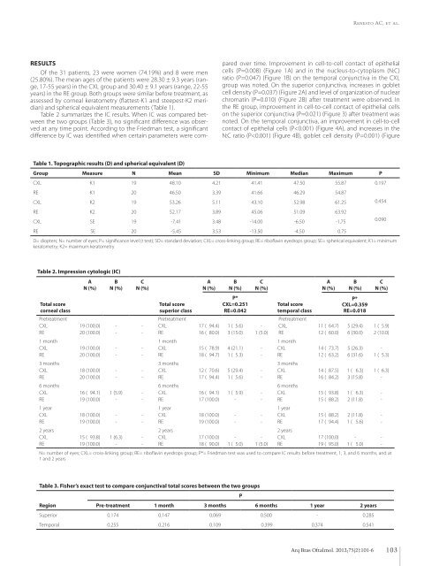
Renesto AC, et al. RESULTS Of the 31 patients, 23 were women (74.19%) and 8 were men (25.80%). The mean ages of the patients were 28.30 ± 9.3 years (range, 17-55 years) in the CXL group and 30.40 ± 9.1 years (range, 22-55 years) in the RE group. Both groups were similar before treatment, as assessed by corneal keratometry (flattest-K1 and steepest-K2 meridian) and spherical equivalent measurements (Table 1). Table 2 summarizes the IC results. When IC was compared bet - ween the two groups (Table 3), no significant difference was observed at any time point. According to the Friedman test, a significant difference by IC was i<strong>de</strong>ntified when certain parameters were compared over time. Improvement in cell-to-cell contact of epithelial cells (P=0.008) (Figure 1A) and in the nucleus-to-cytoplasm (N:C) ratio (P=0.047) (Figure 1B) on the temporal conjunctiva in the CXL group was noted. On the superior conjunctiva, increases in goblet cell <strong>de</strong>nsity (P=0.037) (Figure 2A) and level of organization of nuclear chromatin (P=0.010) (Figure 2B) after treatment were observed. In the RE group, improvement in cell-to-cell contact of epithelial cells on the superior conjunctiva (P=0.021) (Figure 3) after treatment was noted. On the temporal conjunctiva, an improvement in cell-to-cell contact of epithelial cells (P