Nov-Dez - Sociedade Brasileira de Oftalmologia
Nov-Dez - Sociedade Brasileira de Oftalmologia
Nov-Dez - Sociedade Brasileira de Oftalmologia

You also want an ePaper? Increase the reach of your titles
YUMPU automatically turns print PDFs into web optimized ePapers that Google loves.
Axenfeld-Rieger anomaly and corneal endothelial dystrophy: a case series<br />
305<br />
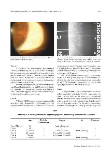
Figure 2 (case 1): Slit lamp evaluation showing a posterior embryotoxon<br />
in the temporal region of left eye.<br />
Case 3<br />
A 25 year-old Caucasian daughter was examined.<br />
The best visual acuity was equal to 20/20 in both eyes.<br />
Slit lamp evaluation presented bilateral nasal and temporal<br />
posterior embryotoxon.This may be an incomplete<br />
form of Axenfeld-Rieger anomaly. No sectorial iris hypoplasia<br />
or atrophy, corectopy, polycoria or systemic signs<br />
and symptoms were <strong>de</strong>tected.<br />
The intraocular pressure (aplannation tonometry)<br />
was 14 mmHg in the right eye and 15 mmHg in the left<br />
eye. Specular microscopic examination was similar to<br />
case 2.The fundoscopy was normal and revealed no signs<br />
of glaucomatous optic nerve damage.<br />
Case 4<br />
A 21 year-old Caucasian son was examined. The<br />
best visual acuity was equal to 20/20 in both eyes. Slit<br />
lamp evaluation presented bilateral nasal and temporal<br />
Figure 3 (case 1): Results of a specular microscopic examination<br />
showing severe guttata associated with corneal e<strong>de</strong>ma (on<br />
biomicroscopic exam), characterizing Fuchs Endothelial dystrophy.<br />
posterior embryotoxon.This may be an incomplete form<br />
of Axenfeld-Rieger anomaly. No sectorial iris hypoplasia<br />
or atrophy, corectopy, polycoria or systemic signs and<br />
symptoms were <strong>de</strong>tected.<br />
for The intraocular pressure (applannation tonometry)<br />
was 16 mmHg in the right eye and 15 mmHg in the<br />
left eye. Specular microscopic examination was similar<br />
to case 2.The fundoscopy was normal and revealed no<br />
signs of glaucomatous optic nerve damage.<br />
Case 5<br />
A 19 year-old Caucasian daughter was examined<br />
and consi<strong>de</strong>red asymptomatic. The best visual acuity was<br />
equal to 20/20 in both eyes. Slit lamp evaluation disclosed<br />
bilateral nasal and temporal posterior embryotoxon and<br />
sectorial iris atrophy with mild corectopy. No polycoria or<br />
systemic signs was observed.The intraocular pressure (applanation<br />
tonometry) was 13 mmHg in the right eye and<br />
Table 1<br />
Clinical signs of a family with anterior segment dysgenesis and central guttata or Fuchs dystrophy<br />
Age Posterior Cornea Iris Glaucoma<br />
Embryotoxon<br />
Case 1 Mother 59 + Fuchs’ Dystrophy - -<br />
Father 61 - - - -<br />
Case 2 27 female + Central Guttata MildCorectopy -<br />
Case 3 25 female + CentralGuttata(mild) - -<br />
Case 4 23 male + CentralGuttata(mild) - -<br />
Case 5 20 female + - MildCorectopy -<br />
Rev Bras Oftalmol. 2008; 67 (6): 303-8