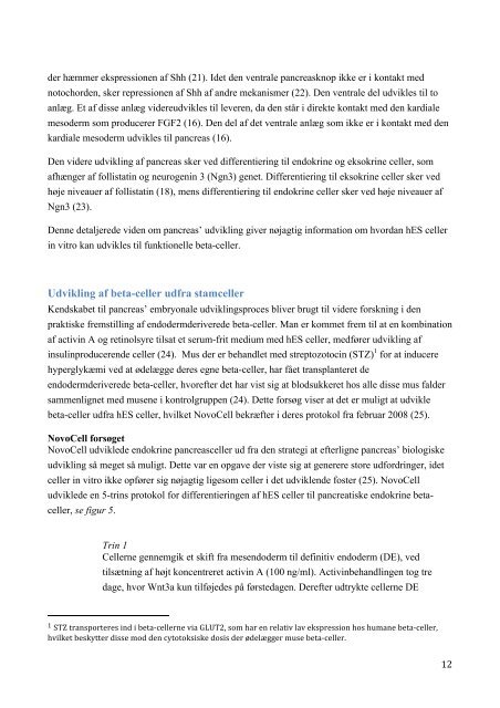
der hæmmer ekspressionen af Shh (21). Idet den ventrale pancreasknop ikke er i kontakt med notochorden, sker repressionen af Shh af andre me<strong>kan</strong>ismer (22). Den ventrale del udvikles til to anlæg. Et af disse anlæg videreudvikles til leveren, da den står i direkte kontakt med den kardiale mesoderm <strong>som</strong> producerer FGF2 (16). Den del af det ventrale anlæg <strong>som</strong> ikke er i kontakt med den kardiale mesoderm udvikles til pancreas (16). Den videre udvikling af pancreas sker ved differentiering til endokrine og eksokrine celler, <strong>som</strong> afhænger af follistatin og neurogenin 3 (Ngn3) genet. Differentiering til eksokrine celler sker ved høje niveauer af follistatin (18), mens differentiering til endokrine celler sker ved høje niveauer af Ngn3 (23). Denne detaljerede viden om pancreas’ udvikling giver nøjagtig information om hvordan hES celler in vitro <strong>kan</strong> udvikles til funktionelle beta-celler. Udvikling af beta-celler udfra stamceller Kendskabet til pancreas’ embryonale udviklingsproces bliver brugt til videre forskning i den praktiske fremstilling af endodermderiverede beta-celler. Man er kommet frem til at en kombination af activin A og retinolsyre tilsat et serum-frit medium med hES celler, medfører udvikling af insulinproducerende celler (24). Mus der er behandlet med streptozotocin (STZ) 1 for at inducere hyperglykæmi ved at ødelægge deres egne beta-celler, har fået transplanteret de endodermderiverede beta-celler, hvorefter det har vist sig at blodsukkeret hos alle disse mus falder sammenlignet med musene i kontrolgruppen (24). Dette forsøg viser at det er muligt at udvikle beta-celler udfra hES celler, hvilket NovoCell bekræfter i deres protokol fra februar 2008 (25). NovoCell forsøget NovoCell udviklede endokrine pancreasceller ud fra den strategi at efterligne pancreas’ biologiske udvikling så meget så muligt. Dette var en <strong>opgave</strong> der viste sig at generere store udfordringer, idet celler in vitro ikke opfører sig nøjagtig lige<strong>som</strong> celler i det udviklende foster (25). NovoCell udviklede en 5-trins protokol for differentieringen af hES celler til pancreatiske endokrine beta- celler, se figur 5. Trin 1 Cellerne gennemgik et skift fra mesendoderm til definitiv endoderm (DE), ved tilsætning af højt koncentreret activin A (100 ng/ml). Activinbehandlingen tog tre dage, hvor Wnt3a kun tilføjedes på førstedagen. Derefter udtrykte cellerne DE 1 STZ transporteres ind i beta-‐cellerne via GLUT2, <strong>som</strong> har en relativ lav ekspression hos humane beta-‐celler, hvilket beskytter disse mod den cytotoksiske dosis der ødelægger muse beta-‐celler. 12
© 2006 Nature Publishing Group http://www.nature.com/naturebiotechnology markørerne CER og CXCR4. Denne overgang til DE var en nødvendighed for den videre udvikling til beta-celler. Trin 2 Activin A blev fjernet fra mediet, hvilket initierede at DE udvikledes til den primitive tarm. Dette blev indikeret ved at DE markørerne CER og CXCR4 faldt, hvorimod den context of an embryo. T<strong>her</strong>efore, our strategy combines an informed approach primitive based tarms on developmental markører biology HNF1B and an og empirical HNF4A approach. steg. Udover at activin A blev fjernet, Previously we described a method for differentiating hES cells to DE tilsattes FGF10 og hedgehog-signalerings hæmmeren KAAD-cyclopamine, <strong>som</strong> havde yderligere indflydelse på resten af processen. Trin 3 Mediet blev <strong>her</strong>efter eksponeret for retinolsyre sammen med FGF10 og KAADcyclopamine. Ved tilsætning af denne kombination til mediet blev høje niveauer af Pdx1 udtrykt, mens den primitive tarms markører fortsat blev udtrykt. Trin 4 Idet der nu sås en ekspression af Pdx1 i mediet, viste det tegn på at pancreatisk epitel var blevet udviklet. Udover Pdx1, blev der også udtrykt Ngn3 <strong>som</strong> er et tegn på differentiering af det pancreatiske epitel til endokrine celler. Trin 5 Efter 15 dage, observeredes endokrine celler, der udskilte henholdsvis insulin, glukagon, <strong>som</strong>atostatin og pancreatiske polypeptid. 24 initial optimization of the protocol, the differentiation process characterized at the level of protein expression by immunofluor western blotting, flow cytometry and ELISA. . Here we show that DE derived from hES cells can be efficiently In stage 1, hES cells are transitioned through mesendoderm differentiated to hormone-expressing endocrine cells through a series using high concentrations of activin A (100 ng/ml) in the co of endoderm intermediates similar to those that occur during pancre- low FBS supplementation, as previously reported atic development. The insulin content of the insulin-expressing cells approaches that of adult islets. In addition, C-peptide release occurs in response to multiple secretory stimuli, although only minimally to glucose, similar to what occurs in the fetal β-cell. RESULTS Directed pancreatic differentiation of hES cells We have developed a five-step protocol for differentiation of hES cells to pancreatic hormone–expressing cells (Fig. 1a and Methods). The protocol was optimized primarily in the CyT203 cell line in a stepwise fashion. We focused first on generating DE, followed by PDX1-expressing cells and, finally, insulin-expressing cells. Study of the developmental biology literature led us to test the action of such factors as activins, Wnt3a, BMPs, TGFβ1, TGFβ inhibitors, FGFs, FGF inhibitors, retinoids, γ-secretase inhibitors, GLPs and analogs, HGF, IGFs, VEGF, nicotinamide and hedgehog inhibitors. The empirical approach involved testing various concentrations, times of administration, lengths of application and combinations of factors as well as assorted medium formulations in which the factors were applied. The effects of various treatments on differentiation were initially evaluated using real-time PCR to detect expression of 30 or more markers of both target and non-target cell types monitored at short intervals throughout the course of differentiation. After 24 . We subse improved this procedure by shortening the activin A treatmen to 3 d and adding Wnt3a during the first day of activin exposure modifications increased the efficiency of mesendoderm spec and the synchrony of DE formation (data not shown). As with th col of ref. 24, the hES cell–derived DE expressed the DE marker (ref. 26) and CXCR4 (refs. 24,27,28), and showed anterior cha indicated by expression of the anterior DE markers CER and (refs. 29,30; Figs. 1b and 2a–c; Supplementary Figs. 1 and 2a o We found that initial specification of hES cells to DE is cr efficient production of pancreatic hormone–expressing cell activin A dose was lowered, t<strong>her</strong>eby altering the quantity and pattern of the DE produced, differentiation to endoderm interm and endocrine cells was considerably reduced (Supplementa online). Moreover, if neural or extraembryonic lineages were s from hES cells, these populations failed to differentiate to endoc or their endoderm precursors (Supplementary Fig. 2a,b onlin In stage 2, activin A is removed, which in our cultures seem essential to allow the transition of DE to a stage resembling the p gut tube. In the mouse embryo, at embryonic day 8 (E8), the tr tion factor Tcf2 (also called Hnf1b) is expressed in the entire p gut tube31,32 . Approximately 12 h later Hnf4a shows a similar p expression33 approach based on developmental biology and an empirical approach. Previously we described a method for differentiating hES cells to DE . In zebrafish, hnf1b is involved in patterning the p 24 initial optimization of the protocol, the differentiation process w characterized at the level of protein expression by immunofluore western blotting, flow cytometry and ELISA. . Here we show that DE derived from hES cells can be efficiently In stage 1, hES cells are transitioned through mesendoderm differentiated to hormone-expressing endocrine cells through a series using high concentrations of activin A (100 ng/ml) in the con of endoderm intermediates similar to those that occur during pancre- low FBS supplementation, as previously reported atic development. The insulin content of the insulin-expressing cells approaches that of adult islets. In addition, C-peptide release occurs in response to multiple secretory stimuli, although only minimally to glucose, similar to what occurs in the fetal β-cell. RESULTS Directed pancreatic differentiation of hES cells We have developed a five-step protocol for differentiation of hES cells to pancreatic hormone–expressing cells (Fig. 1a and Methods). The protocol was optimized primarily in the CyT203 cell line in a stepwise fashion. We focused first on generating DE, followed by PDX1-expressing cells and, finally, insulin-expressing cells. Study of the developmental biology literature led us to test the action of such factors as activins, Wnt3a, BMPs, TGFβ1, TGFβ inhibitors, FGFs, FGF inhibitors, retinoids, γ-secretase inhibitors, GLPs and analogs, HGF, IGFs, VEGF, nicotinamide and hedgehog inhibitors. The empirical approach involved testing various concentrations, times of administration, lengths of application and combinations of factors as well as assorted medium formulations in which the factors were applied. The effects of various treatments on differentiation were initially evaluated using real-time PCR to detect expression of 30 or more markers of both target and non-target cell types monitored at short intervals throughout the course of differentiation. After 24 . We subseq improved this procedure by shortening the activin A treatment to 3 d and adding Wnt3a during the first day of activin exposure 25 modifications increased the efficiency of mesendoderm specif and the synchrony of DE formation (data not shown). As with the col of ref. 24, the hES cell–derived DE expressed the DE markers (ref. 26) and CXCR4 (refs. 24,27,28), and showed anterior chara indicated by expression of the anterior DE markers CER and F (refs. 29,30; Figs. 1b and 2a–c; Supplementary Figs. 1 and 2a on We found that initial specification of hES cells to DE is criti efficient production of pancreatic hormone–expressing cells. activin A dose was lowered, t<strong>her</strong>eby altering the quantity and an pattern of the DE produced, differentiation to endoderm interm and endocrine cells was considerably reduced (Supplementary online). Moreover, if neural or extraembryonic lineages were sp from hES cells, these populations failed to differentiate to endocri or their endoderm precursors (Supplementary Fig. 2a,b online In stage 2, activin A is removed, which in our cultures seem essential to allow the transition of DE to a stage resembling the pr gut tube. In the mouse embryo, at embryonic day 8 (E8), the tran tion factor Tcf2 (also called Hnf1b) is expressed in the entire pri gut tube31,32 . Approximately 12 h later Hnf4a shows a similar pat expression33 . In zebrafish, hnf1b is involved in patterning the po a Stage 1 Stage 2 Stage 3 Stage 4 Stage 5 b Differentiation stage a Definitive Stage endoderm 1 Primitive Stage gut tube 2 Posterior Stage foregut 3 Pancreatic endoderm Stage and 4 Hormone expressing Stage 5 b Antibody \--1--\ 2 \---3---\--4--\------5--- Day of differentiation 2 3 6 Differentiation 8 10 11 12 13 14 stage 15 16 1 Activin + Wnt Activin Definitive endoderm FGF10 + CYC RA + CYC + FGF10 Primitive gut tube Posterior foregut endocrine precursor Pancreatic +/- DAPT; Ex4 endoderm and endocrine cell Hormone +/- Ex4; IGF1; HGF expressing α POUF5F1 Antibody \--1--\ 2 \---3---\--4--\------5 Day of differentiation 2 3 6 8 10 11 12 13 14 15 1 RPMI + 0% FBS RPMI + 0.2% FBS RPMI + 2% FBS DMEM + 1% endocrine B27 precursor CMRL endocrine + 1% B27 cell Activin + Wnt 1–2 days RPMI + 0% FBS Activin 1–2 days RPMI + 0.2% FBS FGF10 + CYC 2–4 days RPMI + 2% FBS RA + CYC + FGF10 +/- DAPT; Ex4 2–4 days 2–3 days DMEM + 1% B27 +/- Ex4; IGF1; HGF 3+ days CMRL + 1% B27 α POUF5F1 α SOX17 ES OCT4 NANOG ES SOX2 ECAD OCT4 NANOG SOX2 1–2 days ME 1–2 days DE 2–4 days PG 2–4 days PF 2–3 days PE 3+ days EN © 2006 Nature Publishing Group http://www.nature.com/naturebiotechnology context of an embryo. T<strong>her</strong>efore, our strategy combines an informed BRA FGF4 ME WNT3 NCAD BRA FGF4 WNT3 SOX17 CERDE FOXA2 CXCR4 SOX17 CER FOXA2 HNF1B HNF4A PG HNF1B HNF4A PDX1 HNF6 PF HLXB9 PDX1 HNF6 HLXB9 NKX6-1 NGN3 PE PAX4 NKX2-2 NKX6-1 NGN3 PAX4 INS CGC EN GHRL SST INS PPY CGC α SOX17 α FOXA2 α FOXA2 α HNF1B α HNF1B α PDX1 Figur 5 GHRL Figur er taget fra Figure ECAD (25). 1 Schematic NCAD of differentiation CXCR4 procedure and protein expression for <strong>som</strong>e key markers NKX2-2 of SST PPY pancreatic differentiation. (a) The differentiation protocol is divided into five stages and the growth α PDX1 α NKX2-2 Skematisk differentieringsprocedure af pancreatisk væv. factors, Figure medium 1 Schematic and range of differentiation of duration for procedure each stage and are protein shown. expression This protocol for <strong>som</strong>e orchestrates key markers differentiation of Dette skema viser through pancreatic de five fem identifiable differentiation. stadier hvori endodermal (a) differentieringen The differentiation intermediates protocol en skete, route is to samt divided production hvilke into of five vækstfaktorer, hormone-expressing stages and the medier growth og tidsrammer der α NKX2-2 α NKX6-1 blev anvendt for endocrine factors, hvert medium cells. stadie. Several and range markers of duration characteristic for each of stage each cell are shown. population This are protocol listed. orchestrates CYC, KAAD-cyclopamine; differentiation Forklaring af RA, forkortelser through all-trans five retinoic identifiable på figuren: acid; endodermal DAPT, RA, γ-secretase retinsyre; intermediates inhibitor; DAPT, en Ex4, route γ-secretase exendin-4; to production ES, inhibitor; hES of hormone-expressing cell; Ex4, ME, mesendoderm; exendin-4; IGF1, Insulin-like α NKX6-1 Growth Factor DE, endocrine 1; definitive HGF, cells. Hepatocyt endoderm; Several PG, markers Growth primitive characteristic Factor; gut tube; ES, of PF, each posterior hES cell celle; population foregut ME, endoderm; are mesendoderm; listed. PE, CYC, pancreatic KAAD-cyclopamine; α PAX6 DE, endoderm definitiv endoderm; PG, and RA, endocrine all-trans precursor; retinoic acid; EN, DAPT, hormone-expressing γ-secretase inhibitor; endocrine Ex4, cells. exendin-4; (b) Western ES, hES blot cell; analyses ME, mesendoderm; show the primitiv tarm; PF, posterior fortarm; PE, pancreatisk endoderm; EN, hormon-udtrykkende endokrine celler. dynamics DE, definitive of protein endoderm; expression PG, for primitive several gut key tube; markers PF, posterior of pancreatic foregut differentiation. endoderm; PE, α, antibody. pancreatic POU5F1 endoderm α PAX6 α C-PEPTIDE (OCT4): and endocrine this marker precursor; of undifferentiated EN, hormone-expressing ES cells gradually endocrine diminishes cells. (b) as Western cells differentiate blot analyses and show becomes the undetectable dynamics of during protein stage expression 3. SOX17: for several the highest key markers levels of of the pancreatic full-length differentiation. SOX17 protein α, (arrow) antibody. are POU5F1 α C-PEPTIDE observed (OCT4): during this marker differentiation of undifferentiated of definitive ES endoderm cells gradually (stage diminishes 1). At later as stages cells it differentiate gradually diminishes, and becomes α GAPDH while undetectable a slightly faster-migrating during stage 3. SOX17: protein the accumulates. highest levels This of corresponds the full-length to the SOX17 truncated protein form (arrow) of SOX17 are (ref. observed 70), and during its presence differentiation throughout of definitive later stages endoderm of differentiation (stage 1). At is later consistent stages with it gradually the continued diminishes, α GAPDH expression while a slightly of SOX17 faster-migrating mRNA (Supplementary protein accumulates. Fig. 2a). See This ref. corresponds 24 for a demonstration to the truncated of SOX17 form of antibody SOX17 specificity. (ref. 70), FOXA2: and its presence the upper throughout band (arrow) later seen stages at all of stages differentiation from the is end consistent of stage with 1 onward the continued corresponds to the 48-kDa FOXA2 protein. We believe tha partial expression degradation of SOX17 results mRNA in the (Supplementary faster-migrating Fig. band 2a). seen See ref. in <strong>som</strong>e 24 for samples. a demonstration HNF1B: the of SOX17 lower band antibody (arrow) seen from stage 2 onward corresponds t 61-kDa specificity. variant FOXA2: of the the HNF1B upper protein. band (arrow) We do seen not currently at all stages know from the the source end of of the stage slower-migrating 1 onward corresponds band seen to the in <strong>som</strong>e 48-kDa samples. FOXA2 PDX1: 13 protein. this We accumulat believe t during partial stage degradation 3 and diminishes results in at the the faster-migrating end of stage 5. band NKX2-2: seen is in present <strong>som</strong>e samples. during stages HNF1B: 4 and the 5. lower NKX6-1: band (arrow) this is detectable seen from stage at the 2 end onward of stage correspond 4 and in 5. 61-kDa PAX6: this variant is most of the strongly HNF1B expressed protein. We during do not stage currently 5. C-peptide: know the although source this of the antiserum slower-migrating is immunoreactive band seen with in <strong>som</strong>e both samples. the fully PDX1: processed this C-peptide accumula the during unprocessed stage 3 and proinsulin, diminishes the size at the of end the band of stage is most 5. NKX2-2: consistent is present with the during 9-kDa stages proinsulin. 4 and C-peptide 5. NKX6-1: + proinsulin this is detectable accumulate at the during end of stage 5. 4 and 5. PAX6: this is most strongly expressed during stage 5. C-peptide: although this antiserum is immunoreactive with both the fully processed C-peptid the unprocessed proinsulin, the size of the band is most consistent with the 9-kDa proinsulin. C-peptide + proinsulin accumulate during stage 5. ESC ESC ARTICL ARTIC