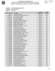
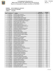
Suplemento 2 Vol 8 No 1.indd - Universidad Veracruzana
Suplemento 2 Vol 8 No 1.indd - Universidad Veracruzana
Suplemento 2 Vol 8 No 1.indd - Universidad Veracruzana

You also want an ePaper? Increase the reach of your titles
YUMPU automatically turns print PDFs into web optimized ePapers that Google loves.
TNF-enhanced NREMS is most often accompanied<br />
by enhanced EEG delta power; this measure is indicative of<br />
sleep intensity and is used to model the S-process in the<br />
two process model of sleep regulation 3 . If TNF is injected<br />
into the pre-optic area (POA) of the anterior hypothalamus, a<br />
sleep regulatory circuit, it also enhances duration of NREMS<br />
and EEG delta power 9 . In contrast, if the TNF soluble receptor,<br />
an inhibitor of TNF, is injected into the POA NREMS duration<br />
is reduced. TNF can also enhance EEG delta power within<br />
local cortical circuits. Thus, unilateral injection of TNF onto<br />
the surface of the cortex results in ipsilateral enhancement<br />
of EEG delta power during NREMS but not during REMS or W.<br />
Conversely, the TNF soluble receptor unilaterally inhibits<br />
sleep loss-enhanced EEG delta power after cortical injections<br />
10 . Similarly, if a TNF si RNA, a substance that decreases TNF<br />
mRNA stability, is unilaterally injected on to the cortex,<br />
it reduces TNF -immuno-reactivity and EEG delta power<br />
unilaterally only during NREMS 11 .<br />
TNF mRNA levels vary with sleep propensity;<br />
daylight levels are about 2 fold higher than nighttime levels.<br />
Similarly, TNF protein in hypothalamus and cortex is about<br />
10 fold higher at the beginning of daylight hours compared<br />
to nighttime levels. TNF mRNA levels also increase in brain<br />
during sleep deprivation in rats 1 . In humans, plasma levels<br />
of TNF in normal healthy individuals co-vary with EEG delta<br />
power. As already mentioned, in a variety of pathologies<br />
that manifest in sleepiness or excess sleep, TNF plasma<br />
levels are enhanced. For instance, after influenza viral<br />
challenge in mice, hypothalamic TNF levels are enhanced at<br />
the time NREMS is also enhanced 12 . Similarly, giving humans<br />
endotoxin, a Gram-negative bacterial cell wall product,<br />
enhances TNF plasma levels and sleep 13 . Collectively<br />
the data discussed in this section clearly implicate TNF in<br />
physiological sleep regulation and in the sleep responses to<br />
sleep loss and pathological challenges.<br />
Activity-dependent induction of TNF; a mechanism for<br />
how the brain keep track of past sleep-wake history<br />
Neurotransmission is characterized by the co-release of ATP<br />
with neurotransmitters 14 . This extracellular ATP signals via<br />
purine receptors P2X and P2Y to release TNF, IL1 and brainderived<br />
neurotrophic factor from glia 15-17 . Some of that<br />
extracellular ATP also rapidly degrades into adenosine and<br />
48<br />
Revista Médica<br />
it in turn acts via P1 receptors. This is posited to be one of<br />
the rapid mechanisms involved in sleep induction (Figure<br />
1). The ATP -released TNF also acts rapidly on cells to alter<br />
membrane potentials and these changes are also likely<br />
involved in sleep regulation 18 . Perhaps more important to<br />
sleep regulation are the long term (hours; Figure 1) actions<br />
of TNF. Thus TNF activates nuclear factor kappa B (NFkB), an<br />
enhancer element involved in the transcription of multiple<br />
substances involved in sleep regulation 19,20 . NFkB enhances<br />
transcription of the adenosine A1 receptor and the glu-R1<br />
component of the glutamate AMPA receptor. Expression<br />
of these receptors alters the sensitivity of the neuron<br />
to adenosine, a hyperpolarizing inhibitory substance,<br />
and glutamate, a depolarizing excitatory substance. As<br />
a consequence, depending upon the relative number of<br />
each of these receptors, the sensitivity of the neuron to<br />
these ligands is altered. Such changes alter network inputoutput<br />
processes and are thought to be a key component in<br />
events such as memory formation and state changes (see<br />
below). This process is called scaling, up-scaling if there is<br />
a relative increase in the AMPA receptors and down-scaling<br />
if there is a relative increase in the adenosine A1 receptors<br />
(Figure 2). There is also direct evidence that TNF is involved<br />
in scaling 21 .<br />
If neurotransmission-associated ATP release is<br />
involved in a long-term mechanism for keeping track of past<br />
brain activity, then ATP agonists or antagonists should alter<br />
sleep. To our knowledge, such substances have not been<br />
characterized for their actions on sleep. Preliminary data<br />
from our laboratory suggest that intracerebroventricular<br />
administration of the ATP agonist, BzATP enhances NREMS<br />
while the ATP antagonist, OXATP, inhibits NREMS (De et al,<br />
unpublished). Further, other preliminary data indicate that<br />
P2 receptor mRNAs (both X7 and Y1) have a diurnal rhythm in<br />
brain and are altered by sleep loss, IL1 and TNF injection, and<br />
changes in ambient temperature (Taishi et al, unpublished).<br />
If such data are confirmed and extended, they will suggest<br />
that the mechanism by which the brain keeps long-term<br />
track of prior activity (states) involves neurotransmissionreleased<br />
ATP that in turn releases brain cytokines and their<br />
subsequent actions on receptor expression and consequent<br />
cell sensitivity to excitatory and inhibitory signals (Table<br />
2). This also strongly suggests, because such a mechanism<br />
Revista Médica de la <strong>Universidad</strong> <strong>Veracruzana</strong> / <strong>Suplemento</strong> 2 <strong>Vol</strong>. 8 núm. 1, Enero - Junio 2008