Caso de implantes angulados como opção reabilitadora ... - ILAPEO
Caso de implantes angulados como opção reabilitadora ... - ILAPEO
Caso de implantes angulados como opção reabilitadora ... - ILAPEO

Create successful ePaper yourself
Turn your PDF publications into a flip-book with our unique Google optimized e-Paper software.
<strong>Caso</strong> <strong>de</strong> <strong>implantes</strong> <strong>angulados</strong><br />
<strong>como</strong> <strong>opção</strong> <strong>reabilitadora</strong><br />
<strong>de</strong> maxilas atróficas<br />
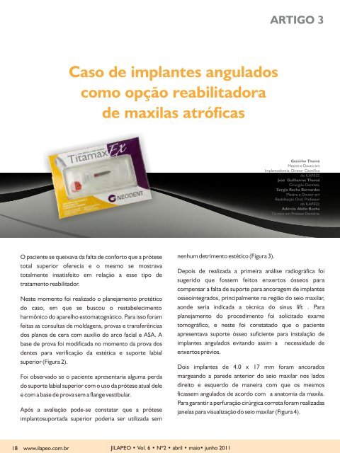
O paciente se queixava da falta <strong>de</strong> conforto que a prótese<br />
total superior oferecia e o mesmo se mostrava<br />
totalmente insatisfeito em relação a esse tipo <strong>de</strong><br />
tratamento reabilitador.<br />
Neste momento foi realizado o planejamento protético<br />
do caso, em que se buscou o restabelecimento<br />
harmônico do aparelho estomatognático. Para isso foram<br />
feitas as consultas <strong>de</strong> moldagens, provas e transferências<br />
dos planos <strong>de</strong> cera com auxílio do arco facial e ASA. A<br />
base <strong>de</strong> prova foi modificada no momento da prova dos<br />
<strong>de</strong>ntes para verificação da estética e suporte labial<br />
superior (Figura 2).<br />
Foi observado se o paciente apresentaria alguma perda<br />
do suporte labial superior com o uso da prótese atual <strong>de</strong>le<br />
e com a base <strong>de</strong> prova sem a flange vestibular.<br />
Após a avaliação po<strong>de</strong>-se constatar que a prótese<br />
implantosuportada superior po<strong>de</strong>ria ser utilizada sem<br />
18 www.ilapeo.com.br<br />
nenhum <strong>de</strong>trimento estético (Figura 3).<br />
J<strong>ILAPEO</strong> • Vol. 6 • Nº2 • abril • maio• junho 2011<br />
ARTIGO 3<br />
Geninho Thomé<br />
Mestre e Douto em<br />
Implantodontia; Diretor Científico<br />
do <strong>ILAPEO</strong>;<br />
José Guilherme Thomé<br />
Cirurgião Dentista;<br />
Sergio Rocha Bernar<strong>de</strong>s<br />
Mestre e Doutor em<br />
Reabilitação Oral; Professor<br />
do <strong>ILAPEO</strong>;<br />
Adércio Abílio Buche<br />
Técnico em Prótese Dentária;<br />
Depois <strong>de</strong> realizada a primeira análise radiográfica foi<br />
sugerido que fossem feitos enxertos ósseos para<br />
compensar a falta <strong>de</strong> suporte para ancoragem <strong>de</strong> <strong>implantes</strong><br />
osseointegrados, principalmente na região do seio maxilar,<br />
aon<strong>de</strong> seria indicada a técnica do sinus lift . Para<br />
planejamento do procedimento foi solicitado exame<br />
tomográfico, e neste foi constatado que o paciente<br />
apresentava suporte ósseo suficiente para instalação <strong>de</strong><br />
<strong>implantes</strong> <strong>angulados</strong> evitando assim a necessida<strong>de</strong> <strong>de</strong><br />
enxertos prévios.<br />
Dois <strong>implantes</strong> <strong>de</strong> 4.0 x 17 mm foram ancorados<br />
margeando a pare<strong>de</strong> anterior do seio maxilar nos lados<br />
direito e esquerdo <strong>de</strong> maneira com que os mesmos<br />
ficassem <strong>angulados</strong> <strong>de</strong> acordo com a anatomia da maxila.<br />
Para garantir a perfuração cirúrgica correta foram realizadas<br />
janelas para visualização do seio maxilar (Figura 4).
Visando a seleção do componente protético angulado<br />
durante as perfurações foram utilizadas peças similares a<br />
paralalômetros cirúrgicos com angulações <strong>de</strong> 17º e 30º<br />
guiando o ato cirúrgico (Figura 5 e 6).<br />
Para essa cirurgia foram utilizados <strong>implantes</strong> especiais com<br />
alto po<strong>de</strong>r <strong>de</strong> perfuração. Os <strong>implantes</strong> Titamax EX (Figura<br />
7) (Neo<strong>de</strong>nt, Curitiba, Brasil) foram instalados logo após a<br />
broca helicoidal <strong>de</strong> 2.0 mm. Esses <strong>implantes</strong>, com interface<br />
protética tipo Cone Morse, possuem em seu projeto um<br />
ápice altamente cortante e roscas piramidais, auxiliando a<br />
inserção cirúrgica com sub-instrumentação. A compressão<br />
óssea realizada pela instalação <strong>de</strong>stes <strong>implantes</strong> resultou<br />
em otimização da estabilida<strong>de</strong> primária (Figura 8).<br />
Além dos <strong>implantes</strong> inclinados foram utilizados mais três<br />
<strong>implantes</strong> Titamax EX <strong>de</strong> 19, 15 e 13 mm <strong>de</strong> comprimento<br />
e 4.0mm <strong>de</strong> plataforma e um titamax CM (Neo<strong>de</strong>nt,<br />
Curitiba, Brasil) na região anterior <strong>de</strong> maxila. Esses<br />
<strong>implantes</strong> foram ancorados buscando bicorticalização e<br />
aproveitando estruturas anatômicas importantes <strong>como</strong> a<br />
eminência canina.<br />
Os ápices dos <strong>implantes</strong> <strong>angulados</strong> se posicionaram na<br />
direção mesial enquanto a plataforma em região distal,<br />
<strong>de</strong>ixando a plataforma dos <strong>implantes</strong> posteriores<br />
emergindo a nível <strong>de</strong> primeiros molares. Essa posição<br />
possibilitou ancoragem <strong>de</strong> <strong>implantes</strong>. Sobre esses foram<br />
instalados minipilares cônicos Cone Morse <strong>angulados</strong> <strong>de</strong><br />
17º e 30º (Neo<strong>de</strong>nt, Curitiba, Brasil) com altura <strong>de</strong> cinta<br />
a<strong>de</strong>quada para cada elemento (Figura 9).<br />
Logo após foi feita a transferência da posição dos<br />
intermediários e registro intermaxilar através do guia<br />
multifuncional confeccionado durante o planejamento<br />
protético (Figura 10).<br />
A barra protética foi feita a partir da técnica da cimentação<br />
em monobloco e os <strong>de</strong>ntes guiados pela montagem em<br />
ASA realizada na fase <strong>de</strong> preparo. Quatro horas <strong>de</strong>pois a<br />
prótese implantosuportada final do caso foi parafusada<br />
(Figura 11).<br />
Quatro meses <strong>de</strong>pois da cirurgia o paciente foi chamado<br />
para consulta <strong>de</strong> manutenção em que não foi constatado<br />
nenhum problema com os <strong>implantes</strong> ou com a prótese<br />
(Figura 12).<br />
FIGURA 1 - Radiogafia inicial<br />
FIGURA 2 - Base <strong>de</strong> prova modificada com os <strong>de</strong>ntes.<br />
FIGURA 3 - A -Posição do lábio superior com a prótese<br />
antiga; B - Posição do lábio com a base <strong>de</strong> prova <strong>de</strong>ntada<br />
sem a flange vestibular.<br />
J<strong>ILAPEO</strong> • Vol. 6 • Nº2 • abril • maio• junho 2011 www.ilapeo.com.br 19
FIGURA 4 - Janela do seio auxiliando no planejamento da<br />
posição do implante.<br />
FIGURA 5 - Medidor <strong>de</strong> Ângulo Neo<strong>de</strong>nt - 17º azul e<br />
30º amarela, para seleção da angulação do componente<br />
protético durante o procedimento cirúrgico.<br />
FIGURA 6 – Guia <strong>de</strong> 30º instalado durante o ato cirúrgico.<br />
FIGURA 7 - Implantes Titamax Ex Neo<strong>de</strong>nt.<br />
20 www.ilapeo.com.br<br />
A<br />
B C<br />
D<br />
J<strong>ILAPEO</strong> • Vol. 6 • Nº2 • abril • maio• junho 2011<br />
FIGURAS 8 A - Ápice do implante em seio maxilar;<br />
B e C - estabilida<strong>de</strong> primária atingida;<br />
D - todos os <strong>implantes</strong> instalados.
FIGURA 9 – Mini-pilares Cônicos <strong>angulados</strong> 17º e 30º.<br />
A<br />
B<br />
FIGURAS 10 A - Guia multifuncional; B - Guia instalado<br />
na boca do paciente;<br />
A<br />
B<br />
C<br />
FIGURAS 11 A -<br />
Prótese finalizada<br />
após 4 horas;<br />
B e C - Panorâmica e<br />
Teleradiografia final.<br />
FIGURA 12 – Manutenção do caso e radiografia após 4<br />
meses.<br />
J<strong>ILAPEO</strong> • Vol. 6 • Nº2 • abril • maio• junho 2011 www.ilapeo.com.br 21
FIGURA 13 – Acompanhamento do caso e radiografia<br />
após 5 anos.<br />
22 www.ilapeo.com.br<br />
CONSIDERAÇÕES FINAIS<br />
J<strong>ILAPEO</strong> • Vol. 6 • Nº2 • abril • maio• junho 2011<br />
Po<strong>de</strong>-se concluir que a técnica aplicada nesse caso <strong>de</strong> reabilitação<br />
oral resultou em sucesso tanto do ponto <strong>de</strong> vista cirúrgico quanto<br />
protético.<br />
Cirurgicamente, o uso <strong>de</strong> <strong>implantes</strong> subinstrumentados reduz o<br />
tempo da cirurgia e promove maior estabilida<strong>de</strong> primária no<br />
momento da instalação. A prótese híbrida que foi confeccionada<br />
utilizando a técnica do guia multifuncional e adaptação passiva<br />
apresentou estética satisfatória, além <strong>de</strong> promover a restauração<br />
da função em um período <strong>de</strong> tempo relativamente curto.<br />
O última manutenção do caso foi após um período <strong>de</strong> 5 anos<br />
<strong>como</strong> po<strong>de</strong>mos ver na figura 13.<br />
REFERÊNCIA<br />
1.A<strong>de</strong>ll R, Lekholm U, Rockler B and Brånemark P-I. A 15-year study of osseointegrated implants in the treatment<br />
of the e<strong>de</strong>ntulous jaw. Int J Oral Surg. 1981. 10: 387–416.<br />
2.Sorní M, Guarinós J, García O, Peñarrocha M. Implant rehabilitation of the atrophic upper jaw: a review of the<br />
literature since 1999. Med Oral Patol Oral Cir Bucal. 2005 1;10 Suppl 1:E45-56.<br />
3.Henry PJ: A review of gui<strong>de</strong>lines for implant rehabilitation of the e<strong>de</strong>ntulous maxilla. J Prosthet Dent 87:281,<br />
2002.<br />
4.Pietrokovski J, Massler M. Alveolar ridge resorption following tooth extraction. J Prosthet Dent. 1967;17(1):21-7.<br />
5.Pietrokovski J, Starinsky R, Arensburg B, Kaffe I. Morphologic characteristics of bony e<strong>de</strong>ntulous jaws. J<br />
Prosthodont. 2007;16(2):141-7.<br />
6.Tallgren A. The continuing reduction of the residual alveolar ridges in complete <strong>de</strong>nture wearers: a mixedlongitudinal<br />
study covering 25 years. 1972. J Prosthet Dent. 2003;89(5):427-35.<br />
7.Johnson, K. A study of the dimensional changes occurring in the maxillae after tooth extraction Part I: Normal<br />
healing, Aust Dent J. 1963;8:428-433.<br />
8.Isaksson S, Alberius P.Maxillary alveolar ridge augmentation with onlay bone-grafts and immediate endosseous<br />
implants. J Craniomaxillofac Surg 1992;20:2–7.<br />
9.Wood R, Moore D. Grafting of the maxillary sinus with intraorally harvested autogenous bone prior to implant<br />
placement. Int J Oral Maxillofac Implants 1988;3:209–214.<br />
10.Summers RB. A new concept in maxillary implant surgery: the osteotome technique. Compendium<br />
1994;15:152, 154-6, 158 passim; quiz 162.<br />
11.Summers RB. The osteotome technique: Part 2. The ridge expansion osteotomy (REO) procedure.<br />
Compendium 1994;15:422, 424, 426, passim; quiz 436.<br />
12.Summers RB. The osteotome technique: Part 3. Less invasive methods of elevating the sinus floor.<br />
Compendium 1994; 15: 698, 700, 702-4 passim; quiz 710.<br />
13.Rambla-Ferrer J, Peñarrocha-Diago M, Guarinos-Carbó J. Analysis of the use of expansion osteotomes for the<br />
creation of implant beds. Technical contributions and review of the literature. Med Oral Patol Oral Cir Bucal<br />
2006;11:E267-71.<br />
14.Isaksson S, Ekfeldt A, Alberius P, Blomqvist J. Early results from reconstruction of severely atrophic (Class IV)<br />
maxillas by immediate endosseous implants in conjunction with bone grafting and Le Fort I osteotomy. J Oral<br />
Maxillofac Surg 1993;22:144–148.<br />
15.Ribeiro-Junior PD, Padovan LE, Gonçales ES, Nary-Filho H. Bone grafting and insertion of <strong>de</strong>ntal implants<br />
followed by Le Fort advancement for correction of severely atrophic maxilla in young patients. Int J Oral Maxillofac<br />
Surg. 2009;38:1101-6.<br />
16.Maló P, Rangert B, Nobre M. All-on-4 immediate function concept with Brånemark System implants for<br />
completely e<strong>de</strong>ntulous maxillae: A 1-year retrospective clinical study. Clin Implant Dent Relat Res 2005;7(suppl<br />
1):588–594.<br />
17.Aparicio C. Tilted implants as an alternative to maxillary sinus grafting: A clinical, radiologic, and Periotest study.<br />
Clin Implant Dent Relat Res 2001;1:39–49.<br />
18.Krogh P. Anatomic and surgical consi<strong>de</strong>rations in the use of osseointegrated implant in the posterior maxilla.<br />
Oral Maxillofac Surg Clin North Am 1991;3:853–868.<br />
19.Balshi S,Wolfinger G, Balshi T. Analysis of 164 titanium-oxi<strong>de</strong> surface implants in completely e<strong>de</strong>ntulous arches<br />
for fixed prosthesis anchorage in the pterygomaxillary region. Int J Oral Maxillofac Implants 2005;20:946–952.<br />
20.Sorní M, Guarinos J, Peñarrocha M. Implants in anatomical buttresses of the upper jaw. Med Oral Patol Oral Cir<br />
Bucal 2005;10:163-8.<br />
21.Krekmanov L. Placement of Posterior Mandibular and Maxillary Implants in Patients with Severe Bone<br />
Deficiency: A Clinical Report of Procedure Int J Oral Maxillofac Implants 2000;15:722–730.<br />
22.Lim TJ, Csillag A, Irinakis T, Nokiani A , Wiebe CB. Intentional angulation of an implant to avoid a pneumatized<br />
maxillary sinus: a case report. J Can Dent Assoc. 2004;70(3):164-8.<br />
23.Peñarrocha M, Carrillo C, Boronat A, Balager J, Peñarrocha M. Palatal positioning of implants in severely<br />
resorbed e<strong>de</strong>ntulous maxillae. Int J Oral Maxillofac Implants 2009;24:527-533.<br />
24.Maló P, Nobre M <strong>de</strong> A, Petersson U, Wigren S. A pilot study of complete e<strong>de</strong>ntulous rehabilitation with<br />
immediate function using a new implant <strong>de</strong>sign: case series. Clin Implant Dent Relat Res. 2006;8(4):223-32.<br />
25.Brånemark P-I, Gröndahl K, Öhrnell L-O, Nilsson P, Petruson B, Svensson B, Engstrand P, Nannmark U. Zygoma<br />
fixture in the management of advanced atrophy of the maxilla: technique and long-term results. Scand J Plast<br />
Reconstr Surg Hand Surg 2004; 38: 70–85<br />
26.Malevez C, Abarca M, Durdu F, Daelemans P. Clinical outcome of 103 consecutive zygomatic implants: a 6–48<br />
months follow-up study. Clin. Oral Impl. Res. 15, 2004; 18–22.<br />
27.Pi-Urgell J, Revilla-Gutiérrez V, Gay-Escoda C. Rehabilitation of atrophic maxilla: A review of 101 zygomatic<br />
implants. Med Oral Patol Oral Cir Bucal. 2008 Jun1;13(6):E363-70.<br />
28.Balshi SF, Wolfinger GJ, Balshi TJ. A Retrospective Analysis of 110 Zygomatic Implants in a Single-Stage<br />
Immediate Loading Protocol Int J Oral Maxillofac Implants 2009;24:335–341.<br />
29.Neves FD, Mendonça G, Fernan<strong>de</strong>s Neto AJ. Analysis of influence of lip line and lip support in esthetics and<br />
selection of maxillary implant-supported prosthesis <strong>de</strong>sign. J Prosthet Dent. 2004 Mar;91(3):286-8.<br />
30.Nikellis I, Levi A, Nicolopoulos C. Immediate loading of 190 endosseous <strong>de</strong>ntal implants: a prospective<br />
observational study of 40 patient treatments with up to 2-year data. Int J Oral Maxillofac Implants. 2004 Jan-<br />
Feb;19(1):116-23.<br />
31.Borges AF, Dias Pereira LA, Thomé G, Melo AC, <strong>de</strong> Mattias Sartori IA. Prostheses removal for suture removal<br />
after immediate load: success of implants. Clin Implant Dent Relat Res. 2010 Sep;12(3):244-8. 2009.<br />
32.Calandriello R, Tomatis M. Simplified treatment of the atrophic posterior maxilla via immediate/early function<br />
and tilted implants: A prospective 1-year clinical study. Clin Implant Dent Relat Res. 2005;7 Suppl 1:S1-12.<br />
33.Degidi M, Nardi D, Piattelli A. Immediate loading of the e<strong>de</strong>ntulous maxilla with a <strong>de</strong>finitive restoration<br />
supported by an intraorally wel<strong>de</strong>d titanium bar and tilted implants. Int J Oral Maxillofac Implants. 2010;25:1175-<br />
82.<br />
34.Hinze M, Thalmair T, Bolz W, Wachtel H. Immediate loading of fixed provisional prostheses using four implants<br />
for the rehabilitation of the e<strong>de</strong>ntulous arch: a prospective clinical study.Int J Oral Maxillofac Implants.<br />
2010;25:1011-8.<br />
35.Krekmanov L, Kahn M, Rangert B, Lindström H. Tilting of posterior mandibular and maxillary implants for<br />
improved prosthesis support. Int J Oral Maxillofac Implants 2000;15:405-14.<br />
36.Shalabi MM, Wolke JG, <strong>de</strong> Ruijter AJ, Jansen JA. Histological evaluation of oral implants inserted with different<br />
surgical techniques into the trabecular bone of goats. Clin Oral Implants Res. 2007; 18:489-95.<br />
37.Aghaloo TL, Moy PK. Which hard tissue augmentation techniques are the most successful in furnishing bony<br />
support for implant placement? Int J Oral Maxillofac Implants. 2007;22 Suppl:49-70.<br />
38.Chiapasco M, Casentini P, Zaniboni M. Bone augmentation procedures in implant <strong>de</strong>ntistry. Int J Oral Maxillofac<br />
Implants. 2009;24 Suppl:237-59.