Descargar - Ilustre Colegio Oficial de Dentistas de Murcia
Descargar - Ilustre Colegio Oficial de Dentistas de Murcia
Descargar - Ilustre Colegio Oficial de Dentistas de Murcia

Create successful ePaper yourself
Turn your PDF publications into a flip-book with our unique Google optimized e-Paper software.
IMPRESIONES<br />
REVISTA OFICIAL DEL COLEGIO DE ODONTÓLOGOS Y ESTOMATÓLOGOS DE LA REGIÓN DE MURCIA • Nº 58 JUNIO 2012<br />
La leyenda <strong>de</strong> los<br />
TRES MONOS SABIOS
Dr. R. Óscar Castro Reino,<br />
Presi<strong>de</strong>nte <strong>de</strong>l <strong>Colegio</strong><br />
<strong>de</strong> <strong>Dentistas</strong> <strong>de</strong> <strong>Murcia</strong><br />
Editorial<br />
EL MONO CIEGO, EL MONO SORDO,<br />
EL MONO MUDO Y EL MONO…<br />
FRUSTRADO Y LENGUARAZ<br />
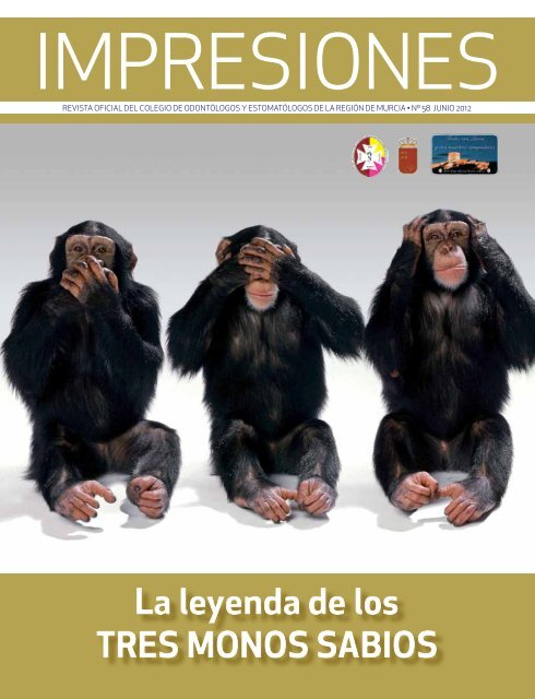
Es célebre el altorrelieve <strong>de</strong>l santuario <strong>de</strong> Toshogu –Japón- que representa a tres monos<br />
exhibiendo gestos humanizados. Uno <strong>de</strong> ellos se tapa los ojos, otro la boca y el último los oídos,<br />
en una presumible alusión a que cada uno <strong>de</strong> ellos carece <strong>de</strong> la facultad <strong>de</strong> la vista, la voz y el<br />
oído, respectivamente. La “Leyenda <strong>de</strong> los tres monos sabios” explica que fueron enviados<br />
por los dioses para que, <strong>de</strong> modo cooperativo, castigaran las malas acciones <strong>de</strong> los hombres.<br />
Debían complementarse para que sus cualida<strong>de</strong>s y sus carencias no fueran un obstáculo a la hora <strong>de</strong><br />
cumplir eficazmente su misión. Partiendo <strong>de</strong>l estudio <strong>de</strong> las distintas posibilida<strong>de</strong>s <strong>de</strong> or<strong>de</strong>nación <strong>de</strong><br />
los monos a la hora <strong>de</strong> transmitirse un mensaje, el profesor Yeray Santana Aday ha concluido que la<br />
única disposición válida es la <strong>de</strong> poner en primer lugar a Kikazaru, el mono sordo, <strong>de</strong>spués a Mizaru,<br />
el mono ciego, y al final a Iwazaru, el mono mudo (no como en la foto <strong>de</strong> portada <strong>de</strong> este número <strong>de</strong><br />
Impresiones). De esta forma el mensaje pasaría <strong>de</strong>l primero al último mono, encargado <strong>de</strong> imponer los<br />
castigos, sin experimentar merma alguna. El resto <strong>de</strong> las opciones aboca a la incomunicación. Santana<br />
ha utilizado la leyenda para teorizar sobre el acto <strong>de</strong> la comunicación en el seno <strong>de</strong> la sociedad.<br />
Todos hemos pa<strong>de</strong>cido en carne propia las interferencias, los ruidos –por utilizar un término <strong>de</strong><br />
semiología- que impi<strong>de</strong>n que el significado real, la intención <strong>de</strong> un enunciado, nos llegue nítido y<br />
veraz. Emisor-canal-mensaje-código-receptor, éstos son los elementos que intervienen en el acto<br />
<strong>de</strong> la comunicación; cuando alguno <strong>de</strong> ellos falla, es difícil la transmisión correcta <strong>de</strong>l mensaje. Así<br />
nos lo enseñaron durante el bachiller, pero sospecho que el espíritu incontaminado que por entonces<br />
disfrutábamos constituía un obstáculo para que los profesores nos dispensaran una enseñanza<br />
completa sobre el proceso. Los santos próceres velaban por que mantuviésemos el mayor tiempo<br />
posible una mirada limpia sobre el mundo. Pero pronto ese mismo mundo empezó a darnos revolcones<br />
y forzó que nuestra mirada <strong>de</strong>jara <strong>de</strong> ser tan limpia –pobre <strong>de</strong>l que no abandonó aquel estado virginal- y<br />
aprendimos a base <strong>de</strong> <strong>de</strong>sengaños que la manipulación, la <strong>de</strong>magogia, la distorsión, el cercenamiento,<br />
la ambigüedad… son elementos tan constitutivos como los primeros <strong>de</strong>l intercambio <strong>de</strong> información.<br />
El lenguaje es un arma po<strong>de</strong>rosísima que pue<strong>de</strong> provocar efectos contrapuestos: acercamiento/<br />
separación, concordia/hostilidad, esclarecimiento <strong>de</strong> la verdad/confusión y doblez. El sujeto cuya<br />
mente supura resentimiento e inquina –aquél que en mayor o menor medida todos hemos pa<strong>de</strong>cido<br />
alguna vez- con su mensaje agre<strong>de</strong> porque inocula en él una dosis <strong>de</strong>l segundo <strong>de</strong> los términos <strong>de</strong> cada<br />
par. Por un extraño complejo <strong>de</strong> inferioridad, la obra bien hecha actúa sobre esta persona agraviándola<br />
por comparación y tiene el po<strong>de</strong>r <strong>de</strong> situarla en un plano <strong>de</strong> nivel inferior. Es entonces cuando el<br />
hemisferio izquierdo se le recalienta y humea, las neuronas que lo conforman se agitan <strong>de</strong>sorientadas<br />
precipitando una caótica vorágine <strong>de</strong> sinapsis, que da como resultado algo parecido a un monólogo<br />
poco inspirado <strong>de</strong> Groucho Marx.<br />
Ese <strong>de</strong>lirio intracraneal se concreta a veces en una ficción bloguera, mixtura <strong>de</strong> frustraciones no<br />
superadas, críticas y valoraciones mal pon<strong>de</strong>radas y otros aliños que por su iniquidad <strong>de</strong>finen a su<br />
creador. Y muy a su pesar, también lo i<strong>de</strong>ntifican. Porque una agria soberbia infantil es su código <strong>de</strong><br />
barras. Aunque lo negara tres veces antes <strong>de</strong> graznar el negro cuervo que aletea en su espíritu, nunca<br />
superaría la prueba <strong>de</strong> la parafina lingüística e i<strong>de</strong>ológica.<br />
Aquellos monos <strong>de</strong>l templo <strong>de</strong> Toshogu quizá un día aprendan a situarse en la posición lógica para<br />
que <strong>de</strong> uno a otro pueda fluir la información y no se empantane y corrompa. Los oscuros recovecos <strong>de</strong><br />
aquella mente, sin embargo, nunca permitirán ese suave discurrir <strong>de</strong>l pensamiento; antes al contrario: si<br />
cierta vez alumbrara una i<strong>de</strong>a honesta y atemperada, pronto acudirían a ella sus particulares duen<strong>de</strong>s<br />
<strong>de</strong>l rencor para enherbolarla. Y si a sus oídos llegara <strong>de</strong> otros un concepto virtuoso, sobre él proyectaría<br />
<strong>de</strong> inmediato una mirada turbia para exhalarlo a los <strong>de</strong>más inficionado y pervertido.<br />
A ese triste personaje le aconsejo bienintencionadamente que se aplique a sí mismo el principio tan en<br />
boga <strong>de</strong> retorno <strong>de</strong> energía, y sopese si el tiempo y la acritud invertidos le reportan suficiente rédito.<br />
No se me ocurre aconsejarle un examen <strong>de</strong> conciencia ni ejercicios espirituales, sean <strong>de</strong>l credo que<br />
sean, pero quizá sí que conviva un tiempo con aquellos primates en el templo japonés (nunca hay que<br />
renunciar a la posibilidad <strong>de</strong> humanizarse).<br />
IMPRESIONES<br />
3
sumario<br />
3 Editorial<br />
6 Entrevista: Dr. Rafael Pacheco Guevara<br />
10 Lorca, 11 <strong>de</strong> Mayo <strong>de</strong> 2012: Primer Aniversario <strong>de</strong>l Terremoto<br />
11 Bone Healing Of Small And…<br />
21 Estudio <strong>de</strong> los Factores <strong>de</strong> Riesgo…<br />
26 Quiste Nasoalveolar…<br />
28 Artritis Séptica <strong>de</strong> La Rodilla…<br />
34 Traducción y Odontología<br />
38 Running para aficionados<br />
42 Premios <strong>de</strong> la Aca<strong>de</strong>mia <strong>de</strong> Medicina…<br />
45 Profesión y Prensa<br />
6<br />
34<br />
10<br />
38<br />
DELEGACIÓN EN MURCIA<br />
Juan Ruiz Parra<br />
968 340 968<br />
619 656 292<br />
juanrparra_4@hotmail.com<br />
Visita nuestra web<br />
www.<strong>de</strong>ntistasmurcia.com<br />
Edita: <strong>Ilustre</strong> <strong>Colegio</strong> <strong>Oficial</strong> <strong>de</strong> Odontólogos y<br />
Estomatólogos <strong>de</strong> la Región <strong>de</strong> <strong>Murcia</strong>.<br />
C/María Zambrano, 4. Edif. Dórico. Bajo. 30007<br />
<strong>Murcia</strong> Tlf. 968 20 16 65 Fax. 968 20 16 69.<br />
E-mail: info@<strong>de</strong>ntistasmurcia.com.<br />
Consejo Editorial: Presi<strong>de</strong>nte: R. Óscar Castro<br />
Reino. Vicepresi<strong>de</strong>nte: José Manuel Granero Marín.<br />
Tesorero: Cristina Cal<strong>de</strong>rón Díaz. Secretario: Antonio<br />
Parra López. Vicesecretario: Celestino García Alfaro<br />
(Distrital <strong>de</strong> Cartagena). Vocales: Carlos García<br />
Ballesta (Presi<strong>de</strong>nte Comisión Científica). Arsenio<br />
Gómez Fernán<strong>de</strong>z (Comarca <strong>de</strong>l Noroeste). Ana Mª.<br />
Hita Velasco. (Presi<strong>de</strong>nte Comisión Jóvenes <strong>Dentistas</strong>).<br />
Jesús Santos Pérez Ortega (Presi<strong>de</strong>nte Comisión Nuevas<br />
Tecnologías). Fernando Sacristán Molpeceres (Distrital<br />
<strong>de</strong> Lorca). Vocales Supernumerarios: Pedro Caballero<br />
Guerrero (Coordinador Activida<strong>de</strong>s Extracolegiales y<br />
apoyo a Nuevos Colegiados), José Luis Calvo Guirado<br />
(Coordinador Formación Continuada), David Sánchez<br />
Espín (Coordinador <strong>de</strong> Convenios Colectivos y ayuda<br />
al Dentista Autónomo).* Presi<strong>de</strong>nte <strong>de</strong> la Comisión<br />
Deontológica: Oscar Erans Richarte.<br />
Redactor jefe: Juan Ruiz Parra.<br />
Editor jefe: Prof. Dr. José Luis Calvo Guirado<br />
Co-Editor: Prof. Dr. Carlos García Ballesta<br />
Comité científico <strong>de</strong> Impresiones: Prof. Dr.<br />
Rafael Delgado Ruiz. Prof. Dr. José Maté Sánchez<br />
<strong>de</strong> Val. Dra. Piedad Ramírez Fernán<strong>de</strong>z. Dra.<br />
Laura López Marí. Dr. Bruno Negri.<br />
Revisores Externos: Prof. Dr. Gerardo Gómez<br />
Moreno. Dr. Javier Guardia Muñoz. Prof. Dr.<br />
Eugenio Velazco Ortega. Prof. Dra. Cristina<br />
Barona Dorado. Prof. Dr. José María Martínez<br />
González. Prof. Juan Carlos Prados Frutos<br />
Edición: Editorial MIC · Delegación en <strong>Murcia</strong>:<br />
Dirección: Juan Ruiz Parra.<br />
Depósito Legal: LE-1087-1995. ISSN: 1695-5269.<br />
Editorial MIC • Artesiano, s/n • León • 24010 • Pol. Ind.<br />
Trobajo <strong>de</strong>l Camino<br />
Teléfono: 902 271 902 • Fax: 902 371 902<br />
E-mail: mic@editorialmic.net • www.editorialmic.com<br />
• Delegaciónen <strong>Murcia</strong>: 968 340 968 • 619 656 292
IMPRESIONES<br />
6<br />
Entrevista<br />
Dr. Rafael Pacheco Guevara,<br />
Profesor <strong>de</strong> Medicina Legal y Bioética <strong>de</strong> la Umu<br />
Presi<strong>de</strong>nte <strong>de</strong>l Comité Ético<br />
Asistencial <strong>de</strong>l Hospital General<br />
Universitario Reina<br />
Sofía y hermano gemelo <strong>de</strong>l<br />
actual Pte. <strong>de</strong> la Audiencia<br />
Provincial <strong>de</strong> <strong>Murcia</strong>, <strong>de</strong>fien<strong>de</strong> una rigurosa<br />
racionalización <strong>de</strong>l gasto sanitario<br />
que no se aleje <strong>de</strong> los principios éticos y<br />
humanitarios que han <strong>de</strong> fundamentar el<br />
sistema sanitario.<br />
La limitación <strong>de</strong> la Tarjeta Sanitaria a<br />
los extranjeros o el abono <strong>de</strong> un porcentaje<br />
<strong>de</strong>l coste <strong>de</strong> los medicamentos,<br />
por ejemplo, ¿representan sólo una<br />
tímida insinuación <strong>de</strong> lo que <strong>de</strong>bería<br />
hacerse para hacer viable la sanidad<br />
pública?<br />
La situación actual no es sostenible.<br />
Cualquier intento para hacerla viable,<br />
siempre que no atente gravemente<br />
contra los fundamentos <strong>de</strong>l sistema sanitario<br />
(altruismo, solidaridad, universalidad,<br />
equidad y “gratuidad”) es <strong>de</strong>fendible,<br />
por necesaria.<br />
En efecto: podría ser consi<strong>de</strong>rada como<br />
una “tímida insinuación <strong>de</strong> lo que <strong>de</strong>bería<br />
hacerse”. Aunque lamentablemente, no<br />
es la mejor. Por un lado, no es suficiente;<br />
a<strong>de</strong>más, atenta contra la solidaridad y la<br />
universalidad y, en la práctica, será imposible<br />
llevarla a cabo.<br />
¿Sólo atención <strong>de</strong> urgencia a los inmigrantes<br />
sin trabajo?... Pronto se presentaran<br />
severas consecuencias: 1-Reventarán<br />
los servicio <strong>de</strong> urgencias, más<br />
<strong>de</strong> lo ya están. 2-Si no hay continuidad<br />
asistencial, estos pacientes serán un<br />
peligro sanitario y social. (¿Qué pasará<br />
El Dr. Pacheco no tiene pelos en la<br />
lengua. Ha <strong>de</strong>sempeñado cargos<br />
relevantes <strong>de</strong> gestión (fue Dir. Médico<br />
<strong>de</strong>l Hospital Los Arcos y <strong>de</strong>l General<br />
Universitario y Dir. Gerente <strong>de</strong>l Reina<br />
Sofía), aunque no parece haber <strong>de</strong>sarrollado<br />
ese lenguaje elíptico y sinuoso<br />
propio <strong>de</strong> quienes viven una experiencia<br />
<strong>de</strong> similar naturaleza. Cuando<br />
has <strong>de</strong> armonizar situaciones que<br />
exigen un alto grado <strong>de</strong> pragmatismo<br />
con los intereses <strong>de</strong>l grupo político<br />
que te ha nombrado, parece que sólo<br />
vienen a la mente –o se eligen minuciosamente-<br />
palabras laxas y vaporosas,<br />
a<strong>de</strong>cuadas para construir un discurso<br />
ambiguo y poco comprometedor. Al<br />
leer la entrevista queda patente que<br />
no es su caso.<br />
Texto: Juan Ruiz Parra<br />
con el VIH+ sin anti-retrovirales o con el<br />
tuberculoso bacilífero activo, sin medicación<br />
específica?). 3-Tras un grave <strong>de</strong>terioro<br />
clínico, aumentaran los ingresos<br />
y las estancias hospitalarias. ¡No se les<br />
va a <strong>de</strong>jar morir en la calle!<br />
Respecto a los medicamentos: Es cierto<br />
que existe frau<strong>de</strong> con las recetas <strong>de</strong><br />
pensionistas, pero… ¡Cuidado con los <strong>de</strong><br />
las pensiones “<strong>de</strong> hambre” y con los que<br />
carecen <strong>de</strong> ellas! Mejor que todos paguemos<br />
algo por las medicinas porque<br />
sólo así se valorarán suficientemente<br />
pero, para ello, habría antes que revisar<br />
esas cantida<strong>de</strong>s <strong>de</strong> subsistencia.<br />
A<strong>de</strong>más, si no van a pagar nada los parados<br />
sin subsidio y, <strong>de</strong>sgraciadamente,<br />
cada vez van a ser más, poco ahorro se<br />
vislumbra; dicho esto <strong>de</strong>s<strong>de</strong> el más pro-
MEJOR QUE TODOS<br />
PAGUEMOS ALGO POR<br />
LAS MEDICINAS PORQUE<br />
SÓLO ASÍ SE VALORARÁN<br />
SUFICIENTEMENTE PERO,<br />
PARA ELLO, HABRÍA ANTES<br />
QUE REVISAR ESAS CANTI-<br />
DADES DE SUBSISTENCIA<br />
fundo respeto ante esa dramática situación<br />
personal y familiar.<br />
¿No le sorpren<strong>de</strong> que no se hayan<br />
adoptado mucho antes medidas racionales<br />
<strong>de</strong> contención <strong>de</strong>l gasto como las<br />
centrales <strong>de</strong> compras o la dispensación<br />
<strong>de</strong> los medicamentos ajustada a la duración<br />
<strong>de</strong> los tratamientos?<br />
Más que sorpren<strong>de</strong>rme, me indigna.<br />
Llevamos un retraso <strong>de</strong> más 20 años.<br />
El Informe Abril data <strong>de</strong> 1991. Yo mismo,<br />
perdón por la inmo<strong>de</strong>stia, publiqué un<br />
artículo en el año 1993, <strong>de</strong>fendiendo la<br />
implantación <strong>de</strong> una tasa sanitaria asistencial,<br />
disuasoria <strong>de</strong>l mal uso y <strong>de</strong>l abuso<br />
(Revista El Médico, nº 492).<br />
¿Cómo se reparte la responsabilidad<br />
por el <strong>de</strong>ficiente estado en que se hallan<br />
actualmente los recursos sanitarios<br />
entre los profesionales <strong>de</strong>l sector,<br />
los gestores-políticos y los propios<br />
usuarios?<br />
Nadie está totalmente exento <strong>de</strong> responsabilidad.<br />
Los causantes <strong>de</strong>l serio<br />
<strong>de</strong>terioro en las cuentas son los políticos,<br />
quienes, llevados por un populismo<br />
suicida, no han tomado a tiempo medidas<br />
correctoras (por miedo a su rechazo<br />
y al correspondiente coste electoral).<br />
Los gestores han (hemos) estado a sus<br />
ór<strong>de</strong>nes, siendo cesados al menor reparo<br />
y sin agra<strong>de</strong>cimiento por los servicios<br />
prestados (bien sé <strong>de</strong> lo que hablo), lo<br />
cual no nos exime <strong>de</strong> haber compartido<br />
la falsa sensación <strong>de</strong> disponibilidad ilimitada<br />
<strong>de</strong> recursos.<br />
Los clínicos son igualmente responsables,<br />
al haber mantenido muchos, hasta<br />
hace bien poco, una absurda actitud, que<br />
se concreta en la frase: “Lo mío es curar<br />
y que sean otros los que hagan las cuentas<br />
y busquen los recursos...”. ¡Vale! : Lo<br />
suyo, lo más importante <strong>de</strong> su misión, es<br />
intentar curar a los enfermos, pero <strong>de</strong>be<br />
saber que, cuando <strong>de</strong>ci<strong>de</strong> un gasto, hace<br />
uso <strong>de</strong> unos fondos escasos y que son<br />
<strong>de</strong> todos. El médico también <strong>de</strong>be implicarse<br />
en la eficiente utilización <strong>de</strong> los<br />
medios diagnósticos y terapéuticos.<br />
Los usuarios (consi<strong>de</strong>rados <strong>de</strong> manera<br />
colectiva) tampoco están excluidos <strong>de</strong><br />
crítica. Las actitu<strong>de</strong>s sociales predominantes<br />
han creado una <strong>de</strong>manda, insaciable<br />
y consumista, <strong>de</strong> asistencia médica<br />
y <strong>de</strong> servicios sanitarios (fascinación<br />
tecnológica). Casi hemos olvidado que<br />
la enfermedad y la muerte son parte <strong>de</strong><br />
la vida, y ésta es un proceso biológico<br />
frágil, vulnerable, limtado e irreversible.<br />
No se ha transmitido bien a la sociedad<br />
que la medicina pública tiene como principal<br />
objetivo garantizar una asistencia<br />
sanitaria <strong>de</strong> calidad… pero no la salud<br />
total, mantenida y dura<strong>de</strong>ra, en todo<br />
momento y <strong>de</strong> manera permanente,<br />
sencillamente porque eso es imposible.<br />
Nuestras leyes garantizan el <strong>de</strong>recho a<br />
una asistencia sanitaria <strong>de</strong> calidad (lo<br />
que no es poco), pero no el “<strong>de</strong>recho a la<br />
salud”, ni tampoco a la felicidad.<br />
Ante una expectativa tan alta, se alcanza<br />
pronto el umbral <strong>de</strong> la frustración, lo que<br />
da lugar a <strong>de</strong>mandas y reclamaciones,<br />
siendo la causa <strong>de</strong> que los agentes sanitarios<br />
caigamos en el riesgo <strong>de</strong> practicar<br />
medicina <strong>de</strong>fensiva, lo que contribuye a<br />
la ruina <strong>de</strong>l servicio nacional <strong>de</strong> salud. Dicho<br />
sea esto, sin cuestionar la existencia<br />
<strong>de</strong> una imprescindible responsabilidad<br />
profesional sanitaria (garantía social<br />
irrenunciable).<br />
Los ciudadanos <strong>de</strong>berán, en el futuro,<br />
implicarse más en la conservación <strong>de</strong> su<br />
estado <strong>de</strong> salud (co-responsabilidad y<br />
auto-cuidado).<br />
¿Cree posible hoy día, tras el camino<br />
<strong>de</strong>scentralizador que llevamos recorrido,<br />
volver a un mo<strong>de</strong>lo rector que<br />
subsane las profundas <strong>de</strong>sigualda<strong>de</strong>s<br />
<strong>de</strong> cobertura sanitaria que a menudo<br />
se producen entre las distintas CC.AA?<br />
Entrevista<br />
Lo consi<strong>de</strong>ro imprescindible. Si ya es<br />
muy difícil justificar el mantenimiento<br />
<strong>de</strong> 17 gobiernos autonómicos, 17 parlamentos<br />
y otras tantas televisiones autonómicas…<br />
creo disparatado afrontar el<br />
gasto <strong>de</strong> 17 servicios sanitarios públicos,<br />
compitiendo entre ellos y dando lugar a<br />
<strong>de</strong>sigualda<strong>de</strong>s. Ahí están las cifras, <strong>de</strong>s<strong>de</strong><br />
la <strong>de</strong>scentralización <strong>de</strong>l INSALD.<br />
Lógico: Todo lo referido a la salud, es<br />
muy valorado por los votantes. Esto,<br />
bien lo supieron los distintos políticos<br />
regionales (<strong>de</strong> uno u otro color político),<br />
que entraron en una dinámica <strong>de</strong> oferta<br />
<strong>de</strong>smesurada, cuyos resultados ahora<br />
pa<strong>de</strong>cemos.<br />
El Plan <strong>de</strong> Atención Dental Infantil supuso<br />
en <strong>Murcia</strong> un medio benefactor<br />
para que personas sin recursos pudieran<br />
acce<strong>de</strong>r a una parcela <strong>de</strong> la sanidad<br />
con cobertura pública tradicionalmente<br />
muy limitada. En este momento corre<br />
el riesgo <strong>de</strong> <strong>de</strong>saparecer o <strong>de</strong> ver<br />
muy recortada su dotación económica.<br />
¿Cómo <strong>de</strong>berían <strong>de</strong> aquilatarse los recursos<br />
para que la población más <strong>de</strong>sfavorecida<br />
no se enfrente a una situación<br />
insostenible?<br />
Regulándolo para que pueda mantenerse…<br />
aunque los usuarios (padres <strong>de</strong>l<br />
niño) tengan que contribuir con una cantidad<br />
simbólica, que hará más valorable<br />
y más viable el servicio recibido por su<br />
hijo. La gratuidad total y absoluta es<br />
perversa, porque genera abusos e ineficiencias.<br />
Muchas veces se habla <strong>de</strong> la presión<br />
que ejercen las gran<strong>de</strong>s corporaciones<br />
empresariales <strong>de</strong>l sector <strong>de</strong> la sanidad<br />
para perpetuar una estructura claramente<br />
inviable <strong>de</strong>s<strong>de</strong> los puntos <strong>de</strong><br />
vista social y económico. ¿Qué hay <strong>de</strong><br />
verdad en ello? ¿Consi<strong>de</strong>ra el clientelismo<br />
un obstáculo difícil <strong>de</strong> vencer?<br />
Las compañías farmacéuticas y las que<br />
fabrican elementos <strong>de</strong> electromedicina<br />
u ortopédicos, no son instituciones<br />
benéficas. Investigan utilizando alta<br />
tecnología, creando fármacos seguros<br />
y eficaces, así como artilugios útiles y<br />
los ven<strong>de</strong>n, persiguiendo un beneficio<br />
económico para sus accionistas y empleados.<br />
Hay que conseguir un a<strong>de</strong>cuado<br />
equilibrio, pagando lo justo y razonable,<br />
IMPRESIONES<br />
7
IMPRESIONES<br />
8<br />
Entrevista<br />
en proporción con el bien real que generan.<br />
Se les <strong>de</strong>ben exigir el seguimiento<br />
<strong>de</strong> códigos <strong>de</strong> buenas prácticas empresariales.<br />
En sanidad, casi todo es complicado.<br />
También lo es permanecer totalmente<br />
al margen <strong>de</strong> la publicidad (muy bien<br />
estructurada), respecto a nuevos remedios<br />
curativos, prometedores <strong>de</strong> más<br />
bienestar y menos sufrimiento… por<br />
eso hay que contar con los gestores<br />
óptimos: los mejor preparados y los más<br />
honestos. La eficiencia es algo tan sencillo<br />
como la suma <strong>de</strong> eficacia y <strong>de</strong>cencia.<br />
Ud. es muy crítico ante la irracionalidad<br />
<strong>de</strong>l actual sistema. En su época <strong>de</strong><br />
gestor, ¿notó el peso <strong>de</strong> una inercia estructural<br />
hacia el <strong>de</strong>spilfarro?<br />
Sí, por parte <strong>de</strong> todos, sin excepción. No<br />
hemos sido suficientemente conscientes<br />
<strong>de</strong> la escasez <strong>de</strong> los recursos, hasta<br />
que la “bomba” nos ha estallado <strong>de</strong>lante.<br />
NO HEMOS SIDO<br />
SUFICIENTEMENTE<br />
CONSCIENTES<br />
DE LA ESCASEZ DE LOS<br />
RECURSOS, HASTA QUE<br />
LA “BOMBA” NOS HA<br />
ESTALLADO DELANTE<br />
La <strong>de</strong>manda ha sido muy exigente y la<br />
oferta ha ido tras ella, en un alocado intento<br />
por satisfacerla. La Medicina y el<br />
SNS han sido víctimas <strong>de</strong> su éxito.<br />
¿Qué papel pue<strong>de</strong>n <strong>de</strong>sempeñar la ética<br />
y la educación en el cambio que Ud.<br />
<strong>de</strong>clara tan necesario en la utilización<br />
<strong>de</strong> los recursos públicos?<br />
Ambas son fundamentales e imprescindibles.<br />
1-Sin ética es imposible el ejercicio <strong>de</strong><br />
cualquiera <strong>de</strong> las activida<strong>de</strong>s sanitarias.<br />
De ahí la conveniencia <strong>de</strong> los colegios.<br />
Son quienes <strong>de</strong>ben velar por la <strong>de</strong>ontología,<br />
que no es otra cosa que la ética específica<br />
<strong>de</strong> los miembros <strong>de</strong> un <strong>de</strong>terminado<br />
colectivo profesional. Valga como<br />
ejemplo, el concepto “secreto”, en todos<br />
los ciudadanos es valorable y en los sanitarios<br />
pasa a ser exigible (el concepto<br />
<strong>de</strong>viene en precepto).<br />
La ética y la <strong>de</strong>ontología no atañen solamente<br />
a los profesionales asistenciales;<br />
también y mucho, a los gestores <strong>de</strong><br />
la salud. Se presentan graves dilemas<br />
éticos en la gestión sanitaria (garantías<br />
<strong>de</strong> equidad, criterios justos y lógicos<br />
para la las listas <strong>de</strong> espera, pérdidas <strong>de</strong><br />
oportunidad, or<strong>de</strong>n <strong>de</strong> priorida<strong>de</strong>s en el<br />
gasto…).<br />
Ante esos dilemas: capacidad, formación,<br />
honra<strong>de</strong>z, pru<strong>de</strong>ncia, valentía y<br />
autocrítica, a<strong>de</strong>más <strong>de</strong> facilidad para<br />
escuchar, compartir, dialogar, comunicar<br />
y explicar las <strong>de</strong>cisiones.<br />
2-La educación es la base <strong>de</strong> todo. Junto<br />
con la evolución, nos diferencia y distancia<br />
<strong>de</strong>l resto <strong>de</strong> los primates. Un país es<br />
lo que la educación <strong>de</strong> su ciudadanía. Sin<br />
educación, no hay futuro. Es imprescindible<br />
una sólida educación sanitaria…<br />
pero, para alcanzarla, tenemos que conseguir<br />
una mejor educación <strong>de</strong> todos<br />
(<strong>de</strong>s<strong>de</strong> la escuela)<br />
Para terminar, permítaseme una reflexión:<br />
Somos como conducimos. Quien<br />
quiera saber sobre la educación colectiva<br />
<strong>de</strong> los españoles, sólo tiene que permanecer<br />
un rato observando en un paso<br />
<strong>de</strong> cebra, en cualquier ciudad <strong>de</strong> nuestro<br />
país…. Conduciendo como lo hacemos…<br />
¿realmente es lógico esperar un<br />
uso racional, serio, a<strong>de</strong>cuado, sensato y<br />
comedido <strong>de</strong>l excelente y “gratuito” servicio<br />
sanitario a nuestro alcance?
IMPRESIONES<br />
10<br />
Vida Colegial<br />
LORCA, 11 DE MAYO DE 2012:<br />
Primer aniversario <strong>de</strong>l Terremoto<br />
Fernando Sacristán Molpeceres. Distrital <strong>de</strong> Lorca.<br />
No obstante, hemos <strong>de</strong> manifestar que nunca nos<br />
hemos sentidos solos. Nuestro colectivo siempre<br />
ha estado respaldado <strong>de</strong>s<strong>de</strong> esos primeros difíciles<br />
momentos por nuestro <strong>Ilustre</strong> <strong>Colegio</strong>, por<br />
su Junta Directiva, por nuestro presi<strong>de</strong>nte Óscar<br />
Castro, que no ha cejado ni un solo día <strong>de</strong> luchar, <strong>de</strong> interesarse<br />
por nuestra situación tanto personal como profesional para<br />
gestionar en la menor mayor brevedad la ayuda que nos posibilitara<br />
la reparación <strong>de</strong> nuestras clínicas.<br />
A día <strong>de</strong> hoy, nos encontramos con una ciudad <strong>de</strong>sencantada,<br />
triste, que unos días siente dudas en lo referente a su futuro, y<br />
otras, cuando observa que las ayudas solicitadas van llegando,<br />
y van paliando sus necesida<strong>de</strong>s más básicas, recupera el optimismo,<br />
ese aliento <strong>de</strong> esperanza que todos <strong>de</strong>seamos para<br />
una pronta recuperación <strong>de</strong> la actividad económica, laboral, en<br />
<strong>de</strong>finitiva, <strong>de</strong> esa vitalidad que con anterioridad tenía la ciudad<br />
<strong>de</strong> Lorca.<br />
En la actualidad, todas las clínicas están funcionando con<br />
“normalidad”. A la crisis financiera, conocida por todos, se<br />
Hoy se cumple un año <strong>de</strong> terremoto que<br />
enmu<strong>de</strong>ció y paralizó toda la comarca <strong>de</strong><br />
Lorca. La memoria común <strong>de</strong> todos los<br />
lorquinos nos hace evocar sufrimiento,<br />
confusión, perplejidad, dolor, muerte…<br />
Nuestro gremio como el resto <strong>de</strong> los lorquinos<br />
soportó el azote terrible <strong>de</strong> esa falla que<br />
atraviesa nuestra ciudad. De las dieciocho<br />
clínicas <strong>de</strong>ntales <strong>de</strong> Lorca, diecisiete<br />
pa<strong>de</strong>cieron <strong>de</strong>terioros importantes en sus<br />
instalaciones. En mayor o menor medida todas<br />
han necesitado significativas reparaciones en<br />
sus infraestructuras, que han sido costosas y<br />
largas, hasta po<strong>de</strong>r reanudar con normalidad<br />
nuestra actividad laboral.<br />
nos ha sumado la crisis particular generada por el terremoto.<br />
La actividad profesional <strong>de</strong> los <strong>de</strong>ntistas <strong>de</strong> Lorca ha<br />
caído <strong>de</strong> manera significativa, muy por <strong>de</strong>bajo <strong>de</strong> la media,<br />
no sólo regional, sino nacional. Por en<strong>de</strong>, apenas cubrimos<br />
los gastos básicos que nos permitan cubrir nuestra actividad<br />
profesional.<br />
Pero ante la adversidad, la fuerza y la unidad <strong>de</strong> los lorquinos<br />
harán que salgamos a<strong>de</strong>lante. Hoy tenemos que<br />
estar con nuestra ciudad, y la mejor manera <strong>de</strong> <strong>de</strong>mostrarlo<br />
es no <strong>de</strong>sfallecer y mantener un servicio odontológico<br />
<strong>de</strong> calidad, para que su saludad buco<strong>de</strong>ntal no se<br />
vea perjudicada y tengan acceso a los tratamientos que<br />
necesiten.<br />
Para finalizar, como dice el popular refrán castellano<br />
–es <strong>de</strong> bien nacido ser agra<strong>de</strong>cido, quisiera agra<strong>de</strong>cer<br />
todo el apoyo tanto económico como moral que hemos<br />
recibido.<br />
Un fuerte abrazo a todos.
Prof. Dr. José Luis Calvo Guirado<br />
Bone healing of small and large<br />
<strong>de</strong>fects filled with bovine and porcine bone<br />
subtitutes: an experimental study in<br />
american fox hound dogs<br />
Corresponding author:<br />
Prof. Dr. José luis Calvo<br />
Guirado<br />
Calle Marques <strong>de</strong> los Velez s/n. Hospital<br />
Morales Meseguer, 2ª Planta, Clinica<br />
Odontológica Integrada <strong>de</strong> Adultos. Faculty<br />
of Medicine and Dentistry. University of<br />
<strong>Murcia</strong>. Spain. Tel 34-868 888 584<br />
Abstract:<br />
Objectives: This experimental study was<br />
<strong>de</strong>signed in or<strong>de</strong>r to compare new bone<br />
formation and crestal bone resorption<br />
after grafting alveolar small and large<br />
extraction sites in canine mandibles with<br />
two different biomaterial types: Endobone<br />
(bovine bone) and MP3 (porcine bone)<br />
at 3 months.<br />
Materials and methods: Six American<br />
Fox Hound dogs were used in the study.<br />
Second premolars (PM2) and first molars<br />
(M1) areas were used as test and<br />
third premolars (PM3) site the extraction<br />
socket was left with its blood clot and interrupted<br />
sututes were used as control<br />
. Those 5 mm premolar area and 10 mm<br />
molar areas <strong>de</strong>fects were filled with the<br />
José Luis Calvo-Guirado * • Rafael Delgado Ruiz * • Bruno Negri * • Laura López Marí*<br />
• Jose Eduardo Maté Sánchez <strong>de</strong> Val* • Maria Piedad Ramirez Fernán<strong>de</strong>z * • Marcus<br />
Abboud**<br />
* Department of General and Implant Dentistry, Faculty of Medicine and Dentistry,<br />
University of <strong>Murcia</strong> Spain<br />
** Department of Prostodontics & Digital Technology. Stony Brook University. New<br />
York.USA.<br />
two biomaterials (Endobone® y MP3®).<br />
The animals were sacrificed four and<br />
eight weeks. After 8 weeks of healing, the<br />
vertical bone loss was <strong>de</strong>termined by placing<br />
a horizontal line passing by the buccal<br />
and lingual bone crest. The mean of<br />
vertical loss of buccal bone plate for large<br />
<strong>de</strong>fects was significantly low of MP3<br />
group was 2.37 ± 0.64 mm, Endobone<br />
group 2.26 ± 0.72 mm compared with control<br />
group 3.11 ± 0.81 (p
IMPRESIONES<br />
12<br />
Divulgación Científica<br />
duces higher quantities of connective<br />
tissue. Endobon material acts as a good<br />
scaffold for new bone formation.<br />
Runnig title: Bone healing in small and<br />
large <strong>de</strong>fects<br />
Key words: biomaterial, bone grafts, bovne<br />
bone, porcine bone<br />
INTRODUCTION<br />
The reconstruction of osseous <strong>de</strong>fects<br />
remains an important and unresolved<br />
issue in oral surgery. Healing processes<br />
following <strong>de</strong>ntal extractions inclu<strong>de</strong><br />
the formation and maturation of blood<br />
clots, the infiltration of immature mesenchymal<br />
cells and the formation of a<br />
provisional bone matrix (Heberer et al.<br />
2008). Immature bone becomes established<br />
quite early on in this process to<br />
be replaced later by mature trabecular<br />
bone (Becker, 2003). Often during this<br />
healing period, bone loss occurs in the<br />
walls surrounding an extraction site<br />
with a reduction to the buccal alveolar<br />
crest (Araújo et al. 2009).<br />
There are various alternatives available<br />
for the treatment of these osseous <strong>de</strong>fects,<br />
the most traditional and long established<br />
of these being autogenous bone<br />
grafts, used to replace the lost bone.<br />
However, this technique has certain disadvantages<br />
given that the quantity of<br />
available bone is always limited and that<br />
it involves a second surgical site and so<br />
increased cost and treatment time and<br />
may lead to further problems at the bone<br />
graft donor site such as bleeding, infection,<br />
and pain. For this reason different<br />
graft materials have been <strong>de</strong>veloped<br />
which are inten<strong>de</strong>d to bring about new<br />
bone formation (Nomura et al. 2005;<br />
Pérez-Sánchez et al. 2010; Schwarz et<br />
al. 2007; Tudor et al. 2007; Busenlechner.<br />
2005). These are used in orthopedic, oral<br />
and maxillofacial surgery, for example,<br />
in cases of skeletal <strong>de</strong>fects or following<br />
surgical procedures (Schmitt et al. 2008;<br />
Schwartz et al. 2008).<br />
It was suggested that the ridge dimensions<br />
could be preserved if implants were<br />
placed in the fresh extraction socket (Denissen<br />
et al. 2000; Paolantonio et al.<br />
2001). The validity of this hypothesis was<br />
processes of hard tissue mo<strong>de</strong>ling and<br />
remo<strong>de</strong>ling following tooth extraction<br />
were studied in the dog mo<strong>de</strong>l (Cardaro-<br />
poli et al. 2003; Araújo & Lindhe 2005). It<br />
was <strong>de</strong>monstrated that the socket was<br />
first occupied by a coagulum that was<br />
replaced with granulation tissue, provisional<br />
connective tissue and woven bone.<br />
This immature hard tissue was subsequently<br />
replaced with lamellar bone and<br />
marrow. During healing, the height of the<br />
buccal bone wall was substantially reduced.<br />
In addition, about 30% of the marginal<br />
portion of the alveolar process of<br />
the extraction site was mo<strong>de</strong>led and lost<br />
(Araújo et al. 2008). In later experiments<br />
using the same mo<strong>de</strong>l, the post-extraction<br />
socket was grafted with Bio-Oss<br />
collagen (Geistlich Pharma) and healing<br />
was monitored in biopsies sampled after<br />
3 months (Araújo et al. 2008) or 6 months<br />
(Araújo & Lindhe 2009) of healing.<br />
However, these synthetic materials were<br />
unable to respond to physiological loads<br />
or biochemical stimuli in the way that living<br />
tissue does (Hench et al. 2002). Over<br />
the last <strong>de</strong>ca<strong>de</strong> research has continued<br />
the search for an i<strong>de</strong>al material for replacing<br />
lost bone, whether this be through<br />
resorption, trauma or fracture.<br />
Bone formation occurs through four processes:<br />
osteogenesis (resulting from the<br />
migration of autogenous osteoblasts and<br />
mesenchymal cells), osteoconduction<br />
(the material works as a passive support<br />
which is absorbed and replaced by bone),<br />
osteoinduction (the material stimulates<br />
osteoblasts and osteoclasts which generate<br />
new bone) (Leite and Ramalho,<br />
2008; Busenlechner et al. 2005; Schwartz<br />
et al. 2008) and osteointegration (the<br />
chemical union with local bone without a<br />
separating soft tissue wall) (Tudor et al.<br />
2007). In this way, a third generation of<br />
materials have come into use that seek<br />
to activate genes stimulating the regeneration<br />
of living tissue (Hench et al. 2002).<br />
One general problem with biomaterials is<br />
the possible transmission of pathogens<br />
and, because of this, they are subjected to<br />
exhaustive processing which can affect<br />
their Aloplastic materials have been <strong>de</strong>veloped<br />
in an attempt to overcome this<br />
problem, offering positive regenerative<br />
properties whilst being readily available,<br />
sterile and of reduced morbidity (Pérez-<br />
Sánchez et al. 2010). In addition to bone<br />
substitutes of allogeneic origin, there are<br />
others of xenogenic origins, synthetically<br />
produced using calcium-based materials<br />
(Calvo-Guirado et al. 2010).<br />
The clinical success of bone substitute<br />
materials <strong>de</strong>pends on their ability to<br />
support primary bone formation and also<br />
to regenerate mature bone. Bone formation<br />
and the quality of the newly formed<br />
bone will be influenced by chemical and<br />
physical properties of the graft materials<br />
(Schwartz et al. 2008).<br />
Two of the most frequently used biomaterials<br />
are <strong>de</strong>rived from bovine and porcine<br />
bone. The bone substitute of bovine<br />
origin (ma<strong>de</strong> up of natural bone mineral<br />
with extensive interconnected pores<br />
[Kim & Kim. 2007]) is <strong>de</strong>rived from trabecular<br />
bone; the porcine bone substitute is<br />
<strong>de</strong>rived from both trabecular bone (80%)<br />
and cortical bone (20%). Both biomaterials<br />
are used in granulated form in or<strong>de</strong>r<br />
to achieve a slower resorption (Figuereido<br />
et al. 2009), although they may be used<br />
in block form (Zambuzzi et al. 2006). They<br />
offer both osteoconductive and osteoinductive<br />
qualities (Zambuzzi et al. 2006).<br />
Bovine bone has a neutral pH, a property<br />
which facilitates healing processes<br />
during the first week (Leite & Ramalho.<br />
2008). It is effective for the regeneration<br />
of bone <strong>de</strong>fects, for sinus elevation and<br />
for repairing periodontal <strong>de</strong>fects prior<br />
to implant placement (Heberer et al.<br />
2008; Schwuarz et al. 2007; Carmagnola<br />
et al. 2008). When biomaterial of bovine<br />
origin is used as a graft material in post<br />
extraction alveoli, a series of processes<br />
occur which inclu<strong>de</strong> innate inflammation,<br />
the formation of granulation tissue and a<br />
provisional matrix, resorption of the graft<br />
material surface, the formation of new<br />
bone and the integration of the biomaterial<br />
into hard tissue (Araújo et al. 2010).<br />
Biomaterial of porcine origin, which has<br />
high biocompatibility, generates the<br />
formation of new bone in contact with<br />
biomaterial particles. This material is<br />
used for maxillary sinus elevation prior to<br />
implant placement (Calvo.Guirado et al.<br />
2009; Calvo-Guirado et al. 2010).<br />
Although studies of bovine bone studies<br />
are numerous, there is a general lack of<br />
research into porcine bone properties in<br />
relation to bone formation and resorption<br />
of its particles when used as a bone<br />
substitute. For this reason, the present<br />
study compares the effects of bovine<br />
and porcine bone graft materials used<br />
to repair small and large post-extraction<br />
alveoli at 8 weeks.
IMPRESIONES<br />
14<br />
Divulgación Científica<br />
Figure1. Composición <strong>de</strong> Endobon ( hueso bovino)<br />
con MEB<br />
Figura 2. Composicion <strong>de</strong> MP3 (porcine bone) con<br />
MEB<br />
MATERIALS AND METHODS<br />
This experimental study was <strong>de</strong>signed<br />
in or<strong>de</strong>r to compare new bone formation<br />
after grafting alveolar extraction sites<br />
in canine mandibles with two different<br />
biomaterial types: Endobone® (bovine<br />
bone) and MP3® (porcine bone).<br />
Six Fox Hound dogs were used in the<br />
study, of one year of age and weighing<br />
between 12 and 13 kg each. The experiment<br />
protocol was <strong>de</strong>signed in accordance<br />
with gui<strong>de</strong>lines laid down by the<br />
Spanish and E.U. authorities for animal<br />
experiments. The University of <strong>Murcia</strong><br />
(Spain) Ethics Committee for Animal<br />
Research approved the experiment,<br />
following the European Union Council<br />
Directive 86/609/EEC, 24th November,<br />
1986.<br />
Surgical Procedure:<br />
During surgical procedures the animals<br />
were anesthetized with Pentothal Natrium®<br />
(30 mg/ ml; Abbot Laboratories,<br />
Chicago, IL, U.S.A.) administered intravenously.<br />
Second premolars (PM2) and first molars<br />
(M1) were hemi-sected and extracted<br />
from both cuadrants of the animal´s<br />
mandibles using extraction needles. Extractions<br />
were carried out by sectioning<br />
teeth with a carbi<strong>de</strong> tungsten drill and<br />
removing the roots with forceps. Sulcular<br />
incisions were ma<strong>de</strong> along the vestibular<br />
and lingual areas adjoining the alveoli, separating<br />
tissues in or<strong>de</strong>r to make crestal<br />
wall hard tissue visible.<br />
GRAFTED ALVEOLOUS<br />
The alveoli corresponding to extracted<br />
teeth were refilled with the two biomaterials<br />
one bovine origin Endobone® (Biomet<br />
3i, Palm Beach Gra<strong>de</strong>ns, FL, USA.<br />
Figure 1) and the other one porcine origin<br />
MP3® (OsteoBiol, Tecnoss Dental, Turin,<br />
Italy. Figure 2) (table 1).<br />
Endobone® was placed in alveoli corresponding<br />
to PM2 (small <strong>de</strong>fect SD) and M1<br />
(large <strong>de</strong>fects LD) of the right hand hemimandible<br />
and MP3® in the PM2 and M1<br />
alveoli of the left hand hemi-mandible.<br />
The tissue flaps left loose by sulcular<br />
incisions were put back in position with<br />
tension free adaptation using interrupted<br />
and horizontal mattress sutures for<br />
wound closure. All procedures were carried<br />
out un<strong>de</strong>r uniform sterility, anesthetic<br />
and operating conditions During the<br />
first week following surgery the animals<br />
were treated with Amoxicyline (500 mg.<br />
twice daily) and Ibuprofen (600mg three<br />
times a day) administered systemically.<br />
Sutures were removed after two weeks.<br />
The animals were sacrificed at four and<br />
eight weeks after graft insertion by<br />
means of an overdose of Pentothal Natrium®<br />
(Laboratorios Abbot) perfused<br />
via the carotid arteries with a fixative<br />
Element (Atomic %) C O P Ca Mg Na Cl Yb S Sb Pt Sn<br />
Composition<br />
Endobon Spectrum-2 29.53 13.18 15.25 56.68 0.15 0,41 0.13 0.13 2.98 1.05 0.64<br />
MP3 Spectrum-5 33.84 15.01 16.63 39.93 0.23 0.25 0.15 0.12 2.68 1.57 0.71<br />
Table 1: Composition of both biomaterial used<br />
Time of<br />
measurement<br />
Connective<br />
Tissue<br />
Mp3<br />
Mean ±SD<br />
%<br />
Connective<br />
Tissue<br />
ENDOBON<br />
Mean ±SD<br />
%<br />
New Bone<br />
Mp3<br />
Mean ±SD<br />
%<br />
New Bone<br />
ENDOBON<br />
Mean ±SD<br />
%<br />
Residual<br />
Graft<br />
Mp3<br />
Mean ±SD<br />
%<br />
Residual<br />
Graft<br />
ENDOBON<br />
Mean ±SD<br />
%<br />
4 Weeks 50,1±0,14 55,2±0,32 32,1±0,22 18,3±0,64 17,8±1,12* 26,5±0,92*<br />
8 Weeks 45,8±0,72 51,6±0,64 38,3±0,81 24,3±0,35 15,9±0,45 24,1±0,84<br />
Table 1. Large <strong>de</strong>fects in Endobon and MP3 grafts were observed. * p< 0.005<br />
Time of<br />
measurement<br />
Connective<br />
Tissue<br />
Mp3<br />
Mean ±SD<br />
%<br />
Connective<br />
Tissue<br />
ENDOBON<br />
Mean ±SD<br />
%<br />
New Bone<br />
Mp3<br />
Mean ±SD<br />
%<br />
New Bone<br />
ENDOBON<br />
Mean ±SD<br />
%<br />
Residual<br />
Graft<br />
Mp3<br />
Mean ±SD<br />
%<br />
Residual<br />
Graft<br />
ENDOBON<br />
Mean ±SD<br />
%<br />
4 Weeks 40,3±0,12 26,6±0,26 37,4±0,41 34,8±0,84 22,3±0,76* 38,6±0,28*<br />
8 Weeks 28,7±0,41 23,1±0,76 52,6±0,84 41,7±0,71 18,7±0,62 35,2±0,54<br />
Table 1. Small <strong>de</strong>fects in Endobon and MP3 grafts were observed. * p< 0.005<br />
containing 5% glutaral<strong>de</strong>hy<strong>de</strong> and 4%<br />
formal<strong>de</strong>hy<strong>de</strong> (Karnovsky, 1965).<br />
Mandibles were dissected and each<br />
alveolar area was removed using a diamond<br />
saw. Biopsies were processed in<br />
sections following methods <strong>de</strong>scribed by<br />
Donath and Breuner (1982).<br />
MORPHOMETRIC MEASUREMENTS:<br />
The composition of the alveolar process<br />
as well as the newly formed tissue in the<br />
control socket site was <strong>de</strong>termined using<br />
a point-counting procedure<br />
Samples were prepared for histomorphometric<br />
analyses to evaluate the percentage<br />
of neo-formed bone, residual graft<br />
material and connective tissue in each<br />
sample. First, they were <strong>de</strong>hydrated using<br />
increasing gra<strong>de</strong>s of ethanol and then em-
Figure 3. Small <strong>de</strong>fect samples were prepared<br />
for histomorphometric analyses to evaluate the<br />
percentage of neo-formed bone, residual graft<br />
material and connective tissue. Each section was<br />
stained with Hematoxylin-Eosin.<br />
IMPRESIONES<br />
16<br />
Divulgación Científica<br />
bed<strong>de</strong>d in paraffin. Sections representing<br />
the experiment alveoli were cut in a buccolingual<br />
ground sections, parallel to their<br />
main axis and each section was stained<br />
with Hematoxylin-Eosin (Fig. 3 and 4).<br />
The mean values and standard <strong>de</strong>viations<br />
of the different variables were calculated<br />
using the dog as the statistical unit.<br />
HISTOMETRIC MEASUREMENTS<br />
The periphery of the extraction socket<br />
(the socket walls and a line connecting<br />
the mesial and the distal crest) was outlined<br />
with the use of a cursor and the area<br />
was assessed. Subsequently, the area occupied<br />
by newly formed bone (hard tissue<br />
and primitive bone marrow) was <strong>de</strong>termined<br />
in an i<strong>de</strong>ntical manner. The amount of<br />
newly formed hard tissue was calculated<br />
and expressed as percentage of the total<br />
socket area.<br />
Microscope and image system: The histologic<br />
examinations and measurements<br />
were performed in a MIP-4.5 (Leica,<br />
Wetzlar, Germany) equipped with an image<br />
system (Q-500 MC; Leica) and a digital<br />
image software (ImageAccess, Imagic,<br />
Glatbrugg, Switzerland).<br />
RESULTS<br />
In all experimental sites, healing was uneventful.<br />
After 8 weks of healing, a<br />
keratinized mucosa was observed to cover<br />
the entrance of the e<strong>de</strong>ntulous part<br />
of all small and lage <strong>de</strong>fect sites.<br />
Histomorphometric measurements<br />
Bone formation was greater with MP3-<br />
LD (32.1 ± 0.22%) and MP3-SD (37.4 ±<br />
0.41 %) than with Endobon-LD (18.3 ±<br />
Figure 4. Large <strong>de</strong>fects filled with porcine (a)<br />
and bovine bone (b). Esch section was stained<br />
with Hematoxilin-Eosin.<br />
0.64 %) and Endobon-SD (34.8 ± 0.84 %)<br />
compared with Control- LD ( 15.01 ± 0.42<br />
%) and Contol-SD (29.86 ± 0.25 %) at 4<br />
weeks. At eight weeks new bone formation<br />
was observed also in MP3-LD (38.3 ±<br />
0.81 %) and MP3-SD (52.6 ± 0.84 %) than<br />
Endobon-LD (24.3 ± 0.35 %) and Endobon<br />
SD (41.7±0.71 %) compared with Control<br />
sites LD ( 34.65 ±0.31) and Control-SD (<br />
33.21 ± 0.54).<br />
Tables one and two shows the percentage<br />
of connective tissue at the extraction<br />
sockets for each study time. It can<br />
be seen that the quantity of connective<br />
tissue was greater in control sites LD<br />
(84,9 ± 0,11 %) than Endobon-LD (55,2<br />
± 0,32 %) and MP3-LD (50,1 ± 0,14 %) at<br />
four weeks. The same results were ob-<br />
tained at small <strong>de</strong>fects in control areas<br />
SD (70,14 ± 0,23 %) MP3-SD (40,3±0,12<br />
%) and Endobon –SD (26,6±0,26) at four<br />
weeks. At eight weeks control sites<br />
LD ( 65,35 ±0.22) and SD ( 66,79 ± 0.18)<br />
were significant higher compared with<br />
MP3-LD (45,8 ± 0,72 %) Endobon–LD<br />
(51,6±0,64) , MP3-SD (28,7±0,41 %) and<br />
Endobon-SD (23,1±0,76),. Differences<br />
were statistically significant (Graphics<br />
1 -2).<br />
HISTOLOGIC ANALYSIS<br />
The apical portion of the extraction sites<br />
harbored areas of newly formed bone<br />
while the central and marginal regions<br />
were occupied by granulation tissue/<br />
provisional matrix that inclu<strong>de</strong>d a large<br />
number of OPN-coated MP3® particles.<br />
Minute amounts of new bone formation<br />
could also be observed along the lateral<br />
walls of the socket. The newly formed<br />
ridges of woven bone were lined by osteoblasts<br />
and enclosed a primary spongiosa.<br />
Some of the MP3® particles were<br />
obviously engaged in the process of <strong>de</strong><br />
novo bone formation .<br />
Thus, woven bone formation were surroun<strong>de</strong>d<br />
Endobone® particles lined by<br />
osteoblasts. Furthermore, in the center<br />
of the biomaterial, foci of mineralized<br />
tissue could be i<strong>de</strong>ntified. Adjacent to<br />
Endobone®, particles that were not yet<br />
engaged in bone tissue formation were<br />
lined by mesenchymal cells
At four weeks of healing the presence<br />
of a hard tissue bridge that sealed the<br />
coronal part of extraction socket was observed.<br />
The bridge was a continued small<br />
amount of trabecular bone with some<br />
areas of mature bone. This marginal bridge<br />
was mainly ma<strong>de</strong> of woven bone with<br />
areas of lamellar bone. Apical of the bridge<br />
the socket was comprised of cancellous<br />
bone dominated by its bone marrow.<br />
Compared with the lingual region the<br />
buccal soft tissue component exhibited a<br />
concave appearance with an invagination<br />
towards the extraction socket. A hard tis-<br />
Figure 5. Evaluation at four weeks: (a) Small<br />
<strong>de</strong>fect filled with MP3 hard tissue bridge that<br />
sealed the coronal part of extraction socket was<br />
observed, (b) Endobon particles being incorporated<br />
into new formed bone in small <strong>de</strong>fects, (c)<br />
large alveolus filled with MP3 partially resorbed<br />
at four weeks, (d) A small part of the biomaterial<br />
was surroun<strong>de</strong>d by connective tissue in particular<br />
at the most coronal part of the former extraction<br />
socket in the endobon large <strong>de</strong>fects group.<br />
Figure 6: Evaluation at eight weeks: (e) At eight<br />
weeks the apical half of the extraction socket<br />
was occupied by newly formed bone and some<br />
MP3 particles were inclu<strong>de</strong>d into the new bone<br />
formation,(f) the woven bone exten<strong>de</strong>d from<br />
the apical and lateral walls of the extraction site<br />
surrounding Endobon particles, (g) MP3® particles<br />
were initially filled with erythrocytes that<br />
were subsequently replaced with mesenchymal<br />
tissue (including cells and vessels) and finally<br />
with mineralized bone,(h) hard tissue formation,<br />
partly mineralized Endobon® particles became<br />
surroun<strong>de</strong>d by ridges of woven bone coming<br />
from the surrounding bone.<br />
Time of<br />
measurement<br />
of<br />
crestal<br />
bone resorption<br />
Endobon<br />
LD<br />
(mm)<br />
sue bridge sealing the former extraction<br />
socket could be <strong>de</strong>tected with Endobon<br />
particles being incorporated. Most of the<br />
Endobone® particles in the coronal and<br />
apical portion were in direct contact with<br />
woven and lamellar bone. A small part of<br />
the biomaterial was surroun<strong>de</strong>d by connective<br />
tissue in particular at the most<br />
coronal part of the former extraction<br />
socket (Fig. 5).<br />
At eight weeks the apical half of the extraction<br />
socket was occupied by newly<br />
formed bone. This woven bone exten<strong>de</strong>d<br />
from the apical and lateral walls of the<br />
extraction site and surroun<strong>de</strong>d Endobon®<br />
and MP3® biomaterial. In the process<br />
of hard tissue formation, several of<br />
the Endobon particles were apparently<br />
enclosed to become part of the newly<br />
formed woven bone. Porcine bone was<br />
partially resorbed at this period of time.<br />
Moreover, the porosities of the MP3®<br />
particles were initially filled with erythrocytes<br />
that were subsequently replaced<br />
with mesenchymal tissue (including cells<br />
and vessels) and finally with mineralized<br />
bone. In the process of hard tissue formation,<br />
partly m i n e ra l i ze d<br />
Endobon® particles became surroun<strong>de</strong>d<br />
by ridges of woven bone (Fig.6).<br />
Vertical measurements<br />
The vertical distance between the coronal<br />
margins of the buccal and lingual bone<br />
walls was <strong>de</strong>termined placing an horizontal<br />
line (HL) was on top of the lingual bone<br />
crest (LBC) perpendicular to the long<br />
axis of the tooth extracted in small and<br />
large <strong>de</strong>fects. Subsequently a perpendicular<br />
vertical line was drawn reaching to<br />
the top of the former buccal bone crest<br />
(BBC) up to horizontal line and buccal<br />
bone crest resorption was measured.<br />
The vertical loss of the buccal bone plate<br />
in control group seems to have a major<br />
consequence for the due to absence of<br />
Divulgación Científica<br />
MP3<br />
LD<br />
(mm)<br />
Control<br />
LD<br />
(mm)<br />
Endobon<br />
SD<br />
(mm)<br />
MP3<br />
SD<br />
(mm)<br />
Control<br />
SD<br />
(mm)<br />
4 weeks 2.14±0.14 2.33±0.32 2.89±0.12 1.7 ±0.64 1.92±1.12 2.36±0.92*<br />
8 Weeks 2.26±0.72 2.37±0.64 3.11±0.81* 1.85±0.35 2.1 ±0.45 2.61±0.84*<br />
Table 2. Buccal Bone resoprtion in milimeters in all groups observed. * p< 0.005<br />
bone support and soft tissue stability. As<br />
the buccal soft tissue occupies the place<br />
of the former buccal bone plate, the place<br />
for new bone formation is reduced, leading<br />
a bucco-lingual shrinkage. It seems that<br />
the osteconductive effect of the porcine<br />
bone substitute is not able to preserve the<br />
buccal bone plate, due to quickly resorption<br />
properties. In the other hand bovine bone<br />
maintain the space but buccal crest resoption<br />
occurs in a slighty manner (Table 2).<br />
DISCUSSION<br />
The present study compares two biomaterials<br />
(MP3® y Endobone®) in or<strong>de</strong>r<br />
to assess bone formation resulting from<br />
their use as well as the quantity of residual<br />
biomaterial present and connective<br />
tissue existing in grafted extraction alveoli<br />
at four and eight weeks.<br />
The experiment <strong>de</strong>monstrated that the<br />
early healing of an extraction socket<br />
that had been grafted with porcine and<br />
bovine bone involved the formation of<br />
a coagulum that was replaced by provisional<br />
matrix in which woven bone could<br />
stablished.<br />
The porous Endobone® and MP3® particles<br />
used in the current study ranged in<br />
size between 0.25 and 1mm and had a pore<br />
diameter that varied between 1 and 500<br />
mm. It was observed that the larger pores<br />
that were initially filled with erythrocytes<br />
became the locus of new bone formation<br />
during later phases of healing. Hence, during<br />
hard tissue mo<strong>de</strong>ling, TCP particles<br />
were inclu<strong>de</strong>d in the newly formed bone.<br />
Some research, such as a study carried<br />
out by Araújo et al. 2010, has analyzed<br />
Bio-Oss (bovine <strong>de</strong>rived) Collagen dynamics.<br />
This study found a percentage of<br />
residual biomaterial present after four<br />
weeks of 16.9%, whilst the evaluations<br />
in the present study were higher for Endobone®<br />
(29.6 % ± 0.1 at four weeks). In<br />
this same study new bone formation was<br />
IMPRESIONES<br />
17
IMPRESIONES<br />
18<br />
Divulgación Científica<br />
found to be 45.1% at four weeks (14.3 %<br />
± 0.4 in the present study), so that new<br />
bone formation at the alveolar sites<br />
was lower in our study than in Araújo et<br />
al.2009.<br />
Araújo & Lindhe. 2009, also ma<strong>de</strong> a comparative<br />
study of alveolar bone formation<br />
at sites grafted with Bio-Oss®, comparing<br />
these with ungrafted alveoli. Bone<br />
formation was greater in alveoli grafted<br />
with the bovine bone material, achieving<br />
60% after six months, at which time residual<br />
graft particles in the area represented<br />
5%. Bone formation would appear<br />
to be greater in the present study but of<br />
course a clear comparison is not possible<br />
given the different time frames used in<br />
each study.<br />
A study ma<strong>de</strong> by Heberer et al. 2008<br />
using Bio-Oss® grafts, found bone formation<br />
at six weeks to be 28%. In the<br />
present study, bone neoformation was<br />
lower for Endobone®, with 14.3 % ± 0.4<br />
neoformed bone at four weeks and 18.9<br />
% ± 0.3 at eight weeks. However, new<br />
bone formation was greater with porcine<br />
bone than with bovine. In Heberer et al.’s<br />
study the quantity of residual biomaterial<br />
was 11% after six weeks but a higher<br />
percentage (less resorption) in our study<br />
with an average after four weeks of 29.6<br />
% ± 0.1 with Endobone® and 20.9 % ± 0.3<br />
at eight weeks. As for connective tissue<br />
present in the areas grafted with biomaterials,<br />
in the present study percentages<br />
were 60.2 % ± 0.2 at four weeks and 56.6<br />
% ± 0.4 at eight weeks, whilst Hebrer<br />
et al. found 54% connective tissue, a<br />
similar result to our own. Another study<br />
(Schwarz et al. 2007) comparing the<br />
effect of Bio-Oss® ceramic bone graft<br />
found that at four and nine weeks there<br />
were increased quantities of new bone;<br />
this is similar to the present study.<br />
Research carried out by Tudor et al. 2008<br />
evaluated bone regeneration after grafting<br />
with autogenous bone, allografts and<br />
bovine graft materials. Complete regeneration<br />
was found in all three groups at<br />
the end of the study period. The percentage<br />
of mineralization at eight weeks was<br />
44.4% with autogenous bone, 46.7% with<br />
allografts and 48.5% with biomaterial of<br />
bovine origin. In the present study new<br />
bone formation with bovine biomaterial<br />
was lower at the eight-week mark (18.9<br />
% ± 0.3). A futher study (Leite & Ramalho.<br />
2008) compared bone formation using<br />
<strong>de</strong>mineralized bovine bone and a polyurethane<br />
<strong>de</strong>rived from castor oil (Ricinus<br />
Comunis). Both graft materials produced<br />
an increase to bone formation although<br />
this was faster with the bovine bone. In<br />
agreement with the present study, this<br />
shows that graft biomateials of bovine origin<br />
produce an increase in bone formation.<br />
Storgard-Jensen et al. 2006 ma<strong>de</strong> a<br />
study comparing bone formation and<br />
resorption of the graft materials (autogenous<br />
graft, inorganic bone of bovine<br />
origin and beta-tricalcium phosphate).<br />
The autogenous graft brought about<br />
faster regeneration and an increase to<br />
mature bone across all study periods.<br />
The other materials also achieved bone<br />
regeneration and were eventually completely<br />
substituted by newly formed<br />
bone.<br />
At four weeks inorganic bovine bone had<br />
achieved bone formation of 24.6% whilst<br />
in our study the figures for Endobone®<br />
at four weeks were 14.3% ± 0.4. At eight<br />
weeks Storgard-Jensen et al. found quantities<br />
of newly formed bone of 41.6%<br />
with inorganic bovine bone; the present<br />
study found 18.9% ± 0.3 with Endobone®<br />
at eight weeks.<br />
Meijn<strong>de</strong>rt et al. 2005 assessed bone<br />
quality resulting from bone graft insertion<br />
using chin bone compared to a bone<br />
substitute of bovine origin (Bio-Oss®).<br />
These authors found that the total bone<br />
volume was less with Bio-Oss® than with<br />
the chin bone graft. Nevertheless, mineralized<br />
tissue levels were similar for both<br />
materials and so were connective tissue<br />
volumes.<br />
In a study by Calvo-Guirado et al. 2009,<br />
grafts of melatonin in combination<br />
with MP3® produced higher levels of<br />
bone formation than melatonin alone<br />
or in the control group (both at two<br />
and four weeks). In the present study<br />
there was also greater new bone formation<br />
with MP3® compared to Endobone®<br />
at four weeks. Calvo-Guirado<br />
et al, 2010, also carried out a study<br />
of porcine bone (MP3®) alone and in<br />
combination with melatonin. In both<br />
cases some bone regeneration occurred<br />
but there was less loss of alveolar<br />
crest height with the use of melatonin<br />
mixed with MP3®. Our study tested<br />
MP3® only without the addition of melatonin<br />
but this achieved greater new<br />
bone formation than Endobone®. With<br />
regard to histological findings in our<br />
study we observed the formation of a<br />
hard tissue bridge with the use of both<br />
biomaterials (Endobone® and MP3®)<br />
at eight weeks of healing. Choi et al.<br />
2009, observed bone bridge formation<br />
comparing in bone graft instead<br />
of hydroxyapatite implants. Takahasi.<br />
2009, conducted a study with two<br />
bone substitutes (collagen sponge and<br />
tricalcium phosphate sponge). Both<br />
in the control group as in the other<br />
two there was a new bone formation<br />
growing from week zero to complete<br />
sealing of the cavity at 8 weeks in the<br />
case of collagen and four weeks in tricalcium<br />
phosphate group. In addition,<br />
the use of these materials showed a<br />
cavity concave appearance in some<br />
cases treated with collagen, but in the<br />
tricalcium phosphate group a convex<br />
appearance was showed. In our study,<br />
a concave shape was observed in two<br />
groups after biomaterial placement in<br />
dog mandible.<br />
It was also observed the formation of<br />
mature bone bridge in the central portion<br />
of interconnecting xenograft particles<br />
used (Osteobiol Genoss) in the<br />
study by Cardaropoli & Cardaropoli.<br />
2008, which also affirms the finding of<br />
newly trabecular and mature formed<br />
bone, in sealed areas with this biomaterial.<br />
Schlegel et al. 2009, conducted a study<br />
to assess bone formation using porous<br />
bovine hydroxyapatite and beta-tricalcium<br />
phosphate. In both cases, bone<br />
formation occurred, greater in the<br />
case of this second material. We also<br />
observed in our study that most of the<br />
particles of both biomaterials in direct<br />
contact with host bone. Cordaro et al.<br />
2008, observed the direct contact between<br />
biomaterial particles (inorganic<br />
bovine bone) and bone walls of ol<strong>de</strong>st<br />
bone<br />
De Vicente et al. 2010, observed new<br />
bone formation in direct contact with<br />
host bone, such us some particles were<br />
even infiltrated by newly formed bone.<br />
We agree in their findings due to new<br />
formed bone was present in MP3® biomaterial<br />
more than bovine bone particles.<br />
Carmagnola et al. 2008 and Kon et<br />
al. 2009 showed that biomaterial small<br />
and large particles were surroun<strong>de</strong>d
y mature or mineralized bridge bone<br />
connecting the particles together with<br />
socket walls.<br />
Araújo et al. 2011 revealed that autologous<br />
bone chips placed in the fresh<br />
extraction socket will (i) neither stimulate<br />
nor retard new bone formation and<br />
(ii) not prevent ridge resorption that<br />
occurs during healing following tooth<br />
extraction.<br />
Araújo et al. 2010, grafted dogs socket<br />
with beta-tricalcium phosphate after<br />
tooth extraction and observed new bone<br />
formation in beta-tricalcium phosphate<br />
particles. In the second and third syudy<br />
in dogs ,highest bone volumen covered<br />
the inorganic bovine particles (Bio Oss®)<br />
compared with <strong>de</strong>mineralized bone matrix<br />
up to 6 weeks (Araujo et al.2010,<br />
Araujo et al.2010).<br />
The current dog mo<strong>de</strong>l was previously<br />
used in several experiments in our laboratory<br />
to study various aspects of<br />
socket healing (e.g. Cardaropoli et al.<br />
2003; Araújo & Lindhe 2005; Araújo et<br />
al. 2009, Araújo et al. 2010, Araújo et<br />
al. 2010, Araújo et al. 2010, Araújo et al.<br />
2010, Araújo et al. 2010 ). In the studies<br />
referred to, woven bone started to<br />
form in the fresh extraction socket after<br />
1 week of healing and after 4 weeks<br />
the socket was more or less filled with<br />
woven bone (about 90% of the socket).<br />
In our study, histological analysis<br />
shows a lower invagination of the soft<br />
tissues into the socket, such as Caneva<br />
et al. 2010 study, which reveals less<br />
socket bone walls resorption. Hence,<br />
it appears that the use of the Endobone®<br />
and MP3® graft may in fact have<br />
retar<strong>de</strong>d bone formation in the dog<br />
mo<strong>de</strong>l<br />
CONCLUSION<br />
In conclusion, the present paper <strong>de</strong>monstrates<br />
that socket preservation techniques<br />
are able to limit , but not avoid,<br />
the contour changes after tooth extraction.<br />
Greater new bone formation and<br />
quicker resorption of the biomaterial<br />
was produced when porcine bone (MP3)<br />
is used. Bovine bone is used to maintain<br />
the alveolar process but do not avoid<br />
crestal bone resorption and it appears<br />
that complete ridge preservation is not<br />
possible with both biomaterials evaluated.<br />
REFERENCES<br />
Araújo, M.G. & Lindhe, J. (2011) Socket<br />
grafting with use of autologous<br />
bone: an experimental study in the<br />
dog. Clinical Oral Implants Research<br />
22: 9-13.<br />
Araújo, M.G., Liljenberg, B., Lindhe, J.<br />
(2010) Beta-tricalcium phosphate in<br />
early phase of socket healing: an experimental<br />
study in the dog. Clinical<br />
Oral Implants Research 21: 445-454.<br />
Araújo, M.G., Liljenberg, B., Lindhe,<br />
J. (2010) Dynamics of Bio-Oss Collagen<br />
incorporation in fresh extraction<br />
wounds: an experimental study in the<br />
dog. Clinical Oral Implants Research<br />
21: 55-64.<br />
Araújo, M.V., Men<strong>de</strong>s, V.C., Chattopadhyay,<br />
P., Davies, J.E. (2010) Lowtemperature<br />
particulate calcium<br />
phosphates for bone regeneración.<br />
Clinical Oral Implants Research 21:<br />
632-641.<br />
Araújo, M.G. & Lindhe, J.(2009) Ridge<br />
preservation with the use of Bio-Oss<br />
collagen: A 6-month study in the dog.<br />
Clinical Oral Implants Research 20:<br />
433-440.<br />
Araújo, M., Lin<strong>de</strong>r, E., Lindhe, J. (2009).<br />
Effect of a xenograft on early bone<br />
formation in extraction sockets: an experimental<br />
study in dogs. Clinical Oral<br />
Implants Research 20: 1-6.<br />
Araújo M., Lin<strong>de</strong>r, E., Wennström, J.,<br />
Lindhe, J. (2008) The influence of Bio-<br />
Oss collagen on healing of an extraction<br />
socket: an experimental study in<br />
the dog. Int J Periodontics Restorative<br />
Dent 28: 123–135.<br />
Araújo, M.G. & Lindhe, J. (2005) Dimensional<br />
ridge alterations following<br />
tooth extraction. An experimental<br />
study in the dog. J Clin Periodontol 32:<br />
212–218.<br />
Becker,W. (2003) Treatment of small<br />
<strong>de</strong>fects adjacent to oral implants<br />
with various biomaterials. Periodontol<br />
2000 33: 26-35.<br />
Busenlechner, D., Kantor, M., Tangl,<br />
S., Tepper, G., Zechner, W., Haas, R.,<br />
Watzek, G. (2005) Alveolar ridge aug-<br />
Divulgación Científica<br />
mentation with a prototype trilayer<br />
membrane and various bone grafs: a<br />
histomorphometric study in baboons.<br />
Clinical Oral Implants Research 16:<br />
220-227.<br />
Calvo-Guirado, J.L., Gómez-Moreno,<br />
G., Barone, A., Cutando, A., Alcaraz-<br />
Baños, M., Chiva, F., López-Marí, L.,<br />
Guardia, J. (2009) Melatonin plus porcine<br />
bone on discrete calcium <strong>de</strong>posit<br />
implant surface stimulates osteointegration<br />
in <strong>de</strong>ntal implants. J. Pineal<br />
Res 47:164-172.<br />
Calvo-Guirado, J.L., Gómez-Moreno,<br />
G., López-Marí, L., Guardia, J., Marínez-González,<br />
J.M., Barone, A., Tresguerres,<br />
I.F., Pare<strong>de</strong>s, S.D., Fuentes-<br />
Breto, L. (2009) Actions of melatonin<br />
mixed with collagenized porcine bone<br />
versus porcine bone only on osteointegration<br />
of <strong>de</strong>ntal implants. J. Pineal<br />
Res 48: 194-203.<br />
Caneva, M., Salata, L.A., <strong>de</strong> Souza,<br />
S.S., Bressan, E., Botticelli, D., Lang,<br />
N.P. (2010) Hard tissue formation adjacent<br />
to implants of various size and<br />
configuration immediately placed<br />
into extracción sockets: and experimental<br />
study in dogs. Clinical Oral Implants<br />
Research 21: 885-890.<br />
Cardaropoli, D., Cardaropoli, G. (2008)<br />
Preservation of the postextraction alveolar<br />
ridge: a clinical and histologic<br />
study. Int J Periodontics Restorative<br />
Dent 28: 469-476.<br />
Cardaropoli, G., Araújo, M.G., Lindhe,<br />
J. (2003) Dynamics of bone tissue<br />
formation in tooth extraction sites.<br />
An experimental study in dogs. J Clin<br />
Periodontol 30: 809–818.<br />
Carmagnola, D., Abati, S., Celestino,<br />
S., Chiapasco, M., Bosshardt, D., Lang,<br />
N.P. (2008) Oral implants placed in<br />
bone <strong>de</strong>fects treated with Bio-Oss,<br />
Ostium-Paste or PerioGlas: an experimental<br />
study in the rabbit tibiae.<br />
Clinical Oral Implants Research 19:<br />
1246-1253.<br />
Choi, SJ, Lee JI, Igawa K, Sugimori O,<br />
Suzuki S, Mochizuki M, Nishimura R,<br />
Chung UI, Sasaki N. (2009) Bone regeneration<br />
within a Taylor-ma<strong>de</strong> tricalcium<br />
phosphate bone implant with<br />
IMPRESIONES<br />
19
IMPRESIONES<br />
20<br />
Divulgación Científica<br />
both horizontal and vertical cylindrical<br />
holes transplanted into the skull<br />
of dogs. J Artif Organs 12: 274-277.<br />
Cordaro, L., Bosshardt, D.D., Palattella,<br />
P., Rao, W., Serino, G., Chiapasco, M.<br />
(2008) Maxillary sinus grafting with<br />
Bio-Oss OR Straumann Bone Ceramic:<br />
histomorphometric results from<br />
a randomized controlled multicenter<br />
clinical trial. Clinical Oral Implants Research<br />
19: 796-803.<br />
Denissen, H., Montanari, C., Martinetti,<br />
R., van Lingen, A., van <strong>de</strong>n Hooff, A.<br />
(2000) Alveolar bone response to submerged<br />
bisphosphonate-complexed<br />
hydroxyapatite implants. J Periodontol<br />
71: 279–286.<br />
<strong>de</strong> Vicente, J.C., Hernán<strong>de</strong>z-Vallejo,<br />
G., Braña-Abascal, P., Peña, I. (2010)<br />
Maxillary sinus augmentation with<br />
autologous bone Harvester from<br />
the lateral maxillary wall combined<br />
with bovine-<strong>de</strong>rived hydroxyapatite:<br />
clinical and histologic observations.<br />
Clinical Oral Implants Research 21:<br />
430-438.<br />
Figueiredo, M., Henriques, J., Martins,<br />
G., Guerra, F., Judas, F., Figueiredo, H.<br />
(2009) Physicochemical characterization<br />
of biomaterials commonly<br />
used in <strong>de</strong>ntistry as bone substitutes<br />
– comparison with human bone. J Biomed<br />
Mater Res B Appl Biomater 92:<br />
409-419.<br />
Heberer, S., Al-Chawaf, B., Hil<strong>de</strong>brand,<br />
D., Nelson, J.J., Nelson, K. (2008) Histomorphometric<br />
analysis of extraction<br />
sockets augmented with Bio-Oss collagen<br />
after a 6-week healing period:<br />
A prospective study. Clinical Oral Implants<br />
Research 19: 1219-1225.<br />
Hench, L.L., Polak, J.M. (2002) Third-<br />
Generation Biomedical materials.<br />
Science 295: 1014-1017.<br />
Jensen, S.S., Broggini, N., Hjørting-<br />
Hansen, E., Schenk, R., Buser, D. (2006)<br />
Bone healing and graft resorption of<br />
autograft, anorganic bovine bone and<br />
beta-tricalcium phosphate. A histologic<br />
and histomorphometric study<br />
in the mandibles of minipigs. Clinical<br />
Oral Implants Research 17: 237-243.<br />
Kim, S.S., Kim, B.S. (2008) Comparison<br />
of Osteogenic Potential Between<br />
Apatite-Coated Poly (Lacti<strong>de</strong>-Co-<br />
Glycoli<strong>de</strong>)/Hydroxiapatite Particulates<br />
and Bio-Oss. Dent Mater J 27:<br />
368-375.<br />
Kon, K., Shiota, M., Ozeki, M., Yamashita,<br />
Y., Kasugai, S. (2009) Bone augmentation<br />
ability of autogenous bone<br />
graft particles with different sizes:<br />
a histological and micro-computed<br />
tomography study. Clinical Oral Implants<br />
Research 20: 1240-1246.<br />
Leite, F.R., Ramalho, L.T.(2008) Bone<br />
regeneration after <strong>de</strong>mineralized<br />
bone matrix and castor oil (Ricinus<br />
Comunis) polyurethane implantation.<br />
J Appl Oral Sci 16: 122-6.<br />
Meijn<strong>de</strong>rt, L., Raghoebar, G.M.,<br />
Schüpbach, P., Meijer, H.J., Vissink, A.<br />
(2005) Bone quality at the implant site<br />
after reconstruction of a local <strong>de</strong>fect<br />
of the maxillary anterior ridge with<br />
chin bone or <strong>de</strong>proteinised cancellous<br />
bovne bone. Int J Oral Maxillofac Surg<br />
34: 877-884.<br />
Nomura, T., Katz, J.L., Powers, M.P.,<br />
Saito, C. (2005) Evaluation of the Micromechanical<br />
Elastic Properties of<br />
Potential Bone-Grafting Materials.<br />
J Biomed Mater Res Part B Appl Biomater<br />
73: 29-34.<br />
Paolantonio, M., Dolci, M., Scarano,<br />
A., d’Archivio, D., Placido, G., Tumini, V.,<br />
Piattelli, A. (2001) Immediate implantation<br />
in fresh extraction sockets. A controlled<br />
clinical and histological study in<br />
man. J Periodontol 72: 1560-1571.<br />
Pérez-Sánchez, M.J., Ramírez-Glindon,<br />
E., Lledó Gil, M., Calvo-Guirado,<br />
J.L., Pérez Sánchez, C. (2010) Biomaterials<br />
for bone regeneration. Com-<br />
parison of the most important. Main<br />
clinical applications. Med Oral Patol<br />
Oral Cir Bucal 15: 517-522.<br />
Schlegel, K.A., Rupprecht, S., Petrovic,<br />
L., Honert, C., Srour, S., von Wilmowsky,<br />
C., Felszegy, E., Nkenke, E.,<br />
Lutz, R. (2009) Preclinical animal<br />
mo<strong>de</strong>l for <strong>de</strong> novo bone formación<br />
in human maxillary sinus. Oral Surg<br />
Oral Med Oral Pathol Oral Radiol<br />
Endod 108: e37-44.<br />
Schmitt, S.C., Wiedmann-Al-Ahmad,<br />
M., Kuschnierz, J., Al-Ahmad, A., Huebner,<br />
U., Schmelzeisen,R., Gutwald, R.<br />
(2008) Comparative in vitro study of<br />
the proliferation and growth of ovine<br />
osteoblast-like cells on various alloplastic<br />
biomaterials manufactured<br />
for augmentation and reconstruction<br />
of tissue or bone <strong>de</strong>fects. J Mater Sci<br />
Mater Med 19: 1441-1450.<br />
Schwarz, F., Herten, M., Ferrari, D.,<br />
Wieland, M., Schmitz, L., Engelhardt,<br />
E., Becker, J. (2007) Gui<strong>de</strong>d bone regeneration<br />
at <strong>de</strong>hiscence-type <strong>de</strong>fects<br />
using biphasic hydroxyapatite + betatricalcium<br />
phosphate (Bone Ceramic)<br />
or a collagen-coated natural bone<br />
mineral (BioOss Collagen): an inmunohistochemical<br />
study in dogs. Int J Oral<br />
Maxillofac Surg 36: 1198-1206.<br />
Takahasi, K. (2009) Effect of New<br />
Bone Substitute Materials Consisting<br />
of Collagen and Tricalcium Phosphate.<br />
Bull Tokyo Dent Coll 50: 1-11.<br />
Tudor, C., Srour, S., Thorwarth, M.,<br />
Stockmann, P., Neukam, F.W., Nkenke,<br />
E., Schlegel, K.A., Felszeghy, E. (2008)<br />
Bone regeneration in osseous <strong>de</strong>fects<br />
– application of particulated<br />
human and bovine materials. Oral<br />
Surg Oral Med Oral Pathol Oral Radiol<br />
Endod 105: 430-436.<br />
Zambuzzi, W.F., Oliveira, R.C., Pereira,<br />
F.L., Cestari, T.M., Taga, R., Granjeiro, J.M.<br />
(2006) Rat subcutaneous response to<br />
macrogranular porous anorganic bovine<br />
bone graft. Braz Dent J 17: 274-278.
Estudio <strong>de</strong> los factores <strong>de</strong> riesgo<br />
en la aparición <strong>de</strong> Alveolitis Post-<br />
Extracción. Revisión <strong>de</strong> la literatura<br />
Autores:<br />
• Ana Huerta García-Tenorio*<br />
• Cristina Barona Dorado**<br />
• Mª <strong>de</strong>l Mar Martínez Sánchez*<br />
• José Mª Martínez-González***<br />
RESUMEN<br />
La alveolitis es un proceso infeccioso localizado<br />
en la pared alveolar, que acontece<br />
tras la extracción <strong>de</strong>ntaria.<br />
Fue <strong>de</strong>scrita por primera vez en 1896<br />
por Crawford y es consi<strong>de</strong>rada una <strong>de</strong><br />
las complicaciones infecciosas más<br />
frecuentes tras la extracción <strong>de</strong>ntaria;<br />
siendo su aparición entre los dos y cuatro<br />
días siguientes. Su localización más frecuente<br />
es a nivel <strong>de</strong> los terceros molares<br />
inferiores retenidos seguidos <strong>de</strong> los segundos<br />
molares inferiores, premolares<br />
superiores, primeros molares superiores,<br />
premolares inferiores y terceros molares<br />
superiores.<br />
Actualmente, se estima que su inci<strong>de</strong>ncia<br />
oscila entre el 3% y el 5% <strong>de</strong> todas las<br />
extracciones.<br />
El propósito con este trabajo, ha sido actualizar<br />
el concepto y revisar los factores<br />
<strong>de</strong> riesgo o predisponentes <strong>de</strong> esta entidad.<br />
Palabras clave: alveolar osteítis or fibrinolytic<br />
alveolitis.<br />
CONCEPTO DE ALVEOLITIS<br />
En la actualidad, existen numerosas <strong>de</strong>finiciones<br />
en la literatura para este proceso.<br />
La más reciente <strong>de</strong>fine alveolitis<br />
como “dolor postoperatorio <strong>de</strong>ntro y<br />
alre<strong>de</strong>dor <strong>de</strong> la zona <strong>de</strong> la extracción que<br />
incrementa en severidad entre el primer<br />
* Licenciada en Odontología. UCM.<br />
** Profesora Asociada <strong>de</strong> Cirugía Bucal. Departamento <strong>de</strong> Medicina y Cirugía Bucofacial.<br />
UCM.<br />
*** Profesor Titular <strong>de</strong> Cirugía Maxilofacial. Departamento <strong>de</strong> Medicina y Cirugía Bucofacial.<br />
UCM.<br />
Figura 1<br />
y el tercer día <strong>de</strong>spués <strong>de</strong> dicha extracción,<br />
acompañado por una parcial o total<br />
<strong>de</strong>sintegración <strong>de</strong>l coágulo <strong>de</strong> sangre <strong>de</strong>l<br />
alveolo con o sin halitosis” (1-4) (Figura 1).<br />
La primera vez que apareció este concepto<br />
en la literatura fue en 1896, por<br />
Crawford (1). Han sido muchos los términos<br />
utilizados como sinónimos <strong>de</strong> alveolitis<br />
seca, entre ellos se encuentran<br />
alveolitis seca dolorosa, alveolalgia,<br />
osteomielitis u osteitis fibrinolítica, osteítis<br />
alveolar, síndrome osteomielítico<br />
postextracción, alveolitis fibrinolítica y<br />
osteitis alveolar localizada.<br />
Muchos autores <strong>de</strong>fien<strong>de</strong>n “alveolitis<br />
fibrinolítica” <strong>de</strong>finida por Birn (5) como<br />
la <strong>de</strong>nominación más a<strong>de</strong>cuada, sin embargo<br />
su uso no está muy extendido en la<br />
literatura.<br />
Es una <strong>de</strong> las complicaciones más frecuentes<br />
tras la extracción <strong>de</strong>ntal, siendo<br />
su pico <strong>de</strong> inci<strong>de</strong>ncia entre los 40-45 años<br />
<strong>de</strong> edad.<br />
Divulgación Científica<br />
Normalmente aparece entre el primer<br />
y tercer día tras la extracción y la duración<br />
varía entre cinco y diez días. Cursa<br />
con dolor, halitosis y mal sabor <strong>de</strong> boca.<br />
Sin embargo, no se suele acompañar <strong>de</strong><br />
fiebre, inflamación, rojez o formación <strong>de</strong><br />
pus. (6)<br />
No se <strong>de</strong>be olvidar que el incremento <strong>de</strong>l<br />
tiempo <strong>de</strong> curación se traduce también<br />
en un incremento <strong>de</strong> coste para el cirujano,<br />
ya que el 45% <strong>de</strong> los pacientes que<br />
<strong>de</strong>sarrollan alveolitis requieren múltiples<br />
visitas hasta lograr la completa duración.<br />
(7,8) .<br />
EPIDEMIOLOGÍA<br />
La alveolitis es una <strong>de</strong> las complicaciones<br />
más comunes tras la extracción <strong>de</strong>ntaria<br />
(9) con un pico <strong>de</strong> inci<strong>de</strong>ncia en el grupo<br />
<strong>de</strong> eda<strong>de</strong>s comprendidas entre 40 y 45<br />
años.<br />
Actualmente, se estima que la inci<strong>de</strong>ncia<br />
<strong>de</strong> alveolitis es <strong>de</strong> entre un 3% y un 5% <strong>de</strong><br />
todas las extracciones (2,3,10-15). Algunos<br />
estudios especifican más y estiman<br />
una inci<strong>de</strong>ncia <strong>de</strong> entre 1 % -4% para las<br />
extracciones simples y entre el 5%-30%<br />
para extracciones <strong>de</strong> los terceros molares<br />
mandibulares (6,10,14-17).<br />
Otro factor importante en la inci<strong>de</strong>ncia<br />
<strong>de</strong> la alveolitis es la razón <strong>de</strong><br />
la extracción. Se ha encontrado en la<br />
literatura una inci<strong>de</strong>ncia <strong>de</strong> alveolitis<br />
<strong>de</strong>l 21,9% <strong>de</strong>spués <strong>de</strong> extracciones<br />
terapéuticas (caries o infección) com-<br />
IMPRESIONES<br />
21
IMPRESIONES<br />
22<br />
Divulgación Científica<br />
parado con un 7,1% en extracciones<br />
profilácticas (18).<br />
La alveolitis es extremadamente rara<br />
<strong>de</strong>spués <strong>de</strong> la extracción <strong>de</strong> dientes <strong>de</strong>ciduos,<br />
es más común en mujeres que en<br />
hombres y tiene gran predilección por la<br />
zona posterior mandibular (13).<br />
ETIOPATOGENIA<br />
La etiología exacta <strong>de</strong> la alveolitis no es<br />
<strong>de</strong>l todo conocida (Tabla 1). Son varias<br />
las teorías publicadas hasta el momento<br />
que intentan justificar este proceso. Estas<br />
teorías incluyen infección bacteriana,<br />
trauma y agentes biomecánicos (6).<br />
Los artículos publicados por Birn (5) intentan<br />
explicar la fisiopatología <strong>de</strong> este<br />
proceso. Según este autor la alveolitis<br />
se <strong>de</strong>sarrolla por un incremento <strong>de</strong> la fibrinólisis<br />
local predisponiendo as a una<br />
<strong>de</strong>sintegración <strong>de</strong>l coágulo, pero esto no<br />
sería posible hasta el segundo día tras<br />
la cirugía, ya que el coágulo contiene antiplasmina,<br />
la cual <strong>de</strong>be ser neutralizada<br />
antes <strong>de</strong> que la disolución <strong>de</strong> dicho coágulo<br />
ocurra (2,6)<br />
A lo largo <strong>de</strong> estos años, son numerosos los<br />
estudios publicados acerca <strong>de</strong> los factores<br />
<strong>de</strong> riesgo o predisponentes a <strong>de</strong>sarrollar<br />
una alveolitis. Estos incluyen: sexo (11), anticonceptivos<br />
orales (19), localización <strong>de</strong>l<br />
diente a extraer, tratamiento tanto durante<br />
la cirugía como en el postoperatorio, y la experiencia<br />
<strong>de</strong>l cirujano (3,19-21). Algunos estudios<br />
también han relacionado el tabaco,<br />
estableciendo una relación entre el número<br />
<strong>de</strong> cigarrillos fumados diariamente y el <strong>de</strong>sarrollo<br />
<strong>de</strong> alveolitis (3).<br />
Parthasarathi y cols (12) en un reciente<br />
estudio analizaron los factores que pue<strong>de</strong>n<br />
predisponer a un paciente a <strong>de</strong>sarrollar<br />
una alveolitis. En sus resultados se<br />
encontró, que la mayor parte <strong>de</strong> las alveolitis<br />
se <strong>de</strong>sarrollaron en la zona posterior<br />
<strong>de</strong> la mandíbula (76.9%) y la mayoría <strong>de</strong><br />
los pacientes eran no fumadores (69.2%)<br />
y presentando todos ellos mala higiene<br />
oral. El 85% <strong>de</strong> las alveolitis se <strong>de</strong>sarrollaron<br />
tras procedimientos no quirúrgicos<br />
y que requirieron más <strong>de</strong> 5 min para<br />
efectuarlos (61.5%). No se produjeron<br />
alveolitis en pacientes medicados con<br />
antibióticos, esteroi<strong>de</strong>s, bisfosfonatos<br />
o anticonceptivos orales, igual que no se<br />
observó en ningún paciente con buena<br />
higiene oral. Este estudio menciona que<br />
los dientes extraídos como consecuen-<br />
cia <strong>de</strong> enfermedad periodontal tienen 7,<br />
5 veces más probabilidad <strong>de</strong> <strong>de</strong>sarrollar<br />
alveolitis. Sugieren también <strong>de</strong>ntro <strong>de</strong> la<br />
buena higiene oral, la realización <strong>de</strong> una<br />
tartrectomía previa y el uso pre y postoperatorio<br />
<strong>de</strong> clorhexidina, como medidas<br />
que disminuyen significativamente el<br />
riesgo <strong>de</strong> alveolitis.<br />
Se ha encontrado también en la literatura<br />
reciente un estudio realizado por Oginni<br />
(13) sobre la prevalencia <strong>de</strong> los factores<br />
<strong>de</strong> riesgo <strong>de</strong> la alveolitis en la población<br />
nigeriana. Este autor llegó a la conclusión<br />
que un control previo <strong>de</strong> la infección, una<br />
buena higiene oral, evitar extracciones<br />
traumáticas y realizar las cirugías a partir<br />
<strong>de</strong>l día 23 <strong>de</strong>l ciclo menstrual <strong>de</strong> las<br />
mujeres medicadas con anticonceptivos<br />
orales, reduce significativamente la inci<strong>de</strong>ncia<br />
<strong>de</strong> alveolitis.<br />
CUADRO CLÍNICO<br />
El cuadro clínico característico <strong>de</strong> este<br />
proceso es un alveolo <strong>de</strong>snudo, sin presencia<br />
<strong>de</strong> coágulo sanguíneo, con las<br />
pare<strong>de</strong>s óseas expuestas y los bor<strong>de</strong>s<br />
gingivales separados (15).<br />
Se pue<strong>de</strong>n encontrar diferentes signos<br />
<strong>de</strong>pendiendo <strong>de</strong> la forma anatomoclínica,<br />
por lo que distinguimos distintos tipos:<br />
• Alveolitis seca, es la más frecuente y<br />
presenta los signos ya mencionados.<br />
• Alveolitis supurada, fungosa o plástica:<br />
el alveolo está ocupado por restos<br />
<strong>de</strong> coágulos esfacelados con tejido <strong>de</strong><br />
granulación y colección purulenta.<br />
• Alveolitis marginal: es una situación<br />
intermedia entre las dos anteriores, se<br />
caracteriza por una ligera infección <strong>de</strong>l<br />
coágulo.<br />
• Alveolitis flemonosa, muy rara en la<br />
actualidad. Se caracteriza por una infección<br />
<strong>de</strong> la pared <strong>de</strong>l coágulo que se<br />
propaga a los alveolos adyacentes.<br />
Como ya se ha mencionado, esta complicación<br />
aparece entre el segundo y tercer<br />
día tras la extracción. El paciente nota un<br />
ligero malestar, seguido <strong>de</strong> una leve mejoría<br />
y un empeoramiento súbito, que es<br />
difícil <strong>de</strong> controlar incluso con analgésicos<br />
potentes (15).<br />
DIAGNÓSTICO Y EXAMENES COM-<br />
PLEMENTARIOS<br />
El diagnóstico es fundamentalmente<br />
clínico, encontrando –como se ha mencionado<br />
anteriormente- distintos signos<br />
<strong>de</strong>pendiendo <strong>de</strong> la forma anatomoclínica<br />
que trate. Como la más común es la<br />
alveolitis seca, el alveolo se encuentra<br />
totalmente <strong>de</strong>snudo, ausente <strong>de</strong> coágulo.<br />
El paciente refiere un dolor insoportable<br />
a la par que tormentoso que le incapacita.<br />
Es infrecuente la aparición <strong>de</strong> a<strong>de</strong>nopatías,<br />
pero no imposible.<br />
Radiológicamente no aparecen alteraciones<br />
importantes, a no ser que estemos<br />
en fases avanzadas, don<strong>de</strong> se podrían<br />
<strong>de</strong>tectar áreas <strong>de</strong> rarefacción que <strong>de</strong>s<strong>de</strong><br />
la cortical pue<strong>de</strong>n alcanzar el tejido óseo<br />
adyacente (15).<br />
PRONÓSTICO Y TRATAMIENTO<br />
Este cuadro cura tras diez o quince días<br />
<strong>de</strong> evolución, con o sin medicación. Pero<br />
tenemos que tener presente realizar<br />
un tratamiento sintomático analgésico,<br />
combinado con antiinflamatorios, haciendo<br />
que el paciente supere el proceso<br />
<strong>de</strong> la forma más confortable posible (15).<br />
Se encuentran publicadas en la literatura<br />
numerosas posibilida<strong>de</strong>s en cuanto al<br />
tratamiento se refiere. Pero es necesario<br />
<strong>de</strong>stacar que el tratamiento es fundamentalmente<br />
paliativo, ya que al no estar<br />
la etiología <strong>de</strong>l todo clara es difícil establecer<br />
un protocolo. Por otro lado, no es<br />
objeto <strong>de</strong> este artículo entrar en profundidad<br />
en las diversas opciones terapéuticas,<br />
por lo que nombraremos las más<br />
utilizadas.<br />
La irrigación es la opción compartida por<br />
la mayoría <strong>de</strong> los autores; se suele hacer<br />
con solución salina o con solución anestésica<br />
seguida <strong>de</strong> una minuciosa aspiración<br />
en el alveolo (14). El legrado es una maniobra<br />
no muy recomendada por algunos<br />
autores (14,15).<br />
En cuanto a la prescripción <strong>de</strong> antibióticos,<br />
hay diversidad <strong>de</strong> opiniones. Hay autores<br />
que no lo recomiendan <strong>de</strong> forma habitual,<br />
sino en los casos más graves con<br />
afectación sistémica. Lo que si se <strong>de</strong>be<br />
recomendar es la toma <strong>de</strong> analgésicos y<br />
antiinflamatorios (10)<br />
La colocación <strong>de</strong> pastas antisépticas intraalveolares<br />
es otra acción terapéutica<br />
en la que no se encuentra unanimidad, ya<br />
que no existen evi<strong>de</strong>ncias claras a favor<br />
<strong>de</strong> ellas en la literatura. Por otra parte, si<br />
hemos encontrado un estudio publicado<br />
por Garibaldi y cols. don<strong>de</strong> comparaban<br />
los apósitos a base <strong>de</strong> eugenol, <strong>de</strong> lido-
caína y el enjuague con clorhexidina al<br />
0,12%, encontrando que el primero produjo<br />
una mayor reducción <strong>de</strong>l tiempo <strong>de</strong><br />
curación (29).<br />
BIBLIOGRAFÍA<br />
1. Crawford JY: Dry socket. Cosmos<br />
929 : 38, 1896.<br />
2. Abu Younis MH, Abu Hantash RO:<br />
Dry socket: frecuency, clinical picture,<br />
and risk factors in a Palestinian <strong>de</strong>ntal<br />
teaching center. Open Dent J. 2011<br />
Feb 7; 5:7-12.<br />
3. Nussair YM, Younis MH: Prevalence,<br />
clinical picture and risk factors of dry<br />
socket in a Jordain <strong>de</strong>ntal teaching<br />
centre. J Contem Dent Prac 8:53, 2007.<br />
4. I. R. Blum: “Contemporary views on<br />
dry socket (alveolar osteitis): a clinical<br />
appraisal of standardization, aetiopathogenesis<br />
and management: a<br />
critical review,” International Journal<br />
of Oral and Maxillofacial Surgery, vol.<br />
31, no. 3, pp.309–317, 2002.<br />
5. Birn H. Bacterial and fibrinolytic<br />
activity in “dry socket”. Acta Odontol<br />
Scand 1970;28:773-83.<br />
6. Ahmad-Reza Noroozi, DDS, Rawle<br />
F. Philbert, DDS: Mo<strong>de</strong>rn concepts in<br />
un<strong>de</strong>rstanding and management of<br />
the “dry socket” syndrome: comprehensive<br />
review of the literature. Oral<br />
Surg Oral Med Oral Pathol Oral Radiol<br />
Endod 2009; 107: 30-35.<br />
7. T. P. Osborn, G. Fre<strong>de</strong>rickson Jr., I. A.<br />
Small, and T. S. Torgerson, “A prospective<br />
study of complications related to<br />
mandibular third molar surgery,” Journal<br />
of Oral a Maxillofacial Surgery,<br />
vol. 43, no. 10, pp. 767–769, 1985.<br />
8. P. E. Larsen, “Alveolar osteitis after<br />
surgical removal of impacted mandibular<br />
third molars: i<strong>de</strong>ntification of<br />
the patient at risk,” Oral SurgeryOral-<br />
Medicine and Oral Pathology vol. 73,<br />
no. 4, pp. 393–397, 1992<br />
9. Colby RC. The general practicioner’s<br />
perspective of the etiology, prevention,<br />
and treatment of dry socket. Gen<br />
Dent 1997; 461-7.<br />
10. Antonia Kolokythas, Eliza Olech,<br />
and Michael Miloro. Alveolar Osteitis:<br />
A Comprehensive Review of Concepts<br />
and Controversies. Int J Dent. 2010;<br />
2010: 249073. Epub 2010 Jun 24.<br />
11. Amaratunga NA, Senaratne CM:<br />
A clinical study of dry sockets in Sri<br />
Lanka. Br J Oral Maxillofac Surg<br />
26:410, 1998.<br />
12. Krishman Parthasarathi, BDSc, Andrew<br />
Smith, BDS and Arun Chandun ,<br />
BDSc. Factors Affecting Inci<strong>de</strong>nce of<br />
Dry Socket: A Prospective Community-Based<br />
Study. J Oral Maxillofac<br />
Surg 69: 1880-1884, 2011.<br />
13. Oginni FO. Dry socket: a prospective<br />
study of prevalent risk factors in a<br />
Nigerian population. J Oral Maxillofac<br />
Surg 2008. Nov; 66 (11): 2290-5.<br />
14. Cardoso CL, Rodrigues MT, Ferreira<br />
Junior O, Garlet GP, <strong>de</strong> Carvalho<br />
PS. Clinical concepts of dry socket. J<br />
Oral Maxillofac Surg. 2010 Aug; 68(8):<br />
1922-32. Epub 2010 May 26. Review.<br />
15. Lagares D, Serrera MA, Romero<br />
MM, Infante P, García M, Gutierrez JL.<br />
Alveolitis seca. Actualización <strong>de</strong> conceptos.<br />
Med Oral Patol Oral Cir Bucal<br />
2005; 10:77-85.<br />
16. Butler DP, Sweet JB. Effect of lavage<br />
on the inci<strong>de</strong>nce of localized<br />
osteitis in mandibular third molar<br />
extraction sites. Oral Surg Oral Med<br />
Oral Pathol Oral Radiol Endod 1977;<br />
44: 14-20.<br />
17. Blum IR. Contemporary views on<br />
dry socket (alveolar osteitis): a clinical<br />
appraisal of standardization, aetiopathogenesis<br />
and management: a<br />
critical review. Int J Oral Maxillof Surg<br />
2002; 31: 309-17.<br />
18. Al-Khateeb TI, El-Marsafi AI, Butler<br />
NP. The relationship between the indications<br />
for the surgical removal impacted<br />
third molars and the inci<strong>de</strong>nce<br />
of alveolar osteitis. Oral Maxillofac<br />
Surg 49: 141, 1991.<br />
19. Hermesch CB, Hilton TJ, Biesbrock<br />
AR. Perioperative use of 0.12% chlorhexidine<br />
gluconate for the prevention<br />
of alveolar osteitis: Efficacy and risk<br />
factor analysis. Oral Surg Oral Med<br />
Divulgación Científica<br />
Oral Pathol Oral Radiol Endod 85: 381,<br />
1988.<br />
20. Ren YF, Malmstrom H: Effectiveness<br />
of antibiotic prophylaxis in third<br />
molar surgery: A meta-analysis of<br />
randomized controlled clinical trials.<br />
J Oral Maxillofac Surg 65: 1909, 2007.<br />
21. Rood JP, Murgatroyd J: Metronidazole<br />
in the prevention of “dry socket”.<br />
Br J Oral Surg 17: 62, 1979.<br />
22. Alexan<strong>de</strong>r RE. Dental extraction<br />
wound management: a case against medicating<br />
postextraction sockets. J Oral<br />
Maxillofac Surg 2000; 58 (5): 538-51.<br />
23. Sweet JB, Butler DP. Predisposing<br />
and operative factors: effect on the<br />
inci<strong>de</strong>nce of localized osteitis in mandibular<br />
third molar surgery. Oral Surg<br />
Oral Med Oral Pathol Oral Radiol Endod<br />
1978; 46: 206-15.<br />
24. J. B. Sweet and D. P. Butler. “Increased<br />
inci<strong>de</strong>nce of postoperative localized<br />
osteitis in mandibular third molar<br />
surgery associated with patients<br />
using oral contraceptive.” American<br />
Journal of Obstetrics and Gynecology,<br />
vol. 127, no. 5, pp. 518–519, 1977.<br />
25. Bascones-Martinez A, Manso<br />
F, González-Moles MA, Bravo M.<br />
Prevention of alveolar osteítis with<br />
azithromycin in women according to<br />
use of tocacco and oral contraceptives.<br />
Quintessence Int 2007; 38: 295-<br />
300.<br />
26. Birn H. Etiology and pathogenesis<br />
of fibrinolytic alveolitis (“dry socket”).<br />
Int J Oral Surg 2: 211, 1973.<br />
27. Jerjes W, El- Maaytah M, Swinson<br />
B, et al. Experience versus complication<br />
rate in third molar surgery. Head<br />
Face Med 2: 14, 2006.<br />
28. Houston JP, McCollum J, Pietz D,<br />
Schneck D. Alveolar osteitis: a review<br />
of its etiology, prevention and<br />
treatment modalities. Gen Dent 2002;<br />
50: 457-63.<br />
29. Garibaldi JA, Greenlaw J, Choi J.<br />
Fotovatjah M. Treatment of post-operative<br />
pain. J Calif Dent Assoc 1995;<br />
23: 71-4.<br />
IMPRESIONES<br />
23
IMPRESIONES<br />
24<br />
Divulgación Científica<br />
Extracción difícil o traumática La mayoría <strong>de</strong> los autores coinci<strong>de</strong>n en que este factor juega un papel fundamental en el<br />
<strong>de</strong>sarrollo <strong>de</strong> la alveolitis. (2,3). Las extracciones traumáticas tienen 10 veces mayor La L La<br />
mayoría <strong>de</strong> los autores coinci<strong>de</strong>n en que este factor juega un papel fundamental en el <strong>de</strong>sarrollo<br />
<strong>de</strong> la alveolitis(2,3). Las extracciones traumáticas tienen 10 veces mayor probabilidad<br />
<strong>de</strong> <strong>de</strong>sarrollar alveolitis (15).<br />
Localización <strong>de</strong>l diente Las zonas posteriores tanto <strong>de</strong>l maxilar como <strong>de</strong> la mandíbula tienen mayor inci<strong>de</strong>ncia <strong>de</strong><br />
alveolitis, siendo la mandíbula la que más lo pa<strong>de</strong>ce. (13,22)<br />
Inexperiencia <strong>de</strong>l cirujano Está relacionado a un mayor trauma quirúrgico y a un incremento <strong>de</strong>l tiempo <strong>de</strong> la cirugía,<br />
que aumentan así el riesgo a pa<strong>de</strong>cer alveolitis (13,22). Sin embargo, hay autores como<br />
Younis (2) que consi<strong>de</strong>ran que no juega un papel importante en el <strong>de</strong>sarrollo <strong>de</strong> la alveolitis<br />
Sexo Autores como Younis (2) no lo consi<strong>de</strong>ran factor <strong>de</strong> riesgo y por el contrario otros como<br />
Sweet y Butler (23) lo consi<strong>de</strong>ran un factor <strong>de</strong> riesgo absoluto, encontrando una relación <strong>de</strong><br />
4,1% <strong>de</strong> mujeres con alveolitis respecto a un 0,5% <strong>de</strong> hombres.<br />
Anticonceptivos Orales Aquí hay controversia, algunos autores lo consi<strong>de</strong>ran como un factor predisponente ya que<br />
el estrógeno que contiene pue<strong>de</strong> elevar la actividad fibrinolítica (6,13,14,24) y otros no encuentran<br />
una relación significativa entre esta medicación y el riesgo <strong>de</strong> pa<strong>de</strong>cer alveolitis.<br />
(12,25)<br />
Tabaco La mayoría <strong>de</strong> los estudios realizados <strong>de</strong>muestran una gran inci<strong>de</strong>ncia <strong>de</strong> alveolitis en pacientes<br />
fumadores (2,25). A<strong>de</strong>más hay estudios que han <strong>de</strong>mostrado una relación proporcional<br />
entre el número <strong>de</strong> cigarrillos fumados al día y el <strong>de</strong>sarrollo <strong>de</strong> la alveolitis (14).<br />
Edad No es un factor muy relevante. La mayoría <strong>de</strong> la literatura revisada apoya el incremento en la<br />
inci<strong>de</strong>ncia <strong>de</strong> alveolitis cuanto mayor es el paciente.(2,6,22)<br />
Participación bacteriana (infección<br />
previa <strong>de</strong>l diente a extraer) H.O<br />
Gran parte <strong>de</strong> los estudios que se han revisado apoyan la infección bacteriana previa como<br />
uno <strong>de</strong> los mayores riesgos para <strong>de</strong>sarrollar una alveolitis. Está <strong>de</strong>l todo <strong>de</strong>mostrado que los<br />
pacientes con pobre HO <strong>de</strong>sarrollan alveolitis con mucha más frecuencia (10,12-14). También<br />
se ha encontrado la pericoronaritis asociada al <strong>de</strong>sarrollo <strong>de</strong> la alveolitis (6).<br />
Extracción única (versus múltiple) La inci<strong>de</strong>ncia <strong>de</strong> alveolitis es mayor tras extracciones <strong>de</strong> una sola pieza que en extracciones<br />
múltiples (2). Nussair (3) publicó una prevalencia <strong>de</strong> 7,3% en exodoncias únicas frente a un<br />
3,4% en exodoncias múltiples.<br />
Fragmentos óseos o radiculares La presencia <strong>de</strong> restos óseos o radiculares tras la extracción <strong>de</strong>ntaria también es consi<strong>de</strong>rada<br />
una causa <strong>de</strong> alveolitis. (26, 27)<br />
Deficiente irrigación Una a<strong>de</strong>cuada irrigación reduce la inci<strong>de</strong>ncia <strong>de</strong> alveolitis tras la extracción <strong>de</strong> terceros molares<br />
impactados (16). Esto se <strong>de</strong>be a que uno <strong>de</strong> sus beneficios es reducir la contaminación<br />
bacteriana en el alveolo (28).<br />
Uso <strong>de</strong> anestésicos El protagonista es el VC que produce isquemia e interviene en la actividad fibrinolítica. Con<br />
respecto a esto hay dos corrientes, unos autores dicen que no influye para nada el uso <strong>de</strong><br />
VC con el <strong>de</strong>sarrollo <strong>de</strong> alveolitis (3,22) y otros que consi<strong>de</strong>ran que <strong>de</strong>bería evitarse su uso<br />
en la mandíbula ya que su menor vascularidad unida al VC predispone a un mayor riesgo <strong>de</strong><br />
infección. Actualmente, la mayoría <strong>de</strong> los autores no consi<strong>de</strong>ran que juegue un papel fundamental<br />
en el <strong>de</strong>sarrollo <strong>de</strong> la alveolitis (10).<br />
Hª médica y enfermeda<strong>de</strong>s sistémicas<br />
Tabla 1. Factores <strong>de</strong> riesgo o predisponentes <strong>de</strong> la alveolitis.<br />
Hay autores que consi<strong>de</strong>ran significativo enfermeda<strong>de</strong>s sistémicas como la diabetes o estados<br />
como la inmunosupresión, en el <strong>de</strong>sarrollo <strong>de</strong> la alveolitis (15). Pero lo cierto es que no<br />
existe evi<strong>de</strong>ncia científica que relacione una enfermedad <strong>de</strong> base con el <strong>de</strong>sarrollo <strong>de</strong> este<br />
proceso (2,10)
IMPRESIONES<br />
26<br />
Divulgación Científica<br />
QUISTE NASOALVEOLAR<br />
Puesta al día y Revisión <strong>de</strong> la literatura<br />
Autores:<br />
• Ana Huerta García-Tenorio*<br />
• Cristina Barona Dorado**<br />
• Isabel Leco Berrocal**<br />
• José Mª Martínez-González***<br />
• Rafael Delgado Ruiz ****<br />
• Jose Eduardo Maté Sánchez<br />
<strong>de</strong> Val ****<br />
• Piedad Ramirez Fernán<strong>de</strong>z *****<br />
• Marta Satorras *****<br />
• Bruno Negri *****<br />
RESUMEN<br />
Los quistes nasoalveolares (QNA) o<br />
nasolabiales son quistes maxilares no<br />
odontogénicos, cuyo origen embriológico<br />
no es <strong>de</strong>l todo claro. Tienen un carácter<br />
benigno y afectan en su mayoría a mujeres<br />
<strong>de</strong> raza negra que se encuentran entre<br />
la cuarta y quinta década <strong>de</strong> vida. Se<br />
consi<strong>de</strong>ran una entidad poco frecuente.<br />
Clínicamente se manifiestan como una<br />
tumefacción redon<strong>de</strong>ada, indolora, raramente<br />
bilateral, en la zona <strong>de</strong>l ala <strong>de</strong> la<br />
nariz y apófisis alveolar cercana a la base<br />
<strong>de</strong>l ala <strong>de</strong> la nariz. Suelen cursar sin dolor,<br />
excepto si se sobreinfectan. Tienen un<br />
tamaño <strong>de</strong> entre 2 y 4 cm, aunque existe<br />
un caso publicado <strong>de</strong> un QNA <strong>de</strong> gran tamaño<br />
que provocó una gran <strong>de</strong>formidad<br />
facial.<br />
El tratamiento <strong>de</strong> elección es la exéresis<br />
quirúrgica via sublabial, sin recidivas en la<br />
mayoría <strong>de</strong> los casos, según la revisión <strong>de</strong><br />
la literatura. Otras posibilida<strong>de</strong>s terapéuticas<br />
son la marsupialización endoscópica<br />
y la rinotomía lateral, procedimientos<br />
menos utilizados.<br />
Palabras clave: Quiste nasoalveolar.<br />
Quiste nasolabial. Quiste <strong>de</strong> Klestadt.<br />
CONCEPTO<br />
El quiste nasoalveolar (QNA) es un quiste<br />
raro, benigno, no odontogénico, <strong>de</strong> crecimiento<br />
lento que se manifiesta como una<br />
tumefacción blanda e indolora, que eleva<br />
el ala <strong>de</strong> nariz. La mayoría <strong>de</strong> las veces es<br />
una lesión unilateral.<br />
La primera <strong>de</strong>scripción <strong>de</strong>l quiste nasolabial<br />
fue hecha por Zuckerkandl en 1882;<br />
* Licenciada en Odontología. UCM.<br />
** Profesora Asociada <strong>de</strong> Cirugía Bucal. Departamento <strong>de</strong> Medicina y Cirugía Bucofacial.<br />
UCM.<br />
*** Profesor Titular <strong>de</strong> Cirugía Maxilofacial. Departamento <strong>de</strong> Medicina y Cirugía Bucofacial.<br />
UCM.<br />
*** Profesor Asociado <strong>de</strong> Patologia y Terapéutica Dental. Facultad <strong>de</strong> Medicina y Odontologia.<br />
Universidad <strong>de</strong> <strong>Murcia</strong><br />
***** Profesor Colaborador Master <strong>de</strong> Implantes . Facultad <strong>de</strong> Medicina y Odontologia.<br />
Universidad <strong>de</strong> <strong>Murcia</strong><br />
también se conoce como quiste nasoalveolar<br />
o quiste <strong>de</strong> Klestadt. (1)<br />
Aunque se trata <strong>de</strong> una entidad clínica<br />
reconocida, se consi<strong>de</strong>ran poco común.<br />
Allard (2) en una revisión <strong>de</strong> 8000 lesiones<br />
quísticas en la cavidad oral en un<br />
periodo <strong>de</strong> aproximadamente diez años<br />
solo encontró 7 casos <strong>de</strong> quistes nasolabiales.<br />
Kuriloff (1) <strong>de</strong>scribió 26 casos<br />
en un período <strong>de</strong> alre<strong>de</strong>dor <strong>de</strong> dieciocho<br />
años. Sin embargo, en los últimos años<br />
se ha sugerido que esta entidad podría<br />
ser más común <strong>de</strong> lo que realmente se<br />
piensa.<br />
EPIDEMIOLOGÍA<br />
La mayoría <strong>de</strong> los estudios indican que<br />
estos quistes afectan <strong>de</strong> manera significativa<br />
con mayor frecuencia al sexo<br />
femenino (2-6). Aunque el rango <strong>de</strong> edad<br />
muy amplio, es más notable entre la cuarta<br />
y la quinta década (3,7). En cuanto a la<br />
raza, la mayoría <strong>de</strong> los estudios publicados<br />
mencionan la prevalencia <strong>de</strong> estos<br />
quistes en población <strong>de</strong> raza negra. Vasconcelos<br />
y cols (6) en un estudio <strong>de</strong> serie<br />
<strong>de</strong> casos <strong>de</strong> QNA, publicaron un 81.8%<br />
<strong>de</strong> raza negra frente a un 18.2% <strong>de</strong> raza<br />
blanca.<br />
ETIOPATOGENIA<br />
Su origen embriológico es incierto, existiendo<br />
dos teorías principales que lo explican:<br />
la primera propuesta por Klestadt<br />
en 1913, y que consi<strong>de</strong>ra que estos quistes<br />
se originan a partir <strong>de</strong> epitelio atrapado<br />
durante el proceso <strong>de</strong> fusión <strong>de</strong> los<br />
procesos maxilar y nasal lateral, <strong>de</strong> esta<br />
teoría surge el término quiste fisural (8).<br />
La segunda es la más aceptada hoy día,<br />
propuesta por Brüggermann en 1920;<br />
afirma que estos quistes se <strong>de</strong>ben a un<br />
atrapamiento <strong>de</strong> una prolongación <strong>de</strong>l<br />
conducto nasolacrimal, por lo que se consi<strong>de</strong>ra<br />
como una lesión <strong>de</strong> partes blandas<br />
(9,10).<br />
CUADRO CLÍNICO<br />
Clínicamente el QNA se presenta como<br />
una tumefacción <strong>de</strong> crecimiento lento,<br />
asintomática, <strong>de</strong> consistencia blanda<br />
y que provoca una elevación <strong>de</strong>l ala <strong>de</strong><br />
la nariz. Su crecimiento suele ser lento,<br />
progresivo e indoloro, aunque se han <strong>de</strong>scrito<br />
casos en los que aparece <strong>de</strong> forma<br />
brusca con dolor y signos inflamatorios<br />
secundarios a la sobreinfección <strong>de</strong>l quiste<br />
(9). Estas lesiones no suelen superar<br />
los 4 centímetros, aunque existen excepciones<br />
como un caso que presentó Cohen<br />
en 1985 en el que un varón <strong>de</strong> 29 años <strong>de</strong><br />
raza negra tenía una gran <strong>de</strong>formidad en<br />
la mitad izquierda <strong>de</strong> la cara <strong>de</strong>bida al<br />
gran tamaño <strong>de</strong>l quiste. (10)<br />
En cuanto a su forma <strong>de</strong> presentación, lo<br />
más frecuente es que sean unilaterales,<br />
aunque se han presentado casos bilaterales<br />
(11-13). Con respecto al predominio<br />
<strong>de</strong> un lado <strong>de</strong> la cara con respecto a otro,<br />
no existe consenso, hay autores que <strong>de</strong>fien<strong>de</strong>n<br />
la prevalencia <strong>de</strong>l lado izquierdo<br />
(12) frente a otros que hablan <strong>de</strong> equivalencia<br />
entre uno y otro.<br />
En una revisión <strong>de</strong> la literatura llevada a<br />
cabo por Chinellato y Damante (14) en<br />
1984 encontraron una mayor prevalencia
<strong>de</strong>l QNA en el lado <strong>de</strong>recho que en el izquierdo<br />
y en los ocho casos que ellos analizaron,<br />
por el contrario, más prevalencia<br />
en el lado izquierdo que en el <strong>de</strong>recho.<br />
DIAGNÓSTICO Y EXÁMENES<br />
COMPLEMENTARIOS.<br />
El diagnóstico <strong>de</strong>l QNA es fundamentalmente<br />
clínico; es en la exploración don<strong>de</strong><br />
po<strong>de</strong>mos ver signos muy característicos<br />
como una prominencia <strong>de</strong>l ala nasal o un<br />
abombamiento <strong>de</strong>l surco gingivolabial.<br />
Debemos tener en cuenta a<strong>de</strong>más, para<br />
su diagnóstico, que los pacientes no suelen<br />
referir ninguna sintomatología como<br />
rinorrea o epistaxis. Después <strong>de</strong> realizar<br />
una exhaustiva exploración, la mayoría <strong>de</strong><br />
los autores recomiendan realizar pruebas<br />
<strong>de</strong> imagen, en concreto una TC, tanto<br />
en proyecciones axiales como coronales<br />
(9). Por otro lado, las radiografías panorámicas<br />
y las periapicales intraorales<br />
pue<strong>de</strong>n ayudar a excluir otros procesos<br />
quísticos tanto odontogénicos como noodontogénicos<br />
(3,14)<br />
En las TC es típico encontrar el quiste en<br />
la parte anterior a la apertura piriforme<br />
(15). La RM también es <strong>de</strong> gran utilidad<br />
no solo para el diagnóstico sino también<br />
para el plan <strong>de</strong> tratamiento quirúrgico.<br />
Kato y cols (16) <strong>de</strong>scribieron las imágenes<br />
<strong>de</strong> RM y TC <strong>de</strong> tres casos <strong>de</strong> QNA. En las<br />
imágenes se podía estudiar la <strong>de</strong>nsidad<br />
e intensidad que <strong>de</strong>pen<strong>de</strong>n <strong>de</strong> la viscosidad<br />
<strong>de</strong>l fluido intraquístico. En cuanto<br />
a la realización <strong>de</strong> ecografías, hay gran<br />
controversia.<br />
La verda<strong>de</strong>ra prueba que confirma el<br />
diagnóstico clínico es el estudio histológico.<br />
El informe anatomopatológico<br />
confirma que el quiste tiene una pared <strong>de</strong><br />
tejido fibroconectivo revestida por tejido<br />
epitelial pseudoestratificado columnar<br />
con células caliciformes (17).<br />
Es importante hacer un diagnóstico diferencial<br />
con otras lesiones como pue<strong>de</strong><br />
ser el absceso <strong>de</strong>ntoalveolar agudo. Un<br />
procedimiento que a veces se ha utilizado<br />
para ayudar a este diagnóstico es la<br />
radiografía <strong>de</strong> contraste, que permite<br />
ver el carácter extraóseo <strong>de</strong>l QNA. El inconveniente<br />
<strong>de</strong> esta prueba, es el riego<br />
<strong>de</strong> infección (3).<br />
TRATAMIENTO<br />
Des<strong>de</strong> el punto <strong>de</strong> vista terapéutico, el<br />
tratamiento <strong>de</strong> elección que se encuentra<br />
en la mayoría <strong>de</strong> los casos publicados<br />
es la realización <strong>de</strong> una exéresis quirúrgica<br />
<strong>de</strong>l quiste en su totalidad, siendo a<strong>de</strong>más<br />
la vía <strong>de</strong> abordaje elegida la sublabial<br />
(6,18,19,20).<br />
Hay casos <strong>de</strong> quistes <strong>de</strong> gran tamaño que<br />
han progresado hasta el orificio piriforme,<br />
don<strong>de</strong> el abordaje alternativo lo constituye<br />
la rinotomía lateronasal modificada (20).<br />
Otro procedimiento terapeútico que se<br />
ha llevado a cabo en los últimos años es la<br />
marsupialización endoscópica. Su y cols<br />
(21) publicaron una serie <strong>de</strong> 17 casos <strong>de</strong><br />
QNA tratados con este procedimiento quirúrgico<br />
bajo anestesia local, los cuales no<br />
presentaron recurrencias tras un período<br />
<strong>de</strong> 16 meses. Ramos y cols. (22) publicaron<br />
dos casos <strong>de</strong> QNA tratados mediante esta<br />
técnica, bajo anestesia local, <strong>de</strong>fendiendo<br />
la rapi<strong>de</strong>z <strong>de</strong> ésta –cada procedimiento<br />
lleva un tiempo estimado <strong>de</strong> 15 minutos- la<br />
hemorragia es mínima y el postoperatorio<br />
sin dolor ni e<strong>de</strong>ma. Por el contrario, algunos<br />
autores afirman que el QNA no respon<strong>de</strong> a<br />
la marsupialización (23,24).<br />
PRONOSTICO<br />
El pronóstico <strong>de</strong> estas lesiones suele ser<br />
bueno, si la técnica empleada para su eliminación<br />
también lo es. El porcentaje <strong>de</strong><br />
recidivas es muy bajo. Sólo hay publicado<br />
en la literatura, un caso <strong>de</strong> malignización<br />
en 1967 por Walsh-Warning.<br />
BIBLIOGRAFÍA<br />
1. Kuriloff DB. The nasolabial cystnasal<br />
hamartoma. Otolaryngol Head<br />
Neck Surg 1978; 96: 268-72.<br />
2. Allard RHB. Nasolabial cyst. Review<br />
of the literature and report of 7 cases.<br />
Int J Oral Surg 1982; 11: 351-9.<br />
3. Van Bruggen AP, Shear M, du Preez<br />
IJ, van Wyk DP, Beyers D, Leeferink<br />
GA: Nasolabial cyst: a report of 10<br />
cases and a review of the literature. J<br />
Dent Assoc S Afr. 1985; 40: 15-19.<br />
4. Waldrep AC Jr, Capodanno JA. Bilateral<br />
nasolabial cysts: Report of case.<br />
J Oral Surg. 1966; 24:347-350.<br />
5. Roed-Peterson BA. Nasolabial<br />
cysts: A presentation of five patients<br />
with a review of the literature. Br J<br />
Oral Surg. 1969; 7:84-95.<br />
6. Vasconcelos RF, Alencar PE, Alves,<br />
R. Retrospective analysis of 15 cases<br />
of nasolabial cyst. Quintessence Int<br />
.1999; 30: 629-632.<br />
7. Roed- Petersen B: Nasolabial cysts.<br />
Br J Oral Surg. 1969; 7: 84-95.<br />
8. Klestadt W. Nasal cysts and the facial<br />
cleft cyst theory. Ann Otol Rhinol<br />
Laryngol.1953; 62:84-86.<br />
9. Rodriguez Domínguez F, Ruiz E,<br />
Castaño I, Esteban T, Díaz F, García J, y<br />
cols. Quistes nasoalveolares: presentación<br />
<strong>de</strong> 5 nuevos casos. An Otorrinolaringol<br />
Ibero Am. 2007; 34(2): 123-33.<br />
Divulgación Científica<br />
10. Cohen MA, Hertzanu Y. Huge<br />
growth potencial of the nasolabial<br />
cyst. Oral Surg, Oral Med, Oral Pathol<br />
1985; 59: 441-5.<br />
11. Marcoviceanu MP, Metzger MC, y<br />
cols. Report of rare bilateral nasolabial<br />
cysts. Journal of Cranio-Maxillofacial<br />
Surgery. 2009; 37 (2): 83-6.<br />
12. Yuen HW, Cheow-Yew L, Chow-Lin<br />
YS. Nasolabial cysts: clinical features,<br />
diagnosis, and treatment. Nasolabial<br />
cysts: clinical features, diagnosis, and<br />
treatment. 2007; 45 (4): 293-7.<br />
13. Choi JH, Cho JH, Kang HJ, Chae SW,<br />
LEE sh, Hwang SJ, Lee HM. Nasolabial<br />
cyst: a retrospective analysis of 18<br />
cases. Ear Nose Throat J. 2002; 81 (2):<br />
94-8.<br />
14. Chinellato LE, Damante JH. Contribution<br />
of radiographs to the diagnosis<br />
of naso-alveolar cyst. Oral Surg<br />
Oral Med Oral Pathol. 1984; 58 (6):<br />
729-35,<br />
15. El-Din K, El-Hamd AA. Nasolabial<br />
cyst: a report of eight cases and a review<br />
of the literatura. J Laryngol Otol.<br />
1999; 113:747-9.<br />
16. Kato H y cols. Nasoalveolar cyst:<br />
imaging findings in three cases. Clin<br />
Imaging. 2007; 31 (3): 206-9.<br />
17. Chrcanovic BR, López Alvarenga<br />
R, Souza LN, De Paula AMB, Freire-<br />
Maia B. Quiste nasolabial: reporte <strong>de</strong><br />
un caso y revisión <strong>de</strong> la literatura. Av.<br />
Odontoestomatol 2011; 27 (3): 123-127.<br />
18. Evaluation of nasoalveolar cysts<br />
for the facial plastic surgeon. Arch Facial<br />
Plast Surg. 2003; 5 (2). 185-8.<br />
19. Lassaletta Atienza L, López-Rios<br />
Moreno F. Quiste nasoalveolar. Aportación<br />
<strong>de</strong> 10 nuevos casos y revisión<br />
<strong>de</strong> la literatura. Acta Otorrinolaring<br />
Esp. 1998; 49 (7): 533-6.<br />
20. Hillman T, Galloway EB, Johnson<br />
LP: Pathology quiz case 1. Arch Otolaryngol<br />
Head Neck Surg 128: 452,<br />
2002.<br />
21. Su CY, Chin CY. A new transnasal<br />
approach to endoscopic marsupialization<br />
of the nasolabial cyst. Laringoscope<br />
1999; 109: 1116-8.<br />
22. Ramos TC y cols. Transnasal Approach<br />
to Marsupialization of the<br />
Nasolabial Cyst: Report of 2 cases.<br />
J Oral Maxillofac Surg. 2007; 65 (6):<br />
1241-3.<br />
23. Crowford W y cols. Nasolabial<br />
cysts: Report of 2 cases. J Oral Surg.<br />
1968; 26: 582.<br />
24. Burtschi TA, Stout RA: Bilateral<br />
nasoalveolar cyst. Oral Surg Oral Med<br />
Oral Pathol.1963; 16: 271.<br />
IMPRESIONES<br />
27
IMPRESIONES<br />
28<br />
Divulgación Científica<br />
Artritis séptica <strong>de</strong> la rodilla<br />
<strong>de</strong>bido a un proceso infeccioso oral<br />
A propósito <strong>de</strong> un caso<br />
Autores:<br />
• Javier Guardia Muñoz 1<br />
• Elena Gómez Nobis 2<br />
• Antonio Aguilar Salvatierra-<br />
Raya 3<br />
• José Luis Calvo-Guirado 4<br />
• Gerardo Gómez-Moreno 2<br />
Correspon<strong>de</strong>ncia:<br />
Prof. Gerardo Gómez Moreno<br />
Facultad <strong>de</strong> Odontología<br />
Campus Universitario <strong>de</strong> Cartuja S/N<br />
E-18071. Granada. España.<br />
958249025<br />
ggomez@ugr.es<br />
INTRODUCCIÓN<br />
La artritis séptica es una inflamación <strong>de</strong><br />
la articulación causada por una infección<br />
bacteriana y que se <strong>de</strong>sarrolla cuando las<br />
bacterias se propagan a través <strong>de</strong>l torrente<br />
sanguineo hasta una articulación o<br />
cuando la articulación se infecta directamente<br />
como consecuencia <strong>de</strong> una cirugía<br />
o un traumatismo.<br />
Las articulaciones que más se afectan<br />
comúnmente son la rodilla y las ca<strong>de</strong>ras.<br />
Entre los factores que incrementan el<br />
riesgo <strong>de</strong> sufrir una artritis séptica son<br />
implantes <strong>de</strong> articulaciones artificiales,<br />
infecciones bacterianas, enfermeda<strong>de</strong>s<br />
crónicas, administración <strong>de</strong> fármacos intravenosos,<br />
medicamentos que inhiben el<br />
sistema inmune, traumatismos recientes<br />
<strong>de</strong> la articulación, artroscopias previas o<br />
cualquier otro procedimiento quirúrgico<br />
que implique a una articulación y artritis<br />
reumatoi<strong>de</strong> (1,2).<br />
La artritis séptica pue<strong>de</strong> presentarse en<br />
cualquier edad y los síntomas iniciales<br />
son generalmente rápidos, con inflamación,<br />
dolor articular intenso, imposibilidad<br />
<strong>de</strong> mover la articulación afectada y<br />
fiebre.<br />
La boca, es una cavidad séptica don<strong>de</strong><br />
encontramos una gran variabilidad <strong>de</strong><br />
1 Doctor en Odontología. Colaborador en Interacciones Farmacológicas en Odontología.<br />
Universidad <strong>de</strong> Granada.<br />
2 Odontóloga. Práctica privada<br />
3 Licenciado en Odontología. Colaborador en Interacciones Farmacológicas en Odontología.<br />
Universidad <strong>de</strong> Granada.<br />
4 Profesor Titular Interino Clínica Odontológica Integrada en Adultos. Universidad <strong>de</strong><br />
<strong>Murcia</strong>.<br />
5 Profesor Titular.Profesor Responsable <strong>de</strong> la asignatura Interacciones farmacológicas<br />
en Odontología. Universidad <strong>de</strong> Granada.<br />
bacterias aerobias y anaerobias. Como<br />
consecuencia <strong>de</strong> un procedimiento <strong>de</strong>ntal<br />
invasivo, pue<strong>de</strong> aparecer una diseminación<br />
por vía hematógena <strong>de</strong> estas<br />
bacterias a otros sitios, como el corazón<br />
(3) o el timo (4). En el siguiente manuscrito,<br />
se presenta un caso <strong>de</strong> un paciente<br />
<strong>de</strong> 40 años <strong>de</strong> edad con una artritis <strong>de</strong><br />
ca<strong>de</strong>ra izquierda que curó hace 35 años<br />
sin secuela ninguna y, que sin tratamiento<br />
<strong>de</strong>ntal invasivo previo, presentó una<br />
artritis <strong>de</strong> rodilla <strong>de</strong>bido a Prevotella<br />
intermedia como consecuencia <strong>de</strong> un<br />
proceso infeccioso en un molar inferior<br />
cariado (Figura 1). Al día siguiente <strong>de</strong> acudir<br />
a la consulta, el paciente se dirigió al<br />
servicio <strong>de</strong> Urgencias con fiebre <strong>de</strong> 37.9<br />
ºC, inflamación <strong>de</strong> la rodilla izquierda, dolor<br />
y malestar general. Previamente se le<br />
prescribió Amoxicilina/ ácido clavulánico<br />
e ibuprofeno para la afección <strong>de</strong>ntaria<br />
que presentaba el paciente y que empezó<br />
a consumir el día <strong>de</strong> la prescripción a las<br />
últimas horas <strong>de</strong>l día.<br />
Se le realizó una analítica sanguínea que<br />
mostró un <strong>de</strong>scenso en el hematocrito alcanzando<br />
un valor <strong>de</strong> 39,80% y un recuento<br />
leucocitario <strong>de</strong> 10.300 u/μL con un 82,10%<br />
<strong>de</strong> neutrófilos polimorfonucleares y un<br />
10,50% <strong>de</strong> linfocitos. Los valores <strong>de</strong> hematíes<br />
y plaquetas se mantuvieron <strong>de</strong>ntro <strong>de</strong><br />
la normalidad (4.580.000 u/μL y 280.000<br />
u/μL respectivamente). La proteína C reactiva<br />
estaba aumentada a 113,30 mg/L. El<br />
resto <strong>de</strong> parámetros bioquímicos en suero<br />
y orina fueron normales (Tabla 1).<br />
Cuatro días <strong>de</strong>spués, el hemograma presentaba<br />
un incremento <strong>de</strong> los leucocitos
TABLA 1.<br />
VALORES EN EL HEMOGRAMA EL DÍA DE ENTRADA EN EL SERVICIO DE URGENCIAS.<br />
Hematología<br />
Bioquímica<br />
Hemograma<br />
Recuento<br />
leucocitario<br />
Suero<br />
Urianálisis<br />
PARÁMETRO VALOR VALOR REFERENCIA UNIDAD<br />
Hematocrito 39,80 (40,00-55,00) %<br />
Hemoglobina 13,40 (13,00-17,50) g/dL<br />
Eritrocitos 4,58·10 6 (4,2·106 - 5,8·10 6) u/μL<br />
IDH 1 12,9 (< 22,0)<br />
VCM 2 86,90 (80,00-101,00) fL<br />
HCM 3 29,26 (25,00-35,00) pg<br />
CHCM 4 33,67 (28,00-37,00) g/dL<br />
Plaquetas 280.000 (130.000-450.000) u/μL<br />
VPM5 8,2 (6,0-11,0) fL<br />
Leucocitos 10.300 (4.500-11.500) u/μL<br />
Eosinófilos 1,00 (< 5,00) %<br />
Basófilos 0,30 (< 1,50) %<br />
Linfocitos 10,50 (20,00-45,00) %<br />
Monocitos 6,10 (0,20-10,00) %<br />
Neutrófilos totales 82,10 (45,00-75,00) %<br />
Eosinófilos 103,00 (< 575,00) u/μL<br />
Basófilos 30,90 (< 175,00) u/μL<br />
Linfocitos 1.081,50 (900-5.175) u/μL<br />
Monocitos 628,30 (45-950) u/μL<br />
Neutrofilos totales 8.456,30 (2.000-8.575) u/μL<br />
Tiempo <strong>de</strong> protrombina 100 100,0 %<br />
INR6 1,00 (0,9-1,20)<br />
Parámetro Valor Valor referencia Unidad<br />
Urea en suero 28 (20-50) mg/dL<br />
Ácido úrico en suero 4,6 (2,5-7,5) mg/dL<br />
Sodio en suero 141,5 (135-148) mEq/dL<br />
Potasio en suero 4,20 (3,50-5,20) mEq/dL<br />
Densidad 1.015 (1.010-1.030) g/L<br />
Acetona Negativo<br />
pH 6,00 (4,50-8,50)<br />
Sangre Negativo<br />
Proteinas Negativo<br />
Glucosa Negativo<br />
Nitritos Negativo<br />
Urobilinógeno 0,20 (0,10-2,00) mg/dL<br />
Urobilina Normal<br />
Leucocitos Negativo<br />
Parámetro Valor Valor referencia Unidad<br />
Proteína C-reactiva 113,30 (< 5,00) mg/L<br />
Divulgación Científica<br />
1 Índice <strong>de</strong> dispersión <strong>de</strong> hematíes. 2 Volumen Celular medio. 3 Hemoglobina celular media. 4 Concentración celular <strong>de</strong> hemoglobina media. 5 Volumen plaquetario<br />
medio. 6 Ratio Internacional normalizado.<br />
IMPRESIONES<br />
29
IMPRESIONES<br />
30<br />
Divulgación Científica<br />
TABLA 2.<br />
VALORES EN EL HEMOGRAMA 4 DÍAS DESPUÉS DE LA ENTRADA EN EL SERVICIO DE URGENCIAS.<br />
Hematología<br />
Hemograma<br />
Recuento<br />
leucocitario<br />
a 12.800 u/μL y <strong>de</strong> los monocitos a 1.036<br />
u/μL. La velocidad <strong>de</strong> sedimentación se<br />
estableció en 39 mm en la primera hora,<br />
66 mm en la segunda hora, y un Índice <strong>de</strong><br />
Katz <strong>de</strong> 36. Aunque la proteína C reactiva<br />
<strong>de</strong>scendió, ésta se situaba en 8,70 mg/μL<br />
en esta segunda <strong>de</strong>terminación (Tabla 1).<br />
Para la <strong>de</strong>terminación biológica, el molar se<br />
aisló usando un dique <strong>de</strong> goma, y el diente<br />
fue perforado hasta la pulpa. Para la toma<br />
<strong>de</strong> la muestra se emplearon tiras <strong>de</strong> papel<br />
estériles (Antaeos, Munich, Germany). Las<br />
tiras se introdujeron en la cavidad pulpar <strong>de</strong>l<br />
diente afectado y permanecieron durante 15<br />
segundos para permitir la impregnación. Las<br />
muestras se introdujeron en un medio <strong>de</strong><br />
transporte reducido con un 25% <strong>de</strong> glucosa.<br />
El aislamiento bacteriano se llevo a cabo en<br />
menos <strong>de</strong> 24 horas para realizar el cultivo y<br />
la i<strong>de</strong>ntificación <strong>de</strong> la muestra.<br />
Una alícuota <strong>de</strong> la muestra se cultivó en<br />
un medio sólido, ágar sangre y ágar chocolate.<br />
PARÁMETRO VALOR VALOR REFERENCIA UNIDAD<br />
Hematocrito 40,60 (40,00-55,00) %<br />
Hemoglobina 13,40 (13,00-17,50) g/dL<br />
Eritrocitos 4,69·106 (4,2·106 - 5,8·10 6) u/μL<br />
IDH1 11,8 (< 22,0)<br />
VCM2 86,57 (80,00-101,00) fL<br />
HCM3 28,57 (25,00-35,00) pg<br />
CHCM4 33,00 (28,00-37,00) g/dL<br />
Plaquetas 371.000 (130.000-450.000) u/μL<br />
VPM5 7,9 (6,0-11,0) fL<br />
Leucocitos 12.800 (4.500-11.500) u/μL<br />
Eosinófilos 1,00 (< 5,00) %<br />
Basófilos 1,30 (< 1,50) %<br />
Linfocitos 35,30 (20,00-45,00) %<br />
Monocitos 8,10 (0,20-10,00) %<br />
Neutrofilos totales 54,30 (45,00-75,00) %<br />
Eosinófilos 128,00 (< 575,00) u/μL<br />
Basófilos 166,40 (< 175,00) u/μL<br />
Linfocitos 4.518,40 (900-5.175) u/μL<br />
Monocitos 1.036,80 (45-950) u/μL<br />
Neutrófilos totales 6.950,40 (2.000-8.575) u/μL<br />
Parámetro Valor Valor referencia Unidad<br />
Proteína C-reactiva 8,70 (< 5,00) mg/L<br />
1 Índice <strong>de</strong> dispersión <strong>de</strong> hematíes. 2 Volumen Celular medio. 3 Hemoglobina celular media. 4 Concentración celular <strong>de</strong> hemoglobina media. 5 Volumen plaquetario medio.<br />
Ambas muestras se incubaron a 35ºC durante<br />
48 horas en una atmósfera al 5% <strong>de</strong> CO2.<br />
Las pruebas <strong>de</strong> las catalasas, oxidasa<br />
e indol fueron todas negativas. Para la<br />
i<strong>de</strong>ntificación <strong>de</strong> Prevotella intermedia<br />
se empleó el sistema Rapid ID 32 A (Bio-<br />
Mérieux, Marcy l’Etoile, France).<br />
Para el análisis <strong>de</strong>l líquido sinovial, se<br />
realizó una artrocentesis <strong>de</strong> la que se obtuvieron<br />
85 cc <strong>de</strong> líquido sinovial y que<br />
presentaba un color turbio, aspecto espeso<br />
y purulento. Esta muestra presentó<br />
una concentración <strong>de</strong> glucosa inferior a<br />
10 mg/dL y <strong>de</strong> 3 mg/dL <strong>de</strong> ácido úrico.<br />
Mientras se obtenían los resultados <strong>de</strong>l<br />
análisis bacteriológico, se administró una<br />
combinación <strong>de</strong> Clindamicina y Cefalexina<br />
por vía parenteral (900 mg + 2 g cada<br />
ocho horas intravenosos) obteniéndose<br />
una mejoría consi<strong>de</strong>rable. El cultivo bacteriológico<br />
se realizó en el <strong>de</strong>partamento<br />
<strong>de</strong> Microbiología <strong>de</strong>l hospital y, 4 días <strong>de</strong>spués<br />
<strong>de</strong> su ingreso, <strong>de</strong>mostró la presencia<br />
<strong>de</strong> la bacteria Prevotella intermedia en el<br />
líquido sinovial. No se halló la presencia<br />
<strong>de</strong> bacteriemia en sangre o en orina.<br />
El tratamiento farmacológico prescrito<br />
fue <strong>de</strong> esomeprazol 20mg (un comprimido<br />
diario durante un mes), <strong>de</strong>xketoprofeno<br />
trometamol 25mg (un comprimido<br />
cada 12 horas durante diez días),<br />
Clindamicina 300mg (una capsula cada<br />
ocho horas durante 21 días) y Cefixima<br />
trihidrato 400mg (una cápsula diaria durante<br />
21 días); todo ello, acompañado <strong>de</strong><br />
reposo mo<strong>de</strong>rado.<br />
DISCUSIÓN<br />
Las bacterias <strong>de</strong>l género Prevotella, Fusobacterium,<br />
Actinobacillus y Eikenella,<br />
están presentes en numerosos sitios <strong>de</strong>l<br />
organismo, incluida la cavidad oral; son<br />
capaces <strong>de</strong> originar bacteriemias como<br />
consecuencia <strong>de</strong> un tratamiento quirúrgico<br />
invasivo. Ésta metástasis bacteriológica<br />
se da con mayor frecuencia en pacien-
tes mayores, <strong>de</strong>bilitados y que poseen un<br />
sitio susceptible <strong>de</strong> <strong>de</strong>pósito (5); sin embargo,<br />
en este manuscrito, presentamos<br />
el caso <strong>de</strong> un paciente adulto joven que no<br />
era susceptible a la infección bacteriana<br />
en la articulación rotuliana y que no se encontraba<br />
en estado <strong>de</strong> inmunosupresión.<br />
Aunque no es común que las bacterias<br />
anaerobias sean causa <strong>de</strong> artritis sépticas,<br />
encontramos evi<strong>de</strong>ncias en la literatura<br />
científica. Fitzgerald en 1982, en<br />
un estudio que realizó en un total <strong>de</strong> 43<br />
pacientes y 46 articulaciones infectadas<br />
por bacterias anaerobias durante un periodo<br />
<strong>de</strong> 3 años y medio, <strong>de</strong>mostró que la<br />
ca<strong>de</strong>ra y la rodilla son las articulaciones<br />
que mas frecuentemente se afectan, alcanzando<br />
valores <strong>de</strong>l 83%.<br />
El 81% <strong>de</strong> los pacientes afectados <strong>de</strong> artritis<br />
séptica por anaerobios, habían sido objeto<br />
<strong>de</strong> una intervención quirúrgica previamente<br />
(artroplastias o cirugías en las extremida<strong>de</strong>s<br />
como consecuencia <strong>de</strong> un trauma importante).<br />
La bacteria que más se aisló en esta<br />
patología fue Peptostreptococcus magnus,<br />
cocos grampositivos anaerobios (64%).Sin<br />
embargo, en ocho pacientes tratados con<br />
esteroi<strong>de</strong>s como consecuencia <strong>de</strong> una enfermedad<br />
<strong>de</strong>bilitante crónica, presentaron<br />
una artritis séptica causada por bacterias<br />
anaerobias gramnegativas, Bacteroi<strong>de</strong>s<br />
fragilis y sin que se les realizara ningún tipo<br />
<strong>de</strong> cirugía (6).<br />
Alegre-Sancho et al (7), <strong>de</strong>mostró en su<br />
publicación que la bacteria anaerobia<br />
Prevotella bivia que se localiza principalmente<br />
en el tracto genital femenino era<br />
capaz <strong>de</strong> originar una artritis séptica en<br />
una paciente inmunosuprimida con bajas<br />
dosis <strong>de</strong> corticoi<strong>de</strong>s como consecuencia<br />
<strong>de</strong> una artritis reumatoi<strong>de</strong>. Este hecho,<br />
está apoyado en el estudio <strong>de</strong> Nolla et al<br />
<strong>de</strong>l año 2000 en el que se hizo una evaluación<br />
<strong>de</strong> 10 casos <strong>de</strong> artritis séptica<br />
en pacientes que tenían una patología<br />
articular autoinmune <strong>de</strong> base como es la<br />
artritis reumatoi<strong>de</strong>; todos los pacientes<br />
estaban con terapia esteroi<strong>de</strong>a para tratar<br />
su enfermedad. Nolla <strong>de</strong>mostró que la<br />
artritis reumatoi<strong>de</strong> es un factor <strong>de</strong> riesgo<br />
en las artritis sépticas (8). Garnetta <strong>de</strong>scribió<br />
un caso <strong>de</strong> artritis séptica en la<br />
articulación esternoclavicular causada<br />
por la bacteria anaerobia gramnegativa,<br />
perteneciente al género Bacteroi<strong>de</strong>s,<br />
Prevotella melaninogenica (9).<br />
Si bien, los procedimientos en <strong>de</strong>ntarios<br />
no están exentos <strong>de</strong> riesgo para el <strong>de</strong>sarrollo<br />
<strong>de</strong> una artritis séptica a pesar <strong>de</strong> la<br />
distancia existente. Sonsale et al, publicó<br />
un caso <strong>de</strong> artritis séptica en la rodilla<br />
causada por Fusobacterium necrophorum<br />
<strong>de</strong>spués <strong>de</strong> realizar un drenaje <strong>de</strong> un<br />
absceso <strong>de</strong>ntario (10). Al igual que Sonsale,<br />
Flesher et al, publicaron un caso <strong>de</strong> artritis<br />
séptica en la misma localización, en<br />
un paciente <strong>de</strong> 22 años y con un historial<br />
previo <strong>de</strong> hemartrosis, esta vez, originada<br />
por las bacterias Eikenella corro<strong>de</strong>ns<br />
y Streptococcus viridans como consecuencia<br />
<strong>de</strong> un tratamiento <strong>de</strong>ntal (11).<br />
Steingruber et al (12), preconiza que el<br />
6% <strong>de</strong> los fallos artroplastias <strong>de</strong> ca<strong>de</strong>ra,<br />
tienen su causa en la patología oral y a<br />
una diseminación <strong>de</strong> las bacterias orales<br />
por vía hematógena. En esta publicación<br />
se i<strong>de</strong>ntifica como bacteria causante a<br />
Prevotella loeschii, una bacteria cuyo hábitat<br />
principal es la cavidad oral.<br />
Contrariamente a los estudios presentados,<br />
don<strong>de</strong> existe una manipulación en la<br />
cavidad oral previa a la aparición <strong>de</strong> los<br />
fenómenos artríticos, en este estudio,<br />
mostramos que es posible que se origine<br />
una artritis sin que se realice ningún tipo<br />
<strong>de</strong> tratamiento odontológico <strong>de</strong> cualquier<br />
tipo. El paciente no fue tratado previamente<br />
por el <strong>de</strong>ntista y tampoco había un<br />
estado <strong>de</strong> inmunosupresión. Este hecho<br />
<strong>de</strong>be tenerse en cuenta cuando se trate a<br />
pacientes que pa<strong>de</strong>zcan o hayan pa<strong>de</strong>cido<br />
algún tipo <strong>de</strong> patología articular.<br />
CONCLUSIONES<br />
Las bacterias <strong>de</strong> la cavidad oral, pue<strong>de</strong>n<br />
diseminarse por vía hematógena y alcanzar<br />
zonas más susceptibles y aunque la<br />
artritis, no es una indicación <strong>de</strong> profilaxis<br />
antibiótica (13), <strong>de</strong>be <strong>de</strong> estar justificada<br />
en aquellos pacientes que sean sometidos<br />
a una intervención quirúrgica <strong>de</strong>ntaria<br />
y que presenten una historia previa <strong>de</strong><br />
artritis reumatoi<strong>de</strong>, patología articular<br />
previa o un estado <strong>de</strong> inmunosupresión.<br />
Creemos que el tratamiento antibiótico<br />
<strong>de</strong> elección <strong>de</strong>be ser <strong>de</strong> amplio espectro<br />
y que abarque a bacterias aerobias y<br />
anaerobias, basándonos en este hecho,<br />
recomendamos como antibiótico <strong>de</strong><br />
elección la combinación <strong>de</strong> amoxicilina/<br />
ácido clavulánico (13).<br />
BIBLIOGRAFÍA<br />
1. Katsoulis IE, Bossi M, Damani N,<br />
Livingstone JI. Arthritis of the sternoclavicular<br />
joint masquerading as<br />
rupture of the cervical oesophagus:<br />
a case report. J Med Case Reports<br />
2009;3:40.<br />
2. Jämsen E, Huhtala H, Puolakka T,<br />
Divulgación Científica<br />
Moilanen T. Risk factors for infection<br />
after knee arthroplasty. A registerbased<br />
analysis of 43,149 cases. J<br />
Bone Joint Surg Am 2009;91:38-47<br />
3. Abranches J, Zeng L, Bélanger M,<br />
Rodrigues PH, Simpson-Haidaris PJ,<br />
Akin D et al.<br />
Invasion of human coronary artery endothelial<br />
cells by Streptococcus mutans<br />
OMZ175. Oral Microbiol Immunol<br />
2009;24:141-5.<br />
4. Lombardi A, Coppola M, Pennica<br />
A, Maglio R, Moscaroli A, Lo Russo M<br />
et al. Splenic abscess due to Streptococcus<br />
anginosus. Case report. G chir<br />
2008;29:359-61.<br />
5. Fe Marqués A, Maestre Vera JR,<br />
Mateo Maestre M, González Romo F,<br />
Castrillo Amores MA. Septic arthritis<br />
of the knee due to Prevotella loescheii<br />
following tooth extraction. Med Oral<br />
Patol Oral Cir Bucal 2008;13:E505-7.<br />
6. Fitzgerald RH Jr, Rosenblatt JE,<br />
Tenney JH, Bourgault AM. Anaerobic<br />
septic arthritis. Clin Orthop Relat Res<br />
1982;164:141-8.<br />
7. Alegre-Sancho JJ, Juanola X, Narvaez<br />
FJ, Roig-Escofet D. Septic arthiritis<br />
due to Prevotella bivia in a patient<br />
with rheumatoid arthritis. Joint<br />
Bone Spine 2000;67:228-9.<br />
8. Nolla JM, Gómez-Vaquero C, Fiter J,<br />
Mateo L, Juanola X, Rodriguez-Moreno<br />
J et al. Pyarthrosis in patients with<br />
rheumatoid arthritis: a <strong>de</strong>tailed analysis<br />
of 10 cases and literature review.<br />
Semin Arthritis Rheum 2000;30:121-6.<br />
9. Garnetta ME. Septic Arthritis of the<br />
Sternoclavicular Joint and Osteomyelitis<br />
of the Proximal Clavicle Caused<br />
by Prevotella melaninogenica. A case<br />
with Several Features Delaying Diagnosis.<br />
J Clin Rheumatol 1999;5:354-9.<br />
10. Sonsale PD, Philipson MR, Bowskill<br />
J. Septic arthritis of the knee due to<br />
Fusobacterium necrophorum. J Clin<br />
Microbiol 2004;42:3369-70.<br />
11. Flesher SA, Bottone EJ. Eikenella<br />
corro<strong>de</strong>ns cellulitis and arthritis of the<br />
knee. J Clin Microbiol 1989;27:2606-8.<br />
12. Steingruber I, Bach CM, Czermak<br />
B, Nogler M, Wimmer C. Infection of<br />
a total hip arthroplasty with Prevotella<br />
loeschii. Clin Orthop Relat Res<br />
2004;418:222-4.<br />
13. Maestre Vera JR, Gómez-Lus Centelles<br />
ML. Antimicrobial prophylaxis<br />
in oral surgery and <strong>de</strong>ntal procedures.<br />
Med Oral Patol Oral Cir Bucal<br />
2007;12:E44-52.<br />
IMPRESIONES<br />
31
YO SOY<br />
COEMU<br />
ILUSTRE COLEGIO OFICIAL DE DENTISTAS DE LA REGIÓN DE MURCIA<br />
>> VENTAJAS DE PERTENECER AL COEMU:<br />
• WEB. En el portal <strong>de</strong>l Coemu<br />
pue<strong>de</strong>s beneficiarte <strong>de</strong> todas las<br />
posibilida<strong>de</strong>s <strong>de</strong> comunicación que<br />
brinda internet. En este portal se<br />
encuentra un acceso restringido<br />
para los colegiados y un área <strong>de</strong><br />
consultoría Jurídica, Laboral y Fiscal<br />
y para el PADI, tablón <strong>de</strong> anuncios,<br />
enlaces a revistas científicas,<br />
normativa jurídica relacionada con<br />
la profesión, noticias <strong>de</strong> actualidad,<br />
buzón <strong>de</strong> sugerencias…<br />
• FORMACIÓN. Las vocalías<br />
Científica y <strong>de</strong> Formación<br />
Continuada programan<br />
cursos durante todo el año<br />
para que puedas actualizar<br />
tus conocimientos en los<br />
últimos avances <strong>de</strong> cualquier<br />
especialidad.
CONVENIOS<br />
• NUEVOS COLEGIADOS.<br />
Los nuevos miembros<br />
<strong>de</strong>l <strong>Colegio</strong> cuentan con<br />
beneficios económicos<br />
y <strong>de</strong> asesoramiento en<br />
los primeros pasos <strong>de</strong> su<br />
andadura profesional.<br />
ENTIDADES FINANCIERAS<br />
• CAJAMURCIA<br />
• BANCO DE SABADELL<br />
• BANKINTER<br />
• CATALUNYACAIXA<br />
COMPAÑÍAS DE SEGUROS<br />
• AMA<br />
• UBK<br />
PROTECCIÓN DE DATOS<br />
• CONVERSIA<br />
• LEGITEC<br />
• REVISTA COLEGIAL. El<br />
<strong>Colegio</strong> edita <strong>de</strong> forma<br />
absolutamente gratuita<br />
para la entidad una <strong>de</strong> las<br />
revistas más prestigiosas<br />
y reconocidas <strong>de</strong>l ámbito<br />
colegial, IMPRESIONES, que<br />
hace llegar nominalmente a<br />
todos sus miembros.<br />
OTROS<br />
• FIDOTEC<br />
Servicios integrales a las clínicas<br />
<strong>de</strong>ntales: Protección Radiológica,<br />
Protección <strong>de</strong> Datos, Prevención<br />
<strong>de</strong> Riesgos, Soluciones Informáticas,<br />
Consultoría <strong>de</strong> Calidad, Formación,<br />
Recogidas <strong>de</strong> Residuos,<br />
Proyectos y Legalizaciones.<br />
• ADANOVA<br />
istemas S.LL. Servicios integrales<br />
a las clínicas <strong>de</strong>ntales. Servicio<br />
<strong>de</strong> Backup Remoto.<br />
• COMPRÁLITAS<br />
Empresa <strong>de</strong>dicada a la comercialización<br />
e intermediación y fundamentalmente<br />
a la <strong>de</strong>fensa <strong>de</strong> la<br />
mejora <strong>de</strong> las condiciones económicas<br />
<strong>de</strong> los clientes en relación a<br />
cualquier tipo <strong>de</strong> proveedor.<br />
>><br />
• CONVENIOS. El <strong>Colegio</strong><br />
ha rubricado convenios <strong>de</strong><br />
colaboración con distintas<br />
empresas para brindar a<br />
sus colegiados ventajas<br />
comerciales en los servicios<br />
que ofrecen estas empresas.<br />
• SANTA APOLONIA. Fiesta<br />
anual en la que el <strong>Colegio</strong><br />
celebra la festividad <strong>de</strong> la<br />
patrona con diversos actos<br />
que persiguen estrechar los<br />
lazos entre los colegiados<br />
(cena <strong>de</strong> gala, jornadas<br />
científicas, torneo <strong>de</strong> pá<strong>de</strong>l,<br />
cata <strong>de</strong> vinos…).
AC<br />
34<br />
Colaboración<br />
SMI MPRESIONES<br />
TRADUCCIÓN Y<br />
ODONTOLOGÍA<br />
José Gambín Asensio es licenciado en Ciencias Biológicas<br />
por la Universidad <strong>de</strong> Valencia y en Traducción e Interpretación por la<br />
Universidad <strong>de</strong> Granada. Ha <strong>de</strong>sarrollado diversas funciones como<br />
gestor <strong>de</strong> proyectos, maquetador, traductor freelance. Des<strong>de</strong> 2002 es<br />
socio fundador <strong>de</strong> AbroadLink y actualmente <strong>de</strong>sarrolla el cargo <strong>de</strong><br />
Director Comercial que compagina con su actividad como traductor.<br />
http://www.abroadlink.com<br />
F<br />
INTRODUCCIÓN<br />
Es muy posible que a lo largo <strong>de</strong> su carrera<br />
profesional haya leído muchas<br />
traducciones, algunas habrán pasado<br />
por usted sin percatarse y otras habrán<br />
<strong>de</strong>jado huella por un uso <strong>de</strong> la<br />
terminología incorrecto, afirmaciones<br />
incorrectas o expresiones extrañas.<br />
Mi primer acercamiento a la traducción<br />
en el campo <strong>de</strong> la odontología se<br />
remonta al año 2000, cuando como<br />
recién licenciado me enfrenté a una<br />
prueba <strong>de</strong> traducción sobre materiales<br />
<strong>de</strong>ntales. Dediqué trece horas a<br />
traducir apenas 300 palabras. En ese<br />
tiempo me pu<strong>de</strong> documentar sobre la<br />
terminología a<strong>de</strong>cuada y enten<strong>de</strong>r con<br />
la suficiente profundidad el texto para<br />
superar la prueba y trabajar en plantilla<br />
para una empresa americana que hacía<br />
las traducciones <strong>de</strong> Dentsply. Quiero<br />
ilustrar en este artículo cómo aborda<br />
un traductor el trabajo terminológico,<br />
espero que pueda servir <strong>de</strong> orientación<br />
a muchos <strong>de</strong>ntistas que en su actividad<br />
profesional pue<strong>de</strong>n necesitar enten<strong>de</strong>r<br />
un texto técnico en inglés o redactarlo,<br />
o a veces enten<strong>de</strong>r unas instrucciones<br />
mal traducidas. El mayor reto <strong>de</strong>ntro<br />
<strong>de</strong> un texto técnico resi<strong>de</strong> en utilizar la<br />
terminología correcta. En este artículo<br />
exploraré, centrándome en el inglés,<br />
técnicas y recursos que pue<strong>de</strong>n ayudar<br />
a muchos profesionales a conseguir<br />
este objetivo.<br />
FUENTES PRIMARIAS Y<br />
SECUNDARIAS<br />
Lo primero que <strong>de</strong>bemos hacer antes<br />
<strong>de</strong> traducir un término que <strong>de</strong>sconocemos<br />
es compren<strong>de</strong>r su significado. Para<br />
esto po<strong>de</strong>mos echar mano <strong>de</strong> fuentes<br />
primarias o secundarias. Con fuentes<br />
primarias nos referimos a textos producidos<br />
por especialistas en un campo<br />
<strong>de</strong> especialización (artículos, libros, manuales,<br />
protocolos…), preferiblemente<br />
en su idioma nativo. Al hablar <strong>de</strong> fuentes<br />
secundarias hacemos referencia básicamente<br />
a diccionarios monolingües o<br />
bilingües. Lo aconsejable, por cuestión<br />
<strong>de</strong> tiempo, es utilizar una fuente secundaria<br />
y confirmarla con una primaria.<br />
Muchos errores <strong>de</strong> traducción se producen<br />
por utilizar la traducción encontrada<br />
en diccionarios bilingües pero no cotejadas,<br />
ya que el término que aparece en un
diccionario se pue<strong>de</strong> referir a un contexto<br />
distinto al nuestro. Por lo tanto, para<br />
po<strong>de</strong>r evitar un error <strong>de</strong> este tipo primero<br />
<strong>de</strong>beremos enten<strong>de</strong>r el término que<br />
queremos traducir, buscar su traducción<br />
y <strong>de</strong>spués buscar fuentes primarias don<strong>de</strong><br />
aparezca el término traducido para<br />
comprobar que el contexto es el mismo.<br />
Cuando no encontremos el término que<br />
buscamos en una fuente secundaria nos<br />
veremos forzados a utilizar otras técnicas<br />
<strong>de</strong> búsqueda. Más abajo expondremos<br />
un par <strong>de</strong> i<strong>de</strong>as que pue<strong>de</strong>n resultar<br />
útiles a este respecto.<br />
¡CUIDADO CON LOS FALSOS AMI-<br />
GOS!<br />
El término “false friend” se utiliza en<br />
inglés para <strong>de</strong>signar aquellas palabras<br />
que coinci<strong>de</strong>n en parte o totalmente en<br />
su grafía en dos idiomas. Por ejemplo,<br />
cuando encontramos el adverbio “actually”<br />
nos po<strong>de</strong>mos ver impelidos a traducirlo<br />
como “actualmente” por su semejanza<br />
a la grafía castellana; sin embargo,<br />
la traducción correcta más habitual es<br />
“realmente”.<br />
En el campo <strong>de</strong> la odontología, había<br />
folletos <strong>de</strong> productos <strong>de</strong> puntas <strong>de</strong> gutapercha<br />
don<strong>de</strong> se <strong>de</strong>jaba la palabra<br />
“regular” para <strong>de</strong>scribir el tamaño, quizá<br />
porque directamente no se traducían o<br />
porque el traductor pensaba, casi siempre<br />
por <strong>de</strong>scuido o por falta <strong>de</strong> conocimiento<br />
o tiempo, que el significado se<br />
transmitiría con esta palabra. Sin embargo,<br />
nada más lejos <strong>de</strong> la realidad. Un<br />
<strong>de</strong>ntista y amigo me contaba la anécdota<br />
<strong>de</strong> que en el transcurso <strong>de</strong> una restauración<br />
un <strong>de</strong>ntista le pidió a su ayudante<br />
que le pasase la gutapercha regular. El<br />
paciente abrió los ojos aprensivo y con<br />
el gesto <strong>de</strong> su cara y la expresión <strong>de</strong> disgusto<br />
le <strong>de</strong>jó enten<strong>de</strong>r al <strong>de</strong>ntista que<br />
interrumpiera su trabajo y le <strong>de</strong>jase hablar.<br />
Entonces dijo: “¡No, no... por favor,<br />
conmigo utilice los mejores materiales<br />
que yo le pago!” Si aquel traductor hubiese<br />
traducido “regular” como “tamaño<br />
intermedio” ahora no estaríamos contando<br />
esta historia. Así que volviendo a<br />
lo expuesto en el apartado anterior, si<br />
no queremos cometer errores <strong>de</strong>bemos<br />
confirmar que nuestra traducción es co-<br />
rrecta consultando fuentes primarias:<br />
¡<strong>de</strong> esta manera <strong>de</strong>scubriremos a los<br />
falsos amigos!<br />
RECURSOS EN INTERNET<br />
Hoy día es difícil enten<strong>de</strong>r el mundo sin<br />
Internet. Sin duda, Internet es la mayor<br />
biblioteca que haya existido hasta la fecha.<br />
Imagínese cuántas bibliotecas <strong>de</strong><br />
Alejandría habrían <strong>de</strong> quemarse para hacerla<br />
<strong>de</strong>saparecer. De hecho, los recursos<br />
que tenemos a disposición para buscar<br />
terminología <strong>de</strong>ntal son muchos más<br />
<strong>de</strong> los que resultan útiles a nivel práctico.<br />
A continuación, expongo algunos recursos<br />
que le resultarán <strong>de</strong> utilidad:<br />
www.onelook.com: Es un buscador <strong>de</strong><br />
diccionarios generales y especializados<br />
en inglés. Es una buena herramienta para<br />
Colaboración<br />
cuando necesitamos enten<strong>de</strong>r un término.<br />
Entre los diccionarios especializados<br />
en los que busca este buscador <strong>de</strong><br />
diccionarios hay varios <strong>de</strong> odontología.<br />
www.wordreference.com: Se trata <strong>de</strong><br />
un diccionario online que nos pue<strong>de</strong> dar<br />
algunas soluciones en campos <strong>de</strong> especialización,<br />
pero que es muy útil como<br />
diccionario general.<br />
http://www.info<strong>de</strong>nt.it: Esta web italiana<br />
cuenta con un diccionario especializado<br />
en odontología en cuatro idiomas<br />
(alemán, español, inglés e italiano). No<br />
es muy riguroso pero nos pue<strong>de</strong> ayudar<br />
a salir <strong>de</strong> un apuro.<br />
Google: Sin duda es la herramienta más<br />
potente que existe en la red. A través <strong>de</strong><br />
búsquedas inteligentes po<strong>de</strong>mos ser capaces<br />
<strong>de</strong> resolver una gran cantidad <strong>de</strong><br />
IMPRESIONES<br />
35
IMPRESIONES<br />
36<br />
Colaboración<br />
cuestiones terminológicas, como encontrar<br />
la traducción correcta para un término<br />
especializado o buscar contextos<br />
don<strong>de</strong> confirmar que la traducción que<br />
hemos encontrado en el diccionario o<br />
por otros medios es la correcta.<br />
Me gustaría exten<strong>de</strong>rme en esta herramienta<br />
por su extrema relevancia y exponer<br />
un caso <strong>de</strong> búsqueda terminológica.<br />
Imaginemos que nos encontramos<br />
con el término en inglés “smear layer” y<br />
que no nos aparece en nuestro diccionario<br />
bilingüe especializado. En Internet<br />
aparecen publicados los resúmenes <strong>de</strong><br />
innumerables artículos científicos <strong>de</strong><br />
todas las disciplinas. Muchas veces el<br />
resumen <strong>de</strong>l artículo en castellano vendrá<br />
acompañado <strong>de</strong>l “abstract”, es <strong>de</strong>cir,<br />
el resumen traducido al inglés. Sabemos<br />
que es una fuente primaria porque, por<br />
lo general, esa traducción la habrán realizado<br />
investigadores especialistas que<br />
son perfectos conocedores <strong>de</strong> la terminología<br />
inglesa. Le animo a que mientras<br />
lee este artículo abra Internet Explorer o<br />
su navegador favorito y vaya a la página<br />
<strong>de</strong> Google. Escriba: site:es resumen abstract<br />
“smear layer”. Al escribir “site:es”,<br />
Google solo busca en webs cuyo domino<br />
acaba en “.es”, se trata <strong>de</strong> un filtro<br />
genérico para buscar aquellas páginas<br />
que tengan más probabilida<strong>de</strong>s <strong>de</strong> ser<br />
españolas y no traducciones. Al escribir<br />
smear layer entre comillas le estamos<br />
diciendo a Google que encuentre solo<br />
páginas don<strong>de</strong> estos dos términos aparezcan<br />
juntos. Finalmente, las palabras<br />
abstract y resumen nos ayudan a <strong>de</strong>limitar<br />
el contexto, precisamente aquellas<br />
páginas don<strong>de</strong> aparezca el resumen <strong>de</strong><br />
un artículo en inglés y que con suerte<br />
contenga el término que buscamos. A<br />
estas palabras podríamos añadir otras<br />
que sabemos que aparecerán en el contexto<br />
como, por ejemplo, microscopio.<br />
<strong>Ilustre</strong>mos ahora otro ejemplo en el que<br />
Google nos pue<strong>de</strong> ayudar <strong>de</strong> manera<br />
sorpren<strong>de</strong>nte. Ya dice el refrán que más<br />
vale una imagen que mil palabras. Estoy<br />
seguro que todos hemos comprobado la<br />
certeza <strong>de</strong> este dicho <strong>de</strong>spués <strong>de</strong> leer la<br />
<strong>de</strong>finición <strong>de</strong> un término en un diccionario.<br />
Si imaginamos los términos probe,<br />
curing light o tray, o cualquier otro ins-<br />
trumental <strong>de</strong> consulta, po<strong>de</strong>mos utilizar<br />
la función <strong>de</strong> buscar imágenes <strong>de</strong> Google<br />
para saber lo que es. Como odontólogo,<br />
es posible que ya no necesite seguir<br />
buscando la traducción porque sabrá <strong>de</strong><br />
qué se trata en cuenta vea las imágenes.<br />
De nuevo, le animo a que haga la búsqueda<br />
<strong>de</strong> <strong>de</strong>ntal probe, <strong>de</strong>ntal curing light y<br />
<strong>de</strong>ntal tray. La palabra “<strong>de</strong>ntal” nos sirve<br />
para <strong>de</strong>limitar nuestro campo <strong>de</strong> búsqueda.<br />
El uso <strong>de</strong> la búsqueda <strong>de</strong> imágenes también<br />
nos pue<strong>de</strong> servir para confirmar<br />
que la traducción <strong>de</strong> un término es la<br />
correcta. Obviamente, la búsqueda <strong>de</strong><br />
imágenes resulta útil cuando estemos<br />
traduciendo un objeto. Utilizando los<br />
ejemplos anteriores veremos que si buscamos<br />
imágenes para sonda <strong>de</strong>ntal, lámpara<br />
<strong>de</strong> polimerización <strong>de</strong>ntal y cubeta<br />
<strong>de</strong>ntal obtendremos unos resultados<br />
idénticos a los <strong>de</strong>l inglés.<br />
Con creatividad e imaginación son muchas<br />
las formas en las que po<strong>de</strong>mos realizar<br />
búsquedas terminológicas a través<br />
<strong>de</strong> Google. Será importante confirmar<br />
y contrastar la información que obtengamos<br />
así como evaluar la calidad <strong>de</strong> la<br />
fuente que utilizamos; quiero <strong>de</strong>cir, no<br />
tiene la misma credibilidad un artículo<br />
publicado por un investigador en el CSIC<br />
que el comentario en un blog <strong>de</strong> un estudiante<br />
<strong>de</strong> odontología.<br />
Existen muchos otros sitios en Internet<br />
que ofrecen recursos interesantes, pero<br />
me gustaría <strong>de</strong>limitar los recursos online<br />
a los expuestos aquí, para que mucha<br />
información no se convierta en <strong>de</strong>sinformación<br />
DICCIONARIOS TRADICIONALES<br />
A pesar <strong>de</strong> los ingentes recursos que<br />
tenemos al alcance <strong>de</strong> la mano con Internet,<br />
los diccionarios especializados bilingües<br />
y monolingües siguen siendo una<br />
fuente muy valiosa, básicamente porque<br />
ya nos dan el trabajo hecho y porque la<br />
calidad <strong>de</strong> estos diccionarios suele ser<br />
muy superior a los que encontramos<br />
online. No obstante, sigue siendo cierto<br />
que a veces los diccionarios contienen<br />
errores y que siempre <strong>de</strong>bemos asegurarnos<br />
<strong>de</strong> que la traducción que encontramos<br />
en el diccionario es la a<strong>de</strong>cuada<br />
a nuestro contexto particular.<br />
Estos son algunos <strong>de</strong> los diccionarios<br />
que pue<strong>de</strong> encontrar en mercado:<br />
Diccionario crítico <strong>de</strong> dudas inglés-español<br />
<strong>de</strong> medicina (2ª ed.). Autor: Fernando<br />
Navarro González. McGraw Hill /<br />
Interamericana <strong>de</strong> España, S.A. Se trata<br />
<strong>de</strong> un diccionario con pocas entradas<br />
pero <strong>de</strong> una calidad excepcional.<br />
Mosby: Diccionario <strong>de</strong> odontología (2ª<br />
ed.) Varios autores. Elsevier España, S.A.<br />
2009. Se trata <strong>de</strong> un diccionario monolingüe,<br />
también existe una versión en<br />
inglés.<br />
Diccionario <strong>de</strong> odontología inglés-español/español-inglés.<br />
Ana Verónica Franscini-Paiva.<br />
Quintaessense, 2004.<br />
Términos técnicos <strong>de</strong> mayor uso en ortodoncia<br />
(2ª ed. digital, 2006). José Colque<br />
Morales. Ebook<strong>de</strong>ntal.com<br />
CONCLUSIÓN<br />
Hoy día resulta simple para el profesional<br />
especializado buscar los equivalentes<br />
correctos entre inglés y castellano<br />
gracias a los recursos que todos tenemos<br />
al alcance <strong>de</strong> la mano. Solo hace<br />
falta un poco <strong>de</strong> ingenio y voluntad para<br />
ser rigurosos y precisos en el uso <strong>de</strong> vocabulario<br />
técnico.<br />
Z
RUNNING PARA<br />
AFICIONADOS<br />
José Luis Tomás Tapia<br />
Llevo corriendo cuatro años<br />
<strong>de</strong> forma continuada, cuatro<br />
o cinco veces por semana.<br />
Desembarqué en este <strong>de</strong>porte<br />
al abandonar la práctica <strong>de</strong><br />
otros por circunstancias que pronto<br />
enten<strong>de</strong>réis:<br />
• Si practicas <strong>de</strong>porte <strong>de</strong> grupo el horario<br />
en muchas ocasiones es difícil<br />
<strong>de</strong> acoplar al <strong>de</strong> nuestra actividad<br />
profesional.<br />
• Si practicas <strong>de</strong>portes <strong>de</strong> pista y ra-<br />
Vida Colegial<br />
queta (tenis, squash, pá<strong>de</strong>l, etc.) has<br />
<strong>de</strong> adaptar tu horario al <strong>de</strong> tu compañero<br />
lo que te hace fallar en más<br />
<strong>de</strong> una ocasión.<br />
• Si te gusta el gimnasio pue<strong>de</strong> que el<br />
plan <strong>de</strong> entrenamiento, el horario o<br />
el coste <strong>de</strong> las cuotas te hagan fallar<br />
también.<br />
• Si te gusta el <strong>de</strong>porte al aire libre<br />
en contacto con la naturaleza y con<br />
poco riesgo, no hay otro como el<br />
running.<br />
Todas estas circunstancias adversas y<br />
otras que me <strong>de</strong>jaré se solucionan con<br />
la práctica <strong>de</strong>l running con sus innumerables<br />
VENTAJAS:<br />
• Las “pistas” siempre están abiertas<br />
en horario continuado <strong>de</strong> mañana,<br />
tar<strong>de</strong> y noche y en perfectas condiciones<br />
<strong>de</strong> uso.<br />
• Las pistas no tienen cuota ni por<br />
hora ni por media, ni si son con luz<br />
o sin luz.<br />
• La pista la tienes al salir <strong>de</strong> casa o<br />
a 5, 10, 20 km. O a 5, 10,1 5 minutos.<br />
Don<strong>de</strong> quieras pue<strong>de</strong>s iniciar tu carrera.<br />
• Pue<strong>de</strong>s elegir la “superficie” que<br />
quieras: monte, campo, asfalto,<br />
huerta, playa, etc.<br />
IMPRESIONES<br />
39
IMPRESIONES<br />
40<br />
Vida Colegial<br />
• Pue<strong>de</strong>s elegir “equipo” pues pue<strong>de</strong>s<br />
correr con alevines, infantiles,<br />
jubilados, fe<strong>de</strong>rados o, como yo,<br />
aficionados <strong>de</strong> cualquier edad y/o<br />
sexo.<br />
• ¡Y qué os voy a <strong>de</strong>cir <strong>de</strong>l equipamiento!<br />
Es el más barato <strong>de</strong> todos los <strong>de</strong>portes:<br />
unas buenas zapatillas y una<br />
camiseta son suficientes.<br />
También pue<strong>de</strong>s correr solo (con o<br />
sin música) evadiéndote <strong>de</strong> los problemas<br />
cotidianos, escuchando tu<br />
corazón, tus pisadas, <strong>de</strong>scubriendo<br />
tus límites. Como compites contra<br />
ti mismo “siempre ganas”. Empiezas<br />
trotando 10, 15 minutos, <strong>de</strong>spués corres<br />
30, 50 ó 1 hora…; al poco tiempo<br />
te atreverás con un 10.000 y más<br />
a<strong>de</strong>lante con una media maratón y, si<br />
tu preparación te lo permite, con la<br />
maratón (42km). Correr una maratón<br />
es una <strong>de</strong> las experiencias más inolvidables<br />
que he tenido en mi vida, y si es<br />
internacional no os podéis imaginar la<br />
sensación <strong>de</strong> compartir impresiones,<br />
sentimientos y emociones con participantes<br />
<strong>de</strong> tantos países diferentes<br />
en un momento en que se borran las<br />
fronteras y todas las diferencias culturales<br />
o i<strong>de</strong>ológicas.<br />
Os animo pues a que os calcéis unas<br />
buenas zapatillas y os pongáis a correr<br />
como terapia contra el estrés profesional<br />
que conlleva nuestra actividad<br />
(y más en estos tiempos <strong>de</strong> crisis). Os<br />
ayudará a <strong>de</strong>scargar la espalda, controlar<br />
el peso, equilibrar el colesterol,<br />
<strong>de</strong>jar el tabaco; <strong>de</strong>scansaréis mejor por<br />
las noches y mejorará vuestra actividad<br />
sexual. Y no os digo nada <strong>de</strong> los amigos<br />
que se hacen por el camino, que te enriquecen<br />
tanto por los consejos <strong>de</strong> los<br />
mayores como por las ilusiones y proyectos<br />
<strong>de</strong> los más jóvenes (a los que,<br />
aunque no les ganes en la carrera, sí en<br />
experiencia y kilometraje <strong>de</strong> la vida).<br />
• El running pues “engancha” por lo<br />
saludable, económico, sencillo y por<br />
la superación que día a día, carrera a<br />
carrera consigues.<br />
¡ÁNIMO A TODOS Y NOS VEMOS CO-<br />
RRIENDO!
IMPRESIONES<br />
42<br />
Vida Colegial<br />
Real aca<strong>de</strong>mia <strong>de</strong> medicina<br />
y cirugía <strong>de</strong> <strong>Murcia</strong><br />
Concurso <strong>de</strong> Premios para el año 2012<br />
La Real Aca<strong>de</strong>mia <strong>de</strong> Medicina<br />
y Cirugía <strong>de</strong> <strong>Murcia</strong>, con<br />
el propósito <strong>de</strong> estimular<br />
entre los profesionales <strong>de</strong> la<br />
Medicina y Ciencias afines la<br />
labor <strong>de</strong> investigación, anuncia la convocatoria<br />
<strong>de</strong> los siguientes premios:<br />
TEMA PRIMERO: SOCIOLOGÍA DE<br />
LA MEDICINA EN LA REGIÓN DE<br />
MURCIA EN CUALQUIERA DE SUS<br />
ASPECTOS.<br />
Premio <strong>de</strong> 1200 €, donados por CAJA-<br />
MURCIA, y Diploma acreditativo.<br />
TEMA SEGUNDO: PREMIO “DR. D.<br />
MANUEL SERRANO MARTÍNEZ”<br />
DEL ILUSTRE COLEGIO DE ODON-<br />
TÓLOGOS Y ESTOMATÓLOGOS DE<br />
LA REGIÓN DE MURCIA.<br />
Un trabajo <strong>de</strong> libre elección sobre<br />
Odontoestomatología en la región <strong>de</strong><br />
<strong>Murcia</strong>. Dotado con 1000 €, donados<br />
por el <strong>Ilustre</strong> <strong>Colegio</strong> <strong>de</strong> Odontólogos<br />
y Estomatólogos <strong>de</strong> la Región <strong>de</strong><br />
<strong>Murcia</strong> y Diploma acreditativo. Para<br />
tomar parte en este concurso será necesario<br />
estar Colegiado o habilitado<br />
en cualquier colegio <strong>de</strong> Odontólogos
y Estomatólogos <strong>de</strong> España, presentando<br />
certificación <strong>de</strong> su colegiación<br />
en la plica correspondiente.<br />
TEMA TERCERO: PREMIO “ENRI-<br />
QUE GELABERT” DEL ILUSTRE<br />
COLEGIO OFICIAL DE FARMACÉU-<br />
TICOS DE LA REGIÓN DE MURCIA<br />
Y DIPLOMA ACREDITATIVO.<br />
Premio <strong>de</strong> 1000 €, y Diploma acreditativo,<br />
a un trabajo sobre Farmacia.<br />
TEMA CUARTO: PREMIO “DOCTOR<br />
ALONSO CARRIÓN”.<br />
Premio <strong>de</strong> 1200 €, donados por la Ilma.<br />
Sra. Dª. Pilar Cáceres Hernán<strong>de</strong>z-Ros,<br />
viuda <strong>de</strong>l Ilmo. Dr. D. Pedro Alonso<br />
Carrión, y Diploma acreditativo, a un<br />
trabajo sobre algún aspecto <strong>de</strong> la Historia<br />
<strong>de</strong> la Medicina o Farmacopea <strong>de</strong><br />
la Región <strong>de</strong> <strong>Murcia</strong>.<br />
TEMA QUINTO: PREMIO DEL ILUS-<br />
TRE COLEGIO OFICIAL DE MÉDI-<br />
COS DE LA REGIÓN DE MURCIA.<br />
Premio <strong>de</strong> 1000 € y Diploma acreditativo,<br />
a un trabajo <strong>de</strong> libre elección<br />
sobre temas médicos.<br />
TEMA SEXTO: PREMIO REAL<br />
ACADEMIA DE MEDICINA<br />
Premio <strong>de</strong> 400 € al mejor expediente<br />
<strong>de</strong> Licenciatura en Medicina correspondiente<br />
al curso 2011-2012.<br />
e-mail: rammurcia@gmail.com<br />
Fax: 968 271534<br />
BASES DEL CONCURSO<br />
Vida Colegial<br />
1º Podrán concursar todos los profesionales <strong>de</strong> Medicina y ciencias afines, con la sola excepción<br />
<strong>de</strong> los Académicos Numerarios <strong>de</strong> esta <strong>de</strong> <strong>Murcia</strong> y sus familiares en primer grado.<br />
2º Los trabajos serán inéditos y no premiados en otro concurso o certamen. Se permitirá que hayan<br />
sido publicados parcialmente (no en su totalidad) en el último año e igualmente que coincidan<br />
en parte con Tesinas <strong>de</strong> Licenciatura, Tesis Doctorales y otro tipo <strong>de</strong> trabajo <strong>de</strong> investigación.<br />
Deberán ser mecanografiados a una cara y dos espacios, y se remitirá original y dos<br />
copias al Secretario General <strong>de</strong> la Aca<strong>de</strong>mia (Plaza Preciosa, Edif. Museo Arqueológico, 30008<br />
MURCIA), por correo certificado. Junto al trabajo se <strong>de</strong>berá incluir un impreso <strong>de</strong> solicitud, que<br />
se facilitará en esa misma dirección o por teléfono (968-23 37 58) <strong>de</strong> 10 a 13 y <strong>de</strong> 17 a 20 horas,<br />
<strong>de</strong> lunes a viernes. El original se presentará con un seudónimo o lema al título y en sobre cerrado<br />
aparte se incluirá el nombre <strong>de</strong>l autor o autores, domicilio y fotocopia <strong>de</strong> los Documentos<br />
Nacionales <strong>de</strong> I<strong>de</strong>ntidad. En el exterior <strong>de</strong>l sobre, que sólo se abrirá <strong>de</strong>spués <strong>de</strong> la adjudicación<br />
<strong>de</strong> los premios, se escribirá el título <strong>de</strong>l trabajo seguido <strong>de</strong>l seudónimo o lema que figure en el<br />
original y el premio al que concurre.<br />
3º Los trabajos premiados quedarán en propiedad <strong>de</strong> la Aca<strong>de</strong>mia, que podrá publicarlos íntegramente<br />
o resumidos por sus autores. En caso <strong>de</strong> no ser publicados por la Aca<strong>de</strong>mia, el autor o<br />
autores podrán solicitar hacerlo en otro lugar.<br />
4º En el caso <strong>de</strong> que el trabajo premiado pertenezca a más <strong>de</strong> un autor, el Diploma se otorgará al<br />
primer firmante y a los <strong>de</strong>más coautores se les proveerá <strong>de</strong>l certificado acreditativo correspondiente.<br />
5º El plazo <strong>de</strong> admisión finalizará el 31 <strong>de</strong> octubre <strong>de</strong> 2012.<br />
6º Los premios serán adjudicados en Sesión <strong>de</strong> Gobierno Plenaria <strong>de</strong> la Aca<strong>de</strong>mia y podrán ser<br />
<strong>de</strong>clarados <strong>de</strong>siertos en el caso <strong>de</strong> no recibirse ningún trabajo que se consi<strong>de</strong>re acreedor <strong>de</strong>l<br />
premio. A cada tema podrá conce<strong>de</strong>rse también un accésit, a cuyo primer firmante se le otorgará<br />
el Diploma Correspondiente.<br />
7º Los premios serán entregados en el Acto <strong>de</strong> apertura <strong>de</strong>l curso 2013 a los autores o personas<br />
por ellos <strong>de</strong>legadas.<br />
8º El hecho <strong>de</strong> solicitar la concurrencia a cualquiera <strong>de</strong> los Premios presupone la total aceptación<br />
<strong>de</strong> las bases.<br />
9º Las resoluciones que adopte la Aca<strong>de</strong>mia en relación con este Concurso tienen el carácter <strong>de</strong><br />
inapelables.<br />
10º Esta Institución se reserva la opción, cuando la calidad <strong>de</strong>l trabajo sea óptima, <strong>de</strong> pasarlo a<br />
otro Tema que no haya tenido solicitantes y coincidan en la temática.<br />
IMPRESIONES<br />
43
IMPRESIONES<br />
44<br />
Profesión y Prensa<br />
Profesión y prensa
IMPRESIONES<br />
46<br />
Profesión y Prensa
Profesión y Prensa<br />
IMPRESIONES<br />
47
IMPRESIONES<br />
48<br />
Profesión y Prensa
25-28 <strong>de</strong> septiembre<br />
CONGRESO MUNDIAL<br />
DENTAL 2012. FEDERA-<br />
CIÓN DENTAL INTER-<br />
NACIONAL (FDI WORLD<br />
DENTAL FEDERATION)<br />
2012 - Geneva, SWITZER-<br />
LAND (25-28 September)<br />
inicio: 25/09/2012 fin:<br />
28/09/2012 país: Suiza<br />
>> 28-29 <strong>de</strong> septiembre<br />
CONGRESO INTERNACIO-<br />
NAL SPROCH CONCEP-<br />
CIÓN<br />
Estetica e implantologia<br />
ORAL<br />
inicio: 28/09/2012 fin:<br />
29/09/2012 país: Chile<br />
>> 3-5 <strong>de</strong> octubre<br />
MEDICAL FAIR MEXICO<br />
2012 - OFICIAL<br />
Octubre 2012 Cd. <strong>de</strong><br />
México<br />
inicio: 03/10/2012 fin:<br />
05/10/2012 país: México<br />
>> 3-5 <strong>de</strong> octubre<br />
VI EXPODENTAL COLOM-<br />
BIA 2012<br />
la muestra especializada<br />
la más completa oferta <strong>de</strong><br />
productos, servicios y los<br />
últimos...<br />
inicio: 03/10/2012 fin:<br />
16-17 <strong>de</strong> noviembre<br />
WEB: WWW.SEPAMADRID2012.COM<br />
Se<strong>de</strong>: Palacio Municipal <strong>de</strong> Congresos<br />
Localidad: Madrid, España<br />
Médicos: Odontología<br />
Tipo: Reuniones<br />
Ámbito/Alcance: Nacional<br />
Organizador: Sociedad Española <strong>de</strong> Periodoncia y Osteointegración<br />
SEPA<br />
Cursos y Congresos<br />
05/10/2012 país: Colombia<br />
>> 18-19 <strong>de</strong> octubre<br />
XI JORNADA INTERNA-<br />
CIONAL, Facultad <strong>de</strong><br />
Odontología, U. <strong>de</strong> Los<br />
An<strong>de</strong>s<br />
18-19 <strong>de</strong> Octubre <strong>de</strong> 2012,<br />
Aula Magna, Edificio Biblioteca,<br />
U. <strong>de</strong> los An<strong>de</strong>s<br />
inicio: 18/10/2012 fin:<br />
19/10/2012 país: Chile<br />
>> 24-27 <strong>de</strong> octubre<br />
31 CONGRESO INTERNA-<br />
CIONAL DEL CÍRCULO<br />
ARGENTINO DE ODONTO-<br />
LOGÍA - 31 Cicao<br />
24 al 27 <strong>de</strong> Octubre <strong>de</strong><br />
2012, Buenos Aires, Argentina<br />
inicio: 24/10/2012 fin:<br />
27/10/2012 país: Argentina<br />
>> 2 <strong>de</strong> noviembre<br />
SEDATION IN PAEDIATRIC<br />
Dentistry Seminar<br />
2nd November 2012 Sydney,<br />
NSW, Australia<br />
inicio: 02/11/2012 fin:<br />
02/11/2012 país: Australia<br />
>> 14-16 <strong>de</strong> noviembre<br />
I CONGRESO INTERNA-<br />
CIONAL DE ORTODONCIA<br />
ANDES 2012<br />
14 al 16 <strong>de</strong> noviembre,<br />
Merida - VENEZUELA<br />
inicio: 14/11/2012 fin:<br />
16/11/2012 país: Venezuela<br />
>> 15-17 <strong>de</strong> noviembre<br />
46ª REUNIÓN ANUAL<br />
SEPA MADRID 2012<br />
15-17 noviembre 2012.<br />
Madrid, España<br />
inicio: 15/11/2012 fin:<br />
17/11/2012 país: España<br />
Descripción<br />
La 46ª Reunión Anual <strong>de</strong> SEPA tendrá lugar en Madrid,<br />
en el Palacio Municipal <strong>de</strong> Congresos en el Campo <strong>de</strong> las<br />
Naciones, entre el 15 y el 17 <strong>de</strong> Noviembre <strong>de</strong> 2012.<br />
La se<strong>de</strong>, Madrid, permite <strong>de</strong>splazamientos confortables<br />
<strong>de</strong>s<strong>de</strong> cualquier lugar <strong>de</strong> España; y la localización <strong>de</strong>l<br />
Palacio <strong>de</strong> Congresos posibilita accesos rápidos <strong>de</strong>s<strong>de</strong><br />
el aeropuerto y <strong>de</strong>s<strong>de</strong> la estación <strong>de</strong> tren <strong>de</strong> Chamartín.<br />
A<strong>de</strong>más, la estación <strong>de</strong> metro <strong>de</strong> Campo <strong>de</strong> las Naciones,<br />
permite llegar al centro en 15 minutos.<br />
El programa científico ha sido diseñado bajo el lema <strong>de</strong><br />
innovación, con el objetivo <strong>de</strong> la actualización <strong>de</strong> vanguardia,<br />
con temas relevantes, actuales y controvertidos.<br />
Se discutirán las opciones terapéuticas en las enfermeda<strong>de</strong>s<br />
peri-implantarias (Lindhe, Ross-Jansaker,van Winkelhoff);<br />
los últimos avances en el diagnóstico y tratamiento<br />
<strong>de</strong> las enfermeda<strong>de</strong>s periodontales (Lalla. Papapanou,<br />
Chapple, Teles); la regeneración ósea (Rochietta, Urban);<br />
el tratamiento <strong>de</strong> las recesiones gingivales (Aroca, <strong>de</strong><br />
Sanctis); y los implantes inmediatos (Blanco, Sanz).<br />
También se <strong>de</strong>batirá sobre las técnicas <strong>de</strong> cirugía periodontal<br />
que nos pue<strong>de</strong>n ayudar a conservar los dientes<br />
(Caffesse, Nart, Naranjo y Zabalegui), la relación con la<br />
diabetes (Lalla, Rodríguez Casanovas, Bullón) o la epi<strong>de</strong>miología<br />
<strong>de</strong> las enfermeda<strong>de</strong>s periodontales en España<br />
(Papapanou, Carasol, Llodra).<br />
Habrá cursos monográficos, en relación con la prótesis<br />
sobre dientes o implantes (Fonzar), o sobre la rehabilitación<br />
<strong>de</strong> pacientes periodontales (Grun<strong>de</strong>r), junto con la<br />
sesión <strong>de</strong> los patrocinadores, los talleres, cursos precongresos,<br />
etc.<br />
IMPRESIONES<br />
49
IMPRESIONES<br />
50<br />
Cursos y Congresos<br />
16-17 <strong>de</strong> noviembre<br />
2ª REUNIÓN ANUAL DE HIGIENE<br />
BUCODENTAL<br />
16-17 noviembre 2012. Madrid,<br />
España<br />
inicio: 16/11/2012 fin: 17/11/2012<br />
país: España<br />
Descripción<br />
La 2ª Reunión Anual <strong>de</strong> Higiene<br />
Buco<strong>de</strong>ntal <strong>de</strong> SEPA tendrá lugar<br />
en Madrid, en el Palacio Municipal<br />
<strong>de</strong> Congresos en el Campo <strong>de</strong><br />
las Naciones, entre el 15 y el 17 <strong>de</strong><br />
Noviembre <strong>de</strong> 2012.<br />
Los simposios incluirán actualización<br />
en relación con los pacientes<br />
especiales que se pue<strong>de</strong>n encontrar<br />
en la consulta <strong>de</strong>ntal, con el papel<br />
<strong>de</strong>l higienista en la cirugía periodontal,<br />
con las innovaciones en el<br />
tratamiento periodontal, con las<br />
enfermeda<strong>de</strong>s peri-implantarias, y<br />
con las interacciones <strong>de</strong> las enfermeda<strong>de</strong>s<br />
periodontales con otras<br />
disciplinas.<br />
A<strong>de</strong>más, se han diseñado talleres<br />
sobre diagnóstico por imagen<br />
en implantología, diagnóstico<br />
avanzado en Periodoncia, fotografía<br />
edición <strong>de</strong> casos, preparación <strong>de</strong>l<br />
campo quirúrgico, investigación,<br />
sistemas avanzados <strong>de</strong> instrumentación<br />
periodontal, afilado,<br />
fi<strong>de</strong>lización <strong>de</strong>l paciente, o cesación<br />
<strong>de</strong> hábito tabáquico.<br />
Habrá a<strong>de</strong>más cursos monográfico<br />
sobre “coaching” o un curso específico<br />
para auxiliares <strong>de</strong> clínica.<br />
La sesión <strong>de</strong> los patrocinadores<br />
permitirá analizar la innovación en<br />
el control <strong>de</strong>l biofilm.