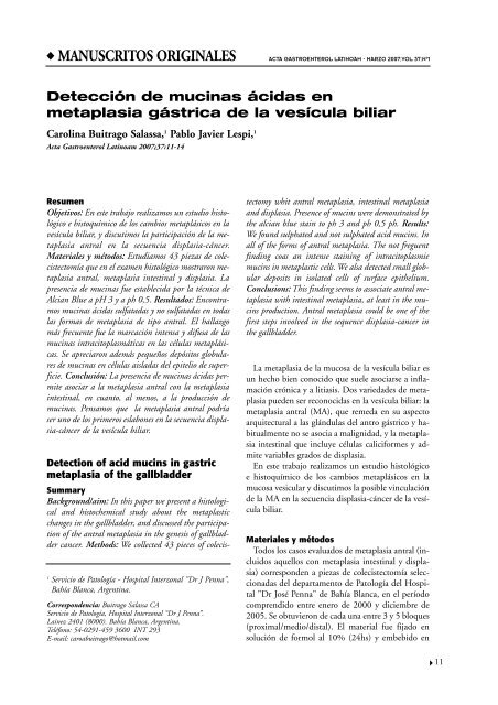
Detección de mucinas ácidas en metaplasia gástrica de la ... - ACTA
Detección de mucinas ácidas en metaplasia gástrica de la ... - ACTA
Detección de mucinas ácidas en metaplasia gástrica de la ... - ACTA
You also want an ePaper? Increase the reach of your titles
YUMPU automatically turns print PDFs into web optimized ePapers that Google loves.
◆ MANUSCRITOS ORIGINALES<br />
Resum<strong>en</strong><br />
Objetivos: En este trabajo realizamos un estudio histológico<br />
e histoquímico <strong>de</strong> los cambios metaplásicos <strong>en</strong> <strong>la</strong><br />
vesícu<strong>la</strong> biliar, y discutimos <strong>la</strong> participación <strong>de</strong> <strong>la</strong> <strong>metap<strong>la</strong>sia</strong><br />
antral <strong>en</strong> <strong>la</strong> secu<strong>en</strong>cia disp<strong>la</strong>sia-cáncer.<br />
Materiales y métodos: Estudiamos 43 piezas <strong>de</strong> colecistectomía<br />
que <strong>en</strong> el exam<strong>en</strong> histológico mostraron <strong>metap<strong>la</strong>sia</strong><br />
antral, <strong>metap<strong>la</strong>sia</strong> intestinal y disp<strong>la</strong>sia. La<br />
pres<strong>en</strong>cia <strong>de</strong> <strong>mucinas</strong> fue establecida por <strong>la</strong> técnica <strong>de</strong><br />
Alcian Blue a pH 3 y a ph 0,5. Resultados: Encontramos<br />
<strong>mucinas</strong> <strong>ácidas</strong> sulfatadas y no sulfatadas <strong>en</strong> todas<br />
<strong>la</strong>s formas <strong>de</strong> <strong>metap<strong>la</strong>sia</strong> <strong>de</strong> tipo antral. El hal<strong>la</strong>zgo<br />
más frecu<strong>en</strong>te fue <strong>la</strong> marcación int<strong>en</strong>sa y difusa <strong>de</strong> <strong>la</strong>s<br />
<strong>mucinas</strong> intracitop<strong>la</strong>smáticas <strong>en</strong> <strong>la</strong>s célu<strong>la</strong>s metaplásicas.<br />
Se apreciaron a<strong>de</strong>más pequeños <strong>de</strong>pósitos globu<strong>la</strong>res<br />
<strong>de</strong> <strong>mucinas</strong> <strong>en</strong> célu<strong>la</strong>s ais<strong>la</strong>das <strong>de</strong>l epitelio <strong>de</strong> superficie.<br />
Conclusión: La pres<strong>en</strong>cia <strong>de</strong> <strong>mucinas</strong> <strong>ácidas</strong> permite<br />
asociar a <strong>la</strong> <strong>metap<strong>la</strong>sia</strong> antral con <strong>la</strong> <strong>metap<strong>la</strong>sia</strong><br />
intestinal, <strong>en</strong> cuanto, al m<strong>en</strong>os, a <strong>la</strong> producción <strong>de</strong><br />
<strong>mucinas</strong>. P<strong>en</strong>samos que <strong>la</strong> <strong>metap<strong>la</strong>sia</strong> antral podría<br />
ser uno <strong>de</strong> los primeros es<strong>la</strong>bones <strong>en</strong> <strong>la</strong> secu<strong>en</strong>cia disp<strong>la</strong>sia-cáncer<br />
<strong>de</strong> <strong>la</strong> vesícu<strong>la</strong> biliar.<br />
Detection of acid mucins in gastric<br />
<strong>metap<strong>la</strong>sia</strong> of the gallb<strong>la</strong>d<strong>de</strong>r<br />
Summary<br />
Background/aim: In this paper we pres<strong>en</strong>t a histological<br />
and histochemical study about the metap<strong>la</strong>stic<br />
changes in the gallb<strong>la</strong>d<strong>de</strong>r, and discussed the participation<br />
of the antral <strong>metap<strong>la</strong>sia</strong> in the g<strong>en</strong>esis of gallb<strong>la</strong>d<strong>de</strong>r<br />
cancer. Methods: We collected 43 pieces of colecis-<br />
<strong>ACTA</strong> GASTROENTEROL LATINOAM - MARZO 2007;VOL 37:Nº1<br />
<strong>Detección</strong> <strong>de</strong> <strong>mucinas</strong> <strong>ácidas</strong> <strong>en</strong><br />
<strong>metap<strong>la</strong>sia</strong> <strong>gástrica</strong> <strong>de</strong> <strong>la</strong> vesícu<strong>la</strong> biliar<br />
Carolina Buitrago Sa<strong>la</strong>ssa, 1 Pablo Javier Lespi, 1<br />
Acta Gastro<strong>en</strong>terol Latinoam 2007;37:11-14<br />
1 Servicio <strong>de</strong> Patología - Hospital Interzonal "Dr J P<strong>en</strong>na",<br />
Bahía B<strong>la</strong>nca, Arg<strong>en</strong>tina.<br />
Correspon<strong>de</strong>ncia: Buitrago Sa<strong>la</strong>ssa CA<br />
Servicio <strong>de</strong> Patología, Hospital Interzonal “Dr J P<strong>en</strong>na”.<br />
Lainez 2401 (8000). Bahía B<strong>la</strong>nca, Arg<strong>en</strong>tina.<br />
Teléfono: 54-0291-459 3600 INT 293<br />
E-mail: caroabuitrago@hotmail.com<br />
tectomy whit antral <strong>metap<strong>la</strong>sia</strong>, intestinal <strong>metap<strong>la</strong>sia</strong><br />
and disp<strong>la</strong>sia. Pres<strong>en</strong>ce of mucins were <strong>de</strong>monstrated by<br />
the alcian blue stain to ph 3 and ph 0,5 ph. Results:<br />
We found sulphated and not sulphated acid mucins. In<br />
all of the forms of antral <strong>metap<strong>la</strong>sia</strong>. The not fregu<strong>en</strong>t<br />
finding coas an int<strong>en</strong>se staining of intracitop<strong>la</strong>smie<br />
mucins in metap<strong>la</strong>stic cells. We alsa <strong>de</strong>tected small globu<strong>la</strong>r<br />
<strong>de</strong>posits in iso<strong>la</strong>ted cells of surface epithelium.<br />
Conclusions: This finding seems to associate antral <strong>metap<strong>la</strong>sia</strong><br />
with intestinal <strong>metap<strong>la</strong>sia</strong>, at least in the mucins<br />
production. Antral <strong>metap<strong>la</strong>sia</strong> could be one of the<br />
first steps involved in the sequ<strong>en</strong>ce disp<strong>la</strong>sia-cancer in<br />
the gallb<strong>la</strong>d<strong>de</strong>r.<br />
La <strong>metap<strong>la</strong>sia</strong> <strong>de</strong> <strong>la</strong> mucosa <strong>de</strong> <strong>la</strong> vesícu<strong>la</strong> biliar es<br />
un hecho bi<strong>en</strong> conocido que suele asociarse a inf<strong>la</strong>mación<br />
crónica y a litiasis. Dos varieda<strong>de</strong>s <strong>de</strong> <strong>metap<strong>la</strong>sia</strong><br />
pue<strong>de</strong>n ser reconocidas <strong>en</strong> <strong>la</strong> vesícu<strong>la</strong> biliar: <strong>la</strong><br />
<strong>metap<strong>la</strong>sia</strong> antral (MA), que remeda <strong>en</strong> su aspecto<br />
arquitectural a <strong>la</strong>s glándu<strong>la</strong>s <strong>de</strong>l antro gástrico y habitualm<strong>en</strong>te<br />
no se asocia a malignidad, y <strong>la</strong> <strong>metap<strong>la</strong>sia</strong><br />
intestinal que incluye célu<strong>la</strong>s caliciformes y admite<br />
variables grados <strong>de</strong> disp<strong>la</strong>sia.<br />
En este trabajo realizamos un estudio histológico<br />
e histoquímico <strong>de</strong> los cambios metaplásicos <strong>en</strong> <strong>la</strong><br />
mucosa vesicu<strong>la</strong>r y discutimos <strong>la</strong> posible vincu<strong>la</strong>ción<br />
<strong>de</strong> <strong>la</strong> MA <strong>en</strong> <strong>la</strong> secu<strong>en</strong>cia disp<strong>la</strong>sia-cáncer <strong>de</strong> <strong>la</strong> vesícu<strong>la</strong><br />
biliar.<br />
Materiales y métodos<br />
Todos los casos evaluados <strong>de</strong> <strong>metap<strong>la</strong>sia</strong> antral (incluidos<br />
aquellos con <strong>metap<strong>la</strong>sia</strong> intestinal y disp<strong>la</strong>sia)<br />
correspon<strong>de</strong>n a piezas <strong>de</strong> colecistectomía seleccionadas<br />
<strong>de</strong>l <strong>de</strong>partam<strong>en</strong>to <strong>de</strong> Patología <strong>de</strong>l Hospital<br />
"Dr José P<strong>en</strong>na" <strong>de</strong> Bahía B<strong>la</strong>nca, <strong>en</strong> el período<br />
compr<strong>en</strong>dido <strong>en</strong>tre <strong>en</strong>ero <strong>de</strong> 2000 y diciembre <strong>de</strong><br />
2005. Se obtuvieron <strong>de</strong> cada una <strong>en</strong>tre 3 y 5 bloques<br />
(proximal/medio/distal). El material fue fijado <strong>en</strong><br />
solución <strong>de</strong> formol al 10% (24hs) y embebido <strong>en</strong><br />
11
12<br />
Acta Gastro<strong>en</strong>terológica Latinoamericana – Vol 37 / N° 1 / Marzo 2007<br />
parafina. Se realizaron cortes seriados <strong>de</strong> 5 micras<br />
que fueron teñidos con hematoxilina y eosina y examinados<br />
con microscopía óptica.<br />
Para <strong>de</strong>tectar <strong>la</strong> pres<strong>en</strong>cia <strong>de</strong> <strong>mucinas</strong> <strong>ácidas</strong> se utilizó<br />
el protocolo histoquímico tradicional con solución<br />
fresca <strong>de</strong> Alcian Blue a pH 3 y 0.5 durante 30<br />
minutos. Secciones <strong>de</strong> colon normal e ileon sirvieron<br />
como testigos.<br />
La MA fue <strong>de</strong>finida como <strong>la</strong> pres<strong>en</strong>cia <strong>de</strong> glándu<strong>la</strong>s<br />
revestidas por célu<strong>la</strong>s <strong>de</strong> citop<strong>la</strong>sma c<strong>la</strong>ro y núcleo<br />
basal, con una apari<strong>en</strong>cia morfológica idéntica<br />
al <strong>de</strong> <strong>la</strong>s célu<strong>la</strong>s <strong>de</strong>l antro pilórico. Glándu<strong>la</strong>s simi<strong>la</strong>res,<br />
pero tapizadas por célu<strong>la</strong>s p<strong>la</strong>nas con escaso citop<strong>la</strong>sma<br />
c<strong>la</strong>ro ubicadas a nivel <strong>de</strong>l cuello vesicu<strong>la</strong>r<br />
fueron <strong>de</strong>scartadas para el estudio. La <strong>metap<strong>la</strong>sia</strong> intestinal<br />
fue i<strong>de</strong>ntificada a partir <strong>de</strong> <strong>la</strong> pres<strong>en</strong>cia <strong>de</strong><br />
célu<strong>la</strong>s caliciformes, <strong>en</strong>terocitos y <strong>en</strong> ciertos casos<br />
célu<strong>la</strong>s <strong>de</strong> Paneth. La disp<strong>la</strong>sia se caracterizó por el<br />
hal<strong>la</strong>zgo <strong>de</strong> seudoestratificación e hipercromatismo<br />
nuclear, pérdida <strong>de</strong> <strong>la</strong> secreción <strong>de</strong> moco e increm<strong>en</strong>to<br />
<strong>en</strong> el número <strong>de</strong> mitosis. Se <strong>la</strong> dividió <strong>en</strong> leve,<br />
mo<strong>de</strong>rada y severa.<br />
Resultados<br />
De <strong>la</strong>s 42 piezas <strong>de</strong> colecistectomía evaluadas, 30<br />
correspondieron a mujeres y 12 a hombres. El rango<br />
etario estuvo compr<strong>en</strong>dido <strong>en</strong>tre 21 y 74 años<br />
(media 47 años). En el exam<strong>en</strong> microscópico <strong>la</strong> MA<br />
mostró glándu<strong>la</strong>s agrupadas revestidas por célu<strong>la</strong>s<br />
con amplio citop<strong>la</strong>sma c<strong>la</strong>ro y núcleo basal. En 28<br />
casos estas glándu<strong>la</strong>s se <strong>en</strong>contraron confinadas a <strong>la</strong><br />
Figura 1. Glándu<strong>la</strong>s con <strong>metap<strong>la</strong>sia</strong> antral.<br />
lámina propia y dispuestas <strong>en</strong> nidos. (figura1)<br />
En 8 casos se ext<strong>en</strong>dían a <strong>la</strong> capa muscu<strong>la</strong>r y <strong>en</strong> 6<br />
se ubicaron <strong>en</strong> <strong>la</strong> interfase <strong>en</strong>tre el epitelio <strong>de</strong> superficie<br />
y <strong>la</strong>s glándu<strong>la</strong>s <strong>de</strong> <strong>la</strong> lámina propia. Solo <strong>en</strong><br />
ocho casos se halló <strong>metap<strong>la</strong>sia</strong> intestinal que incluía<br />
célu<strong>la</strong>s caliciformes y célu<strong>la</strong>s <strong>de</strong> Paneth (figura 2 y<br />
3). La disp<strong>la</strong>sia fue severa solo <strong>en</strong> 1 caso. (figura 4)<br />
Con <strong>la</strong> técnica <strong>de</strong> Alcian Blue a pH 3 y a pH 0,5<br />
Figura 2. Metap<strong>la</strong>sia intestinal <strong>en</strong> el epitelio <strong>de</strong> superficie.<br />
Figura 3. Metap<strong>la</strong>sia intestinal con célu<strong>la</strong>s <strong>de</strong> Paneth.
<strong>Detección</strong> <strong>de</strong> <strong>mucinas</strong> <strong>ácidas</strong> <strong>en</strong> <strong>metap<strong>la</strong>sia</strong> <strong>gástrica</strong> <strong>de</strong> <strong>la</strong> vesícu<strong>la</strong> biliar Carolina Buitrago Sa<strong>la</strong>ssa y col<br />
Figura 4. Disp<strong>la</strong>sia severa <strong>en</strong> área <strong>de</strong> <strong>metap<strong>la</strong>sia</strong> intestinal.<br />
se evi<strong>de</strong>nciaron distintos patrones <strong>de</strong> tinción <strong>en</strong> <strong>la</strong>s<br />
glándu<strong>la</strong>s con <strong>metap<strong>la</strong>sia</strong> <strong>de</strong> tipo antral. A pH 3 se<br />
observaron <strong>de</strong>pósitos focales, difusos e incluso apicales<br />
<strong>de</strong> mucina. Con Alcian Blue a pH 0,5 los patrones<br />
<strong>en</strong>contrados también fueron <strong>la</strong> pres<strong>en</strong>cia <strong>de</strong><br />
pequeños glóbulos intracitop<strong>la</strong>smáticos <strong>de</strong> mucina<br />
<strong>en</strong> forma difusa, focal o <strong>en</strong> <strong>la</strong> región apical <strong>de</strong> <strong>la</strong>s célu<strong>la</strong>s<br />
metaplásicas.<br />
Con ambas técnicas, el hal<strong>la</strong>zgo más frecu<strong>en</strong>te fue<br />
<strong>la</strong> marcación int<strong>en</strong>sa y difusa <strong>de</strong> <strong>la</strong>s <strong>mucinas</strong> intracitop<strong>la</strong>smáticas<br />
<strong>en</strong> <strong>la</strong>s célu<strong>la</strong>s metaplásicas (figura 5).<br />
Se apreciaron a<strong>de</strong>más pequeños <strong>de</strong>pósitos globu<strong>la</strong>res<br />
<strong>de</strong> <strong>mucinas</strong> <strong>en</strong> célu<strong>la</strong>s ais<strong>la</strong>das <strong>de</strong>l epitelio <strong>de</strong> super-<br />
Figura 5. Glóbulos <strong>de</strong> mucina intracitop<strong>la</strong>smática <strong>en</strong><br />
glándu<strong>la</strong>s con MA (Alcian Blue pH o,5).<br />
Figura 6. Mucinas citop<strong>la</strong>smáticas <strong>en</strong> áreas <strong>de</strong> <strong>metap<strong>la</strong>sia</strong><br />
intestinal (Alcian Blue pH 0,5).<br />
ficie. Las célu<strong>la</strong>s caliciformes mostraron una int<strong>en</strong>sa<br />
marcación con <strong>la</strong> coloración <strong>de</strong> Alcian Blue a pH 3<br />
como a ph 0,5. (figura 6)<br />
Discusión<br />
La MA se pres<strong>en</strong>ta <strong>en</strong> el 66 al 84% <strong>de</strong> <strong>la</strong>s piezas<br />
<strong>de</strong> colecistectomía. 1 Se caracteriza por <strong>la</strong> pres<strong>en</strong>cia<br />
<strong>de</strong> glándu<strong>la</strong>s revestidas por célu<strong>la</strong>s columnares <strong>de</strong> citop<strong>la</strong>sma<br />
pálido, idénticas a <strong>la</strong>s <strong>de</strong>l antro pilórico,<br />
dispuestas <strong>en</strong> nidos <strong>de</strong>ntro <strong>de</strong> <strong>la</strong> lámina, que incluso<br />
pue<strong>de</strong>n aparecer <strong>en</strong>tre los haces <strong>de</strong> músculo liso<br />
<strong>de</strong>l órgano y contactar con <strong>la</strong> serosa. 2-4 Des<strong>de</strong> el punto<br />
<strong>de</strong> vista histoquímico <strong>la</strong>s célu<strong>la</strong>s <strong>de</strong> <strong>la</strong> MA conti<strong>en</strong><strong>en</strong><br />
<strong>mucinas</strong> <strong>ácidas</strong> no sulfatadas y <strong>mucinas</strong> neutras.<br />
5-8 Según Tatematsu M y col, a todas <strong>la</strong>s célu<strong>la</strong>s<br />
<strong>de</strong> <strong>la</strong> <strong>metap<strong>la</strong>sia</strong> <strong>de</strong> tipo antral conti<strong>en</strong><strong>en</strong> <strong>mucinas</strong> <strong>de</strong><br />
c<strong>la</strong>se III (sulfo<strong>mucinas</strong>). 9<br />
Laitio M y col, 12 fueron los primeros <strong>en</strong> interpretar<br />
los cambios metaplásicos <strong>de</strong>l epitelio <strong>de</strong> <strong>la</strong> vesícu<strong>la</strong><br />
biliar como precursores <strong>de</strong> tumores vesicu<strong>la</strong>res.<br />
10-12 En <strong>la</strong> misma línea Mukhopadhyay y col, 4 <strong>en</strong>contraron<br />
una asociación significativa <strong>en</strong>tre <strong>la</strong> MA y<br />
<strong>la</strong> <strong>metap<strong>la</strong>sia</strong> intestinal, y sugirieron que <strong>la</strong> MA es<br />
un es<strong>la</strong>bón <strong>en</strong> el <strong>de</strong>sarrollo <strong>de</strong>l carcinoma vesicu<strong>la</strong>r.<br />
Ambos tipos <strong>de</strong> <strong>metap<strong>la</strong>sia</strong> serían, para estos autores,<br />
precursoras <strong>de</strong> disp<strong>la</strong>sia. 4,10-13<br />
Por otra parte, <strong>la</strong> <strong>metap<strong>la</strong>sia</strong> intestinal <strong>de</strong> <strong>la</strong> mucosa<br />
vesicu<strong>la</strong>r es un complejo proceso adaptativo asociado<br />
a colecistitis crónica y colelitiasis. 4,3,7 Se pres<strong>en</strong>ta<br />
<strong>en</strong> el 12 a 52% <strong>de</strong> <strong>la</strong>s piezas <strong>de</strong> colecistecto-<br />
13
14<br />
Acta Gastro<strong>en</strong>terológica Latinoamericana – Vol 37 / N° 1 / Marzo 2007<br />
mía. 1 El hal<strong>la</strong>zgo histológico característico es <strong>la</strong> pres<strong>en</strong>cia<br />
<strong>de</strong> célu<strong>la</strong>s absortivas, célu<strong>la</strong>s caliciformes, célu<strong>la</strong>s<br />
<strong>en</strong>dócrinas y, <strong>en</strong> ais<strong>la</strong>dos casos, célu<strong>la</strong>s <strong>de</strong> Paneth.<br />
2,7,14 La <strong>metap<strong>la</strong>sia</strong> intestinal posee características<br />
morfológicas que permit<strong>en</strong> c<strong>la</strong>sificar<strong>la</strong> como:<br />
completa (o <strong>de</strong> tipo I) cuando pres<strong>en</strong>ta <strong>en</strong>terocitos<br />
y célu<strong>la</strong>s <strong>de</strong> Paneth, e incompleta (o <strong>de</strong> tipo II)<br />
cuando incluye célu<strong>la</strong>s caliciformes y célu<strong>la</strong>s columnares.<br />
14 Otra c<strong>la</strong>sificación <strong>la</strong>s separa <strong>de</strong> acuerdo al tipo<br />
<strong>de</strong> mucina e<strong>la</strong>borada por dichas célu<strong>la</strong>s. En <strong>la</strong><br />
<strong>metap<strong>la</strong>sia</strong> intestinal completa se produce sialomucina,<br />
mi<strong>en</strong>tras que <strong>en</strong> <strong>la</strong> <strong>metap<strong>la</strong>sia</strong> intestinal incompleta<br />
<strong>la</strong>s célu<strong>la</strong>s conti<strong>en</strong><strong>en</strong> sialo<strong>mucinas</strong> y sulfo<strong>mucinas</strong>.<br />
14 Esta última forma <strong>de</strong> <strong>metap<strong>la</strong>sia</strong> intestinal<br />
es <strong>la</strong> que se asocia a disp<strong>la</strong>sia epitelial. 11,12,14 Algunos<br />
autores propon<strong>en</strong> que el proceso <strong>de</strong> <strong>de</strong>sarrollo<br />
<strong>de</strong> <strong>la</strong> <strong>metap<strong>la</strong>sia</strong> intestinal <strong>en</strong> <strong>la</strong> vesícu<strong>la</strong> biliar es<br />
análogo al que ocurre <strong>en</strong> el estómago. 4 Por lo tanto,<br />
es razonable consi<strong>de</strong>rar <strong>la</strong> posibilidad <strong>de</strong> que <strong>la</strong> <strong>metap<strong>la</strong>sia</strong><br />
intestinal <strong>en</strong> <strong>la</strong> vesícu<strong>la</strong> biliar también predisponga<br />
al carcinoma vesicu<strong>la</strong>r. 4,12,13<br />
La disp<strong>la</strong>sia <strong>de</strong>l epitelio <strong>de</strong> <strong>la</strong> vesícu<strong>la</strong> biliar se pres<strong>en</strong>ta<br />
<strong>en</strong> el 1 al 34% <strong>de</strong> <strong>la</strong>s piezas <strong>de</strong> colecistectomia.<br />
1 En el exam<strong>en</strong> histológico se caracterizan por <strong>la</strong><br />
pres<strong>en</strong>cia <strong>de</strong> seudoestratificacion <strong>de</strong>l epitelio, increm<strong>en</strong>to<br />
<strong>de</strong> <strong>la</strong>s mitosis e hipercromatismo nuclear. 12<br />
En cuanto a <strong>la</strong> producción <strong>de</strong> <strong>mucinas</strong>, el epitelio<br />
displásico produce sialo<strong>mucinas</strong>, sulfo<strong>mucinas</strong> y<br />
<strong>mucinas</strong> neutras. Distintos autores propon<strong>en</strong> que <strong>la</strong><br />
disp<strong>la</strong>sia se originaría <strong>de</strong> <strong>la</strong>s áreas con <strong>metap<strong>la</strong>sia</strong> intestinal<br />
o MA. 7,12-15<br />
En nuestro estudio <strong>en</strong>contramos <strong>mucinas</strong> <strong>ácidas</strong><br />
sulfatadas y no sulfatadas <strong>en</strong> todas <strong>la</strong>s formas <strong>de</strong> <strong>metap<strong>la</strong>sia</strong><br />
que al exam<strong>en</strong> histológico correspondieron<br />
arquitecturalm<strong>en</strong>te al tipo antral. Este hal<strong>la</strong>zgo podría<br />
indicar <strong>la</strong> exist<strong>en</strong>cia <strong>de</strong> una fuerte re<strong>la</strong>ción <strong>en</strong>tre<br />
MA y <strong>metap<strong>la</strong>sia</strong> intestinal, <strong>en</strong> cuanto, al m<strong>en</strong>os,<br />
a <strong>la</strong> producción <strong>de</strong> <strong>mucinas</strong>. Es <strong>de</strong>cir, nuestros resultados<br />
sugier<strong>en</strong> que <strong>la</strong> MA está íntimam<strong>en</strong>te vincu<strong>la</strong>da<br />
a <strong>la</strong> <strong>metap<strong>la</strong>sia</strong> intestinal y que, probablem<strong>en</strong>te,<br />
<strong>la</strong> MA sea una variante <strong>de</strong> esta última forma<br />
<strong>de</strong> <strong>metap<strong>la</strong>sia</strong>. Mukhopadhyay y col, 4 <strong>en</strong> su trabajo<br />
sobre precursores <strong>de</strong> disp<strong>la</strong>sia <strong>en</strong> <strong>la</strong> vesícu<strong>la</strong> biliar,<br />
<strong>en</strong>contraron una secu<strong>en</strong>cia etaria <strong>en</strong>tre <strong>la</strong> MA, <strong>la</strong><br />
<strong>metap<strong>la</strong>sia</strong> intestinal y el <strong>de</strong>sarrollo <strong>de</strong> disp<strong>la</strong>sia. A<br />
difer<strong>en</strong>cia <strong>de</strong> nuestro trabajo, no hal<strong>la</strong>ron asociación<br />
<strong>en</strong>tre ambos tipos <strong>de</strong> <strong>metap<strong>la</strong>sia</strong> <strong>en</strong> cuanto a <strong>la</strong>s<br />
<strong>mucinas</strong> producidas por sus célu<strong>la</strong>s.<br />
En resum<strong>en</strong>, <strong>la</strong> MA produce <strong>mucinas</strong> <strong>ácidas</strong> sulfatadas<br />
y no sulfatadas, y este hal<strong>la</strong>zgo parece vincu<strong>la</strong>r<strong>la</strong><br />
muy fuertem<strong>en</strong>te a <strong>la</strong> <strong>metap<strong>la</strong>sia</strong> intestinal más<br />
que a <strong>la</strong>s glándu<strong>la</strong>s <strong>de</strong>l antro gástrico. En refer<strong>en</strong>cia<br />
a esto último, <strong>la</strong> MA podría ser uno <strong>de</strong> los primeros<br />
es<strong>la</strong>bones <strong>en</strong> <strong>la</strong> secu<strong>en</strong>cia disp<strong>la</strong>sia-cáncer <strong>de</strong> <strong>la</strong> vesícu<strong>la</strong><br />
biliar.<br />
Refer<strong>en</strong>cias<br />
1. Scott HS. Gallb<strong>la</strong>d<strong>de</strong>r and extrahepatic biliary tree. Sternberg<br />
S. Sternberg's diagnostic surgical pathology. New<br />
York:Rav<strong>en</strong> Press,1989;1212-1213.<br />
2. Alvores-Saavedra, J; H<strong>en</strong>son, DE. Pyloric g<strong>la</strong>nd <strong>metap<strong>la</strong>sia</strong><br />
with perineural invasion of the gallb<strong>la</strong>d<strong>de</strong>r. A lesion that<br />
can be confused with a<strong>de</strong>nocarcinoma. Cancer1999;86:<br />
2625-2631.<br />
3. Odze, RD; Godblum, JR; Crawford, JM. Surgical Pathology<br />
of the GI Tract, Liver, Biliary Tract and Pancreas<br />
2004;28:621-622.<br />
4. Mukhopadhyay, S; Landas, SK. Putative precursors of gallb<strong>la</strong>d<strong>de</strong>r<br />
dysp<strong>la</strong>sia: a review of 400 routinely resected specim<strong>en</strong>s.<br />
Arch Path Lab Med 2004;129:386-390.<br />
5. Laitio, M. Morphology and histochemistry of non-tumorous<br />
gallb<strong>la</strong>d<strong>de</strong>r epithelium. A series of 103 cases. Pathhol<br />
Res Pract 1980;167:335-345.<br />
6. Laitio, M; Terho, T. Polysacchari<strong>de</strong>s of metap<strong>la</strong>stic mucosa<br />
and carcinoma of the gallb<strong>la</strong>d<strong>de</strong>r. Lab Invest 1975;32: 183-<br />
189.<br />
7. Rosai, J. Ackerman S Surgical Pathology 2004;1:1041-<br />
1048.<br />
8. Frierson, HF. The gross anatomy and histology of the gallb<strong>la</strong>d<strong>de</strong>r,<br />
extrahepatic bile ducts, vaterian system, and minor<br />
papil<strong>la</strong>. Am J Surg Pathol 1989; 13:146-162.<br />
9. Tatematsu, M; Furihata, C; Miki, K; Ichinose, M; and col.<br />
Complete and incomplete pyloric g<strong>la</strong>nd <strong>metap<strong>la</strong>sia</strong> of human<br />
gallb<strong>la</strong>d<strong>de</strong>r. Acta Pathol Jpn 1987;37:39-46.<br />
10. Laitio, M. Early carcinoma of the gallb<strong>la</strong>d<strong>de</strong>r. Beitr Pathol<br />
1976;158:159-172.<br />
11. Laitio, M; Hakkin<strong>en</strong>, I. Intestinal- type carcinoma of gallb<strong>la</strong>d<strong>de</strong>r.<br />
A histochemical and inmonologic study. Cancer<br />
1975;36:1668-1674.<br />
12. Laitio, M. Histog<strong>en</strong>esis of epithelial neop<strong>la</strong>sms of human<br />
gallb<strong>la</strong>d<strong>de</strong>r I. Dysp<strong>la</strong>sia. Pathol Res Pract 1983;178:51-56.<br />
13. Kushima, R; Lohe, B; Borchard, F. Differ<strong>en</strong>tiation towards<br />
gastric foveo<strong>la</strong>r, mucopeptic and intestinal globet cells in<br />
gallb<strong>la</strong>d<strong>de</strong>r a<strong>de</strong>nocarcinoma. Histopathology 1996;29:<br />
443-448.<br />
14. Torres Perez, IJ. Estudio comparativo <strong>en</strong>tre el diagnóstico<br />
morfológico <strong>de</strong>l tipo <strong>de</strong> <strong>metap<strong>la</strong>sia</strong> intestinal con <strong>la</strong> confirmación<br />
histoquímica por el método <strong>de</strong> Gomori-Alcian<br />
Blue. http://sisbib.unmsm.edu.pe/bibvirtual/Tesis/Salud-<br />
/Torres_P_I/indice_Torres.htm.


