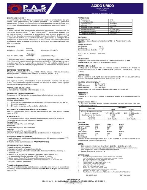
ACIDO URICO - PAS Diagnostica
ACIDO URICO - PAS Diagnostica
ACIDO URICO - PAS Diagnostica
Create successful ePaper yourself
Turn your PDF publications into a flip-book with our unique Google optimized e-Paper software.
SIGNIFICADO CLINICO: 1-3<br />
La cuantificación de Ácido Úrico es comúnmente usada en el diagnóstico de gota.<br />
Niveles altos (hiperuricemia) se pueden presentar en leucemia, policitemia,<br />
arteriosclerosis, diabetes, hipotiroidismo y condiciones asociadas con una reducción en la<br />
función renal; Niveles bajos están presentes en pacientes con la enfermedad Wilson’s.<br />
MÉTODO:<br />
El Ácido Úrico ha sido históricamente determinado por métodos colorimétricos con<br />
variaciones de fosfotungstato 4,5 y reducción de hierro 6,7 . Metodologías recientes usan<br />
las enzimas uricasa y peroxidasa, y un cromógeno para producir un producto final<br />
colorimétrico, demostrando mayor especificidad para la determinación de ácido úrico 8,9 .<br />
El procedimiento de <strong>PAS</strong> DIAGNOSTICA usa uricasa, peroxidasa y el cromógeno TBHB<br />
para producir un producto final colorimétrico. El producto final colorimétrico producido en<br />
esta reacción puede ser medido a 520 nm y es proporcional a la concentración de ácido<br />
úrico en la muestra.<br />
PRINCIPIO:<br />
Uricasa<br />
Acido Urico + O2 + H2O Alantoína + CO2 + H2O2<br />
Peroxidasa<br />
H2O2 + 4-AAP + TBHB Quinoneimina + H2O<br />
El ácido úrico es oxidado a alantoína por la acción de la uricasa con la producción de<br />
H2O2. El peroxido reacciona con la 4-aminoantipirina (4-AAP) y THBH en la presencia de<br />
peroxidasa para formar un colorante de quinoneimina. El producto resultante en la<br />
absorbancia a 520nm (500-550nm) es proporcional a la concentracion de acido urico en<br />
la muestra.<br />
CONTENIDO Y COMPOSICIÓN:<br />
Reactivo: 4-Aminoantipirina 0.5 mM, TBHB 1.75mM, Uricasa >32 U/L, Peroxidasa<br />
(rábano) >1300U/L, Estabilizadores y aditivos no reactivos, pH (7.8 – 8.2).<br />
Estándar: Acido Úrico 5mg/dL.<br />
Evite ingerir el reactivo, la toxicidad no ha sido determinada. Contiene ácida de sodio<br />
(0.05%) como preservativo y puede reaccionar con la tubería de plomo o cobre. Enjuague<br />
las tuberías de drenaje con abundante agua.<br />
PREPARACIÓN DEL REACTIVO:<br />
El monoreactivo y el estándar están listos para su uso.<br />
ESTABILIDAD Y ALMACENAMIENTO:<br />
Conservado de 2-8ºC el reactivo es estable hasta la fecha indicada en la etiqueta.<br />
DETERIORO DEL REACTIVO:<br />
No utilice este reactivo si:<br />
1. El reactivo reconstituido tiene una absorbancia del blanco mayor de 0.5 a 500 nm.<br />
2. El reactivo esta turbio.<br />
3. El reactivo no cumple con los controles establecidos.<br />
RECOLECCIÓN Y CONSERVACIÓN DE LA MUESTRA:<br />
Suero libre de hemolisis, el ácido úrico en suero es estable por 3 días a 2-8°C y hasta 6<br />
meses a -20ºC. 2<br />
INTERFERENCIA:<br />
Los siguientes resultados fueron obtenidos en estudios para determinar el nivel de<br />
interferencia de hemoglobina, bilirrubina, y lipemias.<br />
Hemoglobina:<br />
No interferencia (±10%) hasta 200 mg/L.<br />
Bilirrubina:<br />
No interferencia (±10%) hasta 20.9 mg/dL.<br />
Lipemias:<br />
No interferencia (±10%) hasta 1020 mg/dL.<br />
Un número de drogas y substancias afectan la determinación de Acido Úrico. 10<br />
EQUIPO ADICIONAL REQUERIDO:<br />
1. Analizador de química clínica con longitud de onda de 500 nm y temperatura de 37ºC y<br />
sus consumibles.<br />
2. Material control (suministrados por <strong>PAS</strong> DIAGNOSTICA).<br />
PROCEDIMIENTO DEL ENSAYO:<br />
Procedimiento para uso manual.<br />
1) Atemperar el reactivo a temperatura ambiente.<br />
2) Dispensar 1000 µL del reactivo en cada tubo (Estándar, Controles y muestra) e<br />
incubar a 37ºC.<br />
3) Agregar 20 µL de Estándar, muestra problema y/o suero control al tubo<br />
correspondiente. Mezclar, e incubar a 37ºC durante 10 minutos<br />
4) Ajustar a cero el espectrofotómetro con blanco de reactivo a 500 nm.<br />
5) Leer la absorbancia exactamente a los 10 minutos después de la adición del<br />
estándar, controles y muestras.<br />
Procedimiento automatizado:<br />
Para este procedimiento siga las instrucciones contenidas en el manual de operación del<br />
analizador y las aplicaciones de los reactivos <strong>PAS</strong> DIAGNOSTICA ® para los analizadores<br />
más comunes.<br />
<strong>ACIDO</strong> <strong>URICO</strong><br />
Método Enzimático<br />
Punto Final<br />
Solo para uso diagnostico “In vitro”<br />
PARÁMETROS:<br />
Temperatura 37°C<br />
Longitud de onda 500 nm<br />
Tipo de Ensayo Punto final<br />
Dirección Creciente<br />
Relación: Muestra/Reactivo 1:50<br />
Volumen de Muestra 0.003mL (3 L)<br />
Volumen de Reactivo 0.150 (150 L)<br />
Tiempo de incubación 5 minutos<br />
CALCULOS:<br />
Abs: Absorbancia<br />
Abs muestra x Concentracion del estándar (mg/dL) = C Ácido úrico en mg/dL<br />
Abs estándar<br />
Ejemplo de cálculos:<br />
Abs. Muestra = 0.071<br />
Abs. Estándar = 0.302<br />
Concentración del Estándar = 5.0<br />
0.071 x 5.0 = 1.18 mg/dL ácido úrico<br />
0.302<br />
CALIBRACION:<br />
Esta prueba debe ser calibrada utilizando el Calibrador de Química de <strong>PAS</strong><br />
DIAGNOSTICA (P/N: CAL1-5) o un estándar apropiado.<br />
<strong>PAS</strong> DIAGNOSTICA<br />
CRA 49C No. 76 – 224 BARRANQUILLA-COLOMBIA Tel: (57-5) 3018686 Fax: (57-5) 3580170<br />
www.pasdiagnostica.com<br />
CONTROL DE CALIDAD:<br />
La integridad de la reacción debe ser evaluada usando un control de dos niveles con<br />
valores conocidos como el <strong>PAS</strong> DIAGNOSTICA Control de Química (P/N: CON1-5 y<br />
CON2-5).<br />
LIMITACIONES:<br />
Valores superiores a 25 mg/dL debe ser diluida la muestra 1:1 con solución salina y<br />
evaluada nuevamente, multiplicando el resultado final por 2.<br />
VALORES DE REFERENCIA: 11<br />
Niños: 2.0- 5.5 mg/dL<br />
Adulto Masculino: 3.5 – 7.2 mg/dL<br />
Adulto femenino: 2.6 – 6.0 mg/dL<br />
Se recomienda que cada laboratorio establezca su rango de normalidad 7 .<br />
DESEMPEÑO:<br />
Linealidad:<br />
Es linear de 0.2 a 25 mg/dL, cuando se evalúa de acuerdo a las recomendaciones del<br />
ensayo.<br />
Comparación del Método:<br />
Los siguientes resultados fueron obtenidos mediante estudios realizados entre éste<br />
procedimiento y uno similar:<br />
Concepto<br />
TBHB<br />
Cromógeno<br />
DCHBS<br />
Número de pares de muestras 58 50<br />
Rango de muestras 1.70 – 20.70 (mg/dL) 2.20 – 11.30 (mg/dL)<br />
Coeficiente de Correlación 0.9792 0.9977<br />
Pendiente 1.044 1.117<br />
Intercepto 0.00 (mg/dL) -0.58 (mg/dL)<br />
Precisión:<br />
Entre Corridas Nivel 1 Nivel 2 Nivel 3<br />
Promedio (mg/dL) 6.60 11.31 20.12<br />
S.D. (mg/dL) 0.15 0.24 0.30<br />
C.V. (%) 2.2 2.1 1.5<br />
Entre Series: Nivel 1 Nivel 2 Nivel 3<br />
S.D. (mg/dL) 0.27 0.47 0.51<br />
C.V. (%) 4.1 4.1 2.5<br />
Sensibilidad:<br />
Un factor de calibración aproximado a 58.66 fue obtenido, lo cual es equivalente a una<br />
sensibilidad de 0.01704 ∆ Abs por mg/dL.<br />
NOTAS:<br />
Este ensayo requiere el uso de un Suero Estándar o Estándar apropiado.<br />
Los volúmenes de muestra y reactivo pueden ser modificados proporcionalmente<br />
para adaptarlos a los requisitos de varios instrumentos.<br />
Estudio realizado en Synchron CX.<br />
REFERENCIAS:<br />
1. Searcy RL: Diagnostic Biochemistry. McGraw-Hill, New York, NY, (1969).<br />
2. Henry RJ, Common C, Winkelman JW (eds), Clinical Chemistry: Principles<br />
and Techniques. Harper & Row, Hagerstown, MD, (1974).<br />
3. Balls ME: Adv Clin Chem 18:213, (1976).<br />
4. Folin, D., Dennis, W., J. Biol. Chem. 13:469 (1913).<br />
5. Caraway, W.T., Clin. Chem. 4:239 (1963).<br />
6. Morin, L.G., J. Clin. Path. 60:691 (1973).<br />
7. Morin, L.G., Clin. Chem. 20:51 (1974).<br />
8. Klackar, H.M., J. Biol. Chem. 167:429 (1947).<br />
9. Praetorius, E., Pouldon, H., Scand. J. Clin. Invest 5:273 (1953).<br />
10. Young, D.S., et al. Clin. Chem. 21:1D (1975).<br />
11. Tietz Textbook of Clinical Chemistry, WB Saunders Co., Philadelphia PA, 2 nd Ed (1994).<br />
PI: URI2P.JS02 REV: 04/14/09
CLINICAL SIGNIFICANCE 1-3<br />
Uric Acid measurements are most commonly used in the diagnosis of gout. Increased<br />
levels (hyperuricaemia) may be observed in leukemia, polycythaemia, atherosclerosis,<br />
diabetes, hypothroidism, and conditions associated with decreased renal funtion.<br />
Decreased levels are present in patients with Wilson’s disease.<br />
METHODOLOGY<br />
Uric Acid has historically been determined by variations of colorimetric phosphotungstate<br />
4, 5 and iron reduction 6, 7 methods. Recent methodologies use the enzymes uricase and<br />
hydrogen peroxidase, along with a chromogen to yield a colorimetric end product. These<br />
methods demonstrate better specificity for uric acid, 8, 9 . The <strong>PAS</strong> procedure uses uricase,<br />
peroxidase and he chromogen TBHB to yield a colorimetric end product. The colorimetric<br />
end product produced in this reaction can be measured at 520nm and is proportional to<br />
the uric acid concentration in the sample.<br />
PRINCIPLE<br />
Uricase<br />
Uric Acid + O2 + H2O Allantoin + CO2 + H2O2<br />
Peroxidase<br />
H2O2 +4-Aminoantipyrine+TBHB Quinoneimine + H2O<br />
Uric Acid is oxidized by Uricase to allantoin and hydrogen peroxide. TBHB + 4aminoantipyrine<br />
+ hydrogen peroxide, in the presence of peroxidase, produce a<br />
quinoneimine dye that is measured at 520nm (500-550). The color intensity at 520nm is<br />
proportional to the concentration of Uric Acid in the sample.<br />
COMPOSITION AND CONTENT<br />
Reagent: 4-Aminoantipyrine 0.5 mM, TBHB 1.75 mM, Uricase (Bacillus Sp.) >32<br />
U/L, Peroxidase (Horseradish) >1300 U/L, Non-reactive stabilizers and fillers, pH<br />
(7.8 – 8.2).<br />
Standard: Urid Acid 5mg/dL.<br />
Avoid eating the reagent, the toxicity has not been determined.Reagent contains Sodium<br />
Azide (0.05%) as preservative. In a dry state may react with copper or lead plumbing to<br />
form explosive metal azides. Upon disposal, flush with large amounts of water to prevent<br />
azide build up.<br />
REAGENT PREPARATION<br />
The monoreactivo and standard are ready for use.<br />
STABILITY AND STORAGE<br />
Conserved 2-8 º C, the reagent is stable until the date indicated on the label.<br />
REAGENT DETERIORATION<br />
Do not use the reagent if:<br />
1. The reagent is turbid.<br />
2. The reagent blank has an absorbance of 0.50 or greater at 500nm.<br />
3. The working reagent does not meet stated performance parameters.<br />
SPECIMEN COLLECTION AND STORAGE<br />
Haemolysis-free serum, the serum uric acid is stable for 3 days at 2-8 ° C and up to 6<br />
months at -20 ° C. 2<br />
INTERFERENCE<br />
Studies to determine the level of interference for hemoglobin, bilirubin and lipemia were<br />
carried out, the following results were obtained:<br />
Hemoglobin:No significant interference ( 10%) from hemoglobin up to 200 mg/dL.<br />
Bilirubin:No significant interference ( 10%) from bilirubin up to 20.9 mg/dL.<br />
Lipemia:No significant interference ( 10%) from lipemia up to 1020 mg/dL measured as<br />
triglycerides.<br />
A number of drugs and substances affect the determination of uric acid. 10<br />
ADDITIONAL EQUIPMENT REQUIRED<br />
1. Clinical chemistry analyzer with a wavelength of 500 nm and 37 º C and its<br />
consumables.<br />
2. Control material (supplied by <strong>PAS</strong> DIAGNOSTICA).<br />
ASSAY PROCEDURE<br />
For this procedure follow the instructions in the manual operation of the analyzer and<br />
reagents applications of <strong>PAS</strong> DIAGNOSTICA ® Analyzer most common.<br />
PARAMETERS<br />
Temperature 37°C<br />
Wavelength 500 nm<br />
Assay type End point<br />
Direction Increase<br />
Sample / rgt ratio 1: 50<br />
Sample vol 0.003mL. (3 L)<br />
Reagent vol 0.150 mL (150 L)<br />
Incubation time 5 min<br />
URIC ACID<br />
Enzymatic method<br />
End Point<br />
Only for in vitro diagnostic use<br />
CALCULATIONS:<br />
Abs = Absorbance<br />
Abs (patient) x Concentration of standard = Uric Acid<br />
Abs (standard) (mg/dL) (mg/dL)<br />
Example:<br />
Abs patient = 0.071<br />
Abs standard = 0.302<br />
Concentration of standard = 5.0 mg/dL.<br />
0.071 x 12.1 = 1.18 mg/dL Uric Acid<br />
0.302<br />
<strong>PAS</strong> DIAGNOSTICA<br />
CRA 49C No. 76 – 224 BARRANQUILLA-COLOMBIA Tel: (57-5) 3018686 Fax: (57-5) 3580170<br />
www.pasdiagnostica.com<br />
CALIBRATION<br />
A calibrator such as <strong>PAS</strong> DIAGNOSTICA Chemistry Calibrator (P/N: CAL1-5), or an<br />
appropriate aqueous standard should be used to calibrate this test.<br />
QUALITY CONTROL<br />
The integrity of the reaction should be monitored by use of a two level control with known<br />
values such as <strong>PAS</strong> DIAGNOSTICA Chemistry Controls (P/N: CON1-5 and CON2-5).<br />
LIMITATIONS<br />
Values above 25 mg / dL should be diluted sample 1:1 with saline and tested again,<br />
multiplying the result by 2.<br />
EXPECTED VALUES 12<br />
Child: 2.0 – 5.5 mg/dL<br />
Adult Male: 3.5 - 7.2 mg/dL<br />
Adult Female: 2.6 – 6.0 mg/dL<br />
It is recommended that each laboratory establish its range of normalidad 7 .<br />
PERFORMANCE<br />
Linearity:<br />
When run as recommended the assay is linear from 0.2 to 25.0 mg/dL.<br />
Method Comparison:<br />
Studies performed between this procedure and similar methodologies yielded the<br />
following results:<br />
Concept<br />
TBHB<br />
Chromogen<br />
DCHBS<br />
Number of samples pairs 58 50<br />
Range of samples 1.70 – 20.70 (mg/dL) 2.20 – 11.30 (mg/dL)<br />
Correlation Coefficient 0.9792 0.9977<br />
Slope 1.044 1.117<br />
Intercept 0.00 (mg/dL) -0.58 (mg/dL)<br />
Precision:<br />
Within Run Level 1 Level 2 Level 3<br />
Mean (mg/dL) 6.60 11.31 20.12<br />
S.D. (mg/dL) 0.15 0.24 0.30<br />
C.V. (%) 2.2 2.1 1.5<br />
Total Level 1 Level 2 Level 3<br />
S.D. (mg/dL) 0.27 0.47 0.51<br />
C.V. (%) 4.1 4.1 2.5<br />
Sensitivity:<br />
A calibration factor of approximately 58.66 was obtained, which is equivalent to a<br />
sensitivity of 0.01704 Abs per mg/dL.<br />
NOTES<br />
• This test requires the use of a Standard or Standard Serum appropriate.<br />
• The volumes of sample and reagent can be modified proportionately to fit the<br />
requirements of different instruments.<br />
• Study conducted in Synchron CX.<br />
REFERENCES<br />
1. Searcy RL: Diagnostic Biochemistry. McGraw-Hill, New York, NY, (1969).<br />
2. Henry RJ, Common C, Winkelman JW (eds), Clinical Chemistry: Principles and Techniques. Harper & Row,<br />
Hagerstown, MD, (1974).<br />
3. Balls ME: Adv Clin Chem 18:213, (1976).<br />
4. Folin, D., Dennis, W., J. Biol. Chem. 13:469 (1913).<br />
5. Caraway, W.T., Clin. Chem. 4:239 (1963).<br />
6. Morin, L.G., J. Clin. Path. 60:691 (1973).<br />
7. Morin, L.G., Clin. Chem. 20:51 (1974).<br />
8. Klackar, H.M., J. Biol. Chem. 167:429 (1947).<br />
9. Praetorius, E., Pouldon, H., Scand. J. Clin. Invest 5:273 (1953).<br />
10. Young, D.S., et al. Clin. Chem. 21:1D (1975).<br />
11. Tietz Textbook of Clinical Chemistry, WB Saunders Co., Philadelphia PA, 2 nd Ed (1994).<br />
PI: URI2P.JS01 REV: 04/14/09


