

Regla de Medición de Displasia de Cadera
Regla de Medición de Displasia de Cadera
Regla de Medición de Displasia de Cadera

Create successful ePaper yourself
Turn your PDF publications into a flip-book with our unique Google optimized e-Paper software.
<strong>Regla</strong> <strong>de</strong> <strong>Medición</strong> <strong>de</strong> <strong>Displasia</strong> <strong>de</strong> Ca<strong>de</strong>ra<br />
Permite los siguientes parámetros <strong>de</strong> medición:<br />
Allows the following measurement parameters:<br />
1. Valoración <strong>de</strong>l ángulo formado entre la línea imaginaria que une los dos centros <strong>de</strong> las cabezas femorales <strong>de</strong> la radiografía <strong>de</strong> ca<strong>de</strong>ra <strong>de</strong> cánidos en posición <strong>de</strong><br />
<strong>de</strong>cúbito supino con extensión <strong>de</strong> las extremida<strong>de</strong>s posteriores, y el bor<strong>de</strong> anterior <strong>de</strong>l acetábulo <strong>de</strong> la articulación coxofemoral (Angulo según Norberg). Assessment<br />
of the angle formed between the imaginary line joining the two centers of the femoral heads of hip radiograph of dog in a position to the supine position with extension of the thighs<br />
and the front edge of the acetabulum of the hip joint (as Norberg Angle ).<br />
2. Valoración <strong>de</strong>l ángulo formado entre la diáfisis y el eje <strong>de</strong>l cuello femoral. Measurement of the angle between the axis of the diaphysis and femoral neck<br />
Marcaje <strong>de</strong> centros <strong>de</strong> cabeza femoral<br />
Centrar cada una <strong>de</strong> las circunferencias <strong>de</strong> la porción izquierda <strong>de</strong> la regla sobre cada una <strong>de</strong> las cabezas femorales y marcar con un rotulador un punto sobre cada<br />
centro.<br />
Marking centers of femoral heads.<br />
Focus each of the circumferences of the left portion of the rule on each of the femoral head with a pen and mark a point on each center.<br />
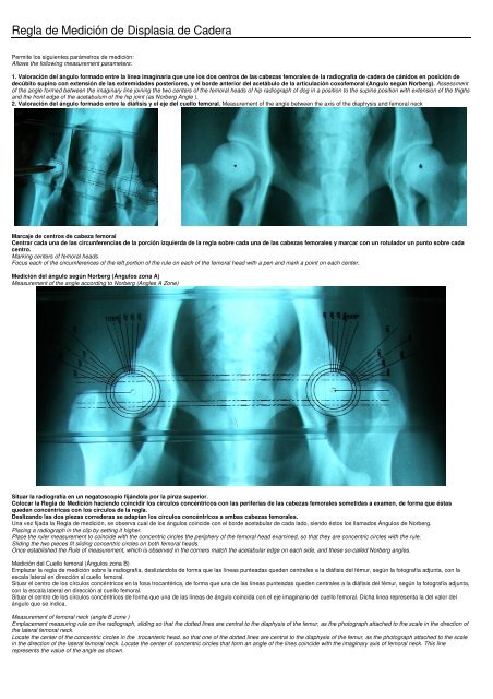
<strong>Medición</strong> <strong>de</strong>l ángulo según Norberg (Ángulos zona A)<br />
Measurement of the angle according to Norberg (Angles A Zone)<br />
Situar la radiografía en un negatoscopio fijándola por la pinza superior.<br />
Colocar la <strong>Regla</strong> <strong>de</strong> <strong>Medición</strong> haciendo coincidir los círculos concéntricos con las periferias <strong>de</strong> las cabezas femorales sometidas a examen, <strong>de</strong> forma que éstas<br />
que<strong>de</strong>n concéntricas con los círculos <strong>de</strong> la regla.<br />
Deslizando las dos piezas corre<strong>de</strong>ras se adaptan los círculos concéntricos a ambas cabezas femorales.<br />
Una vez fijada la <strong>Regla</strong> <strong>de</strong> medición, se observa cual <strong>de</strong> los ángulos coinci<strong>de</strong> con el bor<strong>de</strong> acetabular <strong>de</strong> cada lado, siendo éstos los llamados Ángulos <strong>de</strong> Norberg.<br />
Placing a radiograph in the clip by setting it higher.<br />
Place the ruler measurement to coinci<strong>de</strong> with the concentric circles the periphery of the femoral head examined, so that they are concentric circles with the rule.<br />
Sliding the two pieces fit sliding concentric circles on both femoral heads.<br />
Once established the Rule of measurement, which is observed in the corners match the acetabular edge on each si<strong>de</strong>, and these so-called Norberg angles.<br />
<strong>Medición</strong> <strong>de</strong>l Cuello femoral (Ángulos zona B)<br />
Emplazar la regla <strong>de</strong> medición sobre la radiografía, <strong>de</strong>slizándola <strong>de</strong> forma que las líneas punteadas que<strong>de</strong>n centrales a la diáfisis <strong>de</strong>l fémur, según la fotografía adjunta, con la<br />
escala lateral en dirección al cuello femoral.<br />
Situar el centro <strong>de</strong> los círculos concéntricos en la fosa trocantérica, <strong>de</strong> forma que una <strong>de</strong> las líneas punteadas que<strong>de</strong>n centrales a la diáfisis <strong>de</strong>l fémur, según la fotografía adjunta,<br />
con la escala lateral en dirección al cuello femoral.<br />
Situar el centro <strong>de</strong> los círculos concéntricos <strong>de</strong> forma que una <strong>de</strong> las líneas <strong>de</strong> ángulo coincida con el eje imaginario <strong>de</strong>l cuello femoral. Dicha linea representa la <strong>de</strong>l valor <strong>de</strong>l<br />
ángulo que se indica.<br />
Measurement of femoral neck (angle B zone )<br />
Emplacement measuring rule on the radiograph, sliding so that the dotted lines are central to the diaphysis of the femur, as the photograph attached to the scale in the direction of<br />
the lateral femoral neck.<br />
Locate the center of the concentric circles in the trocanteric head, so that one of the dotted lines are central to the diaphysis of the femur, as the photograph attached to the scale<br />
in the direction of the lateral femoral neck. Locate the center of concentric circles that form an angle of the lines coinci<strong>de</strong> with the imaginary axis of femoral neck. This line<br />
represents the value of the angle as shown.
<strong>Displasia</strong> <strong>de</strong> ca<strong>de</strong>ra. Enfermedad hereditaria <strong>de</strong>l <strong>de</strong>sarrollo.<br />
Descrita por Snelle en Estados Unidos en 1935, la displasia <strong>de</strong> Ca<strong>de</strong>ra es una enfermedad hereditaria relacionada con el <strong>de</strong>sarrollo, que afecta a perros <strong>de</strong> razas <strong>de</strong> trabajo y vigilancia<br />
principalmente.<br />
La displasia <strong>de</strong> ca<strong>de</strong>ra es una malformación que afecta a las articulaciones <strong>de</strong> las cabezas femorales con sus respectivos acetábulos.<br />
Existen diversos métodos <strong>de</strong> evaluación <strong>de</strong> la displasia <strong>de</strong> ca<strong>de</strong>ra basándose en radiografías realizadas dorsoventralmente ; la regla <strong>de</strong> <strong>Medición</strong> <strong>de</strong> Ca<strong>de</strong>ra permite la medición <strong>de</strong>l ángulo <strong>de</strong><br />
Norberg así como el ángulo <strong>de</strong>l Cuello femoral relativo a la diáfisis.<br />
VALORACION DE LA CADERA, CLASIFICACION ORIENTATIVA<br />
Diferentes entida<strong>de</strong>s y asociaciones clasifican la displasia <strong>de</strong> ca<strong>de</strong>ra <strong>de</strong> diferentes formas pero en resumen contienen los criterios citados a continuación:<br />
A. Libre <strong>de</strong> displasia. Ausencia <strong>de</strong> signos radiográficos <strong>de</strong> displasia <strong>de</strong> ca<strong>de</strong>ra.<br />
Ningún signo <strong>de</strong> displasia. Correspon<strong>de</strong>ría a una articulación excelente.<br />
El ángulo acetabular, según Norberg, es > 105º<br />
Perfecta congruencia <strong>de</strong> la cabeza <strong>de</strong>l fémur.<br />
Espacio articular estrecho, uniforme y concéntrico en toda su extensión.<br />
El bor<strong>de</strong> craneolateral ligeramente redon<strong>de</strong>ado, discurre lateral al centro <strong>de</strong> la cabeza femoral.<br />
La parte craneal <strong>de</strong>l acetábulo esta alisada y la parte craneo-lateral es puntiaguda.<br />
B. Correspon<strong>de</strong> a una buena articulación sin ser excelente. De transición.<br />
La cabeza <strong>de</strong>l fémur y el acetábulo son ligeramente incongruente; el espacio articular está ligeramente abierto, y el ángulo acetabular <strong>de</strong> Norberg está alre<strong>de</strong>dor <strong>de</strong> 105º.<br />
El centro <strong>de</strong> la cabeza <strong>de</strong>l fémur, está en posición interna con respecto al bor<strong>de</strong> dorsal <strong>de</strong>l acetábulo, y éste y la cabeza son congruentes.<br />
C. <strong>Displasia</strong> ligera o leve<br />
Ligera incongruencia <strong>de</strong> la cabeza <strong>de</strong>l fémur con respecto al acetábulo.<br />
El ángulo <strong>de</strong> Norberg se encuentra entre 100º y 105º<br />
Encontramos el bor<strong>de</strong> craneo-lateral <strong>de</strong>l acetábulo, ligeramente aplanado y el bor<strong>de</strong> dorsal, se pue<strong>de</strong> encontrar medial al centro <strong>de</strong> la cabeza femoral.<br />
Pue<strong>de</strong>n presentarse irregularida<strong>de</strong>s o ligeros signos <strong>de</strong> cambios osteoartrósicos, <strong>de</strong>l margen acetabular tanto craneal, caudal, dorsal o sobre la cabeza o cuello <strong>de</strong>l fémur, aunque, si existen, éstos son<br />
bastante leves.<br />
D. <strong>Displasia</strong> Media o Mo<strong>de</strong>rada<br />
En este tipo <strong>de</strong> displasia se observa una clara incongruencia entre la cabeza <strong>de</strong>l fémur y el acetábulo; subluxación femoral.<br />
El ángulo acetabular <strong>de</strong> Norberg es algo mayor <strong>de</strong> 90º.<br />
En este grado, también es característicos encontrarse con un aplanamientos <strong>de</strong>l bor<strong>de</strong> craneo-lateral y/o signos <strong>de</strong> osteoartritis.<br />
E. <strong>Displasia</strong> Grave o Severa<br />
La displasia grave se caracteriza por marcados signos displásicos en las articulaciones coxofemorales, con una gran subluxación o luxación total <strong>de</strong> éstas.<br />
El ángulo acetabular es menor <strong>de</strong> 90º y existe una clara <strong>de</strong>formación <strong>de</strong> la cabeza <strong>de</strong>l fémur y aplanamiento <strong>de</strong>l margen acetabular craneal.<br />
Los signos <strong>de</strong> osteoartritis son muy marcados e evi<strong>de</strong>ntes.<br />
VALORACION GENERAL: El dictamen final <strong>de</strong>l grado <strong>de</strong> displasia, vendrá <strong>de</strong>terminado por la articulación que esté más alterada.<br />
Hip dysplasia. Hereditary disease <strong>de</strong>velopment.<br />
Described by Snelle in America in 1935, hip dysplasia is a hereditary disease related to the <strong>de</strong>velopment, which affects dogs and working breeds mainly surveillance.<br />
Hip dysplasia is a malformation that affects the joints of the femoral heads with their respective acetabulum.<br />
Various methods of evaluation of hip dysplasia based on radiographs taken dorsoventrally; the hip measurement rule allows Norberg angle measurement as well as the angle of femoral neck relative to the<br />
shaft.<br />
VALUATION OF THE HIP, CLASSIFICATION GUIDANCE<br />
Different organizations and associations hip dysplasia classified in different ways but in summary containing the criteria listed below:<br />
A. Dysplasia free. No radiographic signs of hip dysplasia<br />
No signs of dysplasia. Correspond to an excellent articulation.<br />
The acetabular angle according to Norberg, is> 105 º<br />
Perfect matching of the femoral head<br />
Narrow joint space, concentric and uniform in its entirety<br />
Craniolateral slightly roun<strong>de</strong>d edge, runs lateral to the center of the femoral head.<br />
The cranial part of the acetabulum is smoothed and the cranio-lateral is pointy<br />
B. A good joint without being excellent. Transition<br />
The femoral head and the acetabulum are slightly incongruent, the joint space is slightly open, and Norberg acetabular angle is about 105 <strong>de</strong>grees.<br />
The center of the femoral head is in internal position with respect to the dorsal edge of the acetabulum, and it and the head are congruent.<br />
C. Slight or mild dysplasia<br />
Femoral head with slight incongruence with their acetabular hole.<br />
Norberg angle is between 100 ° and 105 °<br />
Found cranio-si<strong>de</strong> edge of the acetabulum, slightly flattened and the dorsal edge, can be found at the center of the medial femoral head.<br />
There may be slight signs of irregularities or changes osteoarthrotic, both cranial acetabular margin, caudal, dorsal or on the head or neck of the femur, though, if they exist, they are pretty mild.<br />
D. Mild or mo<strong>de</strong>rate dysplasia<br />
In this type of dysplasia is a clear mismatch between the femoral head and the acetabulum, femoral subluxation.<br />
The acetabular angle according to Norberg is somewhat greater than 90 º.<br />
At this level, it is also characteristic flattening meet a cranio-lateral edge and / or signs of osteoarthritis.<br />
E. Severe dysplasia<br />
Severe dysplasia is characterized by marked signs in dysplastic hip joints, with a large overall subluxation or dislocation thereof.<br />
The acetabular angle is less than 90 ° and a clear <strong>de</strong>formation of the femoral head and acetabular flattening cranial margin.<br />
The signs of osteoarthritis are very marked and obvious.<br />
Overall rating: The final opinion of the <strong>de</strong>gree of dysplasia, is <strong>de</strong>termined by the joint that is more altered.