BBL MGIT - BD
BBL MGIT - BD
BBL MGIT - BD

You also want an ePaper? Increase the reach of your titles
YUMPU automatically turns print PDFs into web optimized ePapers that Google loves.
At the end of six weeks incubation, perform a visual check of all instrument negative tubes. If the tube appears visually positive (i.e., nonhomogenous<br />
turbidity, small grains or clumps) it should be subcultured, acid-fast stained and treated as a presumptive positive, provided the<br />
acid-fast smear result is positive. If the tube shows no signs of positivity, it should be sterilized prior to discarding.<br />
Reprocessing Contaminated <strong>MGIT</strong> tubes: Contaminated <strong>MGIT</strong> tubes may be re-decontaminated and re-concentrated using the procedure in<br />
Appendix E - Supplemental Procedures of the BACTEC <strong>MGIT</strong> Instrument User’s Manual.<br />
User Quality Control: Quality control requirements must be performed in accordance with applicable local, state and/or federal<br />
regulations or accreditation requirements and your laboratory's standard Quality Control procedures. It is recommended that the user<br />
refer to pertinent CLSI guidance and CLIA regulations for appropriate Quality Control practices.<br />
Quality Control Certificates are provided on the <strong>BD</strong> website. Quality Control Certificates list test organisms, including ATCC cultures<br />
specified in the CLSI Approved Standard M22-A3, Quality Control for Commercially Prepared Microbiological Culture Media. 12<br />
NOTE: Middlebrook 7H9 Broth (supplemented) is exempt from User QC testing according to CLSI M22-A3. 12<br />
RESULTS<br />
An instrument-positive sample is determined by the BACTEC <strong>MGIT</strong> instrument and confirmed by an acid-fast smear.<br />
REPORTING OF RESULTS<br />
An instrument positive tube must be confirmed by acid-fast smear. A positive AFB smear result indicates the presence of mycobacteria.<br />
If AFB smear positive, subculture to solid media and report as: Instrument positive, AFB smear positive, ID pending.<br />
If microorganisms other than AFB are present report as: Instrument positive, AFB smear negative. Contaminated.<br />
If no microorganisms are present: Reenter the tube into the instrument as an ongoing negative tube within 5 h of removal. Allow tube<br />
to complete test protocol. No reportable result.<br />
Perform subculture from the <strong>BBL</strong> <strong>MGIT</strong> tube for identification and drug susceptibility testing.<br />
LIMITATIONS OF THE PROCEDURE<br />
Recovery of mycobacteria in the <strong>MGIT</strong> tube is dependent on the number of organisms present in the specimen, specimen collection<br />
methods, patient factors such as presence of symptoms, prior treatment and the methods of processing.<br />
Decontamination with the N-acetyl-L-cysteine Sodium hydroxide (NALC-NaOH) method is recommended. Other decontamination methods<br />
have not been tested in conjunction with the <strong>BBL</strong> <strong>MGIT</strong> medium. Digestant/decontaminant solutions may have harmful effects on<br />
mycobacteria.<br />
Colony morphology and pigmentation can only be determined on solid media. Mycobacteria may vary in acid-fastness depending on<br />
strain, age of culture and other variables. The consistency of microscopic morphology in <strong>BBL</strong> <strong>MGIT</strong> medium has not been established.<br />
An AFB smear-positive <strong>MGIT</strong> tube can be subcultured, to both selective and nonselective mycobacterial media, for isolation to perform<br />
identification and susceptibility testing.<br />
<strong>MGIT</strong> tubes which are instrument-positive may contain other non-mycobacterial species. Non-mycobacterial species may overgrow<br />
mycobacteria present. Such <strong>MGIT</strong> tubes should be re-decontaminated and re-cultured (refer to the BACTEC <strong>MGIT</strong> Instrument User’s<br />
Manual). Reprocessing is strongly recommended if the original specimen source cannot be easily recollected; e.g. tissue specimen.<br />
<strong>MGIT</strong> tubes which are instrument-positive may contain one or more species of mycobacteria. Faster growing mycobacteria may be<br />
detected prior to slower growing mycobacteria; therefore, it is important to subculture positive <strong>MGIT</strong> tubes to ensure proper<br />
identification of all mycobacteria present in the sample.<br />
Due to the richness of the <strong>MGIT</strong> broth and to the non-selective nature of the <strong>MGIT</strong> indicator, it is important to follow the stated<br />
digestion/decontamination procedure to reduce the possibility of contamination. Adherence to procedural instructions, which includes use of<br />
recommended inoculum volume (0.5 mL), is critical for optimum recovery of mycobacteria.<br />
The use of PANTA antibiotic mixture, although necessary for all non-sterile specimens, may have inhibitory effects on some mycobacteria.<br />
Seeded culture studies were performed with twenty-four species (ATCC and wild strains) of mycobacteria using inoculum levels ranging<br />
from 10 1 to 10 2 CFU/mL. The following species were detected as positive in the BACTEC <strong>MGIT</strong> 960 System:<br />
M. avium* M. gordonae* M. nonchromogenicum M. terrae<br />
M. abscessus M. haemophilum† M. phlei M. trivale<br />
M. bovis M. intracellulare M. simiae* M. tuberculosis*<br />
M. celatum M. kansasii* M. scrofulaceum M. xenopi*<br />
M. fortuitum* M. malmoense M. smegmatis<br />
M. gastri M. marinum M. szulgai*<br />
* Species recovered during clinical evaluation of the BACTEC <strong>MGIT</strong> 960 System. In addition, M. mucogenicum was recovered at one of the<br />
clinical sites.<br />
† The M. haemophilum was recovered using the addition of a source of hemin to the <strong>MGIT</strong> tube prior to inoculation.<br />
Clinical studies have demonstrated recovery of mycobacteria from respiratory specimens, gastric aspirates, tissue, stool and sterile body<br />
fluids except blood; recovery of mycobacteria from other body fluids has not been established for this product.<br />
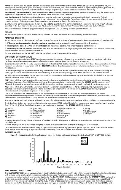
EXPECTED VALUES<br />
Figure 1 –- Frequency distribution of recovery times for clinical trial specimens positive in the BACTEC <strong>MGIT</strong> 960 System<br />
PERFORMANCE CHARACTERISTICS<br />
The BACTEC <strong>MGIT</strong> 960 System was evaluated at six clinical sites including one non-US site, which represented public health laboratories as<br />
well as large acute care hospitals in geographically diverse areas. The site population included patients infected with HIV,<br />
immunocompromised patients and transplant patients. The BACTEC <strong>MGIT</strong> 960 System was compared to the BACTEC 460TB radiometric<br />
system and conventional solid growth media for the detection and recovery of mycobacteria from clinical specimens, except blood. A<br />
total of 3330 specimens were tested during the study. A total of 353 specimens were positive which represented 362 isolates recovered<br />
during the study. The distribution of positives by specimen type is: respiratory (90%), tissue (7%), body fluids (1%), stool (0.85%) and<br />
3