Cystogram - American Society of Radiologic Technologists
Cystogram - American Society of Radiologic Technologists
Cystogram - American Society of Radiologic Technologists

You also want an ePaper? Increase the reach of your titles
YUMPU automatically turns print PDFs into web optimized ePapers that Google loves.
What you<br />
need to know<br />
about…<br />
<strong>Cystogram</strong><br />
A cystogram is an x-ray<br />
examination <strong>of</strong> the urinary bladder,<br />
which is located in the lower<br />
pelvic area. A cystogram can show<br />
the bladder’s position and shape,<br />
and the exam <strong>of</strong>ten is used to<br />
diagnose a condition called reflux.<br />
Reflux occurs when urine in the<br />
bladder moves back up the ureters,<br />
the tubes that transport urine<br />
from the kidneys to the bladder.<br />
This condition can cause repeated<br />
urinary tract infections. A cystogram<br />
may be performed after a<br />
patient has experienced a pelvic<br />
injury to ensure that the bladder<br />
has not torn. <strong>Cystogram</strong>s also are<br />
used to detect polyps or tumors<br />
in the bladder.<br />
Patient Preparation<br />
Before your examination, a<br />
radiographer will explain the<br />
procedure to you and answer<br />
any questions you might have.<br />
A radiographer, also known as<br />
a radiologic technologist, is a skilled<br />
medical pr<strong>of</strong>essional who has specialized<br />
education in the areas <strong>of</strong> radiation<br />
protection, patient care and radiographic<br />
positioning and procedures.<br />
If you are a woman <strong>of</strong> childbearing<br />
age, the radiographer will ask the date <strong>of</strong><br />
your last menstrual period and if there<br />
is any possibility you are pregnant. Next,<br />
the radiographer will ask if you have<br />
any allergies. It is important to list all<br />
allergies to food and medicine, as well as<br />
to let the radiographer know if you have<br />
a history <strong>of</strong> hay fever or asthma. Some<br />
allergies may indicate a possible reaction<br />
to the contrast agent that will be used<br />
during the examination.<br />
You will be asked to put on a hospital<br />
gown and then the radiographer<br />
will direct you to the restroom and ask<br />
you to completely empty your bladder.<br />
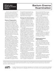
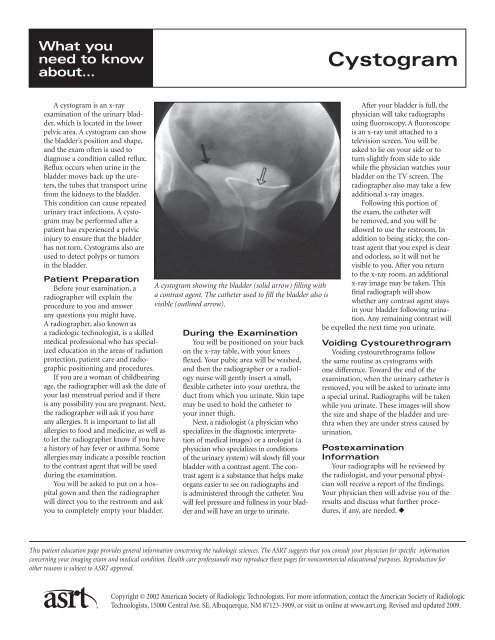
A cystogram showing the bladder (solid arrow) filling with<br />
a contrast agent. The catheter used to fill the bladder also is<br />
visible (outlined arrow).<br />
During the Examination<br />
You will be positioned on your back<br />
on the x-ray table, with your knees<br />
flexed. Your pubic area will be washed,<br />
and then the radiographer or a radiology<br />
nurse will gently insert a small,<br />
flexible catheter into your urethra, the<br />
duct from which you urinate. Skin tape<br />
may be used to hold the catheter to<br />
your inner thigh.<br />
Next, a radiologist (a physician who<br />
specializes in the diagnostic interpretation<br />
<strong>of</strong> medical images) or a urologist (a<br />
physician who specializes in conditions<br />
<strong>of</strong> the urinary system) will slowly fill your<br />
bladder with a contrast agent. The contrast<br />
agent is a substance that helps make<br />
organs easier to see on radiographs and<br />
is administered through the catheter. You<br />
will feel pressure and fullness in your bladder<br />
and will have an urge to urinate.<br />
After your bladder is full, the<br />
physician will take radiographs<br />
using fluoroscopy. A fluoroscope<br />
is an x-ray unit attached to a<br />
television screen. You will be<br />
asked to lie on your side or to<br />
turn slightly from side to side<br />
while the physician watches your<br />
bladder on the TV screen. The<br />
radiographer also may take a few<br />
additional x-ray images.<br />
Following this portion <strong>of</strong><br />
the exam, the catheter will<br />
be removed, and you will be<br />
allowed to use the restroom. In<br />
addition to being sticky, the contrast<br />
agent that you expel is clear<br />
and odorless, so it will not be<br />
visible to you. After you return<br />
to the x-ray room, an additional<br />
x-ray image may be taken. This<br />
final radiograph will show<br />
whether any contrast agent stays<br />
in your bladder following urination.<br />
Any remaining contrast will<br />
be expelled the next time you urinate.<br />
Voiding Cystourethrogram<br />
Voiding cystourethrograms follow<br />
the same routine as cystograms with<br />
one difference. Toward the end <strong>of</strong> the<br />
examination, when the urinary catheter is<br />
removed, you will be asked to urinate into<br />
a special urinal. Radiographs will be taken<br />
while you urinate. These images will show<br />
the size and shape <strong>of</strong> the bladder and urethra<br />
when they are under stress caused by<br />
urination.<br />
Postexamination<br />
Information<br />
Your radiographs will be reviewed by<br />
the radiologist, and your personal physician<br />
will receive a report <strong>of</strong> the findings.<br />
Your physician then will advise you <strong>of</strong> the<br />
results and discuss what further procedures,<br />
if any, are needed. ◆<br />
This patient education page provides general information concerning the radiologic sciences. The ASRT suggests that you consult your physician for specific information<br />
concerning your imaging exam and medical condition. Health care pr<strong>of</strong>essionals may reproduce these pages for noncommercial educational purposes. Reproduction for<br />
other reasons is subject to ASRT approval.<br />
Copyright © 2002 <strong>American</strong> <strong>Society</strong> <strong>of</strong> <strong>Radiologic</strong> <strong>Technologists</strong>. For more information, contact the <strong>American</strong> <strong>Society</strong> <strong>of</strong> <strong>Radiologic</strong><br />
<strong>Technologists</strong>, 15000 Central Ave. SE, Albuquerque, NM 87123-3909, or visit us online at www.asrt.org. Revised and updated 2009.
Lo que usted<br />
necesita saber<br />
acerca de...<br />
Cistograma<br />
El cistograma es un examen de<br />
rayos X de la vejiga urinaria, ubicada<br />
en el área pélvica inferior. El cistograma<br />
logra indicar la posición y la<br />
forma de la vejiga, y el examen se<br />
utiliza a menudo para diagnosticar un<br />
problema llamado reflujo. El reflujo<br />
ocurre cuando la orina en la vejiga<br />
regresa por los uréteres, los tubos que<br />
transportan la orina de los riñones a<br />
la vejiga. Esta condición puede provocar<br />
constantes infecciones de las<br />
vías urinarias. Se puede realizar un<br />
cistograma después de que el paciente<br />
haya sufrido una lesión pélvica,<br />
para asegurar que la vejiga no se haya<br />
desgarrado. También se utilizan los<br />
cistogramas para detectar pólipos o<br />
tumores en la vejiga.<br />
Preparación del<br />
Paciente<br />
Antes de su examen, un tecnólogo<br />
en radiografía le explicará el procedimiento<br />
y responderá a sus preguntas.<br />
El tecnólogo en radiografía, también<br />
conocido como tecnólogo radiológico,<br />
es un pr<strong>of</strong>esional médico capacitado con<br />
estudios especializados en las áreas de<br />
protección contra la radiación, atención<br />
de pacientes y posicionamiento y procedimientos<br />
radiográficos.<br />
Si usted es una mujer en edad fértil,<br />
el tecnólogo en radiografía le preguntará<br />
la fecha de su último período menstrual<br />
y si existe alguna posibilidad de que esté<br />
embarazada. A seguir, el tecnólogo en<br />
radiografía le preguntará si tiene alergias. Es<br />
importante que mencione todas las alergias<br />
a alimentos y medicamentos, así como<br />
informarle al tecnólogo en radiografía si<br />
usted tiene antecedentes de fiebre de heno<br />
o asma. Ciertas alergias pueden indicar una<br />
posible reacción al agente de contraste que<br />
se utilizará durante el examen.<br />
Se le pedirá que vista una bata<br />
hospitalaria; enseguida, el tecnólogo<br />
en radiografía le indicará la ubicación<br />
Un cistograma mostrando la vejiga (flecha maciza)<br />
llenándose con un agente de contraste. El catéter<br />
utilizado para llenar la vejiga también es visible<br />
(flecha con contorno).<br />
del baño y le pedirá que vacíe su vejiga<br />
completamente.<br />
Durante el Examen<br />
Se lo posicionará boca arriba sobre<br />
la mesa de rayos X, con las rodillas<br />
flexionadas. Le lavarán el área púbica;<br />
enseguida, el tecnólogo en radiografía<br />
o una enfermera de radiología insertará<br />
suavemente un catéter pequeño y flexible<br />
en su uretra, el conducto por el<br />
cual usted orina. Se podrá utilizar cinta<br />
adhesiva para sujetar el catéter a la parte<br />
interna de su muslo.<br />
A continuación, un radiólogo (un<br />
médico que se especializa en la interpretación<br />
de imágenes médicas para diagnóstico)<br />
o un urólogo (un médico que<br />
se especializa en problemas del sistema<br />
urinario) llenará su vejiga lentamente<br />
con un agente de contraste. El agente de<br />
contraste es una sustancia que facilita la<br />
visualización de órganos en radiografías<br />
y que se administra a través de un catéter.<br />
Usted sentirá presión y plenitud en la<br />
vejiga y sentirá ganas de orinar.<br />
Cuando su vejiga esté llena, el<br />
médico tomará radiografías a través<br />
de la fluoroscopia. La fluoroscopia es<br />
una unidad de rayos X conectada a<br />
una pantalla de televisión. Se le pedirá<br />
que se acueste de costado o que gire<br />
levemente de lado a lado mientras<br />
el médico observa su vejiga en la<br />
pantalla de TV. El tecnólogo en radiografía<br />
también podrá tomar algunas<br />
imágenes adicionales de rayos X.<br />
Después de esta parte del examen,<br />
se retirará el catéter y se le permitirá<br />
ir al baño. Además de ser pegajoso, el<br />
agente de contraste que usted eliminará<br />
será transparente e inodoro; por<br />
lo tanto, usted no lo verá. Después<br />
de que regrese a la sala de rayos X, se<br />
tomará una imagen de rayos X adicional.<br />
Esta radiografía final mostrará<br />
si aún queda agente de contraste en<br />
su vejiga después de haber orinado.<br />
Todo contraste restante será eliminado la<br />
próxima vez que orine.<br />
Cistouretrograma de<br />
Evacuación<br />
Los cistouretrogramas de evacuación<br />
siguen la misma rutina que los cistogramas,<br />
con una diferencia. Hacia el final<br />
del examen, cuando se retira el catéter urinario,<br />
se le pedirá que orine en un urinal<br />
especial. Se tomarán radiografías mientras<br />
usted orina. Dichas imágenes mostrarán<br />
el tamaño y forma de la vejiga cuando se<br />
encuentra bajo el esfuerzo de la micción.<br />
Información de<br />
Postexamen<br />
Sus radiografías serán analizadas por<br />
el radiólogo, y su médico personal recibirá<br />
un informe de los resultados. Luego,<br />
su médico le informará los resultados y<br />
conversará con usted sobre qué procedimientos<br />
adicionales son necesarios, si<br />
fuera el caso. ◆<br />
Esta página educacional del paciente provée información general en cuanto a la ciencia radiológica. ASRT sugiere que usted consulte con su doctor para obtener información<br />
específica concerniente a su examen de imagen y condiciones medicas. Los pr<strong>of</strong>esionales del cuidado de la salud pueden reproducir estas páginas para ser usadas sin recibir lucro<br />
económico. La reproducción de estos documentos para ser usadas para otros objetivos necesita la autorización del ASRT.<br />
Copyright © 2002 <strong>American</strong> <strong>Society</strong> <strong>of</strong> <strong>Radiologic</strong> <strong>Technologists</strong>. Para más información, contáctese con la Sociedad <strong>American</strong>a de<br />
Radiología Tecnológica, 15000 Central Ave. SE, Albuquerque, NM 87123-3909, o visítenos en la web electrónica: www.asrt.org.