Apport de l'imagerie dans la maladie de Crohn - Gastrospace.com
Apport de l'imagerie dans la maladie de Crohn - Gastrospace.com
Apport de l'imagerie dans la maladie de Crohn - Gastrospace.com

Create successful ePaper yourself
Turn your PDF publications into a flip-book with our unique Google optimized e-Paper software.
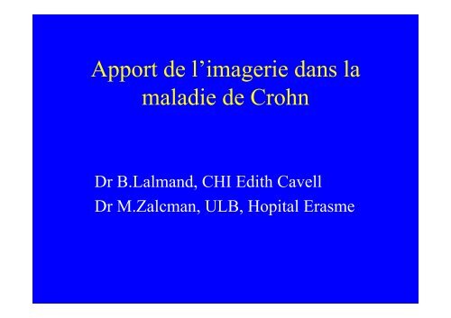
<strong>Apport</strong> <strong>de</strong> l’imagerie <strong>dans</strong> <strong>la</strong><br />
ma<strong>la</strong>die <strong>de</strong> <strong>Crohn</strong><br />
Dr B.Lalmand, CHI Edith Cavell<br />
Dr M.Zalcman, ULB, Hopital Erasme
Imagerie <strong>de</strong> <strong>la</strong> ma<strong>la</strong>die <strong>de</strong> <strong>Crohn</strong><br />
Introduction<br />
• Ma<strong>la</strong>die inf<strong>la</strong>mmatoire du tractus digestif<br />
• Épidémiologie<br />
• Hypothèses étiologiques<br />
Mycobactéries, virus,immunitaire,génétique,<br />
« vasculite mésentérique »,diététique,<br />
psychogénique, …….<br />
• Clinique
Imagerie <strong>de</strong> <strong>la</strong> ma<strong>la</strong>die <strong>de</strong> <strong>Crohn</strong><br />
Introduction<br />
• Distribution :<br />
-55% iléo-colique<br />
-30% grêle seul (3% grêle proximal)<br />
-15% colon seul
Imagerie <strong>de</strong> <strong>la</strong> ma<strong>la</strong>die <strong>de</strong> <strong>Crohn</strong><br />
Images élémentaires<br />
1) Altérations muqueuses<br />
2) Altérations pariétales<br />
3) Manifestations mésentériques<br />
4) Complications
anomalies<br />
du relief villeux<br />
Imagerie <strong>de</strong> <strong>la</strong> ma<strong>la</strong>die <strong>de</strong> <strong>Crohn</strong><br />
Altérations muqueuses<br />
ulcérations et ulcères<br />
lésions polypoï<strong>de</strong>s
plis épaissis<br />
Imagerie <strong>de</strong> <strong>la</strong> ma<strong>la</strong>die <strong>de</strong> <strong>Crohn</strong><br />
Altérations pariétales<br />
sténoses<br />
(inf<strong>la</strong>mm.,séquel…)<br />
saccu<strong>la</strong>tions
Imagerie <strong>de</strong> <strong>la</strong> ma<strong>la</strong>die <strong>de</strong> <strong>Crohn</strong><br />
• Infiltration<br />
• Lipomatose<br />
Manifestations mésentériques
Imagerie <strong>de</strong> <strong>la</strong> ma<strong>la</strong>die <strong>de</strong> <strong>Crohn</strong><br />
Complications<br />
Fistules
Imagerie <strong>de</strong> <strong>la</strong> ma<strong>la</strong>die <strong>de</strong> <strong>Crohn</strong><br />
Complications<br />
Phlegmons et abcès
Imagerie <strong>de</strong> <strong>la</strong> ma<strong>la</strong>die <strong>de</strong> <strong>Crohn</strong><br />
Complications<br />
Péritonite généralisée<br />
- néop<strong>la</strong>sies<br />
- hémorragie<br />
- ma<strong>la</strong>dies hépatiques et squelettiques associées…
Imagerie <strong>de</strong> <strong>la</strong> ma<strong>la</strong>die <strong>de</strong> <strong>Crohn</strong><br />
Modalités d’imagerie<br />
Opacifications : -OED<br />
-transit grêle<br />
-colon par <strong>la</strong>vement<br />
-fistulographie<br />
Tomo<strong>de</strong>nsitométrie<br />
Ultrasons et doppler<br />
IRM<br />
NB:Ponction-drainage sous US et fluoroscopie, ou<br />
sous CT
Imagerie <strong>de</strong> <strong>la</strong> ma<strong>la</strong>die <strong>de</strong> <strong>Crohn</strong><br />
Opacifications<br />
Pierre angu<strong>la</strong>ire traditionnelle <strong>de</strong> l’évaluation<br />
radiologique <strong>de</strong> <strong>la</strong> ma<strong>la</strong>die <strong>de</strong> <strong>Crohn</strong><br />
¤ Diagnostic <strong>de</strong> <strong>la</strong> ma<strong>la</strong>die <strong>de</strong> <strong>Crohn</strong><br />
¤ Diagnostic différentiel <strong>Crohn</strong>/RCUH.<br />
-OED<br />
-grêle : per os (small bowel follow-through) ou entéroclyse<br />
-colon : <strong>la</strong>vement baryté (sc ou dc) ou aux hydrosolubles<br />
-fistulographie
Imagerie <strong>de</strong> <strong>la</strong> ma<strong>la</strong>die <strong>de</strong> <strong>Crohn</strong><br />
Entéroclyse<br />
• Entéroclyse c<strong>la</strong>ssique:<br />
Gold standard pour les pathologie du grêle:<br />
- permet une distension optimale, contrôlée du grêle<br />
- reconnaît les lésions les plus fines – cartographie précise<br />
- diagnostic <strong>de</strong>s lésions causales d’obstructions, même<br />
in<strong>com</strong>plètes<br />
- informations fonctionnelles (distensibilité, souplesse, fixation<br />
<strong>de</strong>s anses)<br />
Limitations:<br />
- informations indirectes sur l’environnement <strong>de</strong>s anses<br />
- superpositions masquant certains segments
Imagerie <strong>de</strong> <strong>la</strong> ma<strong>la</strong>die <strong>de</strong> <strong>Crohn</strong><br />
Lavement baryté versus colonoscopie<br />
- LB =colono<br />
pour le DD <strong>Crohn</strong> /RCUH (spécificité <strong>de</strong> 95% du LBDC)<br />
- Colono >LB<br />
quant à l’extension <strong>de</strong> <strong>la</strong> ma<strong>la</strong>die ssi couplée à <strong>de</strong>s biopsies (zones<br />
macroscopiquement saines)<br />
-LB>colono<br />
profon<strong>de</strong>ur <strong>de</strong>s ulcères et <strong>de</strong>s trajets fistuleux<br />
masses inf<strong>la</strong>mmatoires extrinsèques<br />
-LB>colono<br />
ma<strong>la</strong>die touchant plus souvent le colon D et l’iléon terminal, présentant une<br />
atteinte discontinue
Imagerie <strong>de</strong> <strong>la</strong> ma<strong>la</strong>die <strong>de</strong> <strong>Crohn</strong><br />
CT scanner<br />
Pierre angu<strong>la</strong>ire actuelle <strong>de</strong> l’évaluation radiologique<br />
<strong>de</strong> <strong>la</strong> ma<strong>la</strong>die <strong>de</strong> <strong>Crohn</strong>, <strong>com</strong>plément essentiel <strong>de</strong>s<br />
examens c<strong>la</strong>ssiques<br />
Mise en évi<strong>de</strong>nce par un seul examen <strong>de</strong>s :<br />
-anomalies <strong>de</strong>s parois intestinales<br />
-anomalies mésentériques<br />
-viscères abdominaux et pelviens<br />
(anomalies <strong>de</strong>s tissus mous et osseux)<br />
~30% d’anomalies non suspectées entraînant une modification<br />
<strong>de</strong> l’attitu<strong>de</strong> thérapeutique médicale ou chirurgicale<br />
(Fishman et al. AJR 1987)
Imagerie <strong>de</strong> <strong>la</strong> ma<strong>la</strong>die <strong>de</strong> <strong>Crohn</strong><br />
Rôle du CT scanner: PHASE AIGUE<br />
• Diagnostic différentiel du retentissement<br />
mésentérique <strong>de</strong> <strong>la</strong> ma<strong>la</strong>die suggérée par palpation<br />
ou RX :<br />
Diagnostic et localisation <strong>de</strong>s abcès et fistules,<br />
fibrolipomatose, etc.…<br />
• Évaluation <strong>de</strong>s anomalies pariétales, et leur<br />
retentissement sur le calibres <strong>de</strong>s intestins :<br />
Détection et détermination du siège <strong>de</strong>s obstructions<br />
Caractérisation <strong>de</strong> l’épaississement pariétal
Imagerie <strong>de</strong> <strong>la</strong> ma<strong>la</strong>die <strong>de</strong> <strong>Crohn</strong><br />
Rôle du CT scanner
Imagerie <strong>de</strong> <strong>la</strong> ma<strong>la</strong>die <strong>de</strong> <strong>Crohn</strong><br />
Rôle du CT scanner
Imagerie <strong>de</strong> <strong>la</strong> ma<strong>la</strong>die <strong>de</strong> <strong>Crohn</strong><br />
Recherche <strong>de</strong> techniques d’étu<strong>de</strong>s du grêle<br />
associant les avantages <strong>de</strong> l’entéroclyse<br />
c<strong>la</strong>ssique, mais sans ses limitations:<br />
Entéroclyse par CT
Imagerie <strong>de</strong> <strong>la</strong> ma<strong>la</strong>die <strong>de</strong> <strong>Crohn</strong><br />
Entéroclyse par CT<br />
• Entéroclyse par CT scanner (C+ hydrosoluble)<br />
- performances connues du CT<br />
- reconnaissance d’obstacle subocclusifs :<br />
sensibilité 82% (48% CT c<strong>la</strong>s. )<br />
Mais:<br />
- radiations ionisantes (âge, fréquents examens).<br />
- approche 3D nécessite une exposition plus intense<br />
(coupes fines)<br />
- pas <strong>de</strong> contrôle fluoroscopique au remplissage.
Imagerie <strong>de</strong> <strong>la</strong> ma<strong>la</strong>die <strong>de</strong> <strong>Crohn</strong><br />
Ultrasonographie<br />
Technique peu coûteuse, non irradiante<br />
Doppler US: flux a.mésentérique sup. Activité<br />
Color US: mesure qualitative <strong>de</strong> <strong>la</strong> vascu<strong>la</strong>risation<br />
pariétale follow-up <strong>de</strong> traitement<br />
médical<br />
Hydrocolonic US: extension colique
25 ans: poussée d+<br />
Imagerie <strong>de</strong> <strong>la</strong> ma<strong>la</strong>die <strong>de</strong> <strong>Crohn</strong> :<br />
Ultrasonographie<br />
«target<br />
sign »<br />
Épaissiement pariétal hypoUS<br />
Perte <strong>de</strong> stratification
Imagerie <strong>de</strong> <strong>la</strong> ma<strong>la</strong>die <strong>de</strong> <strong>Crohn</strong><br />
Ultrasonographie: LIMITES<br />
- opérateur dépendante<br />
- artéfacts <strong>de</strong>s gaz intestinaux.<br />
- caractères aspécifiques <strong>de</strong>s épaississements pariétaux<br />
- détection <strong>de</strong>s lésions modérées: sensibilité à 52%<br />
(Pra<strong>de</strong>l, Abdom.Imaging 1997)<br />
Insatisfaisante pour le diagnostic et l’évaluation <strong>com</strong>plète <strong>de</strong><br />
l’extension <strong>de</strong> <strong>la</strong> ma<strong>la</strong>die
Imagerie <strong>de</strong> <strong>la</strong> ma<strong>la</strong>die <strong>de</strong> <strong>Crohn</strong><br />
Imagerie par Résonance Magnétique (IRM)<br />
• Imagerie potentiellement optimale pour le tractus<br />
digestif et en particulier <strong>la</strong> ma<strong>la</strong>die <strong>de</strong> <strong>Crohn</strong>:<br />
-Excellente résolution en contraste pour les tissus mous<br />
-Sensibilité aux processus inf<strong>la</strong>mmatoires<br />
-approche multip<strong>la</strong>naire ( CT multibarette)<br />
-Pas <strong>de</strong> contraste iodé<br />
-Non irradiante patients souvent jeunes, examens itératifs<br />
• Limitations:<br />
-Résolution spatiale<br />
-Artéfacts repiratoires<br />
-mouvements péristaltiques
Imagerie <strong>de</strong> <strong>la</strong> ma<strong>la</strong>die <strong>de</strong> <strong>Crohn</strong> :<br />
IRM<br />
Progrès techniques <strong>de</strong> ces <strong>de</strong>rnières années:<br />
- gating respiratoire<br />
- réduction importante <strong>de</strong>s temps d’acquisitions<br />
(séquences, optimisation <strong>de</strong>s antennes, technique « Sense »,…)<br />
acquisition T2 en (trigger respiratoire: RARE/HASTE…):<br />
résolution anatomique<br />
acquisition T1 en apnée (EG) :C- et C+<br />
(↔inf<strong>la</strong>mmation?)
Imagerie <strong>de</strong> <strong>la</strong> ma<strong>la</strong>die <strong>de</strong> <strong>Crohn</strong> :<br />
IRM<br />
« <strong>Crohn</strong> Disease with Endoscopic Corre<strong>la</strong>tion:<br />
Single-Shot Fast Spin-Echo and Gadolinium<br />
enhanced Fat-suppressed Spoiled Gradient-<br />
Echo MR Imaging » (Russell N. Low, Radiology 2002)
Imagerie <strong>de</strong> <strong>la</strong> ma<strong>la</strong>die <strong>de</strong> <strong>Crohn</strong> :<br />
IRM<br />
1)30 ans. D+ abdominales+++ .Diarrhées : Pancolite aiguë<br />
Axial T2 single-shot fast SE Axial T1 spoiled GRE +Gd<br />
2)50 ans. Atcd iléo-caecectomie. Récidive d+ abdominales et diarrhées : sténose cicatricielle, confirmée par colono<br />
Ileon distal Ileon distal
Imagerie <strong>de</strong> <strong>la</strong> ma<strong>la</strong>die <strong>de</strong> <strong>Crohn</strong><br />
Recherche <strong>de</strong> techniques d’étu<strong>de</strong>s du grêle<br />
associant les avantages <strong>de</strong> l’entéroclyse<br />
c<strong>la</strong>ssique, mais sans ses limitations:<br />
entéroclyse par IRM
Imagerie <strong>de</strong> <strong>la</strong> ma<strong>la</strong>die <strong>de</strong> <strong>Crohn</strong><br />
Entéroclyse par RM<br />
• Mise en p<strong>la</strong>ce <strong>de</strong> <strong>la</strong> son<strong>de</strong><br />
• Administration <strong>de</strong> contraste (eau, solution d’eau<br />
isotonique, méthylcellulose…) par son<strong>de</strong> jéjunale,<br />
<strong>dans</strong> l’aimant<br />
• Contrôle fluoroscopique (jusqu’à 1 image/7sec)<br />
• Examen MR c<strong>la</strong>ssique T2, T1 C- et C+ sous<br />
hypotonie.
Imagerie <strong>de</strong> <strong>la</strong> ma<strong>la</strong>die <strong>de</strong> <strong>Crohn</strong> :<br />
Entéroclyse par RM ∝<br />
• Avantages<br />
- absence irradiation<br />
- approche fonctionnelle plus précise (« fluoroscopie »)<br />
• Limites:<br />
- mise en p<strong>la</strong>ce <strong>de</strong> <strong>la</strong> son<strong>de</strong> sous RX<br />
- résolution spatiale < l’entéroclyse c<strong>la</strong>ssique (∆ superficielles)<br />
- résolution temporelle (fluoroscopie discontinue)<br />
- durée : 30’ à 40’ minimum pour une équipe entraînée<br />
- prix et accessibilité <strong>de</strong>s machines
Imagerie <strong>de</strong> <strong>la</strong> ma<strong>la</strong>die <strong>de</strong> <strong>Crohn</strong> :<br />
Entéroclyse par RM: fluoroscopie<br />
54 ans: remplissage normal<br />
45 ans Obstruction grêle <strong>de</strong> bas gra<strong>de</strong>:<br />
Di<strong>la</strong>tation sacu<strong>la</strong>ire progressive d’une anse<br />
Discrète di<strong>la</strong>tation d’amont
Imagerie <strong>de</strong> <strong>la</strong> ma<strong>la</strong>die <strong>de</strong> <strong>Crohn</strong><br />
Entéroclyse par RM: fluoroscopie<br />
33 ans<br />
Subobstruction grêle <strong>de</strong> haut gra<strong>de</strong>:<br />
Di<strong>la</strong>tation croissante du jéjunum proximal<br />
sur sténoses étagées<br />
Reflux gastrique progressif
Imagerie <strong>de</strong> <strong>la</strong> ma<strong>la</strong>die <strong>de</strong> <strong>Crohn</strong><br />
Entéroclyse par RM<br />
24 ans<br />
Sténose serrée/fistule jéjunocolique<br />
T2 axiale<br />
T2 coronale<br />
Fist. iléoiléale<br />
Masse inf<strong>la</strong>m.<br />
Masse inf<strong>la</strong>m.
Imagerie <strong>de</strong> <strong>la</strong> ma<strong>la</strong>die <strong>de</strong> <strong>Crohn</strong><br />
Entéroclyse par RM<br />
Coronale: True Fisp Coronale: F<strong>la</strong>sh +Gd
Imagerie <strong>de</strong> <strong>la</strong> ma<strong>la</strong>die <strong>de</strong> <strong>Crohn</strong><br />
Complications ano-rectales<br />
• Complications très fréquentes <strong>de</strong> <strong>la</strong> ma<strong>la</strong>die (~90%) :<br />
-anomalies <strong>de</strong>rmatologiques légères<br />
-ulcères et érosions<br />
-hémorroï<strong>de</strong>s<br />
-abcès péri anaux et périnéaux<br />
-fissures et fistules anales(5 à 15% <strong>com</strong>plexes :extension en <strong>de</strong>hors<br />
du sphincter, abcès ischio-rectaux et au-<strong>de</strong>ssus du p<strong>la</strong>n du<br />
m. pubo-rectal, trajets multiples,…)<br />
• Difficultés d’évaluation peropératoire <strong>de</strong> l’extension<br />
secondaire, peut-être en partie à l’origine du nombre<br />
important <strong>de</strong> récidives<br />
• Techniques c<strong>la</strong>ssiques : LB , fistulographie…
Imagerie <strong>de</strong> <strong>la</strong> ma<strong>la</strong>die <strong>de</strong> <strong>Crohn</strong><br />
Complications ano-rectales<br />
• CT scanner: progrès essentiel <strong>dans</strong> <strong>la</strong> mise en<br />
évi<strong>de</strong>nce et l’appréciation <strong>de</strong>s fistules et abcès<br />
péri-anaux<br />
• Écho-endoscopie : - meilleure résolution<br />
- re<strong>la</strong>tions avec les sphincters<br />
mais :<br />
-champ <strong>de</strong> vue réduit<br />
-parfois sous anesthésie
Imagerie <strong>de</strong> <strong>la</strong> ma<strong>la</strong>die <strong>de</strong> <strong>Crohn</strong><br />
Complications ano-rectales<br />
• Intérêt majeur <strong>de</strong> l’IRM :<br />
- excellente résolution en contraste<br />
- excellente résolution anatomique<br />
du p<strong>la</strong>ncher pelvien<br />
<strong>de</strong> l’appareil sphinctérien
Sag. T2<br />
Axial T2<br />
Imagerie <strong>de</strong> <strong>la</strong> ma<strong>la</strong>die <strong>de</strong> <strong>Crohn</strong><br />
Complications ano-rectales<br />
sphincter<br />
Fistule tans-sphinctérienne<br />
Canal anal<br />
fistule<br />
Sph.int.<br />
Sph.ext.<br />
fistule<br />
fistule
f.ss pubo-rectal<br />
Imagerie <strong>de</strong> <strong>la</strong> ma<strong>la</strong>die <strong>de</strong> <strong>Crohn</strong><br />
Complications ano-rectales<br />
2 fistules « en fer-à-cheval »: coupes coronales T2d’arrière en avant<br />
Orifice rectal
Imagerie <strong>de</strong> <strong>la</strong> ma<strong>la</strong>die <strong>de</strong> <strong>Crohn</strong><br />
Ponction-drainage d’abcès
Imagerie <strong>de</strong> <strong>la</strong> ma<strong>la</strong>die <strong>de</strong> <strong>Crohn</strong><br />
Ponction-drainage d’abcès<br />
Sous contrôle US et fluoroscopie<br />
ou<br />
Sous contrôle tomo<strong>de</strong>nsitométrique