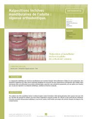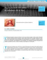
Actinomycose périapicale : à propos d'un cas. - SOP
Actinomycose périapicale : à propos d'un cas. - SOP
Actinomycose périapicale : à propos d'un cas. - SOP

You also want an ePaper? Increase the reach of your titles
YUMPU automatically turns print PDFs into web optimized ePapers that Google loves.
ENDODONTIE<br />
L’actinomycose est une infection chronique granulomateuse,<br />
caractérisée par la formation d’abcès<br />
dont le drainage peut se faire par une fistule. Elle<br />
est causée par les genres actinomyces et propionibacterium,<br />
et survient aussi bien chez l’homme que l’animal<br />
(Mc Ghee et coll., 1982). Chez l’homme, les localis<br />
a t io ns cervic o - fa c iales re p r é s e nt e nt 60 % (Lync h<br />
1977). A. israelii est le ge r me le plus impliqué<br />
(Stashenko 2001). L’actinomycose périapicale semble<br />
être rare (Martin et Harrison, 1984). Les actinomyces<br />
sont des germes commensaux de la cavité buccale. Ils<br />
ont été isolés dans les caries et les canaux des dents<br />
infectées (Sundqvist et Reuterving, 1980 ; Borssen et<br />
Sundqvist, 1981). Dans les lésions périapicales, le diagnostic<br />
est histologique, fondé sur la présence de<br />
grains actinomycosiques. L’examen bactériologique permet<br />
d’identifier la ou les espèces impliquées. Le traitement<br />
consiste en l’exérèse chirurgicale de la lésion, associé<br />
à un traitement médical (Wayman et coll., 1992 ;<br />
O’Grady et Reade, 1988).<br />
Actinomycosis is a chronic granulomatous infection,<br />
characterised by abscess formation and<br />
draining fistulae. Causing agents are actinomyces<br />
and propionibacterium, and can occur as well in man<br />
as in animals (Mc Ghee et al., 1982). In man, cervicofacial<br />
localisation represents 60 % of all lesions (Lynch<br />
1977). A. israelii is the most implicated microorganism<br />
(Stashenko 2001). Apical actinomycosis seems to be<br />
rare (Martin and Harrison, 1984). Actinomyces are commensal<br />
bacteria of the oral cavity. They have been isolated<br />
in caries and root canals of infected teeth (Sundqvist<br />
and Reuterving, 1980 ; Borssen and Sundqvist, 1981). In<br />
the <strong>cas</strong>e of periapical lesions, the diagnosis is made histologically<br />
and shows actinomycetes granules.<br />
Bacteriologic exam allows to identify the involved species.<br />
Treatment consists of surgical resection of the<br />
lesion and medical therapy (Wayman et al., 1992 ;<br />
O’Grady and Reade, 1988)<br />
Présentation du <strong>cas</strong><br />
Case presentation<br />
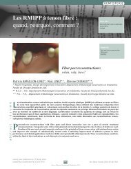
Le patient B.S, âgé de 34 ans, sans antécédents<br />
généraux, s'est présenté au service, pour une fistule en<br />
regard de la 32. Deux ans auparavant (2000), il a subi<br />
un curetage chirurgical périapical antéro-inférieur après<br />
obturation canalaire de la 31, 32, 41 et 42. La 31 a été<br />
extraite à la suite d’un traumatisme.<br />
L’examen exobuccal n’a révélé aucune particularité.<br />
L’examen endobuccal a permis de constater :<br />
■ une mauvaise hygiène ;<br />
■ la présence d’une fistule bourgeonnante productive<br />
entre la 31 et la 32 (Fig. 1) ;<br />
■ une mobilité de degré 1 sur la 32 ;<br />
■ le sondage n’a pas mis en évidence de poche parodontale<br />
;<br />
■ la percussion axiale a révélé une douleur modérée.<br />
La radiographique rétroalvéolaire a révèlé une<br />
image périapicale, radioclaire, à limites irrégulières.<br />
Le repérage du trajet fistuleux, en rapport avec<br />
la 32, a été fait avec un cône de gutta percha (Fig. 2).<br />
Le diagnostic d’abcès chronique fistulisé consécutif<br />
à un traitement endodontique insuffisant a été<br />
retenu.<br />
The patient B.S, 34 year old, without any past<br />
medical history, presented to the department, for a fistula<br />
in front of the 32. Two years ago, he underwent<br />
anterior and inferior periapical surgical curettage, following<br />
root canal obturation of the 31, 32, 41 and 42. The<br />
31 was extracted following trauma.<br />
The extra-oral exam did not reveal any particularities,<br />
whereas the intra-oral exam showed :<br />
■ Poor hygiene ;<br />
■ Presence of a budding productive fistulae between the<br />
31 and 32 (Fig. 1) ;<br />
■ Degree 1 mobility of the 32 ;<br />
■ Probing did not reveal any periodontal pocket;<br />
■ Axial percussion led to moderate pain.<br />
A retroalveolar X ray showed a radiolucent apical<br />
image, with irregular borders.<br />
Fistulae course was followed in relation with the<br />
32 using a gutta percha cone (Fig. 2).<br />
The diagnosis of chronic abscess following<br />
insufficient endodontic treatment was retained.<br />
54<br />
Revue d’Odonto-Stomatologie/février 2004