Actinomycose périapicale : à propos d'un cas. - SOP
Actinomycose périapicale : à propos d'un cas. - SOP
Actinomycose périapicale : à propos d'un cas. - SOP
Create successful ePaper yourself
Turn your PDF publications into a flip-book with our unique Google optimized e-Paper software.
ENDODONTIE<br />
<strong>Actinomycose</strong> périapicale :<br />
à <strong>propos</strong> d’un <strong>cas</strong>.<br />
Mots clés :<br />
Chirurgie endodontique<br />
Actinomyces<br />
Traitement endocanalaire<br />
Reprise traitement<br />
Lésion endo-parodontale<br />
Periapical actinomycosis :<br />
a <strong>cas</strong>e report.<br />
Keywords :<br />
Endodontic surgery<br />
Actinomyces<br />
Intraradicular treatment<br />
Retreatment<br />
Endo-periodontal lesion<br />
Latifa BERREZOUGA*, Zied BACCOUCHE**, Badreddine SRIHA***, Mohamed Semir BELKHIR****<br />
* Maître de conférence agrégé, Service d’odontologie conservatrice, Clinique Dentaire de Monastir, Tunisie<br />
** Assistant hospitalo-universitaire, Service d’odontologie conservatrice, Clinique Dentaire de Monastir, Tunisie<br />
*** Maître de conférence agrégé, Laboratoire de cytologie et d’anatomie pathologique, CHU F.Hached, Sousse, Tunisie<br />
**** Professeur, Service d’odontologie conservatrice, Clinique Dentaire de Monastir, Tunisie<br />
Nous rapportons un <strong>cas</strong> de lésion périapicale fistulisée sur une 32 traitée endodontiquement, chez un patient<br />
âgé de 34 ans. L’ouverture de la dent a permis de retrouver un second canal non obturé. La reprise du traitement<br />
endodontique a été effectuée et les deux canaux ont été obturés. L’évolution a été marquée par la persistance<br />
de la lésion et de la fistule, 11 mois après la reprise du traitement endodontique. La présence de bactéries<br />
extraradiculaires de type actinomyces a été suspectée. L’ablation d’un tissu granulomateux appendu à l’apex de la 32<br />
a été réalisée par voie chirurgicale suivie d’une résection apicale et d’une obturation rétrograde avec une pâte oxyde<br />
de zinc eugénol à prise rapide. L’histopathologie a confirmé le diagnostic d’actinomycose périapicale par la présence<br />
de grains actinomycosiques. L’évolution a été bonne avec une cicatrisation osseuse après 6 mois.<br />
We report a <strong>cas</strong>e of a 34 yo patient with a fistula-forming periapical lesion on an endodontically obturated 32.<br />
Opening of the teeth allowed to find a second non obturated root canal. Endodontic retreatment was performed<br />
and both root canals obturated. The observed lesion and fistulae persisted up to 11 months following<br />
retreatment. The presence of bacteria suggestive of actinomyces around the roots was suspected. Ablation of a granulation<br />
tissue from the 32 apex was performed surgically, followed by an apical resection and a retrograde obturation<br />
with rapidly setting eugenol zinc oxyde paste. The histologic study found actinomyces granules, and confirmed thus the<br />
diagnosis of periapical actinomycosis. Six months later, progression was favourable with bone healing.<br />
soumis pour publication le 01/09/03<br />
accepté pour publication le 21/10/03 Rev Odont Stomat 2004;33:00-00<br />
53<br />
Revue d’Odonto-Stomatologie/février 2004
ENDODONTIE<br />
L’actinomycose est une infection chronique granulomateuse,<br />
caractérisée par la formation d’abcès<br />
dont le drainage peut se faire par une fistule. Elle<br />
est causée par les genres actinomyces et propionibacterium,<br />
et survient aussi bien chez l’homme que l’animal<br />
(Mc Ghee et coll., 1982). Chez l’homme, les localis<br />
a t io ns cervic o - fa c iales re p r é s e nt e nt 60 % (Lync h<br />
1977). A. israelii est le ge r me le plus impliqué<br />
(Stashenko 2001). L’actinomycose périapicale semble<br />
être rare (Martin et Harrison, 1984). Les actinomyces<br />
sont des germes commensaux de la cavité buccale. Ils<br />
ont été isolés dans les caries et les canaux des dents<br />
infectées (Sundqvist et Reuterving, 1980 ; Borssen et<br />
Sundqvist, 1981). Dans les lésions périapicales, le diagnostic<br />
est histologique, fondé sur la présence de<br />
grains actinomycosiques. L’examen bactériologique permet<br />
d’identifier la ou les espèces impliquées. Le traitement<br />
consiste en l’exérèse chirurgicale de la lésion, associé<br />
à un traitement médical (Wayman et coll., 1992 ;<br />
O’Grady et Reade, 1988).<br />
Actinomycosis is a chronic granulomatous infection,<br />
characterised by abscess formation and<br />
draining fistulae. Causing agents are actinomyces<br />
and propionibacterium, and can occur as well in man<br />
as in animals (Mc Ghee et al., 1982). In man, cervicofacial<br />
localisation represents 60 % of all lesions (Lynch<br />
1977). A. israelii is the most implicated microorganism<br />
(Stashenko 2001). Apical actinomycosis seems to be<br />
rare (Martin and Harrison, 1984). Actinomyces are commensal<br />
bacteria of the oral cavity. They have been isolated<br />
in caries and root canals of infected teeth (Sundqvist<br />
and Reuterving, 1980 ; Borssen and Sundqvist, 1981). In<br />
the <strong>cas</strong>e of periapical lesions, the diagnosis is made histologically<br />
and shows actinomycetes granules.<br />
Bacteriologic exam allows to identify the involved species.<br />
Treatment consists of surgical resection of the<br />
lesion and medical therapy (Wayman et al., 1992 ;<br />
O’Grady and Reade, 1988)<br />
Présentation du <strong>cas</strong><br />
Case presentation<br />
Le patient B.S, âgé de 34 ans, sans antécédents<br />
généraux, s'est présenté au service, pour une fistule en<br />
regard de la 32. Deux ans auparavant (2000), il a subi<br />
un curetage chirurgical périapical antéro-inférieur après<br />
obturation canalaire de la 31, 32, 41 et 42. La 31 a été<br />
extraite à la suite d’un traumatisme.<br />
L’examen exobuccal n’a révélé aucune particularité.<br />
L’examen endobuccal a permis de constater :<br />
■ une mauvaise hygiène ;<br />
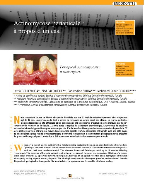
■ la présence d’une fistule bourgeonnante productive<br />
entre la 31 et la 32 (Fig. 1) ;<br />
■ une mobilité de degré 1 sur la 32 ;<br />
■ le sondage n’a pas mis en évidence de poche parodontale<br />
;<br />
■ la percussion axiale a révélé une douleur modérée.<br />
La radiographique rétroalvéolaire a révèlé une<br />
image périapicale, radioclaire, à limites irrégulières.<br />
Le repérage du trajet fistuleux, en rapport avec<br />
la 32, a été fait avec un cône de gutta percha (Fig. 2).<br />
Le diagnostic d’abcès chronique fistulisé consécutif<br />
à un traitement endodontique insuffisant a été<br />
retenu.<br />
The patient B.S, 34 year old, without any past<br />
medical history, presented to the department, for a fistula<br />
in front of the 32. Two years ago, he underwent<br />
anterior and inferior periapical surgical curettage, following<br />
root canal obturation of the 31, 32, 41 and 42. The<br />
31 was extracted following trauma.<br />
The extra-oral exam did not reveal any particularities,<br />
whereas the intra-oral exam showed :<br />
■ Poor hygiene ;<br />
■ Presence of a budding productive fistulae between the<br />
31 and 32 (Fig. 1) ;<br />
■ Degree 1 mobility of the 32 ;<br />
■ Probing did not reveal any periodontal pocket;<br />
■ Axial percussion led to moderate pain.<br />
A retroalveolar X ray showed a radiolucent apical<br />
image, with irregular borders.<br />
Fistulae course was followed in relation with the<br />
32 using a gutta percha cone (Fig. 2).<br />
The diagnosis of chronic abscess following<br />
insufficient endodontic treatment was retained.<br />
54<br />
Revue d’Odonto-Stomatologie/février 2004
1 2<br />
Fig. 1 : Fistule bourgeonnante entre la 31 qui est absente et la 32.<br />
Budding fistula between the 31 (absent) and the 32.<br />
Fig. 2 : petite image périapicale radioclaire. La fistule a été en rapport<br />
avec la 32.<br />
Radiolucent periapical small image. The fistula is in front of the 32.<br />
Traitement<br />
Treatment<br />
Reprise du traitement endodontique sur la 32 :<br />
■ l'ouverture de la dent permet la découverte d’un<br />
second canal non obturé (Fig. 3) ;<br />
■ désobturation de la gutta percha ;<br />
■ irrigation abondante au ClONa 0,5% ;<br />
■ préparation mécanisée par le système Profile ;<br />
■ obturation canalaire par la gutta chaude, utilisant les<br />
systèmes B et Obtura II (Fig. 4). Le matériau de scellement<br />
canalaire a été à base d’oxyde de zinc eugénol<br />
(Pulp Canal Sealer, EWT) ;<br />
■ une reprise de traitement sur la 41. Les mêmes techniques<br />
de préparation et d’obturation ont été effectuées<br />
(Fig. 5).<br />
L'évolution a été caractérisée par la persistance<br />
de la fistule, 11 mois après la reprise du traitement<br />
endodontique (Fig. 6).<br />
Le traitement chirurgical a permis l’exérèse d’un<br />
tissu granulomateux visible à travers la corticale externe<br />
résorbée, et intimement appendu à l’apex de la 32.<br />
Après résection des 2 mm apicaux, la cavité a<br />
retro préparée par des inserts ultrasoniques a été obturée<br />
par une pâte oxyde de zinc eugénol à prise rapide<br />
(Templin®).<br />
Endodontic retreatment of the 32 :<br />
■ tooth opening allows the discovery of a second non<br />
obturated root canal (Fig. 3) ;<br />
■ removal of the gutta percha ;<br />
■ abundant irrigation with NaCl 0.5% ;<br />
■ mechanical preparation using the Profile system ;<br />
■ root canal obturation using hot gutta, with the B and<br />
Obtura II systems (Fig. 4). Root canal cementing<br />
material was based on eugenol zinc oxyde (Pulp<br />
Canal Sealer, EWT) ;<br />
■ Retreatment of the 41 using similar techniques of preparation<br />
and obturation (Fig. 5).<br />
Evolution was characterised by fistulae persistence<br />
11 months following endodontic retreatment<br />
(Fig. 6).<br />
Surgical treatment allowed resection of a granulation<br />
tissue, visible through the resorbed external cortical<br />
and closely appended to the 32 apex.<br />
After resecting the apical 2 mm, the cavity was<br />
obtained with ultrasonic inserts and filled with eugenol<br />
zinc oxide with a fast setting time (Templin®).<br />
55<br />
Revue d’Odonto-Stomatologie/février 2004
ENDODONTIE<br />
3<br />
4<br />
Fig. 3 : Présence d’un deuxième<br />
canal non obturé.<br />
P resence of a second non<br />
obturated root canal.<br />
Fig. 4 : Reprise de l’obturation<br />
endodontique. Noter le<br />
dépassement du matériau<br />
d’obturation.<br />
Obturation with endodontic<br />
retreatment. Note a showing<br />
filling substance.<br />
5 6<br />
Fig. 5 : Reprise du traitement<br />
endodontique sur la 41. Noter<br />
la disparition du matériau<br />
d’obturation.<br />
Endodontic retreatment of the<br />
41. Note the absence of filling<br />
substance.<br />
Fig. 6 : Fistule non bourgeonnante<br />
mais persistante.<br />
Persistent fistula, non bud -<br />
ding.<br />
56<br />
Revue d’Odonto-Stomatologie/février 2004
Le patient a bénéficié <strong>d'un</strong> traitement médical :<br />
Clamoxyl® 500 mg, 1cp x 3/j pendant 7 jours ; Surgam®<br />
200 mg, 1cp x 3/j pendant 5 jours.<br />
Le cliché rétroalvéolaire postopératoire a mis en<br />
évidence la continuité de l'obturation a retro avec l’obturation<br />
canalaire (Fig. 7).<br />
L’évolution a été bonne, marquée par l’absence<br />
de fistule et une diminution nette de la taille de la<br />
lésion (Fig. 8), 6 mois après le retraitement.<br />
L’examen anatomopathologique a confirmé le<br />
diagnostic d’un granulôme inflammatoire périapical sur<br />
la 32, avec présence de grains actinomycosiques.<br />
Le tissu de granulation est polymorphe. Il est<br />
associé à un épithélium malpighien largement ulcéré<br />
(Fig. 9a) et à des travées ostéoïdes remaniées par l'inflammation.<br />
Des grains actinomycosiques ont été révélés au<br />
PAS et avec la coloration de Grocott (Fig. 9b, 9c).<br />
The patient received also antibiotics treatment :<br />
Clamoxyl® 500mg tid x 7 days ; Surgam® 200mg tid x<br />
5 days.<br />
The postoperative retroalveolar radiograph showed<br />
continuity of the retrograde obturation with root<br />
canal filling (Fig. 7).<br />
Progression was favourable without fistula formation<br />
and clear decrease in lesion size after 6 months<br />
(Fig. 8).<br />
Histologic exam confirmed the diagnosis of a<br />
periapical inflammatory granuloma with actinomyces<br />
granules on the 32.<br />
The granulation tissue is polymorph and is associated<br />
to a markedly ulcerated malpighian epithelium<br />
(Fig. 9a) and osteoid span, which have been modified by<br />
the inflammatory process.<br />
PAS (Fig. 9b) and Grocott (Fig. 9c) stains were<br />
positive for actinomyces granules.<br />
7 8<br />
Fig. 7 : cliché rétroalvéolaire après chirurgie endodontique<br />
; noter la continuité de l’obturation a retro<br />
avec l’obturation canalaire.<br />
Postoperative re t roalveolar radiograph showed<br />
continuity of the retrograde obturation with the root<br />
canal filling.<br />
Fig. 8 : cliché rétroalvéolaire postopératoire ; noter la<br />
cicatrisation périapicale à 6 mois.<br />
Postoperative retroalveolar radiograph ; note periapical<br />
healing after 6 months.<br />
57<br />
Revue d’Odonto-Stomatologie/février 2004
ENDODONTIE<br />
9a<br />
9b<br />
9c<br />
Fig. 9a : Epithélium malpighien ulcéré; noter la présence d’un grain<br />
actinimycosique entouré par une réaction inflammatoire (HE x 200).<br />
Ulcerated malpighian epithelium; note the presence of an actinomy -<br />
cetes granule surrounded by an inflammatory reaction (HE x 200).<br />
Fig. 9b : Deux grains actinomycosiques colorés en rouge par le PAS<br />
(PAS x 200).<br />
Two actinomycetes granules staining in red with PAS (PAS x 200).<br />
Fig. 9c : Coloration en noir d’un grain actinomycosique par le colorant<br />
de Grocott (Grocott x 400).<br />
Black staining of an actinomycetes granule with Grocott (Grocott x<br />
400).<br />
Discussion<br />
Discussion<br />
L’actinomycose périapicale est rare, la plupart<br />
des publications ont été rapportées sous forme de <strong>cas</strong><br />
isolés (Martin et Harrison, 1984 ; O’Grady et Reade,<br />
1988). Hirshberg et coll. (2003) ont rapporté une fréquence<br />
de 1,8 %, parmi les 963 biopsies de lésions<br />
périapicales. Les germes le plus souvent retrouvés en<br />
culture bactérienne sont A.israelii, A.naeslundi, A.viscosus<br />
et A.odontolyticus (Sundqvist et Reuterving,<br />
1980 ; Sakellariou 1996 ; Marsh et Martin, 1999 ;<br />
Streckfuss et coll., 1974). Ce sont des bactéries retrouvées<br />
dans la plaque dentaire, les caries, le sulcus gingival,<br />
les poches parodontales. Elles peuvent gagner le<br />
périapex par voie endodontique, par une poche parodontale<br />
ou encore à travers une fistule ouverte. Dans les<br />
<strong>cas</strong> typiques, le pus jaunâtre ou verdâtre est peu abon-<br />
Periapical actinomycosis is a rare affection and<br />
most publications in the literature are <strong>cas</strong>e reports<br />
(Martin and Harrison, 1984 ; O’Grady and Reade,<br />
1988). Hirshberg et al. (2003) have reported a frequency<br />
of 1.8 % from 963 biopsies of periapical lesions.<br />
Bacterial cultures have isolated more frequently A.<br />
israelii, A. naeslundi, A. viscosus and A. odontolyticus<br />
(Sundqvist and Reuterving, 1980 ; Sakellariou 1996 ;<br />
Marsh and Martin, 1999 ; Streckfuss et al., 1974). These<br />
bacteria are found in dental plaque, caries, gingival sulcus<br />
and periodontal pockets. They can reach the periapex<br />
via an endodontic route, a periodontal pocket or<br />
through an open fistula. In typical <strong>cas</strong>es, yellowish or<br />
greenish pus is scarce and contains sulfur granules. As<br />
these are calcified, the mineral source could be the per-<br />
58<br />
Revue d’Odonto-Stomatologie/février 2004
dant et constitué de granules de sulfures. Ces derniers<br />
étant calcifiés, la source minérale peut être l’exsudat<br />
périapical. Cependant, il a été rapporté que les streptocoques<br />
et d’autres bactéries extra-radiculaires peuvent<br />
former des dépôts minéraux intra et extracellulaires<br />
(Borssen et Sundqvist, 1981 ; Sunde et coll., 2002).<br />
L’examen histologique confirme la présence de<br />
grains actinomycosiques par des colorations spéciales :<br />
celles de Grocott et au P.A.S. Ces grains ont été mis en<br />
évidence en microscopie électronique à transmission et<br />
à balayage (Lomcali et coll., 1996 ; Tronstad et coll.,<br />
1987). Il s’agit d’une agrégation très dense d’organismes<br />
filamenteux liés entre eux par une matrice extracellulaire<br />
et entourés par plusieurs couches de cellules<br />
inflammatoires. Dans le <strong>cas</strong> observé, la persistance de<br />
l’infection a été attribuée à l’oubli d’un second canal<br />
non obturé. La reprise du traitement endodontique n’a<br />
pas permis la guérison. Cependant, il est intéressant de<br />
noter que l’élimination du matériau de scellement du<br />
périapex, 3 mois après le retraitement endodontique,<br />
témoigne de la présence d’un système immunitaire suffisamment<br />
efficace pour phagocyter ce corps étranger,<br />
mais aussi suffisamment inefficace pour éliminer des<br />
bactéries.<br />
Certains auteurs (Lin et coll., 1991) ont démontré<br />
sur des biopsies de granulômes périapicaux la présence<br />
de macrophages et de cellules géantes multinuclées<br />
impliquées dans la phagocytose de matériaux d’<br />
obturation canalaire. Il est fort probable que la consistance<br />
lisse du matériau utilisé ait rendue sa dissolution<br />
dans les liquides tissulaires et sa phagocytose possibles,<br />
comparée à la consistance dure des granules de<br />
sulfures, à leur taille importante et à leur surface irrégulière<br />
qui constituent autant de facteurs de résistance<br />
au système immunitaire.<br />
Par ailleurs, les β- l a c t a m i nes (Clamox y l<br />
1,5 g/jour pendant une semaine) prescrites par voie<br />
orale n’ont eu aucun effet sur l’infection fistulisée en<br />
contact permanent avec la flore buccale. Ceci pourrait<br />
faire penser à l’existence de mécanismes de résistance<br />
qui rendent les antibiotiques inefficaces face aux bactéries.<br />
La matrice extracellulaire entourant les bactéries<br />
constitue une sorte de biofilm like qui rend ces dernières<br />
moins sensibles aux anticorps opsonisants et aux<br />
substances antibactériennes. Ce biofilm se comporterait<br />
comme une pompe à efflux empêchant l’antibiotique de<br />
pénétrer. De plus, la capacité de survie des actinomyces<br />
dans le périapex est due au fait que ces bactéries sont<br />
anaérobies ; par conséquent, la phagocytose est altérée<br />
par le bas potentiel redox du milieu.<br />
iapical exudate. However, some authors have reported<br />
that streptococci or other extraradicular bacteria may<br />
form intra and extra-cellular mineral deposits (Borssen<br />
and Sundqvist, 1981 ; Sunde et al., 2002).<br />
Histologic exam confirms the presence of actinomycosis<br />
granules by special Grocott and PAS stains.<br />
Scanning electron microscopy has shown these granules<br />
(Lomcali et al., 1996 ; Tronstad et al., 1987). These<br />
structures are made of highly dense aggregates of filamentous<br />
organisms linked by an extracellular matrix and<br />
surrounded by several layers of inflammatory cells. In<br />
our <strong>cas</strong>e, persistence of the infection was attributed to<br />
the omission of the second unfilled root canal.<br />
Endodontic retreatment was not curative. However, it is<br />
interesting to note that elimination of the periapical sealing<br />
material, 3 months post endodontic retreatment, is<br />
an indication of the efficacy of the immune system in<br />
phagocyting this foreign body, but insufficiently efficacious<br />
in bacterial elimination.<br />
Some periapical granulomas biopsies (Lin et al.,<br />
1991) have shown the presence of macrophages and<br />
giant multinucleated cells involved in the phagocytosis<br />
of root canal sealing material. It is highly likely that the<br />
soft consistency of the used material has made possible<br />
its dissolution into tissue liquids and its phagocytosis,<br />
compared to the hard structure of sulfur granules, and to<br />
their big size and their irregular surface, which build<br />
resistance to the immune system.<br />
On the other hand, β-Lactam antibiotics (clamoxyl<br />
1.5 g/day for one week) per os are not effective<br />
against infected fistula which are in permanent contact<br />
with the oral flora. This leads to think of resistance<br />
mechanisms leading to inefficient antibiotics against<br />
bacteria. The extracellular matrix surrounding bacteria<br />
constitutes a biofilm which protects bacteria from opsonising<br />
antibodies and antibacterial agents. This biofilm<br />
would act like an efflux pump preventing antibiotics<br />
penetration. Moreover, actinomyces survival capacity in<br />
the periapex is due to their anaerobic characteristic ;<br />
thus, phagocytosis is altered by the low medium redox<br />
potential.<br />
59<br />
Revue d’Odonto-Stomatologie/février 2004
ENDODONTIE<br />
La reprise du traitement endodontique a été une<br />
étape primordiale et impérative, permettant d’assurer<br />
une étanchéité canalaire avant d’entreprendre la chirurgie<br />
endodontique. En effet, l’infection persistante à<br />
actinomyces, à des bactéries anaérobies et facultatives<br />
ou à corps étranger, constitue une des indications de la<br />
chirurgie endodontique (Haponen 1986 ; Ford 2001 ;<br />
Machtou 1993). Dans le <strong>cas</strong> présenté, c’est l’exérèse du<br />
tissu granulomateux infecté par des actinomyces qui a<br />
relancé le processus cicatriciel. Le traitement médical a<br />
été de courte durée. Les β-lactamines demeurent les<br />
antibiotiques de choix et sont actives contre la plupart<br />
des germes de la flore buccale isolés des lésions périapicales<br />
persistantes (Pinheiro et coll., 2003).<br />
Endodontic retreatment was a major and imperative<br />
stage, allowing the obtention of a perfectly sealed<br />
root canal before proceeding with surgery. In fact, persistent<br />
infection with actinomyces, with anaerobic bacteria<br />
and other foreign bodies is one of the indications of<br />
endodontic surgery (Haponen 1986 ; Ford 2001 ;<br />
Machtou 1993). In our <strong>cas</strong>e, resection of the infected<br />
granulation tissue has restarted the healing process.<br />
Medical treatment was short term. β-lactams remain the<br />
antibiotics of choice and are efficacious against most<br />
oral flora found at the level of persistent periapical<br />
lesions (Pinheiro et al., 2003).<br />
Demande de tirés-à-part :<br />
Docteur Latifa BERREZOUGA - Clinique dentaire de Monastir - 5019 - TUNISIE.<br />
Traduction : Zeina ANTOUN<br />
BORSSEN E., SUNDQVIST G.<br />
Actinomyces of infected dental root canals. Oral Surg<br />
1981;51:643-648.<br />
FORD P.<br />
Surgical treatment of apical periodontitis. In Orstavik D:<br />
Essential endodontology. Ed: Oxford, Blakwell Sc. 2001.<br />
HAPONEN R.P.<br />
Periapical actinomycosis: A follow-up study of 16 surgically<br />
treated <strong>cas</strong>es. Endo dent Traumat 1986;(2):205-259.<br />
HIRSHBERG A., TSESIS I., METZGER Z., KAPLAN I.<br />
Periapical actinomycosis: A clinicopathologic study. Oral<br />
Surg 2003;95(5):614-620.<br />
LIN L.M., PASCON E.A., SKRIBNER J.E.<br />
Clinic, radiographic and histologic study of endodontic<br />
treatment failures. Oral Surg 1991;(71):603-611.<br />
LOMCALI G., SEN B.H., CANKAYA H.<br />
Scanning electron microscopic observations of apical root<br />
surfaces of teeth with apical periodontitis. Endo dent<br />
Traumat 1996;12:70-76.<br />
LYNCH M.A.<br />
Ed: Burket’s oral medicine. 8th ed. Philadelphia: JB<br />
Lippincot CO., 1977.<br />
MACHTOU P.<br />
Le traitement chirurgical : guide clinique de l’endodontie.<br />
Ed: CdP, Paris 1993.<br />
MARTIN I.C., HARRISSON J.D.<br />
Periapical actinomycosis. Brit dent J 1984;54:169-170.<br />
MARSH P, MARTIN M.V.<br />
Oral microbiology, 4th ed. Ed: Wright, Oxford 1999.<br />
Mc GHEE J.R., MICHALEK S.M., CASSELG.H.<br />
Dental microbiology. Ed: Haper and Row, Philadelphia<br />
1982.<br />
O’GRADYJ.F., READE P.C.<br />
Periapical actinomycosis involving Actinomyces israelii. J<br />
Endo 1988;14(3):147-149.<br />
PINHEIRO E.T., GOMES B.P., FERRAZ C.C., TEIXEIRA<br />
F.B., ZAIAA.A., SOUZAFILHO F.J.<br />
Evaluation of root canal microorganisms isolated from teeth<br />
with endodontic failure and their antimicrobial susceptibility.<br />
Oral Microbiol Immunol 2003;18:100-103.<br />
TRONSTAD L., BARNETT F., RISO K., SLOTS J.<br />
Extraradicular endodontic infections. Endo dent Traumat<br />
1987;(3):86-90.<br />
SAKELLARIOU P.L.<br />
Periapical actinomycosis: report of a <strong>cas</strong>e and review of the<br />
literature. Endo dent Traumat 1996;12:151-154.<br />
STASHENKO P.<br />
Etiology and pathogenesis of pulpitis and apical periodontitis.<br />
In Orstavik D: Essential endodontology. Ed: Blakwell,<br />
Oxford 2001.<br />
STRECKFUSS J.L., SMITH W.N., BROWN L.R.,<br />
CAMPBELLM.M.<br />
Calcification of selected strains of streptococcus mutans<br />
and streptococcus sanguis. J Bacteriol 1974;120:502-506.<br />
SUNDE P.J., OLSEN I., DEBELIAN G.I., TRONSTAD L.<br />
Microbiota of periapical lesions refractory to endodontic<br />
therapy. J Endo 2002;(4):304-310.<br />
SUNDQVISTG., REUTERVING C.O.<br />
Isolation of actinomyces israelii from periapical lesion.<br />
J Endo 1980;(6):602-606.<br />
WAYMAN B.E., MURATA S.M., ALMEIDA ROY J.,<br />
FOWLER C.B.<br />
A bacteriological and histological evaluation of 58 periapical<br />
lesions. J Endo 1992;18(4):152-155.<br />
60<br />
Revue d’Odonto-Stomatologie/février 2004


