Les méthodes de diagnostic des lésions carieuses initiales. - SOP
Les méthodes de diagnostic des lésions carieuses initiales. - SOP
Les méthodes de diagnostic des lésions carieuses initiales. - SOP

Create successful ePaper yourself
Turn your PDF publications into a flip-book with our unique Google optimized e-Paper software.
DENTISTERIE RESTAURATRICE<br />
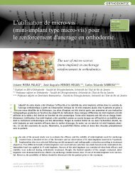
<strong>Les</strong> métho<strong>de</strong>s <strong>de</strong> <strong>diagnostic</strong><br />
<strong>de</strong>s lésions <strong>carieuses</strong> <strong>initiales</strong>.<br />
Mots clés :<br />
Carie émail<br />
Diagnostic précoce<br />
Métho<strong>de</strong> <strong>diagnostic</strong><br />
Diagnostic methods<br />
of initial carious lesions.<br />
Keywords :<br />
Enamel carie<br />
Early diagnosis<br />
Diagnostic method<br />
CHALA S.*, BOUAMARA R.*, ABDALLAOUI F.**<br />
* Rési<strong>de</strong>nte au centre <strong>de</strong> consultation et traitement <strong>de</strong>ntaire - Centre <strong>de</strong> consultation et <strong>de</strong> traitement <strong>de</strong>ntaire -<br />
Faculté <strong>de</strong> mé<strong>de</strong>cine <strong>de</strong>ntaire <strong>de</strong> Rabat - Maroc.<br />
** Professeur d’enseignement supérieur - Centre <strong>de</strong> consultation et <strong>de</strong> traitement <strong>de</strong>ntaire -<br />
Faculté <strong>de</strong> mé<strong>de</strong>cine <strong>de</strong>ntaire <strong>de</strong> Rabat - Maroc.<br />
<strong>Les</strong> lésions <strong>carieuses</strong> <strong>initiales</strong> <strong>de</strong> l’émail diagnostiquées précocement peuvent bénéficier d’une thérapeutique<br />
non invasive. Ce <strong>diagnostic</strong> précoce peut poser un véritable problème en omni-pratique. Le développement <strong>de</strong><br />
moyens <strong>diagnostic</strong> a facilité pour beaucoup cette approche. Le calibrage et l’entraînement <strong>de</strong>s praticiens pour<br />
la recherche <strong>de</strong> ces lésions au cours <strong>de</strong> l’examen clinique reste le moins coûteux et est relativement fiable. Cet article<br />
propose <strong>de</strong> passer en revue les différents moyens disponibles pour le <strong>diagnostic</strong> <strong>de</strong>s lésions <strong>carieuses</strong> <strong>initiales</strong> <strong>de</strong><br />
l’émail.<br />
XInitial carious lesions of the enamel, recent diagnosed, can benefit from non-invasive therapy. However, this diagnosis<br />
is very problematic in general practice. Development of <strong>diagnostic</strong> tools has facilitated this diagnosis for<br />
many <strong>de</strong>ntists. Training of <strong>de</strong>ntists to <strong>de</strong>tect such lesions during a clinical exam remains less costly and relatively<br />
reliable. This manuscript aims to review the different available methods for the diagnosis of enamel initial carious<br />
lesions.<br />
soumis pour publication le 26/08/03<br />
accepté pour publication le 23/06/04 Rev Odont Stomat 2004;33:297-310<br />
297<br />
Revue d’Odonto-Stomatologie/décembre 2004
DENTISTERIE RESTAURATRICE<br />
Le <strong>diagnostic</strong> précoce <strong>de</strong>s altérations <strong>de</strong>s tissus<br />
durs, dues à la maladie carieuse revêt une importance<br />
capitale, afin <strong>de</strong> pouvoir procé<strong>de</strong>r au mieux<br />
à l’instauration <strong>de</strong>s mesures <strong>de</strong> prophylaxie adéquates.<br />
Cette notion est à la base du modèle médical préventif<br />
fondé sur la prévention dans tous les sta<strong>de</strong>s <strong>de</strong> la<br />
maladie.<br />
En effet, les lésions <strong>initiales</strong>, qui correspon<strong>de</strong>nt aux<br />
premiers sta<strong>de</strong>s <strong>de</strong> la carie <strong>de</strong> l’émail, passent le plus<br />
s o u v e nt inaperçues à l’ins p e c t ion clinique (Tr i l l e r<br />
1993), alors que leur <strong>diagnostic</strong> à ce sta<strong>de</strong> permet l’instauration<br />
d’une thérapeutique non invasive permettant<br />
d’assurer la réversibilité <strong>de</strong> la lésion.<br />
Dans ce cadre, le développement <strong>de</strong> nouveaux moyens<br />
<strong>de</strong> <strong>diagnostic</strong> s’est avéré indispensable afin d’ai<strong>de</strong>r le<br />
praticien à répondre aux exigences d’une <strong>de</strong>ntisterie<br />
mo<strong>de</strong>rne basée sur le concept <strong>de</strong> prévention.<br />
Early diagnosis of hard tissues alterations, due to<br />
carious disease, is of capital importance, in or<strong>de</strong>r<br />
to optimise the <strong>de</strong>lay of institution of a<strong>de</strong>quate<br />
preventive measures. This notion is the basis of the preventive<br />
medical mo<strong>de</strong>l which is based on prevention at<br />
all disease stages.<br />
In fact, initial lesions, which correspond to the first<br />
stages of enamel carie, are often un<strong>de</strong>tected during clinical<br />
inspection (Triller 1993), when their diagnosis at<br />
this stage allows initiation of a non invasive therapeutic<br />
measure leading to a reversible lesion.<br />
In this context, <strong>de</strong>velopment of new <strong>diagnostic</strong> methods<br />
was shown to be indispensable in or<strong>de</strong>r to help <strong>de</strong>ntists<br />
face the requirements of mo<strong>de</strong>rn <strong>de</strong>ntistry based on prevention.<br />
Processus carieux initial<br />
L’initiation du processus carieux au niveau d’une<br />
surface <strong>de</strong>ntaire est souvent expliquée par une sucession<br />
<strong>de</strong> phénomènes physico-chimiques mettant en jeu<br />
une série <strong>de</strong> phases <strong>de</strong> dissolution-précipitation dans<br />
lesquels les aci<strong>de</strong>s produits par la plaque bactérienne<br />
induisent une déminéralisation <strong>de</strong>s tissus calcifiés <strong>de</strong> la<br />
<strong>de</strong>nt à la suite d’une chute du pH du milieu (Jaquot et<br />
Fontaine, 1995). Il se produit alors une dissolution <strong>de</strong>s<br />
phosphates <strong>de</strong> calcium qui constituent la phase minérale<br />
<strong>de</strong>s tissus durs <strong>de</strong> la <strong>de</strong>nt : émail, <strong>de</strong>ntine et<br />
cément. Dans un premier temps, les tampons salivaires<br />
tels que les bicarbonates vont neutraliser les aci<strong>de</strong>s présents<br />
et arrêter la fuite <strong>de</strong>s phosphates <strong>de</strong> calcium. Une<br />
précipitation <strong>de</strong>s ions présents dans le milieu ou apportés<br />
par <strong>de</strong>s applications topiques (fluor du <strong>de</strong>ntifrice)<br />
est possible conduisant à la reminéralisation <strong>de</strong> la partie<br />
dissoute.<br />
Lorsque le pH continue <strong>de</strong> baisser, les phénomènes<br />
<strong>de</strong> déminéralisation s'amplifient aux dépens <strong>de</strong> ceux<br />
<strong>de</strong> reminéralisation. Il se produit alors :<br />
■ une dislocation <strong>de</strong>s cristaux superficiels d'hydroxyapatite,<br />
■ un élargissement <strong>de</strong>s espaces intercristallins.<br />
Il en résulte une augmentation <strong>de</strong> la porosité <strong>de</strong> l'émail<br />
qui facilite la diffusion en profon<strong>de</strong>ur <strong>de</strong>s aci<strong>de</strong>s aboutissant<br />
à la formation <strong>de</strong> microchenaux qui s'insinuent<br />
progressivement dans l'émail.<br />
Dans un premier temps, les remaniements histologiques<br />
et les échanges ioniques dus aux variations du<br />
pH ne permettent pas encore la détection <strong>de</strong>s lésions<br />
Initial carious process<br />
The start of the carious process on the <strong>de</strong>ntal surface<br />
is often explained by a series of chemico-physical -<br />
phenomena involving a number of dissolution-precipitation<br />
processes in which acids produced by the bacterial<br />
plaque induce <strong>de</strong>mineralisation of calcified <strong>de</strong>ntal tissues<br />
following a drop of the medium pH (Jaquot and Fontaine,<br />
1995). Dissolution of calcium phosphates which make<br />
the mineral phase of the hard <strong>de</strong>ntal tissues : enamel, <strong>de</strong>ntin<br />
and cement, occurs then. First, salivary buffers such<br />
as bicarbonates, will neutralize the acids and stop calcium<br />
phosphate leakage. Precipitation of ions, which are<br />
present in the medium or brought by topical applications<br />
(fluorinated toothpaste) is possible, leading to remineralization<br />
of the dissolved part.<br />
When the pH drops continuously, the process of<br />
<strong>de</strong>mineralisation is amplified whereas remineralization<br />
<strong>de</strong>creases. This leads to :<br />
■ dislocation of superficial hydroxy-apatite crystals,<br />
■ wi<strong>de</strong>ning of intercrystalline spaces.<br />
As a result, enamel porosity increases and facilitates<br />
<strong>de</strong>ep diffusion of acids, leading to the formation of<br />
microchannels which progressively inva<strong>de</strong> the enamel.<br />
First, histological modifications and ionic<br />
exchanges due to pH variations do not allow yet <strong>de</strong>tection<br />
of carious lesions using traditional <strong>diagnostic</strong><br />
298<br />
Revue d’Odonto-Stomatologie/décembre 2004
<strong>carieuses</strong> par les moyens classiques <strong>de</strong> <strong>diagnostic</strong> (examen<br />
visuel, sondage et radiographie). On parle alors <strong>de</strong><br />
l é s ion infra-clinique ou sub-clinique (Ja q uot et<br />
Fontaine, 1995). Secondairement, les baisses répétitives<br />
du pH font apparaître <strong>de</strong>s chenaux par dissolution<br />
<strong>de</strong>s minéraux sous l’émail <strong>de</strong> surface. La lésion <strong>de</strong>vient<br />
alors cliniquement décelable et l’émail accuse un déficit<br />
minéral <strong>de</strong> 10 % par rapport à un émail sain (Triller<br />
1993 ; Haikel 2001 ; Seguier et Le May, 2002).<br />
L'altération <strong>de</strong>s surfaces cristallines modifie les<br />
indices <strong>de</strong> réfraction lumineuse <strong>de</strong> la surface amélaire,<br />
la lésion <strong>de</strong> sub-surface prend l'aspect d'une tâche blanche<br />
plus ou moins discontinue encore appelée " White<br />
spot ". Cette <strong>de</strong>rnière est cliniquement détectable.<br />
La tâche blanche peut se reminéraliser donnant<br />
naissance à la tâche brune ou " Brown spot ". Cette<br />
couleur est due à l’incorporation <strong>de</strong> pigments lors <strong>de</strong> la<br />
remontée du pH qui favorise la reprécipitation <strong>de</strong>s<br />
phosphates <strong>de</strong> calcium. Par ailleurs, en l'absence <strong>de</strong><br />
maîtrise <strong>de</strong>s facteurs acidogènes, les porosités <strong>de</strong> l'émail<br />
progressent le long <strong>de</strong>s stries <strong>de</strong> Retzius formant<br />
une cavitation amélaire et la zone <strong>de</strong> surface fragilisée<br />
peut alors s'effondrer à plus ou moins court terme<br />
(Triller 1993 ; Haikel 2001).<br />
methods (visual inspection, probing and radiography).<br />
These are called sub-clinical lesions (Jaquot and<br />
Fontaine, 1995). Second, repetitive pH drops lead to<br />
channel formation by the dissolution of minerals un<strong>de</strong>r<br />
the enamel surface . The lesions become then clinically<br />
<strong>de</strong>tectable and the enamel has a 10 % mineral <strong>de</strong>ficit<br />
with respect to healthy enamel (Triller 1993; Haikel<br />
2001; Seguier and Le May, 2002).<br />
Alterations of crystalline surfaces modify the<br />
indices of light reflection on the enamel surface, the<br />
lesion un<strong>de</strong>r the surface has the appearance of white<br />
spot, more or less discontinuous, called "“White spot".<br />
The latter is clinically <strong>de</strong>tectable.<br />
The white spot can re-mineralise and becomes a<br />
tan spot or “ Brown spot”. This colour is due to pigment<br />
incorporation when the pH increases, favouring precipitation<br />
of calcium phosphates. On the other hand, in the<br />
lack of control of acid-producing factors, enamel porosity<br />
progresses along Retzius lines, leading to the formation<br />
of enamel cavities, with a fragile surface which<br />
can break down at a more or less short term (Triller<br />
1993 ; Haikel 2001).<br />
Moyens <strong>de</strong> <strong>diagnostic</strong><br />
<strong>de</strong>s lésions <strong>initiales</strong><br />
Le <strong>diagnostic</strong> <strong>de</strong>s lésions <strong>carieuses</strong> <strong>initiales</strong> <strong>de</strong> l’émail<br />
est plus ou moins difficile en fonction <strong>de</strong> la situation<br />
<strong>de</strong> la lésion (<strong>de</strong>nt antérieure ou postérieure, lésion<br />
occlusale, cervicale ou proximale). Pour faciliter le <strong>diagnostic</strong>,<br />
un certain nombre <strong>de</strong> moyens ont été développés.<br />
Ces moyens sont plus ou moins fiables en fonction<br />
<strong>de</strong> leurs valeurs <strong>de</strong> sensibilité et <strong>de</strong> spécificité qui permettent<br />
aux praticiens <strong>de</strong> choisir l’un plutôt que l’autre.<br />
La sensibilité est définie comme étant la capacité à<br />
détecter les personnes qui présentent la pathologie, et la<br />
spécificité est la capacité à i<strong>de</strong>ntifier correctement ceux<br />
qui ne présentent pas la pathologie.<br />
Inspection clinique<br />
Elle doit être réalisée sur <strong>de</strong>s <strong>de</strong>nts propres, nettoyées<br />
et séchées (Ekstrand et coll.,1998), sous un bon<br />
éclairage, à l’ai<strong>de</strong> d’un miroir. Cette inspection à pour<br />
but <strong>de</strong> détecter toute opacité, coloration ou changement<br />
<strong>de</strong> la translucidité avec ou sans séchage prolongé.<br />
Diagnostic methods<br />
of initial lesions<br />
Diagnosis of initial carious lesions of the enamel<br />
is more or less difficult in function of the lesion location<br />
(anterior or posterior tooth, occlusal, cervical or<br />
approximal lesion). Several methods to simplify the diagnosis<br />
are available to the <strong>de</strong>ntist. Such methods are<br />
more or less reliable <strong>de</strong>pending on their sensitivity and<br />
specificity values, allowing <strong>de</strong>ntists to chose one<br />
method instead of the other. Sensitivity is <strong>de</strong>fined by the<br />
ability to <strong>de</strong>tect subjects with the pathology and specificity<br />
by the ability to properly i<strong>de</strong>ntify those without a<br />
pathology.<br />
Clinical exam<br />
It should be done on clean and dry teeth<br />
(Ekstrand et al., 1998), using a mirror un<strong>de</strong>r good light<br />
source. This exam is aimed to <strong>de</strong>tect any opacity, coloration<br />
or change in translucency with or without prolonged<br />
drying.<br />
299<br />
Revue d’Odonto-Stomatologie/décembre 2004
DENTISTERIE RESTAURATRICE<br />
Tableau 1 - Critères utilisés lors <strong>de</strong> l’examen visuel pour le <strong>diagnostic</strong> <strong>de</strong> caries<br />
Table 1 - Criteria for caries <strong>de</strong>tection during a visual inspection<br />
Score<br />
Critères / Criteria<br />
0 Absence ou léger changement <strong>de</strong> la translucidité <strong>de</strong> l’émail après séchage prolongé > 5s<br />
No or small change in translucency of enamel after prolonged drying > 5s<br />
1 Opacité ou discoloration difficilement visible au niveau d’une surface humi<strong>de</strong>,<br />
mais distinguée visiblement après séchage (Fig. 1)<br />
Poorly visible opaqueness or discoloration of humid surfaces, but visible after drying (Fig. 1)<br />
2 Opacité ou discoloration nettement visible sans séchage<br />
Visible opaqueness or discoloration, without drying<br />
3 Présence d’une cavité amélaire au niveau d’un émail opaque coloré<br />
et/ou discloration grisâtre <strong>de</strong> la <strong>de</strong>ntine sous jacente<br />
Presence of an enamel cavity at the level of a coloured opaque enamel<br />
and/or greyish discoloration of un<strong>de</strong>rlying <strong>de</strong>ntin<br />
4 Cavité au niveau d’un émail opaque ou décoloré exposant la <strong>de</strong>ntine<br />
Cavity at the level of an opaque or discoloured enamel, exposing the <strong>de</strong>ntin<br />
Cependant, c’est un examen à caractère subjectif<br />
d’où l’intérêt d’un système <strong>de</strong> calibrage. Ce système sera<br />
<strong>de</strong> plus en plus adapté pour assurer le suivi ou pilotage<br />
permettant à plusieurs praticiens d’interpréter le bilan<br />
précé<strong>de</strong>mment établi par un autre praticien (Hennequin<br />
et Lasfargues, 1999).<br />
<strong>Les</strong> critères utilisés sont ceux définis par Cortes<br />
et coll. (2000) (Tableau 1).<br />
L’inspection clinique présente les avantages suivants<br />
:<br />
■ familiarité pour le <strong>de</strong>ntiste,<br />
■ possibilité <strong>de</strong> suivi <strong>de</strong>s lésions dans le temps,<br />
■ facilité,<br />
■ rapidité,<br />
■ peu <strong>de</strong> moyen à mettre en œuvre.<br />
Cependant, <strong>de</strong>s problèmes persistent :<br />
■ la difficulté d’accès pour certains sites surtout au<br />
niveau proximal ou l’examen direct <strong>de</strong> cette face s’avère<br />
difficile par une simple inspection,<br />
■ la difficulté d’avoir un bon éclairage au niveau <strong>de</strong>s<br />
zones postérieures. (Pitts 1991).<br />
Le recours à <strong>de</strong>s ai<strong>de</strong>s optiques comme les loupes<br />
a été proposé par certains auteurs (Haak et coll.,<br />
2002) pour faciliter la détection clinique <strong>de</strong> ces caries<br />
débutantes. Cependant, l’étu<strong>de</strong> in vitro réalisée par<br />
Haak en 2002 a montré que ces ai<strong>de</strong>s n’augmentent pas<br />
vraiment la validité du <strong>diagnostic</strong> <strong>de</strong>s caries proximales,<br />
ni même <strong>de</strong>s caries occlusales (Haak et coll., 2002).<br />
However, this exam is subjective and thus a calibrating<br />
system is nee<strong>de</strong>d. This system will be more and<br />
more adapted in or<strong>de</strong>r to ensure surveillance or monitoring,<br />
and to allow several <strong>de</strong>ntists to interpret previously<br />
performed exams (Hennequin and Lasfargues, 1999).<br />
The criteria used are <strong>de</strong>fined by Cortes et al.<br />
(2000) (Table 1).<br />
The clinical exam has the following advantages :<br />
■ <strong>de</strong>ntist familiarisation,<br />
■ possibility of long-term follow-up of lesions ,<br />
■ facility,<br />
■ rapidity,<br />
■ very few tools for implementation.<br />
However, some problems persist :<br />
■ access difficulty to some sites, especially at the<br />
approximal level where a direct exam of this si<strong>de</strong> is<br />
difficult to perform by simple inspection,<br />
■ difficulty to obtain good visibility of the posterior<br />
areas (Pitts 1991)<br />
The use of optic aids such as magnifying visual<br />
aids, was suggested by some authors (Haak et al., 2002)<br />
in or<strong>de</strong>r to facilitate clinical <strong>de</strong>tection of initial caries.<br />
However, Haak’s in vitro study in 2002 have shown that<br />
such aids do not really increase the reliability of<br />
approximal and occlusal caries diagnosis (Haak et al.,<br />
2002).<br />
300<br />
Revue d’Odonto-Stomatologie/décembre 2004
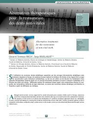
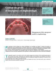
1<br />
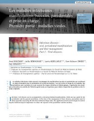
Fig. 1 : Coloration blanchâtre au<br />
niveau mésial <strong>de</strong> la 23 visible<br />
après séchage (score 1).<br />
Whitish discoloration at the mesial<br />
level of the 23 , visible after drying<br />
(score 1).<br />
Sondage<br />
Il nécessite le recours à <strong>de</strong>s son<strong>de</strong>s exploratrices<br />
(son<strong>de</strong>s 6, 17 et 23). La fiabilité <strong>de</strong> cette technique<br />
repose sur la résistance ressentie par l’opérateur pour<br />
enlever une son<strong>de</strong> introduite en force dans une anfractuosité.<br />
De ce fait, cette technique reflète avant tout<br />
le rapport existant entre les caractéristiques géométriques<br />
<strong>de</strong> l’extrémité <strong>de</strong> la son<strong>de</strong> et les critères anatomiques<br />
du sillon, ou du puits dans lequel elle est introduite,<br />
mais ne fournit aucune indication fiable sur la<br />
nature pathologique <strong>de</strong> la zone sondée (Hennequin et<br />
Lasfargues, 1999).<br />
Ces <strong>de</strong>rnières années le sondage a été remis en<br />
question (Mc Comb et Tam, 2001). En effet, la pression<br />
exercée lors d’un sondage rigoureux peut produire <strong>de</strong>s<br />
traumatismes <strong>de</strong> l’émail <strong>de</strong> surface correspondant à <strong>de</strong>s<br />
lésions <strong>de</strong> sub-surface et la fissure peut ainsi <strong>de</strong>venir<br />
plus susceptible à la progression <strong>de</strong> la lésion (Ekstrand<br />
et coll., 1987). De plus, il favoriserait le transport bactérien<br />
d'un site à l'autre et permettrait donc la contamination<br />
<strong>de</strong>s sites sains.(Hennequin et Lasfargues,<br />
1999).<br />
Probing<br />
Probing necessitates the use of sounding probes<br />
(probes 6, 17 and 23). The reliability of this technique<br />
lies on the resistance <strong>de</strong>tected by the <strong>de</strong>ntist to remove a<br />
probe which was pushed insi<strong>de</strong> an anfractuosity. Thus,<br />
this technique reflects an existing relation between geometric<br />
characteristics of the probe tip and anatomic properties<br />
of the line or the well in which it was introduced,<br />
but it does not give any reliable information on the<br />
pathologic nature of the soun<strong>de</strong>d area (Hennequin and<br />
Lasfargues, 1999).<br />
But, the use of probing has been subject to questions<br />
over the last <strong>de</strong>ca<strong>de</strong>s (Mc Comb and Tam, 2001).<br />
In fact, pressure exerted during rigorous probing can<br />
produce trauma at the enamel surface, leading to subsurface<br />
lesions and the fissure can then become more<br />
susceptible to lesion progression (Ekstrand et al., 1987).<br />
Moreover, probing can promote bacterial transport from<br />
one site to another and allow contamination of healthy<br />
sites (Hennequin and Lasfargues, 1999).<br />
Examen radiographique<br />
rétro-coronaire<br />
La radiographie rétrocoronaire (ou bite-wing) est<br />
classée parmi les techniques qui peuvent apporter au<br />
praticien le maximum d’information. La précision et l’orientation<br />
du rayon inci<strong>de</strong>nt font du radiogramme rétrocoronaire<br />
le cliché <strong>de</strong> choix pour le dépistage précoce<br />
<strong>de</strong> la carie, en particulier au niveau <strong>de</strong>s faces proximales.<br />
Il reste limité pour les lésions <strong>initiales</strong> <strong>de</strong> la table<br />
occlusale du fait <strong>de</strong> la superposition d’une gran<strong>de</strong><br />
épaisseur <strong>de</strong> tissus <strong>de</strong>ntaires en vestibulaire et lingual<br />
Retro-coronal<br />
radiographic exam<br />
Bite-wing radiograph is classified amongst the<br />
techniques which can <strong>de</strong>liver a maximum of information<br />
for the <strong>de</strong>ntist. Precision and inci<strong>de</strong>nt ray orientation<br />
makes the bite-wing radiograph an image of choice in<br />
the early caries diagnosis, in particular at the level of<br />
approximal si<strong>de</strong>s. But its use is limited for initial lesions<br />
of the occlusal table due to a superposition of a large<br />
thickness of vestibular and lingual <strong>de</strong>ntal tissues.<br />
301<br />
Revue d’Odonto-Stomatologie/décembre 2004
DENTISTERIE RESTAURATRICE<br />
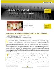
2 3<br />
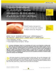
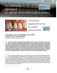
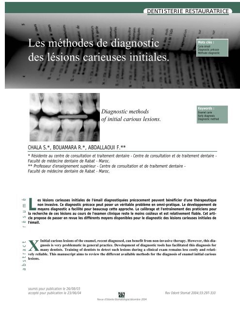
Fig. 2 : Radiographie Bite-wing montrant : une radioclarté score 1 au niveau distal <strong>de</strong> la 15 et 44, score 2 au niveau distal <strong>de</strong> la 45.<br />
Bite-wing radiograph showing : a score 1 radiolucency at the distal level of the 15 and 44, score 2 at the distal level of the 45.<br />
Fig. 3 : Radiographie Bite-wing montrant : une radioclarté score 1 au niveau mésial <strong>de</strong> la 46 et 47, score 2 au niveau mésial <strong>de</strong> la 15, score<br />
3 au niveau distal <strong>de</strong> la 46 et la 45.<br />
Bite-wing radiograph showing : a score 1 radiolucency at the mesial level of the 46 and 47, score 2 at the mesial level of the 15, score 3 at<br />
the distal level of the 46 and 45.<br />
A la lecture <strong>de</strong> l’image, il faut :<br />
■ rechercher une éventuelle solution <strong>de</strong> continuité <strong>de</strong><br />
l’image <strong>de</strong> la ligne <strong>de</strong> contour amélaire,<br />
■ rechercher le présence d’une zone radioclaire au<br />
niveau <strong>de</strong> la jonction amélo-<strong>de</strong>ntinaire,<br />
■ observer au niveau <strong>de</strong> la chambre pulpaire une éventuelle<br />
image <strong>de</strong> réaction, signe possible <strong>de</strong> défense<br />
<strong>de</strong>ntino-pulpaire à une agression (Daudibertiers et<br />
coll., 1993).<br />
L'étu<strong>de</strong> <strong>de</strong> Hintze et coll. (1998) a permis d'établir<br />
une échelle d'évaluation <strong>de</strong>s lésions proximales<br />
selon la profon<strong>de</strong>ur estimée à la radiographie rétrocoronaire<br />
(Fig. 2 et 3). Cette échelle se compose <strong>de</strong><br />
cinq scores (Hintze et coll., 1998) :<br />
■ score 0 : tissus sains (pas <strong>de</strong> radioclarté),<br />
■ score 1 : radioclarté touchant la moitié externe <strong>de</strong> l'émail,<br />
■ score 2 : radioclarté s'étendant à la moitié interne <strong>de</strong><br />
l'émail,<br />
■ score 3 : radioclarté atteignant le tiers externe <strong>de</strong> la<br />
<strong>de</strong>ntine,<br />
■ score 4 : radioclarté s'étendant aux <strong>de</strong>ux tiers internes<br />
<strong>de</strong> la <strong>de</strong>ntine.<br />
Différentes étu<strong>de</strong>s ont tenté d’évaluer cette<br />
technique dans la détection <strong>de</strong> caries débutantes. Selon<br />
Lussi (1993), cette technique possè<strong>de</strong> une sensibilité<br />
<strong>de</strong> 45 % à elle seule et <strong>de</strong> 49 % lorsqu'elle est combinée<br />
à l'examen visuel pour les lésions <strong>carieuses</strong> occlusales<br />
sans cavitation. Au niveau proximal, la sensibilité<br />
<strong>de</strong> cette technique est comprise entre 71 % et 100 % ±<br />
1 %, la spécificité 99 % et 100 % (Vaarkamp et coll.,<br />
2000).<br />
Pour Abdallaoui et coll. (2001) la radiographie<br />
rétro-coronaire doit être réalisée systématiquement<br />
chez tous les patients avant la prise en charge.<br />
Upon image reading, one should :<br />
■ search for an eventual solution of the continuity of the<br />
enamel contour line image,<br />
■ search for the presence of a radiolucent area at the<br />
level of the enamel-<strong>de</strong>ntin junction,<br />
■ observe an eventual reaction image at the level of the<br />
pulpal cavity, a possible sign of <strong>de</strong>ntino-pulpal <strong>de</strong>fence<br />
mechanism against aggression (Daudibertiers et al.,<br />
1993).<br />
The study of Hintze et al. (1998) has established<br />
a new evaluation scale of approximal lesions with<br />
respect to the estimated <strong>de</strong>pth by bite-wing radiographs<br />
(Fig. 2 and 3) . This scale comprises five scores (Hintze<br />
et al., 1998) :<br />
■ score 0 : healthy tissues (no radiolucency),<br />
■ score 1 : radiolucency of enamel external half ,<br />
■ score 2 : radiolucency involving enamel internal half,<br />
■ score 3 : radiolucency involving <strong>de</strong>ntin external third,<br />
■ score 4 : radiolucency involving <strong>de</strong>ntin internal twothirds.<br />
Several studies have tried to assess this technique<br />
in the <strong>de</strong>tection of initial caries. Lussi (1993) stated that<br />
this technique has 45 % sensitivity by itself, which<br />
increases to 49 % when combined with a clinical exam<br />
in the diagnosis of occlusal carious lesions without cavities.<br />
At a approximal level, sensitivity of this technique<br />
is inclu<strong>de</strong>d beetwen 71 % and 100 % ± 1 % and specificity<br />
beetwen 99 % and 100 % (Vaarkamp et al., 2000).<br />
Bite wings radiographs should be performed systematically<br />
for all patients (Abdallaoui et al., 2001).<br />
302<br />
Revue d’Odonto-Stomatologie/décembre 2004
Radiographie numérique<br />
Pour Daudibertiers et coll. (1993), la radiographie<br />
numérique permet une meilleure visualisation <strong>de</strong>s<br />
lésions <strong>carieuses</strong> par augmentation <strong>de</strong>s contrastes, la<br />
mise en évi<strong>de</strong>nce <strong>de</strong>s atteintes superficielles <strong>de</strong> l’émail<br />
ainsi qu’une évaluation quantitative <strong>de</strong>s <strong>de</strong>nsités par<br />
radiométrie (Daudibertiers et coll., 1993). Le contraste<br />
<strong>de</strong> l’image observée peut être réglé <strong>de</strong> manière à révéler<br />
les détails anatomiques recherchés par le praticien<br />
s’ils sont contenus dans le domaine <strong>de</strong>s niveaux <strong>de</strong> gris<br />
les plus forts ou les plus faibles <strong>de</strong> l’image (Le Denmat<br />
et coll., 1994).<br />
Il semblerait, que pour ces systèmes, un grand<br />
contraste soit nécessaire pour améliorer le <strong>diagnostic</strong><br />
<strong>de</strong>s lésions <strong>carieuses</strong>.<br />
Pour Le Denmat et coll. (1994), la résolution est<br />
plus faible que celle d’un film classique.<br />
L’apport <strong>de</strong> la radiographie numérique en matière<br />
<strong>de</strong> <strong>diagnostic</strong> <strong>de</strong> la carie fait objet <strong>de</strong> controverses<br />
dans la littérature ; alors que la plupart <strong>de</strong>s travaux à<br />
ce sujet font état d’une qualité i<strong>de</strong>ntique à celle <strong>de</strong>s<br />
radiographies conventionnelles, certains auteurs rapportent<br />
<strong>de</strong>s résultats moins bons.<br />
Pour Hennequin (1999), les lésions <strong>carieuses</strong><br />
peuvent aussi bien être détectée avec la radiographie<br />
numérique qu’avec un cliché argentique, mais il faut<br />
cependant noter que les images imprimées sur papier<br />
sont <strong>de</strong> moindre qualité <strong>diagnostic</strong> que les films en part<br />
ic u l ier si ces do c u me nts do i v e nt être cons e r v é s<br />
(Hennequin et Lasfargues, 1999 ; Hintze et coll., 1998).<br />
C’est une technique qui présente certains<br />
inconvénients :<br />
■ les capteurs peuvent constituer une gêne pour le<br />
patient ,<br />
■ le coût <strong>de</strong>s systèmes est élevé.<br />
Digital radiography<br />
For Daudibertiers et al. (1993), digital radiography<br />
obtains a better visualisation of carious lesions by<br />
increasing contrast, showing superficial affections of the<br />
enamel as well as a quantitative assessment of <strong>de</strong>nsities<br />
by radiometry (Daudibertiers et al., 1993). The contrast<br />
of the observed image could be controlled so it can<br />
reveal anatomic <strong>de</strong>tails if these are comprised in the area<br />
with the darkest or lightest grey colour of the image (Le<br />
Denmat et al., 1994).<br />
It seems that for all these systems, a big contrast<br />
is nee<strong>de</strong>d to improve the <strong>diagnostic</strong> yield of carious<br />
lesions.<br />
For Denmat et al. (1994), resolution is poorer<br />
than with a traditional film.<br />
The impact of digital radiography in carie diagnosis<br />
is controversial ; even though most published<br />
works reveal an i<strong>de</strong>ntical quality to that obtained with<br />
more conventional techniques, some authors report poorer<br />
results.<br />
For Hennequin (1999), carious lesions can be<br />
<strong>de</strong>tected by visual radiography as well as traditional<br />
films, but it should be noted that edited images on paper<br />
are of less <strong>diagnostic</strong> quality than films, especially if<br />
such documents have to be stored (Hennequin and<br />
Lasfargues, 1999 ; Hintze et al., 1998).<br />
However, this technique has some disadvantages :<br />
■ captors can be a nuisance to the patient,<br />
■ high cost.<br />
Elastiques séparateurs<br />
Il s’agit <strong>de</strong> dispositifs utilisés en orthodontie<br />
pour obtenir un écartement différé <strong>de</strong>s <strong>de</strong>nts, après<br />
mise en place pendant 24 h, afin <strong>de</strong> pouvoir sceller les<br />
bagues orthodontiques.<br />
Dans le cadre du <strong>diagnostic</strong> précoce <strong>de</strong>s lésions<br />
<strong>carieuses</strong> <strong>de</strong>s faces proximales, l’écartement temporaire,<br />
Rubber coins<br />
These are used in orthodontics in or<strong>de</strong>r to obtain<br />
differed teeth separation after 24 hours and in or<strong>de</strong>r to<br />
be able to seal orthodontic brackets.<br />
For the diagnosis of carious lesions of approximal<br />
si<strong>de</strong>s, temporary separation, obtained with rubbers,<br />
303<br />
Revue d’Odonto-Stomatologie/décembre 2004
DENTISTERIE RESTAURATRICE<br />
obtenu avec ces élastiques, permet un examen direct<br />
<strong>de</strong>s surfaces <strong>de</strong>ntaires proximales (Jaquot et Fontaine,<br />
1995) et <strong>de</strong> ce fait la détection <strong>de</strong>s lésions <strong>carieuses</strong><br />
débutantes.<br />
Rimmer et Pitts (1990) rapportent une augmentation<br />
substantielle du nombre <strong>de</strong> lésions diagnostiquées<br />
après écartement <strong>de</strong>ntaire par rapport à l’examen<br />
clinique et l’exa men ra d io g raphique (Ja q uot et<br />
Fontaine, 1995 ; Pitts 1991).<br />
allows a direct exam of approximal <strong>de</strong>ntal surfaces<br />
(Jaquot and Fontaine, 1995) and thus early <strong>de</strong>tection of<br />
initial carious lesions.<br />
Rimmer and Pitts (1990) have reported a substantial<br />
increase of the number of radiographic lesions<br />
following <strong>de</strong>ntal separation with respect to the clinical<br />
and radiographic exam (Jaquot and Fontaine 1995; Pitts<br />
1991).<br />
Fluorescence laser<br />
Cette métho<strong>de</strong> est basée sur la mesure <strong>de</strong> la fluorescence<br />
induite par les <strong>de</strong>nts après irradiation lumineuse<br />
afin <strong>de</strong> différencier entre l’émail carié et l’émail<br />
sain (Tam et Mc Comb, 2001).<br />
Le principe <strong>de</strong> détection est basé sur le changement<br />
<strong>de</strong>s propriétés physiques induites par les lésions<br />
<strong>carieuses</strong>. Parce qu’il y a plus d’eau dans une lésion<br />
carieuse que dans l’émail sain, la longueur <strong>de</strong> trajectoire<br />
libre d’un photon lumineux à l’intérieur d’une lésion<br />
carieuse est plus courte et moins transparente comparativement<br />
à un émail sain (Pinelli et coll., 2002).<br />
L’émail sain possè<strong>de</strong> une fluorescence jaune alors<br />
que les zones déminéralisées para i s s e nt sombre s<br />
(Pinelli et coll., 2002 ; Eggertsson et coll., 1999). On<br />
distingue :<br />
■ La fluorescence laser quantitative : Le terme fluorescence<br />
laser quantitative QLF est appliqué pour<br />
déterminer la fluorescence <strong>de</strong> la <strong>de</strong>nt induite après<br />
l’utilisation d’une lumière laser proche <strong>de</strong> 400 nm<br />
pour quantifier la déminéralisation et la sévérité <strong>de</strong> la<br />
lésion (Tam et Mc Comb 2001). Après corrélation <strong>de</strong>s<br />
résultats expérimentaux et <strong>de</strong>s contrôles histologiques,<br />
plusieurs étu<strong>de</strong>s cliniques ont validé cette<br />
métho<strong>de</strong> et l'ont jugée utilisable pour la détection<br />
<strong>de</strong>s lésions <strong>carieuses</strong> <strong>initiales</strong> et pour l'évaluation <strong>de</strong>s<br />
mesures préventives. Cependant, la QFL ne peut différencier<br />
entre les lésions confinées à l’émail et celles<br />
ayant évolué dans la <strong>de</strong>ntine (Tam et Mc Comb,<br />
2001).<br />
Dans cette optique un instrument <strong>de</strong> détection <strong>de</strong>s<br />
caries basé sur ce principe a été développé. Il s’agit<br />
du DIAGNODENT® (Kavo) qui permet à la fois la détection<br />
et la quantification <strong>de</strong>s caries. <strong>Les</strong> <strong>de</strong>nts sont<br />
illuminées avec une lumière laser <strong>de</strong> longueur d’on<strong>de</strong><br />
655 nm qui est absorbée par la substance organique<br />
et inorganique. La lumière est réémise proche d’une<br />
lumière fluorescente infrarouge. Le changement <strong>de</strong> la<br />
substance <strong>de</strong>ntaire associé à la progression carieuse<br />
Fluorescence laser<br />
This method is based on the fluorescence induced<br />
by teeth which were irradiated with a light source in<br />
or<strong>de</strong>r to differentiate between carious and healthy enamel<br />
(Tam and Mc Comb, 2001).<br />
The concept is based on a change in physical properties<br />
induced by carious lesions. A carious lesion<br />
contains more cater than healthy enamel , and thus, the<br />
length a light photon free trajectory insi<strong>de</strong> such lesions<br />
is shorter and less transparent as compared to healthy<br />
enamel (Pinelli et al., 2002).<br />
Healthy enamel has yellow fluorescence where<br />
as <strong>de</strong>mineralized areas seem to have a dark colour<br />
(Pinelli et al., 2002; Eggertsson et al., 1999). We can distinguish<br />
:<br />
■ Quantitati ve laser fluorescence (QLF) : this word is<br />
used to <strong>de</strong>termine fluorescence of an induced tooth,<br />
using a 400 nm laser light to quantify lesion severity<br />
and <strong>de</strong>mineralization (Tam and Mc Comb, 2001).<br />
After the correlation of experimental results and histological<br />
controls was observed, several clinical studies<br />
have validated this method for its used in initial<br />
carious lesions diagnosis and for the evaluation of preventive<br />
measure. However, QLF cannot differentiate<br />
between enamel lesions and those infiltrating the <strong>de</strong>ntin<br />
(Tam and Mc Comb, 2001).<br />
Thus, a new caries <strong>de</strong>tection instrument based on the<br />
above concept was <strong>de</strong>veloped : “ DIAGNODENT® “<br />
(Kavo) , which allows both <strong>de</strong>tection and quantification<br />
of caries. Teeth are illuminated with a 655 nm<br />
laser light which is absorbed by organic and inorganic<br />
substance. Re-emitted light is close to infrared fluorescent<br />
light. The change in <strong>de</strong>ntal substance associated<br />
with a progressive carious lesion, leads to an<br />
increase in the quantity of fluorescent light (Farge<br />
2000).<br />
304<br />
Revue d’Odonto-Stomatologie/décembre 2004
se traduit par une augmentation <strong>de</strong> la quantité <strong>de</strong><br />
lumière fluorescente (Farge 2000).<br />
■ La fluorescence laser expérimentale avec marqueur<br />
DELF : Le même principe régit ce système à la<br />
différence qu'il repose sur l'utilisation d'un marqueur<br />
afin <strong>de</strong> permettre la détection <strong>de</strong> la lésion initiale<br />
sans pour autant améliorer la quantification.<br />
Eggertsson et coll. (1999), dans une étu<strong>de</strong> in<br />
vitro, ont trouvé les valeurs <strong>de</strong> sensibilité suivantes : la<br />
fluorescence laser 56-74 %, DELF 61-79 % et l’examen<br />
visuel 58-74 %, la spécificité était <strong>de</strong> 67-78 % pour la<br />
fluorescence laser, 86-98 % pour DELF et 83-97 % pour<br />
l’examen visuel. Ainsi, DELF a une valeur <strong>de</strong> sensibilité<br />
favorable qui s’approche <strong>de</strong> celle obtenue pour la fluorescence<br />
laser et l’examen visuel, alors que la spécificité<br />
est meilleure pour DELF et l’examen visuel par rapport<br />
à la fluorescence laser. A l’examen <strong>de</strong>s résultats<br />
obtenus il semble que la fluorescence laser quantitative<br />
n’apporte pas les résultats escomptés en matière <strong>de</strong><br />
<strong>diagnostic</strong> <strong>de</strong>s lésions <strong>initiales</strong>.<br />
Colorants révélateurs<br />
<strong>de</strong> caries<br />
La validité <strong>de</strong> ces colorants pour la détection <strong>de</strong>s<br />
caries amélaires est plus douteuse que pour la carie<br />
<strong>de</strong>ntinaire. Leur principe est basé sur le fait que la fluorescence<br />
du colorant varie avec le <strong>de</strong>gré <strong>de</strong> la perte<br />
minérale occasionnée par la carie.<br />
Vaarkamp et coll. (1997) ont confirmé l’intérêt<br />
limité <strong>de</strong> son usage, lié à une pénétration réduite du<br />
colorant dans la lésion initiale.<br />
Cette métho<strong>de</strong> étant à l'origine <strong>de</strong> nombreux<br />
faux positifs, son application à la pratique quotidienne<br />
pourrait être source <strong>de</strong> surtraitements (Tassery et coll.,<br />
1999 ; Mc Comb 2000).<br />
Transillumination<br />
Ac t u e l l e me nt, la tra ns i l l u m i na t ion par fibre<br />
optique peut se baser sur la technique originelle ou utiliser<br />
l'imagerie numérique.<br />
■ Experimental laser fluorescence with a mar ker<br />
DELF : the same principle gui<strong>de</strong>s this system, with<br />
one difference lying in the use of a marker which<br />
allows initial lesion <strong>de</strong>tection, but without improving<br />
quantification.<br />
In an in vitro study, Eggertsson et al. (1999) have<br />
observed the following sensitivity values : laser fluorescence<br />
56-74 %, DELF 61-79 %, visual inspection 58-<br />
74 %. Specificity values were 67-78 % for laser fluorescence,<br />
86-98 % for DELF and 83-97 % for visual<br />
inspection. DELF has thus <strong>de</strong>monstrated a favourable<br />
sensitivity which is close to that seen with fluorescence<br />
laser and visual inspection, whereas DELF and visual<br />
inspection have a higher specificity than laser fluorescence.<br />
In the light of obtained results, it seems that quantitative<br />
fluorescent laser did not meet the expectations in<br />
terms of initial caries diagnosis.<br />
Caries <strong>de</strong>tector dyes<br />
There is more doubt on the use of such dyes for<br />
enamel caries <strong>de</strong>tection than <strong>de</strong>ntin carie. Their concept<br />
is based on dye fluorescence which varies with the <strong>de</strong>gree<br />
of mineral loss induced by the carious lesion.<br />
Vaarkamp et al. (1997) have confirmed their<br />
limited interest, related to reduced dye penetration in the<br />
initial lesion.<br />
This method leads to an important number of<br />
false positive diagnosis, and is not applicable to daily<br />
care as it may lead to over treatment (Tassery et al., 1999 ;<br />
Mc Comb 2000).<br />
Transillumination<br />
Currently, transillumination with fibre optic is<br />
based on the original technique or on digital imagery.<br />
Transillumination par fibre optique<br />
simple ou FOTI<br />
L'illumination est délivrée via les fibres d’une<br />
source lumineuse halogène placé au niveau <strong>de</strong> la surface<br />
<strong>de</strong>ntaire (Verdonscot et coll., 1992 ; Schnei<strong>de</strong>rman<br />
Tr ansillumination by simple fibr e<br />
optic or FOTI<br />
Illumination is <strong>de</strong>livered to the <strong>de</strong>ntal surface by<br />
the fibres of a halogen light source (Verdonscot et al.,<br />
1992 ; Schnei<strong>de</strong>rman et al., 1997). The specificity of this<br />
305<br />
Revue d’Odonto-Stomatologie/décembre 2004
DENTISTERIE RESTAURATRICE<br />
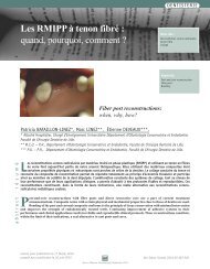
Tableau 2 - Valeurs <strong>de</strong> sensibilité et <strong>de</strong> spécificité <strong>de</strong> la radiographie et la DIFOTI dans le <strong>diagnostic</strong> <strong>de</strong>s caries<br />
Table 2 - sensitivity and specificity of radiography and DIFOTI an caries diagnosis<br />
Caries proximales Caries occlusales Caries <strong>de</strong>s surfaces lisses<br />
Approximal Caries Occlusal Caries Smooth surface Caries<br />
Se Sp Se Sp Se Sp<br />
Radiographie 0,21 à 0,31 0,88 à 0,91 0,18 à 0,20 0,98 à 1,00 0,04 à 0,04 0,96 à 1,00<br />
DIFOTI 0,56 à 0,69 0,73 à 0,76 0,67 à 0,80 0,87 0,41 à 0,43 0,87 à 0,90<br />
et coll., 1997). La spécificité <strong>de</strong> cette technique est<br />
comprise entre 99 et 100 % et <strong>de</strong>meure comparable à<br />
celle <strong>de</strong> la radiographie rétro-coronaire alors que la sensibilité<br />
est inférieure à celle <strong>de</strong> la radiographie ; elle est<br />
comprise entre 50 et 70 %. (Vaarkamp et coll., 2000).<br />
technique is between 99 and 100 % and is comparable to<br />
bite-wing radiography, where as it sensitivity is lower<br />
and is between 50 and 70 % (Vaarkamp et al., 2000).<br />
La transillumination par fibre optique<br />
avec imagerie numérique ou DIFOTI<br />
Ce système est basé sur l'utilisation d'une lumière<br />
<strong>de</strong> radiation visible et non ionisante. La transmission<br />
<strong>de</strong> la lumière à travers les tissus <strong>de</strong>ntaires est fonction<br />
du gradient d'indice réfractaire anisotropique.<br />
<strong>Les</strong> images <strong>de</strong>s <strong>de</strong>nts obtenues par cette technique<br />
peuvent indiquer la présence d'une carie débutante<br />
ou récurrente même lorsque les images radiographiques<br />
échouent dans leur détection. La brillance sur<br />
l'image provient <strong>de</strong> la combinaison <strong>de</strong> la transmission<br />
élevée au niveau <strong>de</strong> l'émail (par rapport à la <strong>de</strong>ntine) et<br />
<strong>de</strong>s vi<strong>de</strong>s causés par la perte structurelle <strong>de</strong> la <strong>de</strong>nsité<br />
amélaire (Schnei<strong>de</strong>rman et coll., 1997).<br />
L’étu<strong>de</strong> <strong>de</strong> Schnei<strong>de</strong>rman et coll. en 1997 a révélé<br />
la supériorité <strong>de</strong> la DIFOTI pour la détection <strong>de</strong>s<br />
caries débutantes aussi bien au niveau <strong>de</strong>s faces proximales,<br />
occlusales ou lisses par rapport à la radiographie.<br />
<strong>Les</strong> valeurs <strong>de</strong> sensibilité et <strong>de</strong> spécificité <strong>de</strong> cette<br />
technique in vitro sont représentées dans le tableau 2<br />
(Schnei<strong>de</strong>rman et coll., 1997).<br />
Tr ansillumination by fibre optic<br />
with digital imagery or DIFOTI<br />
This system is based on light with non ionising<br />
and visible radiation. Light transmission through <strong>de</strong>ntal<br />
tissues is function of the anisotropic refractory in<strong>de</strong>x<br />
gradient.<br />
Teeth images obtained with this technique can<br />
indicate an initial or recurrent carie even when radiography<br />
fail to <strong>de</strong>tect such lesions. The brilliance on the<br />
image came from the association of a high transmission<br />
at the enamel level (with respect to <strong>de</strong>ntin) and void caused<br />
by structural loss of enamel <strong>de</strong>nsity (Schnei<strong>de</strong>rman<br />
et al., 1997).<br />
The study of Schnei<strong>de</strong>rman et al. in 1997 has<br />
<strong>de</strong>monstrated superiority of DIFOTI as compared with<br />
r a d i o g r a p h y, for the <strong>de</strong>tection of initial lesions of<br />
approximal, occlusal or smooth surfaces. In vitro sensitivity<br />
and specificity values of this technique are presented<br />
in table 2 (Schnei<strong>de</strong>rman et al., 1997)<br />
Métho<strong>de</strong>s électriques<br />
<strong>Les</strong> <strong>de</strong>nts possè<strong>de</strong>nt une faible conductivité<br />
électrique liée à la présence <strong>de</strong> l’émail. Lorsque le volume<br />
amélaire est diminué (hypo ou déminéralisation)<br />
ceci s’accompagne par une augmentation <strong>de</strong> la conductivité<br />
électrique (Huysmans et coll., 1998).<br />
Electrical methods<br />
Teeth have low electric conductivity related to<br />
the enamel. When enamel volume is <strong>de</strong>creased (hypo or<br />
<strong>de</strong>mineralisation), this is associated with increased electric<br />
conductivity (Huysmans et al., 1998).<br />
306<br />
Revue d’Odonto-Stomatologie/décembre 2004
Ainsi, le principe <strong>de</strong> cette technique serait basé<br />
sur la détection <strong>de</strong> l’augmentation <strong>de</strong> la conductivité<br />
électrique qui accompagne la réduction du contenu<br />
minéral <strong>de</strong>s lésions <strong>carieuses</strong> (Pine et Bosch, 1996).<br />
Cette augmentation <strong>de</strong> la conductivité est due à la présence<br />
<strong>de</strong> microcavités <strong>de</strong> déminéralisation obturées par<br />
la salive qui joue le rôle d’électrolyte permettant la<br />
transmission du courant électrique.<br />
La mesure électrique montre une sensibilité <strong>de</strong><br />
76 % et une spécificité <strong>de</strong> 76 % (Jaquot et Fontaine,<br />
1995). Dans une étu<strong>de</strong> comparative in vitro avec<br />
l’inspection visuelle et la radiographie Bite-Wing pour<br />
le <strong>diagnostic</strong> <strong>de</strong>s caries occlusales, Huysmans et coll.<br />
1998 ont montré que la précision d’une métho<strong>de</strong> électrique<br />
et <strong>de</strong> la radiographie Bite-Wing est inférieure à<br />
celle <strong>de</strong> l’inspection visuelle.<br />
Thus, the principle of this technique would be<br />
based on the <strong>de</strong>tection of an increased electric conductivity<br />
related to a reduction of the mineral contact of<br />
carious lesions (Pine and Bosch, 1996). This increased<br />
conductivity is due to the presence of <strong>de</strong>mineralisation<br />
microcavities, obturated by saliva which plays the role<br />
of an electrolyte allowing transmission of the electric<br />
current.<br />
Electric measurement has a 76 % sensitivity and<br />
76 % specificity (Jaquot and Fontaine, 1995). In a comparative<br />
in vitro study with visual inspection and Bitwing<br />
radiograph for the diagnosis of occlusal caries,<br />
Huysmans et al., 1998 have shown that the precision of<br />
the electric method and Bite –wing radiography is inferior<br />
to that of the visual inspection.<br />
Métho<strong>de</strong>s endoscopiques<br />
Ces métho<strong>de</strong>s ont été testées en utilisant soit la<br />
lumière blanche ou la fluorescence.<br />
Cette technique fait appel à l’endoscope et une<br />
source <strong>de</strong> lumière blanche qui peut être connectée à<br />
l’appareil par un câble <strong>de</strong> fibre optique. Cependant,<br />
c’est une métho<strong>de</strong> lente, qui nécessite un séchage<br />
rigoureux et une isolation <strong>de</strong>s <strong>de</strong>nts (Pitts 1991 ; Pine<br />
et Bosch, 1996).<br />
<strong>Les</strong> étu<strong>de</strong>s <strong>initiales</strong> sur le bénéfice <strong>de</strong> l’examen<br />
endoscopique soit en lumière blanche soit en fluorescence<br />
montrent une légère augmentation <strong>de</strong> la sensibilité<br />
pour la détection <strong>de</strong>s lésions occlusales <strong>de</strong> l’émail par<br />
rapport à l’inspection visuelle (Jaquot et Fontaine, 1995).<br />
Air abrasion<br />
Goldstein et Parkins, en 1995, ont introduit<br />
cette technique <strong>de</strong> <strong>diagnostic</strong> <strong>de</strong>s lésions <strong>carieuses</strong><br />
débutantes. Le principe est le suivant : si un sillon douteux<br />
est observé, le système d’air abrasion permet l’élimination<br />
d’une coloration ou d’un bouchon organique<br />
par projection d’une poudre d’alumine. Si l’examen suggère<br />
<strong>de</strong> poursuivre la projection d’alumine, seules <strong>de</strong><br />
très petites quantités <strong>de</strong> tissus <strong>de</strong>ntaires déjà déminéralisés<br />
ou infiltrés sont enlevées, révélant ainsi une<br />
lésion sous jacente, invisible auparavant (Blique 2000).<br />
Cependant, cette technique est non spécifique<br />
du <strong>diagnostic</strong> <strong>de</strong> la carie (Mc Comb et Tam, 2001).<br />
Endoscopic methods<br />
These methods were tested by white light or fluorescence.<br />
This technique uses an endoscope and a white<br />
light source which can be connected to the material with<br />
a fibre optic cable. However, it is a slow technique,<br />
which necessitates rigorous drying and teeth isolation<br />
(Pitts 1991; Pine and Bosch, 1996).<br />
Initial studies on the benefits of endoscopic techniques<br />
with white light or fluorescence show a slight<br />
increase of the sensitivity of this technique with respect<br />
to the visual inspection for the diagnosis of enamel<br />
occlusal lesions (Jaquot and Fontaine, 1995).<br />
Air abrasion<br />
This technique was introduced in 1995 by<br />
Goldstein and Parkins for the diagnosis of initial carious<br />
lesions. The concept is as follows : if a suspicious groove<br />
is observed, the air abrasion system allows the elimination<br />
of coloration or an organic plug using a projection<br />
of aluminium oxi<strong>de</strong> pow<strong>de</strong>r. If the exam gives an<br />
indication to continue alumina projection, only small<br />
quantities of <strong>de</strong>ntal tissues, already <strong>de</strong>mineralised or<br />
infiltrated, are removed, revealing thus a previously<br />
invisible, un<strong>de</strong>rlying lesion (Blique 2000).<br />
However, this technique is non specific of carie<br />
diagnosis (Mc Comb and Tam, 2001).<br />
307<br />
Revue d’Odonto-Stomatologie/décembre 2004
DENTISTERIE RESTAURATRICE<br />
Ultrasons<br />
Tout tissu possè<strong>de</strong> une impédance acoustique<br />
qui caractérise son modèle sonore interne. Ainsi, tout<br />
changement <strong>de</strong> ce modèle sonore peut être corrélé à un<br />
changement pathologique <strong>de</strong> ce tissu.<br />
La détection ultrasonore d’une déminéralisation<br />
<strong>de</strong> l’émail a été étudiée par différents auteurs.<br />
Pour Çaliskan Yanikoglu (2000), la comparaison<br />
<strong>de</strong> cette technique avec la radiographie et l’histologie<br />
comme « gold standard » a donné une sensibilité <strong>de</strong> 88<br />
% et une spécificité <strong>de</strong> 86 %. Mais, c’est une métho<strong>de</strong><br />
encore au sta<strong>de</strong> expérimental.<br />
Ultrasounds<br />
Any tissue has acoustic impedance which characterizes<br />
its internal resonance mo<strong>de</strong>l. Thus, any change<br />
in this resonance mo<strong>de</strong>l can be correlated to a pathologic<br />
modification.<br />
Ultrasonic <strong>de</strong>tection of enamel <strong>de</strong>mineralisation<br />
have been evaluated by several authors.<br />
For Caliskan Yanikoglu (2000), comparison of<br />
this technique with radiography and histology as “gold<br />
standards” has yiel<strong>de</strong>d 88 % sensitivity and 86 % specificity.<br />
But, this method is still experimental.<br />
Applications <strong>de</strong> ces techniques<br />
pour le <strong>diagnostic</strong><br />
<strong>de</strong>s lésions <strong>initiales</strong> au<br />
niveau <strong>de</strong>s différents sites<br />
Site occlusale (site 1)<br />
<strong>Les</strong> sta<strong>de</strong>s précoces du développement <strong>de</strong>s lésions<br />
du site 1 sont difficilement détectables du fait que les<br />
déminéralisations progressent le long <strong>de</strong>s gaines prismatiques,<br />
<strong>de</strong> part et d’autre du sillon qui peut paraître<br />
intact alors que sa structure histologique est altérée.<br />
Pour le <strong>diagnostic</strong> <strong>de</strong> ces lésions il est recommandé<br />
<strong>de</strong> recourir à différents moyens. Le praticien doit<br />
commencer par un examen clinique en se référant aux<br />
critères <strong>diagnostic</strong> déjà énoncés (Tableau 1), puis le<br />
compléter, au besoin, et en fonction <strong>de</strong> la disponibilité<br />
par les métho<strong>de</strong>s électriques, la fluorescence laser et<br />
l’air abrasion.<br />
Use of these techniques<br />
for the diagnosis of initial<br />
lesions at different sites<br />
Occlusal site (site 1)<br />
Early stages of site 1 lesions <strong>de</strong>velopment are difficult<br />
to <strong>de</strong>tect due to the fact that <strong>de</strong>mineralisation progresses<br />
along prismatic sheaths, at either si<strong>de</strong> of the<br />
groove which can look intact, but with an altered histological<br />
structure.<br />
It is advised to use different methods for the diagnosis<br />
of such lesions. The <strong>de</strong>ntist should start with a<br />
clinical exam and should refer to already established<br />
<strong>diagnostic</strong> criteria (table 1), which should be completed<br />
as nee<strong>de</strong>d and in function of availability of : electrical<br />
methods, laser fluorescence, dye enhancement and air<br />
abrasion.<br />
Site proximal (site 2)<br />
La radiographie semble plus performante à ce<br />
niveau. D’autres métho<strong>de</strong>s peuvent également être utilisés<br />
tels que : les élastiques séparateurs, la transillumination<br />
par fibre optique.<br />
Proximal site (site 2)<br />
Radiography seems most appropriate at this<br />
level. Other methods can also be used, such as : rubber<br />
coins, fibre optic transillumination.<br />
Site cervical (site 3)<br />
Pour ces lésio ns, l’exa men clinique perme t<br />
d’i<strong>de</strong>ntifier aisément les lésions <strong>initiales</strong>.<br />
Cervical site (site 3)<br />
For lesions at this site, the clinical exam allows<br />
an easy <strong>de</strong>tection of initial lesions.<br />
308<br />
Revue d’Odonto-Stomatologie/décembre 2004
Conclusion<br />
Le <strong>diagnostic</strong> précoce <strong>de</strong>s lésions <strong>initiales</strong> <strong>de</strong> l’émail est très important pour pouvoir instaurer au temps<br />
optimum la thérapeutique adaptée permettant <strong>de</strong> réminéraliser ces lésions. De ce fait, le praticien dans sa pratique<br />
quotidienne doit se doter <strong>de</strong>s moyens lui permettant <strong>de</strong> diagnostiquer à temps ces lésions. A défaut, l’examen<br />
clinique complété par un bilan radiographique rétrocoronaire fournit une ai<strong>de</strong> non négligeable pour peu<br />
que le praticien soit bien entraîné à cet exercice. Enfin, il faut reconnaître qu’il y a peu d’étu<strong>de</strong>s qui ont évalué<br />
l’intérêt <strong>de</strong> l’association <strong>de</strong> plusieurs outils <strong>diagnostic</strong>s sur les valeurs <strong>de</strong> sensibilité et spécificité.<br />
Early diagnosis of initial enamel lesions is very important in or<strong>de</strong>r to institute an adapted treatment in an<br />
optimal timing allowing to remineralize the lesions. Thus, in clinical practice the <strong>de</strong>ntist should possess the necessary<br />
tools that would help him diagnose such lesions on time. When such methods are lacking, the clinical exam<br />
with a bite-wing radiographic evaluation gives a non negligible aid, especially when the <strong>de</strong>ntist has some prior<br />
experience. Finally, few studies have evaluated the combination of several <strong>diagnostic</strong> tools for sensitivity and specificity<br />
values.<br />
Traduction : Zeina ANTOUN<br />
Deman<strong>de</strong> <strong>de</strong> tirés-à-part :<br />
Dr CHALA S. - Faculté <strong>de</strong> Chirurgie Dentaire <strong>de</strong> RABAT - Institut BAB AL IRFANE - BP 6212 Rabat - MAROC.<br />
ABDALLAOUI F.,CHRAIBI B., JAOUHARI ELABRARI<br />
M.<br />
Dépistage précoce <strong>de</strong>s lésions <strong>carieuses</strong> proximales débutantes:<br />
étu<strong>de</strong> clinique et radiographique. Inf Dent (Paris)<br />
2001;(14):1005-1013.<br />
BLIQUE M<br />
Utilisation d’un système d’air abrasion en prophylaxie <strong>de</strong>ntaire<br />
individuelle. Inf <strong>de</strong>nt 2000;(13):933-939.<br />
ÇALISKAN YANIKOGLU F.<br />
Detection of natural white spot caries lesions by an ultrasonic<br />
system. Caries res 2000;34:225-232.<br />
CORTES D.F., EKSTRAND K.R., ELIAS-BOTENAA.R.,<br />
ELLWOOD R.P.<br />
An in vitro comparison of the ability of fibre-optique trans -<br />
illumination, visual inspection and radiographs to <strong>de</strong>tect<br />
occlusal caries and evaluate lesion <strong>de</strong>pth. Caries res<br />
2000;34:443-447.<br />
D A U D I B E RTIERS L., ETIENNE G., BARTHE M.,<br />
CATTÖEN M.<br />
Imagerie <strong>de</strong> la lésion carieuse : traitement et analyse. Rev<br />
Odonto Stomat 1993;22(1):9-21.<br />
EGGERTSSON H., ANALOUI M., VAN DER VEEN<br />
M.H., GONZALEZ-CABEZAS C., ECKERT G.J., STOO-<br />
KEYG.K.<br />
Detection of early interproximal caries in vitro using laser<br />
fluorescence, dye enhanced laser fluorescence and direct<br />
visual examination<br />
Caries res 1999;33,227-233.<br />
EKSTRAND K.R., RICKETTS D.N., KIDD E.A., QVIST<br />
V., SCHOU S.<br />
Detection, diagnosing, monitoring and logical treatment of<br />
occlusal caries in relation to lesion activity and severity: an<br />
in vivo examination with histological validation. Caries res<br />
1998;32:247-254.<br />
EKSTRAND K.R., QVIST V., THYLSTRUPA.<br />
Light microscope study of the effect of probing in occlusal<br />
surfaces. Caries res 1987;21(4):368-374.<br />
FARGE P<br />
Prospectives en cariologie. Réalités clin 2000;11(1):9-18.<br />
HAAK R., WICHT M.J., HELLMICH M., GOSSMANN<br />
A., NOACK M.J.<br />
The validity of proximal caries. Detection using magnifying<br />
visual aids. Caries res 2002;36:249-255.<br />
HAIKEL Y.<br />
Thérapeutique étiopathogénique <strong>de</strong> la carie <strong>de</strong>ntaire.<br />
Encyclop médico-chir 2001;23-010-F-10.<br />
HENNEQUIN M .,LASFARGUES J-J.<br />
La démarche <strong>diagnostic</strong> en cariologie. Réalités clin<br />
1999;10(4):515-539.<br />
HINTZE H., WENZELA., DANIELSEN B., NYVAD B.<br />
Reliability of visual examination, fibre optic transillumination,<br />
and bitewing radiography, and reproductibility of<br />
direct visual examination following tooth separation for the<br />
i<strong>de</strong>ntification of cavitated carious lesions in contacting<br />
approximal surfaces. Caries Res 1998;32:204-209.<br />
309<br />
Revue d’Odonto-Stomatologie/décembre 2004
DENTISTERIE RESTAURATRICE<br />
HUYSMANS M.CH., LONGBOTTOM CH., PITTS N.B.<br />
Electrical methods in occlusal caries diagnosis : an in vitro<br />
comparison with visual inspection and bite-wing radiography.<br />
Caries res 1998,32: 324-329.<br />
JAQUOTB., FONTAINE A.<br />
Étu<strong>de</strong> clinique <strong>de</strong> la carie. Encyclo médico-chir 1995;23-<br />
010-E-10.<br />
LE DENMAT D., LEGRAS A., PELLERINY<br />
Pour <strong>de</strong> nouvelles images radiologiques : nouveaux capteurs<br />
ou filsm conventionnels Inform <strong>de</strong>nt 1994;19-<br />
20:1691-1706.<br />
LUSSI A.<br />
Comparison of different method for the diagnosis of fissure<br />
caries without cavitation. Caries Res 1993;27:409-416.<br />
MC COMB D.<br />
Caries <strong>de</strong>tector dyes- how accurate and useful are they J<br />
Canad <strong>de</strong>nt ass 2000;66:195-198.<br />
MCCOMB D., TAM E.L.<br />
Diagnosis of occlusal caries : part I. conventional method. J<br />
Canad <strong>de</strong>nt ass 2001;67(8):454-457.<br />
PINE C M, BOSCH J.J.T<br />
Dynamics of <strong>diagnostic</strong> methods for <strong>de</strong>tecting small carious<br />
lesions. Caries res 1996;30:381-388.<br />
PINELLI C., CAMPOS SERRA M., MONTEIRO LOF-<br />
FREDO L.<br />
Validity and reproducibility of a laser fluorescence system<br />
for <strong>de</strong>tecting the activity of white spot lesions on free<br />
smooth surface in vivo<br />
Caries res 2002;36:19-24.<br />
PITTS N.B.<br />
Diagnostic methods for caries: what is appropriate when J<br />
<strong>de</strong>nt 1991;19:377-382.<br />
SCHNEIDERMAN A.,ELBAUM M., SCHULTZ M.;<br />
KEEM M., GREENE BAUM M., DRILLER J.<br />
Assesssment of <strong>de</strong>ntal caries with digital fiber - optic transillumination<br />
( DIFOTI) : in vitro study. Caries Res<br />
1997;31:103-110.<br />
SEGUIER S., LE MAY O.<br />
Histopathologie <strong>de</strong> la lésion carieuse <strong>de</strong> l’émail et <strong>de</strong> la <strong>de</strong>ntine.<br />
Encyclopédie médico-chirurgicale 2002;23-010-C-10<br />
TAM E L. MCCOMB D.<br />
DIAGNOSIS of occlusal caries: part II. Recent diagnosis<br />
technologies. J Canad <strong>de</strong>nt ass 2001;67(8):459-463.<br />
TASSERY H., KOUBI N., CHAFAI A., BACCOUCH Z.,<br />
POMELL., DEJOU J.<br />
<strong>Les</strong> révélateurs <strong>de</strong> carie : une ai<strong>de</strong> opératoire Inf <strong>de</strong>nt<br />
(Paris) 1999;(23):1659-1667.<br />
TRILLER M.<br />
Le processus carieux initial. Réalités clin 1993;(4):275-281.<br />
VAARKAMP J., TENBOSCH J., VERDONSCHOL E.H.,<br />
BRONKHORSTE.M.<br />
The real preformance of bitewing radiography and fiberoptic<br />
transillumination in approximal caries diagnosis.<br />
VERDONSCOT E. H., BRONKHORST E.M.,BURGER<br />
SDIJK R.C.W., KONIG KG., SCHAEKEN M. J.M.,<br />
TRUIN G. J.<br />
Performance of some <strong>diagnostic</strong> systems in examinations<br />
for occlusal carious lesions. Caries Res 1992;26:259-264.<br />
310<br />
Revue d’Odonto-Stomatologie/décembre 2004