MILIEU DE BORELLI - Simoco Diagnostics
MILIEU DE BORELLI - Simoco Diagnostics
MILIEU DE BORELLI - Simoco Diagnostics

You also want an ePaper? Increase the reach of your titles
YUMPU automatically turns print PDFs into web optimized ePapers that Google loves.
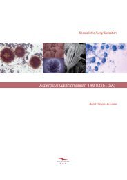
<strong>BORELLI</strong> MEDIUM<br />
Tubed and bottled medium for subculture and identification of dermatophytes<br />
INTEN<strong>DE</strong>D USE<br />
<strong>BORELLI</strong> medium is a convenient method for identification of dermatophytes previously cultured on<br />
media such as Sabouraud. Observation of sporulation and pigmentation during culture from isolated fungi<br />
placed on <strong>BORELLI</strong> Medium allows identification of species.<br />
PRINCIPLE<br />
Cutaneous mycoses are caused by yeasts or filamentous fungi which have an affinity for keratin. Lesions<br />
are confined to the superficial layer of the epidermis, hair, and nails.<br />
Biological diagnosis of mycoses is based on direct examination and sample culture of skin, hair, nails on<br />
Sabouraud medium.<br />
The identification of dermatophytes is based on macroscopic and microscopic characteristics of colonies.<br />
However, dermatophytes cultured on Sabouraud medium do not always show all the characteristics that<br />
allow species identification. A secondary subculture on a medium such as <strong>BORELLI</strong> is required in these<br />
cases<br />
<strong>BORELLI</strong> medium contains all the elements necessary for the growth of dermatophytes and for the<br />
development of those features that allow species identification.<br />
KIT CONTENT<br />
<strong>BORELLI</strong> medium is an agar medium packaged in tubes or bottles. After reconstitution the medium can<br />
be poured onto Petri dishes. Reconstituted medium is suitable for identification of dermatophyte species.<br />
- Prepared bottled medium, 4 bottles of 50 mL (about 20 tests). Reference borelfl20.<br />
Prepared tubed medium, 18 tubes of 10 mL (about 18 tests). Reference borelt18.<br />
MATERIAL REQUIRED BUT NOT PROVI<strong>DE</strong>D<br />
- 50 mm Petri dishes<br />
- Water bath<br />
- Scotch ®<br />
- Microscope<br />
- Microscope slides (76x26 mm)<br />
- Coverslip<br />
- Lactophenol Cotton blue<br />
- Container for contaminated waste<br />
STORAGE CONDITIONS<br />
Stored at + 12°C to +37°C, medium is stable until the expiry date indicated on the box<br />
Do not freeze. Keep the medium below +37°C. Do not keep in intense light.<br />
WARNINGS AND PRECAUTIONS<br />
- For in vitro diagnostic use.<br />
- Only for professional use.<br />
- Do not interchange medium from different kit lots.<br />
- Follow the instructions for use.<br />
- In case of accidental spill of reagent, clean the surface with absorbent paper, bleach and rinse with<br />
water. In case of environmental contamination with culture samples clean with bleach and absorbent<br />
paper.<br />
- The samples, reagents as well as the contaminated materials and products must be disposed of in a<br />
container for contaminated waste, according to the prevailing recommendations and regulations.<br />
- Do not use tubed or bottled medium if they show evidence of microbial contamination.<br />
MEDIUM PREPARATION<br />
- Partially unscrew cap on tube or bottle and put it in a water bath. Media should be heated in the water<br />
bath to ensure complete dissolution. Persistence of medium components in suspension is normal.<br />
- Remove tube or bottle from the water bath, screw cap on tube or bottle.<br />
- Dry tube or bottle and thoroughly homogenise the content of tube or bottle by reversal.<br />
- Dispense bottled <strong>BORELLI</strong> medium into Petri dishes. Volume should be about 10 mL in a 50 mm dish.<br />
Allow the medium to solidify before use. Prepared dishes, sealed in small plastic bags or wrapped with<br />
transparent cellophane adhesive (eg Parafilm®) can be stored at +2°C to +8°C for up to 2 weeks.<br />
- Tubed <strong>BORELLI</strong> medium reconstituted may be placed in an inclined (20°) position or dispensed to 50<br />
mm Petri dishes. Allow to solidify before use.<br />
TEST PROCEDURE<br />
For the subculture push the sample obtained from a colony into <strong>BORELLI</strong> agar. Temperature and<br />
incubation period vary from species to species.<br />
It is advisable to incubate the cultures at +20°C to +25°C for 1 to 4 weeks. Microscopic examination of<br />
sample for identification of species is performed when the size of colony is satisfactory.<br />
The cellotape (Scotch® tape) flag and the lactophenol Cotton blue mount may be used for microscopic<br />
observation of the colony morphology.<br />
The identification of dermatophyte is based on macroscopic aspects of colonies (downy, powdery...),<br />
colonies colour (white, fawn…), colonies pigmentation (red, wine-red, yellow-brown …) and on<br />
microscopic characteristics (aspect, shape, morphology of the microconidia and/or macroconidia,<br />
appendages such as spiral hyphae…).<br />
Standards characteristics specified in literature must be used to identify species of dermatophytes.<br />
PERFORMANCES<br />
A study of 43 dermatophytes isolates has shown that <strong>BORELLI</strong> medium allows growth of all isolates.<br />
Species identification of dermatophytes has been done on the basis of macroscopic and microscopic<br />
characteristics. Identification of dermatophytes species was obtained for 40 dermatophytes isolates i.e.<br />
93%.<br />
BIBLIOGRAPHIE / REFERENCES<br />
- Badillet G. Dermatophyties et dermatophytes. Atlas clinique et biologique. Varia, 1991, 3è ed. 303 p.<br />
- Baran R, Chabasse D et feullade de Chauvin M. Les onychomycoses II Approche diagnostique. J.<br />
Mycol. Méd. 2001, 11:5-13.<br />
- Chabasse D, Guiguen C, Contet-Audonneau. Mycologie médicale. Masson, 1999, 324 p.<br />
- Chabasse D, Bouchara J.P, de Gentile L, BrunZ, Cimon B et Penn P. Les dermatophytes. Cahier de<br />
formation, Biologie Médicale, Vol. 31 Bioforma, 2004.<br />
- Grillot R. Les mycoses humaines : démarche diagnostique. Elsevier, collection Option/Bio, 1996, 392 p.<br />
- Haldane DJ, Robart E. A comparison of calcofluor white, potassium hydroxide, and culture for the<br />
laboratory diagnosis of superficial fungal infection. Diagn Microbiol Infect Dis. 1990, 13:337-339.<br />
- Koenig H. Guide de mycologie médicale. Ellipses, 1995, 284 p.<br />
- Lawry MA, Haneke E, Strobeck K, Martin S, Zimmer B, Romano PS. Methods for diagnosing<br />
onychomycosis: a comparative study and review of the literature. Arch Dermatol. 2000, 136:1112-1116.<br />
- Machouart-Dubach M, LacroixC, Feuilhade de Chauvin M, Le Gall I, Giudicelli C, Lorenzo F, Derouin F.<br />
Rapid Discrimination among Dermatophytes, Scytalidium spp., and Other Fungi with a PCR-Restriction<br />
Fragment Length Polymorphism Ribotyping Method. J Clin Microbiol. 2001, 39: 685–690.<br />
- Monod M, Jaccoud S, Stirnimann R, Anex R, Villa F, Balmer S, Panizzon R. Economical Microscope<br />
Configuration for Direct Mycological Examination with Fluorescence in Dermatology. Dermatology.<br />
2000, 201:246-248<br />
- Monod M, Baudraz-Rosselet F, Ramelet AA, Frenk E : Direct mycological examination in dermatology :<br />
a comparison of different methods. Dermatologica 1989 ; 179 : 183-186.<br />
- Payle B, Serrano L, Bieley HC, Reyes BA. Albert's solution versus potassium hydroxide solution in the<br />
diagnosis of tinea versicolor. Int J Dermatol. 1994, 33:182-183.<br />
- Weinberg JM, Koestenblatt EK, Tutrone WD, Tishler HR, Najarian L. Comparison of diagnostic methods<br />
in the evaluation of onychomycosis. J Am Acad Dermatol. 2003, 49:193-197.