Flore fongique de differents substrats et conditions optimales de ...
Flore fongique de differents substrats et conditions optimales de ...
Flore fongique de differents substrats et conditions optimales de ...
- No tags were found...

You also want an ePaper? Increase the reach of your titles
YUMPU automatically turns print PDFs into web optimized ePapers that Google loves.
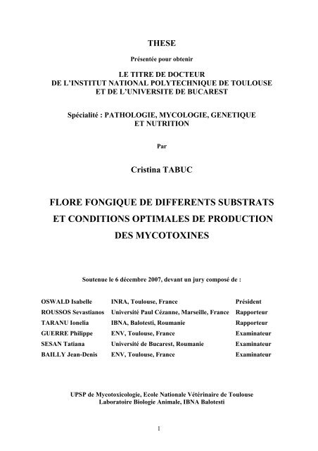
THESEPrésentée pour obtenirLE TITRE DE DOCTEURDE L’INSTITUT NATIONAL POLYTECHNIQUE DE TOULOUSEET DE L’UNIVERSITE DE BUCARESTSpécialité : PATHOLOGIE, MYCOLOGIE, GENETIQUEET NUTRITIONParCristina TABUCFLORE FONGIQUE DE DIFFERENTS SUBSTRATSET CONDITIONS OPTIMALES DE PRODUCTIONDES MYCOTOXINESSoutenue le 6 décembre 2007, <strong>de</strong>vant un jury composé <strong>de</strong> :OSWALD Isabelle INRA, Toulouse, France Prési<strong>de</strong>ntROUSSOS Sevastianos Université Paul Cézanne, Marseille, France RapporteurTARANU Ionelia IBNA, Balotesti, Roumanie RapporteurGUERRE Philippe ENV, Toulouse, France ExaminateurSESAN Tatiana Université <strong>de</strong> Bucarest, Roumanie ExaminateurBAILLY Jean-Denis ENV, Toulouse, France ExaminateurUPSP <strong>de</strong> Mycotoxicologie, Ecole Nationale Vétérinaire <strong>de</strong> ToulouseLaboratoire Biologie Animale, IBNA Balotesti1
REMERCIEMENTSJe tiens d’abord à adresser toute ma gratitu<strong>de</strong> à Jean-Denis Bailly sans lequel c<strong>et</strong>ravail n’aurait pu aboutir. Je le remercie pour sa gentillesse, son soutien <strong>et</strong> pour le fait <strong>de</strong>m’avoir fait partager son expérience. Je lui adresse toute ma reconnaissance pour sa patience,sa disponibilité <strong>et</strong> sa participation active lors <strong>de</strong> la rédaction <strong>de</strong>s articles <strong>et</strong> <strong>de</strong> la thèse.Je remercie également mes directeurs <strong>de</strong> thèse Jean-Denis Bailly (encore une fois)<strong>et</strong> Tatiana Sesan pour leur confiance <strong>et</strong> leur soutien. Merci à Jean-Denis Bailly pour le fait <strong>de</strong>m’avoir accueilli dans son équipe <strong>et</strong> pour ses conseils toujours pertinents. Je remercie TatianaSesan non seulement pour son soutien moral <strong>et</strong> professionnel pendant la thèse mais aussipendant toutes les années d’étu<strong>de</strong>s.J’exprime également mes remerciements <strong>et</strong> ma reconnaissance à Isabelle Oswaldsans laquelle ma venue en France n’aurait pas été possible.Je remercie très sincèrement Sevastianos Roussos <strong>et</strong> Ionelia Taranu qui ontaccepté d’examiner notre travail avec bienveillance.Mes remerciements s’adressent à Philippe Guerre qui nous a inspiré ce suj<strong>et</strong> <strong>de</strong>thèse <strong>et</strong> qui a porté un intérêt tout particulier à notre travail. Qu’il soit assuré <strong>de</strong> ma profon<strong>de</strong>reconnaissance.Merci aussi à Pierr<strong>et</strong>te Le Bars <strong>et</strong> Sylviane Bailly pour leur gentillesse <strong>et</strong> pour tousles secr<strong>et</strong>s dévoilés <strong>de</strong> la mycologie microscopique.Un grand merci pour Didier Tardieu <strong>et</strong> Claudine Condomines (UP, PharmacieToxicologie) qui m’ont appris les métho<strong>de</strong>s d’analyse <strong>de</strong> laboratoire nécessaires pour ce travail.Je leur adresse tout mon respect pour leur disponibilité <strong>et</strong> leur gentillesse.Je tiens à remercier toute l’équipe d’HIDAOA <strong>et</strong> toutes les personnes avec qui j’aipartagé <strong>de</strong>s bons moments <strong>et</strong> qui ont rendu plus agréable mon séjour au sein <strong>de</strong> c<strong>et</strong>te équipe.Je leur serai toujours reconnaissante pour leur ai<strong>de</strong> <strong>et</strong> leurs conseils <strong>et</strong> je pense spécialement àMarie-Rose (pour son dévouement <strong>et</strong> sa gentillesse), Alain (pour sa taquinerie géographique,linguistique <strong>et</strong> culinaire), Monique (pour ses conseils en français), Arl<strong>et</strong>te (pour ses conseils <strong>et</strong>ses encouragements), Jean-Pierre (pour ses histoires <strong>et</strong> sa bonne humeur).2
Également je remercie mon père <strong>et</strong> ma soeur pour les efforts <strong>et</strong> les sacrificesfaits pour que je puisse réaliser mes rêves <strong>et</strong> aussi Françoise Jugie qui m’a énormémentaidée, soutenue <strong>et</strong> abritée pendant tous mes séjours en France.Pour les moments extraordinaires passés ensemble je tiens à remercier mesamis <strong>de</strong> diaspora roumaine: Ciprian, Daniel, Dragos, Dorina, Valentin, Mihaela,Loredana, Rodica, Marian, Carmen <strong>et</strong> Nicu.Un grand merci aux collègues roumains Daniela, Radu <strong>et</strong> Cornelia égalementpour leur soutien <strong>et</strong> aussi à Lucia, Nina, Anca, George, Vova, Gina, Lumi, … pour leurfidèle soutien pendant les années d’étu<strong>de</strong>s <strong>et</strong> pour tous les excellents souvenirs.A tous ce que j’ai oubliés, mais qui se reconnaîtront ici.Enfin, merci à Chucky, la crème <strong>de</strong>s chiens, dédicace ridicule <strong>et</strong> sans intérêt pourcertains, mais sans doute la plus méritée pour tout ce que je lui ai fait subir <strong>et</strong> tout ce qu’il<strong>de</strong>vra supporter encore longtemps !3
Ce travail a fait l’obj<strong>et</strong> <strong>de</strong>s publications suivantes:Tabuc C., Bailly J.D., Bailly S., Querin A., Guerre P., 2004 : Toxigenic potential ofPenicillium strains isolated from dry cured meat products and stability of produced toxins,Rev. Med. V<strong>et</strong>.; 155, 5, 287-291Bailly J.D., Tabuc C. Querin A. Guerre P., 2005 : Production and stability of Patulin,OTA, Citrinin and CPA in dry cured ham, J. Food Prot., 68 (7), 1516-1520Trung T.S., Tabuc C., Bailly S., Querin A., Guerre P., Bailly J.D., 2007 : Fungalmycoflora and contamination of maize from Vi<strong>et</strong>nam with fumonisin B1 and aflatoxin B1,World Mycotoxin Journal, sous presseTabuc C., Marin D., Guerre P., Sesan T, Bailly J.D., 2007 : Aflatoxin B1,<strong>de</strong>oxynivalenol and zearalenone contamination of cereals in South-East Romania, J. Food.Prot., soumisFinancementCes travaux ont été financés en partie dans le cadre <strong>de</strong> RéseauxFormation Recherche N o : 3654
Liste <strong>de</strong>s abréviationsADNARNPCRSIDAHIVPDAAci<strong>de</strong> désoxyribonucléiqueAci<strong>de</strong> ribonucléiquePolymerase chain reactionSyndrome immunodéficitaire acquisHuman immuno<strong>de</strong>ficiency virusPotato <strong>de</strong>xtrose agarAFB1 Aflatoxine B 1AFB2 Aflatoxine B 2AFG1 Aflatoxine G 1AFG2 Aflatoxine G 2AFM1 Aflatoxine M 1OTA Ochratoxine AFB1 Fumonisine B 1FB2 Fumonisine B 2FB3 Fumonisine B 3T-2 Toxine T-2HT-2 Toxine HT-2DAS DiacétoxyscirpénolDON DéoxynivalénolNIV NivalénolFX Fusarenone X3aDON 3 Acétyl-déoxynivalénol15aDON 15 Acétyl-déoxynivalénolZEA ZéaralénoneRAL Aci<strong>de</strong> résorcyliqueSCOOP Scientific Cooperation on Questions relating to FoodsELISA Enzyme Linked Immuno Sorbent AssayLOD Limit of <strong>de</strong>tectionLOQ Limit of quantificationHPLC High Performance Liquid ChromatographyCCM Chromatographie sur Couche Mince5
Liste <strong>de</strong>s figures <strong>et</strong> <strong>de</strong>s tableauxFigure 1. Mo<strong>de</strong>s <strong>de</strong> formation <strong>de</strong>s conidies pag. 19Figure 2. Mo<strong>de</strong>s <strong>de</strong> groupement <strong>de</strong>s conidies pag. 20Figure 3. Classification <strong>de</strong>s champignons pag. 24Figure 4. Principaux caractères morphologiques <strong>de</strong>s Aspergillus pag. 27Figure 5. Aspergillus flavus pag. 29Figure 6. Aspergillus fumigatus pag. 30Figure 7. Aspergillus niger pag. 31Figure 8. Aspergillus ochraceus pag. 31Figure 9. Aspergillus oryzae pag. 32Figure 10. Caractères du thalle <strong>de</strong> genre Penicillium pag. 36Figure 11. Caractères morphologiques <strong>de</strong>s Penicillium pag. 36Figure 12. Caractères morphologiques <strong>de</strong>s Fusarium pag. 40Figure 13. Fusarium culmorum pag. 41Figure 14. Fusarium graminearum pag. 42Figure 15. Fusarium oxysporum pag. 42Figure 16. Fusarium verticilloi<strong>de</strong>s pag. 43Figure 17. Voies <strong>de</strong> biosynthèse <strong>de</strong>s mycotoxines pag. 65Figure 18. L’ochratoxine A pag. 71Figure 19. Structure générale <strong>de</strong>s fumonisines pag. 75Figure 20. Structure chimique générale <strong>de</strong>s principaux trichothécènes<strong>de</strong> groupes A <strong>et</strong> B pag. 79Figure 21. Structure moléculaire <strong>de</strong> la zéaralènone pag. 84Figure 22. Fusarium graminearum pag. 160Figure 23 : niveau <strong>de</strong> production du DON après 5 semaines <strong>de</strong> culturesur riz, maïs <strong>et</strong> blé pag. 161Figure 24. Production <strong>de</strong> ZEA après 6 semaines <strong>de</strong> culture sur riz <strong>et</strong> maïsgrossièrement broyé pag. 162Figure 25. Cinétique <strong>de</strong> la production <strong>de</strong> DON en fonction du temps<strong>et</strong> <strong>de</strong> la température <strong>de</strong> culture pag. 163Tableau 1. Principales mycotoxines <strong>et</strong> espèces <strong>fongique</strong>s productrices pag. 13Tableau 2. Les Aspergillus producteurs <strong>de</strong> mycotoxines pag. 33Tableau 3. Les Penicillium producteurs <strong>de</strong>s mycotoxines pag. 37Tableau 4. Les Fusarium producteurs <strong>de</strong>s mycotoxines pag. 44Tableau 5. Influence <strong>de</strong> pH sur la croissance <strong>de</strong> Fusarium proliferatum pag. 46Tableau 6. Présence <strong>de</strong>s Aspergillus dans les céréales pag. 50Tableau 7. Présence <strong>de</strong>s Penicillium dans les céréales pag. 51Tableau 8. Présence <strong>de</strong>s Fusarium dans les céréales pag. 52Tableau 9. Présence <strong>de</strong> moisissures dans les aliments composés pour lesanimaux pag. 54Tableau 10. Présence <strong>de</strong> moisissures dans les produits alimentairesà base <strong>de</strong> céréales pag. 56Tableau 11. Présence <strong>de</strong>s espèces <strong>fongique</strong>s toxinogènes dans les légumes pag. 57Tableau 12. Présence <strong>de</strong>s espèces <strong>fongique</strong>s toxinogènes dans les produitslaitiers pag. 58Tableau 13. Espèces <strong>fongique</strong>s présentes dans les produits carnés pag. 60Tableau 14. Influence <strong>de</strong> pH sur la production <strong>de</strong> fumonisine B1 pag. 62Tableau 15. Influence <strong>de</strong> température sur l’élaboration <strong>de</strong>zéaralénone <strong>et</strong> déoxynivalénol par Fusarium graminearum pag. 63Tableau 16. Les principaux représentants <strong>de</strong> famille d’aflatoxines pag. 67Tableau 17. Présence <strong>de</strong>s aflatoxines dans <strong>de</strong>s matières premières <strong>et</strong><strong>de</strong>s produits d’origine végétaleTableau 18. Présence <strong>de</strong> l’aflatoxine M 1 dans le lait <strong>et</strong> les produits laitierspag. 68pag. 69Tableau 19. Présence <strong>de</strong> l’ochratoxine A dans céréales pag. 726
Tableau 20. Présence <strong>de</strong> l’ochratoxine A dans <strong>de</strong>s produits végétaux pag. 73Tableau 21. Les principales fumonisines pag. 75Tableau 22. Présence <strong>de</strong> fumonisines dans <strong>de</strong>s céréales pag. 76Tableau 23. Présence <strong>de</strong> fumonisines dans <strong>de</strong>s produits à baze <strong>de</strong> céréales pag. 77Tableau 24. Présence <strong>de</strong> trichothécènes dans <strong>de</strong>s céréales pag. 81Tableau 25. Présence <strong>de</strong> trichothécènes dans <strong>de</strong>s produits à base <strong>de</strong> céréales pag. 82Tableau 26. Présence <strong>de</strong> zéaralénone dans les céréales pag. 857
RésuméLes moisissures sont <strong>de</strong>s contaminants fréquents <strong>de</strong> nombreux <strong>substrats</strong> végétaux <strong>et</strong> <strong>de</strong>certains produits d’origine animale. Leur présence peut améliorer les qualités organoleptiquesdu produit ou, au contraire, l’altérer <strong>et</strong> conduire à l’accumulation <strong>de</strong> métabolites secondairestoxiques : les mycotoxines.L’objectif <strong>de</strong> ce travail a été <strong>de</strong> caractériser la flore <strong>fongique</strong> <strong>de</strong> différents <strong>substrats</strong> (céréales<strong>et</strong> produits <strong>de</strong> salaison) <strong>et</strong> d’étudier le potentiel toxinogène <strong>de</strong>s souches isolées afin d’évaluerle risque mycotoxicologique associé à la consommation <strong>de</strong> ces aliments.Nous avons aussi caractérisé les <strong>conditions</strong> <strong>optimales</strong> <strong>de</strong> production <strong>de</strong> certaines mycotoxines.L’objectif était double : les comparer avec les <strong>conditions</strong> naturelles <strong>et</strong> déterminer lesparamètres nécessaires à la production <strong>de</strong> gran<strong>de</strong>s quantités <strong>de</strong> toxines partiellement purifiées.Ce <strong>de</strong>rnier point est un préalable nécessaire à l’étu<strong>de</strong> <strong>de</strong> l’impact <strong>de</strong> ces contaminants sur lasanté animale <strong>et</strong> la qualité <strong>de</strong>s produits d’origine animale.SummaryMoulds are common contaminants of a wi<strong>de</strong> vari<strong>et</strong>y of veg<strong>et</strong>al and animal <strong>de</strong>rived foods.Their presence can improve organoleptic properties or, contrary, lead to food spoilage andaccumulation of toxic compounds: mycotoxins.The aim of this study was to characterize the fungal flora of several substrates (cereals anddry cured meat products) and to <strong>de</strong>termine the toxigenic potential of isolated strains in or<strong>de</strong>rto appreciate the risk associated with consumption of such food products.We also characterized the optimal <strong>conditions</strong> for some mycotoxin production. The objectiveswere double: to compare them with natural <strong>conditions</strong> and to be able to produce largequantities of partially purified toxins. This later point is necessary to investigate effects ofthese contaminants on both animal health and quality of animal <strong>de</strong>rived products8
SOMMAIREPagesIntroduction 12Première partie : Données bibliographiques 15Les moisissures: <strong>conditions</strong> <strong>de</strong> développement <strong>et</strong> <strong>de</strong> toxinogénèse1. I<strong>de</strong>ntification <strong>et</strong> classement <strong>de</strong>s moisissures 161.1. I<strong>de</strong>ntification <strong>de</strong>s moisissures 161.1.1. I<strong>de</strong>ntification morphologique 171.1.1.1. Critères d’i<strong>de</strong>ntification macroscopique 171.1.1.2. Critères d’i<strong>de</strong>ntification microscopique 181.1.2. I<strong>de</strong>ntification génétique 221.2. Classement <strong>de</strong>s moisissures 231.3. Principaux genres <strong>fongique</strong>s 261.3.1. Le genre Aspergillus 261.3.1.1. Caractères culturaux généraux 281.3.1.2. Morphologie microscopique 281.3.1.3. Les principales espèces 291.3.1.4. Importance du genre Aspergillus 331.3.2. Le Genre Penicillium 351.3.2.1. Caractères culturaux généraux 351.3.2.2. Morphologie microscopique 361.3.2.3. Importance du genre Penicillium 371.3.3. Le genre Fusarium 381.3.3.1. Caractères culturaux généraux 391.3.3.2. Morphologie microscopique 391.3.3.3. Principales espèces <strong>de</strong> Fusarium 401.3.3.4. Importancedu genre Fusarium 449
2. Contamination <strong>de</strong>s aliments par les moisissures 452.1. Conditions <strong>de</strong> développement <strong>de</strong>s moisissures 452.1.1. Activité en eau (Aw) 462.1.2. pH 462.1.3. Présence d’oxygène 472.1.4. Température 472.1.5. Lumière 482.1.6. Interactions microbiennes 482.1.7. Présence d’insectes 482.2. Contamination <strong>de</strong>s aliments par les moisissures 492.2.1. Contamination <strong>de</strong> céréales <strong>et</strong> produits végétaux 502.2.1.1. Les céréales 502.2.1.2. Aliments composés pour les animaux 532.2.1.3. Produits alimentaires à base <strong>de</strong> céréales 552.2.1.4. Autres végétaux 562.2.1.5. Produits affinés d’origine animale 573. Les mycotoxines 613.1 Conditions <strong>de</strong> toxinogènese 613.1.1. Activité en eau (Aw) 613.1.2. pH 613.1.3. Présence d’oxygène 623.1.4. Température 623.1.5. Composition du substrat 633.1.6. Intéractions microbiennes 633.2. Nature <strong>et</strong> origine <strong>de</strong>s mycotoxines 643.2.1. Biogénèse <strong>de</strong>s mycotoxines 643.2.2. Structure <strong>de</strong>s mycotoxines 653.3. Les principales mycotoxines 663.3.1. Les aflatoxines 663.3.1.1. Contamination <strong>de</strong>s aliments 663.3.1.2. Eff<strong>et</strong>s toxiques d’aflatoxines 693.3.2. L’ochratoxine A 703.3.2.1. Contamination <strong>de</strong>s aliments 713.3.2.2. Eff<strong>et</strong>s toxiques <strong>de</strong> l’ochratoxine 733.3.3. Les fumonisines 743.3.3.1. Contamination <strong>de</strong>s aliments 753.3.3.2. Eff<strong>et</strong>s toxiques <strong>de</strong>s fumonisines 773.3.4. Les trichothécènes 7810
3.3.4.1. Contamination <strong>de</strong>s aliments 803.3.4.2. Eff<strong>et</strong>s toxiques <strong>de</strong>s trichothécènes 823.3.5. La zéaralénone 843.3.5.1. Contamination <strong>de</strong>s aliments 853.3.5.2. Eff<strong>et</strong>s toxiques <strong>de</strong> zéaralénone 86Objectifs <strong>de</strong> la thèse 87Deuxième partie : Données expérimentales 88Analyse <strong>de</strong> la flore <strong>fongique</strong> <strong>de</strong> différents <strong>substrats</strong>Article 1 : Contamination Fongique <strong>et</strong> mycotoxique <strong>de</strong> céréalesproduites dans le Sud Est <strong>de</strong> la Roumanie 89Article 2 : Contamination <strong>fongique</strong> <strong>et</strong> mycotoxique du maïs vi<strong>et</strong>namien 112Article 3 : <strong>Flore</strong> <strong>fongique</strong> <strong>de</strong>s salaisons sèches commercialisées en France 136Article 4 : Production <strong>et</strong> stabilité <strong>de</strong> la patuline, l’ochratoxine A,la citrinine <strong>et</strong> l’aci<strong>de</strong> cyclopiazonique sur le jambon sec 146Détermination <strong>de</strong>s <strong>conditions</strong> <strong>optimales</strong> <strong>de</strong> productiondu déoxynivalénol <strong>et</strong> <strong>de</strong> la zéaralénone 155Introduction 156Matériel <strong>et</strong> métho<strong>de</strong>s 157Résultats <strong>et</strong> discussion 160Conclusions générales 165Références bibliographiques 16711
IntroductionLes moisissures représentent un groupe hétérogène <strong>de</strong> champignons microscopiquessaprophytes <strong>et</strong>, parfois parasites, caractérisés par :- la nature chimique <strong>de</strong> paroi cellulaire, riche en chitine ;- la reproduction par spores sexuées ou asexuées ;- la présence <strong>de</strong> glycogène, comme substance <strong>de</strong> réserve ;- l’absence <strong>de</strong> la chlorophylle.Ce sont <strong>de</strong>s organismes eucaryotes, thallophytes car l’appareil végétatif est un thalle constituépar <strong>de</strong>s filaments mycéliens à croissance apicale, dans toutes les directions à la même vitesse.Dépourvues <strong>de</strong> pigments assimilateurs, les moisissures sont <strong>de</strong>s microorganismeshétérotrophes dépendants d’une source <strong>de</strong> carbone organique. Globalement peu exigeants surles <strong>conditions</strong> environnementales du substrat, ces champignons peuvent contaminer lesmilieux les plus divers comme : les céréales, les produits d’origine animale (lait, vian<strong>de</strong>) maisaussi le papier, les tissus, les matières organiques en décomposition, où elles trouvent unesource <strong>de</strong> carbone <strong>et</strong> d’azote accessible.La contamination <strong>fongique</strong> d’un substrat ou d’un aliment provoque <strong>de</strong>s modificationsphysiques (aspect, goût, o<strong>de</strong>ur) <strong>et</strong> <strong>de</strong>s modifications chimiques (modification <strong>de</strong>s qualitésnutritives). On peut distinguer <strong>de</strong>ux grands types <strong>de</strong> moisissures :Les moisissures utiles qui sont utilisées dans l’industrie pour conférer aux produits <strong>de</strong>spropriétés organoleptiques <strong>et</strong> technologiques supérieures comme le Penicillium camenberti <strong>et</strong>Penicillium roqueforti en fromagerie, Penicillium jensenii ou nalgiovense en salaisonnerie.Les moisissures nuisibles qui peuvent se développer sur différents <strong>substrats</strong> <strong>et</strong> entraîner unealtération <strong>de</strong>s qualités nutritionnelles <strong>et</strong> diététiques <strong>de</strong>s produits. Ainsi, on estime que ledéveloppement incontrôlé <strong>de</strong> micromycètes est à l’origine <strong>de</strong> la perte <strong>de</strong> 5 à 10% <strong>de</strong>s récoltesmondiales (Filtenborg <strong>et</strong> al., 1996).12
Par ailleurs, dans <strong>de</strong>s <strong>conditions</strong> propices <strong>de</strong> température, humidité, pH, composition <strong>de</strong>substrat, les moisissures peuvent synthétiser <strong>de</strong>s métabolites secondaires toxiques : lesmycotoxines.Parmi la centaine <strong>de</strong> mycotoxines i<strong>de</strong>ntifiées à l’heure actuelle, une trentaine sontvéritablement importantes pour la santé humaine <strong>et</strong> animale à cause <strong>de</strong> leur fréquence ou <strong>de</strong>leur toxicité (Benn<strong>et</strong>t <strong>et</strong> Klich, 2003). Les toxines majeures (Tableau 1) sont produites par <strong>de</strong>ssouches <strong>fongique</strong>s appartenant aux genres Aspergillus, Penicillium <strong>et</strong> Fusarium (AFSSA,2006).Tableau 1 : Principales mycotoxines <strong>et</strong> espèces <strong>fongique</strong>s productrices.MycotoxinesPrincipales moisissures productricesMycotoxines réglementées ou en cours <strong>de</strong> réglementationAflatoxines B1, B2, G1, G2Aspergillus flavus, A. parasiticus, A. nomiusOchratoxine APenicillium verrucosumAspergillus ochraceus, A. carbonariusPatulinePenicillium expansum, Aspergillus clavatusByssochlamys niveaFumonisines B1, B2, B3Fusarium verticillioi<strong>de</strong>s, F. proliferatumTrichothécènes (DON) Fusarium graminearum, F. culmorum, F.crookwellense, F. sporotrichioi<strong>de</strong>s, F. poae, F.tricinctum, F. acuminatumZéaralènone Fusarium graminearum, F. culmorum, F.crookwellense.Alcaloï<strong>de</strong>s d’ergot (dit ergot du seigle) Claviceps purpurea, C. paspali, C. africanaAutres mycotoxinesCitrinineAspergillus terreus, A. carneus, A. niveusPenicillium verrucosum, P. citrinum, P. expansumToxines d’Alternaria (alternariol, Alternaria alternata, Alternaria solanialternariol méthyl éther…)Aci<strong>de</strong> cyclopiazoniqueAspergillus flavus, A. versicolor, A. tamariiPenicillium dont P. camembertiStérigmatocystineAspergillus nidulans, A. versicolor, A. flavusSpori<strong>de</strong>sminesPithomyces chartarumStachybotryotoxinesStrachybotrys chartarumToxines d’endophytesNeotyphodium coenophialum, N. lolii(ergovaline, lolitrème B)PhomopsinesPhomopsis leptostromiformisToxines trémorgènes Penicillium roquefortii, P. crustosum, P.puberrelumAspergillus clavatus, A. fumigatusToutefois, il n’existe pas <strong>de</strong> relation directe entre espèce <strong>fongique</strong> <strong>et</strong> mycotoxine. En eff<strong>et</strong>,une molécule peut-être produite par plusieurs espèces <strong>fongique</strong>s <strong>et</strong>, au sein d’une espèce13
toxinogène, toutes les souches n’ont pas forcément la capacité à produire la (les)mycotoxine(s) (Castegnaro <strong>et</strong> Pfohl-Leszkovicz, 2002). Par conséquent, l’évaluation du risquelié à la contamination <strong>fongique</strong> <strong>de</strong>s aliments <strong>de</strong> l’homme <strong>et</strong> <strong>de</strong>s animaux nécessite, d’une partd’i<strong>de</strong>ntifier les espèces susceptibles <strong>de</strong> contaminer ce substrat <strong>et</strong> <strong>de</strong> déterminer si, dans les<strong>conditions</strong> <strong>de</strong> préparation, les <strong>conditions</strong> environnementales peuvent entraîner la synthèse <strong>et</strong>l’accumulation <strong>de</strong> toxines dans l’aliment. Ces travaux sont un préalable nécessaire àl’établissement <strong>de</strong> plans <strong>de</strong> contrôles mycotoxiques pertinents, prenant en considération lesparticularités <strong>de</strong>s différents aliments.L’objectif <strong>de</strong> ce travail a donc été <strong>de</strong> caractériser la flore <strong>fongique</strong> <strong>de</strong> différents typesd’aliments d’origine végétale <strong>et</strong> animale, <strong>de</strong> caractériser le potentiel toxinogène <strong>de</strong>s souches<strong>fongique</strong>s isolées <strong>et</strong> <strong>de</strong> caractériser les <strong>conditions</strong> <strong>optimales</strong> <strong>de</strong> production <strong>de</strong> certainesmycotoxines.14
1 ère partieDonnées Bibliographiques15
Les moisissures :<strong>conditions</strong> <strong>de</strong> développement <strong>et</strong> <strong>de</strong> toxinogénèse1- I<strong>de</strong>ntification <strong>et</strong> classement <strong>de</strong>s moisissuresLes champignons, ou les mycètes, sont <strong>de</strong>s organismes eucaryotes uni- ou pluricellulaires,incluant <strong>de</strong>s espèces macroscopiques (macromycètes) <strong>et</strong> d’autres microscopiques(micromycètes) d’aspect filamenteux ou lévuriforme. Ces <strong>de</strong>rniers peuvent <strong>de</strong>venir visibleslorsque leur développement est important. Ces champignons sont appelés couramment« moisissures », véritables agglomérats <strong>de</strong> filaments mycéliens <strong>et</strong> d’organes fructifèrescapables <strong>de</strong> coloniser <strong>de</strong>s <strong>substrats</strong> très divers (végétaux, papier, cuir, murs….).Il s’agitd’organismes hétérotrophes (nécessitant une source <strong>de</strong> carbone <strong>et</strong> d’azote pour leurdéveloppement) <strong>et</strong> ubiquistes.Une caractéristique majeure <strong>de</strong>s champignons est leur mo<strong>de</strong> <strong>de</strong> reproduction ; ils produisentun grand nombre <strong>de</strong> spores, ce qui leur assure un pouvoir <strong>de</strong> contamination considérable. Lesspores sont issues <strong>de</strong> plusieurs modalités <strong>de</strong> reproduction sexuée ou asexuée qui représententle principal critère <strong>de</strong> leur classification.1.1- I<strong>de</strong>ntification <strong>de</strong>s moisissuresL’i<strong>de</strong>ntification <strong>de</strong>s très nombreuses espèces <strong>fongique</strong>s susceptibles <strong>de</strong> coloniser les aliments<strong>et</strong> d’en altérer les qualités, voire <strong>de</strong> produire <strong>de</strong>s mycotoxines est une étape indispensable àl’évaluation du risque mycotoxique.C<strong>et</strong>te i<strong>de</strong>ntification a pendant longtemps été exclusivement basée sur l’observation <strong>de</strong>scaractères culturaux <strong>et</strong> morphologiques <strong>de</strong> l’espèce. Les progrès récents <strong>de</strong> la biologiemoléculaire ont permis <strong>de</strong> proposer <strong>de</strong>s outils d’ai<strong>de</strong> à l’i<strong>de</strong>ntification. Toutefois, lacomplexité du règne <strong>fongique</strong> fait, qu’à l’heure actuelle, ces outils ne peuvent pas encoreremplacer complètement l’examen morphologique, qui reste la base <strong>de</strong> l’i<strong>de</strong>ntification.16
1.1.1- I<strong>de</strong>ntification morphologiqueL’i<strong>de</strong>ntification d’une espèce <strong>fongique</strong> repose sur l’analyse <strong>de</strong> critères culturaux (température<strong>et</strong> vitesse <strong>de</strong> croissance, milieux favorables) <strong>et</strong> morphologiques. Ces <strong>de</strong>rniers sont constitués<strong>de</strong>s paramètres macroscopiques (aspect <strong>de</strong>s colonies, <strong>de</strong> leur revers) <strong>et</strong> microscopique (aspectdu mycélium, <strong>de</strong>s spores, <strong>de</strong>s phiali<strong>de</strong>s, <strong>de</strong>s conidiophores,…) (Cahagnier <strong>et</strong> Richard-Molard,1998).1.1.1.1- Critères d’i<strong>de</strong>ntification macroscopiqueL’aspect <strong>de</strong>s colonies représente un critère d’i<strong>de</strong>ntification. Les champignons filamenteuxforment <strong>de</strong>s colonies duv<strong>et</strong>euses, laineuses, cotonneuses, veloutées, poudreuses ougranuleuses ; parfois certaines colonies peuvent avoir une apparence glabre (l’absence oupauvr<strong>et</strong>é du mycélium aérien).Le relief <strong>de</strong>s colonies : il peut être plat ou plissé <strong>et</strong> la consistance <strong>de</strong>s colonies peut êtrevariable (molle, friable, élastique ou dure).La taille <strong>de</strong>s colonies: Elle peut-être très variable en fonction <strong>de</strong>s genres <strong>fongique</strong>s : p<strong>et</strong>itescolonies (Cladosporium) ou au contraire, colonies étendues, envahissantes (Mucor,Rhizopus).La couleur <strong>de</strong>s colonies est un élément très important d’i<strong>de</strong>ntification ; les couleurs les plusfréquentes sont le blanc, le crème, le jaune, l’orange, le rouge allant jusqu’au viol<strong>et</strong> ou lebleue, le vert, le brun allant jusqu’au noir. Les pigments peuvent être localisés au niveau dumycélium (Aspergillus, Penicillium) ou diffuser dans le milieu <strong>de</strong> culture (Fusarium).Les structures <strong>de</strong> fructification : la présence ou l’absence, au centre <strong>de</strong> la colonie, <strong>de</strong>sstructures <strong>de</strong> fructification sexuée (cléistothèces) ou asexuée (pycni<strong>de</strong>s) est aussi un élémentimportant <strong>de</strong> diagnose (Botton <strong>et</strong> al., 1990).17
1.1.1.2- Critères d’i<strong>de</strong>ntification microscopiqueL’examen microscopique d’une colonie <strong>fongique</strong> se fait après réalisation d’un étalement entrelame <strong>et</strong> lamelle <strong>et</strong> coloration <strong>de</strong> la préparation au Bleu Cotton. Généralement, un examen àl’objectif 40 est suffisant pour m<strong>et</strong>tre en évi<strong>de</strong>nce la plupart <strong>de</strong>s éléments importants <strong>de</strong>diagnose (Cahagnier <strong>et</strong> Richard-Mollard, 1998).Le thalle : tous les champignons possè<strong>de</strong>nt un appareil végétatif constitué <strong>de</strong> filaments(hyphes) qui, ensemble, forment le thalle filamenteux ou le mycélium ; le thalle peut êtresiphoné ou septé :- Le thalle siphoné, constitué d’éléments tubulaires peu ou pas ramifié, <strong>de</strong> diamètre large <strong>et</strong>irrégulier (5-15 µm), non cloisonné est caractéristique <strong>de</strong>s Zygomycètes ;- Le thalle septé ou cloisonné, constitué <strong>de</strong> filaments <strong>de</strong> diamètre étroit (2-5 µm) <strong>et</strong> régulier,divisé par <strong>de</strong>s cloisons en articles uni ou pluricellulaires est caractéristique <strong>de</strong>s Ascomycètes,Basidiomycètes <strong>et</strong> Deutéromycètes (Badill<strong>et</strong> <strong>et</strong> al., 1987).Les sporesLes spores qui sont le produit <strong>de</strong> la reproduction asexuée peuvent être endogènes ouexogènes :- Les spores endogènes (endospores) sont produites à l’intérieur d’un sac fermé (sporange),porté par un filament spécialisé (sporangiophore). Ces spores, que l’on observe par exemplechez les Mucorales, sont libérées par le déchirement <strong>de</strong> la paroi <strong>de</strong> sporange à maturité.- Les spores exogènes (conidies), r<strong>et</strong>rouvées chez les Ascomycètes, Basidiomycètes <strong>et</strong>Deutéromycètes, sont formées par bourgeonnement à partir d’une cellule spécialisé (celluleconidiogène).L’examen <strong>de</strong>s spores <strong>et</strong> <strong>de</strong> leur organisation est une étape importante <strong>de</strong> l’i<strong>de</strong>ntification<strong>fongique</strong> (Campbell <strong>et</strong> al., 1996).Aspect <strong>de</strong>s sporesD’après la forme <strong>et</strong> les modalités <strong>de</strong> septation, on distingue 5 groupes <strong>de</strong> spores1) les amérospores : spores unicellulaires <strong>de</strong> p<strong>et</strong>ite taille (Penicillium,Aspergillus)2) les didymospores : spores bicellulaires (Trichothecium) ;18
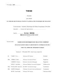
3) les phragmospores : spores pluricellulaires à cloisons transversales(Curvularia) ;4) les dictyospores : spores pluricellulaires à cloisons transversales <strong>et</strong>longitudinales (Alternaria) ;5) les scolécospores : spores étroites, effilées, souvent incurvées <strong>et</strong>cloisonnées transversalement (Fusarium).Mo<strong>de</strong>s <strong>de</strong> formation <strong>de</strong>s conidiesLe mo<strong>de</strong> thallique : la formation <strong>de</strong>s spores s’effectue à partir d’éléments préexistants duthalle. On en distingue <strong>de</strong>ux variantes principales :- le type thallique solitaire, ex: Chrysosporium- le type thallique arthrique, ex: GeotrichumLe mo<strong>de</strong> blastique : les spores sont formées par bourgeonnement à partir <strong>de</strong> cellulesconidiogènes différenciées ou pas, puis une cloison se forme à l’émergence <strong>de</strong> bourgeon <strong>et</strong> lacellule fille (la spore) se sépare <strong>de</strong> la cellule mère. On en distingue plusieurs variantes :- le type blastique acropète, ex: Cladosporium, Alternaria- le type blastique sinchrone, ex: Botrytis- le type blastique sympodial, ex: BeauveriaFigure 1. Mo<strong>de</strong>s <strong>de</strong> formation <strong>de</strong>s conidies1. Formation thallique : a : solitaire (Chrysogenum), b : arthritique (Geotrichum)2. Formation blastique : c : acropète (Cladosporium), d : sinchrone (Botrytis), e : sympodial(Beauveria), f : regressif (Trichothecium), g : annelidique (Scopulariopsis), h :phialidique (Penicillium), i : poric (Curvularia).19
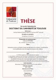
- le type blastique régressif, ex: Trichothecium- le type blastique percurrent (annellidique), ex : Scopulariopsis- le type blastique phialidique, ex: Aspergillus, Penicillium- le type blastique porique, ex: Alternaria, Curvularia (Figure 1) (Botton <strong>et</strong> al., 1990).Mo<strong>de</strong> <strong>de</strong> groupement <strong>de</strong>s conidiesLes conidies sont, en général, regroupées à l’extrémité <strong>de</strong> la cellule conidiogène.L’organisation <strong>de</strong> ce regroupement est aussi un facteur d’i<strong>de</strong>ntification. Les principaux typessont :- grappes, ex. Beauveria, Trichothecium- masse, ex. Botrytis- têtes ou balles, ex. Acremonium, Tricho<strong>de</strong>rma- chaînes basipètes, ex. Scopulariopsis, Aspergillus, Penicillium- chaînes acropètes, ex. Cladosporium, Alternaria (Figure 2) (Botton <strong>et</strong> al., 1990).Figure 2. Mo<strong>de</strong>s <strong>de</strong> groupement <strong>de</strong>s conidies <strong>de</strong>s champignons filamenteux1. grappes (Beauveria), 2. masses (Botrytis), 3. têtes (Acremonium),4. chaînes basipètes (Aspergillus), 5. chaînes acropètes (Alternaria).Mo<strong>de</strong> d’implantation <strong>de</strong>s cellules conidiogènesLes cellules conidiogènes peuvent naître <strong>de</strong> structures plus ou moins élaborées issues dumycélium végétatif. Ceci est utilisé pour l’i<strong>de</strong>ntification <strong>de</strong> genres <strong>et</strong> d’espèces (<strong>de</strong> Hoog <strong>et</strong>Guarro, 1995).20
Les cellules conidiogènes non différenciées sont intégrées dans les hyphes, intercalaires ousituées dans une position terminale (ex : Aureobasidium).Les cellules conidiogènes sont différenciées. Elles peuvent alors être :- directement insérées sur les filaments végétatifs (ex : Acremonium, Fusarium) ;- bien distinctes <strong>de</strong>s filaments végétatifs, portées par <strong>de</strong>s conidiophores dispersés sur l<strong>et</strong>halle végétatif :a) regroupées à l’extrémité dilatée du conidiophore, formant une tête (ex :Aspergillus) ;b) regroupées en verticille au somm<strong>et</strong> du conidiophore, formant un pinceau (ex :Penicillium) ;c) disposées en verticille le long du conidiophore (ex : Verticillium) ;- bien distinctes <strong>de</strong>s filaments végétatifs, portées par <strong>de</strong>s conidiophores groupés :- conidiophores disposés parallèlement les uns aux autres, agrégés en une gerbe sporifèrenommée corèmie (ex : Graphium) ;- conidiophores agrégés en coussin<strong>et</strong>s superficiels nommé sporodochie (ex : Myrothecium).Présence <strong>de</strong> structures protectrices issues <strong>de</strong> la reproduction asexuée ou sexuéeLes structures protectrices issues <strong>de</strong> la reproduction asexuée sont les pycni<strong>de</strong>s <strong>et</strong> lesacervules :- Les pycni<strong>de</strong>s sont <strong>de</strong>s nodules mycéliens, creux, composés d’une paroi épaisseformée par un feutrage compact <strong>de</strong> filaments mycéliens. La face interne <strong>de</strong> la paroi esttapissée <strong>de</strong>s conidiophores produisant <strong>de</strong>s conidies qui sont libérées à maturité par l’ostiole(Phoma).- Les acervules sont <strong>de</strong>s agrégats <strong>de</strong> filaments mycéliens enchevêtrés, soli<strong>de</strong>mentattachés sur un végétal délimitant une cavité avec une ouverture. A l’intérieur, on r<strong>et</strong>rouveune assise <strong>de</strong> conidiophores produisant les conidies.Sur les milieux <strong>de</strong> culture seules les pycni<strong>de</strong>s sont visibles, les acervules ne se formant quedans les tissus <strong>de</strong> l’hôte végétal (<strong>de</strong> Hoog <strong>et</strong> Guarro, 1995).Les structures protectrices issues <strong>de</strong> la reproduction sexuée peuvent être observées chez lesAscomycètes ; l’ascocarpe, qui protège l’asque peut être <strong>de</strong> plusieurs types:a) les apothècies : l’ascocarpe est ouvert, en forme <strong>de</strong> coupe, portant les asques ensurface ;21
) les cléistothèques : l’ascocarpe est arrondi <strong>et</strong> lisse ; il n’y a pas <strong>de</strong> réseaux mycélienspériphériques ; il est clos <strong>et</strong> sa paroi se fissure à maturité pour libérer les asquessphériques octosporées (ex : Emericella) ;c) les périthèces : l’ascocarpe à la forme d’une bouteille avec, à l’extrémité rétrécie, uneouverture (ostiole) ; le périthèce renferme <strong>de</strong>s asques allongés, entourés d’une paroi àsimple membrane (unituniqué) ou à <strong>de</strong>ux membranes (bituniqué) <strong>et</strong> contenant chacun8 ascospores (Cha<strong>et</strong>omium) ;Présence <strong>de</strong>s chlamydosporesLes chlamydospores sont <strong>de</strong>s éléments <strong>de</strong> résistance qui sont formés à partir du filamentmycélien ou à son extrémité. Elles ont une paroi épaisse. Contrairement aux autres spores, leschlamydospores ne possè<strong>de</strong>nt pas <strong>de</strong> mécanismes <strong>de</strong> libération perm<strong>et</strong>tant leur disséminationà maturité. Bien que peu spécifiques puisqu’elles se r<strong>et</strong>rouvent pratiquement chez toutes lesespèces lorsque les <strong>conditions</strong> sont défavorables, elles peuvent cependant constituer une ai<strong>de</strong>dans l’i<strong>de</strong>ntification lorsqu’elles apparaissent précocement (comme chez certaines espèces <strong>de</strong>Fusarium).1.1.2- I<strong>de</strong>ntification génétiqueL'i<strong>de</strong>ntification d'espèces <strong>fongique</strong>s est traditionnellement fondée sur les caractéristiquesculturales <strong>et</strong> morphologiques macroscopiques <strong>et</strong> microscopiques. C<strong>et</strong>te i<strong>de</strong>ntificationnécessite donc, en général, plusieurs jours <strong>de</strong> culture (7 à 10 jours le plus souvent). La culturesur <strong>de</strong>s milieux spécifiques peut être nécessaire pour obtenir la formation <strong>de</strong> conidies <strong>et</strong>, danscertains cas, l’absence d’apparition <strong>de</strong> conidies rendra impossible l’i<strong>de</strong>ntification dumycélium. Par conséquent, <strong>de</strong> nombreuses étu<strong>de</strong>s ont visé à développer <strong>de</strong>s métho<strong>de</strong>s outilsd’i<strong>de</strong>ntification reposant sur l’étu<strong>de</strong> <strong>de</strong>s aci<strong>de</strong>s nucléiques (ADN <strong>et</strong> ARN) <strong>et</strong> ne nécessitantplus obligatoirement un examen morphologique (P<strong>et</strong>erson, 2006 ; Hinrikson <strong>et</strong> al., 2005 ;Feuilha<strong>de</strong> <strong>de</strong> Chauvin, 2005 ; Jin <strong>et</strong> al., 2004 ; Reiss <strong>et</strong> al., 1998).Les métho<strong>de</strong>s les plus intéressantes sont basées sur l’amplification par PCR (polymerasechain reaction) <strong>de</strong> certaines régions spécifiques comme le gène codant pour la sous-unité 28Sribosomale (région D1-D2) <strong>et</strong> <strong>de</strong>s régions ITS1 <strong>et</strong> ITS2 (Hinrikson <strong>et</strong> al., 2005).L’i<strong>de</strong>ntification moléculaire d’espèces <strong>fongique</strong>s est, à l’heure actuelle, surtout appliquée enmycologie médicale pour différencier les espèces d’intérêt. En eff<strong>et</strong>, les infections <strong>fongique</strong>senvahissantes sont <strong>de</strong> plus en plus i<strong>de</strong>ntifiées comme cause primaire <strong>de</strong> morbidité <strong>et</strong> <strong>de</strong>22
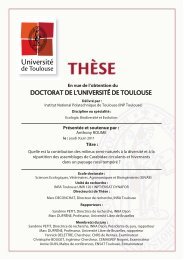
mortalité, particulièrement chez les immunodéficients (<strong>de</strong> Aguire, <strong>et</strong> al., 2004).L’i<strong>de</strong>ntification moléculaire peut perm<strong>et</strong>tre une différenciation rapi<strong>de</strong> <strong>de</strong>s différentes espècesd’Aspergillus ainsi que celle d'autres moisissures ou levures, pathogènes opportunistes (Dial,2007 ; <strong>de</strong> Aguire, <strong>et</strong> al., 2004 ; Schabereiter-Gurtner <strong>et</strong> al., 2007 ; Paterson <strong>et</strong> al., 2003). Lesson<strong>de</strong>s d'oligonucléoti<strong>de</strong>, dirigées vers la région ITS 2 <strong>de</strong> l'ADN ribosomal <strong>de</strong> l’Aspergillusflavus, A. fumigatus, A. nidulans, A. niger, A. terreus, A. ustus, <strong>et</strong> A. versicolor ont permis ladifférenciation <strong>de</strong> 41 isolats <strong>et</strong> n'ont donné aucune réaction faussement positive avec 33espèces d'Acremonium, Exophiala, Candida, Fusarium, Mucor, Paecilomyces, Penicillium,Rhizopus, ou d'autres espèces d’Aspergillus (<strong>de</strong> Aguire, <strong>et</strong> al., 2005).C<strong>et</strong>te métho<strong>de</strong> est aussi utilisée pour différencier <strong>et</strong> i<strong>de</strong>ntifier les moisissures responsables <strong>de</strong>l’altération <strong>de</strong>s aliments, principalement les espèces <strong>de</strong> Penicillium (Boysen <strong>et</strong> al., 2000 ;Hageskal <strong>et</strong> al., 2006).En ce qui concerne les Fusarium, les métho<strong>de</strong>s moléculaires existantes donnent <strong>de</strong>s résultatsfréquemment peu concluants <strong>et</strong> l’examen morphologique classique semble encore unemétho<strong>de</strong> indispensable à l’i<strong>de</strong>ntification <strong>de</strong>s espèces appartenant à ce genre <strong>fongique</strong> (Healy<strong>et</strong> al., 2005).De nombreux travaux visent aussi à utiliser les progrès <strong>de</strong> la biologie moléculaire afin <strong>de</strong>dépister rapi<strong>de</strong>ment les souches <strong>fongique</strong>s toxinogènes (pour revue : Niessen, 2007).Si à l’heure actuelle les outils <strong>de</strong> d’i<strong>de</strong>ntification moléculaire ne semblent pas en mesure <strong>de</strong>remplacer l’i<strong>de</strong>ntification morphologique classique, il est probable que dans les années àvenir, ces métho<strong>de</strong>s représenteront <strong>de</strong>s outils particulièrement utiles pour la détection <strong>et</strong>l’i<strong>de</strong>ntification <strong>fongique</strong> dans les aliments.1.2- Classement <strong>de</strong>s moisissuresL’ensemble <strong>de</strong>s caractéristiques morphologiques évoquées précé<strong>de</strong>mment a permis leclassement <strong>de</strong>s moisissures. Il s’agit d’un véritable règne comprenant <strong>de</strong>s divisions, ellesmêmessubdivisées en classes ; les classes englobent <strong>de</strong>s ordres qui rassemblent <strong>de</strong>s familles ;une famille peut comprendre un ou plusieurs genres <strong>et</strong> le genre, à son tour, rassemble une ouplusieurs espèces. Chaque champignon porte un nom qui suit les règles <strong>de</strong> la nomenclaturebinominale (genre <strong>et</strong> espèce) énoncées au XVIII ème siècle par Carl von Linné.La classification <strong>de</strong> Kwon Chung <strong>et</strong> Benn<strong>et</strong>t (1992) modifiée par <strong>de</strong> Hoog <strong>et</strong> Guarro (1995)est la plus utilisée actuellement (Figure 3). On estime à plus <strong>de</strong> 100 000 le nombre d’espèces23
<strong>fongique</strong>s, plus <strong>de</strong> 1000 d’entre elles pouvant contaminer les aliments (Castegnaro <strong>et</strong> Pfohl-Leskowicz, 2002).DeutéromycotinaChytridiomycotinaBasidiomycotinaChampignonsZygomycotinaAsco mycotin aOomycotinaFigure 3. Classification <strong>de</strong>s champignons.Les AscomycètesCe groupe comprend un grand nombre d’espèces pathogènes pour l’homme <strong>et</strong> les animaux(levures ascosporées ou champignons filamenteux comme les Aspergillus). L’appareilvégétatif est un thalle à mycélium septé.Les Ascomycètes présentent une structure caractéristique appelée asque, formé au cours <strong>de</strong> lareproduction sexuée, qui renferme un nombre défini d’ascospores Ce sporocyste, globuleux,cylindrique ou plus ou moins claviforme, avec une paroi simple ou double représente unimportant critère d’i<strong>de</strong>ntification.Les ascospores, hyalines ou colorées, globuleuses, elliptiques, cylindriques, vermiculaires,unicellulaires, cloisonnées transversalement ou cloisonnées transversalement <strong>et</strong>longitudinalement, lisses ou ornementées sont utilisées aussi pour l’i<strong>de</strong>ntification <strong>de</strong>s genres<strong>et</strong> <strong>de</strong>s espèces.Souvent les asques sont produits, en grand nombre, dans <strong>de</strong>s structures <strong>de</strong> fructification,nommés ascocarpes, divisés en 3 catégories : les cléistothèques (ascocarpes globuleux, clos,rencontrés chez Eurotium <strong>et</strong> Emericella), les périthèces (ascocarpes plus ou moins en forme<strong>de</strong> bouteille présentant un ostiole par lequel les spores sont expulsées, rencontré chezCha<strong>et</strong>omium) <strong>et</strong> les apothècies (ascocarpes ouverts, en forme <strong>de</strong> coupe, portant <strong>de</strong>s asques ensurface). Certains Ascomycètes présentent <strong>de</strong>s asques nus (levures) ou enveloppés dans unfeutrage mycélien lâche (Byssochlamys).A côté <strong>de</strong> c<strong>et</strong>te forme sexuée, ascogène (forme parfaite ou téléomorphe), la majoritéd’Ascomycètes se reproduisent par multiplication asexuée (forme imparfaite ou anamorphe) àl’ai<strong>de</strong> <strong>de</strong>s conidies ((Botton <strong>et</strong> al., 1990 ; Sutton <strong>et</strong> al., 1998).24
Les Ascomycètes ont une gran<strong>de</strong> importance économique. Certaines espèces sont <strong>de</strong>s agents<strong>de</strong> biodégradation <strong>de</strong>s <strong>substrats</strong> cellulosiques. Les Ascomycètes sont souvent r<strong>et</strong>rouvés comme<strong>de</strong>s parasites redoutables <strong>de</strong> végétaux (l’ergot du seigle parasité par Claviceps purpurea) ou<strong>de</strong> l’homme (histoplasmose). Certaines espèces peuvent entraîner <strong>de</strong>s altérations aux <strong>de</strong>nréesalimentaires <strong>et</strong> élaborer <strong>de</strong>s toxines ; d’autres interviennent dans <strong>de</strong>s processus <strong>de</strong>fermentations <strong>de</strong>s industries agro-alimentaires.Les Deutéromycètes (Champignons imparfaits ou Fungi imperfecti)Les Deutéromycètes réunissent le plus grand nombre d’espèces pathogènes pour l’homme <strong>et</strong>les animaux. C<strong>et</strong>te division, très hétérogène, englobe toutes les espèces <strong>de</strong> champignons, pourlesquelles la reproduction sexuée n’est pas connue. La majorité <strong>de</strong> Deutéromycètes sont <strong>de</strong>sformes imparfaites d’Ascomycètes.Ces champignons sont unicellulaires ou à thalle filamenteux septé. Les Deutéromycètes sontdivisés en 3 classes :- les Blastomycètes qui réunissent <strong>de</strong>s levures ;- les Hyphomycètes : champignons filamenteux, stériles ou produisant <strong>de</strong>s sporesdirectement sur les hyphes ou sur <strong>de</strong>s conidiophores simples ou agrégés(Moniliales) ;- les Coelomycètes : Champignons auxquelles les conidies sont produites dans <strong>de</strong>sstructures <strong>de</strong> protection : les pycni<strong>de</strong>s (Sphaeropsidales) <strong>et</strong> les acervules(Mélanconiales) ;Les Hyphomycètes <strong>et</strong> les Coelomycètes comprennent <strong>de</strong>s agents <strong>de</strong> biodégradation mais aussi<strong>de</strong>s moisissures utiles aux biotechnologies.Les Hyphomycètes sont <strong>de</strong>s champignons pour lesquels les conidies naissent <strong>de</strong> cellulesbanales ou <strong>de</strong> cellules spécialisées souvent portées par un filament différencié, leconidiophore. Certaines hyphomycètes (Rhizoctonia, Sclerotium) ne forment jamais <strong>de</strong>spores ; ils sont classés dans le groupe Micellia sterilia.Les spores peuvent être :a) <strong>de</strong>s chlamydospores, spores <strong>de</strong> résistance, souvent à paroi épaisse, terminales ouintercalaires, différenciées par transformation <strong>de</strong> cellules du mycélium(Tricho<strong>de</strong>rma, Fusarium) ou <strong>de</strong> conidies (Fusarium) ;b) <strong>de</strong>s arthrospores (arthroconidies) formées par fragmentation d’hyphes nondifférenciées.25
c) <strong>de</strong>s conidies proprement dites, souvent bourgeonnées par <strong>de</strong>s cellules spécialiséesnommées phiali<strong>de</strong>s.La forme, les dimensions, la couleur, la structure (unicellulaires, bicellulaires, …),l’ornementation <strong>de</strong>s conidies représentent un ensemble <strong>de</strong> caractères très varié qui constitue<strong>de</strong>s critères d’i<strong>de</strong>ntification <strong>de</strong> genres <strong>et</strong> d’espèces.Les cellules conidiogènes ou les phiali<strong>de</strong>s bourgeonnent à l’apex <strong>de</strong>s spores en successionbasipétales réunies en glomérules (Phialophora, Stachybotrys) ou en chaînes (Aspergillus,Penicillium). Les cellules conidiogènes sont diverses comme forme (ampulliformes, ovées,lancéolées, subulées, …), solitaires ou groupées en verticille (Tricho<strong>de</strong>rma) ou en têtes(Aspergillus, Penicillium).Les sclérotes, présents sous la forme <strong>de</strong> masses mycéliennes compactes, souvent dures,globuleuses, ellipsoïdales ou allongées, sont considérés comme <strong>de</strong>s organes <strong>de</strong> conservation(Botrytis, Aspergillus, Penicillium).Les Coelomycètes sont <strong>de</strong>s champignons auxquels les cellules conidiogènes sont renferméesdans <strong>de</strong>s structures <strong>de</strong> fructification dont la morphologie perm<strong>et</strong> <strong>de</strong> différencier :a) Les Sphaeropsidales, pour lesquelles les cellules conidiogènes sont dans <strong>de</strong>sorganes globuleux ou en forme <strong>de</strong> bouteille, appelés pycni<strong>de</strong>s (Phoma) ;b) Les Melanconiales, généralement parasites <strong>de</strong> végétaux, forment <strong>de</strong>s structures <strong>de</strong>fructification aplaties (acervules), sous l’épi<strong>de</strong>rme ou la cuticule <strong>de</strong>s cellules <strong>de</strong> laplante-hôte (Botton <strong>et</strong> al., 1990 ; Sutton <strong>et</strong> al., 1998).1.3- Principaux genres <strong>fongique</strong>sLes genres les plus importants <strong>de</strong> point <strong>de</strong> vue économique <strong>et</strong> médical sont les Aspergillus,les Penicillium <strong>et</strong> les Fusarium qui représentent les contaminants les plus fréquents <strong>de</strong>saliments. On les r<strong>et</strong>rouve principalement dans les céréales, mais aussi dans <strong>de</strong> nombreuxautres produits végétaux <strong>et</strong> d’origine animale.1.3.1- Le genre AspergillusC’est un genre appartenant à la classe <strong>de</strong>s Ascomycètes. Le thalle, hyalin ou coloré, présenteun mycélium cloisonné portant <strong>de</strong> nombreux conidiophores dressés, terminés en vésicule(Figure 4) (Raper <strong>et</strong> Fennell, 1965).26
Ce genre comprend environ 185 espèces réparties en 18 groupes morphologiquement,génétiquement <strong>et</strong> physiologiquement proches (Raper <strong>et</strong> Fennell, 1965 ; Botton <strong>et</strong> al., 1990 ;Roquebert, 1998). Une vingtaine d’espèces est impliquée dans <strong>de</strong>s pathologies animales <strong>et</strong>humaines.Les Aspergillus ont une large répartition géographique, mais sont plus souvent associés auxrégions à climat chaud (Castegnaro <strong>et</strong> Pfohl-Leszkowicz, 2002); ils se développent sur lamatière organique en décomposition, dans le sol, le compost, les <strong>de</strong>nrées alimentaires, lescéréales. De nombreuses espèces d’Aspergillus sont présentes dans l’environnement humain,notamment dans la poussière <strong>et</strong> l’air (Morin, 1994).Certaines espèces peuvent être directement pathogènes pour l’homme <strong>et</strong> les animaux en étantcapable d’envahir les tissus vivants <strong>et</strong> provoquer <strong>de</strong>s aspergilloses (Aspergillus fumigatusresponsable <strong>de</strong> mycoses pulmonaires ; Aspergillus niger responsable d’aspergillose duconduit auditif) (Morin, 1994).Figure 4. Principaux caractères morphologiques <strong>de</strong>s Aspergillus.De nombreuses espèces d’Aspergillus sont aussi connues pour leur capacité à produire <strong>de</strong>smycotoxines responsables <strong>de</strong> pathologies animales <strong>et</strong> humaines.27
Enfin, certaines espèces d’Aspergillus sont utilisées dans l’industrie agro-alimentaire <strong>et</strong> dansl’industrie <strong>de</strong>s produits biotechnologiques notamment pour la production d’enzymes, d’aci<strong>de</strong>sorganiques (Botton <strong>et</strong> al., 1990).1.3.1.1- Caractères culturaux générauxLes Aspergillus présentent une croissance rapi<strong>de</strong> sur les milieux <strong>de</strong> culture classiques (géloseau malt, Sabouraud) additionnés d’antibiotiques. Après 48 heures d’incubation, on observe<strong>de</strong>s colonies plates, formées <strong>de</strong> courts filaments aériens, blancs ; après 96 heures d’incubation,les colonies vont prendre leur teinte caractéristique, brune, verte, jaune ou noire selon lesespèces. La majorité <strong>de</strong>s Aspergillus poussent à 22-25°C ; les espèces thermophiles (A.fumigatus) se développent à 37-40°C est parfois jusqu’à 57°C (Badill<strong>et</strong> <strong>et</strong> al., 1987 ; Morin,1994).Les Aspergillus forment <strong>de</strong>s colonies souvent poudreuses ou granuleuses. La couleur <strong>de</strong>colonies perm<strong>et</strong> une orientation rapi<strong>de</strong> dans l’i<strong>de</strong>ntification d’espèces : gris-vert pour A.fumigatus, vert-jaune pour A. flavus <strong>et</strong> les espèces du groupe A. glaucus, vert foncé à chamoispour A. nidulans, brun canelle pour A. terreus, chamois clair, jaune <strong>et</strong> rose pour A. versicolor,jaune puis noir pour A. niger <strong>et</strong> blanche pour A. candidus. Le revers <strong>de</strong> la colonie est incoloreou jaune, mais il peut brunir ou rougir avec l’âge (Cherm<strong>et</strong>te <strong>et</strong> Bussieras, 1993).1.3.1.2- Morphologie microscopiqueLes Aspergillus sont caractérisés par un appareil végétatif (thalle) formé <strong>de</strong> filamentsmycéliens hyalins, <strong>de</strong> diamètre fin <strong>et</strong> régulier, septés <strong>et</strong> ramifiés. Sur les filaments végétatifsprennent naissance <strong>de</strong>s filaments dressés, non cloisonnés (conidiophores) qui se terminent parune vésicule <strong>de</strong> forme variable sur la quelle sont disposées les cellules conidiogènes ouphiali<strong>de</strong>s. Les phiali<strong>de</strong>s peuvent être insérées directement sur la vésicule (têtes unisériées) ouportées par <strong>de</strong>s p<strong>et</strong>its structures insérés sur la vésicule (têtes bisériées) nommées métules oustérigmates (Figure 4) (Badill<strong>et</strong> <strong>et</strong> al., 1987 ; Raper <strong>et</strong> Fennell, 1965). Les conidies, sèches,disposées en chaînes divergentes ou associées en colonnes compactes, sont toujoursunicellulaires, globuleuses, sub-globuleuses ou elliptiques, lisses ou ornementées, hyalines oupigmentées en jaune, vert, brun ou noir.L’ensemble vesicule ± métules + phiali<strong>de</strong>s + conidies constitue la tête aspergillairecaractéristique du genre Aspergillus.28
Pour certaines espèces, <strong>de</strong>s formations sexuées apparaissent parfois en culture. Il s’agit <strong>de</strong>cléistotèques qui contiennent <strong>de</strong>s asques arrondis renfermant chacun 8 ascospores.Les « hülle cells » ou les cellules en nois<strong>et</strong>te, sont <strong>de</strong>s formations arrondies, réfringentes àparoi épaisse qui accompagnent souvent les formes sexuées, mais que l’on peut égalementobserver isolément, indépendamment <strong>de</strong> la reproduction sexuée.1.3.1.3- Les principales espècesi) Aspergillus flavus :Caractères culturaux : Le champignon se développe rapi<strong>de</strong>ment sur les milieux classiques(géloses au malt <strong>et</strong> Sabouraud) à 22-25°C. La température optimale <strong>de</strong> croissance est 37°C.Sur le milieu <strong>de</strong> culture A. flavus forme <strong>de</strong>s colonies duv<strong>et</strong>euses à poudreuses, d’abordblanches, puis jaune, puis vert-jaune. Le revers peut être incolore, rosâtre ou brun-rouge foncépour les souches productrices <strong>de</strong> sclérotes (Figure 5).Figure 5. Aspergillus flavus(culture <strong>de</strong> 7 jours sur gélose au malt à 25 o C <strong>et</strong> aspect microscopique).Morphologie microscopique : Les têtes conidiennes, unisériées ou bisériées, d’abord radiées,puis reparties en plusieurs colonnes <strong>de</strong> spores mal individualisées, jaunâtres au début, puisvert-jaune foncé. Les conidiophores hyalins, verruqueux, atteignent 1 à 2,5 mm <strong>de</strong> long. Lesvésicules sont sub-globuleuses, <strong>et</strong> mesurent 25 (10-65) à 45 µm <strong>de</strong> diamètre. Les phiali<strong>de</strong>s (6-10 x 4-5,5 µm) sont insérées directement sur la vésicule (unisériées) ou portées par <strong>de</strong>smétules. Les conidies sont globuleuses à sub-globuleuses, <strong>de</strong> 3-6 µm <strong>de</strong> diamètre, <strong>de</strong> couleurverte pâle, verruqueuses. Les sclérotes, fréquents dans les isolats récents, sont globuleux àsub-globuleux, d’abord blanc puis virant au brun-rouge foncé <strong>et</strong> au noir (Figure 5).29
ii) Aspergillus fumigatus :Caractères culturaux : mycélium à croissance rapi<strong>de</strong> sur les milieux <strong>de</strong> culture classiques à37°C. A. fumigatus est une espèce thermtolérante dont la température <strong>de</strong> croissance estcomprise entre 15 <strong>et</strong> 48°C ; la température optimale étant située aux alentours <strong>de</strong> 40 <strong>et</strong> 42°C.C<strong>et</strong>te espèce peut se développer jusqu’à 57°C (Morin, 1994).A. fumigatus forme <strong>de</strong>s colonies d’abord blanches, puis bleu-vertes <strong>et</strong> enfin vert foncé à grisnoirâtre. Le revers peut être incolore, jaune, vert ou brun-rouge suivant les souches.Figure 6. Aspergillus fumigatus(culture <strong>de</strong> 7 jours sur gélose au malt à 25 o C <strong>et</strong> aspect microscopique).Morphologie microscopique : Les têtes conidiennes, strictement unisériées, en colonnecompacte sont d’abord bleu-vert puis virant au vert bronze. Les conidiophores sont courts(300-500 µm), lisses, s’élargissent insensiblement au somm<strong>et</strong> en formant <strong>de</strong>s vésicules subhémisphériques.Ces <strong>de</strong>rnières (20-30 µm en diamètre), vertes, sont fertiles dans leur moitiésupérieure. Les phiali<strong>de</strong>s dressées, sont <strong>de</strong>nsément groupées, <strong>de</strong> couleur verte. Les conidiessont globuleuses à sub-globuleuses, mesurent 2(2,5)-3 (3,5) µm <strong>de</strong> diamètre, <strong>et</strong> sontéchinulées (Figure 6).iii) Aspergillus niger :Caractères culturaux : Ce champignon pousse rapi<strong>de</strong>ment (2-3 jours) sur les milieux <strong>de</strong>culture classiques (géloses au malt <strong>et</strong> Sabouraud). La température optimale <strong>de</strong> croissancevarie généralement entre 25 <strong>et</strong> 30°C, mais A. niger peut se développer jusqu’à 42°C.Les colonies d’A. niger sont granuleuses, blanches au début, puis jaunes <strong>et</strong>, à maturité, elles<strong>de</strong>viennent noires. Le revers <strong>de</strong>s colonies est incolore ou jaune pâle. Sur le milieu Czapek, A.niger forme <strong>de</strong>s colonies à mycélium blanc ou jaune, <strong>et</strong> revers souvent incolore (Figure 7).30
Morphologie microscopique : Les têtes conidiennes, bisériées, radiées, sont disposées enplusieurs colonnes brunâtres ou noires. Les conidiophores sont longs atteignant 1,5-3 mm,lisses, hyalins ou brunâtres dans leur moitié supérieure. Les vésicules sont globuleuses <strong>et</strong>entièrement fertiles. Les phiali<strong>de</strong>s (7-10 x 3-3,5 µm) sont portées par <strong>de</strong>s métules brunâtres,<strong>de</strong> dimensions variables. Les conidies sont habituellement globuleuses, parfois légèrementaplaties. Elles mesurent 3,5-5 µm <strong>de</strong> diamètre, sont brunes, échinulées à très verruqueuses.Les sclérotes parfois différenciés, sont crème à chamois foncé au début, puis virent auchamois vinacé (Figure 7).Figure 7. Aspergillus niger(culture <strong>de</strong> 7 jours sur gélose au malt à 25 o C <strong>et</strong> aspect microscopique).iv) Aspergillus ochraceus :Caractères culturaux : Ce champignon pousse rapi<strong>de</strong>ment (2-3 jours) sur les milieux <strong>de</strong>culture classiques à une température <strong>de</strong> 25 à 30°C.Les colonies d’A. ochraceus sont poudreuses ou granuleuses, blanches au début, puis jaunesou ocre-jaunes à chamois. Le revers <strong>de</strong> colonies est incolore au jaune pâle (Figure 8).Figure 8. Aspergillus ochraceus(aspect microscopique).31
Morphologie microscopique : Les têtes conidiennes sont bisériées, d’abord globuleuses puisse séparent en 2 ou 3 colonnes divergentes, bien individualisées, <strong>de</strong> couleur jaune, ocre-jauneou chamois. Les conidiophores sont rugueux, jaunes à brun pâle, longs atteignant 1,5 mm. Lesvésicules sont globuleuses, hyalines, 30-50 µm en diamètre. Les phiali<strong>de</strong>s (7-10 x 2-3,5 µm)sont portées par <strong>de</strong>s métules, <strong>de</strong> dimensions variables. Les conidies sont sub-globuleuses àglobuleuses. Elles mesurent 2,5-3 (3,5) µm <strong>de</strong> diamètre, sont finement échinulées ou lisses.Les sclérotes, souvent présents, <strong>de</strong> couleur lavan<strong>de</strong> à pourpre, sont globuleux, ovales oucylindriques, <strong>et</strong> atteignent 1mm <strong>de</strong> diamètre (Figure 8).v) Aspergillus oryzae :Caractères culturaux : Le champignon se développe rapi<strong>de</strong>ment sur les milieux classiques(géloses au malt <strong>et</strong> Sabouraud) à 22-25°C.Sur milieu <strong>de</strong> culture A. oryzae forme <strong>de</strong>s colonies duv<strong>et</strong>euses à poudreuses, d’abord <strong>de</strong>couleur blanche, puis jaune, <strong>et</strong> enfin vert-jaunâtre à vert olive. Le revers peut être incolore oujaunâtre (Figure 9).Morphologie microscopique : A. oryzae peut souvent être confondu avec A. flavus. Les têtesunisériées ou bisériées (les <strong>de</strong>ux aspects pouvant cohabiter sur une même souche), sontFigure 9. Aspergillus oryzae(culture <strong>de</strong> 7 jours sur gélose au malt à 25 o C).radiées, jaunâtre au début puis jaune verdâtre à vert-olive, puis évoluent vers <strong>de</strong>s tons bruns.Les conidiophores, hyalins, sont souvent longs (2,5-5 mm), plus ou moins verruqueux suivantles souches. Les vésicules sont sub-globuleuses, à paroi mince, mesurent 40-75 µm <strong>de</strong>diamètre. Les phiali<strong>de</strong>s (8-15 x 3-5 µm) sont insérées directement sur la vésicule ou portéespar <strong>de</strong>s métules. Les conidies d’abord piriformes à elliptiques <strong>de</strong>viennent sub-globuleuses àglobuleuses, <strong>de</strong> taille très variable, lisses ou finement verruqueuses (Figure 9).32
1.3.1.4- Importance du genre Aspergillusi) Potentiel toxinogèneDe nombreuses espèces appartenant au genre Aspergillus (Tableau 2) sont connues pour leurcapacité à produire certaines mycotoxines (Pitt, 2000).Aspergillus flavus <strong>et</strong> A. parasiticus sont les principaux producteurs d'aflatoxines. L’aflatoxineB1 est classée comme cancérigène chez l’homme <strong>et</strong> l’animal (IARC, 1993).Aspergillus fumigatus synthétise plusieurs métabolites très toxiques comme la fumagiline,l’aci<strong>de</strong> helvolique, la gliotoxine, les dérivés quinoniques, <strong>de</strong>s alcaloï<strong>de</strong>s voisins <strong>de</strong> ceux <strong>de</strong>l’ergot <strong>de</strong> seigle.Aspergillus niger peut produire <strong>de</strong> l’aci<strong>de</strong> oxalique, <strong>de</strong>s malformines <strong>et</strong>, certaines souches,<strong>de</strong>s aflatoxines.Aspergillus ochraceus est le principal producteur d'ochratoxine A. Il colonise lui aussi <strong>de</strong> trèsnombreux <strong>substrats</strong>.Aspergillus terreus élabore <strong>de</strong>s substances antibactériennes, <strong>de</strong> toxicité variable (flavipine,terréine, citrinine, erdine <strong>et</strong> molécules voisines, clavacine) (Botton <strong>et</strong> al., 1990).Tableau 2 : Les Aspergillus producteurs <strong>de</strong> mycotoxines.Espèces d’AspergillusAspergillus candidusAspergillus carneusAspergillus clavatusAspergillus flavusAspergillus fumigatusAspergillus nigerAspergillus nomiusAspergillus oachraceusAspergillus oryzaeAspergillus parasiticusAspergillus sydowiiAspergillus terreusAspergillus verzicolorAspergillus wentiiMycotoxines produitescandidulinecitrinineaci<strong>de</strong> kojique, patuline, xanthocilineaflatoxines B1 <strong>et</strong> B2, aci<strong>de</strong> aspergillique, aci<strong>de</strong>cyclopiazonique, aci<strong>de</strong> kojiquefumigaclavine, fumagiline, fumitoxine, fumitremorgine A<strong>et</strong> C, gliotoxinemalformine, naftoquinoneaflatoxines B1 <strong>et</strong> B2, G1 <strong>et</strong> G2, aci<strong>de</strong> aspergilliqueaci<strong>de</strong> kojique, aci<strong>de</strong> neoaspergillique, ochratoxine, aci<strong>de</strong>penicillique, aci<strong>de</strong> sécalonique Aaci<strong>de</strong> cyclopiazonique, aci<strong>de</strong> kojiqueaflatoxines B1 <strong>et</strong> B2, G1 <strong>et</strong> G2, aci<strong>de</strong> aspergillique, aci<strong>de</strong>kojiquesterigmatocystine, griséofulvinecitréoviridine, citrinine, gliotoxine, patuline, terréine,aci<strong>de</strong> terréique, terrétonine, territrem, terrami<strong>de</strong> Astérigmatocystineaci<strong>de</strong> kojique33
ii) Pouvoir pathogèneCertaines espèces d’Aspergillus sont <strong>de</strong>s pathogènes opportunistes ; leur développementnécessite <strong>de</strong>s <strong>conditions</strong> locales favorables (cavernes tuberculeuses, cancer bronchopulmonaire,broncho-pneumopathies chroniques obstructives, emphysèmes,mucoviscidose…) ou générales (corticothérapies prolongées, hémopathies malignes,chimiothérapies aplaisantes, SIDA…). (Badill<strong>et</strong> <strong>et</strong> al., 1987 ; Morin, 1994)Les principales espèces responsables <strong>de</strong> mycoses (aspergilloses) sont :- Aspergillus fumigatus considéré comme le principal agent d’aspergillose aviaire <strong>et</strong>humaine (représentant 80-90% <strong>de</strong>s aspergilloses humaines) (Morin, 1994) ;- Aspergillus flavus responsable d’aspergilloses pulmonaires ou généraliséesprincipalement chez les patients immunodéprimés (Baculard <strong>et</strong> Tournier, 1995) ;- Aspergillus niger rarement rencontré chez l’immunodéprimé ; chez le suj<strong>et</strong> nonimmunodéprimé il peut provoquer <strong>de</strong>s aspergilloses, <strong>de</strong>s otites <strong>et</strong> <strong>de</strong>s sinusites ; il est aussi àl’origine d’infections cutanées, pulmonaires <strong>et</strong> généralisées (Morin, 1994) ;- Aspergillus sydowi impliqué dans <strong>de</strong>s cas d’endocardite humaine (Botton <strong>et</strong> al.,1990) ;- Aspergillus terreus est un agent important d’aspergilloses pulmonaires <strong>et</strong> cérébraleschez les patients immunodéficitaires ; il est souvent isolé <strong>de</strong>s expectorations chez les patientsatteints <strong>de</strong> mucoviscidose (Baculard <strong>et</strong> Tournier, 1995 ; Khan <strong>et</strong> al., 1999) ; c’est aussi uneespèce fréquemment isolée dans <strong>de</strong>s prélèvements <strong>de</strong> peau <strong>et</strong> <strong>de</strong> phanères. Il est considérécomme le principal agent d’onychomycoses (Morin, 1994).iii) Espèces utilesCertaines espèces d’Aspergillus sont utilisées dans l’industrie agro-alimentaire <strong>et</strong> dansl’industrie <strong>de</strong>s produits biotechnologiques notamment pour la fermentation <strong>de</strong> divers <strong>substrats</strong><strong>et</strong> la production d’enzymes ou d’aci<strong>de</strong>s organiques :- Aspergillus awamori, agent lipolytique d’oléagineux, est utilisé fréquemment auJapon pour la fermentation alcoolique.- Aspergillus niger est utilisé dans les processus biotechnologiques pour la synthèse <strong>de</strong>différents aci<strong>de</strong>s comme l’aci<strong>de</strong> citrique <strong>et</strong> l’aci<strong>de</strong> gluconique ainsi que pour la productiond’enzymes : alpha-amylase, b<strong>et</strong>a-glucanase, catalase, glucose oxydase, lipase, pectinase,polygalacturonase.- Aspergillus oryzae est utilisé, dans les pays asiatiques, pour la fabrication <strong>de</strong>produits fermentés à base <strong>de</strong> soja. Il est utilisé aussi dans <strong>de</strong>s processus biotechnologiques34
pour la production <strong>de</strong> certaines enzymes comme : alpha-amylase, b<strong>et</strong>a-glucanase, lipase(Botton <strong>et</strong> al., 1990).1.3.2- Le Genre PenicilliumCe genre réunit <strong>de</strong>s champignons filamenteux, appartenant au phylum <strong>de</strong>s Ascomycètes. Cegenre comprend environ 227 espèces définies essentiellement d’après les caractères du thalle,<strong>de</strong>s pénicilles <strong>et</strong> <strong>de</strong>s spores (Pitt, 1988). Les Penicillium sont <strong>de</strong>s champignons pour la pluparttrès communs dans l’environnement, polyphages, pouvant être responsables <strong>de</strong> nombreusesdégradations. Ils ont pour habitat naturel le sol, les <strong>de</strong>nrées alimentaires, les matièresorganiques en décomposition, le compost, les céréales.Les espèces du genre Penicillium se développent normalement dans <strong>de</strong>s milieux ou l’activité<strong>de</strong> l’eau est plus élevée que celle perm<strong>et</strong>tant la croissance <strong>de</strong>s Aspergillus, à <strong>de</strong>s températuresplus basses. Il s’agit <strong>de</strong> contaminants fréquents <strong>de</strong>s régions tempérées.1.3.2.1- Caractères culturaux générauxLes Penicillium se développent rapi<strong>de</strong>ment <strong>et</strong> facilement sur les milieux <strong>de</strong> culture utilisés enroutine (géloses au malt, Sabouraud). Ils se développent à <strong>de</strong>s températures modérées <strong>de</strong>l’ordre <strong>de</strong> 20-27°C. Après 2 jours d’incubation, on observe <strong>de</strong>s p<strong>et</strong>ites colonies plates,formées <strong>de</strong> courts filaments aériens, habituellement blancs. Après 3-4 jours d’incubation, lasporulation va conférer aux colonies leur teinte, le plus souvent dans les tons vert, vert bleu,vert-gris, vert-jaune, gris-bleu mais aussi, pour certaines espèces, jaune, orange, chamois,rose, ou rouge. C<strong>et</strong>te couleur perm<strong>et</strong> une première orientation dans l’i<strong>de</strong>ntification d’espèces :vert-gris pour P. citrinum, P. cyclopium, P. italicum, vert-jaune pour P. chrysogenum, vertsombre pour P. roquefortii, P. fellutatum, jaune pâle, chamois pour P. nalgiovense, jaune vif àrouge pour P. purpurogenum, mélange d’orange <strong>et</strong> verdâtre pour P. islandicum, <strong>et</strong> blanchepour P. camembertii. Le revers <strong>de</strong> colonies peut être incolore, jaune, rouge, brun ou noir <strong>et</strong>parfois le pigment diffuse dans le milieu <strong>de</strong> culture (Cherm<strong>et</strong>te <strong>et</strong> Bussieras, 1993).Les Penicillium peuvent former <strong>de</strong>s colonies floconneuses (P. camembertii, P. nalgiovense),funiculeuses (P. funiculosum), granuleuses (P. expansum, P. griseofulvum), veloutées (P.chrysogenum, P. citrinum, P. roquefortii…) (Figure 10).35
Figure 10. Caractères du thalle <strong>de</strong> genre Penicillium (Botton <strong>et</strong> al., 1990).1.3.2.2- Morphologie microscopiqueAu point <strong>de</strong> vue morphologique les Penicillium se distinguent par leur organisation enpinceau. Le thalle, formé <strong>de</strong> filaments mycéliens septés <strong>et</strong> hyalins, porte <strong>de</strong>s conidiophoreslisses ou granuleux, simples ou ramifiés qui se terminent par un pénicille. Les conidiophorespeuvent être isolés, groupés en faisceaux lâches ou agrégés en corémies bien individualisés(Figure 11).Figure 11. Caractères morphologiques <strong>de</strong>s Penicillium.36
Les phiali<strong>de</strong>s sont disposées en verticilles à l’extrémité <strong>de</strong>s conidiophores. Les phiali<strong>de</strong>speuvent être insérées directement (Penicillium monoverticillé) ou par l’intermédiaire d’unerangée <strong>de</strong> métules (Penicillium biverticillé) ; <strong>de</strong> <strong>de</strong>ux rangées successives <strong>de</strong> métules(Penicillium triverticillé) ; parfois <strong>de</strong> trois rangées <strong>de</strong> métules (Penicillium quadriverticillé)sur les conidiophores. Les phiali<strong>de</strong>s, ampulliformes ou lancéolées, sont serrées les unes contreles autres donnant à l’ensemble un aspect <strong>de</strong> pinceau. Les phiali<strong>de</strong>s (cellules conidiogènes)donnent naissance à <strong>de</strong>s conidies disposées en longues chaînes.Les conidies sont <strong>de</strong>s spores unicellulaires, globuleuses, elliptiques, cylindriques oufusiformes, lisses ou rugueuses, hyalines, grisâtres ou verdâtres (Botton <strong>et</strong> al., 1990). Lescaractères <strong>de</strong>s pénicilles servent à la différenciation <strong>de</strong>s groupes <strong>et</strong> <strong>de</strong>s espèces. Certainesespèces peuvent présenter une reproduction sexuée avec la formation d’ascocarpes.1.3.2.3.- Importance du genre Penicilliumi) Potentiel toxinogèneLa majorité d’espèces du genre Penicillium (Tableau 3) sont capables <strong>de</strong> produire <strong>de</strong>smycotoxines : l’aci<strong>de</strong> cyclopiazonique (Penicillium chrysogenum), l’aci<strong>de</strong> pénicillique(Penicillium cyclopium) ; la patuline ou la clavacine (Penicillium expansum, Penicilliumgriseofulvum), la citrinine (Penicillium expansum), l’ochratoxine A (Penicillium verrucosum)(Pitt, 2000).Tableau 3. Les Penicillium producteurs <strong>de</strong>s mycotoxines.Espèces <strong>de</strong> Penicillium Mycotoxines produitesPenicillium chrysogenum aci<strong>de</strong> cyclopiazonique, roquefortine C,Penicillium citreonigrum citréoviridinePenicillium crustosum pénitrem A, roquefortine C,Penicillium expansum citrinine, patuline, roquefortine CPenicillium griseofulvum aci<strong>de</strong> cyclopiazonique, griséofulvine, patuline, roquefortine CPenicillium nalgiovense pénicilline(sin. jensenii)Penicillium oxalicum roquefortine C, aci<strong>de</strong> sécalonique DPenicilliu puberulum griséofulvine, aci<strong>de</strong> kojique(sin. lanosum)Penicillium roquefortii aci<strong>de</strong> pénicillique, roquefortine C, PR toxinePenicillium rubrum rubratoxinePenicillium rugulosum rugulosine, skirinePenicillium simplicissimum aci<strong>de</strong> pénicillique, verrucologènePenicillium vatiabile rugulosinePenicillium verrucosum citrinine, ochratoxine A37
ii) Pouvoir pathogèneCes champignons sont <strong>de</strong>s contaminants fréquemment isolés au laboratoire. Par contre lesPenicillium sont très rarement incriminés en pathologie animale <strong>et</strong> humaine, parce que latempérature <strong>de</strong> croissance <strong>de</strong> la plupart <strong>de</strong>s espèces est inférieure à 30°C. (Hennequin <strong>et</strong>Lavar<strong>de</strong>, 1998).Les infections sont habituellement provoquées par l'inhalation <strong>de</strong>s spores. Les premiers signessont souvent pulmonaires Des espèces <strong>de</strong> Penicillium sont responsables <strong>de</strong> kératomycose(inflammation <strong>de</strong> la cornée), d'otomycose (infection <strong>de</strong> l'oreille externe), d'onychomycose(infection <strong>de</strong>s ongles) <strong>et</strong> parfois d’infections profon<strong>de</strong>s (Hennequin <strong>et</strong> Lavar<strong>de</strong>, 1998).Une seule espèce, Penicillium marneffei, rencontrée exclusivement en Asie du Sud-Est(Chine, Thaïlan<strong>de</strong>, Laos, Birmanie) a pu être isolée chez <strong>de</strong>s personnes immunodéprimées,notamment les patients infectés par le HIV; c<strong>et</strong>te espèce est alors responsable d’infectionssystémiques touchant la peau <strong>et</strong> les organes profonds (foie, rate, ganglions, os,…) (Rosenthal<strong>et</strong> al., 2000).iii) Espèces utilesDe nombreuses espèces <strong>de</strong>s Penicillium sont utilisées au niveau industriel pour la fabrication<strong>de</strong> fromages <strong>et</strong> salaisons ou pour la production <strong>de</strong>s différents métabolites d’intérêt :- Penicillium camembertii est utilisé dans la fromagerie pour la fabrication <strong>de</strong>sfromages à pâte molle <strong>et</strong> croûte fleurie ;- Penicillium roquefortii pour l’affinage <strong>de</strong>s fromages à pâte persillée ;- Penicillium nalgiovense pour l’amélioration <strong>de</strong>s qualités organoleptiques <strong>de</strong>ssaucissons ;- Penicillium chrysogenum, Penicillium griseofulvum, Penicillium notatum,Penicillium jensenii (P. nalgiovense), sont utilisés pour l’obtention <strong>de</strong> différentessubstances antibiotiques (Botton <strong>et</strong> al., 1990).1.3.3- Le genre FusariumCe genre inclue <strong>de</strong>s champignons imparfaits appartenant à la classe <strong>de</strong>s Deutéromycètes. Lesformes parfaites ou téléomorphes <strong>de</strong> quelques espèces <strong>de</strong> Fusarium sont connues, <strong>et</strong>appartiennent à la classe <strong>de</strong>s Ascomycètes (ordre <strong>de</strong>s Hyphocreales, famille <strong>de</strong>s Nectriaceae,genres Gibberella, Calonectria <strong>et</strong> Nectria). Pour plusieurs espèces <strong>de</strong> Fusarium, le sta<strong>de</strong>38
parfait n’est pas connu. Le genre comprend près <strong>de</strong> 40 espèces souvent largement répandues(Nelson <strong>et</strong> al., 1983).Sur le plan économique le genre Fusarium est très important parce qu’il regroupe beaucoupd’espèces phytopathogènes, susceptibles d’induire <strong>de</strong>s maladies (fusarioses) chez <strong>de</strong>nombreuses plantes. De plus, beaucoup d’espèces saprophytes sont capables <strong>de</strong> se développeren tant que pathogènes secondaires sur <strong>de</strong>s tissus végétaux sénescents. Les espèces du genreFusarium peuvent ainsi attaquer les céréales (maïs, blé, orge, avoine), <strong>de</strong>s légumes, lesplantes ornementales <strong>et</strong> beaucoup d’arbres fruitiers. La majorité <strong>de</strong>s espèces <strong>de</strong> Fusarium sontsusceptibles <strong>de</strong> produire <strong>de</strong>s mycotoxines <strong>et</strong> sont ainsi impliquées dans <strong>de</strong>s intoxications chezles animaux d’élevage.1.3.3.1- Caractères culturaux générauxLes Fusarium poussent sur milieu Sabouraud, mais se développent mieux sur gélose au maltou sur milieu PDA (potato-<strong>de</strong>xtrose-agar). Leur température optimale <strong>de</strong> croissance estcomprise entre 22 <strong>et</strong> 37°C.Sur les milieux <strong>de</strong> culture, les Fusarium forment <strong>de</strong>s colonies duv<strong>et</strong>euses ou cotonneuses <strong>de</strong>couleur variable (blanche, crème, jaune, rose, rouge, viol<strong>et</strong>te ou lilas) selon les espèces. Lerevers peut être crème, rouge à pourpre, lilas ou viol<strong>et</strong>. Les pigments diffusent souvent dans lagélose (Cherm<strong>et</strong>te <strong>et</strong> Bussieras, 1993).1.3.3.2- Morphologie microscopiqueLe principal caractère morphologique <strong>de</strong>s Fusarium est la présence <strong>de</strong> macroconidiesfusiformes <strong>et</strong> cloisonnées (le nom <strong>de</strong> Fusarium vient du latin « fusus » car les spores <strong>de</strong> cesmoisissures sont en forme <strong>de</strong> fuseau). Dans la figure 12 sont présentés les principauxcaractères morphologiques <strong>de</strong> genre Fusarium.Les conidiophores, parfois très ramifiés, forment sur le thalle <strong>de</strong>s coussin<strong>et</strong>s (sporodochies) <strong>et</strong>portent <strong>de</strong>s masses <strong>de</strong> spores.39
Figure 12. Caractères morphologiques <strong>de</strong>s Fusarium.Les phiali<strong>de</strong>s, plus ou moins allongées, présentent, le plus souvent, un site <strong>de</strong>bourgeonnement unique (monophiali<strong>de</strong>) situé à l’extremité d’un col allongé (F. solani) oucourt <strong>et</strong> trapu (F. oxysporum). Chez d’autres espèces comme F. proliferatum, les phiali<strong>de</strong>sprésentent plusieurs sites <strong>de</strong> bourgeonnement (polyphiali<strong>de</strong>s).Les phiali<strong>de</strong>s produisent <strong>de</strong>ux types <strong>de</strong> conidies :- microconidies – uni- ou bicellulaires, piriformes, fusiformes, cylindriques ouovoï<strong>de</strong>s, isolées, solitaires ou groupées, disposées en verticille ou plus rarement enchaîn<strong>et</strong>tes (F. verticilloi<strong>de</strong>s) ;- macroconidies – conidies pluricellulaires à cloisons seulement transversales,souvent groupées en paqu<strong>et</strong>s. Les macroconidies sont fusiformes, souventcourbées, avec une cellule basale pédicellée, formant un sort <strong>de</strong> talon plus oumoins visible.Les chlamydospores, sont parfois présentes, en position terminale ou intercalaire (Roquebert,1998).1.3.3.3- Principales espèces <strong>de</strong> FusariumCompte tenu <strong>de</strong> leur fréquence dans les différents <strong>substrats</strong>, notamment les céréales, <strong>de</strong> leurpotentiel toxinogène <strong>et</strong> <strong>de</strong> leur pouvoir pathogène, les principales espèces <strong>de</strong> Fusarium sont :F. culmorum, F. graminearum, F. oxysporum <strong>et</strong> F. verticilloi<strong>de</strong>s (F. moniliforme).40
i) Fusarium culmorumCaractères culturaux: C<strong>et</strong>te moisissure pousse rapi<strong>de</strong>ment sur géloses PDA <strong>et</strong> au malt. Lescolonies sont duv<strong>et</strong>euses, d’abord blanches à jaunâtres ou roses puis ocracées à rougesbrunâtre. Le revers est rouge à pourpre.Morphologie microscopique: Les phiali<strong>de</strong>s, courtes <strong>et</strong> larges, formées sur le mycélium aérien,sont groupées en sporodochies. Les microconidies sont absentes. Les macroconidies sontfusiformes, courbées <strong>et</strong> septées (5 cloisons en moyenne, 3-8). La cellule apicale est courte <strong>et</strong>pointue (26-50 x 4-7 μm). Les chlamydospores, intercalaires ou terminales, formées par lemycélium ou par les conidies, sont sub-globuleuses, brunâtres, lisses ou verruqueuses (9-14μm <strong>de</strong> diamètre). Dans la figure 13 sont présentés les principaux caractères morphologiques<strong>de</strong> F. culmorum.Figure 13. Fusarium culmorumCaractères morphologiques:a: macrophiali<strong>de</strong>s <strong>et</strong> macroconidies;b: macroconidies;c: chlamydospores.ii) Fusarium graminearum (forme parfaite: Gibberella zeae)Caractères culturaux: Ce champignon se développe vite sur les géloses PDA <strong>et</strong> au malt. Lescolonies, floconeuses, sont au début roses grisâtres ou rouges à pourpres, puis <strong>de</strong>viennentbrun vineux. Le revers est rouge à pourpre. Le pigment diffuse dans la gélose.Morphologie microscopique: Les phiali<strong>de</strong>s (10-14 x 3,5-5 μm) peuvent s’agréger ensporodochies. Les microconidies sont absentes. Les macroconidies sont fusiformes, courbées,<strong>et</strong> présentent 3 à 7 septum. La cellule terminale est longue <strong>et</strong> pointue (25-62 x 2,5-5 μm). Leschlamydospores, intercalaires, formées par le mycélium rarement dans les conidies, sontglobuleuses, hyalines à brun pâle (8-12 μm en diamètre) (figure 14).41
Figure 14. Fusarium graminearum(culture <strong>de</strong> 7 jours sur milieu PDA à 25 o C <strong>et</strong> caractères microscopiques:a: macrophiali<strong>de</strong>s <strong>et</strong> macroconidies; b: macroconidies; c: asque octosporé).Dans la nature, F. graminearum est capable <strong>de</strong> former <strong>de</strong>s périthèces, superficiels, ovoï<strong>de</strong>s,garnis <strong>de</strong> tubercules (140-250 μm) sur un grand nombre <strong>de</strong> graminées. Les asques, clavés,contiennent 8 ascospores, hyalines ou brun très clair, fusiformes, triseptées.iii) Fusarium oxysporumCaractères culturaux: Ce champignon à une vitesse <strong>de</strong> croissance modérée sur les milieux<strong>de</strong> culture utilisés au laboratoire. Les colonies, sont blanches, pêches, roses saumon à viol<strong>et</strong>.Le revers est pourpre.Figure 15. Fusarium oxysporum(culture <strong>de</strong> 7 jours sur milieu PDA à 25 o C <strong>et</strong> aspect microscopique).42
Morphologie microscopique: Les microphiali<strong>de</strong>s (10-14 x 3,5-5 μm) peuvent s’agréger ensporodochies. Les microconidies sont ellipsoïdales, isolées ou potées par <strong>de</strong>s conidiophorescourts <strong>et</strong> ramifiés. Les microconidies sont abondantes, ovoï<strong>de</strong>s. Les macrophiali<strong>de</strong>s sontéparses ou groupées en sporodochies (Figure 15). Les macroconidies sont fusiformes, plus oumoins courbées, pointues aux <strong>de</strong>ux extrémités, présentant 3 à 5 septum. Elles mesurent 27-65x 3-5 μm. Les chlamydospores, terminales ou intercalaires, formées dans le mycélium <strong>et</strong> dansles conidies, sont sub-globuleuses <strong>et</strong> hyalines (5-15 μm <strong>de</strong> diamètre).iv) Fusarium verticillioi<strong>de</strong>s (anciennement F. moniliforme) (forme parfaite: Gibberellafujikuroi)Caractères culturaux: Ce champignon se développe rapi<strong>de</strong>ment sur les milieux classique <strong>de</strong>culture (PDA <strong>et</strong> gélose au malt). Le thalle, d’abord blanc à pêche ou rose saumun, <strong>de</strong>vientvinacé à viol<strong>et</strong>. Le mycélium aérien est <strong>de</strong>nse, floconeux d’aspect poudreux. Le revers peutêtre viol<strong>et</strong> foncé, lilas, vinacé ou crème.Figure 16. Fusarium verticillioi<strong>de</strong>s Sin. F. moniliforme(culture <strong>de</strong> 7 jours sur milieu PDA à 25 o C <strong>et</strong> aspect microscopique).Morphologie microscopique: Les microphiali<strong>de</strong>s, subulées, sont formées sur le mycéliumaérien (20-30 x 2,5-4 μm). Les microconidies ellipsoïdales sont disposées en chaînes (Figure16). Elles ont un aspect fusiformes, unicellulaires ou parfois uniseptées. Leur taille estd’environ 5-12 x 1,5-2,5 μm. Les macrophiali<strong>de</strong>s sont groupées par 2 ou 3 sur uneramification courte. Les macroconidies sont rares, en fuseau allongé, avec 3 à 7 cloisons. Leschlamydospores sont absents.43
Les périthèces, qui peuvent être formés lors du développement <strong>de</strong> c<strong>et</strong>te espèce sur <strong>de</strong>svégétaux morts, sont superficiels, bleu noir, globuleux, <strong>et</strong> mesurent 250-350 μm. Les asques,clavés contiennent 4-8 ascospores hyalines, elliptiques, le plus souvent uniseptées, parfoistriseptées.1.3.3.4- Importance du genre Fusariumi) Potentiel toxinogèneLe genre Fusarium comprend <strong>de</strong>s espèces capables <strong>de</strong> produire <strong>de</strong> nombreuses mycotoxines :les trichothécènes, la zéaralénone <strong>et</strong> les fumonisines (Tableau 4).- Fusarium poae, F. sporotrichioi<strong>de</strong>s, F. crookwellense, F. culmorum, F.graminearum produisent <strong>de</strong>s trichothécènes <strong>de</strong> types A <strong>et</strong> B ;- Fusarium verticillioi<strong>de</strong>s (moniliforme) <strong>et</strong> F. proliferatum synthétisent lesfumonisines ;- Fusarium culmorum, F. graminearum F. oxysporum <strong>et</strong> F. sporotrichioi<strong>de</strong>sproduisent <strong>de</strong> la zéaralénone (Pitt, 2000).Tableau 4. Les Fusarium producteurs <strong>de</strong>s mycotoxinesEspèces <strong>de</strong> FusariumFusarium acuminatumFusarium anthophilumFusarium avenaceumFusarium chlamydosporumFusarium cerealis(sin. crookwellense)Fusarium culmorumFusarium graminearumFusarium oxysporumFusarium pallidoroseum(sin. semitectum)Fusarium poae(sin. tricinctum)Fusarium proliferatumFusarium sacchariFusarium sambucinumFusarium solaniFusarium sporotrichoi<strong>de</strong>sFusarium verticillioi<strong>de</strong>s(sin. moniliforme)Mycotoxines produitesmoniliformine, trichotécènes type Amoniliforminefusarine C, moniliforminemoniliformineculmorine, fusarine C, trichotécènes type Bculmorine, fusarine C, trichotécènes type B, zéaralénon<strong>et</strong>richotécènes type B, zéaralénoneaci<strong>de</strong> fusarique, moniliformine, oxysporinemoniliformine, zéaralénonefusarine C, trichotécènes type Amoniliforminemoniliforminefusarine C, trichotécènes type Aaci<strong>de</strong> fusarique, naftoquinonefusarine C, trichotécènes type A, zéaralénonefumonisines, fusarine C, gibberelines, moniliformine,naftoquinone44
ii) Pouvoir pathogèneLes Fusarium sont, principalement, <strong>de</strong>s phytopathogènes. Ces champignons contaminent lescéréales, les légumes, les arbres fruitiers provoquant <strong>de</strong>s maladies nommées fusarioses. LesFusarium sont généralement impliqués dans la pourriture <strong>de</strong>s racines, tiges <strong>et</strong> fruits ; dans ladégradation du système vasculaire (Trenholm <strong>et</strong> al., 1988).Le pouvoir pathogène chez l’homme <strong>et</strong> les animaux est varié. Certaines espèces sont àl’origine <strong>de</strong>s kératites <strong>et</strong> endophtalmies. D’autres espèces (F. solani, F. moniliforme) sontimpliquées dans <strong>de</strong>s infections systémiques (Guarro <strong>et</strong> Gene, 1992)- Fusarium verticillioi<strong>de</strong>s peut être un agent <strong>de</strong> fusarioses disséminées chez les patientsinfectés par le HIV (Duran <strong>et</strong> al., 1989).- Fusarium oxysporum est un agent d’onyxis, <strong>de</strong> kératites, d’endophtalmies, <strong>de</strong> péritonites <strong>et</strong>d’infections disséminées chez les patients atteints d’hemopathie maligne (Thomas <strong>et</strong>Geraldine, 1992).- Fusarium solani est l’espèce la plus commune, impliquée dans les fusarioses rencontréesaux patients diabétiques. Il peut également être responsable <strong>de</strong>s ulcères cornéens (<strong>de</strong>l Palacio<strong>et</strong> al., 1985 ; Gari-Toussaint <strong>et</strong> al., 1997).2- Contamination <strong>de</strong>s aliments par les moisissures2.1- Conditions <strong>de</strong> développement <strong>de</strong>s moisissuresLes moisissures sont <strong>de</strong>s microorganismes hétérotrophes: elles peuvent se développerseulement si le milieu lui apporte les éléments nutritifs nécessaires. La paroi rigi<strong>de</strong> <strong>de</strong> lacellule <strong>fongique</strong> l’empêche <strong>de</strong> phagocyter les substances nutritives complexes du milieu; lamoisissure est obligée les transformer préalablement en molécules simples absorbables. Ceciest rendu possible grâce à <strong>de</strong>s dépolymérases qui sont excrétées dans l’environnement. Sousleurs actions les polymères complexes sont transformés en molécules simples(monosacchari<strong>de</strong>s, aci<strong>de</strong>s gras, aci<strong>de</strong>s aminés).Comme tout micro-organisme, la croissance <strong>fongique</strong> est aussi dépendante d’un certainnombre <strong>de</strong> paramètres intrinsèques <strong>et</strong> extrinsèques du milieu.45
2.1.1- Activité en eau (Aw)L'Aw d'un aliment dépend <strong>de</strong> sa composition chimique, c'est-à-dire <strong>de</strong> la quantité d'eaur<strong>et</strong>enue par les sels, sucres <strong>et</strong> protéines, ainsi que <strong>de</strong> ses caractéristiques physiques (porosité,polarité). Ce paramètre peut varier <strong>de</strong> 0 (pour les <strong>substrats</strong> dans lesquels toute l'eau estr<strong>et</strong>enue) à 1 (pour l’eau pure).Les moisissures sont, <strong>de</strong> façon schématique, plus xérotolérantes que les autresmicroorganismes (bactéries, levures). La plupart <strong>de</strong>s moisissures se développent bien pour <strong>de</strong>sactivités en eau voisines <strong>de</strong> 0,85. Par conséquent, beaucoup <strong>de</strong> produits dont l’activitéhydrique ne perm<strong>et</strong> pas la croissance bactérienne peuvent être colonisés par les moisissures.Les moisissures appartenant aux genres Aspergillus <strong>et</strong> Penicillium sont généralement capablesse développer à <strong>de</strong>s Aw voisines <strong>de</strong> 0,7 à 25°C ; elles peuvent donc se développer dans lesaliments pauvres en eau comme les céréales au cours <strong>de</strong> stockage, les fruits secs, les produitsdont l’activité hydrique a été réduite (produits <strong>de</strong> salaison sèche, confitures,…) (Castegnaro,Pfohl-Leszkowicz, 2002). Par comparaison, les Fusarium ne peuvent pas se développer quepour <strong>de</strong>s Aw supérieurs à 0,9. Il s’agit donc d’espèces se développant au champ, sur lesplantes vivantes (Castegnaro, Pfohl-Leszkowicz, 2002).2.1.2- pHLes micromycètes peuvent se développer dans une large gamme <strong>de</strong> pH ; elles se développentnormalement pour <strong>de</strong>s pH compris entre 3 <strong>et</strong> 8, leur croissance étant normalement optimaleentre 5 <strong>et</strong> 6. En raison <strong>de</strong> leur acidité (pH < 6) <strong>de</strong> nombreux aliments comme les légumes <strong>et</strong>les fruits sont beaucoup plus exposés à une altération <strong>fongique</strong> que bactérienne.En 1997 Keller a étudié l’eff<strong>et</strong> <strong>de</strong> pH sur la croissance <strong>de</strong> Fusarium proliferatum (Tableau 5).La croissance <strong>de</strong> champignon était maximale à un pH=5,6 (Keller <strong>et</strong> al., 1997).Tableau 5. Influence <strong>de</strong> pH sur la croissance <strong>de</strong> Fusarium proliferatum.Keller, 1997pHBiomasse (g/l)2,2 11,7 ± 2,72,6 11,1 ± 1,23,0 12,0 ± 2,63,7 13,8 ± 1,44,2 16,7 ±1,65,6 24,4 ± 2,046
2.1.3- Présence d’oxygèneLes champignons sont <strong>de</strong>s microorganismes aérobies ; ils ont besoin d’oxygène pour unecroissance normale. Toutefois, leur développement est peu affecté par <strong>de</strong>s teneurs <strong>de</strong> 10 foisplus faibles (2,1%) que celle <strong>de</strong> l’atmosphère. En conséquence, certaines espèces <strong>de</strong>moisissures pourront se développer sur les <strong>de</strong>nrées alimentaires conservées dans uneatmosphère pauvre en oxygène.Par exemple, Aspergillus flavus peut se développer dans une atmosphère qui contient 61,7%CO 2 , 8,7% O 2 <strong>et</strong> 29,7% N 2 ; la moisissure est, en fait, plus sensible à la concentration <strong>de</strong> CO 2qu’à celle en O 2 <strong>et</strong> en N 2 . Elle est en eff<strong>et</strong> capable <strong>de</strong> se développer en présence <strong>de</strong> gran<strong>de</strong>sconcentrations <strong>de</strong> N 2 (99%) ou <strong>de</strong> faibles concentrations d’O 2 (0,5%), mais une concentration<strong>de</strong> 80% CO 2 inhibe sa croissance. De même, le développement d’Aspergillus ochraceus estcompl<strong>et</strong>ement inhibé par une teneur <strong>de</strong> 80% <strong>de</strong> CO 2 (Pfohl-Leszcowicz, 2001).Penicillium verrucosum peut supporter <strong>de</strong>s concentrations <strong>de</strong> CO 2 allant jusqu’à 25% sansmodification significative <strong>de</strong> son développement; à partir <strong>de</strong> 25% <strong>de</strong> CO 2 , la croissance duchampignon est diminué <strong>de</strong> 40% <strong>et</strong> <strong>de</strong> presque 75% dans une atmosphère contenant 50% <strong>de</strong>CO 2 (Cairns-Fuller <strong>et</strong> al., 2005).Par contre Fusarium proliferatum est, quant à lui, dépendant <strong>de</strong> la présence d’oxygène(Keller <strong>et</strong> al., 1997) <strong>et</strong> l’absence d’O 2 réduit considérablement la biomasse <strong>fongique</strong>.Certaines espèces peuvent se développer en anaérobiose : c’est le cas du Byssochlamys quicontamine les jus <strong>de</strong> fruits conservés par pasteurisation (Pfohl-Leszkowicz, 2001).2.1.4-. TempératureLes moisissures sont généralement mésophiles : la croissance <strong>de</strong>s hyphes est optimale à 20-25°C. En <strong>de</strong>hors <strong>de</strong> c<strong>et</strong> intervalle <strong>de</strong> température les hyphes se développent plus lentement.Les spores <strong>de</strong> moisissures mésophiles ne peuvent pas germer à une température inférieure à5°C, mais elles peuvent résister longtemps aux basses températures allant jusqu’à -20°C(Pfohl-Leszkowicz, 2001).Il existe aussi <strong>de</strong>s espèces psychrophiles, comme, par exemple, Penicillium expansum, P.verrucosum, P. viridicatum. Elles peuvent se développer, lentement, à <strong>de</strong>s températuresbasses, inférieures à 4°C. Ces espèces sont responsables <strong>de</strong>s altérations d’aliments conservésau froid (Pfohl-Leszcowicz, 2001).Les espèces thermophiles sont plus rares. C’est le cas <strong>de</strong> l’Aspergillus flavus. La températureoptimale pour sa croissance est comprise entre 25 <strong>et</strong> 35°C, mais c<strong>et</strong>te moisissure peut sedévelopper bien dans un intervalle plus large (15-45°C) <strong>et</strong> parfois jusqu'à 50°C (Castegnaro,47
Pfohl-Leszkowicz, 2002). Ces caractéristiques physiologiques (besoin d’une faible teneur eneau <strong>et</strong> développement possible dans un large intervalle <strong>de</strong> température) font que c<strong>et</strong>te espèc<strong>et</strong>rès fréquemment impliquée dans la détérioration <strong>de</strong>s matières premières <strong>et</strong> <strong>de</strong>s aliments(Castegnaro, Pfohl-Leszkowicz, 2002).Il faut souligner que les moisissures sont, pour la très gran<strong>de</strong> majorité, sensibles à uneélévation <strong>de</strong> la température <strong>et</strong> un traitement <strong>de</strong> cuisson ou <strong>de</strong> pasteurisation perm<strong>et</strong> le plussouvent <strong>de</strong> détruire les mycéliums ainsi que les spores <strong>fongique</strong>s (Pfohl-Leszkowicz, 2001).Toutefois, les mycotoxines sont, elles, pour la plupart thermorésistantes (voir plus loin).2.1.5- LumièreLa lumière favorise la maturation <strong>de</strong>s conidies <strong>et</strong> la germination <strong>de</strong>s spores. Les moisissuressont, généralement, indifférentes à l’action <strong>de</strong> lumière. Toutefois, certaines espèces (lesTuberales) ne supportent pas la lumière <strong>et</strong> se développent dans <strong>de</strong>s endroits obscurs (grottes);Inversement, d’autres se développent sur les versants <strong>de</strong> montagne ensoleillés en permanenceou dans les régions désertiques (les Discomyc<strong>et</strong>es) (Pfohl-Leszkowicz, 2001).2.1.6- Intéractions microbiennesLa compétition pour les nutriments <strong>et</strong> l’espace est un phénomène rencontré fréquemment dansle mon<strong>de</strong> vivant. La présence simultanée <strong>de</strong> plusieures espèces <strong>de</strong> microorganismes dans lemême milieu détermine <strong>de</strong>s interactions entre les différentes espèces. Les <strong>conditions</strong>environmentales peuvent favoriser certaines espèces <strong>et</strong> défavoriser les autres. Par exemple, lesMucoraceae, caractérisés par une vitesse <strong>de</strong> croissance apicale élevée envahissent rapi<strong>de</strong>mentle milieu en inhibant le développement <strong>de</strong>s espèces se développant plus lentement. Lasynthèse <strong>de</strong>s substances toxiques (mycotoxines) <strong>et</strong> leur accumulation dans le milieu peut aussiavoir un eff<strong>et</strong> inhibiteur sur le développement d’autres espèces (Pfohl-Leszkowicz, 2001).2.1.7- Présence d’insectesLes insectes représentent les pricipaux vecteurs <strong>de</strong> spores <strong>de</strong> moisissures au champ <strong>et</strong> dans leslieux <strong>de</strong> stockage (Pfohl-Leszkowicz, 2001). Les insectes, en dégradant la paroi <strong>de</strong>s grains,favorisent la contamination par les moisissures <strong>et</strong> la production <strong>de</strong>s mycotoxines.Les acariens sont <strong>de</strong>s vecteurs important <strong>de</strong> spores, ils vivent sur les grains moisis, récupèrent<strong>et</strong> transportent ensuite les spores sur la surface <strong>de</strong> leur corps <strong>et</strong> dans leur tube digestif(Castegnaro, Pfohl-Leszkowicz, 2002).48
Les charançons, dont les larves se développent dans l’environnement <strong>de</strong>s grains, transportentaussi les spores <strong>de</strong> champignon dans les lieux <strong>de</strong> stockage <strong>de</strong>s grains (Pfohl-Leszkowicz,2001).Les chenilles <strong>et</strong> les coléoptères sont souvent associés avec la contamination du maïs par lesaflatoxines (Hubert <strong>et</strong> al., 2004). De même, la contamination d’arachi<strong>de</strong>s avec Aspergillusflavus avant la récolte est souvent associée avec l’attaque d’insectes (Pfohl-Leszkowicz,2001).De manière générale, <strong>de</strong> nombreux insectes qui attaquent les plantes contribuent à leurinfestation par <strong>de</strong>s moisissures, avant même la formation <strong>de</strong> l’épi.En 1996, Sobek a ainsi démontré expérimentalement le rôle <strong>de</strong>s larves d’insectes dansl’infestation du maïs par Fusarium verticillioi<strong>de</strong>s (Sobek, 1996).Plus récemment, un expérimentation similaire effectuée par Hubert (2004) en RépubliqueTchèque a mis en évi<strong>de</strong>nce le rôle <strong>de</strong> coléoptères (mites) dans l’infestation <strong>fongique</strong>d’aliments à base <strong>de</strong> céréales ainsi que leur rôle dans la production <strong>de</strong> mycotoxines (Hubert <strong>et</strong>al., 2004). L’analyse <strong>de</strong>s spores <strong>fongique</strong>s présentes sur la surface du corps <strong>de</strong>s mites <strong>et</strong> dansleur intestin a révélé la présence <strong>de</strong> spores d’Aspergillus flavus, <strong>de</strong> Penicilliumbrevicompactum, <strong>de</strong> P. chrysogenum, <strong>de</strong> P. cyclopium, <strong>de</strong> P. griseofulvum, <strong>de</strong> Botrytiscinerea, <strong>de</strong> Cladosporium herbarum, <strong>et</strong> d’Alternaria alternata. Ces différentes espèces<strong>fongique</strong>s ont, ensuite, été r<strong>et</strong>rouvées dans les échantillons d’aliments analysés (Hubert <strong>et</strong> al.,2004).Les insectes interviennent aussi dans les locaux <strong>de</strong> stockage: les échantillons <strong>de</strong> maïs <strong>et</strong>d’orge qui contiennent <strong>de</strong>s charançons sont aussi généralement contaminés par une population<strong>fongique</strong> importante (Pfohl-Leszkowicz, 2001).Il faut souligner que, pendant le stockage <strong>de</strong> céréales, les oiseaux <strong>et</strong> les rongeurs peuventavoir le même rôle <strong>de</strong> vecteur <strong>de</strong> spores <strong>fongique</strong>s (Pfohl-Leszkowicz, 2001).2.2- Contamination <strong>de</strong>s aliments par les moisissuresLes moisissures sont <strong>de</strong>s microorganismes ubiquistes qui peuvent se développer sur unegran<strong>de</strong> variété <strong>de</strong> <strong>substrats</strong>. Les espèces <strong>de</strong> moisissures les plus fréquentes r<strong>et</strong>rouvées dans lesaliments appartiennent aux genres Aspergillus, Penicillium <strong>et</strong> Fusarium. Ces moisissurespeuvent proliférer <strong>et</strong> produire <strong>de</strong>s mycotoxines avant ou après la récolte, pendant le stockage,le transport, ou la transformation <strong>de</strong>s matières premières <strong>et</strong> <strong>de</strong>s aliments.49
2.2.1- Contamination <strong>de</strong> céréales <strong>et</strong> produits végétaux2.2.1.1- Les céréalesLes céréales sont sans doute les <strong>de</strong>nrées alimentaires les plus fréquemment contaminées parles moisissures. La contamination peut avoir lieu avant la récolte, au champ, au cours duséchage, du stockage <strong>et</strong> après transformation <strong>de</strong>s graines.Si <strong>de</strong> très nombreuses étu<strong>de</strong>s sont disponibles concernant la contamination mycotoxique <strong>de</strong>scéréales, les enquêtes sur la contamination <strong>fongique</strong> <strong>de</strong> ces matières premières sont plus rares.De manière schématique, elles m<strong>et</strong>tent en évi<strong>de</strong>nce une relation entre la flore <strong>fongique</strong> <strong>et</strong> les<strong>conditions</strong> climatiques pendant le développement <strong>et</strong>/ou le stockage <strong>de</strong>s grains.Tableau 6. Présence <strong>de</strong>s Aspergillus dans les céréales <strong>de</strong>s <strong>differents</strong> pays.Pays Céréales Espècesi<strong>de</strong>ntifiésNombred’échantillonsFréquence <strong>de</strong>contamination(%)78RéférencesArgentine maïs A. flavusEtcheverry, 1999A. parasiticus21Brésil Aspergillus spp. 150 27,7 Ono, 1999Espagne Aspergillus spp. 60 14,19 Arino, 2007Etats Unis A. ochraceus 85 68,4 Trucksses, 1999Ghana Aspergillus spp. 50 76,4 Kpodo, 2000In<strong>de</strong>A. flavusA. candidusA. fumigatusA. terreus197 55-6230-3318-3310-11Janardhana, 1999Iran Aspergillus spp. 50 8,7 Ghiasian, 2004Nigeria A. flavus 89 49,4 Bankole, 2004VenezuelaA. flavus80 Medina-Martinez,A. parasiticus2000Lituanie blé Aspergillus spp. 33 1,4-3 Baliukoniene, 2003PologneAspergillus spp. 20 76,5 Krysinska-Traczyc,2001Espagne orge Aspergillus spp 184 82.3 Medina, 2006Etats UnisA. ochraceus 85 93,6 Trucksses, 1999Lituanie seigle .Aspergillus spp. 22 6, 3-6,8 Baliukoniene, 2003Corée riz A. candidus 88 26Park, 2005A. flavus17A. niger18A. ochraceus7A. versicolor20Vi<strong>et</strong>namA.s flavusA. fumigatusA.oryzae25 402311Tran, 200150
Ainsi, les Aspergillus sont souvent r<strong>et</strong>rouvés dans les céréales issus <strong>de</strong>s régions chau<strong>de</strong>s,parfois à <strong>de</strong>s fréquences très élevées : 82.3% <strong>de</strong>s échantillons d’orge en Espagne (Medina <strong>et</strong>al., 2006). 76,4%, 78% <strong>et</strong> 82,3% <strong>de</strong>s échantillons <strong>de</strong> maïs produits au Ghana (Kpodo <strong>et</strong> al.,2000), en Argentine (Etcheverry <strong>et</strong> al., 1999) <strong>et</strong> au Venezuela (Medina-Martinez <strong>et</strong> Martinez,2000) ont été trouvés contaminés par <strong>de</strong>s Aspergillus. Les principaux résultats concernant lacontamination <strong>de</strong>s céréales par les Aspergillus sont regroupés dans le Tableau 6.Les espèces du genre Penicillium prolifèrent en présence d’une activité hydrique relativementfaible <strong>et</strong> à basse température. Ces espèces sont fréquemment isolées <strong>de</strong>s céréales produitesdans les pays tempérés. On les r<strong>et</strong>rouve aussi dans les grains stockés dans <strong>de</strong>s pays dont leclimat est plus chaud (Amérique du Sud, Asie). Par exemple, 57% <strong>de</strong>s échantillons d’orge enEspagne contenaient <strong>de</strong>s Penicillium (Medina <strong>et</strong> al., 2006), mais aussi 38 à 61% <strong>et</strong> 33,4 à54% <strong>de</strong>s échantillons <strong>de</strong> blé <strong>et</strong> <strong>de</strong> seigle en Lituanie (Baliukoniene <strong>et</strong> al., 2003), 67%, 50 à83%, 93 à 100% <strong>de</strong>s échantillons <strong>de</strong> maïs d’Argentine, d’In<strong>de</strong> <strong>et</strong> <strong>de</strong> Brésil (Etcheverry <strong>et</strong> al.,1999 ; Janardhana <strong>et</strong> al., 1999 ; Ono <strong>et</strong> al., 1999). Les principaux résultats concernant lacontamination <strong>de</strong>s céréales par les Penicillium sont présentés dans le tableau 7. Il fautsouligner que, compte tenu <strong>de</strong> la difficulté <strong>de</strong> l’i<strong>de</strong>ntification <strong>de</strong>s Penicillium, les données neprécisent pas la nature exacte <strong>de</strong>s espèces présentes (Tableau 7).Tableau 7. Présence <strong>de</strong>s Penicillium dans les grains <strong>de</strong> céréales pour <strong>differents</strong> pays.Pays Céréales Espècesi<strong>de</strong>ntifiéesNombred’échantillonFréquence(%)RéférencesArgentine maïs Penicillium spp. 67 Etcheverry, 1999Brésil Penicillium spp. 150 93-100 Ono, 1999Espagne Penicillium spp. 60 9,4 Arino, 2007Ghana Penicillium spp. 50 19,9 Kpodo, 2000In<strong>de</strong> Penicillium spp. 197 50-83 Janardhana, 1999IranPenicillium spp. 50 4,8 Ghiasian, 2004Lituanie blé Penicillium spp. 33 38-61 Baliukoniene, 2003PolognePenicillium spp. 20 3,4 Krysinska-Traczyc,2001Espagne orge Penicillium spp. 184 57.8 Medina, 2006Lituanie seigle Penicillium spp. 22 33,4-54 Baliukoniene, 2003Corée riz P. citrinumP. islandicumP. verrucosum88 271511Park, 2005Vi<strong>et</strong>namPenicillium spp. 25 10,94 Tran, 200151
Les espèces <strong>de</strong> Fusarium se développent, <strong>de</strong> préférence, sous <strong>de</strong>s climats moins chauds <strong>et</strong>plus humi<strong>de</strong>s. Une étu<strong>de</strong> effectuée sur 4 ans, en Russie, a mis en évi<strong>de</strong>nce la présenced’espèces <strong>de</strong> Fusarium dans 5 à 100% <strong>de</strong>s échantillons <strong>de</strong> blé analysés (Tutelyan, 2004). En1998, au Brésil, <strong>de</strong>s espèces <strong>de</strong> Fusarium ont été r<strong>et</strong>rouvées dans 98,7 à 100% <strong>de</strong>séchantillons analysés (Ono <strong>et</strong> al., 1999). Les espèces les plus fréquentes sont, en général, F.graminearum, dans l’orge (Usleber <strong>et</strong> al., 1996) <strong>et</strong> le maïs (Park <strong>et</strong> al., 1996 ; Molto <strong>et</strong> al.,1997), F. verticilloi<strong>de</strong>s dans le maïs (Ono <strong>et</strong> al., 1999 ; Kpodo <strong>et</strong> al., 2000 ; Ke<strong>de</strong>ra <strong>et</strong> al.,1999 ; Bankole <strong>et</strong> Mabekoje, 2004 ; Janardhana <strong>et</strong> al., 1999). Un récapitulatif <strong>de</strong>s donnéesrelatives à la contamination <strong>de</strong>s céréales par les Fusarium est présenté dans le Tableau 8.Tableau 8. Présence <strong>de</strong>s Fusarium dans les grains <strong>de</strong> céréales pour différents pays.Pays Céréale Espèces i<strong>de</strong>ntifiées Nombred’échantillonArgentine maïs F. moniliformeF. nygamaiF. proliferatumF. graminearumF. sporotrichioi<strong>de</strong>sF. semitectumF. solaniF. equs<strong>et</strong>iF. h<strong>et</strong>erosporumF. oxysporumFréquence(%)42561,842 6411,911,94,72,32,32,3RéférencesEtcheverry, 1999Molto, 1997BrésilF. verticilloi<strong>de</strong>s 84 55 Almeida, 2002Fusarium spp. 150 98,7-100 Ono, 1999F. verticilloi<strong>de</strong>s85,9Espagne F. verticilloi<strong>de</strong>s 60 50 Arino, 2007Etats UnisF. poaeF. sporotrichioi<strong>de</strong>sF. graminearumF. subglutinansF. moniliformeF. proliferatum98 44,756,567,157,622,441,2Park, 1996Ghana Fusarium spp. 50 26 Kpodo, 2000In<strong>de</strong> F. moniliforme 197 30-80 Janardhana, 1999IndonésieF. moniliformeF. nygamaiF. proliferatumF. <strong>de</strong>cemcellulare60 5061238Ali, 1998Iran Fusarium spp. 50 38,5 Ghiasian, 2004KenyaF. graminearumF. solaniF. moniliformeF. subglutinansF. oxysporum153 31,3718,360,133,25,2Ke<strong>de</strong>ra, 1999NigeriaFusariumverticilloi<strong>de</strong>s89 89 Bankole, 200452
VenezuelaF. moniliformeF. proliferatum80 Medina-Martinez, 2000Argentine blé F. poaeF.sporotrichioi<strong>de</strong>s50 7,87,8Moreno-Contreras, 2000Lituanie Fusarium spp. 33 10-18 Baliukoniene,2003PologneF. avenaceumF. culmorumF. graminearumF. sporotrichioi<strong>de</strong>sF. poae20 5010202040Krysinska-Traczyc, 2001RussieCanadaorgeFusarium spp. 5652 5-100 Tutelyan, 2004F. graminearum 596 71,81 Usleber, 1996F. sporotrichioi<strong>de</strong>s3,1F. poae3,3F. avenaceum10EspagneFusarium spp. 184 27,8 Medina, 2006F. verticilloi<strong>de</strong>s61,3RussieFusarium spp. 5652 0-16 Tutelyan, 2004Lituanie seigle Fusarium spp. 22 13,7-17,2 Baliukoniene,2003RussieFusarium spp. 5652 0-27 Tutelyan, 2004Corée riz F. graminearum 88 10 Park, 2005F. oxysporum11F. proliferatum16F. semitectum7Vi<strong>et</strong>namFusarium spp. 25 21,88 Tran, 20012.2.1.2- Aliments composés pour les animauxLes aliments composés peuvent être contaminés par les spores qui étaient initialementprésentes dans les céréales ou dans les autres ingrédients (oléagineux) qui entrent dans leurcomposition. Ils peuvent aussi être contaminés au cours du processus <strong>de</strong> fabrication oupendant le stockage. Les espèces <strong>de</strong> moisissures les plus fréquemment r<strong>et</strong>rouvées dans lesaliments composés appartiennent aux mêmes genres que ceux contaminant les céréales :Aspergillus, Penicillium, Fusarium.Les étu<strong>de</strong>s disponibles concernant l’évaluation du niveau <strong>de</strong> contamination <strong>fongique</strong> <strong>de</strong>saliments pour animaux concernent en général les aliments composés pour animauxmonogastriques (volailles, porcs…) qui sont plus sensibles à l’action <strong>de</strong>s mycotoxines que lesruminants.Malgré un processus technologique perm<strong>et</strong>tant souvent la réduction <strong>de</strong> la contamination<strong>fongique</strong> à cause <strong>de</strong> l’élévation <strong>de</strong> température (pendant la granulation par exemple), lesrésultats disponibles montrent souvent une contamination fréquente par <strong>de</strong> nombreusesespèces <strong>fongique</strong>s.53
Par exemple, une enquête menée en Slovaquie a montré que 89% <strong>de</strong>s échantillons contenaient<strong>de</strong>s Penicillium, 69% <strong>de</strong>s Aspergillus <strong>et</strong> 42% <strong>de</strong>s Fusarium (Labuda <strong>et</strong> Tancinova, 2006). Cesfréquences <strong>de</strong> contaminations sont comparables avec celles obtenues dans <strong>de</strong>s enquêtesréalisées en Argentine (Dalcero <strong>et</strong> al., 1997 ; Dalcero <strong>et</strong> al., 1998). Lorsque l’analyse vajusqu’à la détermination <strong>de</strong> l’espèce, les espèces les plus fréquentes sont, en général: A.ochraceus, r<strong>et</strong>rouvé dans 78-100% <strong>de</strong>s échantillons d’aliments composés pour lapins, porcs <strong>et</strong>volailles en Argentine (Dalcero <strong>et</strong> al., 2002) <strong>et</strong> F. verticilloi<strong>de</strong>s r<strong>et</strong>rouvé dans 73% dans lesaliments composés pour les volailles (Dalcero <strong>et</strong> al., 1997). Les données relatives à lacontamination <strong>fongique</strong> <strong>de</strong>s aliments composés pour animaux sont répertoriées dans leTableau 9.Tableau 9. Présence <strong>de</strong> moisissures dans les aliments composés pour les animaux.Pays Destination Espèces <strong>fongique</strong>si<strong>de</strong>ntifiéesArgentineBrésilSlovaquievolailleslapinsporcsvolaillesvolaillesRoumanie volaillesA. flavusA. nigerA. ochraceusNombred’échantillons96Fréquence <strong>de</strong>contamination(%)12-14Aspergillus spp. 130 85Fusarium spp. 70Penicillum spp.41Aspergillus spp. 130 85Fusarium spp. 70Penicillum spp.41A.ochraceus 130 78-100A. niger61A. japonicus38Penicillium spp. 41.26Aspergillus spp. 33.33Fusarium spp. 20.63Penicillium spp. 5089Aspergillus spp. 69Fusarium spp.42F.culmorumF. graminearumF.moniliforme42 ,8F.oxysporumF.sporotrichioi<strong>de</strong>sP. brevicompactumP. citrinumP. purpurogenumA. flavusA. fumigatusA. niger2233,3314,2RéférencesRosa, 2006Dalcero,1998Dalcero,1997Dalcero,2002Oliveira,2006aLabuda,2006Tabuc, 200754
2.2.1.3- Produits alimentaires à base <strong>de</strong> céréalesLes céréales représentent une matière première importante pour l’alimentation. Elles sonttransformées en farines à partir <strong>de</strong>squelles sont élaborés <strong>de</strong> nombreux aliments finis commeproduits issus <strong>de</strong> la panification, <strong>de</strong> la biscuiterie, les céréales pour le p<strong>et</strong>it déjeuner, lesgâteaux… Ces produits, comme les aliments composés pour les animaux, peuvent êtrecontaminés soit par les spores qui se trouvent initialement dans les céréales, soit plus tard,pendant le stockage. Les principales espèces <strong>de</strong> moisissures qui les contaminent appartiennenttoujours aux genres Aspergillus, Penicillium <strong>et</strong> Fusarium.Les données concernant la contamination <strong>fongique</strong> <strong>de</strong> produits alimentaires à base <strong>de</strong> céréalesne sont pas nombreuses. En eff<strong>et</strong>, les recherches sont, en général, orientées directement versl’analyse <strong>de</strong> la présence <strong>de</strong> mycotoxines dans ces produits. De plus, la nature <strong>de</strong>s procédés <strong>de</strong>fabrication implique souvent la réalisation <strong>de</strong> traitements thermiques suffisants pour détruireles spores <strong>fongique</strong>s présentes. Il peut néanmoins se poser la question <strong>de</strong> recontamination <strong>de</strong>ces produits après le traitement thermique <strong>et</strong> le développement <strong>de</strong>s moisissures pendant lestockage.Des espèces <strong>fongique</strong>s toxinogènes ont été r<strong>et</strong>rouvées dans <strong>de</strong>s farines (Lugauskas <strong>et</strong> al.,2006 ; Etcheverry <strong>et</strong> al., 1999), dans le riz (Park <strong>et</strong> al., 2005). Des investigationsmycologiques effectuées au Nigeria (A<strong>de</strong>bajo <strong>et</strong> al., 1994) ont démontré <strong>de</strong>s niveaux <strong>de</strong>contaminations élevés dans <strong>de</strong>s gâteaux <strong>de</strong> maïs. Les résultats disponibles sur lacontamination <strong>fongique</strong> <strong>de</strong>s produits alimentaires à base <strong>de</strong> céréales sont récapitulés dans leTableau 10.55
Tableau 10. Présence <strong>de</strong> moisissures dans les produits alimentaires à base <strong>de</strong> céréales.PaysArgentineCoréeLituanieNature <strong>de</strong>l’alimentriz raffinéfarine <strong>de</strong> bléfarine <strong>de</strong> seiglefarine <strong>de</strong> maïsfarine d’orgeEspèces <strong>fongique</strong>si<strong>de</strong>ntifiéesFusarium spp 68Aspergillus spp.Aspergillus parasiticus3538Penicillium spp. 21Penicillium citrinum 27Aspergillus versicolor 26Aspergillus niger20Fusarium proliferatum 16Fusarium oxysporum 3Fusarium graminearum 3Fusarium semitectum 3Penicillium expansumPenicillium viridicatumPenicillium oxalicumFréquence <strong>de</strong>contamination(%)>40Alternaria alternataFusarium sporotrichioi<strong>de</strong>s >40Aspergillus spp.Aspergillus oryzaeAspergillus versicolorAspergillus flavusPenicillium expansumPenicillium verrucosumPenicillium variabileFusarium poaeFusarium oxysporumFusarium avenaceumFusarium moniliformeFusarium tricinctumAspergillus fumigatusPenicillium PenicilliumverrucosumPenicillium viridicatumNigeria gâteaux <strong>de</strong> maïs Aspergillus spp.Penicillium spp.Fusarium spp.>50>50909488RéférencesEtcheverry, 1999Park, 2005Lugauskas, 2006A<strong>de</strong>bajo, 19942.2.1.4- Autres végétauxLes moisissures sont fréquemment r<strong>et</strong>rouvées dans les céréales, mais elles peuvent aussi sedévelopper sur d’autres végétaux <strong>de</strong>stinés à l’alimentation humaine ou animale (légumes,fruits). En 2003-2004 en Lituanie, Lugauskas <strong>et</strong> al. a analysé la contamination mycologique<strong>de</strong> légumes nouvellement récoltés puis après stockage. Le but <strong>de</strong> l'étu<strong>de</strong> était <strong>de</strong> détecter <strong>de</strong>sespèces <strong>fongique</strong>s capables <strong>de</strong> synthétiser les métabolites toxiques. Des échantillons <strong>de</strong>56
carottes, d’oignons <strong>et</strong> <strong>de</strong> choux ont été prélevés dans <strong>de</strong>s entrepôts. Penicillium expansum, P.nalgiovense <strong>et</strong> P. verrucosum ont été r<strong>et</strong>rouvés sur les carottes, P. expansum sur les oignons <strong>et</strong>Aspergillus niger, Botrytis cinerea, Penicillium funiculosum <strong>et</strong> P. expansum sur les choux. Lafréquence <strong>de</strong> contamination <strong>de</strong>s légumes était importante (Lugauskas <strong>et</strong> al., 2005).Le café est aussi un produit souvent contaminé par <strong>de</strong>s moisissures ; les espèces <strong>fongique</strong>sr<strong>et</strong>rouvées dans le café appartiennent principalement au genre Aspergillus (A. ochraceus A.niger, A. flavus). Des analyses mycologiques effectuées sur <strong>de</strong>s échantillons <strong>de</strong> café vertmontrent une contamination importante par <strong>de</strong>s espèces d’Aspergillus : A. niger (83,3% <strong>de</strong>séchantillons), A. ochraceus (53 ,3%) <strong>et</strong> A. flavus (25%) dans le café portugais <strong>et</strong> A.ochraceus (10%) <strong>et</strong> A. niger (22,9%) dans le café brésilien (Martins <strong>et</strong> Martins, 2003 ; Urbano<strong>et</strong> al., 2001) (Tableau 11).Tableau 11. Présence <strong>de</strong>s espèces <strong>fongique</strong>s toxinogènes dans les légumes.Pays Echantillons Espèces <strong>fongique</strong>si<strong>de</strong>ntifiéesLituaniePortugalBrésilcarotteoignonchoucaféPenicillium viridicatumPenicillium nalgiovenseFusarium oxysporum -Aspergillus spp. -Penicillium expansum 22,22Penicillium jenseniiAspergillus restrictus -Aspergillus nigerPenicillium expansum 39,1Penicillium funiculosumPenicillium cyclopiumPenicillium martensiiAspergillus nigerFusarium avenaceumAspergillus nigerAspergillus ochraceusAspergillus flavusPenicillium spp.Aspergillus ochraceusAspergillus nigerFréquence <strong>de</strong>contamination(%)10067--83,353,3251010,322,9RéférencesLugauskas, 2005Martins, 2003Urbano, 20012.2.1.5- Produits affinés d’origine animaleLes moisissures participent directement au processus <strong>de</strong> fabrication <strong>de</strong> nombreux alimentstransformés d’origine animale. Ainsi, il s’agit d’auxiliaires <strong>de</strong> fabrication indispensable à la57
fabrication fromagère ainsi qu’à l’acquisition <strong>de</strong>s qualités organoleptiques <strong>de</strong>s produits <strong>de</strong>salaison sèche. Par conséquent, ces aliments représentent, par définition, un substrat favorableau développement <strong>fongique</strong> <strong>et</strong> posent donc un double problème en termes <strong>de</strong> santé publique :- l’innocuité <strong>de</strong>s souches <strong>fongique</strong>s utilisées en tant que ferment (absence <strong>de</strong> pouvoirtoxinogène notamment).- le développement possible d’une flore contaminante potentiellement capable d’altérer leproduit <strong>et</strong>/ou <strong>de</strong> produire <strong>de</strong>s métabolites toxiques.i) Le lait <strong>et</strong> les produits laitiersLe lait est, en général, peu contaminé. En eff<strong>et</strong>, les <strong>conditions</strong> <strong>de</strong> pasteurisation <strong>et</strong>/oustérilisation, <strong>de</strong> conditionnement <strong>et</strong> <strong>de</strong> stockage ne perm<strong>et</strong>tent pas <strong>de</strong> contaminationsultérieures.Les produits laitiers comme les yaourts ou les fromages sont, eux, beaucoup plus sensiblesaux contaminations <strong>fongique</strong>s. Les espèces qui contaminent les yaourts <strong>et</strong> le lait en poudreappartiennent aux genres Mucor, Fusarium, Penicillium, Alternaria, Aspergillus <strong>et</strong> Rhizopus.Le beurre peut rancir, suite à une contamination par Aspergillus repens (Pfohl-Leszkowicz,2001). Parfois les moisissures sont r<strong>et</strong>rouvées dans les produits laitiers (Tableau 12).Tableau 12. Présence <strong>de</strong>s espèces <strong>fongique</strong>s toxinogènes dans les produits laitiers.PaysNature <strong>de</strong>l’alimentEspèces <strong>fongique</strong>si<strong>de</strong>ntifiéesItalie fromages Penicillium sp.Geotrichum sp.Aspergillus sp.Mucor sp.NorvegeNorvegia cheeseJarlsberg cheesePenicillium roquefortiP. communeP. palitansP. solitumTurquie Kuflu cheese Penicillium communeP. roquefortiP. verrucosumP. expansumP. chrysogenumFréquence <strong>de</strong>contamination(%)72,9%7,3%4,2%4,2%99,1%69,8%70.25%RéférencesMontagna, 2004Kure, 2001Hayaloglu, 200758
ii) Les produits carnésLes produits carnés séchés sont obtenus après un long processus <strong>de</strong> maturation. Lespopulations <strong>fongique</strong>s, qui se développent à leur surface, peuvent être essentielles audéveloppement <strong>de</strong>s qualités organoleptiques caractéristiques <strong>de</strong> ces produits. Le plus souvent,la flore <strong>fongique</strong> <strong>de</strong> ces aliments est complexe <strong>et</strong> comporte <strong>de</strong> nombreuses espèces, parmilesquels on note souvent la présence d’espèces potentiellement toxinogènes. Le problème estalors d’évaluer la conséquence possible d’un développement incontrôlé <strong>de</strong> ces espèces aucours <strong>de</strong> la pério<strong>de</strong> d’affinage, qui dure en général plusieurs semaines à plusieurs mois.Les analyses mycologiques réalisées sur <strong>de</strong>s échantillons <strong>de</strong> jambon Istrian produit dans lesud <strong>de</strong> la Croatie, ont mis en évi<strong>de</strong>nce la présence <strong>de</strong> 17 espèces <strong>fongique</strong>s appartenant auxgenres Penicillium, Aspergillus, Eurotium <strong>et</strong> Mucor. Les espèces <strong>de</strong> Penicillium, Aspergillus<strong>et</strong> Eurotium représentaient plus <strong>de</strong> 97% d’espèces isolées (Comi <strong>et</strong> al, 2004).En Egypte, l’i<strong>de</strong>ntification <strong>de</strong>s moisissures présentes sur <strong>de</strong>s tranches <strong>de</strong> vian<strong>de</strong> <strong>de</strong> porc amontré la présence d’espèces appartenant aux genres Aspergillus (A. flavus <strong>et</strong> A. niger),Penicillium (P. chrysogenum), Rhisopus <strong>et</strong> Mucor (Izmail <strong>et</strong> Zaki, 1999).En Espagne, l’i<strong>de</strong>ntification <strong>de</strong> la flore <strong>fongique</strong> présente sur <strong>de</strong>s produits carnés affinés amontré que les espèces <strong>fongique</strong>s présentes appartiennent en majorité au genre Penicillium(Lopez-Diaz <strong>et</strong> al., 2001, Nunez <strong>et</strong> al., 1996). Des micromycètes appartenant au genreAspergillus (groupe glaucus) ont aussi été isolés sur le jambon sec salé (Hernan<strong>de</strong>z <strong>et</strong> Huerta,1993)Les résultats <strong>de</strong>s analyses <strong>fongique</strong>s réalisées sur <strong>de</strong>s produits carnés affinés sont répertoriésdans le Tableau 13.Par conséquent, les moisissures sont <strong>de</strong>s microorganismes ubiquistes, peu exigents quant àleur environnement <strong>et</strong> capables <strong>de</strong> se développer dans une large gamme <strong>de</strong> <strong>conditions</strong>environnementales <strong>et</strong> sur <strong>de</strong> nombreux <strong>substrats</strong>.Le développement incontrôlé <strong>de</strong> ces contaminants peut entraîner une altération <strong>de</strong> l’aspect <strong>de</strong>saliments <strong>et</strong> en modifier la qualité organoleptique. Dans certaines <strong>conditions</strong>, cedéveloppement <strong>fongique</strong> peut aussi conduire à la production <strong>et</strong> à l’accumulation dans lesaliments <strong>de</strong> métabolites secondaires zootoxiques: les mycotoxines.59
Tableau 13. Espèces <strong>fongique</strong>s présentes dans les produits carnés.Pays Produit Espèces <strong>fongique</strong>s i<strong>de</strong>ntifiées RéférencesCroatie Jambon Istrian Penicillium frequentansComi, 2004Penicillium verrocosumPenicillium lanoso-coeruleumPenicillium lanoso-griseumPenicillium citrinumPenicillium chrysogenumPenicillium communePenicillium expansumAspergillus flavusAspergillus repensAspergillus parasiticusAspergillus candidusAspergillus ochraceusFusarium graminearumFusarium culmorumMucor racemosusEurotium repensEgypte Vian<strong>de</strong> <strong>de</strong> porc Aspergillus flavusIzmail, 1999Aspergillus nigerPenicillium chrysogenumPenicillium aurantiogriseumPenicillium oxalicumRhisopus stoloniferMucor circineloi<strong>de</strong>sCladosporium sphaerospermumAlternaria alternataEspagne SaucissonPenicillium nalgiovenseLopez-Diaz, 2001Penicillium olsoniiPenicillium communeJambon Ibérien Penicillium communePenicillium chrysogenumPenicillium aurantiogriseumPenicillium expansumPenicillium echinulatumNunez, 1996Eurotium herbariorumEurotium repensJambon sec salé Aspergillusgroupe glaucusHernan<strong>de</strong>z, 199360
3- Les mycotoxinesLes mycotoxines sont <strong>de</strong>s métabolites secondaires toxiques produits par certaines souches <strong>de</strong>moisissures dans les milieux où elles se développent, principalement dans les matièrespremières d’origine végétales (céréales, légumes, fruits). Plusieurs centaines <strong>de</strong> mycotoxinesont pu être i<strong>de</strong>ntifiées <strong>et</strong> environ une trentaine <strong>de</strong> ces molécules a une véritable importance entermes <strong>de</strong> santé animale <strong>et</strong> humaine (Castegnaro <strong>et</strong> Pfohl-Leszkowicz, 2002).3.1 Conditions <strong>de</strong> toxinogèneseLa production <strong>de</strong> mycotoxines est directement liée à la croissance <strong>fongique</strong>. Par conséquent,les facteurs capables d’influencer la croissance <strong>fongique</strong> vont aussi jouer un rôle sur latoxinogénèse. De manière générale, les <strong>conditions</strong> environnementales nécessaires à laproduction <strong>de</strong> mycotoxines sont plus étroites que celles perm<strong>et</strong>tant la croissance <strong>fongique</strong> <strong>et</strong>sont, le plus souvent, proches <strong>de</strong>s <strong>conditions</strong> <strong>optimales</strong> <strong>de</strong> développement <strong>de</strong> l’espèceconsidérée.3.1.1- Activité en eau (Aw)L’activité hydrique nécessaire à la toxinogénèse est supérieure à celle perm<strong>et</strong>tant la croissance<strong>fongique</strong> (Pfohl-Leszcowicz, 2001). Par exemple, Penicillium verrucosum peut se développerà partir d’une Aw <strong>de</strong> 0,80; par contre la production d’OTA n’est possible que lorsque l’Awest ≥ 0,85 (Cairns-Fuller <strong>et</strong> al., 2005). De même, Fusarium graminearum peut se développerdans <strong>de</strong>s <strong>substrats</strong> dont l’activité hydrique est <strong>de</strong> l’ordre <strong>de</strong> 0,93. La production <strong>de</strong>déoxynivalénol, elle, est importante pour <strong>de</strong>s Aw <strong>de</strong> 0,995 (Ramirez <strong>et</strong> al., 2006).3.1.2- pHComme pour l’Aw, la gamme <strong>de</strong> pH perm<strong>et</strong>tant la toxinogénèse est plus restreinte que celleperm<strong>et</strong>tant la croissance <strong>fongique</strong>. En 1997 Keller a caractérisé l’eff<strong>et</strong> du pH sur la croissance<strong>de</strong> Fusarium proliferatum <strong>et</strong>, en parallèle, sur la production <strong>de</strong> fumonisine B1. Les résultatssont présentés dans le Tableau 14.61
Tableau 14. Influence <strong>de</strong> pH sur la production <strong>de</strong> fumonisine B1 par Fusariumproliferatum.Keller, 1997pHFumonisine B1 (ppm)2,2 9,4 ± 4,52,6 33,3 ± 10,23,0 261,6 ± 38,13,7 436,7 ± 118,04,2 432,3 ± 66,95,6 16,9 ± 9,23.1.3- Présence d’oxygèneGénéralement la production <strong>de</strong>s mycotoxines est plus sensible à la variation <strong>de</strong> composition<strong>de</strong> l’air que la croissance <strong>fongique</strong>. Une concentration en oxygène inférieure à 1% <strong>et</strong> <strong>de</strong>sconcentrations élevées <strong>de</strong> CO 2 empêchent l’élaboration <strong>de</strong> mycotoxines (Cairns-Fuller <strong>et</strong> al.,2005; Keller <strong>et</strong> al., 1997).3.1.4- TempératureLa température optimale pour l’élaboration <strong>de</strong> mycotoxines est généralement proche <strong>de</strong> latempérature optimale <strong>de</strong> croissance, mais, le plus souvent, légèrement inférieure. C’estnotamment le cas pour l’élaboration <strong>de</strong>s aflatoxines (Aspergillus flavus), <strong>de</strong> l’ochratoxine A(A. ochraceus), <strong>de</strong> la stérigmatocystine (A. versicolor), <strong>de</strong> la patuline (Penicilliumgranulatum), <strong>de</strong> l’aci<strong>de</strong> pénicillique <strong>et</strong> <strong>de</strong> l’aci<strong>de</strong> cyclopiazonique (Penicillium granulatum)(Pfohl-Leszkowicz, 2001).La température optimale pour la croissance <strong>de</strong> Fusarium graminearum est <strong>de</strong> 25°C, mais lasynthèse <strong>de</strong> la zéaralénone peut avoir lieu à 15°C.La température peut aussi influencer la proportion <strong>de</strong> toxines produites par une souchesusceptible <strong>de</strong> synthétiser plusieures molécules. Par exemple, Fusarium graminearum(Tableau 15) peut produire préférentiellement <strong>de</strong> la zéaralénone à 25°C, alors que c’est ledéoxynivalenol qui sera majoritairement produit à 28°C (Pfohl-Leszkowicz, 2001).62
Tableau 15. Influence <strong>de</strong> la température sur l’élaboration <strong>de</strong> zéaralénone <strong>et</strong> <strong>de</strong>déoxynivalénol par Fusarium graminearum.Pfohl-Leszkowicz, 2001TempératureConcentration en ppmzéaralénonedéoxynivalénol19,5 57,7 ± 7 6,1 ± 0,625 120 ± 13 149 ± 1428 98 ±34 365 ± 153.1.5- Composition du substratLa composition qualitative <strong>et</strong> quantitative <strong>de</strong>s substances nutritives (<strong>de</strong>s gluci<strong>de</strong>s,principalement) peut influencer la production <strong>de</strong> mycotoxines. La présence <strong>de</strong> certainesmolécules dans le substrat peut aussi influencer la production <strong>de</strong> mycotoxines. Ainsi, l’aci<strong>de</strong>phytique diminue la synthèse d’aflatoxine par Aspergillus parasiticus <strong>et</strong> Aspergillus flavusalors que la proline stimule c<strong>et</strong>te production. De même, la proline <strong>et</strong> l’aci<strong>de</strong> glutamiquestimulent la synthèse d’ochratoxine A par Aspergillus ochraceus (Pfohl-Leszkowicz, 2001).3.1.6- Intéractions microbiennesLa présence simultanée <strong>de</strong> plusieures espèces <strong>de</strong> microorganismes dans le même milieuentraîne une diminution <strong>de</strong> la production <strong>de</strong> mycotoxines par chacun <strong>de</strong>s microorganismesproducteurs.Ainsi, la quantité d’aflatoxine B1 produite est réduite quand une souche d’Aspergillus flavusest introduite dans une culture en même temps qu’une souche d’Aspergillus parasiticus, <strong>et</strong> ce,même si la souche d’ Aspergillus parasiticus est une souche non toxinogène (Pfohl-Leszkowicz, 2001).La présence <strong>de</strong> Fusarium verticilloi<strong>de</strong>s sur les épis protège le maïs d’une contaminationultérieure avec l’Aspergillus flavus <strong>et</strong> réduit la quantité d’aflatoxine produite (Zummo <strong>et</strong>Scott, 1992).En 1988 Mislivec a démontré expérimentalement que la culture simultanée d’Aspergillusparasiticus <strong>et</strong> d’Aspergillus flavus ne modifie pas la production d’aflatoxines par ce <strong>de</strong>rnier,alors que la présence d’espèces <strong>de</strong> Penicillium diminuent la production <strong>de</strong> c<strong>et</strong>te mycotoxine(Mislivec <strong>et</strong> al., 1988). Dans le même esprit, la production d’aflatoxines par Aspergillusflavus est inhibée par la présence d’Aspergillus niger (Horn <strong>et</strong> Wicklow, 1983).63
3.2- Nature <strong>et</strong> origine <strong>de</strong>s mycotoxinesLa formation <strong>de</strong> métabolites toxiques dans un substrat à la suite <strong>de</strong> leur attaque par <strong>de</strong>smoisissures peut être le résultat <strong>de</strong> trois mécanismes différents :1 - Le champignon, en parasitant un végétal vivant, peut entraîner, soit uneexacerbation <strong>de</strong> certaines réactions métaboliques <strong>de</strong> la plante, conduisant à <strong>de</strong>s concentrationsanormalement élevées d’un constituant habituel, soit la formation par le végétal <strong>de</strong> produitstoxiques n’existant pas dans la plante saine.2 - Le champignon peut transformer un composé peu ou pas toxique en un produittoxique par le jeu <strong>de</strong>s bioconversions. Ainsi, l’aci<strong>de</strong> coumarique, présent en faibleconcentration peut être transformé par différentes moisissures en 4-hydroxycoumarine, puisen dicoumarol, anticoagulant puissant.3 - La toxine est un métabolite propre <strong>de</strong> champignon: aflatoxine, zéaralénone (LeBars, 1988)3.2.1- Biogénèse <strong>de</strong>s mycotoxinesLes mycotoxines font partie <strong>de</strong>s métabolites secondaires, qui ne jouent pas <strong>de</strong> rôle évi<strong>de</strong>ntdans l’économie du microorganisme. Contrairement au métabolisme primaire, qui estfondamentalement le même pour tous les êtres vivants, le métabolisme secondaire dépend <strong>de</strong>l’espèce considérée, <strong>et</strong> très souvent <strong>de</strong> la souche. Le métabolisme secondaire, très importantchez les moisissures, aboutit à une gran<strong>de</strong> diversité <strong>de</strong> molécules, dont les mycotoxines.Les métabolites secondaires sont très souvent élaborés par familles <strong>de</strong> produits chimiquementvoisins (les aflatoxines, les trichothécènes, <strong>et</strong>c.). La nature <strong>de</strong> ces produits, très hétérogènes,dépend <strong>de</strong>s caractères individuels <strong>de</strong> la souche <strong>et</strong> <strong>de</strong>s <strong>conditions</strong> environnementales.Les voies <strong>de</strong> biosynthèse sont longues <strong>et</strong> complexes <strong>et</strong> les réactions sont catalysées par <strong>de</strong>senzymes <strong>de</strong> spécificité différente <strong>de</strong> celles du métabolisme primaire. La détermination <strong>de</strong>sschémas métaboliques <strong>de</strong> biosynthèse <strong>de</strong> certaines mycotoxines a été rendu possible grâce àl’utilisation d’inhibiteurs enzymatiques <strong>et</strong> <strong>de</strong> précurseurs métaboliques (Luchese <strong>et</strong> Harrigan,1993 ; Steyn, 1980).Les mycotoxines ont trois origines biosynthétiques principales : la voie <strong>de</strong>s polyacétates, celle<strong>de</strong>s terpènes <strong>et</strong> celle <strong>de</strong>s aci<strong>de</strong>s aminés. Les principales voies connues <strong>de</strong> biosynthèse <strong>de</strong>smycotoxines sont présentées dans la Figure 17.64
Figure 17. Voies <strong>de</strong> biosynthèse <strong>de</strong>s mycotoxines.3.2.2- Structure <strong>de</strong>s mycotoxinesLa diversité <strong>de</strong>s voies <strong>de</strong> synthèse <strong>et</strong> <strong>de</strong>s espèces productrices fait qu’il existe <strong>de</strong> trèsnombreuses molécules, <strong>de</strong> structure relativement différentes les unes <strong>de</strong>s autres (voir plusloin).Le plus souvent, les mycotoxines sont <strong>de</strong> molécules <strong>de</strong> faible poids moléculaire: <strong>de</strong> 154 Dpour la patuline qui est l’une <strong>de</strong>s plus p<strong>et</strong>ites, à 466 D pour la toxine T2 qui est l'une <strong>de</strong>s plusgrosses.Les mycotoxines sont, pour la plupart, <strong>de</strong>s composés hétérocycliques insaturés. La présence<strong>de</strong> doubles liaisons C = C joue souvent un rôle dans la toxicité <strong>et</strong> les propriétés cancérigènes.C'est notamment le cas pour les aflatoxines dont la double liaison à l'extrémité <strong>de</strong>sgroupements furanes perm<strong>et</strong> l'addition d'O 2 <strong>et</strong> la formation d'un cycle triangulaire époxy<strong>de</strong>extrêmement toxique (Benn<strong>et</strong>t <strong>et</strong> Klich, 2003). Pour le DON <strong>et</strong> la toxine T2 qui sont <strong>de</strong>s 12-13 époxytrichothécènes, le cycle époxy<strong>de</strong> est constitutif.Un certain nombre <strong>de</strong> ces molécules sont fluorescentes sous lumière U.V. (aflatoxines B1, B2<strong>et</strong> G1, G2, ochratoxine A, zéaralénone,…). C<strong>et</strong>te caractéristique est importante dansl’élaboration <strong>de</strong>s métho<strong>de</strong>s <strong>de</strong> détection <strong>et</strong> <strong>de</strong> dosage.65
3.3- Les principales mycotoxinesLes principales mycotoxines peuvent être produites par 5 types <strong>de</strong> champignons : Aspergillus,Penicillium, Fusarium, Claviceps <strong>et</strong> Alternaria. Compte tenu <strong>de</strong> leurs propriétés toxiqueschez l’homme <strong>et</strong> l’animal <strong>et</strong> <strong>de</strong> leur fréquence <strong>de</strong> contamination <strong>de</strong>s matières premières <strong>et</strong> <strong>de</strong>saliments, les mycotoxines les plus importantes sont les aflatoxines, l’ochratoxine A, lesfumonisines, les trichothécènes <strong>et</strong> la zéaralénone.3.3.1- Les aflatoxinesTrois espèces d’Aspergillus sont connues pour leur capacité à synthétiser <strong>de</strong>s aflatoxines. A.flavus produit principalement l’aflatoxine B 1 <strong>et</strong> l’aflatoxine B 2 , A. parasiticus, produit les 4aflatoxines (B1 ; B2, G1, G2) <strong>et</strong> A. nomius, une souche rare, proche <strong>de</strong> A. flavus, est capable<strong>de</strong> produire <strong>de</strong>s aflatoxines (Castegnaro <strong>et</strong> Pfohl-Leszkowicz, 2002). Les <strong>conditions</strong> les plusfavorables à la production d’aflatoxines sont une activité en eau relativement faible (0,84 -0,86) <strong>et</strong> une température élevée, comprise entre 25 <strong>et</strong> 40 o C (Pfohl-Leszkowicz, 2001;Castegnaro <strong>et</strong> Pfohl-Leszkowicz, 2002).La famille <strong>de</strong>s aflatoxines compte 13 substances. Les principales aflatoxines sont présentéesdans le Tableau 16.3.3.1.1- Contamination <strong>de</strong>s alimentsCompte tenu <strong>de</strong> leurs <strong>conditions</strong> <strong>de</strong> synthèse, les aflatoxines sont généralement trouvées dans<strong>de</strong>s aliments en provenance <strong>de</strong> régions chau<strong>de</strong>s <strong>et</strong> humi<strong>de</strong>s (Amérique <strong>de</strong> Sud, Afrique, Asie).Elles ont été détectées dans les céréales (maïs, blé, orge, avoine, seigle, riz) <strong>et</strong> les produits àbase <strong>de</strong> céréales, <strong>de</strong>s oléagineux (soja), <strong>de</strong>s nois<strong>et</strong>tes <strong>et</strong> leurs dérivés (arachi<strong>de</strong>s, beurred’arachi<strong>de</strong>, pistache), <strong>de</strong>s légumes (pommes <strong>de</strong> terre, lentilles, piments) <strong>et</strong> fruits secs (figues)<strong>et</strong> bière. Dans le Tableau 17 sont présentées quelques données concernant la contamination<strong>de</strong>s aliments par les aflatoxines dans le mon<strong>de</strong>.66
Tableau 16. Les principaux représentants <strong>de</strong> famille d’aflatoxines.Dénomination Formule brute Structure Masse moléculaireg/molAflatoxine B1 C17H12O6 312,3Aflatoxine B2 C17H14O6 314,3Aflatoxine G1 C17H12O7 328,3Aflatoxine G2 C17H14O7 330,3Aflatoxine M1 C17H12O7 328,367
Tableau 17. Présence <strong>de</strong>s aflatoxines dans <strong>de</strong>s matières premières <strong>et</strong> <strong>de</strong>s produitsd’origine végétale pour <strong>differents</strong> pays.Pays Produit Toxine Niveau <strong>de</strong> Référencescontamination (µg/kg)CéréalesArgentine maïs AFB150Nepote, 1997AFB230Bostwana sorgho aflatoxines totales 0,3 Siame, 1998BrésilmaïsAFB1 0,2-129 Vargas, 2001AFB1+AFG1 12-906 Hennigen, 1995AFB248-180AFG26-11Corée riz AFB1 4,3 Park, 2005Etats Unis maïs aflatoxines totales 0-35 Abbas, 2002Ethiopie blé orge sorgho AFB1 26 Ayalew, 2006France maïs AFB1 4-34 Garon, 2006GrandBr<strong>et</strong>agneriz AFB1 28 Scudamore,1998bIn<strong>de</strong> maïs AFB1 0-26,8 Janardhana, 1999Indonesie maïs aflatoxines totales 119 Ali, 1998Kenya maïs AFB1 52.91 Lewis, 2005Nigeria maïs AFB1 22 Bankole, 2004Pologne avoine blé maïsorge seigleaflatoxines totales 5-1140 Juszkiewicz,1992Turquie avoine blé maïs aflatoxines totales 0.03–3.16 Baydar, 2005orge seiglesoja AFB1 0,94Vi<strong>et</strong>nam maïs AFB1 28 Wang, 1995Aliments composée pour les animauxArgentine AC volailles AFB1 17-197 Dalcero, 199710-123 Dalcero, 1998AC volailles AFB1 181-14545 Magnoli, 1998AFB2 6-3640Brésil AC volailles AFB1 1,2-17,5 Oliveira, 2006aAliments d’origine végétale <strong>de</strong>stinés à l’alimentation humaineArgentineAfrique <strong>de</strong>SudBostwanaQuatarTurquiepolentacorn flakesmaïs malt sorgho pourbièrearachi<strong>de</strong>beurre d’arachi<strong>de</strong>pistachepiment (chilli)arachi<strong>de</strong>sfigues sèchespommes <strong>de</strong> terrelentillesnois<strong>et</strong>tes (crème)abricots séchésaflatoxines totales >2 Solovey, 1999aflatoxines totales 200-400 Odhav, 2002aflatoxines totales 0,1-64 Siame, 1998AFB1 0,23-81,645,60-69,280,17-2,130,7-11,8AFB1 0,110,57AFB1 1,071,44Abdulkadar, 2004Baydar, 2005Günsen, 200268
L’aflatoxine B1 présente dans les aliments ingérés par <strong>de</strong>s vaches laitières est partiellementmétabolisée au niveau hépatique <strong>et</strong> transformée en son dérivé 4-hydroxy, connu sous le nomd'aflatoxine M1 qui est excrétée dans le lait. C<strong>et</strong>te molécule est stable <strong>et</strong> peut ensuite êtrer<strong>et</strong>rouvée dans les produits à base <strong>de</strong> lait (yaourts, fromages). Le Tableau 18 présentequelques données concernant la présence <strong>de</strong> l'aflatoxine M 1 dans le lait <strong>et</strong> les produits laitièrs.Tableau 18. Présence <strong>de</strong> l’aflatoxine M 1 dans le lait <strong>et</strong> les produits laitiersPays Nature <strong>de</strong>s échantillons Contamination en RéférencesAFM1 (ng/l)LaitBrésil lait cru 73-370 <strong>de</strong> Sylos, 1996lait UHTlait pasteurisé50-240 Garrido, 2003lait UHT 11-251 Oliveira, 2006bColombie lait cru 10,6- 288,9 Diaz, 2006Corée lait pasteurisé18Kim, 2000lait en poudrelait pour les nourrissons20046Espagne lait cru 10-80 Rodriguez-Velasco, 2003Grèce lait pasteuriséRoussi, 2002lait UHTlait concentré< 50Italie lait cru 2-108 Bognano, 2006lait cru 1-23,5 Galvano, 2001Japon lait pasteurisé 1-29 Nakajima, 2004Kuwait lait frais 0,21 Srivastava, 2001Libye lait cru 0,03-3,13 Elgerbi, 2004Maroc lait pasteurisé 1-117 Zinedine, 2007aMexique lait cru 0-8350 Carvajal, 2003Portugal lait cru5-50Martins, 2000lait UHT21-50Turquie lait cru < 50 Oruc, 2001lait UHT 108.17 Unusan, 2006Produits laitiersCorée yaourt 29 Kim, 2000Italie fromage 5-250 Peitri, 1997yaourt 1-32,1 Galvano, 2001Portugal Yaourt44Martins, 2004Yaourt +fraises51,12Turquie fromage 810 Oruc, 2001fromage 142,2 Günsen, 20023.3.1.2- Eff<strong>et</strong>s toxiques d’aflatoxinesLes eff<strong>et</strong>s <strong>de</strong>s aflatoxines sur la santé animale varient suivant l'espèce, l'âge, le sexe, l'étatphysiologique <strong>de</strong> l'animal, le mo<strong>de</strong> d'administration, la composition <strong>de</strong> l'alimentation.69
L'AFB 1 est la plus toxique suivie, par ordre décroissant <strong>de</strong> toxicité, par l'AFM 1 , l'AFG 1 ,l'AFB 2 <strong>et</strong> l'AFG 2 . La toxicité <strong>de</strong>s aflatoxines G 1 , B 2 <strong>et</strong> G 2 sont respectivement 50, 80 <strong>et</strong> 90 %moindre que celle <strong>de</strong> l'AFB 1 (Cole <strong>et</strong> Cox, 1981).Ingérée en gran<strong>de</strong> quantité, l’aflatoxine peut être responsable <strong>de</strong> toxicités aiguës. Elle secaractérise généralement par la mort rapi<strong>de</strong> <strong>de</strong>s animaux. Ils présentent alors un foie décoloré<strong>et</strong> augmenté <strong>de</strong> volume (hépatotoxicité) ; les reins présentent <strong>de</strong>s signes <strong>de</strong> glomérulonéphrite<strong>et</strong> les poumons sont congestionnés.Les aflatoxines sont tératogènes (Arora <strong>et</strong> al., 1981). L'eff<strong>et</strong> tératogène est bien décrit chezles embryons <strong>de</strong> poul<strong>et</strong> pour lesquels on note un r<strong>et</strong>ard <strong>de</strong> développement, une microcéphalie,une anophthalmie, un palais fendu (bec <strong>de</strong> lièvre) <strong>et</strong> une déformation <strong>de</strong>s maxillaires (Vesely<strong>et</strong> al., 1983).Toutefois, la propriété toxique majeure <strong>de</strong> l’AFB1 est son pouvoir cancérigène. En eff<strong>et</strong>, c<strong>et</strong>temolécule est responsable <strong>de</strong> l’apparition d’hépatocarcinomes chez les hommes <strong>et</strong> les animaux.Pour c<strong>et</strong>te raison, elle est classée dans le groupe I <strong>de</strong>s molécules cancérigènes chez l’hommepar le IARC (Autrup <strong>et</strong> al., 1991; Vainio <strong>et</strong> al., 1992).3.3.2- L’ochratoxine ALa famille <strong>de</strong>s ochratoxines comprend une dizaine <strong>de</strong> molécules connues, mais l’ochratoxineA est le représentant le plus important. L’ochratoxine A ou OTA (Figure 18) est produite par<strong>de</strong>s espèces d’Aspergillus (A. ochraceus) <strong>et</strong> <strong>de</strong> Penicillium (P. verrucosum, P. viridicatum) cequi en fait un contaminant pouvant être produit dans <strong>de</strong>s <strong>conditions</strong> assez variables. En eff<strong>et</strong>,la température optimale <strong>de</strong> production <strong>de</strong> l’OTA par l’Aspergillus ochraceus est <strong>de</strong> 28 °C,c<strong>et</strong>te production étant fortement réduite à 15 °C ou 37 °C. Au contraire, Penicilliumviridicatum se développe <strong>et</strong> peut produire <strong>de</strong> l’OTA dans une gamme <strong>de</strong> températures quivarie <strong>de</strong> 4 à 30 °C. Dans les régions froi<strong>de</strong>s, l'OTA est donc plutôt produite par <strong>de</strong>sPenicillium, alors que dans les régions chau<strong>de</strong>s, ce sont plutôt les Aspergillus qui lasynthétisent (Pohland <strong>et</strong> al., 1992 ; Varga <strong>et</strong> al., 1996).70
Formule brute:Masse moléculaire :C2OH18ClNO6.403,8 g/molFigure 18. L’ochratoxine A.3.3.2.1- Contamination <strong>de</strong>s alimentsL’ochratoxine A est r<strong>et</strong>rouvée essentiellement dans les céréales (blé, maïs, seigle, orge,avoine...), mais aussi dans le riz, le soja, le café, le cacao, les haricots, les pois, les cacahuètes<strong>et</strong> les fruits secs (figues, raisins). Elle est présente aussi dans les produits dérivés <strong>de</strong>s céréalescomme la farine, le pain, les pâtes (Majerus <strong>et</strong> al., 1993), dans la bière (El-Dessouki, 1992) <strong>et</strong>même dans le vin <strong>et</strong> les jus <strong>de</strong> raisin (Zimerli <strong>et</strong> Dick, 1996).Contrairement aux aflatoxines, r<strong>et</strong>rouvée plus souvent dans <strong>de</strong>s céréales issus <strong>de</strong>s régionschau<strong>de</strong>s, l’ochratoxine A est r<strong>et</strong>rouvée dans les céréales <strong>de</strong> toutes les régions car elle peut êtreproduite par l’Aspergillus ochraceus dans les régions chau<strong>de</strong>s <strong>et</strong> par les Penicillium dans lesclimats tempérés (Tableau 19).L’ochratoxine A, se r<strong>et</strong>rouve dans les aliments composés pour les aliments, conséquenced’utilisation <strong>de</strong> matières premières contaminés ou <strong>de</strong> mauvaises <strong>conditions</strong> <strong>de</strong> stockage. C<strong>et</strong>temycotoxine a été r<strong>et</strong>rouvée à <strong>de</strong>s niveaux pouvant atteindre 30 µg/kg dans les alimentscomposés pour les porcs (Dalcero <strong>et</strong> al, 2002) <strong>et</strong> jusqu’à 25 µg/kg dans les alimentscomposés pour les porcs, les volailles <strong>et</strong> les lapins produits en Argentine (Magnoli <strong>et</strong> al.,1998).Les produits alimentaires à base <strong>de</strong> céréales contiennent parfois <strong>de</strong> l’ochratoxine A ; lesquantités r<strong>et</strong>rouvées sont en générale faibles (Ngundi <strong>et</strong> al., 2006). Les Aspergillus <strong>et</strong> lesPenicillium prolifèrent le plus souvent en surface <strong>de</strong>s grains <strong>et</strong> une gran<strong>de</strong> quantité <strong>de</strong>mycotoxine est éliminée pendant les processus technologiques.71
Tableau 19. Présence <strong>de</strong> l’ochratoxine A dans les grains <strong>de</strong> céréalespour différents pays.Pays Echantillon Niveau <strong>de</strong>Référencescontamination (µg/kg)Corée riz 3,9 Park, 2005Côte d’Ivoire mill<strong>et</strong> 3-1738Sangare-Tigori, 2006maïsriz9-920,6-64Croatie maïs 1,47 Domijan, 2005Ethiopie blé orge 54,1 Ayalew, 2006sorghoEtats Unis blé60Ngundi, 2006GrandBr<strong>et</strong>agneHoungarieorge85maïs 2 Scudamore, 1998ariz 3-12 Scudamore, 1998bmaïs 1,5 Scudamore, 2000blé orge avoine 5,2-231 MacDonald, 2004blé0,3-62,8Fazekas, 2002maïs1,9-8,3orge0,14-212In<strong>de</strong> maïs 0-20 Janardhana, 1999Italie blé 0-4,07 Castoria, 2005Lituanie blé 1,77-3,19Baliukoniene, 2003orge 0,37-0,92Maroc riz 0,02-32,4 Zinedine, 2007Pologne blé 0,5 Krysinska-Traczyk, 2001Portugal riz 0,09-3,52 Pena, 2005RussieTurquieblé, seigle,orge, avoineblémaïsrizsojaavoineseigleorge200-33300 Aksenov, 20060,36-2,230,470,270,574,073,693,45Baydar, 2005Les figues <strong>et</strong> raisins secs (Zinedine <strong>et</strong> al., 2007b ; Tjamos <strong>et</strong> al., 2006), les arachi<strong>de</strong>s(Zinedine <strong>et</strong> al., 2007b), le café (Fujii <strong>et</strong> al., 2006 ; Fazekas <strong>et</strong> al., 2002; Robledo <strong>et</strong> al., 2001)sont souvent contaminés par l’ochratoxine A. On trouve aussi <strong>de</strong>s p<strong>et</strong>ites quantités <strong>de</strong> c<strong>et</strong>temycotoxine dans les pommes <strong>de</strong> terre <strong>et</strong> les lentilles (Baydar <strong>et</strong> al., 2005). Ces données <strong>de</strong>contamination sont présentées dans le Tableau.20.72
Tableau 20. Présence <strong>de</strong> l’ochratoxine A dans <strong>de</strong>s produits végétauxPays Produit Niveau <strong>de</strong>Référencescontamination(µg/kg)Brésil café 0,84-7,3 Fujii, 2006Grèce raisins 16-25 Tjamos, 2006Houngarie café 0,17-1,3 Fazekas, 2002Marocfigues séchsraisins séchsarachi<strong>de</strong>s0,01-1,420,05-4,950,1-2,36Zinedine, 2007bMexique café vert 30,1 Robledo, 2001Portugal café 0,2-7,3 Martins, 2003Tunisie raisins 0,59-2,57 Lasram, 2007Turquie pommes <strong>de</strong> terre 0,32lentilles 0,83Baydar, 2005L’ochratoxine A a aussi pu être r<strong>et</strong>rouvée dans les abats <strong>et</strong> les vian<strong>de</strong>s d’animaux recevant <strong>de</strong>saliments contaminés (Jorgensen, 1998). Elle a été mise en évi<strong>de</strong>nce dans le sang <strong>et</strong> les tissus<strong>de</strong>s animaux d’élévage où elle s’accumule au niveau rénal <strong>et</strong> hépatique (Terplan <strong>et</strong> Wenzel,1993 ; Mac Donald <strong>et</strong> al., 1993 ; Gareis, 1996).3.3.2.2- Eff<strong>et</strong>s toxiques <strong>de</strong> l’ochratoxineL’organe cible pour l’OTA est le rein. (Pohland <strong>et</strong> al., 1992 ; Marquardt <strong>et</strong> Fröhlich, 1992).Toutefois, la toxicité <strong>de</strong> c<strong>et</strong>te mycotoxine peut varier en fonction <strong>de</strong> l'espèce, du sexe, <strong>de</strong> lavoie d'administration. Di Paolo <strong>et</strong> coll. (1993) ont décrit un cas d'intoxication aiguë parinhalation d'Aspergillus ochraceus, ayant provoqué une atteinte rénale (oligurie <strong>et</strong>tubulonécrose) (Pohland <strong>et</strong> al., 1992; Marquardt <strong>et</strong> Frohlich, 1992). L’OTA est aussi trèsfacilement <strong>et</strong> rapi<strong>de</strong>ment absorbée par le tractus respiratoire (Breitholz-Emanuelsson <strong>et</strong> al.,1995).L'OTA est potentiellement néphrotoxique chez toutes les espèces testées, à l'exception <strong>de</strong>sruminants adultes (Ribelin <strong>et</strong> al., 1978). Des étu<strong>de</strong>s effectuées au Danemark, en Hongrie, enScandinavie <strong>et</strong> en Pologne, ont montré que l'OTA peut jouer un rôle majeur dans l’étiologie<strong>de</strong> la néphropathie porcine. Des lésions rénales chez le poul<strong>et</strong> ont aussi été associées àl'ingestion <strong>de</strong> c<strong>et</strong>te mycotoxine (Hamilton <strong>et</strong> al., 1982).L'OTA administrée à divers animaux provoque <strong>de</strong>s eff<strong>et</strong>s variables au niveau <strong>de</strong> la moelleosseuse <strong>et</strong> <strong>de</strong> la réponse immunitaire. Elle peut être à l'origine <strong>de</strong> lymphopénie, <strong>de</strong> régression73
du thymus <strong>et</strong> <strong>de</strong> suppression <strong>de</strong> la réponse immunitaire (Singh <strong>et</strong> al., 1990, Lea <strong>et</strong> al., (1989,Luster <strong>et</strong> al., 1987).L'OTA est tératogène chez l'animal. Elle provoque <strong>de</strong>s anomalies morphologiques diverseschez le rat, la souris, le hamster, le porc <strong>et</strong> les embryons <strong>de</strong> poul<strong>et</strong>. Celles ci incluent : unemortalité fœtale augmentée, <strong>de</strong>s malformations fœtales, une perte <strong>de</strong> poids <strong>de</strong>s fœtus, uneproportion anormale <strong>de</strong> fœtus présentant <strong>de</strong>s hémorragies, une réduction <strong>de</strong> la taille <strong>de</strong>sportées <strong>et</strong> <strong>de</strong>s r<strong>et</strong>ards <strong>de</strong> croissance, anomalies <strong>de</strong>s viscères <strong>et</strong> du squel<strong>et</strong>te (Pfohl-Leszkowicz,2001).Chez l’homme, l’OTA est suspecte d’être impliquée dans la néphropathie endémique <strong>de</strong>sBalkans (Abouzied <strong>et</strong> al., 2002 ; Vrabceva <strong>et</strong> al., 2000 ; Vrabceva <strong>et</strong> al., 2004). Elle estclassée dans le groupe 2B <strong>de</strong>s molécules cancérigènes chez l’animal est possiblementcancérigènes chez l’homme (Vainio <strong>et</strong> al., 1992).3.3.3- Les fumonisinesLes fumonisines sont un groupe <strong>de</strong> mycotoxines caractérisées à la fin <strong>de</strong>s années 80 <strong>et</strong>produites par Fusarium verticilloi<strong>de</strong>s, une moisissure présente dans le mon<strong>de</strong> entier <strong>et</strong>fréquemment r<strong>et</strong>rouvée sur le maïs. Plusieurs souches <strong>de</strong> F. verticilloi<strong>de</strong>s isolées d’autres<strong>substrats</strong> comme le sorgho (Ayalew <strong>et</strong> al., 2006) <strong>et</strong> l’avoine (Sugita-Konishi <strong>et</strong> al., 2006)produisent <strong>de</strong>s quantités importantes <strong>de</strong> fumonisines.Ces toxines peuvent aussi être produites par F. proliferatum (Norred <strong>et</strong> al., 1992) <strong>et</strong> F.nygamai qui parasite principalement le sorgho <strong>et</strong> le mill<strong>et</strong> (Nelson <strong>et</strong> al., 1992). On a aussimontré que les fumonisines peuvent être élaborées par F. oxysporum (Abbas <strong>et</strong> al., 1995a) <strong>et</strong>F. polyphialidicum (Abbas <strong>et</strong> Ocamb, 1995) ; A. alternata sp. Lycopersi peut synthétiseraussi <strong>de</strong>s fumonisines (Abbas <strong>et</strong> al., 1997).Aujourd’hui la famille <strong>de</strong>s fumonisines comprend 15 molécules différentes. Les fumonisinesB1, B2 <strong>et</strong> B3 (Figure 19) sont les plus répandues dans le mon<strong>de</strong> comme <strong>de</strong>s contaminantsnaturels <strong>de</strong>s céréales. Les principales fumonisines sont présentés dans le Tableau 21.74
Figure 19. Structure générale <strong>de</strong>s fumonisines.Tableau 21. Les principales fumonisines.Denomination R1 R2 Formule brute Masse moleculaireFumonisine B1 OH OH C34H59NO15 721,838Fumonisine B2 OH H C34H59NO14 705,839Fumonisine B3 H OH C34H59NO14 705,839Fumonisine B4 H H C34H59NO13 689,840Les Fusarium sont <strong>de</strong>s champignons endophytes qui colonisent la plante en croissance sansaucun signe visible. Ils peuvent infester les racines, les jeunes feuilles <strong>et</strong> les partiesvégétatives ou directement les grains. Les épis <strong>de</strong> maïs peuvent contenir <strong>de</strong> fortes teneurs enfumonisines sans aucune altération macroscopique.3.3.3.1- Contamination <strong>de</strong>s alimentsLes fumonisines sont <strong>de</strong>s contaminants très fréquents <strong>de</strong>s aliments à base <strong>de</strong> maïs en Afrique,en Chine, en France, en Indonésie, en Italie, en Amérique du Sud, en Thaïlan<strong>de</strong> <strong>et</strong> aux Etats-Unis. Les niveaux <strong>de</strong> contamination observés sont parfois très importants: 3600-11600 µgFB1/kg <strong>de</strong> maïs au Kenya (Ke<strong>de</strong>ra <strong>et</strong> al., 1999) <strong>et</strong> 2117 μg FB1/kg dans le sorgho enEthiopie (Ayalew <strong>et</strong> al., 2006), 6100 µg FB1/kg dans le maïs au Brésil (Vargas <strong>et</strong> al., 2001),18500 µg FB1/kg dans du maïs en Corée (Ah Seo <strong>et</strong> Lee, 1999), ce maïs contenant aussi <strong>de</strong> laFumonisine B2 (5600 μg/kg) <strong>et</strong> <strong>de</strong> la fumonisine FB3 (2500 μg/kg). Par comparaison, le maïsproduit en Europe présente <strong>de</strong>s niveaux <strong>de</strong> contamination plus faibles (Arino <strong>et</strong> al., 2007).Ces données <strong>de</strong> contamination sont répertoriées dans le Tableau 22.75
Tableau 22. Présence <strong>de</strong> fumonisines dans <strong>de</strong>s céréales pour différents pays.Pays Echantillon Fumonisines Niveau <strong>de</strong> Référencescontamination(µg/kg)Bostwana maïs FB1 20-1270 Siame, 1998Brésil maïs FB1 200-6100 Vargas, 2001Coréemaïs FB1FB2FB323,27,56,3Sohn, 1999maïsFB1FB2FB31850056002500Ah Seo, 1999riz FB1 54,4 Park, 2005Croatie maïs FB1 459,5 Domijan, 2005Espagne maïs FB143Arino, 2007FB222Etats Unis maïs fumonisines totales 140-609 Abbas, 2002Ethiopie sorgho fumonisines totales 2117 Ayalew, 2006Grand Br<strong>et</strong>agne maïs FB1 8-32 Scudamore, 1998bIndonésie maïs FB1 855 Ali, 1998Italie blé FB1 0-70 Castoria, 2005JaponmaïsavoineFB1FB2FB33549464Sugita-Konishi,2006Kenya maïs FB1 3600-11600 Ke<strong>de</strong>ra, 1999Mexique maïs FB1 2541 Robledo, 2001Nigérie maïs FB1 70-1780 Bankole, 2004Thaîlan<strong>de</strong> maïs FB1FB21790251Yoshizawa, 1996Les fumonisines sont essentiellement présentes dans les aliments à base <strong>de</strong> maïs <strong>de</strong>stinés auxhommes ou aux animaux. En Italie <strong>de</strong>s quantités importantes <strong>de</strong> fumonisines se r<strong>et</strong>rouventdans les produits <strong>de</strong> panification ; les échantillons <strong>de</strong> pain, pâtes, biscuits, céréales pour lep<strong>et</strong>it déjeuner, analysés contenaient <strong>de</strong> la fumonisine B1 entre 10 <strong>et</strong> 2870 μg/kg <strong>et</strong> <strong>de</strong> lafumonisine B2 entre 10 <strong>et</strong> 790 μg/kg (Cirillio <strong>et</strong> al., 2003). Plus récemment, une étu<strong>de</strong>effectuée sur les aliments à base <strong>de</strong> céréale pour les enfants, en Russie, a démontré uneimportante contamination ; les échantillons analysés présentaient en eff<strong>et</strong> un taux <strong>de</strong>FB1+FB2 allant <strong>de</strong> 10 à 9200 μg/kg (Sedova <strong>et</strong> Tutelyan, 2006). Ces données sontrépertoriées dans le Tableau 23.76
Tableau 23. Présence <strong>de</strong> fumonisines dans <strong>de</strong>s produits à base <strong>de</strong> céréales.Pays Echantillon Fumonisines Concentration(µg/kg)Argentine farine <strong>de</strong> maïsFB1 60-2860corn flakesFB2 61-1090BrésilItalieRussiefarine <strong>de</strong> maïsproduits à base <strong>de</strong> maïspain, pâtes, biscuits,céréals p<strong>et</strong>it déjeunerproduits à base <strong>de</strong>céréalesproduits à base <strong>de</strong>céréales <strong>et</strong> laitFB3FB1FB2FB318-10152242437FB1 10-2870FB2 10-790FB1+FB2 30-435010-9200ReferencesSolovey, 1999<strong>de</strong> Castro,2004Cirillo, 2003Sedova, 20063.3.3.2- Eff<strong>et</strong>s toxiques <strong>de</strong>s fumonisinesLa toxicité <strong>de</strong>s fumonisines est caractérisée par l’apparition <strong>de</strong> signes clinqiues très différentsen fonction <strong>de</strong>s espèces. Chez les équins, la fumonisine est responsable, d’une maladieconnue sous la dénomination <strong>de</strong> : Equine LeukoEncephaloMalacia (ELEM) qui se traduit parune liquéfaction <strong>de</strong> la substance blanche cérébrale (Bailly <strong>et</strong> al., 1996). En plus <strong>de</strong>s lésions auniveau cérébral, <strong>de</strong>s anomalies histopathologiques du foie <strong>et</strong> <strong>de</strong>s reins apparaissent chez <strong>de</strong>schevaux ayant ingéré <strong>de</strong> la FB 1 pure ou du maïs naturellement contaminé (Ross <strong>et</strong> al., 1993 ;Wilson <strong>et</strong> al., 1992).Les porcins nourris avec <strong>de</strong>s cultures <strong>de</strong> F. verticilloi<strong>de</strong>s développent un œdème pulmonaire(Osweiler <strong>et</strong> al., 1992 ; Ross <strong>et</strong> al., 1992). Les doses plus faibles engendrent <strong>de</strong>shépatotoxicoses aiguës (Osweiler <strong>et</strong> al., 1992 ; Colvin <strong>et</strong> al., 1993). La FB1 provoque aussi<strong>de</strong>s altérations morpho-physiologiques au niveau du pancréas, du cœur, <strong>de</strong>s reins, <strong>de</strong>l’œsophage <strong>et</strong> <strong>de</strong> l’endothélium alvéolaire. Les expériences effectuées par Becker <strong>et</strong> al.(1995), montrent que la FB 1 ne se transm<strong>et</strong> pas dans le lait <strong>et</strong> n’affecte donc pas les porcel<strong>et</strong>s.Les volailles sont beaucoup plus résistantes à la FB1. C<strong>et</strong>te molécule n’entraîne en généralqu’une perte <strong>de</strong> poids <strong>et</strong> <strong>de</strong>s diarrhées à forte dose (Bucci <strong>et</strong> Howard, 1996). D’après Bucci <strong>et</strong>Howard (1996), la fumonisine B1 ne passe pas dans les œufs.Lebepe-Mazur <strong>et</strong> al. (1995) ont montré que la FB 1 affecte les fœtus chez les rattes. On noteune diminution du poids <strong>de</strong>s p<strong>et</strong>its <strong>et</strong> <strong>de</strong>s malformations fœtales (Lepe<strong>de</strong>-Mazur <strong>et</strong> al., 1995 ;Floss <strong>et</strong> al., 1994 a <strong>et</strong> b). Ces molécules sont aussi immunotoxiques <strong>et</strong> diminuent la77
prolifération lymphocytaire ainsi que la synthèse <strong>de</strong> certaines immunoglobulines (Osweiler <strong>et</strong>al., 1993, Rotter <strong>et</strong> al., 1996 ; Martinova <strong>et</strong> Merrill, 1995).L’administration prolongée <strong>de</strong> FB1 à <strong>de</strong>s rongeurs <strong>de</strong> laboratoire entraîne l’apparition <strong>de</strong>tumeurs hépatiques (Gel<strong>de</strong>rblom <strong>et</strong> al., 2001). Chez l’homme, la FB1 est suspecte d’êtreimpliquée dans la forte prévalence <strong>de</strong>s cancers <strong>de</strong> l’œsophage observée dans certains payscomme en Afrique du Sud <strong>et</strong> en Chine (Luo <strong>et</strong> al., 1990 ; Yoshizawa <strong>et</strong> al., 1994). Pour cesraisons, la FB1 est classée dans le groupe 2B <strong>de</strong>s molécules cancérigènes chez l’animal <strong>et</strong>potentiellement cancérigènes chez l’homme (IARC, 2000).3.3.4- Les trichothécènesLe groupe <strong>de</strong>s trichothécènes compte approximativement 60 molécules biologiquementactives. Les trichothécènes sont produites, principalement, par <strong>de</strong>s espèces <strong>de</strong> genre Fusariumqui contaminent les céréales, particulièrement le maïs. Des trichothécènes peuvent aussi êtreproduites par <strong>de</strong>s espèces appartenant aux genres Myrothecium, Tricho<strong>de</strong>rma, Trichothecium,Stachybotrys.Les trichothécènes appartiennent au groupe <strong>de</strong>s sesquiterpènoï<strong>de</strong>s. Ils possè<strong>de</strong>nt un squel<strong>et</strong>t<strong>et</strong>ricyclique formé par un cyclopentane, un cyclohexane, un cycle à six chaînons oxygénés <strong>et</strong>quatre groupements méthyles. Ce squel<strong>et</strong>te est appelé trichothécane. Tous les trichothécènesnaturels possè<strong>de</strong>nt une double liaison (ou pont oléfinique) en C9, 10 ainsi qu’un groupementépoxy en C12,13 caractéristique <strong>de</strong>s 12,13 époxy-trichothécènes.On classe les trichothécènes en 4 groupes, les groupes A <strong>et</strong> B (figure 4) étant les plusimportants en termes <strong>de</strong> prévalence naturelle :- Groupe A : constitué par les trichothécènes qui n'ont pas <strong>de</strong> fonction cétone en C8. Les plusimportants sont la toxine T-2, la toxine HT-2 <strong>et</strong> le diacétoxyscirpénol (DAS) ;- Groupe B : constitué par les trichothécènes ayant une fonction cétone en C8. Les plusimportants sont le déoxynivalénol (DON) <strong>et</strong> ses formes acétylées, le nivalénol (NIV), <strong>et</strong> lafusarénone-X (FX) ;- Groupe C : constitué par les trichothécènes ayant un époxy<strong>de</strong> supplémentaire en C7 commela crotocine ;- Groupe D : constitué par les trichothécènes ayant un macrocycle entre C4 <strong>et</strong> C15. Les plusimportants sont les verrucarines, les roridines <strong>et</strong> les satratoxines.La structure <strong>de</strong>s principales trichothécènes <strong>de</strong>s groupes A <strong>et</strong> B est représentée dans la Figure20.78
Dénomination R1 R2 R3 R4 R5 FormulebruteMassemoléculaireg/molToxine T2 OH OAc OAc H OCOCH 2 CH(CH 3 ) 2 C24H34O9 466,50Toxine HT2 OH OAc OAc H OCOCH 2 CH(CH 3 ) 2 C22H32O8 424,5Diacétoxyscirpénol OH OAc OAc H H C19H26O7 366,41DASNivalénol OH OH OH OH =O C15H20O7 312,32NIVDéoxynivalénolDONOH H OH OH =O C15H20O6 296,36Figure 20. Structure chimique générale <strong>de</strong>s principaux trichothécènes<strong>de</strong> groupes A <strong>et</strong> B.Fusarium sporotrichioi<strong>de</strong>s, F. poae (tricinctum) <strong>et</strong> F. equis<strong>et</strong>i sont les principales espèces quiproduisent les trichothécènes du groupe A ; les principaux représentants <strong>de</strong> ce groupe sont latoxine T-2 (T-2) <strong>et</strong> la toxine HT-2 (HT-2) ; la toxine T-2 est considérée la molécule la plustoxique (Pfohl-Leszkowicz, 2001).Les trichothécènes du groupe B sont produites, principalement, par Fusarium graminearum,F. nivale <strong>et</strong> F. culmorum. Les principales mycotoxine du groupe B sont le nivalénol (NIV) <strong>et</strong>le déoxynivalénol (DON). Le déoxynivalénol (encore appelé) vomitoxine est reconnu commela mycotoxine la plus répandue (DiMello <strong>et</strong> al., 1997).Les trichothécène <strong>de</strong> groupes A <strong>et</strong> B peuvent être produites aussi par d’autres espèces <strong>de</strong>Fusarium: F. acuminatum <strong>et</strong> F. sambuccinum produisent <strong>de</strong>s trichothécènes du groupe A <strong>et</strong> F.croockwellense <strong>de</strong>s trichothécènes du groupe B.Le groupe C réuni <strong>de</strong>s trichothécènes produites par les espèces <strong>de</strong> genres Tricho<strong>de</strong>rma <strong>et</strong>Trichothecium.Les trichothécènes du groupe D sont produites par les espèces <strong>de</strong> genres Myrothecium <strong>et</strong>Stachybotrys; les trichothécènes <strong>de</strong> ce groupe, les plus connues, sont la roridine, laverrucarine <strong>et</strong> les satratoxines (DiMello <strong>et</strong> al., 1997; Placinta <strong>et</strong> al., 1999)79
3.3.4.1- Contamination <strong>de</strong>s alimentsLes trichothécènes sont largement répandues dans les régions tempérés <strong>et</strong> humi<strong>de</strong>s.La toxine T-2 (T-2) est produite sur les céréales dans <strong>de</strong> nombreuses parties du mon<strong>de</strong> <strong>et</strong> saprésence est habituellement associée à une pério<strong>de</strong> prolongée d’humidité pendant la moisson.La toxine T-2 <strong>et</strong> la toxine HT-2 se r<strong>et</strong>rouvent souvent dans l’avoine.Le déoxynivalénol (DON) contamine diverses céréales, en particulier le maïs <strong>et</strong> le blé, mais ila également été détecté, en teneur plus faible, dans d’autres céréales comme l’orge, l’avoine,le seigle <strong>et</strong> le riz. (Tableau 24)Les trichothécènes sont essentiellement présentes dans les aliments à base <strong>de</strong> maïs <strong>de</strong>stinésaux hommes ou aux animaux. Le DON est la mycotoxine la plus répandue dans les céréales <strong>et</strong>elle est souvent r<strong>et</strong>rouvée dans les farines, les pâtes <strong>et</strong> les céréales pour le p<strong>et</strong>it déjeuner(Tableau 25)80
Tableau 24. Présence <strong>de</strong> trichothécènes dans les céréales.Pays Céréale Trichothécènes Niveau <strong>de</strong>contamination (µg/kg)Allemagne seigleDON0,5-64,6NIV17,6-30,4bléDON167-7353aDON948NIV2-73DAS17BulgarieavoineDONT2HT2Chine blé DONNIV15aDON18-256-115-23RéférencesGang, 1998Muller, 2001Schollenberger, 2005blé DON 180 Vrabcheva, 1996T2 552850Li, 200257859-1800Corée maïs DON 0-1500 Abbas, 2002Etats Unis blé orge avoine DON 100-600 MacDonald, 2004Ethiopie orge sorgho ble DON 40-2340 Ayalew, 2006France maïs DON 100-213 Garon, 2006In<strong>de</strong>maïsT2 12-30 Jabnardhana, 1999DON 17-21Indonésie maïs DON21Ali, 1998NIV49Mexique maïs T2 7 Robledo, 2001Nigérie maïs DON3aDONDAS226,217,316A<strong>de</strong>jumo, 2007NorvègePologneavoineorgeblébléHT2T2DONNIVHT2T2DONNIVHT2T2DONDONNIV11560104567385155302020530-9500-1280Langs<strong>et</strong>h, 1999Kryszinska-Traczyc,2001RoumanieRussieorge DON 0,1-156,6 Perkowski, 1998bléDON3aDON15aDONT28806615013Curtui, 1998maïsbléorgeseigleDON3aDON15aDONT2DAS890180620632,6DON 0-8600-9100-1110Tutelyan, 2004Uruguay orge DON 1900-10000 Pan, 200781
Tableau 25. Présence <strong>de</strong> trichothécènes dans <strong>de</strong>s produits à base <strong>de</strong> céréales.Pays Echantillon Trichothécènes Niveau <strong>de</strong>contamination(µg/kg)Danemark farine <strong>de</strong> bléDON 191Etats Unisfarine <strong>de</strong> seiglefarine d’avoinebléorge99DON 118065RéférencesRasmunssen,2003Ngundi, 2006Italie pain pâtes biscuits DON 7-930 Cirillo, 2003céréales p<strong>et</strong>it déjeunerPortugal céréales p<strong>et</strong>it déjeuner DON 103-6400 Martins, 2001Aliments composés pour les animauxSlovaquie AC volailles DON3aDON15aDONDAST2HT20-12300-14970-2290-50-1300-173Labuda, 2005Les Fusarium sont <strong>de</strong>s espèces endophytes; elles se développent seulement sur <strong>de</strong>s plantesvivantes ou elles peuvent produire <strong>de</strong>s mycotoxines. Le trichothécènes, tout comme lesfumonisines, ne sont transférées que <strong>de</strong> manière très limitée à la vian<strong>de</strong>, au lait <strong>et</strong> aux œufs, <strong>et</strong>la contribution <strong>de</strong> la nourriture d’origine animale à l’exposition totale <strong>de</strong> l’homme à cestoxines est donc insignifiante (Tobias <strong>et</strong> al., 1992).3.3.4.2- Eff<strong>et</strong>s toxiques <strong>de</strong>s trichothécènesi) Trichothécènes du groupe ALa plupart <strong>de</strong>s étu<strong>de</strong>s toxicologiques ont été réalisées avec la toxine T-2. On estime que latoxicité <strong>de</strong> la HT-2 est équivalente à celle <strong>de</strong> la T-2.Les eff<strong>et</strong>s observés lors d'étu<strong>de</strong> <strong>de</strong> la toxicité aiguë chez l'animal sont principalement <strong>de</strong>ssymptômes non spécifiques comme la perte <strong>de</strong> poids, la perte d’appétit, <strong>de</strong>s <strong>de</strong>rmatites, <strong>de</strong>svomissements, <strong>de</strong>s diarrhées, <strong>de</strong>s hémorragies <strong>et</strong> <strong>de</strong>s nécroses <strong>de</strong> l'épithélium gastrique <strong>et</strong>intestinal <strong>et</strong> <strong>de</strong> la moelle osseuse, <strong>de</strong> la rate, <strong>de</strong>s testicules <strong>et</strong> <strong>de</strong>s ovaires (Coppock <strong>et</strong> al.,1985). L'organe cible <strong>de</strong> la toxine T-2, après exposition à une ou plusieurs doses, est le tissuhématopoïétique, au sein <strong>de</strong> la moelle osseuse (Diaz <strong>et</strong> Boermans, 1994, Harvey <strong>et</strong> al., 1994).82
Les étu<strong>de</strong>s <strong>de</strong> toxicité subchronique chez le rat, la souris, le porc <strong>et</strong> le singe rapportent <strong>de</strong>smodifications hématologiques <strong>et</strong> immunologiques (Vidal, 1990).Les eff<strong>et</strong>s hématotoxiques se manifestent par <strong>de</strong>s leucopénies qui apparaissent aprèsexposition à ces trichothécènes chez <strong>de</strong> nombreuses espèces. L’apparition d’hémorragies estun <strong>de</strong>s symptômes caractéristiques <strong>de</strong>s intoxications par les trichothécènes chez le rat, lasouris, le porc, les bovins, les ovins <strong>et</strong> l'homme. Ces hémorragies sont dues à une diminutiondu nombre <strong>de</strong> plaqu<strong>et</strong>tes dans le sang circulant <strong>et</strong> à <strong>de</strong>s dysfonctionnements <strong>de</strong> celles-ci(Coppock <strong>et</strong> al., 1989 ; Gentry <strong>et</strong> al., 1984).La myélotoxicité concerne les troubles induits sur la formation <strong>de</strong>s cellules sanguines lors <strong>de</strong>l'hématopoïèse qui se déroule dans la moelle osseuse. Les atteintes <strong>de</strong> la moelle osseuse parces trichothécènes ont été rapportées chez plusieurs espèces (mouton, souris, poul<strong>et</strong> <strong>et</strong>cobaye). Elles sont caractérisées par <strong>de</strong>s hypoplasies résultant <strong>de</strong> la nécrose <strong>de</strong>s cellulesmédullaires. Dans tous les cas, la toxine T-2 est le plus myélotoxique <strong>de</strong>s trichothécènes(Wang <strong>et</strong> al., 1998).Concernant les eff<strong>et</strong>s immunotoxiques, l’exposition aux trichothécènes du groupe A induitune diminution du nombre <strong>de</strong> splénocytes, <strong>de</strong> thymocytes, <strong>de</strong> lymphocytes circulants <strong>et</strong> unedéplétion <strong>de</strong>s lymphocytes B dans le foie fo<strong>et</strong>al <strong>de</strong> souris. Les eff<strong>et</strong>s immunotoxiques <strong>de</strong> latoxine T-2 sont attribués à la déplétion du nombre <strong>de</strong> lymphocytes T due audysfonctionnement <strong>de</strong>s macrophages liés aux lymphocytes T (Pestka <strong>et</strong> Bondy, 1994 ; Oswal<strong>de</strong>t Comera , 1998 ; Islam <strong>et</strong> al., 1998)Dans les étu<strong>de</strong>s <strong>de</strong> toxicité chronique <strong>et</strong> <strong>de</strong> cancérogenèse, <strong>de</strong>s lésions <strong>de</strong> l'oesophage ont étérapportées chez la souris <strong>et</strong> le rat ; une augmentation <strong>de</strong> l'inci<strong>de</strong>nce d’adénomes pulmonaires<strong>et</strong> hépatocellulaires <strong>et</strong> une hyperplasie <strong>de</strong> l'épithélium gastrique dose-dépendante ontégalement été décrites aux plus fortes doses testées (Coulombe, 1993).Les étu<strong>de</strong>s visant à rechercher <strong>de</strong>s eff<strong>et</strong>s génotoxiques <strong>de</strong> ces trichothécènes présentent <strong>de</strong>srésultats contradictoires <strong>et</strong> ne perm<strong>et</strong>tent pas <strong>de</strong> conclure quant à leur génotoxicité.ii) Trichothécènes du groupe BLes eff<strong>et</strong>s toxiques observés dans les étu<strong>de</strong>s <strong>de</strong> toxicité aiguë <strong>et</strong> subaiguë sont <strong>de</strong>svomissements, <strong>de</strong>s refus <strong>de</strong> s'alimenter, <strong>de</strong>s pertes <strong>de</strong> poids <strong>et</strong> <strong>de</strong>s diarrhées. Aprèsintoxication aiguë, une nécrose tissulaire est observée au niveau du tractus intestinal, <strong>de</strong> lamoelle osseuse <strong>et</strong> <strong>de</strong>s tissus lymphoï<strong>de</strong>s (Marpegan <strong>et</strong> al., 1988).Lors <strong>de</strong>s étu<strong>de</strong>s <strong>de</strong> toxicité subchronique par voie orale, les eff<strong>et</strong>s indésirables observés sontune réduction <strong>de</strong> la consommation alimentaire, une diminution du gain <strong>de</strong> poids <strong>et</strong> <strong>de</strong>s83
perturbations <strong>de</strong> certains paramètres sanguins dont le taux d'immunoglobulines sériques(Zhou <strong>et</strong> al., 1998 ; Yamamura <strong>et</strong> al., 1989 ; Li <strong>et</strong> al., 1997 ; Li <strong>et</strong> al., 2000). Les eff<strong>et</strong>simmunotoxiques <strong>de</strong> l’exposition aux trichothécènes du groupe B se traduit par une réductiondu nombre <strong>de</strong>s cellules <strong>de</strong>s différentes lignées cellulaires immunitaires ainsi qu'une baisse <strong>de</strong>la résistance à l'infection (Hinoshita <strong>et</strong> al., 1997 ; Oswald <strong>et</strong> Comera , 1998 ; Pestka <strong>et</strong> Bondy,1994; Ouyang <strong>et</strong> al., 1996).Une diminution du nombre <strong>de</strong> cellules sanguines circulantes due à une myélotoxicité estégalement décrite avec les trichothécènes du groupe B. Cependant, ces troubleshématologiques semblent avoir une amplitu<strong>de</strong> plus faible que ceux induits par lestrichothécènes du groupe A (Benn<strong>et</strong>t <strong>et</strong> Klich, 2003).3.3.5- La zéaralénoneLa zéaralénone (ZEA) ou toxine F-2 est produite par les espèces apaprtenant au genreFusarium, en particulier F. graminearum, F. semitectum, F. equis<strong>et</strong>i, F. crookwellense <strong>et</strong> F.culmorum. Elle peut être également être synthétisée par F. tricinctum, F. oxysporum, F.sporotrichoï<strong>de</strong>s <strong>et</strong> F. laterium. La production <strong>de</strong> c<strong>et</strong>te mycotoxine est favorisée lorsque lestempératures sont situées entre 10 <strong>et</strong> 15°C.La zéaralènone a été i<strong>de</strong>ntifiée comme une lactone <strong>de</strong> l’aci<strong>de</strong> résorcylique ou RAL (Figure21)Formule brute : C 18 H 22 O 5Poids moleculaire: 318 DaFigure 21. Structure moléculaire <strong>de</strong> la zéaralènone.Le noyau <strong>de</strong> la molécule, constitué par la lactone <strong>de</strong> l’aci<strong>de</strong> résorcylique (zéaralone),caractérise toute une famille <strong>de</strong> produits naturels ou dérivés par synthèse chimique.Les dérivés α <strong>et</strong> β zéaralènols, métabolites naturels, sont <strong>de</strong>s produits du métabolisme animalou humain. Ils peuvent être également détectés dans les céréales contaminées (Schollenberger<strong>et</strong> al., 2005 ; Schollenberger <strong>et</strong> al., 2006).84
3.3.5.1- Contamination <strong>de</strong>s alimentsLa contamination <strong>de</strong>s céréales par la ZEA est un phénomène mondial car les espèces <strong>de</strong>Fusarium productrices <strong>de</strong> c<strong>et</strong>te mycotoxine se développent très facilement dans tous les typesclimatiques.La zéaralénone est une toxine fréquente dans le maïs <strong>et</strong> les produits <strong>de</strong> maïs, mais sa présencea été aussi détectée dans le soja <strong>et</strong> diverses céréales <strong>et</strong> graines, ainsi que dans leurs sousproduits.La présence <strong>de</strong> zéaralénone est régulièrement observée en association avec d'autresfusariotoxines, en particulier le déoxynivalénol, le nivalénol <strong>et</strong> les fumonisines.La ZEA a été détectée comme contaminant naturel du maïs, <strong>de</strong> l'orge, du blé <strong>et</strong> du sorgho(Siame <strong>et</strong> al., 1998; Vargas <strong>et</strong> al., 2001; Fazekas <strong>et</strong> Tar, 2001; Odhav <strong>et</strong> Naicker, 2002), maisaussi <strong>de</strong> l'avoine <strong>et</strong> du foin (Coulombe, 1993), du riz <strong>et</strong> <strong>de</strong> la noix <strong>et</strong> du tabac (Maghraby <strong>et</strong>Ab<strong>de</strong>l-Sater, 1993).Ainsi, elle a été mise en évi<strong>de</strong>nce dans <strong>de</strong>s aliments à base <strong>de</strong> céréales <strong>de</strong>stinées à l'homme à<strong>de</strong>s concentrations pouvant atteindre 289 µg/g (Kim <strong>et</strong> al., 1993 ; Yuwai <strong>et</strong> al., 1994).Ces données sont présentées dans le Tableau 26.Tableau 26. Présence <strong>de</strong> zéaralénone dans les céréales.Pays Céréale Niveau <strong>de</strong>Référencescontamination(µg/kg)Afrique <strong>de</strong> Sud malt sorgho 3-2340 Odhav, 2002Allemagne blé 13-37 Muller, 2001Bostwana maïs 40 Siame, 1998Brésil maïs 36,8-719 Vargas, 2001Bulgarie blé 17 Vrabcheva, 1996Chine blé 10-217 Li, 2002Corée maïs 20,6 Sohn, 1999Ethiopie sorgho 32 Ayalew, 2006France maïs 23-41 Garon, 2006Grand Br<strong>et</strong>agne maïs 500 Scudamore, 1998bHongrie maïs orge blé 30 Fazekas, 2001In<strong>de</strong> maïs 30-41 Janardhana, 1999Indonésie maïs 11 Ali, 1998Iran maïs 100-212 Hadiani, 2002Italie maïs 0,1-10 Visconti, 1998Lituanieblé 1,77-5,01Baliukoniene, 2003orge 0,34-0,58Mexique maïs 1610 Robledo, 2001Roumanie blé 10Curtui, 1998maïs 25085
Peu d'étu<strong>de</strong>s ont été réalisées en vue <strong>de</strong> déterminer la teneur en résidus <strong>de</strong> ZEA dans les tissusanimaux. La ZEA peut se r<strong>et</strong>rouver dans les organes <strong>de</strong> volailles. Cependant les données neconcernent que <strong>de</strong>s expériences faites avec <strong>de</strong>s contaminations artificielles par <strong>de</strong> fortes doses<strong>de</strong> ZEA. Ainsi <strong>de</strong>s dindons ayant absorbés 800 ppm sur une pério<strong>de</strong> <strong>de</strong> 2 semaines ont 258ppb dans les poumons, 295 ppb dans le cœur, 599 ppb dans le rein <strong>et</strong> 2 991 ppb dans le foie(Olsen <strong>et</strong> al., 1986).3.3.5.2- Eff<strong>et</strong>s toxiques <strong>de</strong> zéaralénoneDepuis son isolement en 1962, on a observé <strong>de</strong>s eff<strong>et</strong>s toxiques <strong>de</strong> la zéaralenone dansplusieurs espèces dont les porcs (Swamy <strong>et</strong> al., 2002a; Greene <strong>et</strong> al., 1990), les vaches(Diekman <strong>et</strong> Green, 1992), les agneaux (Hufstedler <strong>et</strong> al., 1996), les poul<strong>et</strong>s (Swamy <strong>et</strong> al.,2002b ; Chi <strong>et</strong> al. 1980), les din<strong>de</strong>s (Allen <strong>et</strong> al., 1983), le cheval (Minervini <strong>et</strong> al., 2006), lesrongeurs (Yang <strong>et</strong> al., 2006 ; Perez-Martinez <strong>et</strong> al, 1997). C<strong>et</strong>te mycotoxine induit un hyperoestrogénismechez les animaux (Rainey <strong>et</strong> al., 1991), les suines étant l’espèce la plussensible (Dickman <strong>et</strong> Green, 1992). Les signes cliniques observés sont <strong>de</strong>s œdèmes du vagin,<strong>de</strong>s atrophies <strong>de</strong>s ovaires (Kaliamurthy <strong>et</strong> al., 1997), <strong>de</strong>s hypertrophies <strong>de</strong>s glan<strong>de</strong>smammaires, <strong>de</strong>s pseudo-gestations, une baisse du niveau <strong>de</strong> testostérone <strong>et</strong> <strong>de</strong> la quantité <strong>de</strong>sperme, l'apparition <strong>de</strong> stérilité (Etienne <strong>et</strong> Dourmad, 1994). Les volailles semblent trèsrésistantes à <strong>de</strong> fortes contaminations en ZEA puisque <strong>de</strong>s taux <strong>de</strong> 800 ppm n'entraînentaucun eff<strong>et</strong> (Allen, 1980 ; Allen <strong>et</strong> al., 1981). Le métabolisme <strong>de</strong> c<strong>et</strong>te molécule est un facteurclé <strong>de</strong> la différence <strong>de</strong> sensibilité inter-spécifique (Zinedine <strong>et</strong> al., 2007c).La ZEA n'est pas considérée comme tératogène ; les doses modérées provoquent quelquesmalformations mineures du squel<strong>et</strong>te, du essentiellement à un r<strong>et</strong>ard d’ossification. À <strong>de</strong>sdoses supérieures aux contaminations naturelles, chez le porc, on peut m<strong>et</strong>tre en évi<strong>de</strong>nce uneff<strong>et</strong> tératogène caractérisé par une altération du développement <strong>de</strong>s membres (Diekman <strong>et</strong>Green, 1992).86
Objectifs <strong>de</strong> la thèsePar conséquent, les mycotoxines sont <strong>de</strong>s métabolites secondaires pouvant être produits par<strong>de</strong> nombreuses espèces <strong>fongique</strong>s. Leur diversité structurale se traduit par une gran<strong>de</strong> variété<strong>de</strong> mécanisme d’action <strong>et</strong> d’eff<strong>et</strong>s toxiques. Ces molécules sont <strong>de</strong>s contaminants naturels <strong>de</strong>saliments <strong>et</strong> principalement <strong>de</strong>s <strong>de</strong>nrées végétales comme les céréales. La production <strong>de</strong> cesmolécules est directement liée au développement <strong>fongique</strong> <strong>et</strong>, par conséquent, peutgran<strong>de</strong>ment varier en fonction <strong>de</strong>s paramètres environnementaux qui influent sur la croissance<strong>fongique</strong>.La diversité <strong>de</strong>s situations climatiques, écologiques, <strong>de</strong>s procédés culturaux, <strong>de</strong> conservation<strong>et</strong> <strong>de</strong> transformation <strong>de</strong>s aliments fait qu’il n’existe pas <strong>de</strong> relation directe <strong>et</strong> systématiqueentre un substrat – une (<strong>de</strong>s) espèce(s) <strong>fongique</strong>(s) – une (<strong>de</strong>s) mycotoxine(s). L’objectif <strong>de</strong>notre travail a donc été <strong>de</strong> caractériser la flore <strong>fongique</strong> <strong>et</strong> la contamination mycotoxique <strong>de</strong>divers <strong>substrats</strong>, provenant <strong>de</strong> divers pays.87
2 ème partieDonnées expérimentales88
ANALYSE DE LA FLORE FONGIQUE DE DIFFERENTS SUBSTRATSArticle 1Contamination <strong>fongique</strong> <strong>et</strong> mycotoxique <strong>de</strong> céréales produitesdans le Sud est <strong>de</strong> la Roumanie89
IntroductionLes céréales sont <strong>de</strong>s <strong>substrats</strong> naturellement favorables au développement <strong>fongique</strong>. C<strong>et</strong>tecroissance <strong>de</strong>s micromycètes peut avoir plusieurs conséquences néfastes : une altération <strong>de</strong>l’aspect <strong>et</strong> <strong>de</strong>s propriétés technologiques <strong>de</strong>s matières premières, le développement <strong>de</strong>mycoses ou d’allergies (Benn<strong>et</strong>t <strong>et</strong> al., 2003) <strong>et</strong> la production <strong>et</strong> l’accumulation <strong>de</strong>mycotoxines. On estime ainsi que, chaque année, 5 à 10% <strong>de</strong>s récoltes mondiales sontperdues à cause d’un développement incontrôlé <strong>de</strong> moisissures.En Europe, compte tenu du climat tempéré, les Fusarium sont en général les contaminantsmajeurs <strong>de</strong>s céréales (Domijan <strong>et</strong> al., 2005 ; Klich, 2002 ; Krysinska-Traczyk <strong>et</strong> al., 2007).Leur développement peut conduire à la contamination <strong>de</strong>s grains par <strong>de</strong>s toxines telles que lestrichothecènes, <strong>et</strong> plus particulièrement le déoxynivalenol (Mankeviciene <strong>et</strong> al., 2007 ; Pi<strong>et</strong>ri<strong>et</strong> al., 2004 ; Schothorst <strong>et</strong> al., 2004 ; Schollenberger <strong>et</strong> al., 2006), la zearalenone (Cervero <strong>et</strong>al., 2007 ; Mankeviciene <strong>et</strong> al., 2007 ; Schollenberger <strong>et</strong> al., 2006) <strong>et</strong> les fumonisines (Arino<strong>et</strong> al., 2007 ; Domijan <strong>et</strong> al., 2005 ; Engelhardt <strong>et</strong> al., 2006 ; Fandohan <strong>et</strong> al., 2005 ; Pi<strong>et</strong>ri <strong>et</strong>al., 2004). Compte tenu <strong>de</strong>s particularités écophysiologiques <strong>de</strong>s Fusarium, le développement<strong>fongique</strong> <strong>et</strong> la mycotoxinogénèse associée ont en général lieu au champ, sur la plante vivante,où en pério<strong>de</strong> péri-récolte (Miller, 2002). Le séchage <strong>de</strong>s grains avant stockage perm<strong>et</strong>d’arrêter le développement <strong>de</strong> ces espèces <strong>fongique</strong>s hygrophiles.Pendant le stockage, le développement <strong>de</strong> Penicillium, comme par exemple P. verrucosum,peut entraîner une contamination secondaire <strong>de</strong>s grains par d’autres mycotoxines commel’ochratoxine A (Magan <strong>et</strong> al., 2005).Plus rarement en Europe, le développement d’espèces <strong>fongique</strong>s appartenant au genreAspergillus comme l’A. flavus ou l’A. parasiticus, peut conduire à la contamination <strong>de</strong>scéréales par l’aflatoxine B1. En général, un tel développement est rapporté après <strong>de</strong>s étésexceptionnellement chauds, comme en 2003 en Italie (Giorni <strong>et</strong> al., 2007 ; Martins <strong>et</strong> al.,2007 ; Pi<strong>et</strong>ri <strong>et</strong> al., 2004).La présence <strong>de</strong> ces contaminants est importante en termes <strong>de</strong> santé publique puisque certaines<strong>de</strong> ces molécules sont cancérigènes chez l’homme <strong>et</strong> l’animal (AFB1) (IARC, 1993) <strong>et</strong> qued’autres sont cancérigènes chez l’animal <strong>et</strong> suspectes d’être carcinogènes chez l’homme (FB1,OTA).En Europe, <strong>de</strong>s travaux ont été menés afin d’évaluer l’exposition humaine à ces contaminantsnaturels (proj<strong>et</strong>s SCOOP). L’objectif <strong>de</strong> ces enquêtes était d’évaluer la contamination <strong>de</strong>smatières premières <strong>et</strong> <strong>de</strong>s aliments en Europe ainsi que d’estimer l’exposition alimentaire <strong>de</strong>s90
habitants <strong>de</strong> l’Union Européenne. Cependant, les <strong>de</strong>rniers pays entrant dans la communautéeuropéenne, dont la Roumanie, n’ont pas été inclus dans ces étu<strong>de</strong>s. De plus, compte tenu duclimat continental qui règne dans ce pays, avec <strong>de</strong>s hivers très froids <strong>et</strong> <strong>de</strong>s étés chauds <strong>et</strong>secs, la contamination <strong>fongique</strong> <strong>et</strong> mycotoxique <strong>de</strong>s céréales produites dans ce payspourraient être différentes <strong>de</strong> celles observées dans les autres pays européens.Part conséquent, l’objectif <strong>de</strong> c<strong>et</strong>te première étu<strong>de</strong> a été <strong>de</strong> caractériser la flore <strong>fongique</strong> <strong>et</strong> lacontamination mycotoxique <strong>de</strong> céréales produites en Roumanie, dans la région <strong>de</strong> Bucarest(Région Sud-Est du pays). Afin <strong>de</strong> m<strong>et</strong>tre en évi<strong>de</strong>nce d’éventuelles variations annuelles <strong>de</strong>la flore <strong>fongique</strong> <strong>et</strong> du niveau <strong>de</strong> contamination mycotoxique, c<strong>et</strong>te étu<strong>de</strong> a été réalisée surtrois ans, à partir d’échantillons récoltés en 2002, 2003 <strong>et</strong> 2004.91
Aflatoxin B1, <strong>de</strong>oxynivalenol and zearalenone contamination of cereals inSouth-East Romania.Tabuc C. 1* , Marin D. 1 , Guerre P. 2 , Sesan T 3 , Bailly JD. 21: IBNA, Calea Bucuresti, n°1, 077015 Balotesti, Romania2: Mycotoxicology unit, National V<strong>et</strong>erinary School of Toulouse, 23 chemin <strong>de</strong>s capelles, 31076 Toulousece<strong>de</strong>x, France.3 : Faculty of Biology, University of Bucharest, 36-46 M. Kogalniceanu Bd, 70709, Bucharest, Romania*: corresponding author: cristinatabuc@yahoo.fr, phone: + 33 561 193 901, fax: +33 561 491 26392
SummaryFungal mycoflora and mycotoxin contamination were <strong>de</strong>termined in 110 cereals samples (54maize, 35 wheat, 21 barley), coming from the south–eastern part of Romania during the 2002to 2004 period.The most frequent fungal contaminants belong to Aspergillus and Fusarium genus, maizebeing the most contaminated cereal. The main toxinogenic species i<strong>de</strong>ntified were A. niger, A.flavus, A. fumigatus,F. graminearum, F. culmorum in all cereals and F. verticillioi<strong>de</strong>s inmaize. Although cereal mycoflora remains constant over the period of analysis, theprevalence of each species varies from a year to another. Aflatoxin B1 (AFB1),<strong>de</strong>oxynivalenol (DON), zearalenone (ZEA), fumonisins and ochratoxin A (OTA) contentswere analyzed by ELISA. More than 90 % of samples were found contaminated by at leastone toxin. Around 30% of maize samples were contaminated with AFB1 and 20% excee<strong>de</strong>dEU regulation. Depending on the year and the cereal, 57 to 100% of cereals samples werecontaminated by DON and ZEA. and 48 and 42% of samples excee<strong>de</strong>d E.U. regulation froDON and ZEA respectively. Neither fumonisins nor OTA were found, whatever the year andthe cereal.These results <strong>de</strong>monstrate that strong differences could be noted in the prevalence of fungalspecies and the mycotoxin content <strong>de</strong>pending on the cereal and the year, confirming the needof regular surveys of cereals in this country.Key words:Fungal mycoflora, Aspergillus, Fusarium, Aflatoxin B1, Deoxynivalenol, Zearalenone,Fumonisins, Ochratoxin A, cereals, RomaniaRunning title:Fungal flora and mycotoxins of Romanian cereals.93
INTRODUCTIONFungal <strong>de</strong>velopment in alimentary substrates can lead to different <strong>de</strong>trimental effects:alterations of aspect and technological properties, modifications of nutritive value,<strong>de</strong>velopment of mycosis and allergy agents, and production of mycotoxins (3). The presenceof mycotoxins in foods and feeds is regulated in many countries (14). In Europe, due to th<strong>et</strong>emperate climate, Fusarium fungi are usually found as major contaminants of cereals (8, 22,23). Their <strong>de</strong>velopment may lead to contamination of raw material with toxins suchtrichothecenes, mainly <strong>de</strong>oxynivalenol (27, 35, 39, 40) and zearalenone (5, 27, 40) andfumonisines (1, 9, 10, 13, 35). The <strong>de</strong>velopment of Penicillium species, such as P.verrucosum may lead to Ochratoxin A contamination (8, 23, 25). More rarely in Europe, the<strong>de</strong>velopment of Aspergillus species such as A. flavus and A. parasiticus, can lead to thepresence of aflatoxin B1, especially after warm summers (16, 29, 35).Mycotoxins are usually consi<strong>de</strong>red as relevant for public health, some of them beingconsi<strong>de</strong>red as carcinogenic. Within this context, efforts to assess human exposure in Europehave been un<strong>de</strong>rtaken within SCOOP projects (Scientific Cooperation on Questions relatingto Foods). They aimed to evaluate food and feeds contamination and intake of mycotoxins byEU inhabitants (39). However, Romania was not inclu<strong>de</strong>d in these surveys and only few dataare available concerning mycotoxin contamination of cereals produced in this new EUmember state (6). Moreover due to its continental climate with very cold winter and hot anddry summers, fungal mycoflora and mycotoxin contamination of cereals produced in maydiffer from that reported in other European countries.The aim of this study was to analyse fungal mycoflora and mycotoxin contamination ofcereals produced in the South East part of Romania during the 2002 to 2004 years.94
MATERIAL and METHODSSolvents and reagents.All solvents and reagents were purchased from VWR international (Fontenay sous bois,France) and were of analytical gra<strong>de</strong>.Samples:110 cereal samples (54 corn samples, 35 wheat samples and 21 barley samples), coming fromSouth Eastern part of Romania were send by producers to IBNA b<strong>et</strong>ween 2002 and 2004 forfungal and mycotoxin analysis before commercialisation. All samples were thus analysedafter an 8-10 months storage period in silos. Mycological and mycotoxin analysis wereperformed as soon as the sample was received by IBNA.Fungal count, i<strong>de</strong>ntification and toxigenic potential <strong>de</strong>terminationTwenty grams of sample were dispersed in 180 ml of 0.05% Tween 80 solution using awarring blendor mixer. 1 ml of each <strong>de</strong>cimal dilution was plated on both malt agar medium(2% agar, 2% malt, 50 ppm chloramphenicol) and NaCl ad<strong>de</strong>d malt agar medium (malt agar +6% NaCl). Typical fungal colonies were counted after 3, 5 and 7 days of culture at 25 and31°C. Aspergillus and Fusarium were then i<strong>de</strong>ntified to species level according to Pitt (36)Raper and Fennell (38) and Botton (4). Aspergillus, Fusarium and certain Penicillium strainswere isolated from plates by several planting out on both malt agar medium and more specificmedium: Potato Dextrose agar for Fusarium strains, Czapek medium for Penicillium andAspergillus.Mycotoxin quantification:300 g of samples were finely grin<strong>de</strong>d into pow<strong>de</strong>r. A 20g sub-sample was then extracted bymechanical agitation for 3 min in a warring blen<strong>de</strong>r using 100 ml of 70% m<strong>et</strong>hanol foraflatoxin B1, zearalenone and fumonisins, 50% m<strong>et</strong>hanol for ochratoxin A, and <strong>de</strong>-ionizedwater for <strong>de</strong>oxynivalenol. Extract was filtered on Whatman n°1 filter.The concentration of the monitored mycotoxins (ochratoxin A, aflatoxin B1, <strong>de</strong>oxynivalenol,zearalenone and fumonisins) was <strong>de</strong>termined using immunoenzymatic ELISA kits (veratox,Neogen, Scotland) as recommen<strong>de</strong>d by the manufacturer. The optical <strong>de</strong>nsity was read within95
20 min on a TEKAN <strong>de</strong>nsitom<strong>et</strong>er at 650 nm. Limits of <strong>de</strong>tection (LOD) and quantification(LOQ) were respectively 1 and 2 µg/kg for ochratoxin A, 2 and 5 µg/kg for aflatoxin B1, 25and 25 µg/kg for <strong>de</strong>oxynivalenol, 5 and 25 µg/kg for zearalenone, 0.2 and 1 mg/kg forfumonisinsExpression of resultsFor mycotoxin contamination evaluation and mean <strong>de</strong>termination, samples foundcontaminated with levels < LOD were consi<strong>de</strong>red to be contaminated with half the LOD.Samples with levels comprised b<strong>et</strong>ween LOD and LOQ were consi<strong>de</strong>red contaminated withhalf the LOQ.96
RESULTSFungal population of cereal samplesThe fungal contamination of maize, wheat and barley samples <strong>de</strong>pending on the year ofharvest was presented in Figure 1. The overall analysis of these results reveals that maize wasthe most contaminated cereal with a mean fungal count of 51 x10 3 CFU/g followed by wheatand barley that presented respective mean fungal counts of 37.9 and 26.6 x 10 3 CFU/g.However, fungal counts greatly varied from one year to another. The total fungalcontamination registered in 2002 was significantly higher than that reported in the followingtwo years, whatever the cereal consi<strong>de</strong>red. For example, a 10-fold difference was observed infungal contamination of barley b<strong>et</strong>ween 2002 and 2003 (51.3 10 3 and 5.5 10 3 CFU/grespectively) (figure 1).Analysis of fungal mycoflora at the genus level was carried out on agar medium after 3, 5 and7 days of culture at 25 and 31°C (Figure 2). Only weak differences <strong>de</strong>pending on the nature ofthe cereals were observed. Aspergillus was the predominant contaminant fungal genus,always found in more than 50% of samples, followed by Fusarium and Penicillium..Aspergillii were found in 67, 68 and 52%, Fusarium in 30, 23 and 52%, and Penicilliumspecies in 39, 26 and 33% of maize, wheat and barley samples respectively.Because mycotoxins contamination <strong>de</strong>pends on the fungal species <strong>de</strong>veloping on thesubstrate, a precise analysis of Aspergillus, Fusarium and Penicillium strains was done byseveral planting out. Table 1 reports the <strong>de</strong>tailed results as a function of the cereal and of theyear. Among Aspergillus species, A. niger was the most frequently found in all tested cereals,followed by A. flavus, A. versicolor and A. parasiticus. A. fumigatus was also frequentlyfound in wheat whereas it was never isolated from barley. Other i<strong>de</strong>ntified Aspergillus specieswere A. candidus, A. terreus, and A. restrictus. No strain of A. ochraceus was isolated fromanalysed samples. Although mean overall contamination with Aspergillii showed only mild<strong>de</strong>crease over the consi<strong>de</strong>red period, important variations could be noted at the species level.For example, on maize, A. flavus was isolated from more than 68% of samples harvested in2002 whereas its prevalence was reduced to 10% in 2004. By contrast, A. parasiticus and A.fumigatus were never found in 2002 samples but contaminated 31 and 21% of maize samplesin 2004 (table 1). Similar differences can be observed on wheat and barley.The Fusarium species found in cereal samples mainly belong to F. culmorum and F.graminearum species. In maize, F. verticillioi<strong>de</strong>s was also found as a contaminant of 13% of97
samples. However, the prevalence of Fusarium species greatly differed from one year toanother. For example, on maize, F. verticillioi<strong>de</strong>s was not i<strong>de</strong>ntified in 2003 whereas inwheat, Fusarium species were observed in 60% of samples in 2002 and no strain was isolatedin 2004 (table 1).Penicillium isolates observed mainly belong to P. brevicompactum, P. citrinum, P.griseofulvum and P. purpurogenum species. As it was observed for Aspergillus and Fusarium,the prevalence of each species differ from the cereal analyzed and the year of sampling.Mucor, Rhizopus and other fungi (mainly Chladosporium and Epicoccum) were alsoi<strong>de</strong>ntified. Contamination greatly varies, <strong>de</strong>pending on the cereal and the year of sampling.Because these fungi are not known to be toxigenic, no strain analysis was performed.Taken tog<strong>et</strong>her these results suggest that although the global fungal mycoflora was the samefrom a year to another whereas the prevalence of each fungal species greatly varies.Many fungal species isolated from cereal samples are known to be potentially toxigenic.Therefore, mycotoxin content of samples was analysed.Mycotoxin contamination of cereal samplesCereal samples were analysed for aflatoxin B1 (AFB1), <strong>de</strong>oxynivalenol (DON), zearalenone(ZEA), ochratoxin A (OTA) and fumonisins contamination. Results are reported in figure 3.AFB1 contamination was mainly observed in maize, with a mean frequency of 29% ofcontaminated samples. Mean level of contamination was about 7 µg/kg, the highestcontamination levels observed being about 45 µg/kg. For wheat and barley, AFB1contamination appeared very rare, only 4 samples out of 56 analyzed exceed the regulatoryEU limit of 5 µg/kg (14), the level of AFB1 being always below 7 µg/kg (figure 3A).Deoxynivalenol was found in more than 57% of samples, whatever the cereal. Contaminationlevels observed in the three cereals often exceed several thousand of µg/kg, with highestlevels around 20000 µg DON/kg (table 2). Levels of contamination appeared often higher inmaize compared to wheat and barley (figure 3B). 48, 56 and 61% of maize, wheat and barleysamples respectively excee<strong>de</strong>d 1750 µg/kg that is E.U. regulation for DON (12)Zearalenone was the most frequent mycotoxin of analysed cereals since always more than83% of samples were found contaminated. Surprisingly all barley samples were foundcontaminated, and the level of ZEA was more important in barley than in maize (figure 3C).Contamination level was usually ranging from several tens to one hundred µg/kg and 33, 4098
and 65% of maize, wheat and barley samples excee<strong>de</strong>d 100 µg/kg that corresponds to E.U.regulation for ZEA (12)None of the analysed samples presented contamination with OTA or fumonisins exceedingthe <strong>de</strong>tection limits of analytical m<strong>et</strong>hod used (1 and 200µg/kg respectively).As for fungal contamination, differences in mycotoxin contamination could be noted<strong>de</strong>pending on the year. In<strong>de</strong>ed, 19, 31 and 37% of maize samples were found contaminatedwith AFB1 in 2002, 2003 and 2004 respectively. The mean contamination observed alsovaried, being of 5.3 µg/kg in 2002; 7.1 µg/kg in 2003 and 8.3 µg/kg in 2004 (figure 3A). ForDON, samples from 2003 appeared more contaminated than those from 2002 and 2004,whatever the cereal consi<strong>de</strong>red (figures 3B). Zearalenone content appeared more constantthroughout the study, mean levels of contamination being quite similar during the threeanalyzed years, especially for wheat and barley (figure 3C).DISCUSSIONThis study reports for the first time the fungal contamination and the mycotoxin contents ofcereals harvested in South East part of Romania over three years of sampling. Many surveysdone worldwi<strong>de</strong> previously <strong>de</strong>monstrated that these raw materials could be contaminated withvarious fungal species, and that both fungal and mycotoxins contaminations vary <strong>de</strong>pendingon the climate. In<strong>de</strong>ed, whereas the Fusarium species <strong>de</strong>velop in the field on the living plants,Aspergillus and Penicillium mainly grow during storage (31, 42). In all cases, mould<strong>de</strong>velopment and subsequent mycotoxin production is directly related to hydrothermic<strong>conditions</strong> (20, 28, 37). Because Romania is located in the South East part of Europe, acontinental climate characterized by dry and cold winter and hot summer prevails in thiscountry. This climate may influence fungal species able to <strong>de</strong>velop on cultures grown in thiscountry, and both fungal and mycotoxins contamination of cereals may differ from thosereported in other European countries. Because climatic <strong>conditions</strong> vary b<strong>et</strong>ween years,samplings were performed during three years. Moreover, because fungal growth varies<strong>de</strong>pending on the substrate, tog<strong>et</strong>her maize, wheat and barley were analyzed.We showed that Aspergillus fungi were very frequent contaminants of maize, wheat andbarley. This is in agreement with other study done in countries where climate during springand summer may be comparable such as Italy or Spain (16, 30). Aspergillus i<strong>de</strong>ntification at99
the species level revealed that Aspergillus niger was the most frequent contaminant of cereals.This species is a very frequent fungal contaminant found worldwi<strong>de</strong> on various substratessuch as cereals, but also grapefruits, or coffee bean (2, 24, 26). It has been shown to produceochratoxin A (11), but usually at low level (17). By contrast, Aspergillus ochraceus, the mostimportant producer of ochratoxin A (34), was never isolated from analysed samples. OTAwas monitored in all cereal samples but this mycotoxin was never found, in agreement withthe results of fungal i<strong>de</strong>ntification.Also, Aspergillus flavus and Aspergillus parasiticus, known to produce the carcinogenicmycotoxin AFB1 (18, 32), were frequent contaminants of cereals in Romania. Surprisingly,more than 20% of maize samples analyzed were found to exceed E.U. regulation for AFB1 inhuman food. Although the presence of AFB1 in maize produced in Europe is surprising, thisis not the first report of such a contamination. In<strong>de</strong>ed, recent surveys in Italy <strong>de</strong>monstrated thepresence of AFB1 contamination of foods and feeds, (16, 35). However, whereas the resultsobtained in Italy follow the exceptional dry and hot summer 2003, AFB1 contamination inRomania seems to occur each year. No AFB1 was found on wheat and barley, confirming thatAspergillus flavus and A. parasiticus toxinogenesis potential <strong>de</strong>pends on the substrate wher<strong>et</strong>hey grow (21, 30).Fusarium species were also frequently isolated from Romanian cereals samples, as reportedin other European countries. F. graminearum and F. culmorum were present on the threekinds of cereals and more than 65% of samples were found contaminated with DON or ZEA.These results are in agreement with those reported in other regions of Romania (6). They arealso in agreement with results observed in countries located in north part of Europe such asthe North of France, Germany, Norway, Belgium, Poland or N<strong>et</strong>herlands (19, 23, 39, 40).Among the three cereals tested maize appeared the most contaminated with DON, inagreement with results form SCOOP task (39). Levels of contamination by <strong>de</strong>oxynivalenoloften excee<strong>de</strong>d EU regulation for cereals inten<strong>de</strong>d for human food (64% of samplescontaminated with more than 1750 µg/kg) (12). These levels of contamination in Romania aremuch higher than those reported in other European countries where the percentage of samplesexceeding 750 µg/kg are usually less than 10% (39). Zearalenone is also an importantcontaminant since 44% of analysed samples excee<strong>de</strong>d the content of 100 µg/kg that is EUregulation value for this toxin (12). Once again these contamination levels appear higher thanthose usually reported in other European countries (15, 27). Barley was found to be the mostcontaminated cereal in agreement with data from the north of Europe (27).100
F. verticilloi<strong>de</strong>s was i<strong>de</strong>ntified in maize samples. The frequency of contamination of maizegrains with this fungal species is in agreement with occurrence observed in near countries,such as Croatia (8) but appeared less important than that usually reported in other countries.In<strong>de</strong>ed, Fusarium verticillioi<strong>de</strong>s prevalence was less than 30% in maize samples, whereasresults coming from Spain, Argentina or Benin report a Fusarium verticillioi<strong>de</strong>s prevalencehigher than 60% (1, 13, 33). Fumonisins were never found in analysed maize samples. Thisfinding is in agreement with previous studies reporting only very low level of fumonisincontamination of maize cultivated in Eastern Europe (6, 41). It differs from results obtained inthe south of France and Italia where high frequencies of positive samples were reported (7,35, 41).Taken tog<strong>et</strong>her, these results <strong>de</strong>monstrate that cereals produced in Romania present aparticular pattern of fungal mycoflora and mycotoxin contamination. In<strong>de</strong>ed, DON and ZEAappear important contaminant as reported for cereals produced in North and Central part ofEurope. Also, AFB1 in maize often overtakes E.U regulation in human food, as observed afterexceptional dry and hot summer in some countries of south Europe. This last point speciallyhighlights that the carcinogenic mycotoxin AFB1 has still to be monitored in Europe, sincelocal or temporary climatic <strong>conditions</strong> can lead to contamination of food and feeds.REFERENCES1- Arino, A., T. Juan, G. Estopanan, and J.F. Gonzalez-Cabo. 2007. Natural occurrence ofFusarium species, fumonisin production by toxigenic strains and concentration of fumonisinsB1, and B2 in conventional and organic maize grown in Spain. J. Food Prot. 70: 151-156.2- Battilani, P., C. Barbano, S. Marin, V. Sanchis, Z. Kozakiewicz, and N. Magan. 2006.Mapping of Aspergillus section Nigri in southern Europe and Israel based on geostatisticalanalysis. Int. J. Food Microbiol. 111 : S72-S823- Benn<strong>et</strong>t, J.W., and M. Klich. 2003. Mycotoxins. Clin. Microbiol. Rev. 16 : 497-516.4- Botton, B. <strong>et</strong> al. 1990. Moisissures utiles <strong>et</strong> nuisibles: importance industrielle, 2 ème ed.Masson ed, Paris.5- Cervero, M.C., M.A. Castillo, R. Montes, and E. Hernan<strong>de</strong>z. 2007. D<strong>et</strong>ermination oftrichothecenes, zearalenone and zearalenols in commercially available corn-based foods inSpain. Rev. Iberoam. Micol. 24: 52-55.101
6- Curtui, V, E. Usleber, R. Di<strong>et</strong>rich, J. Lepschy, and E. Martlbauer. 1998. A survey on theoccurrence of mycotoxins in wheat and maize from western Romania. Mycopathologia 143 :97-103.7- Direction Générale <strong>de</strong> l’Alimentation. 2005. Bilan <strong>de</strong>s plans <strong>de</strong> surveillance <strong>et</strong> <strong>de</strong> contrôle.Avalaible at http://www.observatoirepestici<strong>de</strong>s.gouv.fr/upload/bibliotheque/838038044692232700299451445037/bilan2004_plans_surveillance_DGAL.pdf8- Domijan, A.M., M. Peraica, B. Cvj<strong>et</strong>kovic, S. Turcin, Z. Jurjevic, and D. Ivic. 2005. Mouldcontamination and co-occurrence of mycotoxins in maize grain in Croatia. Acta Pharm. 55:349-356.9- Domijan, A.M., M. Peraica, Z. Jurjevic, D. Ivic, and B. Cvj<strong>et</strong>kovic. 2005. Fumonisin B1,fumonisin B2, zearalenone and ochratoxinA contamination of maize in Croatia. Food Addit.Contam. 22: 677-680.10- Engelhardt, G., J. Barthel, and D. Sparrer. 2006. Fusarium mycotoxins and ochratoxin Ain cereals and cereal product : results from the Bavarian Health and Food Saf<strong>et</strong>y Authority in2004. Mol. Nutr. Food Res. 50: 401-405.11- Esteban, A., M.L. Abarca, M.R. Bragulat, and F.J. Cabanes. 2006. Effect of pH onochratoxin A production by Aspergillus niger aggregate species. Food Addit. Contam. 23:616-622.12- European Union. 2005. Commission regulation 856/2005 amending Regulation (EC) No466/2001 as regards Fusarium toxins. Off. J. E. U. 143: 3-8.13- Fandohan, P., B. Gnonionfin, K. Hell, W.F. Marasas, M.J. Wingfield. 2005. Naturaloccurrence of Fusarium and subsequent fumonisin contamination in preharvest and storedmaize in Benin. Int. J. Food Microbiol. 99, 173-183.14- F.A.O., worldwi<strong>de</strong> regulations for mycotoxins in food and feed in 2003. FAO food andnutrition papers, Rome, Italy, 81, 2004.15- Gareis, M., R. Schothorst, A. Vidnes, C. Bergsten, and B. Paulsen. 2003. Collection ofoccurrence data of Fusarium toxins in food and assessment of di<strong>et</strong>ary intake by the populationof E.U. member states. Report of SCOOP task 3.2.10. Available athttp://ec.europa.eu/food/fs/scoop/task3210.pdf16- Giorni, P., N. Magan, A. Pi<strong>et</strong>ri, T. Bertuzzi, and P. Battilani. 2007. Studies on Aspergillussection flavi isolated from maize in northern Italy. Int. J. Food Microbiol. 113: 330-338.17- Hajjaji, A., M. El Otmani, D. Bouya, A. Bous<strong>et</strong>a, F. Mathieu, S. Collin, and A. Lebrihi.2006. Occurrence of mycotoxins (ochratoxin A, <strong>de</strong>oxynivalenol) and toxigenic fungi inMoroccan wheat grains: impact of ecological factors on the growth and ochratoxin Aproduction. Mol. Nutr. Food Res. 50: 494-499.102
18- I.A.R.C. 1993. Some naturally occurring substances, food items and constituents,h<strong>et</strong>erocyclic aromatic amines and mycotoxins. In Monographs on the evaluation ofcarcinogenic risks to human. World Health Organization, Lyon, France.19- Isebaert, S., G. Haesaert, R. Devreese, P. Maene, F. Fremaut, and G. Vlaemynck. 2005.Fusarium spp and Fusarium mycotoxins in maize: a problem for Flan<strong>de</strong>rs? Commun. Agric.Appl. Biol. Sci. 70: 129-136.20- Jimenez, M., M. Manez, and E. Hernan<strong>de</strong>z. 1996. Influence of water activity andtemperature on the production of zearalenone in corn by three Fusarium species. Int. J. FoodMicrobiol. 29, 417-421.21- Juszkiewicz, T., and J. Piskorska-Pliszczynska. 1992. Occurrence of mycotoxins inanimal feeds. J. Environ. Pathol. Toxicol. Oncol. 11: 211-215.22- Klich, M.A. 2002. Biogeography of Aspergillus species in soil and litter. Mycologia, 94:21-27.23- Krysinska-Traczyk, E., J. Perkowski, and J. Dutkiewicz. 2007. Levels of fungi andmycotoxins in the samples of grain and grain dust collected from five various cereal crops ineastern Poland, Ann. Agric. Environ. Med. 14: 159-167.24- Leong, S.L., L.T. Hien, T.V. An, N.T. Trang, A.D. Hocking, and E.S. Scott. 2007.Ochratoxin A producing Aspergilli in Vi<strong>et</strong>namese green coffee bean. L<strong>et</strong>t. Appl. Microbiol.45: 301-306.25- Magan, N., and D. Aldred. 2005. Conditions of formation of ochratoxin A in drying,transport and in different commodities. Food Addit. Contam. 22: 1-10.26- Magnoli, C.E., A.L. Astoreca, S.M. Chiacchiera, and A.M. Dalcero. 2007. Occurrence ofochratoxin A and ochratoxigenic mycoflora in corn and corn based foods and feeds in someSouth American countries. Mycopathologia, 163 : 249-260.27- Mankeviciene, A., B. Butkute, Z. Dabkevicius, and S. Suproniene. 2007. Fusariummycotoxins in lithanian cereals from the 2004-2005 harvests. Ann. Agric. Environ. Med. 14:103-107.28- Marin, S., N. Magan, A.J. Ramos, and V. Sanchis. 2004. Fumonisin producing strains ofFusarium: a review of their ecophysiology. J. Food Prot. 67, 1792-1805.29- Martins, H.M., M.M. Men<strong>de</strong>s Guerra, and F.M. d’Almeida Bernardo. 2007. Occurrenceof Aflatoxin B1 in diary cow feeds over 10 years in Portugal (1995-2004). Rev. Iberom.Micol. 24 : 69-71.30- Medina, A., F.M. Valle-Algarra, R. Mateo, J.V. Gimeno-A<strong>de</strong>lntado, F. Mateo, and M.Jimenez. 2006. Survey of the mycobiota of Spanish malting barley and evaluation of themycotoxin producing potential of species of Alternaria, Aspergillus and Fusarium. Int. J.Food Microbiol. 108: 196-203.103
31- Miller, J.D. 2002. Aspects of the ecology of Fusarium toxins in cereals. Adv. Exp. Med.Biol. 504: 19-27.32- Moreno Romo, M.A., and G. Suarez Fernan<strong>de</strong>z. 1986. Aflatoxin producing potential ofAspergillus flavus strains isolated from Spanish poultry feeds. Mycopathologia 95: 129-132.33- Ono, E.Y., Y. Sugiura, M. Homechin, M. Kamogae, E. Vizzoni, Y. Ueno, and E.Y.Hirooka. 1999. Effect of climatic <strong>conditions</strong> on natural mycoflora and fumonisins in freshlyharvested corn of the state of Panama, Brazil. Mycopathologia 147: 139-148.34- Pardo, E., S. Marin, A.J. Ramos, and V. Sanchis. 2006. Ecophysiology of ochratoxigenicAspergillus ochraceus and Penicillium verrucosum isolates. Predictive mo<strong>de</strong>ls for fungalspoilage prevention: a review. Food Addit. Contam. 23 : 398-410.35- Pi<strong>et</strong>ri, A., T. Bertuzzi, L. Pallaroni, and G. Piva. 2004. Occurrence of mycotoxins an<strong>de</strong>rgosterol in maize harvested over 5 years in northern Italy. Food Addit. Contam. 21: 479-487.36- Pitt, J.L. 1988. Laboratory gui<strong>de</strong> to common Penicillium species. Aca<strong>de</strong>mic Press,London,37- Ramirez, M.L., S. Chulze, and N. Magan. 2006. Temperature and water activity effects ongrowth and temporal <strong>de</strong>oxynivalenol production by two Argentinean strains of Fusariumgraminearum on irradiated wheat grains. Int. J. Food Microbiol., 106, 291-296.38- Rapper, K.B., and D.I. Fennel. 1965. The genus Aspergillus, Williams & Wilkins,Baltimore, Maryland.39- Schothorst, R.C., and H.P. Van Egmond. 2004. Report from SCOOP task 3.2.10“collection of occurrence data of Fusarium toxins in food and assessment of di<strong>et</strong>ary intake bythe EU member states”. Subtask: trichothecenes. Toxicol. t. 153: 133-143.40- Shollenberger, M., H.M. Muller, M. Rufle, S. Suchy, S. Plank, and W. Drochner. 2006.Natural occurrence of 16 Fusarium toxins in grains and feedstuffs of plant origin fromGermany. Mycopathologia 161: 43-52.41- Visconti, A. 1996. Fumonisins in maize genotypes grown in various geographic areas.Adv. Exp. Med. Biol. 392: 193-204.42- Wilson, D.M., W. Mubatanhema, and Z. Jurjevic. 2002. Biology and ecology ofmycotoxigenic Aspergillus species as related to economic and health concerns. Adv. Exp.Med. Biol. 504: 3-17.104
Figure 1: Fungal contamination of maize (ν), wheat (ν) and barley (ν) samples as a functionof the year of harvest. Results are expressed as mean ± S.E.105
Figure 2: Aspergillus, Fusarium and Penicillium prevalence in Romanian cereals during the2002-2004 period106
Figure 3: mean mycotoxin contamination of cereal samples in 2002 (ν),2003 (ν) and 2004(ν). A: AFB1; B : DON, C : ZEA ; : E.U. regulation ; : limit of <strong>de</strong>tection107
Table 1: fungal species isolated from maize, wheat and barley samplesAspergillusA. nigerA. flavusA. versicolorA. parasiticusA. fumigatusOthersFusariumF. graminearumF. culmorumF. verticillioi<strong>de</strong>sOthersPenicillium sp.P. griseovulvumP. purpurogenumP. citrinumP. brevicompactumOthersMucorRhizopusMicelleaneous*2002(n =16)87.587.568.7568.750037.530302503062.562.556503156557519Maize Wheat Barley2003 2004 2002 2003 2004 2002 2003(n=19) (n=19) (n=10) (n=13) (n=12) (n=8) (n=6)63 52 100 69 67 75 5058 42.1 70 30 50 75 5026.3 10 100 54 25 62.5 042 31.5 20 23 25 0 16.742 31 30 23 41 50 00 21 70 38 0 0 063 42 60 60 50 62.5 037372131026.32116161516634221262110.521031.52126162116472110603040030605060605050206010464630.707.730.8303025302015.446.27.8000008080886717087.57575037.550373750303762.537.5255033.316.70000000033.3002004(n=7)28.528.201415028.528.514.5140042.828.5430142842.842.814.3Results are expressed as % of contaminated samples for each species.*: Alternaria, Cladosporium, Epicoccum, Tricho<strong>de</strong>rma108
109
DiscussionIl est clairement établi que la contamination <strong>fongique</strong> <strong>de</strong>s céréales au champ ou pendant lestockage est directement lié aux <strong>conditions</strong> hydrothermiques (Miller, 2002 ; Wilson <strong>et</strong> al.,2002). A ce titre, la localisation géographique <strong>de</strong> la Roumanie lui confère un climat <strong>de</strong> typecontinental, caractérisé par <strong>de</strong>s pério<strong>de</strong>s hivernales froi<strong>de</strong>s <strong>et</strong> sèches <strong>et</strong> <strong>de</strong>s étés chauds. Notreétu<strong>de</strong> a permis <strong>de</strong> montrer que, dans ces <strong>conditions</strong> climatiques, les Aspergillus sont <strong>de</strong>scontaminants fréquents du maïs, mais aussi <strong>de</strong> l’orge <strong>et</strong> du blé produits dans c<strong>et</strong>te région. Laflore <strong>fongique</strong> <strong>de</strong> ces matières première se rapproche <strong>de</strong> celles observées dans <strong>de</strong>s pays où leclimat est proche en été comme l’Italie ou l’Espagne (Giorni <strong>et</strong> al., 2007 ; Medina <strong>et</strong> al.,2006).Parmi les espèces <strong>fongique</strong>s appartenant au genre Aspergillus, il faut souligner la gran<strong>de</strong>prévalence <strong>de</strong> l’Aspergillus flavus. C<strong>et</strong>te fréquence <strong>de</strong> contamination importantes’accompagne aussi <strong>de</strong> la production d’aflatoxine B1 puisque 20% <strong>de</strong>s échantillons ontprésenté un niveau <strong>de</strong> contamination supérieur à la législation européenne en vigueur. Si c<strong>et</strong>teprésence d’Aflatoxine est surprenante, ce n’est pas la première fois qu’une tellecontamination est observée en Europe. En eff<strong>et</strong>, <strong>de</strong>s enquêtes récentes menées en Italie ontdémontré la présence d’aflatoxine B1 dans les certaines matières premières <strong>et</strong> aliments pouranimaux (Pi<strong>et</strong>ri <strong>et</strong> al., 2004). Toutefois, alors que ces résultats ont été obtenus après un étéexceptionnellement chaud <strong>et</strong> sec en 2003, il semble que la contamination du maïs parl’aflatoxine B1 survienne <strong>de</strong> façon beaucoup plus régulière en Roumanie. Par contre, aucunecontamination n’a été observée sur l’orge <strong>et</strong> le blé, ce qui montre bien l’importance <strong>de</strong> lanature du substrat sur la mycotoxinogénèse (Juskiewicz <strong>et</strong> al., 1992 ; Pi<strong>et</strong>ri <strong>et</strong> al., 2004).L’analyse <strong>de</strong> la flore <strong>fongique</strong> <strong>de</strong>s cérélaes produites en Roumanie a aussi mis en évi<strong>de</strong>nce laprésence fréquente d’espèces appartenant au genre Fusarium, comme il est classiquementrapporté dans d’autres pays européens.Fusarium graminearum <strong>et</strong> Fusarium culmorum sont r<strong>et</strong>rouvés sur les trois types <strong>de</strong> céréalesétudiés <strong>et</strong> plus <strong>de</strong> 65% <strong>de</strong>s échantillons analysés étaient contaminés par le déoxynivalénol<strong>et</strong>/ou la zéaralénone, ce qui confirme <strong>de</strong>s résultats obtenus précé<strong>de</strong>mment dans ce pays(Curtui <strong>et</strong> al., 1998). Ces résultats sont aussi en accord avec ceux classiquement obtenus dansles pays du nord <strong>de</strong> l’Europe comme le nord <strong>de</strong> la France, l’Allemagne, la Norvège, laBelgique, la Pologne ou les Pays-Bas (Isebaert <strong>et</strong> al., 2005 ; Krysinska-Traczyk <strong>et</strong> al., 2007 ;Shothorst <strong>et</strong> al., 2004 ; Schollenberger <strong>et</strong> al., 2006).110
Le niveau <strong>de</strong> contamination <strong>de</strong>s céréales par le DON apparaît important puisque 64% <strong>de</strong>séchantillons présentent une teneur en mycotoxine supérieure à la réglementation européennepour l’alimentation humaine (1750 µg/kg) (E.U., 2005). Un tel niveau <strong>de</strong> contamination estlargement supérieur à ce qui est classiquement rapporté lors <strong>de</strong>s enquêtes européennes(Schothorst <strong>et</strong> al., 2004).De même, la zéaralénone est un contaminant fréquent <strong>de</strong>s céréales produites en Roumanie <strong>et</strong>plus <strong>de</strong> 40% <strong>de</strong>s échantillons dépasse la teneur réglementaire <strong>de</strong> 100 µg/kg.L’analyse <strong>de</strong> la flore <strong>fongique</strong> <strong>de</strong>s échantillons <strong>de</strong> maïs a aussi permis <strong>de</strong> m<strong>et</strong>tre en évi<strong>de</strong>ncela présence <strong>de</strong> Fusarium verticilloi<strong>de</strong>s. La fréquence <strong>de</strong> contamination par c<strong>et</strong>te espèceapparaît similaire à celle rapportée dans <strong>de</strong>s pays voisins (Domijan <strong>et</strong> al., 2005) mais sembleplus faible que celle habituellement rencontrée ailleurs en Europe ou dans le mon<strong>de</strong> (Gareis <strong>et</strong>al., 2003 ; Mankeviciene <strong>et</strong> al., 2007). C<strong>et</strong>te observation est en accord avec les étu<strong>de</strong>smontrant un très faible niveau <strong>de</strong> contamination du maïs produit en Europe <strong>de</strong> l’est par lesfumonisines (Curtui <strong>et</strong> al., 1998 ; Visconti, 1996).Dans l’ensemble, nos résultats montrent que les céréales produites en Roumanie présentent unprofil particulier à la fois en ce qui concerne la flore <strong>fongique</strong> <strong>et</strong> la contaminationmycotoxique. En eff<strong>et</strong>, le DON <strong>et</strong> la ZEA apparaissent être <strong>de</strong>s contaminants importants,comme ce qui est classiquement décrit pour les céréales produites en Europe du nord <strong>et</strong> enEurope centrale. En parallèle, la contamination par l’Aflatoxine B1 est souvent supérieure à laréglementation européenne, comme il a pu être décrit dans <strong>de</strong>s pays au climat chaud, ou enEurope, après <strong>de</strong>s étés exceptionnellement chauds dans le sud <strong>de</strong> l’Europe.Ce <strong>de</strong>rnier point souligne l’importance <strong>de</strong> continuer la surveillance <strong>de</strong> ce contaminant enEurope, où les <strong>conditions</strong> climatiques locales peuvent perm<strong>et</strong>tre la production <strong>et</strong>l’accumulation <strong>de</strong> ce composé carcinogène dans les aliments <strong>de</strong>stinés à l’homme ou l’animal.111
Article 2Contamination <strong>fongique</strong> <strong>et</strong> mycotoxique du maïs vi<strong>et</strong>namien112
IntroductionSi le climat tempéré qui prévaut en Europe est particulièrement favorable au développementd’espèces <strong>fongique</strong>s du genre Fusarium sur les céréales, les matières premières cultivées dansles zones tropicales comme l’Amérique du Sud, l’Afrique équatoriale ou l’Asie sont, quant àelle, plus particulièrement exposées à la contamination par <strong>de</strong>s espèces appartenant au genreAspergillus, qui sont thermo-préférentes. Ainsi, la plupart <strong>de</strong>s enquêtes menées dans cesrégions ont mis en évi<strong>de</strong>nce la prédominance <strong>de</strong>s espèces <strong>fongique</strong>s du genre Aspergillus dansces <strong>substrats</strong> (Takahashi <strong>et</strong> al., 2004 ; Erlich <strong>et</strong> al., 2007 ; Gao <strong>et</strong> al., 2007). Malgrél’importance alimentaire <strong>de</strong>s céréales <strong>et</strong> les risques associés <strong>de</strong> contamination <strong>fongique</strong> <strong>et</strong>mycotoxique (climat, <strong>conditions</strong> <strong>de</strong> récolte, <strong>de</strong> séchage <strong>et</strong> <strong>de</strong> stockage,…) peu <strong>de</strong> donnéesétaient disponibles sur la contamination <strong>fongique</strong> <strong>et</strong> mycotoxique <strong>de</strong>s céréales produites auVi<strong>et</strong>nam (Wang <strong>et</strong> al., 1995). Or, le maïs représente la secon<strong>de</strong> production céréalière auVi<strong>et</strong>nam, après le riz. Il sert d’aliment <strong>de</strong> base, en substitution au riz, lorsque ce <strong>de</strong>rnier vientà manquer, particulièrement dans les zones rurales <strong>et</strong> montagneuses du pays. Il s’agit aussi <strong>de</strong>la principale matière première <strong>de</strong> l’alimentation animale dans ce pays. La production <strong>de</strong> maïsa ainsi progressé <strong>de</strong> plus <strong>de</strong> 160% au cours <strong>de</strong>s 10 <strong>de</strong>rnières années (Tran Ha <strong>et</strong> al., 2004).L’objectif <strong>de</strong> c<strong>et</strong>te secon<strong>de</strong> étu<strong>de</strong> a donc été <strong>de</strong> caractériser la flore <strong>fongique</strong> <strong>et</strong> mycotoxiqued’échantillons <strong>de</strong> maïs produits au Vi<strong>et</strong>nam.Ce pays se présente comme une longue ban<strong>de</strong> <strong>de</strong> près <strong>de</strong> 2000 km du nord au sud surseulement 200 km <strong>de</strong> large. C<strong>et</strong>te particularité géographique fait qu’il existe <strong>de</strong>s différencesnotables <strong>de</strong> climat entre différentes régions du pays. Dans le nord du pays, les températuressont relativement constantes, avec <strong>de</strong>s moyennes <strong>de</strong> l’ordre <strong>de</strong> 24°C toute l’année. Dans lesud, le climat est <strong>de</strong> type sub-équatorial avec <strong>de</strong>ux saisons marquées : une saison sèchependant laquelle les températures peuvent monter jusqu’à 40°C, <strong>et</strong> une saison <strong>de</strong>s pluies, <strong>de</strong>Mai à Novembre. La région centrale du pays présente un climat intermédiaire.Pour c<strong>et</strong>te raison, nous avons analysé <strong>de</strong>s échantillons prélevés dans différentes régions dupays, afin <strong>de</strong> m<strong>et</strong>tre en évi<strong>de</strong>nce d’éventuelles différences <strong>de</strong> flore <strong>fongique</strong>.113
Fungal mycoflora and contamination of maize from Vi<strong>et</strong>nam withfumonisin B1 and aflatoxin B1Trung T.S. 1 , Tabuc C. 1 , Bailly S. 2 , Querin A. 1 , Guerre P. 1 , Bailly J.D. 1 *1 : Equipe <strong>de</strong> mycotoxicologie, Ecole Nationale Vétérinaire <strong>de</strong> Toulouse, BP 87614, 23 chemin <strong>de</strong>s capelles,31076 Toulouse ce<strong>de</strong>x2 : Laboratoire Myco 2B, BP4, 31170 Tournefeuille* : corresponding author. E-mail: jd.bailly@envt.fr114
SummaryTwenty five samples of maize inten<strong>de</strong>d for human or animal consumption coming from north,central and south of Vi<strong>et</strong>nam were analysed for fungal contamination and for the presence ofaflatoxin B1 and fumonisin B1. The total fungal load was found to be greater in maizeinten<strong>de</strong>d for animal feed than that for human with average levels of 4.10 6 and 7.10 5 CFU/grespectively. I<strong>de</strong>ntification of fungal strains revealed that Aspergillus was the most frequentgenus, and was found in all maize samples, whatever their inten<strong>de</strong>d use or their geographicorigin. Among Aspergillus species, A. flavus was the most frequent contaminant, observed inmore than 90% of samples. Other fungal strains found in Vi<strong>et</strong>namese maize samples belongedto the Penicillium, Fusarium Genus and Mucorales and were found in 53, 23 and 23 % ofsamples respectively. All samples were tested for both aflatoxins and fumonisinscontamination. Aflatoxin B1 was found in 17 samples out of 25 (68%). Twelve samples werefound contaminated by concentrations of mycotoxin ranging from 11 to 128 µg/kg includingtwo samples with contamination levels of 98 and 126 µg/kg, respectively. All samplescontaining more than 50 µg/kg were <strong>de</strong>stined for animal feed. Maize for human consumptionwas found frequently contaminated (10 out of 13 samples) but the level of contamination waslower. Fumonisin B1 contamination was found in eight out of 25 samples (30%). D<strong>et</strong>ectedamounts of Fumonisin ranged from 0.5 to 3.2 mg/kg.Key Words:Maize, Vi<strong>et</strong>nam, Aflatoxin B1, Fumonisin B1, fungal mycofloraRunning tittle:Fungal flora and mycotoxin contamination of Vi<strong>et</strong>namese maize115
IntroductionFungal <strong>de</strong>velopment in alimentary substrates has <strong>de</strong>trimental effects: changes in appearance,changes in technological properties, changes in nutritive value, the <strong>de</strong>velopment of mycosisand allergy agents, and the production of refuse factors and zootoxic compounds calledmycotoxins (Benn<strong>et</strong>t and Klich, 2003).Maize is a very good substrate for fungal growth and toxinogenesis. Many surveys conductedworldwi<strong>de</strong> showed that this food can be contaminated by mycotoxins such as aflatoxins(Wang <strong>et</strong> al., 2006; Zinedine <strong>et</strong> al., 2007), ochratoxin A (Domijan <strong>et</strong> al., 2005; Sangare-Tigori <strong>et</strong> al., 2006,), trichothecenes (Schothorst and van Egmond, 2004; Pi<strong>et</strong>ri <strong>et</strong> al., 2004),fumonisins (Nikiema <strong>et</strong> al., 2004; Pi<strong>et</strong>ri <strong>et</strong> al., 2004; Domijan <strong>et</strong> al., 2005; Fandohan <strong>et</strong> al.,2005; Arino <strong>et</strong> al., 2007). All these molecules have been shown to be toxic in ro<strong>de</strong>nts andmay be responsible for toxic acci<strong>de</strong>nts in farm animals when concentrations in feeds are high(Le Bars and Le Bars, 1996; Benn<strong>et</strong>t and Klich, 2003). More frequently, they are responsiblefor a <strong>de</strong>crease in breeding performance and subsequent economic losses for farmers (Husseinand Brasel, 2001). The presence of such molecules may also be a public health concern.Aflatoxin B1 (AFB1) has been classified as carcinogenic in humans, leading to theappearance of hepatocarcinoma (IARC, 1993). Other mycotoxins such as ochratoxin A(OTA) and fumonisin B1 (FB1) display carcinogenic properties in laboratory animals andhave therefore been classified by IARC in the group 2B of molecules that are carcinogenic inanimals and possibly carcinogenic in humans (IARC, 1993; Gel<strong>de</strong>rblom <strong>et</strong> al., 2001; Pfohl-Leszkowicz and Man<strong>de</strong>rville, 2007).In addition, these molecules do not all have the same mechanism of action. For example,AFB1 is genotoxic. It induces the formation of DNA adducts or changes in DNA (Macé <strong>et</strong> al.,1997) and is consi<strong>de</strong>red to be a cancer initiator. By contrast, FB1 does not interact with DNAbut more probably modifies the cell <strong>de</strong>ath and proliferation mechanisms, acting as a cancerpromoter (Riley <strong>et</strong> al., 2001). As a result, the co-contamination of foods by severalmycotoxins, with different mechanisms of action raises the problem of a possible synergyb<strong>et</strong>ween these toxic agents. That is why the presence of mycotoxins in foods and feeds isimportant and is regulated in many countries (FAO, 2004).Maize is the second most important food crop in Vi<strong>et</strong>nam, after rice (Than Ha <strong>et</strong> al., 2004). Itis the substitute staple in periods of rice shortage, especially for people in the rural areas andmountainous regions. Maize is also the primary source of feed for Vi<strong>et</strong>nam’s livestockindustry. Maize production has risen sharply since 1990, and increased by 161% b<strong>et</strong>ween116
1990 and 1999 (Than Ha <strong>et</strong> al., 2004).Despite the alimentary importance of maize as a food in Vi<strong>et</strong>nam, and the existence of themycotoxic risk, mainly linked to climatic <strong>conditions</strong> and crop handling and storageprocedures, only one survey on the fungal and mycotoxic contamination of maize in Vi<strong>et</strong>namwas published more than 10 years ago (Wang <strong>et</strong> al., 1995). The two aims of this study were to<strong>de</strong>termine fungal contamination of maize samples from Vi<strong>et</strong>nam, and to evaluatecontamination of this very important foodstuff by mycotoxins.Material and m<strong>et</strong>hodsSolvents and reagentsAll solvents and reagents were purchased from VWR international (Fontenay sous bois,France) and were of analytical gra<strong>de</strong>. Mycotoxins standards (aflatoxin B1, fumonisin B1)were purchased from Sigma (Saint-Quentin Fallavier, France). Aflatoxin B1 was dissolved intoluene-ac<strong>et</strong>onitrile (98:2), fumonisin B1 in acentonitrile-water (70:30) to obtain 1 mg/mlstock solutions which was stored at –20°C.SamplingAll samples were taken in mark<strong>et</strong>s, in the three main regions of Vi<strong>et</strong>nam (figure 1). 500 gwere randomly sampled and i<strong>de</strong>ntified according to their use for human food or animal feed.All samples were of “commercial gra<strong>de</strong>”. Samples were collected in July 2005 andcorrespon<strong>de</strong>d to 2004 harvests.Fungal count and i<strong>de</strong>ntificationTwenty grams of sample were dispersed in 180 ml of 0.05% Tween 80 solution using aWarring Blen<strong>de</strong>r. 1 ml of each <strong>de</strong>cimal dilution was plated on both malt agar medium (2%agar, 2% malt, 50 ppm chloramphenicol) and NaCl ad<strong>de</strong>d malt agar medium (malt agar + 6%NaCl). Typical fungal colonies were counted after 3, 5 and 7 days of culture at 25 and 31°C.Aspergillus and Fusarium were then i<strong>de</strong>ntified to species level according to Pitt (1988) Raperand Fennell (1965) and Botton (1990). Aspergillus, Fusarium and certain Penicillium strainswere isolated from plates by plating out several times. In or<strong>de</strong>r to evaluate the <strong>de</strong>pth of thefungal contamination, grains were superficially disinfected with diluted bleach (10%) andthen dried before analysis.117
Quantification of mycotoxins:Aflatoxin B1 quantification was performed by fluorim<strong>et</strong>ry after separation by thin layerchromatography according to ISO m<strong>et</strong>hod 6651:2001 (ISO, 2001). Briefly, aflatoxins wereextracted by 100 ml ac<strong>et</strong>onitrile-KCl (5% in water) (90:10). 25 ml of the filtered extract was<strong>de</strong>-fatted twice with 12.5 ml of isooctane. New extract was then extracted with chloroform(vol:vol). After filtration on phase separator, the chloroformic phase was evaporated todryness un<strong>de</strong>r a gentle stream of nitrogen. The dry extract was dissolved in tolueneac<strong>et</strong>onitrile(98:2) and 3µl were spotted on TLC plates. Extracts were separated by<strong>de</strong>velopment of TLC plates in Ether-m<strong>et</strong>hanol-water (6:3:1, vol/vol/vol), and quantified byfluoro<strong>de</strong>nsitom<strong>et</strong>ry at 365 nm using a Shimadzu CS930 fluoro<strong>de</strong>nsitom<strong>et</strong>er (Shimadzu Corp.,Kyoto, Japan). Quantification was done by comparison with known amounts of standardspotted on the same plate (Shotwell <strong>et</strong> al., 1981). The limit of quantification was 2.5 ng/g.Fumonisin B1 was quantified in samples by HPLC according to AFNOR Norm NF EN 13585as previously <strong>de</strong>scribed (AFNOR, 2002; Bailly <strong>et</strong> al., 2005). For that, 25 µl of the extracts or25 µl of standard were <strong>de</strong>rivatized with a mixture of 25 µl of borate buffer (pH 8.3), 25 µl ofwater and 25 µl of O-phtaldial<strong>de</strong>hy<strong>de</strong> (15 mM) and separated by HPLC using an M2200(ICS, Toulouse, France) pump, a Prontosil C18, 5 µm, 250x4mm column equipped with apre-column (ICS, Toulouse, France), and a 8450 fluorescence HPLC-monitor (Shimadzu,Kyoto, Japan). The operating <strong>conditions</strong> were as follows: liquid phase: NaH 2 PO 4 (0.1M pH3.3)/ M<strong>et</strong>hanol 25/75 (v/v); flow rate: 1 ml/min; injection volume: 10 µl; Excitation 335 nmand Emission 440 nm. Quantification was done by measuring peak area with a Pic3 datasystem from ICS (Toulouse, France) and comparing with standard calibration curve. Themean r<strong>et</strong>ention time was 7, 5 min. Limits of quantification was 100 ng/g for FB1 (Rice <strong>et</strong> al.,1995; AFNOR, 2002).118
ResultsFungal mycofloraMould counts of samples are listed in Tables 1 and 2. Total fungal contamination was about7x10 5 and 4x10 6 CFU/g for maize inten<strong>de</strong>d for human and animal consumption respectively.Significant individual variations in mould contamination were observed b<strong>et</strong>ween samples butthere was no correlation with their geographic origin.I<strong>de</strong>ntification of fungal strains revealed that Aspergillus was the most frequent genus, foundin all maize samples (100%), whatever their geographic origin or their inten<strong>de</strong>d use (tables 3and 4). Eight different species of Aspergillus were found: A. flavus, A. niger, A. ornatus, A.glaucus, A. candidus, A. restrictus and A. ochraceus.Aspergillus flavus was the most frequent species, found in all but two samples. Levels ofcontamination by A. flavus varied consi<strong>de</strong>rably <strong>de</strong>pending on the samples but there was noclear correlation with other known param<strong>et</strong>ers of the samples. For other Aspergillus species,the frequency of contamination was lower and some differences were observed <strong>de</strong>pending onthe inten<strong>de</strong>d use of the maize. For example, A. wentii was observed in 46% of samples ofmaize <strong>de</strong>stined for animal feed whereas it was never found in samples <strong>de</strong>stined for humanconsumption. In contrast, A. ornatus was found in more than 60% of maize <strong>de</strong>stined forhuman consumption whereas it was present in only 16% of maize <strong>de</strong>stined for animal feed. A.glaucus was also observed more frequently in maize for human consumption than for animalfeed (54 and 33% respectively).Other fungal strains were found in Vi<strong>et</strong>namese maize samples at a lower frequency. Althoughthree samples showed high levels of contamination by Penicillium, this fungal genus was onlyfound in 53% of samples (tables 3 and 4).Fusarium strains were observed in 23% of samples. Superficial disinfection of maize grainsdid not allow the isolation of other Fusarium species. The average level of contaminationwith Fusarium verticilloi<strong>de</strong>s was of 10 4 CFU/g.Mucorales were observed in 23% of samples and yeasts were found in about 40% of samples.Mycotoxin contaminationThe results of aflatoxin B1 and fumonisin B1 quantification in samples are listed in Tables 1and 2.Aflatoxin B1 was found in 17 samples out of 25 (68%). Five samples presented aflatoxin B1levels below 10 µg/kg. Ten samples were found contaminated by concentrations of mycotoxin119
anging from 11 to 50 µg/kg. Two samples showed higher contamination levels of 98 and 126µg/kg. All samples containing more than 50 µg/kg were <strong>de</strong>stined for animal feed. Ten out of13 samples of maize inten<strong>de</strong>d for human consumption were contaminated. The level ofcontamination appeared to be lower since only one sample was contaminated with more than15 µg/kg AFB1. Generally, the samples that were highly contaminated with aflatoxins wer<strong>et</strong>hose in which the fungal count was the highest. Two samples showed high levels ofcontamination with 47.2 and 31.1 ng AFB1/g but only mo<strong>de</strong>rate fungal counts of 705 and 3.210 3 CFU/g respectively. But in these two cases Aspergillus flavus was the most frequentspecies, representing more than 95% of the fungal flora of these samples.Fumonisin B1 contamination was found in eight out of 25 samples (tables 1 and 2). Measuredamounts of Fumonisin ranged from 0.5 to 3.2 mg/kg, with an average level of 1.1 mg/kg.There was no clear correlation with geographic origin or with the inten<strong>de</strong>d use of samples.DiscussionVi<strong>et</strong>nam is located at the extremity of the Indochina peninsula, bor<strong>de</strong>red by Laos and the gulfof Tonkin. It is thus located in both tropical and sub-tropical zones and its climate is as awhole characterized by both strong sunshine and high rainfall. This kind of climate is knownto be very favourable to mould <strong>de</strong>velopment, especially of thermo-tolerant species such asAspergillus (Rapper and Fennell, 1965). These species are usually the main fungalcontaminants in Asia and are responsible for both crop spoilage and mycotoxin contaminationof foodstuffs (Takahashi <strong>et</strong> al., 2004; Erlich <strong>et</strong> al. 2007; Gao <strong>et</strong> al., 2007). However, Vi<strong>et</strong>namis a long narrow that extends nearly 2000 km from north to south but only 200 km from eastto west, and there are consequently consi<strong>de</strong>rable differences in climate <strong>de</strong>pending on theregion. In the north of Vi<strong>et</strong>nam, the temperatures are quite constant throughout the year, witha mean air temperature of 24°C. In the south of Vi<strong>et</strong>nam, the climate is sub-equatorial, withtwo main seasons: a dry season, in which temperatures can go up to 40°C with a very humidatmosphere; and a rainy season, from May till November. The climate in central Vi<strong>et</strong>nam isintermediate b<strong>et</strong>ween the north and the south (Sullivan, 2006). As a result, Vi<strong>et</strong>nam is ofspecial interest to evaluate the effect of climate on fungal contamination. For this reason, w<strong>et</strong>ook samples in the north, the centre, and the south (see figure 1). Surprisingly, no markeddifferences were observed b<strong>et</strong>ween the samples with respect to both total fungalcontamination and fungal species. These results are in agreement with those of another study120
done on Vi<strong>et</strong>namese rice (Tran <strong>et</strong> al., 2001). In contrast, fungal mycoflora of maize samplesfrom Vi<strong>et</strong>nam differed consi<strong>de</strong>rably from those reported in the few available studiesconcerning the mycological quality of cereals grown in Europe, but also America. Thisobservation may be related to both climatic <strong>conditions</strong> during plant growth and harvest, and todrying and storage procedures during the post-harvest period. In<strong>de</strong>ed, Fusarium is usually themain genus observed in maize grown in Europe or USA (Gutema <strong>et</strong> al., 2000; Domijan <strong>et</strong> al.,2005; Arino <strong>et</strong> al., 2007) whereas Aspergillus fungi were found to be major contaminant ofVi<strong>et</strong>namese maize, as reported in other hot and w<strong>et</strong> regions of the world (Asia, SouthAmerica and equatorial Africa) (Lee <strong>et</strong> al., 1986; Janardhana <strong>et</strong> al., 1999; Pacin <strong>et</strong> al., 2003;Kaaya and Kyamuhangire, 2006; Magnoli <strong>et</strong> al., 2006). Aspergillus flavus was found to be themajor contaminant of maize samples, whatever their geographical origin.Nevertheless, differences in fungal mycoflora were noted b<strong>et</strong>ween samples inten<strong>de</strong>d foranimal feed or for human consumption. In maize <strong>de</strong>stined for animal feed, A. wentii wasfrequently found whereas it was never found in maize for human consumption. Conversely, A.ornatus and A. glaucus were found more often in samples for human consumption. Thesedifferences may be related to differences in drying procedures and storage after harvest. A.glaucus and A. ornatus are xerophilic species which <strong>de</strong>velop more easily on dry substrateswhereas A. wentii needs more moisture to grow (Rapper and Fennell, 1965).Fusarium was found to be a minor contaminant of Vi<strong>et</strong>namese maize, being observed in only23% of samples, whereas in Europe, a prevalence of near 100% of contamination with thismould genus is often reported. Moreover the only Fusarium species found in the samples weanalysed was Fusarium verticillioi<strong>de</strong>s whereas the presence of other Fusarium species such asF. graminearum, F. culmorum or F. equis<strong>et</strong>i is often reported in other part of the world (Pacin<strong>et</strong> al., 3003; Domijan <strong>et</strong> al., 2005; Isebaert <strong>et</strong> al., 2005; Arino <strong>et</strong> al., 2007; A<strong>de</strong>jumo <strong>et</strong> al.,2007; Morales-Rodriguez <strong>et</strong> al., 2007).Because it is known that a high proportion of A. flavus strains is able to produce aflatoxins(Moreno and Suarez-Fernan<strong>de</strong>z, 1986; Gabal <strong>et</strong> al., 1994; Giorni <strong>et</strong> al., 2007) and Fusariumverticillioi<strong>de</strong>s is known to be able to produce fumonisins during the peri-harvest period (LeBars <strong>et</strong> al., 1994; Miller, 2001; Marin <strong>et</strong> al., 2004), aflatoxin B1 and fumonisin B1 were alsoquantified in our maize samples.Aflatoxin B1 was quantified in all samples using thin layer chromatography. For that, we useda m<strong>et</strong>hod normalized in 2001. It allows a limit of quantification of 2.5 ng/g that is bellow themost constraining regulatory limit for AFB1 in cereals (FAO, 2004). Moreover, TLC has theadvantage of permitting <strong>de</strong>tection over a wi<strong>de</strong> range of concentrations.121
The prevalence of contamination and the levels of aflatoxin B1 we observed in our maizesamples from Vi<strong>et</strong>nam are in agreement with ol<strong>de</strong>r data available on cereal contamination bythis mycotoxin in this country (Wang <strong>et</strong> al., 1995; Son <strong>et</strong> al., 1998) and in other countrieswith the same climatic <strong>conditions</strong> (Yoshizawa <strong>et</strong> al., 1996; Bhat <strong>et</strong> al., 1997; Ali <strong>et</strong> al., 1998;Toteja <strong>et</strong> al., 2006; Wang <strong>et</strong> al., 2006) However, it is important to note that in Vi<strong>et</strong>nam, theregulatory limits for this mycotoxin are 10 µg/kg in whereas in Europe, they are 2 µg/kg formaize inten<strong>de</strong>d for human and 20 µg/kg for animal consumption (FAO, 2004). Consequently,many samples corresponding to maize used for human food should be withdrawn. However,due to the <strong>de</strong>mand for cereals for human consumption, it is unlikely that such screening isregularly performed. The contamination of maize inten<strong>de</strong>d for human consumption withaflatoxin B1 is a great public health concern and it is important to highlight that primary livercancer is one of the most frequent malignancies in Vi<strong>et</strong>nam, more than 90% of liver cancerbeing hepatocellular carcinoma (Ha, 1997).In the same way, many samples inten<strong>de</strong>d for animal feed excee<strong>de</strong>d regulatory values.Moreover, samples containing 50 to 100 µg/kg AFB1 could lead to subacute toxicity if fed toanimals (Dilkin <strong>et</strong> al., 2003; Hamilton, 1971; Hamilton, 1975).Prevalence of Fumonisin B1 contamination was less frequent than Aflatoxin B1, only 30% ofsamples being found contaminated with this mycotoxin. This result is in agreement with dataavailable on maize contamination by fumonisins in Southeast Asia region, the frequency ofcontamination being around 50% of samples (Yamashita <strong>et</strong> al., 1995; Wang <strong>et</strong> al., 1995;Yoshizawa <strong>et</strong> al., 1996). These results contrast with those reported in studies done in otherpart of the world, and especially in European countries or USA, where fumonisincontamination prevalence usually ranges from 70 to 100% of samples analysed (Castello <strong>et</strong>al., 1998; Gonzalez <strong>et</strong> al., 1999; Solovey <strong>et</strong> al., 1999; Gutema <strong>et</strong> al., 2000; Nikiema <strong>et</strong> al.,2004, Pi<strong>et</strong>ri <strong>et</strong> al., 2004; Domijan <strong>et</strong> al., 2005; Engelhardt <strong>et</strong> al., 2006). Moreover, theamounts of Fumonisin <strong>de</strong>tected can be consi<strong>de</strong>red as mo<strong>de</strong>rate (ranging from 0.5 to 3.2mg/kg) in comparison to levels reported in other countries (Doko and Visconti, 1994; Hirooka<strong>et</strong> al., 1996; Castello <strong>et</strong> al., 1998). For most of the samples analysed, levels of contaminationare in agreement with JECFA recommendations concerning human exposure to fumonisins(JECFA, 2001).Nevertheless, it should be noted that seven samples were found co-contaminated by aflatoxinB1 and fumonisin B1. Since the former is an important cancer initiator and the later, at leastin animals, may act as a cancer promoter, the simultaneous exposure to these two toxic agents122
may be of public health concern and raises the question of the possible addition or synergy ofthe two mechanisms of action (Mc Kean <strong>et</strong> al., 2006).In conclusion, this study <strong>de</strong>monstrated that Aspergillus flavus is a very frequent contaminantof Vi<strong>et</strong>namese maize and may lead to contamination of this food by AFB1. Due to the levelsof mycotoxin observed, it is important to un<strong>de</strong>rtake controls and to sort maize for human oranimal feed <strong>de</strong>pending on the mycotoxin content.AcknowledgmentsThis work was financed by a grant from the AUF (Agence Universitaire <strong>de</strong> la Francophonie):PSCI n°: 6313 PS578.ReferencesA<strong>de</strong>jumo, T.O., H<strong>et</strong>twer, U., and Karlovsky, P. 2007. Occurrence of Fusarium species andtrichothecenes in Nigerian maize. International Journal of Food Microbiology 116: 350-357.AFNOR NF EN 13585, 2002. Dosage <strong>de</strong>s fumonisines B1 <strong>et</strong> B2 dans le maïs, AFNOR ed.,Paris.Ali, N., Yamashita, A., and Yoshizawa, T., 1998. Natural co-occurrence of aflatoxins andfusarium mycotoxins (fumonisins, <strong>de</strong>oxynivalenol, nivalenol and zearalenone) in corn fromIndonesia. Food Additives & Contaminants 15: 377-384.Arino, A., Juan, T., Estopanan, G., and Gonzalez-Cabo, J.F., 2007. Natural occurrence ofFusarium species, fumonisin production by toxigenic strains and concentration of fumonisinsB1, and B2 in conventional and organic maize grown in Spain. Journal of Food Protection 70:151-156.Bailly, J.D., Querin, A., Tardieu, D., and Guerre, P., 2005. Production and purification offumonisins from a highly toxigenic strain of Fusarium verticillioi<strong>de</strong>s strain. Revue <strong>de</strong>Mé<strong>de</strong>cine Vétérinaire 156: 547-554.Benn<strong>et</strong>t, J.W., and Klich M., 2003. Mycotoxins. Clinical Microbiology Reviews 16: 497-516.Bhat, R.V., Vasanthi, S., Rao, B.S., Rao, V.S., Nagaraja, K.V., Bai, R.G., Prasad, C.A.,Vanchinathan, S., Roy, R., Saha, S., Mukherjee, A., Ghosh, P.K., Toj<strong>et</strong>a, G.S., and Saxena,B.N., 1997. Aflatoxin B1 contamination in maize samples collected from differentgeographical regions of India. Food Additives & Contaminants 14 : 151-156.Botton, B. <strong>et</strong> al, 1990. Moisissures utiles <strong>et</strong> nuisibles: importance industrielle, 2 èmeMasson ed, Paris.ed.123
Castello, M.M., Sumner, S.S., and Bullerman, L.B., 1998. Occurrence of fumonisins in cornbasedfood products. Journal of Food Protection 61: 704-707.Dilkin, P., Zorz<strong>et</strong>e, P., Mallmann, C.A., Gomes, J.D., Utiyama, C.E., O<strong>et</strong>ting, L.L., andCorrea, B., 2003. Toxicological effects of chronic low doses of aflatoxin B1 and fumonisinB1 containing Fusarium moniliforme culture material in weaned pigl<strong>et</strong>s. Food ChemicalToxicology 41: 1345-1353.Doko, M.B., and Visconti, A., 1994. Occurrence of fumonisins B1 and B2 in corn and cornbasedhuman foodstuffs in Italy. Food Additives & Contaminants 11: 433-439.Domijan, A.M., Peraica, M., Cvj<strong>et</strong>kovic, B., Turcin, S., Jurjevic, Z., and Ivic, D., 2005.Mould contamination and co-occurrence of mycotoxins in maize grain in Croatia. ActaPharmacologica 55: 349-356.Domijan, A.M., Peraica, M., Jurjevic, Z., Ivic, D., and Cvj<strong>et</strong>kovic, B., 2005. Fumonisin B1,fumonisin B2, zearalenone and ochratoxinA contamination of maize in Croatia. FoodAdditives & Contaminants 22: 677-680.Ehrlich, K.C., Kobbeman, K., Montalbano, B.G., and Cotty, P.J., 2007. Aflatoxin producingAspergillus species from Thailand. International Journal of Food Microbiology 114: 153-159.Fandohan, P., Gnonionfin, B., Hell, K., Marasas, W.F., and Wingfield, M.J., 2005. Naturaloccurrence of Fusarium and subsequent fumonisin contamination in preharvest and storedmaize in Benin. International Journal of Food Microbiology 99: 173-183.FAO, 2004. Worldwi<strong>de</strong> regulations for mycotoxins in food and feed in 2003. FAO food andnutrition papers, Rome, Italy, 81, 183 pp.Gabal, M.A., Hegasi, S.A., and Hassanin, N., 1994. Aflatoxin production by Aspergillusflavus field isolates. V<strong>et</strong>erinary and Human Toxicology 36: 519-521.Gao, J., Liu, Z., and Yu, J., 2007. I<strong>de</strong>ntification of Aspergillus section flavi in maize innortheastern China. Mycopathologia, 11: 91-95.Gel<strong>de</strong>rblom, W.C., Abel, S., Smuts, C.M., Marnewick, J., Marasas, W.F., Lemmer, E.R., andRamijak, D., 2001. Fumonisin induced hepatocarcinogenesis: mechanisms related to cancerinitiation and promotion. Environmental Health Perspectives 109: 291-300.Giorni, P., Magan, N., Pi<strong>et</strong>ri, A., Bertuzzi, T., and Battilani, P., 2007. Studies on Aspergillussection flavi isolated from maize in northern Italy. International Journal of FoodMicrobiology 113: 330-338.Gonzalez, H.H., Martinez, E.J., Pacin, A.M., Resnik, S.L., and Sy<strong>de</strong>nham, E.W., 1999.Natural co-occurrence of fumonisins, <strong>de</strong>oxynivalenol, zearalenone and aflatoxins in field trialcorn in Argentina. Food Additives & Contaminants 16: 565-569.Gutema, T., Munimbazi, C., and Bullerman, L.B., 2000. Occurrence of fumonisins andmoniliformin in corn and corn-based food products of U.S. origin. Journal of Food Protection63:1732-1737.124
Ha, M.V., 1997. Some particularities of hepatobiliary diseases in Vi<strong>et</strong>nam. Journal ofGastroenterology and Hepatology 12: S15-18.Hamilton, P.B., 1971. A natural and extremely severe occurrence of aflatoxicosis in layinghens. Poultry Science 50: 1880-2.Hamilton, P.B., 1975. Proof of mycotoxicoses being a field problem and a simple m<strong>et</strong>hod fortheir control. Poultry Science 54: 1206-1208.Hirooka, E.Y., Yamaguchi, M.M., Aoyama, S., Sugiura, Y., and Ueno, Y., 1996. The naturaloccurrence of fumonisins in Brazilian corn kernels. Food Additives & Contaminants 13: 173-183.Hussein, H.S., and Brasel, J.M., 2001. Toxicity, m<strong>et</strong>abolism and impact of mycotoxins onhuman and animals. Toxicology 167: 101-134.IARC, 1993. Some naturally occuring substances, food items and constituents, h<strong>et</strong>erocyclicaromatic amines and mycotoxins, in Monographs on the evaluation of carcinogenic risks tohumans. World health organization , Lyon, 56, 245 pp.Isebaert, S., Haesaert, G., Devreese, R., Maene, P., Fremaut, F., and Vlaemynck, G., 2005.Fusarium spp and Fusarium mycotoxins in maize: a problem for Flan<strong>de</strong>rs? Communicationsin Agricultural and Applied Biological Sciences 70: 129-136.International Standardization Organisation, 2001. Norm 6651:2001. Dosage semi-quantitatif<strong>de</strong> l’aflatoxine B1. ISO publishing, Switzerland, 13 pp.Janardhana, G.R., Raveesha, and K.A., and Sh<strong>et</strong>ty, H.S., 1999. Mycotoxin contamination ofmaize grown in Karnataka (India). Food and Chemical Toxicology 37: 863-868.JECFA, 2001. Saf<strong>et</strong>y evaluation of certain mycotoxins in food. Fifty-six report. WHOTechnical Report Series, Geneva, 47.Kaaya, A.N., and Kyamuhangire, W., 2006. The effect of storage time and agroecologicalzone on mould inci<strong>de</strong>nce and aflatoxin contamination of maize from trai<strong>de</strong>rs in Uganda.International Journal of Food Microbiology 110: 217-223.Le Bars, J., Le Bars, P., Dupuy, J., Boudra, H., and Cassini, R., 1994. Biotic and abioticfactors in fumonisin B1 production and stability, Journal of AOAC International 77: 517-521.Le Bars, J., and Le Bars, P., 1996. Recent acute and subacute mycotoxicoses recognized inFrance. V<strong>et</strong>erinary Research 27: 383-394.Lee, U.S., Jang, H.S., Tanaka, T., Toyasaki, N., Sugiura, Y., Oh, Y.J., Cho, C.M., and Ueno,Y., 1986. Mycological survey of Korean cereals and production of mycotoxins by Fusariumisolates. Applied and Environmental Microbiology 52: 1258-1260.Mace, K., Aguilar, F., Wand, J.S., Vautravers, P., Gomez-Lechon, M., Gonzalez, F.J.,Groopman, J., Harris, C.C., and Pfeifer, A.M., 1997. Aflatoxin B1 induced adduct formation125
and p53 mutations in CYP450 expressing human liver cell lines. Carcinogenesis 18: 1291-1297.Magnoli, C., Hallak, C., Astoreca, A., Ponsone, L., Chiacchiera, S., and Dalcero, A.M. 2006.Occurrence of ochratoxin A producing fungi in commercial corn kernels in Argentina.Mycopathologia, 161: 53-58.Marin, S., Magan, N., Ramos, A.J., and Sanchis, V., 2004. Fumonisin producing strains ofFusarium: a review of their ecophysiology. Journal of Food Protection 67: 1792-1805.Mc Kean, C., Tang, L., Billam, M., Wang, Z., Theodorakis, C.W., Kendall, R.J., and Wand,J.S., 2006. Comparative acute and combinative toxicity of aflatoxin B1 and fumonisin B1 inanimals and human cells. Food Chemical Toxicology 44: 868-876.Miller, J.D., 2001. Factors that affect the occurrence of fumonisin. Environmental HealthPerspectives 109: 321-324.Morales-Rodriguez, I., Yanez-Morales, M<strong>de</strong> J., Silva-Rojas, H.V., Garcia <strong>de</strong> Los Santos, G.,and Guzman <strong>de</strong> Pena, D.A., 2007. Biodiversity of Fusarium species in Mexico associatedwith ear rot in maize and their i<strong>de</strong>ntification using a phylogenic approach. Mycopathologia163: 31-39.Moreno Romo, M.A., and Suarez Fernan<strong>de</strong>z, G., 1986. Aflatoxin producing potential ofAspergillus flavus strains isolated from Spanish poultry feeds. Mycopathologia 95: 129-132.Nikiema, P.N., Worrillow, L., Traore, A.S., Wild, C.P., and Turner, P.C., 2004. Fumonisincontamination of maize in Burkina Faso, West Africa. Food Additives & Contaminants 21:865-870.Pacin, A.M., Gonzalez, H.H., Etcheverry, M., Restnik, S.L., Vivas, L., and Espin, S., 2003.Fungi associated with food and feed commodities from Ecuador. Mycopathologia 156: 87-92.Pfohl-Leszkowicz, A., and Man<strong>de</strong>rville, R.A., 2007. Ochratoxin A toxicity: an overview ontoxicity and carcinogenicity in animals and humans. Molecular Nutrition & Food Research51: 61-69.Pi<strong>et</strong>ri, A., Bertuzzi, T., Pallaroni, L., and Piva, G., 2004. Occurrence of mycotoxins an<strong>de</strong>rgosterol in maize harvested over 5 years in northern Italy. Food Additives & Contaminants21: 479-487.Pitt, J.L., 1988. Laboratory gui<strong>de</strong> to common Penicillium species. Aca<strong>de</strong>mic Press, London,Rapper, K.B., and Fennell, D.I., 1965.The genus Aspergillus, Williams & Wilkins, Baltimore,Maryland.Rice, L.G., Ross, P.F., and Dejong, J., 1995. Evaluation of a liquid chromatographic m<strong>et</strong>hodfor the <strong>de</strong>termination of fumonisins in corn, poultry feed, and Fusarium culture material.Journal of AOAC International 78: 1002-1009.126
Riley, R.T., Enongene, E., Voss, K.A., Norred, W.P., Meredith, F.I., Sharma, R.P.,Spitsbergen, J., Williams, D.E., Carlson, D.B., and Merrill, A.H., 2001. Sphingolipidperturbation as mechanism for fumonisin carcinogenesis. Environmental Health Perspectives109: 301-308.Sangare-Tigori, B., Dem, A.A., Kouadio, H.J., B<strong>et</strong>be<strong>de</strong>r, A.M., Dano, D.S., Moukha, S., andCreppy, E.E., 2006. Co-occurrence of aflatoxin B1, fumonisin B1, ochratoxin A andzearalenone in cereals and peanuts from Cote d'Ivoire. Human & Experimental Toxicology25: 211-216.Schothorst, R.C., and Van Egmond, H.P., 2004. Report from SCOOP task 3.2.10 “collectionof occurrence data of Fusarium toxins in food and assessment of di<strong>et</strong>ary intake by the EUmember states”. Subtask: trichothecenes. Toxicology L<strong>et</strong>ters 153: 133-143.Shotwell, O.L., Burg, W.R., and Diller, T., 1981. Thin layer chromatographic <strong>de</strong>terminationof aflatoxin in corn dust. Journal of AOAC international 64: 1060-1063.Solovey, M.M., Somoza, C., Cano, G., Pacin, and A., Resnik, S., 1999. A survey offumonisins, <strong>de</strong>oxynivalenol, zearalenone and aflatoxins contamination in corn based foodproducts in Argentina. Food Additives & Contaminants 16: 325-329.Son, C.P.N., Anh, P.T., Hao, L.B., Huong, H.T., Duy, K.H.D., Anh, L.Q., and Blanc, M.,1998. Enquête sur la contamination par les aflatoxines <strong>de</strong>s produits agricoles <strong>et</strong> agroalimentairesau sud vi<strong>et</strong>nam. Revue <strong>de</strong> Me<strong>de</strong>cine V<strong>et</strong>erinaire 149 : 705.Sullivan, J., 2006. Vi<strong>et</strong>nam. National Geographic Publishers, 400 pp.Tabahashi, H., Kamimura, H., and Ichinoe, M., 2004. Distribution of aflatoxin producingAspergillus flavus and Aspergillus parasiticus in sugarcane fields in the southernmost islandof Japan. Journal of Food Protection 67: 90-95.Thanh Ha, D., Dinh Thao, T., Tri Khiem, N., Xuan Trieu, M., Gerpacio, R.V., and Pingali,P.L., 2004. Maize in Vi<strong>et</strong>nam: Production Systems, Constraints, and Research Priorities.Mexico, D.F., 50 pp.Toj<strong>et</strong>a, G.S., Mukherjee, A., Diwakar, S., Singh, P., Saxena, B.N., Sinha, K.K., Sinha, A.K.,Kumar, N., Nagaraja, K.V., Bai, G., Prasad, C.A., Vanchinathan, S., Roy, R., and Parkar, S.,2006. Aflatoxin B1 contamination in wheat grain samples collected from differentgeographical regions of India: a multicenter study. Journal of Food Protection 969: 1463-1467.Tran, S.T., Bailly, J.D., Querin, A., Le Bars, P., and Guerre, P., 2001. Fungal contaminationof rice from south Vi<strong>et</strong>nam, mycotoxinogenesis of selected strains and residues in rice. Revue<strong>de</strong> Me<strong>de</strong>cine V<strong>et</strong>erinaire 152: 555-560.Wang, D.S., Liang, Y.X., Nguyen, T.C., Le, D.D., Tanaka, T., and Ueno, Y., 1995. Naturalco-occurrence of Fusarium toxins and aflatoxin B1 in corn for feed in North Vi<strong>et</strong>nam. NaturalToxins 3: 445-449.127
Wang, J., and Liu, X.M., 2006. Surveillance on contamination of total aflatoxins in corn,peanut, rice, walnut and pine nut in several Area in China. Zhonghua Yu Fang Yi Xue Za Zhi40: 33-37.Yamashita, A., Yoshizawa, T., Aiura, Y., Sanchez, P.C., Dizon, E.I., Arim, R.H., andSardjono, S., 1995. Fusarium mycotoxins (fumonisins, nivalenol, and zearalenone) andaflatoxins in cron from Southeast Asia. Bioscience Biotechnology & Biochemistry 59 : 1804-1807.Yoshizawa, T., Yamashita, A., and Chok<strong>et</strong>haworn, N., 1996. Occurrence of fumonisins andaflatoxins in corn from Thailand. Food Additives & Contaminants 13: 163-168.Zinedine, A., Juan, C., Soriano, J.M., Molto, J.C., Idrissi, L., and Manes, J., 2007. Limitedsurvey for the occurrence of aflatoxins in cereals and poultry feeds from Rabat, Morocco.International Journal of Food Microbiology 115: 124-127.128
Figure 1: sampling sites in Vi<strong>et</strong>nam. Maize samples inten<strong>de</strong>d for human (ν) or animal (λ)consumption were taken randomly from public mark<strong>et</strong>s.129
130
131
132
133
DiscussionNotre étu<strong>de</strong> a montré que la flore <strong>fongique</strong> du maïs produit au Vi<strong>et</strong>nam est proche <strong>de</strong> celledécrite dans les enquêtes réalisées sur ce substrat dans d’autres pays d’Asie ou dans <strong>de</strong>s paysproches <strong>de</strong> la zone équatoriale (Amérique du Sud, Afrique équatoriale) (Janardhana <strong>et</strong> al.,1999 ; Pacin <strong>et</strong> al., 2003 ; Takahashi <strong>et</strong> al., 2004 ; Kaaya and Kyamuhangire, 2006 ; Magnoli<strong>et</strong> al., 2006 ; Erlich <strong>et</strong> al., 2007 ; Gao <strong>et</strong> al., 2007). Ainsi, la flore <strong>fongique</strong> est dominée par laprésence d’espèces <strong>fongique</strong>s appartenant au genre Aspergillus, <strong>et</strong> plus particulièrementAspergillus flavus. Il faut souligner que, <strong>de</strong> façon assez surprenante, nous n’avons pas pum<strong>et</strong>tre en évi<strong>de</strong>nce <strong>de</strong> différence marquante <strong>de</strong> la flore <strong>fongique</strong> en focntion <strong>de</strong> l’originegéographique <strong>de</strong>s échantillons, <strong>et</strong> ce, malgré les différences climatiques pouvant exister entrele nord <strong>et</strong> le sud du pays.Par contre, <strong>de</strong>s différences ont pu être observées en fonction <strong>de</strong> l’utilisation attendue <strong>de</strong> c<strong>et</strong>tematière première. A titre d’illustration, A. wentii apparaît être un contaminant fréquent dumaïs <strong>de</strong>stiné à l’alimentation animale alors qu’il n’est jamais présent dans le maïs <strong>de</strong>stiné àl’alimentation humaine. A l’inverse, <strong>de</strong>s espèces comme A. glaucus <strong>et</strong> A. ornatus semblentplus fréquent dans les échantillons <strong>de</strong>stinés à l’alimentation humaine. Ces différences peuventêtre liées à <strong>de</strong>s différences dans les procédures <strong>de</strong> séchage puis <strong>de</strong> stockage <strong>de</strong> ces aliments.En eff<strong>et</strong>, A. glaucus <strong>et</strong> A. ornatus sont <strong>de</strong>s espèces xérophiles qui se développent plusfacilement sur <strong>de</strong>s <strong>substrats</strong> dont l’activité hydrique est faible (Rapper <strong>et</strong> Fennell, 1965).L’analyse <strong>de</strong> la flore <strong>fongique</strong> du maÏs produit au Vi<strong>et</strong>nam a aussi montré que les Fusariumsont <strong>de</strong>s contaminants moins importants dans ce pays par rapport à ce que l’on peut observeren Europe. De plus, la seule espèce <strong>de</strong> Fusarium que nous avons mis en évi<strong>de</strong>nce était leFusarium vertcillioi<strong>de</strong>s alors que, généralement, le maïs est aussi fréquemment contaminé pard’autres espèces telles que F. graminearum, F. culmorum ou F. equis<strong>et</strong>i (Pacin <strong>et</strong> al., 2003 ;Domijan <strong>et</strong> al., 2003 ; Isebaert <strong>et</strong> al., 2005 ; Arino <strong>et</strong> al., 2007 ; A<strong>de</strong>jumo <strong>et</strong> al., 2007 ;Morales-Rodriguez <strong>et</strong> al., 2007).Compte tenu <strong>de</strong> la nature <strong>de</strong> la flore <strong>fongique</strong> présente sur les échantillons analysés, nousavons quantifié la contamination du maïs par l’aflatoxine B1 <strong>et</strong> la fumonisine B1.Ces analyses ont montré que <strong>de</strong> nombreux échantillons sont contaminés par l’aflatoxine B1(68%), à <strong>de</strong>s niveaux supérieurs aux réglementations nationales (Vi<strong>et</strong>nam) ou internationales(Europe). Ce résultat est particulièrement important en termes <strong>de</strong> santé publique puisque <strong>de</strong>nombreux échantillons <strong>de</strong>stinés à l’alimentation humaine ne sont pas conformes à c<strong>et</strong>telégislation <strong>et</strong> ne <strong>de</strong>vraient donc pas être utilisés. Toutefois, il est fort probable que les134
nécessités d’approvisionnement <strong>de</strong>s marchés en céréales ne perm<strong>et</strong>tent pas un tel tri <strong>de</strong>smatières premières en fonction <strong>de</strong> leur teneur en mycotoxine. Il faut d’ailleurs souligner queles cancers primaires du foie sont une pathologie majeure au Vi<strong>et</strong>nam <strong>et</strong> que 90% <strong>de</strong> cestumeurs sont <strong>de</strong>s carcinomes hépatocellulaires, qui sont les types tumoraux rapportés aprèsexposition prolongée à l’AFB1 (Ha, 1997, IARC, 1993).La fréquence <strong>de</strong> contamination par la fumonisine B1 apparaît plus faible (30% <strong>de</strong>séchantillons). Nos résultats sont en accord avec <strong>de</strong>s étu<strong>de</strong>s antérieures sur la prévalence <strong>de</strong>c<strong>et</strong>te molécule dans le maïs produit en Asie du Sud Est (Yamashita <strong>et</strong> al., 1995 ; Wang <strong>et</strong> al.,1995 ; Yoshizawa <strong>et</strong> al., 1996). De plus, le niveau <strong>de</strong> contamination observé apparaît commemodéré puisque, pour la plupart <strong>de</strong>s échantillons, il est en accord avec les recommandationsdu JECFA concernant l’exposition humaine aux fumonisines (JECFA, 2001).Toutefois, il convient <strong>de</strong> noter que 7 échantillons ont été trouvés contaminés à la fois parl’aflatoxine B1 <strong>et</strong> la fumonisine B1. Puisque l’AFB1 est un puissant initiateur <strong>de</strong> cancers <strong>et</strong>que la FB1 semble avoir <strong>de</strong>s propriétés promotrice <strong>de</strong> tumeur, l’exposition simultanée à cesmolécules soulève la question d’un possible eff<strong>et</strong> synergique <strong>de</strong> ces composés toxiques (McKean <strong>et</strong> al., 2006).135
Article 3<strong>Flore</strong> <strong>fongique</strong> <strong>de</strong>s salaisons sèches commercialisées en France136
IntroductionLe problème <strong>de</strong> la contamination <strong>fongique</strong> <strong>et</strong> mycotoxique <strong>de</strong>s produits céréaliers <strong>et</strong> leurimplication dans l’exposition humaine à ces contaminants est bien connu. Le problème <strong>de</strong> lacontamination <strong>fongique</strong> <strong>et</strong> mycotoxique <strong>de</strong>s produits d’origine animale <strong>et</strong> leur possible rôledans l’exposition directe <strong>de</strong>s consommateurs est moins souvent évoqué.En eff<strong>et</strong>, le développement <strong>fongique</strong> observé sur certains aliments comme les fromages ou lesproduits <strong>de</strong> salaison sèche est particulièrement important pour l’acquisition <strong>de</strong>s qualitésorganoleptiques. Ces microorganismes ont un rôle antioxydant <strong>et</strong> participent au maintien <strong>de</strong> lacouleur. Ils protègent la surface <strong>de</strong>s aliments <strong>et</strong> limitent les phénomènes <strong>de</strong> déshydratation ouau contraire <strong>de</strong> prolifération bactérienne. Ils participent au développement <strong>de</strong>s caractéristiquesorganoleptiques du produit fini, grâce à leur potentiel enzymatique (lipolyse, protéolyse).Enfin, ils confèrent aussi au produit son aspect typique, recherché par les consommateurs(Desmazeaud <strong>et</strong> al., 1976 ; Philips <strong>et</strong> al., 1988 ; Rodriguez <strong>et</strong> al., 1998).Ce développement <strong>fongique</strong> indispensable pose donc le problème <strong>de</strong> la synthèse potentielle <strong>de</strong>mycotoxines sur ce type <strong>de</strong> <strong>substrats</strong>.Dans le cas <strong>de</strong> la fromagerie, les produits sont ensemencés avec <strong>de</strong>s souches pures dontl’absence <strong>de</strong> potentiel toxinogène est contrôlé avant utilisation (Le Bars <strong>et</strong> al., 1979 ; Engel <strong>et</strong>al., 1989; Le Bars <strong>et</strong> al., 1998). Aussi, tout développement d’une flore contaminante est, leplus souvent, facilement détectable <strong>et</strong> entraîne la mise en place <strong>de</strong> mesures correctives.La flore <strong>fongique</strong> <strong>de</strong>s produits <strong>de</strong> salaison est en général plus complexe. En eff<strong>et</strong>, les raresétu<strong>de</strong>s ayant visé à analyser la flore <strong>fongique</strong> <strong>de</strong> ces produits montrent, en général, lacoexistence <strong>de</strong> nombreuses espèces appartenant aux genres Aspergillus <strong>et</strong> Penicillium (Wu <strong>et</strong>al., 1974 ; Rojas <strong>et</strong> al., 1991 ; Nunez <strong>et</strong> al., 1996 ; Lopez-Diaz <strong>et</strong> al., 2001 ; Mizakova <strong>et</strong> al.,2002). Ils s’agit <strong>de</strong> microorganismes présents dans les locaux d’affinage <strong>et</strong> qui vont pouvoirse développer <strong>de</strong> manière plus ou moins importante sur les produits carnés en fonction <strong>de</strong>s<strong>conditions</strong> environnementales (température, hygrométrie <strong>de</strong>s locaux) <strong>et</strong> du procédé <strong>de</strong>fabrication (durée <strong>de</strong> l’affinage, teneur en sel, utilisation <strong>de</strong> ferments <strong>fongique</strong>s, nature <strong>de</strong>slocaux d’affinage,…) (Battilani <strong>et</strong> al., 2007).Malgré l’importance <strong>de</strong>s produits <strong>de</strong> salaison sèche dans les habitu<strong>de</strong>s culinaires françaises,aucune étu<strong>de</strong> n’avait été faîte pour caractériser la flore <strong>fongique</strong> <strong>de</strong>s produits fabriqués enFrance, les étu<strong>de</strong>s antérieures ayant été réalisées sur <strong>de</strong>s productions espagnoles oualleman<strong>de</strong>s.137
L’objectif <strong>de</strong> ce travail a donc été <strong>de</strong> caractériser la flore <strong>fongique</strong> <strong>de</strong> produits <strong>de</strong> salaisonsèche commercialisés en France <strong>et</strong> <strong>de</strong> déterminer le potentiel toxinogène <strong>de</strong>s souches<strong>fongique</strong>s isolées. Compte tenu du nombre d’échantillons analysés, c<strong>et</strong>te étu<strong>de</strong> ne prétend pasêtre exhaustive <strong>et</strong> représentative <strong>de</strong> la flore <strong>fongique</strong> <strong>de</strong> l’ensemble <strong>de</strong>s produitscommercialisés en France. Elle se veut être une étu<strong>de</strong> préliminaire <strong>de</strong>stinée à évaluer lapertinence <strong>et</strong> la nécessité éventuelle <strong>de</strong> réaliser <strong>de</strong>s enquêtes plus approfondies dans cedomaine.138
139
140
141
142
143
DiscussionL’analyse <strong>de</strong> la flore <strong>fongique</strong> <strong>de</strong> produits <strong>de</strong> salaison sèche commercialisés en France apermis <strong>de</strong> m<strong>et</strong>tre en évi<strong>de</strong>nce la présence <strong>de</strong> nombreuses espèces <strong>fongique</strong>s différentes,appartenant exclusivement au genre Penicillium.Les isolats appartiennent à 12 espèces différentes <strong>de</strong> Penicillium, l’espèce prépondérante étantP. nalgiovensis. Il s’agit d’une espèce couramment utilisée en salaisonnerie comme« starter », notamment pour l’affinage <strong>de</strong>s saucisses <strong>et</strong> saucissons secs. C<strong>et</strong>te flore estcomparable celle décrite dans les rares travaux ayant poussé l’i<strong>de</strong>ntification <strong>de</strong>s Penicilliumjusqu’à la détermination <strong>de</strong>s espèces.L’analyse <strong>de</strong> la flore <strong>fongique</strong> a montré que, parmi les espèces <strong>de</strong> Penicillium présentes, uncertain nombre sont connues comme étant potentiellement toxinogènes. Par exemple, P.viridicatum, P. cyclopium, P. expansum, P. granulatum sont <strong>de</strong>s espèces connues pour êtrepotentiellement productrices <strong>de</strong> certaines mycotoxines comme la patuline, l’ochratoxine A, lacitrinine ou l’aci<strong>de</strong> cyclopiazonique (Skrinjar <strong>et</strong> al., 1992 ; Escoula, 1992 ; Kokkonen <strong>et</strong> al.,2005 ; Morales <strong>et</strong> al., 2007).Par contre, notre étu<strong>de</strong> n’a permis d’isoler qu’une seule espèce non représentatived’Aspergillus (A. fischeri), <strong>et</strong> ce, contrairement à ceux qui été rapporté dans les étu<strong>de</strong>sréalisées sur les produits commercialisés en Espagne (Rojas <strong>et</strong> al., 1991 ; Nunez <strong>et</strong> al., 1996).Ces différences peuvent probablement s’expliquer par les <strong>conditions</strong> <strong>de</strong> production <strong>et</strong>d’affinage <strong>de</strong> ce type <strong>de</strong> produits dans ces <strong>de</strong>ux pays. En eff<strong>et</strong>, en France, les produits secssont souvent affinés dans <strong>de</strong>s zones montagneuses assez froi<strong>de</strong>s qui sont défavorables audéveloppement <strong>de</strong>s Aspergillus.La détermination du potentiel toxinogène <strong>de</strong> l’ensemble <strong>de</strong>s isolats appartenant à <strong>de</strong>s espècesconnues pour produire <strong>de</strong>s mycotoxines a montré que certaines <strong>de</strong> ces souches étaientcapables <strong>de</strong> synthétiser <strong>de</strong>s molécules toxiques après croissance sur milieu <strong>de</strong> culture. Ainsi,nous avons pu i<strong>de</strong>ntifier une souche productrice <strong>de</strong> faibles <strong>de</strong> niveau d’ochratoxine A <strong>et</strong> troissouches capables <strong>de</strong> synthétiser <strong>de</strong> l’aci<strong>de</strong> cyclopiazonique à <strong>de</strong>s niveaux <strong>de</strong> plusieurs mg/kg.Ce <strong>de</strong>rnier résultat vient confirmer une étu<strong>de</strong> réalisée sur <strong>de</strong>s saucisses sèches <strong>et</strong> qui avaitmontré la production d’aci<strong>de</strong> cylcopiazonique par certains isolats <strong>de</strong> Penicillium (Lopez-Diaz<strong>et</strong> al., 2001).Néanmoins, la présence <strong>de</strong> moisissures toxinogènes sur les produits <strong>de</strong> salaison sèche nesignifie pas forcément que ces souches soient capables <strong>de</strong> synthétiser ces composés toxiquessur ces <strong>substrats</strong> particuliers, dans les <strong>conditions</strong> normales <strong>de</strong> production. Aussi, nous nous144
sommes ensuite attachés à étudier la production <strong>et</strong> la stabilité <strong>de</strong> certaines mycotoxines sur lejambon sec.145
Article 4Production <strong>et</strong> stabilité <strong>de</strong> la patuline, l’ochratoxine A, la citrinine<strong>et</strong> l’aci<strong>de</strong> cyclopiazonique sur le jambon sec146
IntroductionNos travaux précé<strong>de</strong>nts <strong>et</strong> plusieurs étu<strong>de</strong>s antérieures ont démontré la présence possible <strong>de</strong>souches <strong>fongique</strong>s potentiellement toxinogènes sur les produits <strong>de</strong> salaison sèche.Ces souches sont capable <strong>de</strong> produire <strong>de</strong>s aflatoxines (El Kady <strong>et</strong> al., 1994 ; Rojas <strong>et</strong> al.,1991), <strong>de</strong> la patuline (Mintzlaff <strong>et</strong> al., 1972), <strong>de</strong> l’ochratoxin A (Escher <strong>et</strong> al., 1973), <strong>de</strong> lacitrinine (An<strong>de</strong>rsen, 1995 ; Ciegler <strong>et</strong> al., 1997), ou <strong>de</strong> l’aci<strong>de</strong> cyclopiazonique (Lopez-Diaz<strong>et</strong> al., 2001 ; Tabuc <strong>et</strong> al., 2004) après culture in vitro. Par aileurs, certains auteurs rapportentla présente d’aflatoxine B1 <strong>et</strong> d’ochratoxine A dans <strong>de</strong>s produits carnés affinés (Jimenez <strong>et</strong>al., 2001 ; Chiavaro <strong>et</strong> al., 2002 ; Refai <strong>et</strong> al., 2003) <strong>et</strong> la consommation <strong>de</strong> ces produits estsuspectée d’être impliquée dans l’exposition alimentaire <strong>de</strong>s consommateurs (Kuiper-Goodman, 1991 ; Ruprich <strong>et</strong> al., 1993 ; Skaug <strong>et</strong> al., 2001 ; Thuvan<strong>de</strong>r <strong>et</strong> al., 2001).Cependant, bien qu’il soit très clairement établi que la production <strong>de</strong> mycotoxine estdirectement dépendante <strong>de</strong> la nature du substrat sur lequel les souches <strong>fongique</strong>s sedéveloppent (Kokkonen <strong>et</strong> al., 2005) seules quelques étu<strong>de</strong>s ont cherché a évaluer le potentieltoxinogène <strong>de</strong> souches <strong>fongique</strong>s directement sur un substrat carné (Bullerman <strong>et</strong> al., 1969 ;Escher <strong>et</strong> al., 1973, Wu <strong>et</strong> al., 1974).Par ailleurs, la contamination mycotoxique d’un aliment peut être considérée comme lerésultat <strong>de</strong> l’équilibre qui s’établit entre la production <strong>et</strong> la dégradation naturelle <strong>de</strong> cescomposés. Par conséquent, l’analyse <strong>de</strong> la stabilité <strong>de</strong>s mycotoxines dans les produits <strong>de</strong>salaison sèche est importante car il s’agit d’aliments susceptibles d’être conservés longtempssans précaution particulière.L’objectif <strong>de</strong> ce travail a donc été <strong>de</strong> caractériser la production <strong>et</strong> la stabilité <strong>de</strong> la patuline,l’ochratoxine A, la citrinine <strong>et</strong> l’aci<strong>de</strong> cyclopiazonique sur le jambon sec.147
148
149
150
151
152
DiscussionDans c<strong>et</strong>te étu<strong>de</strong>, nous avons montré que la souche utilisée <strong>de</strong> Penicillium patulum n’était pascapable <strong>de</strong> produire <strong>de</strong> la patuline sur le jambon sec alors qu’elle en produit 475 mg/kg aprèsculture sur milieu YES liqui<strong>de</strong>. Ceci confirme l’influence directe du substrat sur latoxinogénèse, comme cela a déjà été clairement démontré (Le Bars, 1982 ; Nunez <strong>et</strong> al.,2000). C<strong>et</strong>te absence <strong>de</strong> synthèse peut s’expliquer par la forte teneur en protéine <strong>de</strong> ce type <strong>de</strong>substrat. En eff<strong>et</strong>, la faculté <strong>de</strong> la patuline à se lier aux groupements sulhydryl entraîne unedisparition analytique rapi<strong>de</strong> <strong>de</strong> la molécule (Lieu <strong>et</strong> Bullerman, 1978).De la même façon, la souche utilisée d’Aspergillus ochraceus n’a pas produit <strong>de</strong> niveaudétectable d’OTA sur le jambon. Ce résultat peut être lié à l’absence <strong>de</strong> production <strong>de</strong> lamolécule sur ce substrat ou bien à une dégradation rapi<strong>de</strong> <strong>de</strong> la toxine formée. Dans notreétu<strong>de</strong>, l’OTA présente une <strong>de</strong>mi-vie <strong>de</strong> 120h sur le jambon sec, ce qui suggère que c<strong>et</strong>t<strong>et</strong>oxine puisse être dégradée ou convertie en une autre molécule sur ce type <strong>de</strong> substrat, sansque nous soyons à même <strong>de</strong> préciser la signification toxicologique <strong>de</strong> ce type <strong>de</strong> conversion.Par comparaison, les souches <strong>fongique</strong>s utilisées dans l’étu<strong>de</strong> ont été capables <strong>de</strong> produire <strong>de</strong>la citrinine <strong>et</strong> <strong>de</strong> l’aci<strong>de</strong> cyclopiazonique sur le substrat jambon sec.En ce qui concerne la citrinine, aucune donnée n’est disponible sur la possible présence <strong>de</strong>c<strong>et</strong>te mycotoxine dans les produits carnés, <strong>et</strong> ce malgrè son implication suspectée dans lanéphropathie endémique <strong>de</strong>s balkans (Pfohl-Leszkowicz <strong>et</strong> al., 2002). Toutefois, les étu<strong>de</strong>s <strong>de</strong>stabilité réalisées à 20 <strong>et</strong> 4°C montrent que c<strong>et</strong>te molécule n’est que partiellement stable dansce substrat avec une <strong>de</strong>mi-vie <strong>de</strong> l’ordre <strong>de</strong> 6h. Ces résultats sont en accord avec ceux obtenussur d’autres <strong>substrats</strong> comme le fromage ou les céréales (Bailly <strong>et</strong> al., 2002 ; Harwig <strong>et</strong> al.,1977).Notre étu<strong>de</strong> sur la flore <strong>fongique</strong> <strong>de</strong>s produits <strong>de</strong> salaison sèche ainsi qu’une étu<strong>de</strong> espagnole(Lopez-Diaz <strong>et</strong> al., 2001) a montré la présence <strong>de</strong> souche <strong>de</strong> Penicillium productrice d’aci<strong>de</strong>cyclopiazonique sur les produits <strong>de</strong> salaison. Dans c<strong>et</strong>te étu<strong>de</strong>, nous avons démontré que cerssouches pouvaient effectivement produire c<strong>et</strong>te toxine sur le substrat jambon <strong>et</strong> que, <strong>de</strong> plus,la moélcule formée est stable dans ce milieu puisque plus <strong>de</strong> 80% <strong>de</strong> la contamination initialesont r<strong>et</strong>rouvés après 8 jours d’incubation. Ce résultat souligne l’importance <strong>de</strong> tester lepotentiel toxinogène <strong>de</strong>s souches <strong>fongique</strong>s utilisées en production <strong>de</strong> produits carnés,comme cela a déjà été recommandé en production fromagère (Le Bars <strong>et</strong> Le Bars, 1998). Parailleurs, l’existence possible d’une flore toxinogène sauvage sur <strong>de</strong>s produits comme lejambon sec nécessite la réalisation <strong>de</strong> contrôles <strong>de</strong> la flore <strong>fongique</strong> <strong>de</strong>s salaisons sèches153
pendant la pério<strong>de</strong> d’affinage afin d’éviter tout risque <strong>de</strong> contamination mycotoxique duproduit final.154
DÉTERMINATION DES CONDITIONS OPTIMALES DEPRODUCTION DU DEOXYNIVALENOL ET DE LA ZEARALENONE155
IntroductionLe développement incontrôlé <strong>de</strong> la flore <strong>fongique</strong> contaminant un aliment, qu’il soit d’originevégétale ou animale, peut aboutir à la production <strong>et</strong> à l’accumulation <strong>de</strong> métabolites toxiques,les mycotoxines. L’évaluation du risque lié à la présence <strong>de</strong> ces contaminants naturels dansles aliments nécessite la réalisation d’intoxications expérimentales afin <strong>de</strong> caractériser latoxicité <strong>de</strong> ces molécules <strong>et</strong> <strong>de</strong> pouvoir définir les doses sans eff<strong>et</strong> <strong>et</strong> les doses tolérables dansles aliments.Si la toxicité <strong>de</strong> nombreuses mycotoxines est, désormais, bien caractérisée chez les rongeurs<strong>de</strong> laboratoire, leurs eff<strong>et</strong>s chez les animaux <strong>de</strong> production sont en général moins biendocumentés, notamment les conséquences d’expositions chroniques. Les animaux dits « <strong>de</strong>rente » sont pourtant particulièrement exposés à ces contaminants du fait <strong>de</strong> leurs <strong>conditions</strong>d’élevage <strong>et</strong> <strong>de</strong> leur régime alimentaire, le plus souvent riche en céréales. Par ailleurs, l’étu<strong>de</strong><strong>de</strong> la toxicité chronique <strong>de</strong>s mycotoxines dans ces espèces animales est importante puisqu’ellepeut perm<strong>et</strong>tre <strong>de</strong> m<strong>et</strong>tre en évi<strong>de</strong>nce d’eff<strong>et</strong>s zootechniques qui, sans m<strong>et</strong>tre en péril la santé<strong>de</strong>s animaux, peut avoir <strong>de</strong>s répercussions économiques importante pour l’élevage <strong>et</strong> altérer laqualité <strong>de</strong>s produits d’origine animale.Ce constat peut s’expliquer par la quantité nécessaire <strong>de</strong> toxine nécessaire pour réaliser <strong>de</strong>telles étu<strong>de</strong>s <strong>et</strong> le coût <strong>de</strong>s toxines pures vendues commercialement. En eff<strong>et</strong>, la réalisationd’une étu<strong>de</strong> <strong>de</strong> toxicité chronique chez <strong>de</strong>s animaux <strong>de</strong> gran<strong>de</strong> taille (plusieurs kilogrammes)nécessite en général plusieurs dizaines <strong>de</strong> grammes <strong>de</strong> toxine.Une stratégie alternative peut être d’utiliser <strong>de</strong>s aliments ou <strong>de</strong>s matières premièrenaturellement contaminés pour effectuer ces étu<strong>de</strong>s. Il se pose alors le problème <strong>de</strong> la polycontaminationfréquente <strong>de</strong> ces aliments qui rend difficile l’interprétation <strong>de</strong>s résultatsobtenus <strong>et</strong> pratiquement impossible la reproduction d’un protocole expérimental.Afin <strong>de</strong> pouvoir caractériser la toxicité <strong>de</strong>s fusariotoxines chez les volailles, nous avonsdécidé <strong>de</strong> produire expérimentalement les toxines au laboratoire. Pour cela, après avoir isoléou nous être procuré <strong>de</strong>s souches <strong>fongique</strong>s fortement toxinogènes, nous avons caractérisé les<strong>conditions</strong> <strong>optimales</strong> <strong>de</strong> production <strong>de</strong> ces métabolites.Notre première étu<strong>de</strong> sur la contamination <strong>fongique</strong> <strong>et</strong> mycotoxique <strong>de</strong>s céréales produites enRoumanie a mis en évi<strong>de</strong>nce l’importance <strong>et</strong> la fréquence <strong>de</strong> la contamination <strong>de</strong> ces matièrespremières par le déoxynivalénol <strong>et</strong> la zéaralénone. Nous nous sommes donc attaché àdéterminer les <strong>conditions</strong> <strong>optimales</strong> <strong>de</strong> production <strong>de</strong> ces <strong>de</strong>ux molécules. Pour cela nous156
avons étudié l’impact d’un certain nombre <strong>de</strong> paramètres environnementaux comme latempérature, l’Aw, le temps <strong>de</strong> culture, le substrat,…sur la production <strong>de</strong> mycotoxine.Matériel <strong>et</strong> métho<strong>de</strong>sSolvants <strong>et</strong> réactifsLes solvants utilisés étaient tous <strong>de</strong> qualité HPLC <strong>et</strong> ont été ach<strong>et</strong>é chez Prolabo, (Paris,France). Les mycotoxines pures utilisées comme standard pour la quantification proviennent<strong>de</strong> Sigma (Saint Louis MO).Souches <strong>fongique</strong>s- La souche <strong>de</strong> Fusarium graminearum F6G10 productrice <strong>de</strong> DON nous a été fournie par leDocteur La<strong>et</strong>icia Pinson <strong>de</strong> l’unité INRA Mycsa <strong>de</strong> Bor<strong>de</strong>aux. C<strong>et</strong>te souche a été isolée <strong>de</strong> blétendre (variété Soisson) en 2001 en France. La souche a été purifiée par culture monosporale<strong>et</strong> i<strong>de</strong>ntifiée en utilisant selon la taxonomie classique sur la base <strong>de</strong> ses caractèresmorphologiques.La caractérisation du potentiel toxinogène <strong>de</strong> c<strong>et</strong>te souche a été réalisée in vitro dans les<strong>conditions</strong> suivantes: culture pendant 21 jours sur blé stérile, à 25°C <strong>et</strong> à l’obscurité, Aw dumilieu > 0,9. Dans ces <strong>conditions</strong>, le chemotype <strong>de</strong> c<strong>et</strong>te souche est le suivant: production invitro <strong>de</strong> 3476 ppm <strong>de</strong> DON <strong>et</strong> 13,5 ppm <strong>de</strong> 15-ADON <strong>et</strong> d’une faible quantité <strong>de</strong> Nivalénol(2,4 ppm) (Bakan <strong>et</strong> al., 2001).C<strong>et</strong>te souche est désormais référencée par l’ARS (Agricultural Research Service CultureCollection, Peoria, Illinois) sous la dénomination Fusarium graminearum F6G10- La souche <strong>de</strong> Fusarium graminearum productrice <strong>de</strong> zéaralénone a été obtenue auprès <strong>de</strong> laDSMZ (Deutsche Sammlung von Mikroorganismen und Zellkulturen, Braunschweig,Allemagne) où elle est référencée sous le numéro DMSZ 1095. C<strong>et</strong>te souche a initialementété isolée du maïs. La caractérisation initiale <strong>de</strong> son potentiel toxinogène a été effectuée, invitro, sur milieu soli<strong>de</strong> (maïs), incubé à 24°C pendant 14 jours (Bacon, 1977).Culture <strong>fongique</strong>Au laboratoire, les <strong>de</strong>ux souches toxinogènes ont été cultivées sur milieu PDA. Des tubes enpente, contenant du milieu PDA ont été ensemencés <strong>et</strong> conservés à 4°C. Ces tubes sontrepiqués tous les <strong>de</strong>ux mois pour s’assurer <strong>de</strong> la viabilité <strong>et</strong> <strong>de</strong> la pur<strong>et</strong>é <strong>de</strong>s souches<strong>fongique</strong>s.157
Pour les essais <strong>de</strong> toxinogénèse, les souches sont mises en culture sur boîte <strong>de</strong> P<strong>et</strong>ri contenantdu milieu PDA. Après 1 semaine, ces boîtes sont utilisées pour l’inoculation <strong>de</strong>s différentsmilieux <strong>de</strong>stinés à tester la toxinogénèse. Pour cela, 3 morceaux <strong>de</strong> 1 cm 2 <strong>de</strong> gélose sontdécoupés <strong>et</strong> placés sur le milieu à tester: blé, maïs ou riz.Les milieux <strong>de</strong> toxinogénèse sont composés <strong>de</strong> 50g <strong>de</strong> céréale (blé, riz ou maïs grossièrementbroyé) auxquels sont ajoutés 50 ml d’eau distillée. Ce mélange est ensuite placé dans <strong>de</strong>sboites <strong>de</strong> P<strong>et</strong>ri en verre <strong>de</strong> 15 cm <strong>de</strong> diamètre ou dans <strong>de</strong>s flacons <strong>de</strong> 500 cm 3 . Ces milieuxsont stérilisés à 121°C pendant 30 min puis refroidis avant inoculation par les souches<strong>fongique</strong>s.Les cultures ont ensuite été incubées à différentes températures (<strong>de</strong> 15 à 35°C) pendant <strong>de</strong>sdurées variables (<strong>de</strong> 1 à 8 semaines). Chaque semaine elles ont été contrôlées pour vérifierl’absence <strong>de</strong> développement d’une flore contaminante. En cas <strong>de</strong> développement <strong>de</strong> coloniessuspectes, un examen morphologique microscopique a été effectué pour i<strong>de</strong>ntifier les espèces<strong>fongique</strong>s présentes (Botton <strong>et</strong> al, 1990 ; Nelson <strong>et</strong> al. (1983), Pitt (2000), <strong>et</strong>. Raper <strong>et</strong> Fennell(1965)). En cas <strong>de</strong> développement <strong>fongique</strong> indésirable, les boîtes ont été détruites.Extraction <strong>et</strong> purification du déoxynivalénol <strong>et</strong> <strong>de</strong> la zéaralénoneL’extraction du DON <strong>et</strong> <strong>de</strong> la ZEA a été réalisée par un mélange acétonitrile:eau distilée, 4%NaCl (84:16) ou avec un mélange méthanol: eau distilée, 1% NaCl (40:60) (Scott <strong>et</strong> al.,1986 ; Trucksses <strong>et</strong> al., 1987 ; Martins <strong>et</strong> Martins, 2002 ; Mateo <strong>et</strong> al., 2002). Les <strong>de</strong>uxmycotoxines ont été extraites à partir <strong>de</strong>s 50 g <strong>de</strong> milieu <strong>de</strong> culture <strong>fongique</strong> auxquels ont étéajoutés 200 ml du mélange <strong>de</strong> solvant. Le mélange a été agités fortement puis filtrés surpapier filtre Whatman no. 1.Le ren<strong>de</strong>ment d’extraction a été aprécié en utilisant <strong>de</strong>s échantillons <strong>de</strong> blé, maïs <strong>et</strong> rizin<strong>de</strong>mnes <strong>de</strong> mycotoxine <strong>et</strong> artificiellement contaminés par <strong>de</strong>s quantités connues <strong>de</strong> DON(10 µg DON/kg) <strong>et</strong> <strong>de</strong> ZEA (20 <strong>et</strong> 200 µg ZEA/kg). Pour le DON, le ren<strong>de</strong>ment <strong>de</strong>l’extraction était <strong>de</strong> 89±4% en utilsant le mélange acétonitrile:eau distillée <strong>et</strong> <strong>de</strong> 87±9% avecla solution méthanol:eau distillée. Pour la ZEA, les ren<strong>de</strong>ments d'extraction ont été <strong>de</strong> 90±3%dans <strong>et</strong> <strong>de</strong> 88±6% en utlisant respectivement les mélanges acétonitrile:eau distillée <strong>et</strong>méthanol:eau distillée.La purification du DON produit a été effectuée selon trois métho<strong>de</strong>s: colonnecharbon:Al 2 O 3 :célite, 07:05:03 (Fernan<strong>de</strong>z <strong>et</strong> al., 1994; Trucksses <strong>et</strong> al., 1984, 1987);colonne charbon:Al 2 O 3 , 0,75:0,75 (Tutelyan 1991, 2004) <strong>et</strong> colonne MycoSep (Malone, 1998;Berger <strong>et</strong> al., 1999; Royer <strong>et</strong> al., 2004). Pour les premières <strong>de</strong>ux métho<strong>de</strong>s, les colonnes <strong>de</strong>158
purification ont été préparées par passage <strong>de</strong> 10 ml d’acétonitrile:eau distilée, (84:16) puis 10ml <strong>de</strong> filtrat ont été mis sur la colonne <strong>et</strong> elle a été éluées avec 10 ml d’acétonitrile:eau distilée(84:16); pour la troisième métho<strong>de</strong>, 5 ml <strong>de</strong> filtrat ont été directement passés sur la colonneMycoSep.Le ren<strong>de</strong>ment <strong>de</strong> l’étape <strong>de</strong> purification du DON a été calculé en utilisant <strong>de</strong>s extraitscontenant une quantité fixée <strong>de</strong> DON (10 mg DON/kg).La purification <strong>de</strong> la ZEA a été réalisée selon <strong>de</strong>ux protocoles : colonne C18 <strong>et</strong> colonne <strong>de</strong>Romer (charbon-Al2O3-celite). Pour cela, les colonnes ont été conditionnées par 5 mld'acétonitrile-eau (90:10), puis 5 ml d'extrait ont été passés <strong>et</strong> la toxine a été éluée par 10 mld’ac<strong>et</strong>onitrile-eau (90:10). Le ren<strong>de</strong>ment <strong>de</strong> purification pour la ZEA a été calculé en utilisant<strong>de</strong>s extraits contenant <strong>de</strong>s quantités connues <strong>de</strong> zéaralénone pure (20 <strong>et</strong> 200 µg/kg).Les extraits purifiés ont été évaporés à sec sous un faible flux d’azote, puis re-dissous dansune solution acétonitrile:eau distilée (90:10). Chaque extrait a été dilué ou concentré pour êtrecompatible avec la courbe <strong>de</strong> calibration.Quantification du déoxynivalénol <strong>et</strong> <strong>de</strong> la zéaralenoneLe déoxynivalénol a été quantifié par chromatographie sur couche mince (Eppley <strong>et</strong> al., 1984,1986; Frayssin<strong>et</strong> <strong>et</strong> Fremi, 1991, Tutelyan 1991, 2004) <strong>et</strong> par HPLC (Fernanez <strong>et</strong> al., 1994,Berger <strong>et</strong> al., 1999; Yoshizawa 2001).Pour la chromatographie en couche mince (CCM), 5μl <strong>de</strong>s extraits filtrés <strong>et</strong> purifiés ont étédéposés sur les plaques <strong>de</strong> silice (Merck nr 5553, VWR, Fontenay sous bois, France). Ledéveloppement a été exécutée dans <strong>de</strong>ux bains successifs: éther éthylique:méthanol:eaudistilée (96:3:1) puis, après séchage à température ambiante, toluène:éthyle acétate:aci<strong>de</strong>formique : (60:30:10). Après séchage à température ambiante les plaques ont été pulvériséesavec du chlorure d’aluminium (20% en solution éthanolique 50%) <strong>et</strong> chauffées 10 min à110 o C. La quantification a été réalisée par fluoro<strong>de</strong>nsitométrie en utilisant unFluoro<strong>de</strong>nsitomètre Schimadzu CS930 (Shimadzu Corp., Kyoto, Japonia) à 330 nm. Laquantité <strong>de</strong> DON a été calculée par comparaison à une gamme étalon déposée sur la mêmeplaque. La limite <strong>de</strong> détection <strong>de</strong> c<strong>et</strong>te métho<strong>de</strong> est <strong>de</strong> 25-50 μg DON/g pour les extraitspurifiés.Pour la quantification par HPLC, 30 μl d’extrait ou <strong>de</strong> standard ont été injectés. La séparationa été éffectuée en utilisant une pompe M 2200 (ICS, Toulouse, France), une colonne ProntosilC18, 5 μm, 250x4 mm équipée avec une pré-colonne (ICS, Toulouse, France). La détectionest réalisée en utilisant un Fluorimètre 8450 (Shimadzu Corp., Kyoto, Japonia), à 330 nm. Les159
<strong>conditions</strong> opératoires pour le DON ont été les suivantes: phase mobile acétonitrile:eaudistilée (10:90), débit 1 ml/min, Ex 221 nm <strong>et</strong> Em 330 nm. Le temps <strong>de</strong> rétention a été <strong>de</strong> 3,5min. La quantification a été obtenue par la mesure <strong>de</strong> l’aire <strong>de</strong>s pics (systeme Pic3, ICS,Toulouse, France) <strong>et</strong> par comparaison avec les résultats obtenus avec les standards. La limite<strong>de</strong> quantification <strong>de</strong> c<strong>et</strong>te métho<strong>de</strong> est <strong>de</strong> 0,2 μg/g pour le DON.La zéaralénone a, elle aussi, été quantifiée par chromatographie sur couche mince (Hadiani <strong>et</strong>al., 2003) <strong>et</strong> HPLC (Llorens <strong>et</strong> al., 2002; Rosenberg <strong>et</strong> al., 1998).Pour la CCM, 3 µl d’extraits filtrés <strong>et</strong> purifiés ont été déposés sur les plaques <strong>de</strong> silice. Ledéveloppement a été effectué dans un mélange éther éthylique:méthanol:eau distilée (6:3:1,v/v/v). La quantification <strong>de</strong> la toxine a été réalisée par fluoro<strong>de</strong>nsitométrie à 313 nm pour laZEA. La limite <strong>de</strong> détection <strong>de</strong> la zéaralénone dans les extraits <strong>de</strong> culture est <strong>de</strong> 40-50 µg/g.Pour la quantification par HPLC, 20 μl d’extrait ou <strong>de</strong> standard ont été injectés dans le mêmesystème que celui utilisé pour le DON. Les dérivés alpha- <strong>et</strong> b<strong>et</strong>a-zéaralenol ont aussi étéquantifiés dans les extraits. Pour ces dosages, les <strong>conditions</strong> opératoires étaient les suivantes :phase mobile acétonitrile:eau distilée (45:55), débit 1.1 ml/min, Ex 235 nm <strong>et</strong> Em 440 nm. Laquantification a été obtenue <strong>de</strong> la même façon que pour le DON. Les temps moyen <strong>de</strong>rétention ont été <strong>de</strong> 6 min (ZEA), 8 min (alpha-zéaralénol) <strong>et</strong> 16 min (b<strong>et</strong>a-zéaralénol). Lalimite <strong>de</strong> quantification <strong>de</strong> c<strong>et</strong>te métho<strong>de</strong> est <strong>de</strong> 0,1 μg/g pour la ZEA <strong>et</strong> ses dérivés.Résultats <strong>et</strong> discussionI<strong>de</strong>ntification <strong>de</strong> souchesLes caractéristiques macroscopiques <strong>et</strong> microscopiques <strong>de</strong>s souches utilisées sontcaractéristiques <strong>de</strong> l’espèce Fusarium graminearum : abondance <strong>de</strong>s macroconidiesfusiformes, courbes <strong>et</strong> septées avec une cellule terminale longue <strong>et</strong> pointue. Le mycéliumdéveloppe une couleur rose au début puis rouge à pourpre (Figure 22).Figure 22. Fusarium graminearum(culture <strong>de</strong> 7 jours sur milieu PDA à 25 o C).160
Production <strong>de</strong> déoxynivalénol <strong>et</strong> zéaralénonePendant les 5 à 6 semaines d’incubation, les boîtes P<strong>et</strong>ri <strong>et</strong> les flacons ont été contrôléeshebdomadairement pour confirmer l’absence <strong>de</strong> contamination par d’autres espèces<strong>fongique</strong>s. En eff<strong>et</strong>, il a été démontré que le <strong>de</strong>veloppement d’autres espèces sur un mêmemilieu <strong>de</strong> culture peut diminuer la production <strong>de</strong> mycotoxine (Velluti <strong>et</strong> al., 2000).Influence du substrat sur la production <strong>de</strong> DON <strong>et</strong> ZEALe blé, le maïs <strong>et</strong> le riz ont été utilisés comme substrat pour la production <strong>de</strong> DON. La Figure23 présente les niveaux <strong>de</strong> DON obtenus avec sur trois milieux pendant les 5 semainesd’incubation. Sur le plan du développement <strong>fongique</strong>, aucune différence notable <strong>de</strong>développement du mycélium n’a pas pu être notée. La quantité <strong>de</strong> DON produite a étésupérieure apr ès culture sur le maïs. Ces résultats sont en accord avec <strong>de</strong>s étu<strong>de</strong>s antérieursayant comparé plusieurs milieux <strong>de</strong> culture pour la production <strong>de</strong> DON (Mateo <strong>et</strong> al., 2002).Concentrations (mg/kg)3500300025002000150010005000sem2 sem 3 sem 4 sem 5Temps d'incubation (semaines)Figure 23. Niveau <strong>de</strong> production du DON après 5 semaines <strong>de</strong> culture sur maïs (ν),riz (ν) <strong>et</strong> blé (ν).Pour la production <strong>de</strong> zéaralénone, du riz <strong>et</strong> du maïs grossièrement broyé ont été utilisés.Après 6 semaines d’incubation, la production <strong>de</strong> ZEA obtenue est supérieure dans le maïs(Figure 24).161
12002500Concentration (mg/kg10008006004002000S2 S3 S4 S5 S6 S7SemainesConcentration (mg/kg)20001500100050001 2 3 4 5 6SemainesFigure 24. Production <strong>de</strong> ZEA après 6 semaines <strong>de</strong> culturesur riz (A) <strong>et</strong> maïs grossièrement broyé (B).Les cultures ont été réalisés sur boite <strong>de</strong> P<strong>et</strong>ri (ν) ou en flacon (ν).La Figure 24 montre également que la production <strong>de</strong> ZEA est plus importante dans les boitesP<strong>et</strong>ri (1783 μg/g milieu <strong>de</strong> culture) que dans les flacons (1645 μg/g milieu <strong>de</strong> culture). Cephénomène est probablement à relier à l’aération, supérieure dans les boites <strong>de</strong> P<strong>et</strong>ri comparéaux flacons.Influence du couple durée/température <strong>de</strong> cultureLa Figure 3 montre l’évolution <strong>de</strong> la production <strong>de</strong> DON en fonction du temps <strong>et</strong> <strong>de</strong> latempérature <strong>de</strong> culture.La cinétique <strong>de</strong> production est biphasique : la teneur en DON dans le mileiu <strong>de</strong> cultureaugmente pendant les 6 premières semaines <strong>de</strong> culture puis diminue progressivement. Cerésustat est en accord avec ceux observés pour d’autres mycotoxines (Fernan<strong>de</strong>z <strong>et</strong> al., 1994 ;Bailly <strong>et</strong> al., 2005).La production <strong>de</strong> DON est maximale pour <strong>de</strong>s températures <strong>de</strong> 20 à 25°C. La toxinogénèse,bien que plus faible reste importante à 15°C. Par contre, l’élévation <strong>de</strong> la température (>30°C) entraîne une inhibition <strong>de</strong> la synthèse <strong>de</strong> toxine.162
concentrations (ppm)70006000500040003000200010000-10000 1 2 3 4 5 6 7 8temps d'incubation (semaines)15° C20°C25°C30°C35° CFigure 25. Cinétique <strong>de</strong> la production <strong>de</strong> DON en fonction du temps <strong>et</strong> <strong>de</strong> latempérature <strong>de</strong> culture.La cinétique <strong>de</strong> production <strong>de</strong> la ZEA apparaît comparable avec une augmentationprogressive pendant les 5 à 6 premières semaines d’incubation avant <strong>de</strong> décroitre dans lemilieu (résultats non montrés). De même, la température influe sur la production <strong>de</strong> la toxinedans les mêmes proportions que pour le DON. La production est optimale à 20-25°C puisdécroit rapi<strong>de</strong>ment si la températre s’élève au <strong>de</strong>la <strong>de</strong> 30°C.Dans les <strong>conditions</strong> <strong>optimales</strong> <strong>de</strong> production, nos souches <strong>fongique</strong>s ont permis <strong>de</strong> produire<strong>de</strong>s quantités d’environ 5000 mg DON/kg <strong>de</strong> milieu <strong>de</strong> culture <strong>et</strong> <strong>de</strong> 1700 mg ZEA/kg milieu<strong>de</strong> culture. Ces concentrations sont compatibles avec la production <strong>de</strong> quantité <strong>de</strong> toxinessuffisantes pour poiuvoir en étudier l’eff<strong>et</strong> chez <strong>de</strong>s animaux <strong>de</strong> rente. Toutefois, il convient<strong>de</strong> souligner que nous avons eu beaucoup <strong>de</strong> difficultés à reproduire ces résultats, au moinsen ce qui concerne le DON. Nous n’avons à ce jour pas pu i<strong>de</strong>ntifier les facteurs qui sont àl’origine <strong>de</strong> ces difficultés. Il semble que les souches <strong>de</strong> Fusarium soit particulièrementdifficiles à conserver longtemps dans les <strong>conditions</strong> <strong>de</strong> laboratoire <strong>et</strong> qu’elles per<strong>de</strong>nt, aprèsquelques mois, leur potentiel toxinogène.Les extraits obtenus contiennent <strong>de</strong> gran<strong>de</strong>s quantités <strong>de</strong> toxines. Toutefois, un certain nombred’impur<strong>et</strong>és sont aussi co-extraites avec les molécules d’intérêt. Outre le fait que cesmolécules co-extraites peuvent gêner la quantification <strong>de</strong>s mycotoxines, elles peuvent aussiavoir une activité biologique susceptible d’interferer avec l’eff<strong>et</strong> <strong>de</strong>s mycotoxines. Aussi,163
nous nous sommes attachés à déterminer les <strong>conditions</strong> <strong>optimales</strong> <strong>de</strong> purification <strong>de</strong> cesextraits.Purifications <strong>de</strong>s extraitsPour le DON, trois protocoles <strong>de</strong> purification ont été testés. Les trois métho<strong>de</strong>s perm<strong>et</strong>tentd’éliminer les pigments présents dans les extraits. Le ren<strong>de</strong>ment <strong>de</strong> purification obtenu était<strong>de</strong> 72-83% après passage sur colonnes charbon:Al 2 O 3 :célite (07:05:03) <strong>et</strong> <strong>de</strong> 76-89% enutilisant les colonnes charbon:Al 2 O 3 (0,75:0,75); Avec les colonnes MycoSep, le ren<strong>de</strong>ment<strong>de</strong> la purification <strong>de</strong> l’extrait était seulement <strong>de</strong> 65-67%. L’explication <strong>de</strong> ce faible ren<strong>de</strong>mentpeut être la gran<strong>de</strong> quantité <strong>de</strong> toxine présente dans les extraits. En eff<strong>et</strong>, ces colonnes sont<strong>de</strong>stinées à la purification d’extraits naturellement contaminés <strong>et</strong> il est probable que nosextraits ont saturé les colonnes.Pour la ZEA, la purification <strong>de</strong>s extraits par la colonne <strong>de</strong> charcoal-Al2O3-celite <strong>et</strong> la colonneC18 ont permis d’éliminer une gran<strong>de</strong> partie <strong>de</strong>s substances co-extraites avec les toxines. Leren<strong>de</strong>ment <strong>de</strong> purification est très élevé avec la colonne C18 (91,07±4%). Pour la colonne <strong>de</strong>charcoal-Al2O3-celite la qualité <strong>de</strong> la purification est similaire mais le ren<strong>de</strong>ment duprocessus est seulement <strong>de</strong> 55-56%.En conclusion, la culture <strong>de</strong> souches fortement toxinogènes <strong>de</strong> Fusarium graminearumpendant 5-6 semaines à 20°C sur riz (DON) ou maïs (ZEA) perm<strong>et</strong> <strong>de</strong> produire <strong>de</strong>s quantités<strong>de</strong> toxines compatibles avec la réalisation d’intoxications expérimentales. Les <strong>conditions</strong>hydro-thermiques <strong>optimales</strong> à c<strong>et</strong>te synthèse mycotoxique sont fréquemment rencontrées enEurope, <strong>et</strong> correspon<strong>de</strong>nt tout particulièrement aux <strong>conditions</strong> climatiques rencontrées auprintemps <strong>et</strong> en début d’été en Roumanie, ce qui explique la prévalence <strong>de</strong> ces contaminantdans les céréales produites dans ce pays.L’utilisation <strong>de</strong> colonnes Charbon-Al 2 O 3 (DON) <strong>et</strong> C18 perm<strong>et</strong> d’obtenir <strong>de</strong>s extraits purifiés,in<strong>de</strong>mne <strong>de</strong> tout pigment ou substance co-extraite.164
CONCLUSION GENERALELes moisissures sont <strong>de</strong>s microorganismes ubiquistes susceptibles <strong>de</strong> contaminer <strong>de</strong>nombreux produits <strong>de</strong>stinés à l’alimentation <strong>de</strong>s animaux ou <strong>de</strong>s hommes.Les céréales sont, sans doute, les matières premières les plus exposées à la contamination<strong>fongique</strong>. Le développement <strong>fongique</strong> sur ces <strong>substrats</strong> peut avoir plusieurs conséquences :altération <strong>de</strong>s propriétés organoleptiques, diminution <strong>de</strong>s qualités nutritives, apparitions <strong>de</strong>maladies (allergies <strong>et</strong> mycoses) ou accumulation <strong>de</strong> composés toxiques (mycotoxines).Les espèces les plus fréquentes appartiennent aux genres Aspergillus, Penicillium <strong>et</strong> Fusarium<strong>et</strong> leur proportion respective peut gran<strong>de</strong>ment varier en fonction <strong>de</strong>s <strong>conditions</strong> climatiquesdans lesquelles ces végétaux sont cultivés <strong>et</strong> récoltés ainsi que <strong>de</strong>s <strong>conditions</strong> hydrothermiquesobservées pendant le stockage. Les moisissures peuvent aussi se développer surles produits d’origine animale pendant la pério<strong>de</strong> d’affinage, après que le séchage ait entraînéune diminution <strong>de</strong> l’activité hydrique <strong>et</strong> un arrêt du développement bactérien.Nos travaux ont permis <strong>de</strong> m<strong>et</strong>tre en évi<strong>de</strong>nce la flore <strong>fongique</strong> <strong>de</strong> différents <strong>substrats</strong>végétaux ou animaux. De façon générale, nos résultats sont en accord avec les données préexistantesdans la littérature. Toutefois, nous avons aussi mis en évi<strong>de</strong>nce, sur les céréalescultivées en Roumanie une forte prévalence d’espèces <strong>fongique</strong>s appartenant au genreAspergillus, flore plus généralement associée aux pays chauds.La conséquence possible d’un développement <strong>fongique</strong> incontrôlé est la production <strong>et</strong>l’accumulation <strong>de</strong> mycotoxines dans les aliments. Nous avons montré que les céréalespouvaient être contaminées par certaines mycotoxines, parfois à <strong>de</strong>s teneurs élevées,supérieures aux normes réglementaires en vigueur en Europe. Nos travaux ont notammentpermis <strong>de</strong> m<strong>et</strong>tre en évi<strong>de</strong>nce une contamination fréquente <strong>et</strong> régulière <strong>de</strong>s céréales produitesen Roumanie avec l’Aflatoxine B1. Ces résultats, associés aux différentes alertes rapportéesnotamment en Italie après <strong>de</strong>s étés chauds, justifient que l’on se préoccupe toujours <strong>de</strong> cecontaminant, y compris en Europe, où le climat peut parfois être favorable à l’apparition <strong>de</strong>c<strong>et</strong> agent cancérigène.Nos travaux ont aussi montré que les aliments carnés affinés ne représentent pas un milieufavorable à la production <strong>de</strong> mycotoxines. Toutefois, nous avons montré que ledéveloppement <strong>de</strong> certaines espèces <strong>fongique</strong> sur ces <strong>substrats</strong> pouvait conduire à165
l’accumulation d’aci<strong>de</strong> cyclopiazonique, molécule ensuite très stable sur ces <strong>substrats</strong>. Ilexiste peu <strong>de</strong> données sur le niveau <strong>de</strong> contamination <strong>de</strong>s aliments par c<strong>et</strong>te molécules <strong>et</strong>, si latoxicité aiguë <strong>de</strong> c<strong>et</strong>te mycotoxine est certainement plus faible que celles <strong>de</strong>s toxines les plusétudiées, il semblerait important <strong>de</strong> disposer <strong>de</strong> données sur la toxicité chronique, à longterme <strong>de</strong> ce contaminant afin <strong>de</strong> pouvoir évaluer le risque d’une éventuelle contamination <strong>de</strong>saliments <strong>de</strong> façon précise.166
REFERENCES BIBLIOGRAPHIQUESAbbas H.K., Williams W.P., Windham G.L., Pringle H.C. 3 rd , Xie W., Shier W.T.,(2002), Aflatoxin and fumonisin contamination of commercial corn (Zea mays) hybrids inMississippi, J. Agric. Food Chem., 50 (18), 5246-5254Abbas H.K,, Ocamb C.M., (1995), First report of production of fumonisin B 1 by Fusariumpolyphialidium collected from seeds of Pinus strobes, Plant Dis., 79, 642-645Abbas H.K., Ocamb C.M., Xie W.P., Mirocha C.J., Shier W.T., (1995), First report offumonisins B 1 , B 2 and B 3 produced by Fusarium oxysporum var redolens, Plant Dis., 79,968-973Abbas H.K., Smeda R.J., Duke S.O., Shier, W.T., (1997), Fumonisin-Plant interactions,Bull<strong>et</strong>ins of the Institute for Comprehensive Agricultural Sciences, Kinki University, 5, 63-73.Abdulkadar A.H.W., Abdulla A. Al-Ali, Afrah M. Al-Kildi, Jassim H. Al-Jedah, (2004),Mycotoxins in food products available in Qatar, Food Control, 15, 543–548Abouzied M.M., Horvath A.D., Podlesny P.M., Regina N.P., M<strong>et</strong>odiev V.D., Kamenova-Tozeva R.M., Niagolova N.D., Stein A.D., P<strong>et</strong>ropoulos E.A., Ganev V.S., (2002),Ochratoxin A concentrations in food and feed from a region with Balkan En<strong>de</strong>micNephropathy, Food Addit Contam, 19 (8), 755-764A<strong>de</strong>bajo L.O., Idowu A.A., A<strong>de</strong>sanya O.O., (1994), Mycoflora, and mycotoxins productionin Nigerian corn and corn-based snacks, Mycopathologia, 126 (3); 183-192A<strong>de</strong>jumo T.O., H<strong>et</strong>twer U., Karlovsky P., (2007), Ocurrence of Fusarium species andtrichothecenes in Nigerian maize, Int. J. Food Microbiol., 116 (3), 350-357AFSSA, (2006), “Evaluation <strong>de</strong>s risques liés à la présence <strong>de</strong> mycotoxines dans les chaînesalimentaires humaine <strong>et</strong> animale. Rapport synthétique”Ah Seo J., Lee Y.W., (1999), Natural Occurrence of the C Series of Fumonisins in Moldy Corn,Appl. Environ. Microbiol, 65 (3), 1331-1334Aksenov I.V., Eller K.I., Tutelyian V.A., (2006), Ochratoxin A contamination of cerealgrain harvested in 2003 and 2004 years, Vopr Pitan., 75 (1), 43-47Ali N., Sardjono, Yamashita A., Yoshizawa Y., (1998), Natural co-occurrence of aflatoxinand Fusarium mycotoxins (fumonisins, <strong>de</strong>oxynivalenol, nivalenol and zearalenone) in cornfrom Indonesia, Food Addit. Contam., 15 (4), 377-384Allen N.K., Peguri A., Mirocha C.J., Newman J.A., (1983), Effects of Fusarium cultures,T-2 toxin and zearalenone on reproduction of turkey females, Poult. Sci., 62 (2), 282-289Allen N.K., (1980), Effect of zearalenone on reproduction of chickens, Poult. Sci., 59, 1577167
Allen N.K., Mirocha C.J., Weaver G., Aakhus-Allen S., Bates F., (1981), Effects ofzearalenone on finishing broiler chickens and young turkey poults, Poult. Sci., 60, 124-127.Almeida A.P., Fonseco H., Fancelli A.L., Direilo G.M., Ortega G.M., Correa B., (2002),Mycoflora and fumonisins in brazilian corn from sowing to harvest, J. Agric. Food. Chem.,50 (13), 3877-3882Arino A., Juan T., Estopanan G., Gonzales-Cabo J.F., (2007), Natural Occurrence ofFusarium Species, Fumonisin Production by Toxigenic Strains, and Concentrations ofFumonisins B1 and B2 in Conventional and Organic Maize Grown in Spain, J. Food Prot., 70(1), 151-156Arora R.G., Frölen H, Nilsson Z.A., (1981), Interference of mycotoxins with prenatal<strong>de</strong>velopment of the mouse. I Influence of aflatoxin B1, ochratoxin A and zearalenone, ActaV<strong>et</strong>. Scand., 22, 524-534.Autrup, J.L., Schmidt, J., Serem<strong>et</strong>, T., Autrup, H., (1991), D<strong>et</strong>ermination of exposure toaflatoxins among Danish workers in animal-feed production through the analysis of aflatoxinB1 adducts to serum albumin. Scand. J. Work Environ. Health, 17, 436-440Ayalew A., Fehrmann H., Lepschy J., Beck R., Abate D., (2006), Natural occurrence ofmycotoxins in staple cereals from Ethiopia, Mycopathologia, 162 (1), 57-63Bacon C.Z., Robbins J.D., Porter K.J., (1977), Media for i<strong>de</strong>ntification of Gibberella zeaeand production of F-2 (zearalenone), Appl. Environ. Microbiol., 33 (2), 445-449Baculard A., Tournier G., (1995), Aspergilloses broncho-pulmonaires <strong>et</strong> mucoviscidose,Rev. Pneumol. Clin., 51, 159-162Badill<strong>et</strong> G., <strong>de</strong> Briève C., Guého E., (1987), Champignons contaminants <strong>de</strong>s cultures,champignons opportunistes, Atlas clinique <strong>et</strong> biologique, vol II, Ed VARIA, ParisBailly J.D, Raymond I., Le Bars P., Guyomard Y., Abadie J., Le Bars J., Guerre P.,Delverdier M., Burgat V., (1996), Leucoencéphalomalacie <strong>de</strong>s équidés : cas rapportés auCNITV, Rev. Méd. Vét., 147, 787-796.Bailly J.D., Querin A., Tardieu D., Guerre P., (2005), Production and purification offumonisins from a highly toxigenic Fusarium verticiloi<strong>de</strong>s strain, Rev. Med. V<strong>et</strong>.,156 (2), xxxxxx.Bakan B., Pinson L., Cahagnier B., Melcion D., Semon E., Richard-Molard D., (2001),Toxigenic potential of Fusarium culmorum strains isolated from French wheat, Food Addit.Contam., 18 (11) , 998 - 1003Baliukoniene V., Bakutis B., Stankevicius H., (2003), Mycological and Mycotoxicologicalevaluation of grain, Ann. Agric. Environ. Med., 10, 223–227Bankole S.A., Mabekoje O.O., (2004), Occurrence of aflatoxin and fumonsin in preharvestmaize from south-western Nigeria, Food Addit. Contam., 21 (3), 251-255168
Battilani P., Pi<strong>et</strong>ri V.A., Giorni P., Formenti S., Bertuzzi T., Toscani T., Virgili R.,Kozakiewicz Z. (2007), Penicillium populations in dry-cured ham manufacturing plants, J.Food Prot., 70, 975-980.Baydar T., Engin A.B., Girgin G., Aydin S., sahim G., (2005), Aflatoxin and ochratoxin invarious types of commonly consumed r<strong>et</strong>ail ground samples in Ankara, Turkey, Ann. Agric.Environ. Med., 12, 193–197Becker B.A., Pace L., Rottinghaus G.E., Shelby R., Misfeldt M., Ross P.F., (1995), Effectof feeding fumonisin B 1 in lactating sows and their suckling pigs, Am. J. V<strong>et</strong>. Res., 56, 1253-1258Bejaoui H., Mathieu F., Taillandier P., Lebrihi A.., (2006), Black aspergilli and ochratoxinA production in French vineyards, Int. J. Food Microbiol., 111 (1), 46-52Benn<strong>et</strong>t J.W., Klich M., (2003), Mycotoxins, Clin. Microbiol. Rev., 16, 497-516Berger U., Oehme M., Kuhn F., (1999), Quantitative <strong>de</strong>termination and structure elucidationof type A and B trichothecenes by HPLC/IO trap mulriple mass spectrom<strong>et</strong>ry, J. Chromatogr.A, 47 (10), 4240-4245Bognanno M., La Fauci L., Ritieni A., Tafuri A., De Lorenzo A., Micari P., Di Renzo L.,Ciappellano S., Sarullo V., Galvano F., (2006), Survey of the occurrence of Aflatoxin M1in ovine milk by HPLC and its confirmation by MS, Mol. Nutr. Food Res., 50 (3), 300-305Botton B., Br<strong>et</strong>on A., Fèvre M., Gauthier S., Guy P., Larpent J.P., Reymond P., SanglierJ.J., Vayssier Y., Veau P., (1990), Moisissures utiles <strong>et</strong> nuisibles, Importance industrielle,Ed. Masson, ParisBoysen M.E., Jacobsson K.G., Schnürer J., (2000), Molecular I<strong>de</strong>ntification of Speciesfrom the Penicillium roqueforti Group Associated with Spoiled Animal Feed, Appl. Environ.Microbiol., 66 (4), 1523-1526Breitholtz-Emanuelsson A., Fuchs R., Hult K., (1995), Toxicokin<strong>et</strong>ics of ochratoxin A inrat following intratracheal administration, Natural Toxins, 3, 101-103Bucci T.J., Howard P.C., (1996), Effect of fumonisin mycotoxins in animals, J. Toxicol.-Toxin rev., 15, 293-302.Cahagnier B., Richard-Molard D., (1998), Analyse mycologique in Moisissures <strong>de</strong>saliments peu hydratés, Ed. Tec & Doc, p 140-158Cairns-Fuller V., Aldred D., Magan N., (2005) Water, temperature and gas compositioninteractions affect growth and ochratoxin A production by isolates of Penicillium verrucosumon wheat grain, J. Appl. Microbiol., 99, 1215-1221Campbell C.K., Johnson E.M., Philpot C.M., Warnock D.W., (1996), I<strong>de</strong>ntification ofpathogenic fungi, Public Health Laboratory ServiceCarvajal M., Bolaños A., Rojo F., Mén<strong>de</strong>z I., (2003), Aflatoxin M1 in pasteurized andultrapasteurized milk with different fat content in Mexico, J. Food Prot., 66 (10), 1885-1892169
Castegnaro M., Pfohl-Leszkowicz A., (2002), Les mycotoxines : contaminants omniprésentsdans l’alimentation animale <strong>et</strong> humaine, dans La sécurité alimentaire du consommateur,Lavoisier, Tec&DocCastoria R., Lima G., Ferracane R., Ritieni A., (2005), Occurrence of mycotoxin in Farrosamples from southern Italy, J Food Prot., 68 (2), 416-420Chatterjee D., Mukherjee S.K., (1994), Contamination of Indian maize with fumonisin B 1and its effects on chicken macrophage, L<strong>et</strong>t. Appl. Microbiol., 18, 251-253.Chatterjee D., Mukherjee S.K., Dey A., (1995), Nuclear disintegration in chicken peritonealmacrophage exposed to fumonisin B 1 from indian maize, L<strong>et</strong>t. Appl. Microbiol., 20, 184-185.Cherm<strong>et</strong>te R., Bussieras J., (1993), Parasitologie vétérinaire. Mycologie, Edité par leService <strong>de</strong> Parasitologie <strong>de</strong> l’Ecole Nationale Vétérinaire <strong>de</strong> Maisons-AlfortChi M.S., Mirocha C.J., Weaver G.A., Kurtz H.J., (1980), Effect of zearalenone on femaleWhite Leghorn hens, Appl. Environ. Microbiol., 39 (5), 1026-1030Chulze S.N., Magnoli C.E., Dalcero A.M., (2006), Occurrence of ochratoxin A in wine andochratoxigenic mycoflora in grapes and dried vine fruits in South America, Int J FoodMicrobiol., 111 (1), 5-9Cirillo T., Ritieni A., Galvano F., Amodio Cocchieri R.., (2003), Natural co-occurrence of<strong>de</strong>oxynivalenol and fumonisins B1 and B2 in Italian mark<strong>et</strong>ed foodstuffs, Food AdditContam., 20 (6), 566-571Cole R.J., Cox R.H., (1981), Handbook of Toxic Fungal M<strong>et</strong>abolites, New York, Aca<strong>de</strong>micPressColvin B.M., Cooley M., Beaver R.W., (1993), Fumonisin toxicosis in swine: clinical andpathologic findings, J. V<strong>et</strong>. Diagn. Invest, 5, 232-241.Comi G., Orlic S., Redzepovic S., Urso R., Iacumin L., (2004), Moulds isolated fromIstrian dried ham at the pre-ripening and ripening level, Int. J. Food Microbiol., 96, 29-34Coppock R.W., Swanson S.P., Gelberg H.B., Koritz G.D., Hoffman W.E., Buck W.B.,Veson<strong>de</strong>r B.S., (1985), Preliminary study of the pharmacokin<strong>et</strong>ics and toxicopathy of<strong>de</strong>oxynivalenol (vomitoxin) in swine, Am. J. V<strong>et</strong>. Res., 46 (1), 169-174Coulombe R.A., (1993), Biological action of mycotoxins, J. Dairy Sci,. 76, 880-891Curtui V., Usleber E., Di<strong>et</strong>rich R., Lepschy J., Martlbauer E., (1998), A survey of theoccurrence of mycotoxins in wheat and maize from western Romania, Mycopathol, 143, 97-103Dacasto M., Rolando P., Nachtmann C., Ceppa L., Nebbia C., (1995), Zeralenonemycotoxicosis in pigl<strong>et</strong>s sucling sows fed contaminated grain, V<strong>et</strong>. Human Toxicol., 37, 359-361.170
Dalcero A., Magnoli C., Chiacchiera S., Palacios G., Reynoso M., (1997), Mycoflora andinci<strong>de</strong>nce of aflatoxin B1, zearalenone and <strong>de</strong>oxynivalenol in poultry feeds in Argentina,Mycopathologia, 137 (3), 179-184Dalcero A., Magnoli C., Hallak C., Chiacchiera S.M., Palacio G., Rosa C.A., (2002),D<strong>et</strong>ection of ochratoxin A in animal feeds and capacity to produce this mycotoxin byAspergillus section Nigri in Argentina, Food Addit Contam., 19(11), 1065-1072Dalcero A., Magnoli C., Luna M., Ancasi G., Reynoso M.M., Chiacchiera S., Miazzo R.,Palacio G., (1998), Mycoflora and naturally occurring mycotoxins in poultry feeds inArgentina, Mycopathologia, 141 (1), 37-43De Aguirre L., Hurst S.F., Choi J.S., Shin J.H., Hinrikson H.P., Morrison C.J., (2004),Rapid Differentiation of Aspergillus Species from Other Medically Important OpportunisticMolds and Yeasts by PCR-Enzyme Immunoassay, J. Clin. Microbiol., 42 (8), 3495-3504De Castro M.F., Shephard G.S., Sewram V., Vicente E., Mendonça T.A., Jordan AC.,(2004), Fumonisins in Brazilian corn-based foods for infant consumption, Food AdditContam., 21 (7), 693-699De Hoog G.S., Guarro J., (1995), Atlas of clinical fungi, Centraalbureau voorSchimmelcultures, Baarn, Pays-BasDe Sylos C.M., Rodriguez-Amaya D.B., Carvalho P.R., (1996), Occurrence of aflatoxinM1 in milk and dairy products commercialized in Campinas, Brazil, Food Addit. Contam., 13(12), 169-172Del Palacio H.A., Guttierez A., Guttierez E., (1985), Ulcera corneal por Fusarium solani,Rev. Iber. Micol., 2, 29-35Desmazeaud M.J., Gripon J.C., Le Bars D., Bergere J.L., Etu<strong>de</strong> du rôle <strong>de</strong>s microorganismes<strong>et</strong> <strong>de</strong>s enzymes au cours <strong>de</strong> la maturation <strong>de</strong>s fromages, Lait, 1976, 56, 379-396.Di Paolo N., Guarnieri A., Loi F., Sacchi G., Mangiarotti A.M., Di Paolo M., (1993),Acute renal failure from inhalation of mycotoxins, Nephron, 64, 621-625Dial S.M., (2007), Fungal diagnostics: current techniques and future trends, V<strong>et</strong>. Clin. NorthAm. Small Anim. Pract., 37(2):373-92Diaz G.J., Boermans H.J., (1994), Fumonisin toxicosis in domestic animals: a review, V<strong>et</strong>.Hum. Toxicol, 36, 548-555.Diaz G.J., Espitia E., (2006), Occurrence of aflatoxin M1 in r<strong>et</strong>ail milk samples fromBogotá, Colombia, Food Addit. Contam., 23 (8), 811-815Diekman M.A., Green M.L., (1992), Mycotoxins and reproduction in domestic livestock, J.Anim. Sci., 70, 1815-1827DiMello J.P.F., Parker J.K., MacDonald A.M.C., Placinta C.M., (1997), Fusariummycotoxins in DiMello J.P.F., Ed. “Handbook of plant and fungal toxicants“, CRC Press,Boca Raton FL, 287-301171
Domijan A.M., Peraica M., Jurjevic Z., Ivic D., Cvj<strong>et</strong>kovic B., (2005), Fumonisin B1,fumonisin B2, zearalenone and ochratoxin A contamination of maize in Croatia, Food AdditContam., 22 (7), 677-680Domijan A.M., Peraica M., Cvj<strong>et</strong>kovic B., Turcin S., Jurjevic Z., Ivic D., (2005), Mouldcontamination and co-occurrence of mycotoxins in maize grain in Croatia. Acta Pharm. 55,349-356.Duran J.A., Malvar A., Pereiro M., Pereiro Jr. M., (1989), Fusarium moniliforme keratitis,Acta ophthalmol., Scand. 67, 710-713Ehrlich K.C., Kobbeman K., Montalbano B.G., Cotty, P.J., (2007), Aflatoxin producingAspergillus species from Thailand. Intern. J. of Food Microbiol. 114, 153-159.El-Dessouki S., (1992), Ochratoxin A in bier, Deutsche Lebensmittel-Rundschau, 11, 354-355.Elgerbi A.M., Aidoo K.E., Candlish A.A., Tester R..F, (2004), Occurrence of aflatoxin M1in randomly selected North African milk and cheese samples, Food Addit. Contam., 21 (6),592-597Engel G., Teuber M., Toxic m<strong>et</strong>abolites from fungal cheese starter cultures (Penicilliumcamemberti and P. roqueforti). In Mycotoxins in dairy products, Van Egmond (H.P.), ElsevierAppl. Sci., London and N.Y., 1989, 163-192Engelhardt G., Barthel J., Sparrer D., (2006), Fusarium mycotoxins and ochratoxin A incereals and cereal product: results from the Bavarian Health and Food Saf<strong>et</strong>y Authority in2004, Mol. Nutr. Food Res., 50, 401-405Eppley R.M., Trucksess M.W., Nesheim S., Thorpe C.W., Wood G.E., Pohland A. E.,(1984), Deoxynivalenol in winter wheat: thin layer chromatographic m<strong>et</strong>hod and survey, J.Assoc. Off. Anal. Chem., 67 (1), 43-45Eppley R.M., Trucksses M.W., Nesheim S., Thorpe C.W., Pohland A.E., (1986), Thinlayer chromatographic m<strong>et</strong>hod for <strong>de</strong>termination of <strong>de</strong>oxynivalenol in wheat: collaborativestudy, J. Assoc. Off. Anal. Chem., 69 (1), 37-40Escoula L., (1992), Patulin production by Penicillium granulatum and inhibition of ruminalflora, J. Environ. Pathol. Toxicol. Oncol., 11, 45-48.Espada Y., Ruiz <strong>de</strong> Gopegui R., Cuadradas C., Cabanes F.J., (1997), Fumonsinmycotoxicosis in broilers : plasma proteins and coagulation modifications, Avian Dis., 41, 73-79.Etcheverry M., Nesci A., Barros G., Torres A., Chulze S. , (1999), Occurrence ofAspergillus section flavi and aflatoxin B1 in corn genotypes and corn meal in Argentina,Mycopathologia, 147 (1), 37-44Etienne M., Dourmad J.Y., (1994), Effects of zearalenone or glucosinolates in the di<strong>et</strong> onreproduction in sows: A review, Livestock Production Sci., 40, 99-113.European Union. 2005. Commission regulation 856/2005 amending Regulation (EC) No466/2001 as regards Fusarium toxins. Off. J. E. U. 143 : 3-8.172
Fandohan P., Gnonionfin B., Hell K., Marasas W.F., Wingfield M.J., (2005), Naturaloccurrence of Fusarium and subsequent fumonisin contamination in preharvest and storedmaize in Benin, Int. J. Food Microbiol. 99, 173-183Ferdan<strong>de</strong>z C., Stack M.E., Musser S.M..(1994), D<strong>et</strong>ermination of the <strong>de</strong>oxynivalenol in1991 US winter and spring wheat by high performance thin layer chromatography, J. Assoc.Off. Anal. Chem., 77 (3), 628-630Fazekas B., Tar A., (2001), D<strong>et</strong>ermination of zearalenone content in cereals and feedstuffsby immunoaffinity column coupled with liquid chromatography, J. AOAC Int, 84 (5), 1453-1459Fazekas B., Tar A.K., Zomborszky-Kovács M., (2002), Ochratoxin a contamination ofcereal grains and coffee in Hungary in the year 2001, Acta V<strong>et</strong> Hung., 50 (2), 177-188Feuilha<strong>de</strong> <strong>de</strong> Chauvin M., (2005), New diagnostic techniques, J. Eur. Acad. Dermatol.Venereol., 19 (1), 20-24Filtenborg O., Frisvad J.C., Thrane U., (1996), Moulds in food spoilage, Int. J. FoodMicrobiol., 33(1), 85-102Floss J.L., Casteel S.W., Johnson G.C., Rottinghaus G.E., Krause G.F., (1994 a)Developmental toxicity in hamsters of an aqueous extract of Fusarium moniliforme culturematerial containing know quantities of fumonisin B 1 , V<strong>et</strong>. Hum. Toxicol., 36, 5-10.Floss J.L., Casteel S.W., Johnson G.C., Rottinghaus G.E., Krause G.F., (1994 b)Developmental toxicity of fumonisin in Syrian hamsters. Mycopathologia, 128, 33-38Frayssin<strong>et</strong> C., Fremi J.M., (1991), Dosage <strong>de</strong>s mycotoxines. Techniques d’analyse <strong>et</strong> <strong>de</strong>contrôle dans les industries agroalimentaires: Analyse <strong>de</strong>s constituants alimentaires, Ed.Lavoisier Tec. Et Doc., Paris, 349 - 370Fujii S., Ribeiro R.M., Scholz M.B., Ono E.Y., Pr<strong>et</strong>e C.E., Itano E.N., Ueno Y.,Kawamura O., Hirooka E.Y., (2006), Reliable indirect comp<strong>et</strong>itive ELISA used for a surveyof ochratoxin A in green coffee from the North of Paraná State, Brazil, Food Addit Contam.,23 (9), 902-909Galvano F., Galofaro V., Ritieni A., Bognanno M., De Angelis A., Galvano G., (2001),Survey of the occurrence of aflatoxin M1 in dairy products mark<strong>et</strong>ed in Italy: second year ofobservation, Food Addit. Contam., 18(7), 644-646Gang G., Miedaner T., (1998), Deoxynivalenol and nivalenol production by Fusariumculmorum isolates differing in aggressiveness toward winter rye, Phytopathol., 88 (9), 879-884Gao J., Liu Z., Yu J., (2007), I<strong>de</strong>ntification of Aspergillus section flavi in maize innortheastern China, Mycopathologia, 11, in press.Gareis M., (1996), Fate of ochratoxin A on processing of meat products, Food Addit.Contam, 13, 35-37Gareis M., Schothorst R., Vidnes A., Bergsten C., Paulsen B., (2003), Collection ofoccurrence data of Fusarium toxins in food and assessment of di<strong>et</strong>ary intake by the population173
of E.U. member states, Report of SCOOP task 3.2.10, Available athttp://ec.europa.eu/food/fs/scoop/task3210.pdfGari-Toussaint M., Leguay J.M., Zur C., Michiels J.F., Ferraen L., Negre S., Le FichouxY., (1997), Keratite à Fusarium solani chez une patiente diabétique, J. Mycol. Med., 7, 227-231Garon D., Richard E., Sage L., Bouchart V., Pottier D., Lebailly P., (2006), Mycofloraand multimycotoxin <strong>de</strong>tection in corn silage: experimental study, J. Agric. Food Chem., 54(9), 3479-3484Garrido N.S., Iha M.H., Santos Ortolani M.R., Duarte Fávaro R.M., (2003), Occurrenceof aflatoxins M(1) and M(2) in milk commercialized in Ribeirão Pr<strong>et</strong>o-SP, Brazil, Food AdditContam., 20 (1), 70-73Gel<strong>de</strong>rblom W.C., Galendo D., Abel S., Swanevel<strong>de</strong>rS., Marasas W.F., Wild C.P.,(2001), Cancer initiation by fumonisin B1 in rat liver: role of cell proliferation, Cancer L<strong>et</strong>t.,169, 127-137Gentry P.A., Ross M.L., Chan P.K.C., (1984), Effect of T-2 toxin on hematological andserum enzymes param<strong>et</strong>ers, V<strong>et</strong> Hum Toxicol., 26 (1), 24-28.Ghiasian S.A., Kord-Bacheh P., Rezayat S.M., Maghsood A.H., Taherkhani H. , (2004),Mycoflora of Iranian maize harvested in the main production areas in 2000, Mycopathologia,158 (1), 113-121Giorni P., N. Magan, Pi<strong>et</strong>ri A, Bertuzzi T., Battilani P., (2007) Studies on Aspergillussection flavi isolated from maize in northern Italy, Int. J. Food Microbiol., 113, 330-338Greene M.L., Diekman L.A., Malayer J.R., Scheidt A.B., Long G.G., (1990), Effect ofprepuberal consumption of zearalenone on puberty and subsequent reproduction of gilts, J.Anim. Sci., 68, 171-178Guarro J., Gene J., (1992), Fusarium infections, Criteria for the i<strong>de</strong>ntification of theresponsible species, Mycoses, 35, 109-114Günsen U., Büyükyörük I., (2002), Aflatoxins in r<strong>et</strong>ail food products in Bursa, Turkey, V<strong>et</strong>Hum Toxicol., 44 (5), 289-290Ha M.V., (1997), Some particularities of hepatobiliary diseases in Vi<strong>et</strong>nam, J. ofGastroenterology and Hepatology, 12, S15-18.Hadiani M.R., Yazdanpanah H., Ghazi-Khansari M., Cheraghali A.M., Goodarzi M.,(2003), Survey of the natural occurrence of zearalenone in maize from northen Iran by thinlayer chromathography <strong>de</strong>nsitom<strong>et</strong>ry, Food Addit. Contam., 20 (4), 380-385Hageskal G., Knutsen A.K., Gaustad P., <strong>de</strong> Hoog G.S., Skaar1 I., (2006), Diversity andSignificance of Mold Species in Norwegian Drinking Water, Appl. Environ. Microbiol., 72(12), 7586-7593174
Harvey R.B., Kubena L.F., Elissal<strong>de</strong> M.H., Rottinghaus G.E., Corrier D.E., (1994),Administration of ochratoxin A and T-2 toxin to growing swine, Am. J. V<strong>et</strong>. Res., 55 (12),1757-1761Hayaloglu A.A., Kirbag S., (2007), Microbial quality and presence of moulds in Kuflucheese, Int. J. Food Microbiol., 115(3), 376-80Healy M., Reece K., Walton D., Huong J., Frye S., Raad II, Kontoyiannis D.P., (2005),Use of the DiversiLab System for Species and Strain Differentiation of Fusarium SpeciesIsolates, J. Clin. Microbiol., 43 (10), 5278-5280Hennequin C., Lavar<strong>de</strong> V., (1998), Infections à Penicillium, Encycl. Med. Chir. (Elsevier,Paris), Maladies Infectieuses, 850-A-11Hennigen M.R., Dick T., (1995), Inci<strong>de</strong>nce and abundance of mycotoxins in maize in RioGran<strong>de</strong> do Sul, Brazil, Food. Addit. Contam., 12 (5), 677-681Hernan<strong>de</strong>z E., Huerta T., (1993), Evolution of the microbiological param<strong>et</strong>ers of cured ham,Microbiologia, 9, 10-19Hinoshita F., Suzuki Y., Yokoyama K., Hara S., Yamada A., Ogura y., Hashimoto H.,Tomura S., Marumo F., Ueno Y., (1997), Experimental IgA nephropathy induced by lowdoses enviromental mycotoxin, nivalenol, Nephron., 75, 469-478Hinrikson H.P., Hurst S.F., De Aguirre L., Morrison C.J,. (2005), Molecular m<strong>et</strong>hods forthe i<strong>de</strong>ntification of Aspergillus species, 43 (1), 129-137Horn B.W., Wicklow D.T., (1983), Factors influencing the inhibition of aflatoxin productionin corn by Aspergillus niger, Can. J. Microbiol., 29, 1087-1091Hubert J., Stejskal V., Munzbergová Z., Kubátová A., Vánová M., Zd'árková E. (2007),Mites and fungi in heavily infested stores in the Czech Republic, J. Econ. Entomol., 97(6),2144-2153Hufstedler G.D., Gillman P.L., Corstens G.E., Green L.W., Turner N.D., (1996),Physiological and hormonal response of lambs repeatedly implanted with zeranol andprovi<strong>de</strong>d two levels of feed intake, J. Anim. Sci., 74, 2376-2384I.A.R.C. (1993), Some naturally occurring substances, food items and constituents,h<strong>et</strong>erocyclic aromatic amines and mycotoxins, In Monographs on the evaluation ofcarcinogenic risks to human, World Health Organization, Lyon, France.IARC (2000) IARC Monographs on the Evaluation of Carcinogenic Risks to Humans. Listof IARC evaluations. IARC, LyonIsebaert S., Haesaert G., Devreese R., Maene P., Fremaut F., and Vlaemynck G., (2005),Fusarium spp and Fusarium mycotoxins in maize: a problem for Flan<strong>de</strong>rs?, Commun. Agric.Appl. Biol. Sci., 70, 129-136.Islam Z., Nagase M., Yoshizawa T., Yamauchi K., Sakato N., (1998), T-2 toxin inducesthymic apoptosis in vivo in mice, Toxicol Appl Pharmacol., 148 (2), 205-14.175
Ito Y., Ohtsubo K., (1997), Effects of neonatal administration of zearalenone on the genitalorgans of female mice, Cereal Res. Commun, 25, 3, 453-454.Izmail M.A., Zaki Z.M., (1999), Evaluation of the mycological status of luncheon meat withspecial reference to aflatoxigenic moulds and aflatoxin residues, Mycopathologia, 146 (3),147-154Janardhana G.R., Raveesha K.A., Sh<strong>et</strong>ty H.S., (1999), Mycotoxin contamination of maizegrains grown in Karnataka (India), Food Chem. Toxicol., 37, 863-868JECFA, (2001), Saf<strong>et</strong>y evaluation of certain mycotoxins in food, Fifty-six report. WHOTechnical Report Series, Geneva, 47.Jin J., Lee Y.K., Wickes B.L., (2004), NOTES Simple Chemical Extraction M<strong>et</strong>hod for DNAIsolation from Aspergillus fumigatus and Other Aspergillus Species, J. Clin. Microbiol., 42(9), 4293-4296Jorgensen K., (1998), Survey of pork, poultry, coffee, bier and pulses for ochratoxin A, FoodAddit. Contam, 5, 550-554.Juszkiewicz T., Piskorska-Pliszczynska J., (1992), Occurrence of mycotoxins in animalfeeds, J. Environ. Pathol. Toxicol. Oncol., 11 (4), 211-215Kaaya A.N., Kyamuhangire W., (2006), The effect of storage time and agroecological zoneon mould inci<strong>de</strong>nce and aflatoxin contamination of maize from trai<strong>de</strong>rs in Ugenda, Intern. J.of Food Microbiol., 110, 217-223.Kaliamurthy J., Geraldine P., Thoams P.A., (1997), Effect of zeralenone on foodconsumption, growth rate, organ weight and serum testosterone level in male rats, J.Environ.Biol., 18, 115-120.Ke<strong>de</strong>ra C.J., Plattner R.D., Desjardins A.E., (1999), Inci<strong>de</strong>nce of Fusarium spp. andLevels of Fumonisin B1 in Maize in Western Kenya, Appl. Environ. Microbiol., 65, (1), 41–44Keller S.E., Sullivan T.M., Chirtel S., (1997), Factors aff<strong>et</strong>ing the growth of Fusariumproliferatum and the production of fumonisin B1: oxygen and pH, Indust. Microbiol,Biotechnology, 19, 305-309Khan Zu, Kortom M., Marouf R., Jamal W., Chandy R., (1999), Fatal pulmonaryaspergillosis due to Aspergillus terreus, J. Mycol. Med., 9, 166-169Kim E.K., Shon D.H., Ryu D., Park J.W., Hwang H.J., Kim Y.B., (2000), Occurrence ofaflatoxin M1 in Korean dairy products <strong>de</strong>termined by ELISA and HPLC, Food Addit.Contam., 17 (1), 59-64Kim J.C., Kang H.J., Lee D.H., Lee Y.W., Yoshizawa T., (1993), Natural occurence ofFusarium mycotoxins (trichothecenes and zearalenone) in barley and corn in Korea, Appl.Environ. Microbiol, 59, 3798-3802.Klich M.A., (2002), Biogeography of Aspergillus species in soil and litter, Mycologia, 94,21-27.176
Kokkonen M., Jestoi M., Rizzo A., (2005), The effect of substrate on mycotoxin productionof selected Penicillium strains, Int. J. Food Microbiol., 99, 207-214.Kpodo K., Thrane U., Hald B., (2000), Fusaria and fumonisins in maize from Ghana andtheir co.occurrence with aflatoxins, Int. J. Food Microbiol., 61 (33), 147-157Krysinska-Traczyk E., Kiecana I., Perkowski J., Dutkiewicz J., (2001), Levels of fungiand mycotoxins in samples of grain and grain dust collected on farms in Eastern Poland, Ann.Agric. Environ. Med., 8 (2), 269-274Krysinska-Traczyk E., Perkowski J., Dutkiewicz J., (2007), Levels of fungi andmycotoxins in the samples of grain and grain dust collected from five various cereal crops ineastern Poland, Ann. Agric. Environ. Med., 14, 159-167.Kure C.F., Skaar I., (2001), Mould contaminants àn Jarlsberg and Norvegian cheese blocksfrom four factories, Int. J. Food Microbiol., 62 (1-2), 133-137Kwon Chung K.J., Benn<strong>et</strong>t J.E., (1992), Medical mycology, Lea and Febiger, London,Labuda R., Tancinová D., (2006), Fungi recovered from Slovakian poultry feed mixturesand their toxinogenity, Ann. Agric. Environ. Med., 13 (2), 193-200Langs<strong>et</strong>h W., Rudberg<strong>et</strong> T., (1999), The occurrence of HT-2 toxin and other trichothecenesin Norwegian cereals, Mycopathologia, 147 (3), 157-165Lasram S., Bellí N., Chebil S., Nahla Z., Ahmed M., Sanchis V., Ghorbel A., (2007),Occurrence of ochratoxigenic fungi and ochratoxin A in grapes from a Tunisian vineyard, IntJ Food Microbiol., 114 (3), 376-379Le Bars J., (1988),”Toxicogenesis as a fonction of the ecological <strong>conditions</strong> of thegrain/microorganims system. In "Presrvation and storage of grains, seeds and their byproducts", J.L.MULTON, Lavoisier pub. New-York, Paris, 347-366.Le Bars J., Le Bars P., (1998), Strategy for safe use of fungi and fungal <strong>de</strong>rivatives in foodprocessing, Rev. Med. V<strong>et</strong>., 149, 493-500.Le Bars J., (1979), Cyclopiazonic acid production by Penicillium camemberti Thom andnatural occurrence of this mycotoxin in cheese, Appl. Environ. Microbiol., 38, 1052-1055.Lea T., Steien K., Stormer C., (1989), Mechanism of ochratoxin A inducedimmunosuppression, Mycopathologia, 107, 153-159Lebepe-Mazur S., Bal H., Hopmans E., Murphy P., Hendrich S., (1995), Fumonisin B 1 isf<strong>et</strong>otoxic in rats, V<strong>et</strong>. Hum. Toxicol., 37, 126-130.Lewis L., Onsongo M., Njapau H., Schurz-Rogers H., Luber G., Kieszak S., Nyamongo J, Backer L , Dahiye A D , Misore A , De Cock K, Rubin C., (2005), AflatoxinContamination of Commercial Maize Products during an Outbreak of Acute Aflatoxicosis inEastern and Central Kenya, Environ. Health Perspectives, 113, 1763-1767177
Li F.Q., Li Y.W., Luo X.Y., Yoshizawa T.,(2002), Fusarium toxins in wheat from an area inHenan Province, PR China, with a previous human red mould intoxication episo<strong>de</strong>, FoodAddit Contam., 19 (2), 163-167Li S., Ouyang Y.L., Dong W., Pestka J.J., (1997), Superinduction of IL-2 gene expressionby vomitoxin (<strong>de</strong>oxynivalenol) involves increased mRNA stability, Toxicol Appl Pharmacol.,147 (2), 331-42.Li S., Ouyang Y., Yang G.H., Pestka J.J., (2000), Modulation of transcription factor AP-1activity in murine EL-4 thymoma cells by vomitoxin (<strong>de</strong>oxynivalenol), Toxicol ApplPharmacol., 163 (1), 17-25.Lorens A., Mateo R., Mateo J.J., Jimenez M., (2002), Comparison of extraction and cleanupprocedures for analysis of zearalenone in corn, rice and wheat grains by high-performanceliquid chromatography with photodio<strong>de</strong> array and fluorescence <strong>de</strong>tection, Food Addit.Contam., 19 (3), 272-281Lopez-Diaz T.M., Santos J.A., Garcia-Lopez M.L., Otero A., (2001), Surface mycofloraof a Spanish fermented meat sausage and toxigenity of Penicillium isolates, Int.J., FoodMicrobiol., 68, 69-74Luchese R.H., Harrigan W.F., (1993), Biosynthesis of aflatoxin – the role of nutritionalfactors, J. Appl. Bacteriol., 74, 5-14Lugauskas A., Raila A., Railiene M., Raudoniene V., (2006), Toxic micromyc<strong>et</strong>es in grainraw material during its processing, Ann. Agric. Environ. Med., 13, 147–161Lugauskas A., Repeckiene J., Novosinskas H., (2005), Micromyc<strong>et</strong>es, producers of toxins,<strong>de</strong>tected on stored veg<strong>et</strong>ables, Ann. Agric. Environ. Med., 12, 253–260Luo Y., Yoshizawa T., Katayama T., (1990), Comparative study on the natural occurrenceof Fusarium mycotoxins (trichothecenes and zearalenone) in corn and wheat from high- andlow-risk areas for human esophageal cancer in China, Appl. Environ. Microbiol., 56 (12),3723-3726Luster M.I., Germolec D.R., Burleson G.R., Jameson C.W., Ackermann M.F., LammK.R., Hayes H.T., (1987), Selective immunosuppressions in mice of natural killer cellactivity by ochratoxin A, Cancer Res., 47, 2259-2263Mac Donald S., Sharman M., Castle L., Gilbert J., (1993), Ochratoxin A in foodstuffs andhuman plasma” in Occurrence and significance of mycotoxins, Scudamore ed., CentralLaboratory MAFF, U.K., 208-213.MacDonald S., Prick<strong>et</strong>t P.J, Wil<strong>de</strong>y K.B., Chan D., (2004), Survey of ochratoxin A and<strong>de</strong>oxynivalenol in stored grains from the 1999 harvest in the UK, Food Addit. Contam., 21(2), 172-181Magan N., Aldred D., (2005), Conditions of formation of ochratoxin A in drying, transportand in different commodities, Food Addit. Contam. 22, 1-10.Maghraby O.M., Ab<strong>de</strong>l-Sater M.A., (1993), Mycoflora and natural occurrence ofmycotoxins in tabacco from cigar<strong>et</strong>tes in Egypt, Zentral. Mikrobiol, 148, 253-264.178
Magnoli C., Dalcero A.M., Chiacchiera S.M., Miazzo R., Saenz M.A., (1998),Enumeration and i<strong>de</strong>ntification of Aspergillus group and Penicillium species in poultry feedsfrom Argentina, Mycopathologia, 142 (1), 27-32Magnoli C., Hallak C., Astoreca A., Ponsone L., Chiacchiera S.M., Palacio G., DalceroA.., (2005), Surveillance of toxigenic fungi and ochratoxin a in feedstuffs from córdobaprovince, Argentina, V<strong>et</strong>. Res. Commun., 29 (5), 431-445Majerus P., Cutko I., Dreyer A., El-Dessouki S., Eyrich W., Reusch H., Schurer B.,Waiblinger H.U., (1993), Zur Belastungssituation von Ochratoxin A, in Lebensmittelnpflanzlichen Ursprungs, Deutesche Lebensmitteln-Rundschau, 89, 112-113.Malone B.R., Humphrey C.W., Romer T.R., Richard J.L., (1998), One-step solid-phaseextraction cleanup and fluorom<strong>et</strong>ric analysis of <strong>de</strong>oxynivalenol in grains, J. Assoc. Off. Anal.Chem., 81 (2), 448-452Mankeviciene A., Butkute B., Dabkevicius Z., and Suproniene S., (2007), Fusariummycotoxins in lithanian cereals from the 2004-2005 harvests, Ann. Agric. Environ. Med. 14,103-107Marijanovic D.R., Holt P., Norred W.P., Bacon C.W., Voss K.A., Stancel P.C., (1991),Immunosuppressive effects of Fusarium moniliforme corn cultures in chickens, Poultry Sci.,70, 1895-1901Marpegan M.R., Perfumo C.J., Godoy H.M., Sala <strong>de</strong> Miguel M., Diaz E., Risso M.A.,(1988), Feed refusal of pigs caused by Fusarium mycotoxins in Argentina, J. V<strong>et</strong>. Med., A,35, 610-616Marquardt R.R., Frohlich A.A., (1992), A review of recent advances in un<strong>de</strong>rstandingochratoxicosis, J. Anim. Sci., 70, 3968-3988Martinova E.A., Merrill A.H. Jr, (1995), Fumonisin B 1 alters sphingolipid m<strong>et</strong>abolism andimmune function in BALB/c mice, Mycopathologia, 130, 163-170.Martins M.L., Martins H.M., (2000), Aflatoxin M1 in raw and ultra high temperatur<strong>et</strong>reatedmilk commercialized in Portugal, Food Addit. Contam., 17 (10), 871-874Martins M.L., Martins H.M., (2001), D<strong>et</strong>ermination of <strong>de</strong>oxynivalenol in wheat-basedbreakfast cereals mark<strong>et</strong>ed in Portugal, J Food Prot., 64 (11), 1848-1850.Martins M.L., Martins H.M., (2002), Influence of water activity, temperature andincubation time on the stimulaneous production of <strong>de</strong>oxynivalenol and zearalenone in corn in(Zea mays) by Fusarium graminearum, Food Chem., Article in press.Martins M.L., Martins H.M., Gimeno A., (2003), Inci<strong>de</strong>nce of microflora and of ochratoxinA in green coffee beans (Coffea arabica), Food Addit Contam., 20 (12), 1127-1131Martins M.L., Martins H.M., (2004), Aflatoxin M1 in yoghurts in Portugal, Int J FoodMicrobiol., 91 (3), 315-317179
Martins H.M., Men<strong>de</strong>s Guerra M.M., D’Almeida Bernardo F.M., (2007), Occurrence ofAflatoxin B1 in diary cow feeds over 10 years in Portugal (1995-2004). Rev. Iberom. Micol.24, 69-71.Mateo J.J., Mateo R., Hinojo M.J., Llorens A., Jimenes M., (2002), Liquidchromatographic <strong>de</strong>termination of toxigenic secondary m<strong>et</strong>abolites produced by Fusariumstrains, J. Chromatogr. A, 955 (2), 245 - 256.Mc Kean C., Tang L., Billam M., Wang Z., Theodorakis C.W., Kendall, R.J., Wand J.S.,(2006), Comparative acute and combinative toxicity of aflatoxin B1 and fumonisin B1 inanimals and human cells, Food Chemical Toxicology , 44, 868-876.Medina A., Jiménez M., Gimeno-A<strong>de</strong>lantado J.V., Valle-Algarra F.M., Mateo R., (2005),D<strong>et</strong>ermination of ochratoxin A in beer mark<strong>et</strong>ed in Spain by liquid chromatography withfluorescence <strong>de</strong>tection using lead hydroxyac<strong>et</strong>ate as a clean-up agent, J Chromatogr A.,1083(1-2), 7-13.Medina A., Valle-Algarra F.M. , Mateo R., Gimeno-A<strong>de</strong>lantado J.V., Fernando MateoF., Jiménez M., (2006), Survey of the mycobiota of Spanish malting barley and evaluationof the mycotoxin producing potential of species of Alternaria, Aspergillus and Fusarium , IntJ Food Microbiol, 108 (2), 196-203Medina-Martinez M.S., Martinez A.J., (2000), Mold occurrence and aflatoxin B (1) andFumonisin B (1) <strong>de</strong>termination in corn samples in Venezuela, J. Agric. Food Chem., 47 (7),2833-2836Merrill AH, Schroe<strong>de</strong>r JJ, (1993), Lipid modulation of cell function, Annu. Rev. Nutr., 13,539-559Miller, J.D., (2002), Aspects of the ecology of Fusarium toxins in cereals, Adv. Exp. Med.Biol. 504, 19-27Minervini F., Giannoccaro A., Fornelli F., Deii’Aquila M.E., Minoia P., Visconti A.,(2006), Influence of mycotoxin zearalenole and its <strong>de</strong>rivatives (alpha and b<strong>et</strong>a zearalenol) onapoptosis and proliferation of cultured granulosa cells from equine ovaries reproductive, Biol.Endocrinology, 4 (62), 1-9Mislivec P.B., Trucksses M.W., Stoloff L., (1988), Effect of other toxigenic mold species onaflatoxin production by Aspergillus flavus in sterile broth shake culture, J. Food Prot, 51,449-451Mizakova A., Pipova M., Turek P. (2002), The occurrence of moulds in fermented raw meatproducts, Czech J. Food Sci, 20, 89-94.Molto G.A., Gonzales H.H., Resnik S.L., Pereyra Gonzales A., (1997), Production oftrichothecenes and zearalenone by isolates of Fusarium spp. From Argentinian maize, FoodAddit. Contam., 14 (3), 263-268Montagna M.I., Santacroce M.P., Spilotros G., Napoli C., Minervini F., Papa A.,Dragoni I., (2004), Investigation of fungal contamination in sheep and goat cheeses insouthern Italy, Mycopathologia, 158 (2), 245-249180
Morales H., Marin S., Rovira A., Ramos A.J., Sanchis V., (2007), Patulin accumulation inapples by Penicillium expansum during postharvest stages, L<strong>et</strong>t. Appl. Microbiol., 44, 30-35.Morales-Rodriguez I., Yanez-Morales M <strong>de</strong> J., Silva-Rojas H.V., Garcia <strong>de</strong> Los SantosG., Guzman <strong>de</strong> Pena D.A., (2007) Biodiversity of Fusarium species in Mexico associatedwith ear rot in maize and their i<strong>de</strong>ntification using a phylogenic approach, Mycopathologia,163, 31-39.Moreno-Contreras M.C., Martinez-Yepez A.J., Raybaudi-Martinez R., (2000),D<strong>et</strong>erminacion <strong>de</strong> <strong>de</strong>oxinivalenol (DON) en trigo, cebada y maiz y su relacion con los niveles<strong>de</strong> mohos totales, Fusarium spp., porcentahe <strong>de</strong> colonizacion y actividad <strong>de</strong> agua, Arch.Latinoam. Nutr., 50, 183-186Morin O., (1994), Aspergillus <strong>et</strong> aspergilloses: biologie, Ed. Techniques Encyl. Med. Chir.(Elsevier, Paris), Maladies infectieuses 8-600-A-10Muller H.M., Reiman J., Schumacher U., Schwadorf K., (2001), Further survey of theoccurrence of Fusarium toxins in wheat grown in southwest Germany, Arch Tierenahr, 54(2), 173-182Nakajima M., Tabata S., Akiyama H., Itoh Y., Tanaka T., Sunagawa H., Tyonan T.,Yoshizawa T., Kumagai S., (2004), Occurrence of aflatoxin M1 in domestic milk in Japanduring the winter season, Food Addit. Contam., 21(5), 472-478Nelson P.E., Plattner R.D., Shackelford D.D., Desjardins A.E., (1992), Fumonisin Blproduction by Fusarium species other than F moniliforme in section Liseola and by somerelated species, Appl. Environ. Microbiol, 58, 986-989.Nelson P.E., Toussoun T.A., Marasas W.F.O., (1983), Fusarium species: an illustratedmanual for i<strong>de</strong>ntification. Pennsylvania state Univ. editorNepote M.C., Piontelli E., Saubois A., (1997), Occurrence of Aspergillus flavus strains incorn from Santa Fe, Argentina, Arch. Latinoam. Nutr., 47 (3), 262-264Ngundi M.M., Shriver-Lake L.C., Moore M.H., Ligler F.S., Taitt C.R., (2006),Multiplexed <strong>de</strong>tection of mycotoxins in foods with a regenerable array, J Food Prot., 69 (12),3047-3051Niessen L., (2007), PCR based diagnosis and quantification of mycotoxin producing fungi,Int. J. Food Microbiol.Norred W., Plattner R.D., Veson<strong>de</strong>r R.F., Bacon C.W., Voss K.A., (1992), Effect ofselected secondary m<strong>et</strong>abolites of Fusarium moniliforme on unscheduled synthesis of DNAby rat primary hepatocytes, Food Chem. Toxicol., 30, 233-237Nunez F., Rodriguez M.M., Bermu<strong>de</strong>z M.E., Cordoba J.J., Asensio M.A., (1996),Composition and toxigenic potential of the mould population on dry-cured Iberian ham, Int.J., Food Microbiol., 32 (1-2), 185-197181
Odhav B., Naicker V., (2002), Mycotoxins in South African traditionally brewed beers,Food Addit. Contam., 19 (1), 55-61Oliveira C.A., Rosmaninho J., Rosim R., (2006b), Aflatoxin M1 and cyclopiazonic acid influid milk tra<strong>de</strong>d in São Paulo, Brazil, Food Addit Contam., 23 (2), 196-201Oliveira G.R., Ribeiro J.M., Fraga M.E., Cavaglieri L.R., Direito G.M., Keller K.M.,Dalcero A.M., Rosa C.A., (2006a), Mycobiota in poultry feeds and natural occurrence ofaflatoxins, fumonisins and zearalenone in the Rio <strong>de</strong> Janeiro State, Brazil, Mycopathologia,162 (5), 355-362Olsen M., Mirocha C.J., Abbas H.K., Johansson E., (1986), M<strong>et</strong>abolism of highconcentrations of di<strong>et</strong>ary zearalenone by young male turkey poults, Poult. Sci., 65, 1905-1910.Ono E.Y., Sugiura Y., Homechin M., Kamogae M., Vizzoni E., Ueno Y., Hirooka E.Y.,(1999), Effect of climatic <strong>conditions</strong> on natural mycoflora and fumonisins in freshly harvestedcorn of the State of Panama, Brazil, Mycopathologia, 147 (3), 139-148Oruc H.H., Sonal S., (2001), D<strong>et</strong>ermination of aflatoxin M1 levels in cheese and milkconsumed in Bursa, Turkey, V<strong>et</strong>. Hum. Toxicol., 43 (5), 292-293Oswald I.P., Comera C., (1998), Immunotoxicity of mycotoxins, Rev. Med., V<strong>et</strong>., 149, 585-590Osweiler G.D., Kehrli M.E., Stabel J.R., Thurston J.R., Ross P.F., Wilson T.M., (1993),Effects of fumonisin-contaminated corn screenings on growth and health of fee<strong>de</strong>r calves, J.Anim. Sci., 71, 459-466Osweiler G.D., Ross P.F., Wilson T.M., Nelson P.E., Witte S.T., Carson T.L., Rice L.G.,Nelson H.A., (1992), Characterization cf an epizootic of pulmonary e<strong>de</strong>ma in swineassociated with fumonisin in corn screenings, J. V<strong>et</strong>. Diagn. Invest., 4, 53-59Ouyang I.L., Azcona-Olivera J.I., Murtha J., Pestka J.J., (1996), Vomitoxin mediated Il-2, Il-4 and Il-5 superinduction in murine CD4+ T cells stimulated with phorbol esther andcalcium ionophore: relation to kin<strong>et</strong>ics of proliferation, Toxicol. Appl. Pharmacol., 147, 331-342Pacin A.M., Gonzalez H.H., Etcheverry M., Restnik S.L., Vivas L., Espin S., (2003),Fungi associated with food and feed commodities from Ecuador, Mycopathologia 156, 87-92.Pan D., Bonsignore F., Rivas F., Perera G., B<strong>et</strong>tucci L., (2007), Deoxynivalenol in barleysamples from Uruguay, Int J Food Microbiol., 114 (2), 149-152Park J.J., Smalley E.B., Chu F.S., (1996), Natural Occurrence of Fusarium Mycotoxins inField Samples from the 1992 Wisconsin Corn Crop, Appl. Environ. Microbiol., 62 (5), 1642-1648Park J.W., Choi S.Y., Hwang H.J., Kim Y.B., (2005), Fungal mycoflora and mycotoxins inKorean polished rice <strong>de</strong>stined for humans, Int. J. Food Microbiol., 103 (3), 305-314182
Paterson P.J., Seaton S., McLaughlin J., Kibbler C.C., (2003), Development of molecularm<strong>et</strong>hods for the i<strong>de</strong>ntification of Aspergillus and emerging moulds in paraffin wax embed<strong>de</strong>dtissue sections, J. Clin. Pathol., 56, 368-370Peitri A. , Bertuzzi T. , Bertuzzi P. Piva G., (1997), Aflatoxin M1 occurrence in samples ofGrana Padano cheese, Food Addit. Contam., 14 (4), 341-344Pena A. , Cerejo F. , Lino C. , Silveira I., (2005), D<strong>et</strong>ermination of ochratoxin A inPortuguese rice samples by high performance liquid chromatography with fluorescence<strong>de</strong>tection, Anal Bioanal Chem., 382 (5), 1288-1293Perkowski J. , (1998), Distribution of <strong>de</strong>oxynivalenol in barley kernels infected by Fusarium,Nahrung, 42 (2), 81-83Perkowski J. , Basiński T., (2002), Natural contamination of oat with group A trichothecenemycotoxins in Poland, Food Addit Contam., 19 (5), 478-482Perkowski J. , Kiecana I. , Stachowiak J. , Basiński T., (2003), Natural occurrence ofscirpentriol in cereals infected by Fusarium species, Food Addit Contam., 20 (6), 572-578Pestka J.J., Bondy G.S., (1994), Immunotoxic effects of mycotoxins, in Miller J.D.,Trenholm H.D., eds. “Mycotoxins in grain”, Eagon Press, Minnesota, 339-358P<strong>et</strong>erson S.W., (2006), Multilocus sequence analysis of Penicillium and Europenicilliumspecies, Rev. Iberoam Micol., 23(3), 134-8.Pfohl-Leszkowicz A., (2001), Définition <strong>et</strong> origines <strong>de</strong>s mycotoxies in Les mycotoxines dansl’alimentation: évaluation <strong>et</strong> gestion du risque, Ed. Tec & Doc, 3-14Philipp S., Pe<strong>de</strong>rsen P.D., (1988), Mould cultures for the food industry, Dan. Dairy FoodInd., 6, 10-12.Pier A.C., Varman M.J., Dahlgren R.R., Bel<strong>de</strong>n E.L., Maki L.R., (1986), Aflatoxicsuppression of cell-mediated immune response and interactions with T-2 toxin, in :Mycotoxins and Phycotoxins, Steyn, P.S. and Vleggar, R., eds, Elsevier, Amsterdam, pp 423-427.Pi<strong>et</strong>ri A., T. Bertuzzi L. Pallaroni, G. Piva. (2004), Occurrence of mycotoxins an<strong>de</strong>rgosterol in maize harvested over 5 years in northern Italy, Food Addit. Contam., 21, 479-487.Pitt J.I., (1988), Laboratory gui<strong>de</strong> to common Penicillium species, Aca<strong>de</strong>mia Press editor,LondonPitt J.I., (2000), Toxigenic fungi and mycotoxins, Br. Med. Bull., 56 (1), 184 - 192.Placinta C.M., D’Mello J.P.F., MacDonald A.M.C., 1999, A review of worldwi<strong>de</strong>contamination of gereal grain and animal feed with Fusarium mycotoxins, Anim. Feed Sci.Technol., 78, 21-37Pohland A.E., Nesheim S., Friedman L., (1992), Ochratoxin A, a review, Pure, Appl.Chem., 64, 1029-1046183
Rainey M.R., Tubbs R.C., Hardin D.K., Cox N.M., (1991), Clinical manifestations ofprepubertal exposure to zearalenone In : gilts, Agri-practice, 12, 35-41.Ramirez M., Chulze S., Magan N., (2006), Temperature and water activity effects ongrowth and temporal <strong>de</strong>oxinivalenol production by two argentinian strains of Fusariumgraminearum on irradied wheat grain, Int. J. Food Microbiol., 106, 291-296Raper K., Fennell D.J., (1965), The genus Aspergillus”, Williams and Wilkins editors,BaltimoreRasmussen P.H., Ghorbani F., Berg T., (2003), Deoxynivalenol and other Fusarium toxinsin wheat and rye flours on the Danish mark<strong>et</strong>, Food Addit Contam., 20 (4), 396-404Reiss E., Tanaka K., Bruker G., Chazal<strong>et</strong> V., Coleman D., Debeaupuis J.P., HanazawaR., Latgé J.P., Lortholary J., Makimura K., Morrison C.J., Murayama S.Y., Naoe S.,Paris S., Sarfati J., Shibuya K., Sullivan D., Uchida K., Yamaguchi H., (1998), Moleculardiagnosis and epi<strong>de</strong>miology of fungal infections, 36 (1), 249-257Ribelin W.E., Fukushima K., Still P., (1978), The toxicity of ochratoxin A to ruminantsCanadian J. Comp. Med., 42, 172-176.Robledo M.L., Marin S., Ramos A.J., (2001), Contamination natural con micotoxinas enmaiz forrajero y granos <strong>de</strong> café ver<strong>de</strong> en el Estado Nayarit (Mexico), Rev. Iberoam. Micol.,18, 141-144Rodriguez M., Nunez F., Cordoba J.J., Bermu<strong>de</strong>z M.E., Asensio M.A., (1998), Evaluationof proteolytic activity of micro-organisms isolated from dry cured ham, J Appl. Microbiol.,85, 905-912.Rodríguez Velasco M.L., Calonge Delso M.M., Ordónez Escu<strong>de</strong>ro D., (2003), ELISA andHPLC <strong>de</strong>termination of the occurrence of aflatoxin M(1) in raw cow's milk, Food Addit.Contam., 20 (3), 276-280Rojas F.J., Jodral M., Gosalvez F., Pozo R., (1991), Mycoflora and toxigenic Aspergillusflavus in Spanish dry-cured ham. Int. J. Food Microbiol., 13, 249-256.Roquebert M.F, (1998), Taxonomie <strong>de</strong>s moisissures ; Métho<strong>de</strong>s <strong>de</strong> culture <strong>et</strong> techniquesd’observation ; I<strong>de</strong>ntification”, in “Moisissures <strong>de</strong>s aliments peu hydratés”, Ed. Tec & Doc,39-95Rosa C.A.R., Ribeiro J.M.M., Fraga M.J., Gatti M., Cavaglieri L.R., Magnoli C.E.,Dalcero A.M., Lopes C.W.G., (2006), Mycoflora of poultry feeds and ochratoxin-producingability of isolated Aspergillus and Penicillium species, V<strong>et</strong> Microbiol., 113(1-2), 89-96Rosenberg E., Krska R., Wissiack R., Km<strong>et</strong>ov V., Josephs R., Razzazi E., GrasserbauerM., 1998, „High-performance liquid chromatography-atmospheric-pressure chemicalionization mass spectrom<strong>et</strong>ry as a new tool for the <strong>de</strong>termination of the mycotoxinzearalenone in food and feed”, J. Chromatogr. A, 81 (1-2), 277-288Rosenthal E., Marty P., Ferrero C., le Fichoux Y., Cassuto J.P., (2000), Infection àPenicillium marneffei chez un patient infecté par le HIV, Presse Med., 29, 363-364184
Ross P.F., Le<strong>de</strong>t A.E., Owens D.L., Rice L.G., Nelson H.A., Osweiler G.D., Wilson T.M.,(1993), Experimental equine leukoencephalomalacia, toxic hepatosis, and encephalopathycaused by corn naturally contarninated with fumonisins, J. V<strong>et</strong>. Diagn. Invest., 5, 69-74.Ross P.F., Rice L.G., Osweiler G.D., Nelson P.E., Richard J.L., Wilson T.M., (1992), Areview and update of animal toxicoses associated with fumonisin-contaminated feeds andproduction of fumonisins by Fusarium isolates, Mycopathologia, 117, 109-114.Rotter B.A., Thompson B.K., Prelusky D.B., Trenholm H.L., Stewart B., Miller J.D.,Savard M.E., (1996), Response of growing swine to pure di<strong>et</strong>ary fumonisin B 1 during an 8week period: growth and clinical param<strong>et</strong>ers, Nat. Toxins, 4, 42-50.Roussi V., Govaris A., Varagouli A., Botsoglou N.A., (2002), Occurrence of aflatoxin M(1)in raw and mark<strong>et</strong> milk commercialized in Greece, Food Addit Contam.,19 (9), 863-868Royer D., Humpasf H.U., Guy P.A., (2004),Quantitative analysis of Fusarium mycotoxinsin maize using accelerated solvent extraction before liquid chromatography/atmosphericpressure chemical ionization tan<strong>de</strong>m mass-spectrom<strong>et</strong>ry, Food Addit. Contam., 21 (7), 678-692Sangare-Tigori B., Dem A.A., Kouadio H.J., B<strong>et</strong>be<strong>de</strong>r A.M., Dano D.S., Moukha S.,Creppy E.E., (2006), Preliminary survey of ochratoxin A in mill<strong>et</strong>, maize, rice and peanuts inCôte d'Ivoire from 1998 to 2002, Hum Exp Toxicol., 25 (4), 211-216Schabereiter-Gurtner C., Selitsch B., Rotter M.L., Hirschl A.M., Willinger B., (2007),Development of Novel Real-Time PCR Assays for D<strong>et</strong>ection and Differentiation of ElevenMedically Important Aspergillus and Candida Species in Clinical Specimens, J. Clin.Microbiol., 45 (3), 906-914Schollenberger M., Műller H.M., Rűfle M., Suchy S., Planck S., Drochner W., (2005),Survey of Fusarium toxins in foodstuffs of plant origin mark<strong>et</strong>ed in Germany, Int. J. FoodMicrobiol., 97 (3), 317-326Schollenberger M., Muller H.M., Rufle M., Suchy S., Plank S., Drochner W., (2006),Natural occuence of 16 Fusarium toxins in grains and feedstuffs of plant origin fromGermany, Mycopathologia, 161 (1), 43-52Schollenberger M., Müller H.M., Rüfle M., Terry-Jara H., Suchy S., Plank S., DrochnerW., (2007), Natural occurrence of Fusarium toxins in soy food mark<strong>et</strong>ed in Germany, Int JFood Microbiol., 113 (2), 142-146Schothorst, R.C., Van Egmond H.P., (2004), Report from SCOOP task 3.2.10 “collection ofoccurrence data of Fusarium toxins in food and assessment of di<strong>et</strong>ary intake by the EUmember states, Subtask: trichothecenes, Toxicol. L<strong>et</strong>t., 153, 133-143.Scott P.M., Kanhere S.R., Tarter E.J., (1986), D<strong>et</strong>ermination of nivalenol and<strong>de</strong>oxynivalenol in cereals by electron capture gas chromatography, J. Assoc. Off. Anal.Chem., 69 (5), 889-893.Scudamore K.A., Nawaz S., H<strong>et</strong>manski M.T., (1998a), Mycotoxins in ingredients of animalfeeding stuffs: II. D<strong>et</strong>ermination of mycotoxins in maize and maize products, Food Addit.Contam., 15 (1), 30-55185
Scudamore K.A., Nawaz S., H<strong>et</strong>manski M.T., Rainbird S.C., (1998b), Mycotoxins iningredients of animal feeding stuffs: III. D<strong>et</strong>ermination of mycotoxins in rice bran, FoodAddit. Contam., 15 (2), 185-194Scudamore K.A., Patel S., (2000), Survey for aflatoxins, ochratoxin A, zearalenone andfumunisins in maize imported into the United Kingdom, Food Addit. Contam., 17 (5), 407-416Sedova I.B., Tutelyian V.A., (2006), Study of fumonisins B1 and B2 contamination of babyfood, Vopr Pitan., 75 (6), 67-71.Serafini M., Foddai S., Pier<strong>et</strong>ti S., Tomasini L., Nicol<strong>et</strong>ti M., (1991), Effect of ochratoxinA on aflatoxin production by Aspergillus parasiticus, Can. J. Bot., 69, 16-17Serra R., Mendonca C., Venancio A., (2006), Ochratoxin A occurrence and formation inPortuguese wine grapes at various stages of maturation, Int J Food Microbiol., 111 (l), 35-39Siame B.A., Mpuchane S.F., Gashe B.A., Allotey J., Teffera G., (1998), Occurrence ofaflatoxins, fumonisin B1 and zearalenone in foods and feeds in Bostwana, J. Food Prot., 61(12), 1670-1673Singh G.S., Chauhan H.V., Jha G.J., Singh K.K., (1990), Immunosuppression due tochronic ochratoxicosis in broiler chicks, Pathol, 103, 399-410Smith J.F., di Menna M.E., McGowan L.T., (1990), Reproductive performance ofcoopworth ewes following oral doses of zeralenone before and after mating, J. Reprod. Fert.,89, 99-106.Smith J.F., Wesselink C., Parr J., Sprosen J.M., Fowke E.A., Towers N.R., LaboyrieN.R., (1995), Effect of zeralenone on ewe pregnancy rates In Garthwaite, L., ed, Toxinologyand food saf<strong>et</strong>y research report 1992-1995 AgResearch : Hamilton, New Zealand, 41-43.Sobek E.A., (1996), Association b<strong>et</strong>ween corn insects and symptomatic and asymptomaticcorn kernel infection by Fusarium moniliforme, MS Thesis, Iowa State University, AmesSohn H.B., Seo J.A., Lee Y.W., (1999), Co-occurrence of Fusarium mycotoxins in mouldyand healthy corn from Korea, Food Addit. Contam., 16 (4), 153-158Solovey M.M., Somoza C., Cano G., Pacin A., Resnik S., (1999), A survey of fumonisins,<strong>de</strong>oxynivalenol, zearalenone and aflatoxins contamination in corn-based food products inArgentina, Food Addit. Contam., 16 (8), 325-329Srinjar M., Dimic G., (1992), Ochratoxigenicity of Aspergillus ochraceus group andPenicillium verrucosum var. cyclopium strains on various media, Acta Microbiol. Hung., 39,257-261.Srivastava V.P., Bu-Abbas A, Alaa-Basuny , Al-Johar W., Al-Mufti S., Siddiqui M.K.,(2001), “Aflatoxin M1 contamination in commercial samples of milk and dairy products inKuwait”, Food Addit. Contam., 18 (11), 993-997Steyn P.S., (1980), The biosynthesis of mycotoxins: a study of secondary m<strong>et</strong>abolism,Aca<strong>de</strong>lic Press, INC186
Sugita-Konishi Y., Nakajima M., Tabata S., Ishikuro E., Tanaka T., Norizuki H., ItohY., Aoyama K., Fujita K., Kai S., Kumagai S., (2006), Occurrence of aflatoxins, ochratoxinA, and fumonisins in r<strong>et</strong>ail foods in Japan, J Food Prot., 69 (6), 1365-1370Sutton D.A., Fothergill A.W., Rinaldi M.G., (1998), Gui<strong>de</strong> to clinically significant fungi,Baltimore, Williams and WillkinsSwamy H.V.L.N., Smith T.K., Cotter P.F., Boermans H.J., Sefton A.E., (2002a), Effectsof feeding blends, of grains naturally contaminated with Fusarium mycotoxins on productionand m<strong>et</strong>abolism in broilers, Poult. Sci., 81 (7), 966 - 975.Swamy H.V.L.N., Smith T.K., Mac Donald E.J., (2002b), Effects of feeding blends, ofgrains naturally contaminated with Fusarium mycotoxins on brain regional neurochemistry ofstarter pigs and broiler chickens, J. Anim. Sci., 82, 2131-2139Tabahashi H., Kamimura H., Ichinoe M., (2004), Distribution of aflatoxin producingAspergillus flavus and Aspergillus parasiticus in sugarcane fields in the southernmost islandof Japan, J. of Food Protection, 67, 90-95.Tabuc C., (2007), Inci<strong>de</strong>nce of Fusarium species and of their toxins in the compound feedsfor poultry, International Scientific symposium: Performances and comp<strong>et</strong>itiveness in animalproduction, 26-27 avril 2007, Iasi, RoumanieTam J., Mankotia M., Mably M., Pantazopoulos P., Neil R.J., Calway P., Scott P.M.,(2006), Survey of breakfast and infant cereals for aflatoxins B1, B2, G1 and G2, Food AdditContam., 23 (7), 693-699Terplan G., Wenzel S., (1993), Untersuchungen zum einfluss <strong>de</strong>s Mykotoxins Ochratoxin Aauf die Tiergesundheit und auf das Rückstandsverhalten beim Schwein und aus daraushergestellten Wurstwaren, Archiv für Lebensmittelhygiene, 44, 129-152.Thanh Ha D., Dinh Thao T., Tri Khiem N., Xuan Trieu M., Gerpacio R.V., Pingali P.L.,(2004), Maize in Vi<strong>et</strong>nam: Production Systems, Constraints, and Research Priorities. Mexico,D.F.Thomas P.A., Geraldine P., (1992), Fungal keratitis due Fusarium and other fungi, J. Mycol.Med., 2, 121-131Tjamos S.E., Antoniou P.P., Tjamos E.C., (2006), Aspergillus spp., distribution,population composition and ochratoxin A production in wine producing vineyards in Greece,Int J Food Microbiol., 111 (1), 61-66Tobias S., Rajic I., Vanyi A., (1992), Effect of T-2 toxin on egg production and hatchabilityin laying hens, Acta V<strong>et</strong> Hung., 40 (1-2), 47-54Towers N.R., (1992), Zearalenone induced infertility in sheep, Proc. 22nd Sheep and beefcattle seminar, Publication 145 v<strong>et</strong>erinary continuing education, Masey university :Palmerston, New Zealand, 159-178Towers N.R., Sprosen J.M., (1992), Fusarium mycotoxins in pastoral farming : zearalenoneinduced infertility in sheep, In : Gopalakrishnakon, P. & Tan, C.K., eds, Recent advances intoxinology research, vol 3, Singapore, 272-284187
Visconti A., Pascale M., (1998), D<strong>et</strong>ermination of zearalenone in corn by means ofimmunoaffinity clean-up and high-performance liquid chromatography with fluorescence<strong>de</strong>tection, J Chromatogr. A, 815 (1), 133-140Visconti, A., (1996), Fumonisins in maize genotypes grown in various geographic areas. Adv.Exp. Med. Biol. 392, 193-204.Vrabcheva T., Gessler R., Usleber E., Martlbauer E., (1996), First survey on the naturalocurrence of Fusarium mycotoxinx in Bulgarian wheat, Mycopathologia, 136 (1), 47-52Vrabcheva T., Usleber E., Di<strong>et</strong>rich R., Märtlbauer E., (2000), Co-occurrence ofochratoxin A and citrinin in cereals from Bulgarian villages with a history of Balkan en<strong>de</strong>micnephropathy, J Agric Food Chem., 48 (6), 2483-2488Vrabcheva T., P<strong>et</strong>kova-Bocharova T., Grosso F., Nikolov I., Chernozemsky I.N.,Castegnaro M;, Dragacci S., (2004), Analysis of ochratoxin A in foods consumed byinhabitants from an area with balkan en<strong>de</strong>mic nephropathy: a 1 month follow-up study, JAgric Food Chem., 52 (8), 2404-2410Wang D.S., Liang Y.X., Nguyen T.C., Le D.D., Tanaka T., Ueno Y., (1995), Natural cooccurrenceof Fusarium toxins and aflatoxin B1 in corn for feed in north Vi<strong>et</strong>nam, Nat.Toxins, 3 (6), 445-449Wang J., Fitzpatrick D.W., Wilson J.R., (1998), Effects of the trichothecene mycotoxin T-2toxin on neurotransmitters and m<strong>et</strong>abolites in discr<strong>et</strong>e areas of the rat brain, Food ChemToxicol., 36 (11), 947-53.Wilson T.M., Ross P.F., Owens D.L., Rice L.G., Green S.A., Jenkins S.J., Nelson H.A.,(1992), Experimental reproduction of ELEM. A study to <strong>de</strong>termine the minimum toxic dosein ponies, Mycopathologia, 117, 115-120.Wilson, D.M., Mubatanhema W., Jurjevic Z., (2002), Biology and ecology ofmycotoxigenic Aspergillus species as related to economic and health concerns, Adv. Exp.Med. Biol., 504, 3-17.Wu M.T., Ayres J.C., Koehler P.E., (1974), Toxigenic Aspergilli and Penicillia isolatedfrom aged cured meats, Applied Microbiol., 28, 1094-1096.Yamamura H., Kobayashi T., Ryu J.C., Ueno Y., Nakamura K., Izumiyama N., OhtsuboK., (1989), Subchronic feeding studies with nivalenol in C57BL/6 mice, Food Chem Toxicol.,27 (9), 585-90.Yamashita, A., Yoshizawa, T., Aiura, Y., Sanchez, P.C., Dizon, E.I., Arim, R.H.,Sardjono, S., (1995), Fusarium mycotoxins (fumonisins, nivalenol, and zearalenone) andaflatoxins in corn from Southeast Asia. Bioscience Biotechnology & Biochemistry 59, 1804-1807.Yang J., Zhang Y., Wang Y., Cui S., (2006), Toxic effects of zearalenone and α-zearalenolon the regulation of steroidogenesis and testosterone production in mouse Leydig cells,Toxicol. In vitro, Article in Press189
Tran Sy Tung, Bailly J.D., Querin A., Le Bars P., Guerre P., (2001), Fungalcontamination of rice from south Vi<strong>et</strong>nam, mycotoxinogenesis of selected strains and residuesin rice, Rev. Méd. Vét., 152, 7, 555-560Trenholm H.L., Prelusky D.B., Young J.C., Miller J.D., (1988), Reducing Mycotoxins inAnimal Feeds, Agriculture Canada, A63, 1827-1988Trucksess M.W., Nesheim S., Eppley R.M., (1984), Thin layer chromatographic<strong>de</strong>termination of <strong>de</strong>oxynivalenol in wheat and corn, J. Assoc. Off. Anal. Chem., 67 (1), 40-43.Trucksess M.W., Flood M.T., Mossoba M.M., Page S.W., (1987), High performance thinlayer chromatographic <strong>de</strong>termination of <strong>de</strong>eoxynivalenol, fusarenon X, nivalenol in barley,corn and wheat, J. Agric. Food Chemistry, 35 (4) , 445 - 448.Trucksses M.W., Giler J., Young K., Whitw K.D., Page S.W., (1999), D<strong>et</strong>ermination andsurvey of ochratoxin A in wheat, barley and coffe, J.A.O.A.C., 82 (1), 85-89Tutelyan V.A., (2004), Deoxynivalenol in cereals in Russia, Toxicol. L<strong>et</strong>t., 153, 173-179Uhlig S., Torp M., Jarp J., Parich A., Gutleb A.C., Krska R.., (2004), Moniliformin inNorwegian grain, Food Addit Contam., 21 (6), 598-606Unusan N., (2006), Occurrence of aflatoxin M1 in UHT milk in Turkey, Food Chem.Toxicol., 44 (11), 1897-1900Urbano G.R., Taniwaki M.H., Leitão M.F., Vicentini M.C., (2001), Occurrence ofochratoxin A-producing fungi in raw Brazilian coffee, J Food Prot., 64 (8), 1226-1230Usleber E., Abramson D., Gessler R., Smith D.M., Clear R.M., Märtlbauer E., (1996),Natural Contamination of Manitoba Barley by 3, 15-Diac<strong>et</strong>yl<strong>de</strong>oxynivalenol and Its D<strong>et</strong>ectionby Immunochromatography, Appl. Environ. Microbiol., 62 (10), 3858-3860Vainio H., Magee P., McGregor D., McMichael A., (1992), Mechanisms of Carcinogens inRisk I<strong>de</strong>ntification, IARC Scientific Publications No 116, IARC, Lyon.Varga J., Kevei E., Rimyu E., Teren J., Kazakiewicz Z., (1996), Ochratoxin production byAspergillus species, Appl. Environ. Microbiol., 62, 4461-4464Vargas E.A., Preis R.A., Castro L., Silva C.M., (2001), Co-occurrence of aflatoxins B1,B2, G1, G2, zearalenone and fumonisin B1 in Brazilian corn, Food Addit. Contam., 18 (11),981-986Velluti A., Marin S., B<strong>et</strong>tucci L., Ramos A.J., Sanchis V., (2000), The effect of fungalcomp<strong>et</strong>ition on colonization of maize grain by Fusarium moniliforme, F. proliferatum and F.graminearum and on fumonisin B1 and zearalenone formation, Int. J., Food Microbiol., 59,59-66Vesely D., Vesela D., Jelinek R., (1983), Comparative assessment of the aflatoxin B 1 , B 2 , G 1 ,G 2 and M1 embryotoxicity in the chick embryo, Toxico. L<strong>et</strong>ters, 15, 297-302.Vidal D.R., (1990), Propriétés immunosuppressives <strong>de</strong>s mycotoxines du groupe <strong>de</strong>strichotécènes, Bull. Inst. Pasteur, 88, 159-192.188
Yoshizawa T., Yamashita A., Luo Y., (1994), Fumonisin occurnece in corn from high andlow areas from esophageal cancer in China, Applied Envriron. Microbiol., 60, 1626-1629.Yoshizawa T, Yamashita A, Chok<strong>et</strong>haworn N, (1996), Occurrence of fumonisins andaflatoxins in corn from Thailand, Food Addit Contam, 13 (2), 163-168Yoshizawa T., (2001), Chromatographic m<strong>et</strong>hods for trichorhecenes, M<strong>et</strong>hods Mol. Biol.,157, 115 – 129.Young LG, Ping H, King GJ, (1990), Effects of feeding zearalenone to sows on rebreedingand pregnancy, J. Anim. Sci., 68, 15-20Yuwai KE, Rao KL, Singh K, Tanaka T, Ueno Y, (1994), Occurence of nivalenol,<strong>de</strong>oxynivalenol and zearalenone in imported cereals in Papua, New Guinea., Nat. Toxins, 2,19-21.Zhou HR, Yan D, Pestka JJ., (1998), Induction of cytokine gene expression in mice afterrepeated and subchronic oral exposure to vomitoxin (Deoxynivalenol): differential toxininducedhyporesponsiveness and recovery, Toxicol Appl Pharmacol., 151 (2), 347-58.Zimmerli B, Dick R, (1996), Ochratoxin A in table wine and grape juice : occurrence andrisk assessment, Food Addit. Contam, 13, 655-668.Zinedine A, González-Osnaya L, Soriano JM, Moltó JC, Idrissi L, Mañes J, (2007a),Presence of aflatoxin M1 in pasteurized milk from Morocco, Int J Food Microbiol., 114 (1),25-29Zinedine A, Soriano JM, Juan C, Mojemmi B, Moltó JC, Bouklouze A, Cherrah Y,Idrissi L, El Aouad R, Mañes J., (2007b), Inci<strong>de</strong>nce of ochratoxin A in rice and dried fruitsfrom Rabat and Salé area, Morocco, Food Addit Contam., 24 (3), 285-291Zinedine A., Soriano M.J., Molto J.C., Mañes J., 2007c, “Review on the toxicity,occurrence, m<strong>et</strong>abolism, <strong>de</strong>toxification, regulations and intake of zearalenone: An oestrogenicmycotoxin”, Food Chem. Toxicol., 45, 1-18Zummo N, Scott GE, (1992), Interaction of Fusarium moniliforme and Aspergillus flavus onkernel infection and aflatoxin contamination in maize ears, Plant. Dis., 76, 771-773190