admission et prise en charge des patients au dechocage
admission et prise en charge des patients au dechocage
admission et prise en charge des patients au dechocage
You also want an ePaper? Increase the reach of your titles
YUMPU automatically turns print PDFs into web optimized ePapers that Google loves.
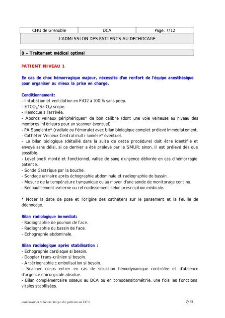
CHU de Gr<strong>en</strong>oble DCA Page: 7/12L’ADMISSION DES PATIENTS AU DECHOCAGE8 – Traitem<strong>en</strong>t médical optimalPATIENT NIVEAU 1En cas de choc hémorragique majeur, nécessite d’un r<strong>en</strong>fort de l’équipe anesthésiquepour organiser <strong>au</strong> mieux la <strong>prise</strong> <strong>en</strong> <strong>charge</strong>.Conditionnem<strong>en</strong>t:- Intubation <strong>et</strong> v<strong>en</strong>tilation <strong>en</strong> FiO2 à 100 % sans peep.- ETCO 2 /Sa O 2 /scope.- Hémocue à l’arrivée.- Abords veineux périphériques* de bon calibre (dont une voie veineuse <strong>au</strong> nive<strong>au</strong> <strong>des</strong>membres inférieurs pour un scanner év<strong>en</strong>tuel).- PA Sanglante* (radiale ou fémorale) avec bilan biologique compl<strong>et</strong> prélevé immédiatem<strong>en</strong>t.- Cathéter Veineux C<strong>en</strong>tral multi-lumiére* év<strong>en</strong>tuel.- Le bilan biologique (détaillé dans la suite de c<strong>et</strong>te procédure) doit être id<strong>en</strong>tifié <strong>et</strong><strong>en</strong>voyé sans délai, si ce dernier a été prélevé par le SMUR; sinon, il est prélevé dès quepossible.- Level one® monté <strong>et</strong> fonctionnel, valise de sang d’urg<strong>en</strong>ce délivrée <strong>en</strong> cas d’hémorragiepat<strong>en</strong>te.- Sonde Gastrique par la bouche.- Sondage urinaire après échographie abdominale <strong>et</strong> radiographie de bassin.- Mesure de la température tympanique ou <strong>au</strong> moy<strong>en</strong> d’une sonde de monitorage continu.- Réch<strong>au</strong>ffem<strong>en</strong>t externe ou refroidissem<strong>en</strong>t selon prescription médicale.* Noter la date de pose <strong>et</strong> l’origine <strong>des</strong> cathéters sur le pansem<strong>en</strong>t <strong>et</strong> la feuille dedéchocage.Bilan radiologique immédiat:- Radiographie de poumon de face.- Radiographie du bassin de face.- Echographie abdominale.Bilan radiologique après stabilisation :- Echographie cardiaque si besoin.- Doppler trans-crâni<strong>en</strong> si besoin.- Artériographie ± embolisation si besoin.- Scanner corps <strong>en</strong>tier <strong>en</strong> cas de situation hémodynamique contrôlée <strong>et</strong> d’abs<strong>en</strong>ced’urg<strong>en</strong>ce chirurgicale absolue.- Bilan complém<strong>en</strong>taire osseux <strong>au</strong> DCA ou <strong>en</strong> tomod<strong>en</strong>sitométrie, une fois les fonctionsvitales stabilisées.Admission <strong>et</strong> <strong>prise</strong> <strong>en</strong> <strong>charge</strong> <strong>des</strong> pati<strong>en</strong>ts <strong>au</strong> DCA 7/13


