Card per test Mononucleosi (Sangue intero/Siero ... - Intermedical.it
Card per test Mononucleosi (Sangue intero/Siero ... - Intermedical.it
Card per test Mononucleosi (Sangue intero/Siero ... - Intermedical.it

Create successful ePaper yourself
Turn your PDF publications into a flip-book with our unique Google optimized e-Paper software.
<strong>Card</strong> <strong>per</strong> <strong>test</strong><br />
<strong>Mononucleosi</strong> (<strong>Sangue</strong> <strong>intero</strong>/<strong>Siero</strong>/Plasma)<br />
Metodica<br />
C-35 Italiano<br />
Test rapido <strong>per</strong> la determinazione qual<strong>it</strong>ativa degli anticorpi eterofili anti<br />
<strong>Mononucleosi</strong> Infettiva (IM) nel sangue <strong>intero</strong>, nel siero o nel plasma<br />
umano.<br />
Solo <strong>per</strong> uso diagnostico professionale in v<strong>it</strong>ro.<br />
USO PREVISTO<br />
La card MONO <strong>per</strong> il <strong>test</strong> della <strong>Mononucleosi</strong> Infettiva è un <strong>test</strong> rapido<br />
immunocromatografico <strong>per</strong> la determinazione qual<strong>it</strong>ativa degli anticorpi<br />
eterofili anti <strong>Mononucleosi</strong> Infettiva, da usarsi quale ausilio <strong>per</strong> la<br />
diagnosi dell’infezione da IM.<br />
RIEPILOGO<br />
La <strong>Mononucleosi</strong> Infettiva (IM) è causata dal virus di Epstein -<br />
Barr,appartenente alla famiglia degli Herpes Virus.Sintomi della IM sono<br />
febbre,mal di gola e ingrossamento delle ghiandole linfatiche. Solo in casi rari<br />
si possono verificare problemi cardiaci o al sistema nervoso centrale. La<br />
diagnosi di IM si basa sulla presenza degli anticorpi eterofili. Gli anticorpi<br />
eterofili della <strong>Mononucleosi</strong> Infettiva derivano da quelli di classe IgM. Sono<br />
presenti nell’80-90% dei casi di IM in fase acuta e possono essere rilevati nel<br />
60-70% dei pazienti durante la prima settimana di sintomi clinici 1,2,3,4.<br />
La card <strong>per</strong> il <strong>test</strong> MONO è un <strong>test</strong> semplice che utilizza un estratto di<br />
er<strong>it</strong>roc<strong>it</strong>i bovini <strong>per</strong> la determinazione selettiva qual<strong>it</strong>ativa degli anticorpi<br />
esterofili anti <strong>Mononucleosi</strong> Infettiva nel sangue <strong>intero</strong>,siero o plasma in<br />
alcuni minuti.<br />
PRINCIPIO<br />
La card <strong>per</strong> il <strong>test</strong> MONO (<strong>Sangue</strong> <strong>intero</strong>/<strong>Siero</strong>/Plasma) è un <strong>test</strong><br />
immunologico a flusso laterale <strong>per</strong> la determinazione qual<strong>it</strong>ativa degli anticorpi<br />
eterofili <strong>Mononucleosi</strong> Infettiva nel sangue <strong>intero</strong>, nel siero o nel plasma. In<br />
questo <strong>test</strong>, l’antigene estratto da er<strong>it</strong>roc<strong>it</strong>i bovini è legato sulla card nella zona<br />
reattiva del <strong>test</strong>. Durante l’esecuzione del <strong>test</strong> il campione reagisce con<br />
l’antigene estratto da er<strong>it</strong>roc<strong>it</strong>i bovini legato alle particelle che sono state<br />
applicate alla membrana assorbente. Questa miscela migra<br />
cromatograficamente lungo la membrana e reagisce con l’antigene estratto da<br />
er<strong>it</strong>roc<strong>it</strong>i bovini immobilizzato. Se il campione contiene anticorpi esterofili anti<br />
IM, nella zona reattiva del <strong>test</strong> comparirà una banda colorata, indice di un<br />
risultato pos<strong>it</strong>ivo. Se invece il campione non contiene anticorpi eterofili anti<br />
IM, in questa zona non comparirà alcuna banda colorata, indice di un risultato<br />
negativo. Come controllo procedurale, comparirà sempre una banda colorata<br />
nella zona di controllo a indicare che il <strong>test</strong> è stato esegu<strong>it</strong>o correttamente. Se la<br />
banda colorata non compare, il risultato del <strong>test</strong> non è valido.<br />
REAGENTI<br />
La card contiene antigene estratto da er<strong>it</strong>roc<strong>it</strong>i bovini legato alle particelle e<br />
antigene estratto da er<strong>it</strong>roc<strong>it</strong>i bovini legato alla membrana.<br />
PRECAUZIONI<br />
Solo <strong>per</strong> uso diagnostico professionale in v<strong>it</strong>ro. Non utilizzare dopo la data<br />
di scadenza.<br />
Conservare la card nell’involucro chiuso fino al momento dell’uso.<br />
Non mangiare, bere o fumare nell’area in cui vengono manipolati i<br />
campioni o i k<strong>it</strong>.<br />
Non utilizzare il <strong>test</strong> se il sacchetto è danneggiato.<br />
Manipolare tutti i campioni come potenzialmente infettivi. Osservare le<br />
adeguate precauzioni contro i rischi microbiologici in tutte le fasi d’analisi<br />
e seguire le procedure standard <strong>per</strong> il corretto smaltimento dei campioni.<br />
Il plasma umano utilizzato <strong>per</strong> i controlli pos<strong>it</strong>ivo e negativo è stato <strong>test</strong>ato<br />
con metodo ELISA <strong>per</strong> la presenza di anticorpi <strong>per</strong> HIV 1&2,<strong>per</strong> l’antigene<br />
HbsAg e <strong>per</strong> gli anticorpi anti-HCV con es<strong>it</strong>o negativo.Ciò non toglie la<br />
necess<strong>it</strong>à di assumere precauzioni nel maneggiare ed eliminare questo<br />
materiale.<br />
Durante l’analisi dei campioni indossare abbigliamento protettivo: camice da<br />
laboratorio, guanti monouso e protezione <strong>per</strong> gli occhi.<br />
Umid<strong>it</strong>à e tem<strong>per</strong>atura possono influire negativamente sui risultati.<br />
CONSERVAZIONE E STABILITÀ<br />
Conservare nel sacchetto sigillato a tem<strong>per</strong>atura ambiente o in frigorifero<br />
(2-30°C). La card è stabile sino alla data di scadenza indicata sul sacchetto<br />
sigillato. Conservare la card nel sacchetto sigillato sino al momento<br />
dell’utilizzo. NON CONGELARE. Non utilizzare dopo la data di scadenza.<br />
PRELIEVO E PREPARAZIONE DEI CAMPIONI<br />
La card <strong>per</strong> il <strong>test</strong> MONO (<strong>Sangue</strong> <strong>intero</strong>/<strong>Siero</strong>/Plasma) può essere<br />
utilizzata con sangue <strong>intero</strong>,siero o plasma.<br />
Separare sub<strong>it</strong>o il siero o il plasma dal sangue <strong>per</strong> ev<strong>it</strong>are l’emolisi.<br />
Utilizzare solo campioni trasparenti, non emolizzati.<br />
Effettuare il <strong>test</strong> sub<strong>it</strong>o dopo aver prelevato i campioni. Non lasciare i<br />
campioni a tem<strong>per</strong>atura ambiente <strong>per</strong> lunghi <strong>per</strong>iodi. I campioni possono<br />
essere conservati a una tem<strong>per</strong>atura di 2-8°C <strong>per</strong> 3 giorni. Per <strong>per</strong>iodi di<br />
conservazione più lunghi, congelare i campioni a una tem<strong>per</strong>atura inferiore<br />
a -20°C.<br />
Portare i campioni a tem<strong>per</strong>atura ambiente prima dell’analisi. Lasciar<br />
scongelare completamente i campioni congelati e miscelarli accuratamente<br />
prima dell’analisi. Non congelare e scongelare i campioni più volte.<br />
In caso di spedizione dei campioni, imballarli secondo le vigenti norme<br />
locali <strong>per</strong> la spedizione di agenti eziologici.<br />
<strong>Card</strong><br />
Tampone<br />
Contagocce<br />
COMPOSIZIONE DELLA CONFEZIONE<br />
Controllo<br />
negativo(plasma<br />
Materiale Forn<strong>it</strong>o<br />
umano dilu<strong>it</strong>o,0,09%<br />
sodio azide)<br />
Controllo pos<strong>it</strong>ivo(plasma<br />
umano dil. contenente<br />
anticorpi esterofili<br />
dil.,0,09% sodio azide)<br />
Conten<strong>it</strong>ori <strong>per</strong> la raccolta<br />
campioni<br />
Materiale Necessario Ma Non Forn<strong>it</strong>o<br />
Centrifuga<br />
Contag.<br />
PROCEDURA<br />
Timer<br />
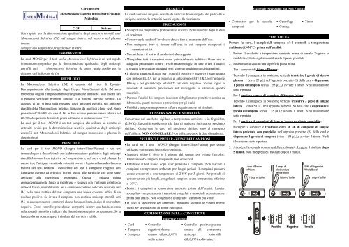
Portare la card, i campioni,il tampone e/o i controlli a tem<strong>per</strong>atura<br />
ambiente (15-30°C) prima dell’analisi.<br />
1. Portare il sacchetto a tem<strong>per</strong>atura ambiente prima di aprirlo. Togliere la<br />
card dal sacchetto sigillato e utilizzarla il prima possibile.<br />
2. Posizionare la card su una su<strong>per</strong>ficie piana pul<strong>it</strong>a.<br />
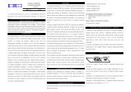
Per i campioni di <strong>Siero</strong> o Plasma:<br />
Tenendo il contagocce in posizione verticale trasferire 1 goccia di siero o<br />
plasma (circa 25 L) nell’appos<strong>it</strong>o pozzetto (S) della card e dispensare<br />
1 goccia di tampone (circa 55 L),e avviare il timer. Vedi illustrazione<br />
sotto riportata.<br />
Per il prelievo venoso di campioni di <strong>Sangue</strong> Intero:<br />
Tenendo il contagocce in posizione verticale trasferire 2 gocce di sangue<br />
<strong>intero</strong> (circa 50 L) nell’appos<strong>it</strong>o pozzetto (S) della card e dispensare 1<br />
goccia di tampone (circa 55 L),e avviare il timer. Vedi illustrazione<br />
sotto riportata.<br />
Per il prelievo di campioni di <strong>Sangue</strong> Intero mediante pungid<strong>it</strong>o:<br />
Riempire il capillare e trasferire circa 50 L di campione di sangue<br />
<strong>intero</strong> prelevato con pungid<strong>it</strong>o nell’appos<strong>it</strong>o pozzetto (S) della card e<br />
dispensare 1 goccia di tampone (circa 55 L),e avviare il timer. Vedi<br />
illustrazione sotto riportata.<br />
3. Attendere l’eventuale comparsa della/e colorata/e. Leggere il risultato dopo<br />
5 minuti. Non interpretare il risultato dopo 10 minuti.
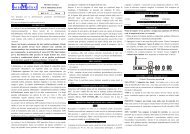
INTERPRETAZIONE DEI RISULTATI<br />
(Vedere illustrazione precedente)<br />
POSITIVO:* Compaiono due bande colorate distinte, una nella zona di<br />
controllo (C), l’altra nella zona reattiva (T).<br />
*NOTA: L’intens<strong>it</strong>à del colore nella zona reattiva (T) varia a seconda della<br />
concentrazione degli anticorpi esterofili della IM presenti nel campione.<br />
Ogni sfumatura di colore nella zona reattiva (T) deve quindi essere r<strong>it</strong>enuta<br />
indice di risultato pos<strong>it</strong>ivo.<br />
NEGATIVO: Compare una banda colorata nella zona di controllo (C).<br />
Non si nota alcuna banda colorata nella zona reattiva (T).<br />
NON VALIDO: Non compare alcuna banda nella zona di controllo. Le<br />
ragioni più probabili della mancata comparsa della banda di controllo sono<br />
un volume insufficiente di campione o una non corretta esecuzione della<br />
procedura. Rivedere la procedura e ripetere il <strong>test</strong> con una nuova card. Se il<br />
problema <strong>per</strong>siste, non utilizzare più il k<strong>it</strong> e contattare il distributore locale.<br />
CONTROLLO DI QUALITÀ<br />
Ogni <strong>test</strong> è forn<strong>it</strong>o di controllo interno della procedura. La banda rossa che<br />
compare nella zona di controllo (C) è un controllo interno della procedura e<br />
conferma il prelievo di un volume di campione sufficiente e la correttezza<br />
della procedura.<br />
Con il k<strong>it</strong> vengono forn<strong>it</strong>i standard di controllo; si raccomanda di analizzarli<br />
e riscontrarne la corrispondenza ai dati attesi quale corretta pratica di<br />
laboratorio atta a confermare la procedura anal<strong>it</strong>ica e verificare la corretta<br />
esecuzione del <strong>test</strong>.<br />
LIMITI<br />
1. La card <strong>per</strong> il <strong>test</strong> MONO (<strong>Sangue</strong> Intero/<strong>Siero</strong>/Plasma) è esclusivamente<br />
<strong>per</strong> uso diagnostico in v<strong>it</strong>ro. Il <strong>test</strong> deve essere impiegato <strong>per</strong> la<br />
determinazione degli anticorpi anti-IM solo in campioni di sangue <strong>intero</strong>,<br />
siero o plasma. Questo <strong>test</strong> qual<strong>it</strong>ativo non è in grado di determinare né il<br />
valore quant<strong>it</strong>ativo né la <strong>per</strong>centuale di aumento della concentrazione di<br />
anticorpi anti-IM.<br />
2. La card <strong>per</strong> il <strong>test</strong> MONO (<strong>Sangue</strong> Intero/<strong>Siero</strong>/Plasma) indica<br />
unicamente la presenza di anticorpi anti-IM nel campione e non deve<br />
essere usata come unico cr<strong>it</strong>erio di diagnosi dell’infezione da<br />
<strong>Mononucleosi</strong> Infettiva.<br />
3. Come <strong>per</strong> tutti i <strong>test</strong> diagnostici, i risultati devono essere interpretati<br />
alla luce del quadro clinico complessivo del paziente.<br />
4. Se il <strong>test</strong> risulta negativo e i sintomi clinici <strong>per</strong>sistono, si raccomanda di<br />
eseguire altri <strong>test</strong> clinici. Un risultato negativo non esclude comunque la<br />
possibil<strong>it</strong>à di infezione da IM.<br />
VALORI ATTESI<br />
Un’infezione da Virus di Epstein-Barr (EBV) produce durante l’adolescenza<br />
o nei giovani adulti una IM nel 35-50% dei casi riportati 1,5 .<br />
Non ci sono riscontri di stagional<strong>it</strong>à nell’infezione da EBV. Il <strong>per</strong>iodo di<br />
incubazione è tra i 10 e i 60 giorni,mentre è tra i 7 e i 14 giorni in bambini e<br />
adolescenti.<br />
PERFORMANCE<br />
Sensibil<strong>it</strong>à<br />
La card <strong>per</strong> il <strong>test</strong> MONO (<strong>Sangue</strong> Intero/<strong>Siero</strong>/Plasma) è stata valutata con<br />
campioni confermati pos<strong>it</strong>ivi o negativi con un <strong>test</strong> disponibile in commercio<br />
in vetrino <strong>per</strong> agglutinazione.I risultati mostrano che la sensibil<strong>it</strong>à della card<br />
MONO è > 99.9% comparata con il <strong>test</strong> in vetrino <strong>per</strong> agglutinazione.<br />
Specific<strong>it</strong>à<br />
La card <strong>per</strong> il <strong>test</strong> MONO (<strong>Sangue</strong> Intero/<strong>Siero</strong>/Plasma) utilizza un antigene<br />
altamente specifico <strong>per</strong> gli anticorpi anti IM nel sangue <strong>intero</strong>,siero o<br />
plasma. I risultati mostrano che la specific<strong>it</strong>à della card MONO (<strong>Sangue</strong><br />
Intero/<strong>Siero</strong>/Plasma) è 98.6% comparata con il <strong>test</strong> in vetrino <strong>per</strong><br />
agglutinazione.<br />
MONO<br />
Test <strong>Card</strong><br />
<strong>Card</strong> <strong>per</strong> <strong>test</strong> MONO vs. Vetrino <strong>per</strong> agglutinazione<br />
Metodo<br />
Vetrino <strong>per</strong><br />
agglutinazione<br />
Risultati Pos<strong>it</strong>ivi Negativi<br />
Risultati<br />
Totali<br />
Pos<strong>it</strong>ivi 52 1 53<br />
Negativi 0 69 69<br />
Totale risultati 52 70 122<br />
Sensibil<strong>it</strong>à Relativa:> 99,9% (93,2%-100,0%)* Specific<strong>it</strong>à Relativa: 98,6%<br />
(92,3%-100,0%)*<br />
Accuratezza relativa: 99,2% (95,5%-100,0%)* * Intervallo<br />
di confidenza 95%<br />
Intra-Seduta<br />
Precisione<br />
La precisione intra-seduta è stata determinata analizzando 3 replicati di tre<br />
campioni: uno negativo, uno basso pos<strong>it</strong>ivo e uno alto pos<strong>it</strong>ivo. I valori dei<br />
campioni negativo, basso pos<strong>it</strong>ivo e alto pos<strong>it</strong>ivo sono stati identificati<br />
correttamente in oltre il 99% dei casi.<br />
Inter-Seduta<br />
La precisione inter-seduta è stata determinata tram<strong>it</strong>e 10 analisi indipendenti<br />
sugli stessi tre campioni: uno negativo, uno basso pos<strong>it</strong>ivo e uno alto<br />
pos<strong>it</strong>ivo. Sono stati analizzati tre diversi lotti della card <strong>per</strong> il <strong>test</strong> MONO<br />
(<strong>Sangue</strong> Intero/<strong>Siero</strong>/Plasma) con campioni negativi, basso pos<strong>it</strong>ivi e alto<br />
pos<strong>it</strong>ivi. I campioni sono stati identificati correttamente in oltre il 99% dei<br />
casi.<br />
Reattiv<strong>it</strong>à Crociata<br />
Campioni pos<strong>it</strong>ivi <strong>per</strong> RF,HbsAg,HbeAg,HbcAb,HCV,TB e verso la sifilide<br />
sono stati <strong>test</strong>ati con il <strong>test</strong> rapido su card MONO. Non sono state osservate<br />
reattiv<strong>it</strong>à crociate che stanno a indicare un elevato grado di specific<strong>it</strong>à del<br />
<strong>test</strong> rapido su card <strong>per</strong> MONO<br />
BIBLIOGRAFIA<br />
1. Hickey SM, Strasburger VC.What every Pediatrician Should know<br />
About infectious <strong>Mononucleosi</strong>s In Adolescents.Pediatr Clin North<br />
Am.1997;44(6):1541-56<br />
2. Omori M. <strong>Mononucleosi</strong>s.2002<br />
http:/7www.emedicine.com/EMERg/topic309htm.<br />
3. Linde A. Diagnosis of Epstein-Barr virus- related diseas.Scand J Infect<br />
Dis Suppl 1996;100:<br />
83-8.<br />
4 Papesch M., Watkins R. Epstein-Barr virus infectious<br />
mononucleosis.Clin. Otolaryngol. 2001;<br />
26(1):3-8.<br />
5. CDC National Center for Infectious Diseas<br />
EBV&IM:http://www.cdc.gov./ncidod/diseas/<br />
ebv.htm.<br />
Attenzione,<br />
consultare le<br />
istruzioni <strong>per</strong> l’uso<br />
Solo <strong>per</strong> uso<br />
diagnostico in<br />
v<strong>it</strong>ro<br />
Conservare a 2-<br />
30°C<br />
Fabbricante<br />
Indice dei simboli<br />
N°determinaz<br />
ioni <strong>per</strong> k<strong>it</strong><br />
Usare entro<br />
Numero di<br />
lotto<br />
INTERMEDICAL s.r.l.<br />
Via A.Genovesi,13<br />
80010 Villaricca(Na)-ITALY<br />
Fabbricante<br />
Non<br />
riutilizzare<br />
REF Codice #<br />
Data: 2008-10
MONO<br />
<strong>Mononucleosi</strong>s Rapid Test Device<br />
(Whole blood/Serum/Plasma)<br />
Package Insert<br />
C-35 Italiano<br />
A rapid <strong>test</strong> for the diagnosis of Infectious <strong>Mononucleosi</strong>s (IM) to detect<br />
infectious mononucleosis heterophile antibodies qual<strong>it</strong>atively in whole<br />
blood, serum or plasma.<br />
For professional in v<strong>it</strong>ro diagnostic use only.<br />
INTENDED USE<br />
The MONO <strong>Mononucleosi</strong>s Rapid Test Device (Whole<br />
Blood/Serum/Plasma) is a rapid chromatographic immunoassay for the<br />
qual<strong>it</strong>ative detection of Infectious <strong>Mononucleosi</strong>s heterophile antibodies in<br />
whole blood, serum or plasma as an aid in the diagnosis of Infectious<br />
<strong>Mononucleosi</strong>s.<br />
SUMMARY<br />
Infectious <strong>Mononucleosi</strong>s (IM) is caused by the Epstein-Barr virus, which is<br />
a member of the herpesvirus family. Symptoms of IM are fever, sore throat<br />
and swollen lymph glands. In very rare cases, heart or central nervous<br />
system problems may occur. Diagnosis of IM is made based on the presence<br />
of heterophile antibodies. Infectious <strong>Mononucleosi</strong>s heterophile antibodies<br />
belong to the IgM class. They are present in 80-90% of acute IM cases and<br />
can be detected in 60-70% of patients during the first week of clinical<br />
illness.1,2,3,4<br />
The MONO <strong>Mononucleosi</strong>s Rapid Test Device (Whole Blood/Serum/<br />
Plasma) is a simple <strong>test</strong> that utilizes an extract of bovine erythrocytes to<br />
qual<strong>it</strong>atively and selectively detect Infectious <strong>Mononucleosi</strong>s heterophile<br />
antibodies in whole blood, serum or plasma in just minutes.<br />
PRINCIPLE<br />
The MONO <strong>Mononucleosi</strong>s Rapid Test Device (Whole<br />
Blood/Serum/Plasma) is a qual<strong>it</strong>ative membrane strip based immunoassay<br />
for the detection of IM heterophile antibodies in whole blood, serum or<br />
plasma. In this <strong>test</strong> procedure, bovine erythrocyte extracted antigen is<br />
immobilized in the <strong>test</strong> line region of the device. The specimen reacts w<strong>it</strong>h<br />
bovine erythrocyte extracted antigen coated particles that have been applied<br />
to the label pad. This mixture migrates chromatographically along the length<br />
of the <strong>test</strong> strip and interacts w<strong>it</strong>h the immobilized bovine erythrocyte<br />
extracted antigen. If the specimen contains IM heterophile antibodies, a<br />
colored line will appear in the <strong>test</strong> line region indicating a pos<strong>it</strong>ive result. If<br />
the specimen does not contain IM heterophile antibodies, a colored line will<br />
not appear in this region indicating a negative result. To serve as a<br />
procedural control, a colored line will always appear in the control line<br />
region, indicating that the pro<strong>per</strong> volume of specimen has been added and<br />
membrane wicking has occurred.<br />
REAGENTS<br />
The <strong>test</strong> device contains bovine erythrocyte extracted antigen-coated<br />
particles and bovine erythrocyte extracted antigen-coated membrane.<br />
PRECAUTINS<br />
For professional in v<strong>it</strong>ro diagnostic use only. Do not use after expiration<br />
date.<br />
Do not eat, drink or smoke in the area where the specimens or k<strong>it</strong>s are<br />
handled.<br />
Do not use <strong>test</strong> if pouch is damaged<br />
Handle all specimens as if they contain infectious agents. Observe<br />
established precautions against microbiological hazards throughout <strong>test</strong>ing<br />
and follow standard procedures for pro<strong>per</strong> disposal of specimens.<br />
Wear protective clothing such as laboratory coats, disposable gloves and eye<br />
protection when specimens are being <strong>test</strong>ed.<br />
The used <strong>test</strong> should be discarded according to local regulations.<br />
Humid<strong>it</strong>y and tem<strong>per</strong>ature can adversely affect results.<br />
STORAGE AND STABILITY<br />
Store as packaged in the sealed pouch at room tem<strong>per</strong>ature or refrigerated<br />
(2-30°C). The <strong>test</strong> device is stable through the expiration date printed on the<br />
sealed pouch. The <strong>test</strong> device must remain in the sealed pouch until use. DO<br />
NOT FREEZE. Do not use beyond the expiration date.<br />
SPECIMEN COLLECTION AND PREPARATION<br />
The MONO <strong>Mononucleosi</strong>s Rapid Test Device (Whole Blood/<br />
Serum/Plasma) can be <strong>per</strong>formed using whole blood (from venipuncture or<br />
fingerstick), serum or plasma.<br />
To collect Venipuncture Whole Blood specimens: Collect anti-coagulated<br />
blood specimen (sodium or l<strong>it</strong>hium heparin, potassium or sodium EDTA,<br />
sodium oxalate, sodium c<strong>it</strong>rate) following standard laboratory procedures.<br />
To collect Fingerstick Whole Blood specimens:<br />
Wash the patient’s hand w<strong>it</strong>h soap and warm water or clean w<strong>it</strong>h an alcohol<br />
swab. Allow to dry.<br />
Massage the hand w<strong>it</strong>hout touching the puncture s<strong>it</strong>e by rubbing down the<br />
hand towards the fingertip of the middle or ring finger.<br />
Puncture the skin w<strong>it</strong>h a sterile lancet. Wipe away the first sign of blood.<br />
Gently rub the hand from wrist to palm to finger to form a rounded drop of<br />
blood over the puncture s<strong>it</strong>e.<br />
Add the Fingerstick Whole Blood specimen to the <strong>test</strong> device by using a<br />
capillary tube:<br />
Touch the end of the capillary tube to the blood until filled to approximately<br />
50 µL. Avoid air bubbles.<br />
Place the bulb onto the top end of the capillary tube, then squeeze the bulb to<br />
dispense the whole blood to the specimen well (S) of the <strong>test</strong> device.<br />
Separate serum or plasma from blood as soon as possible to avoid<br />
hemolysis. Use only clear, non-hemolyzed specimens.<br />
Testing should be <strong>per</strong>formed immediately after specimen collection. Do not<br />
leave the specimens at room tem<strong>per</strong>ature for prolonged <strong>per</strong>iods. Serum and<br />
plasma specimens may be stored at 2-8°C for up to 3 days. For long-term<br />
storage, specimens should be kept below -20°C. Whole blood collected by<br />
venipuncture should be stored at 2-8°C if the <strong>test</strong> is to be run w<strong>it</strong>hin 2 days<br />
of collection. Do not freeze whole blood specimens. Whole blood collected<br />
by fingerstick should be <strong>test</strong>ed immediately.<br />
Bring specimens to room tem<strong>per</strong>ature prior to <strong>test</strong>ing. Frozen specimens<br />
must be completely thawed and mixed well prior to <strong>test</strong>ing. Specimens<br />
should not be frozen and thawed repeatedly.<br />
If specimens are to be shipped, they should be packed in compliance w<strong>it</strong>h<br />
local regulations covering the transportation of etiologic agents.
Materials Provided<br />
MATERIALS<br />
Test devices Buffer Drop<strong>per</strong>s<br />
Negative control (Diluted human plasma, 0.09% Package insert<br />
sodium azide)<br />
Pos<strong>it</strong>ive control (Diluted human plasma containing IM heterophile<br />
antibodies, 0.09% NaN3)<br />
Workstation<br />
Materials Required But Not Provided<br />
Specimen collection containers (for venipuncture Timer<br />
whole blood)<br />
Lancet (for fingerstick whole blood only) Centrifuge<br />
Heparinized capillary tubes and dispensing bulb (for fingerstick whole blood<br />
only)<br />
DIRECTIONS FOR USE<br />
Allow the <strong>test</strong> device, specimen, buffer, and/or controls to equilibrate to<br />
room tem<strong>per</strong>ature (15-30°C) prior to <strong>test</strong>ing.<br />
Remove the <strong>test</strong> device from the foil pouch and use <strong>it</strong> as soon as possible.<br />
Best results will be obtained if the assay is <strong>per</strong>formed w<strong>it</strong>hin one hour.<br />
Place the <strong>test</strong> device on a clean and level surface.<br />
For Serum or Plasma specimens:<br />
Hold the drop<strong>per</strong> vertically and transfer 1 drop of serum or plasma<br />
(approximately 25 L) to the specimen well (S) of the <strong>test</strong> device, and add 1<br />
drop of buffer (approximately 55 L), then start the timer. See illustration<br />
below.<br />
For Venipuncture Whole Blood specimens:<br />
Hold the drop<strong>per</strong> vertically and transfer 2 drops of whole blood<br />
(approximately 50 L) to the specimen well (S) of the <strong>test</strong> device, and add 1<br />
drop of buffer (approximately 55 L), then start the timer. See illustration<br />
below.<br />
For Fingerstick Whole Blood specimens:<br />
To use a capillary tube: Fill the capillary tube and transfer approximately 50<br />
L of fingerstick whole blood specimen to the specimen well (S) of the <strong>test</strong><br />
device, then add 1 drop of buffer (approximately 55 L) and start the timer.<br />
See illustration below.<br />
Wa<strong>it</strong> for the colored line(s) to appear. The result should be read at 5 minutes.<br />
Do not interpret the result after 10 minutes.<br />
(Please refer to the illustration)<br />
INTERPRETATION OF RESULTS<br />
POSITIVE: * Two distinct colored lines appear. One line should be in the<br />
control line region (C) and another line should be in the <strong>test</strong> line region (T).<br />
*NOTE: The intens<strong>it</strong>y of the color in the <strong>test</strong> line region (T) will vary<br />
depending on the concentration of IM heterophile antibodies present in the<br />
specimen. Therefore, any shade of color in the <strong>test</strong> line region (T) should be<br />
considered pos<strong>it</strong>ive.<br />
NEGATIVE: One colored line appears in the control line region (C). No<br />
apparent colored line appears in the <strong>test</strong> line region (T).<br />
INVALID: Control line fails to appear. Insufficient specimen volume or<br />
incorrect procedural techniques are the most likely reasons for control line<br />
failure. Review the procedure and repeat the <strong>test</strong> w<strong>it</strong>h a new <strong>test</strong> device. If<br />
the problem <strong>per</strong>sists, discontinue using the <strong>test</strong> k<strong>it</strong> immediately and contact<br />
your local distributor.<br />
QUALITY CONTROL<br />
A procedural control is included in the <strong>test</strong>. A colored line appearing in the<br />
control line region (C) is considered an internal procedural control. It<br />
confirms sufficient specimen volume, adequate membrane wicking and<br />
correct procedural technique.<br />
In add<strong>it</strong>ion to your laboratory’s standard qual<strong>it</strong>y control procedures, <strong>it</strong> is<br />
recommended that a pos<strong>it</strong>ive and negative external control be <strong>test</strong>ed at least<br />
once w<strong>it</strong>hin each <strong>test</strong> k<strong>it</strong> and by each o<strong>per</strong>ator <strong>per</strong>forming <strong>test</strong>ing w<strong>it</strong>hin a<br />
k<strong>it</strong>. This will verify that the reagents and <strong>test</strong> are working pro<strong>per</strong>ly and the<br />
o<strong>per</strong>ator is able to correctly <strong>per</strong>form the <strong>test</strong> procedure. External pos<strong>it</strong>ive and<br />
negative controls are supplied in the k<strong>it</strong>.<br />
Procedure for External Qual<strong>it</strong>y Control Testing<br />
1. Holding the bottle vertically, add 1 full drop (approximately 40<br />
L) of pos<strong>it</strong>ive or negative control solution to the specimen well (S) of the<br />
<strong>test</strong> device, and add 1 drop of buffer (approximately 55 L).<br />
2. Continue w<strong>it</strong>h Step 3 of Directions For Use.<br />
3. If the controls do not yield the expected results, do not use the<br />
<strong>test</strong> results. Repeat the <strong>test</strong> or contact your distributor.<br />
LIMITATIONS<br />
The MONO <strong>Mononucleosi</strong>s Rapid Test Device (Whole<br />
Blood/Serum/Plasma) is for in v<strong>it</strong>ro diagnostic use only. The <strong>test</strong> should be<br />
used for the detection of Infectious <strong>Mononucleosi</strong>s antibodies in whole<br />
blood, serum or plasma specimens only. Ne<strong>it</strong>her the quant<strong>it</strong>ative value nor<br />
the rate of increase in Infectious <strong>Mononucleosi</strong>s antibody concentration can<br />
be determined by this qual<strong>it</strong>ative <strong>test</strong>.<br />
The MONO <strong>Mononucleosi</strong>s Rapid Test Device (Whole<br />
Blood/Serum/Plasma) will only indicate the presence of infectious<br />
mononucleosis antibodies in the specimen and should not be used as the sole<br />
cr<strong>it</strong>eria for the diagnosis of Infectious <strong>Mononucleosi</strong>s infection.<br />
As w<strong>it</strong>h all diagnostic <strong>test</strong>s, all results must be interpreted together w<strong>it</strong>h<br />
other clinical information available to the physician.<br />
If the <strong>test</strong> result is negative and clinical symptoms <strong>per</strong>sist, add<strong>it</strong>ional <strong>test</strong>ing<br />
using other clinical methods is recommended. A negative result does not at<br />
any time preclude the possibil<strong>it</strong>y of Infectious <strong>Mononucleosi</strong>s infection.<br />
EXPECTED VALUES<br />
Epstein-Barr virus infection during adolescence or young adulthood causes<br />
Infectious <strong>Mononucleosi</strong>s in 35% to 50% of reported cases.1, 5<br />
The incidence of EBV-associated Infectious <strong>Mononucleosi</strong>s in the USA has<br />
been estimated at 45 <strong>per</strong> 100,000 and is highest in adolescent and young<br />
adults-about 2 out of 1,000. No seasonal pattern of EBV infection exists.<br />
The incubation <strong>per</strong>iod is 10 to 60 days, though 7 to 14 days is common for<br />
children and adolescents.
Sens<strong>it</strong>iv<strong>it</strong>y<br />
PERFORMANCE CHARACTERISTICS<br />
The MONO <strong>Mononucleosi</strong>s Rapid Test Device (Whole Blood/Serum/<br />
Plasma) has been evaluated w<strong>it</strong>h specimens confirmed by a leading<br />
commercial latex agglutination <strong>test</strong>. The latex agglutination <strong>test</strong> served as<br />
the reference method for the MONO <strong>Mononucleosi</strong>s Rapid Test Device<br />
(Whole Blood/Serum/Plasma). The result shows that the sens<strong>it</strong>iv<strong>it</strong>y of the<br />
MONO <strong>Mononucleosi</strong>s Rapid Test Device (Whole Blood/Serum/Plasma) is<br />
>99.9% relative to the latex agglutination <strong>test</strong>.<br />
Specific<strong>it</strong>y<br />
The MONO <strong>Mononucleosi</strong>s Rapid Test Device (Whole<br />
Blood/Serum/Plasma) uses an antigen that is highly specific for IM<br />
antibodies in whole blood, serum or plasma. The results show that the<br />
specific<strong>it</strong>y of the MONO <strong>Mononucleosi</strong>s Rapid Test Device (Whole<br />
Blood/Serum/ Plasma) is 98.6% relative to the latex agglutination <strong>test</strong>.<br />
MONO Rapid Test Device vs. Latex Agglutination<br />
Method Latex Agglutination<br />
MONO<br />
Test Device<br />
Results Pos<strong>it</strong>ive Negative<br />
Pos<strong>it</strong>ive 52 1 53<br />
Negative 0 69 69<br />
Total<br />
Results<br />
Total Results 52 70 122<br />
Relative Sens<strong>it</strong>iv<strong>it</strong>y: >99.9% (93.2% -100.0%)* Relative Specific<strong>it</strong>y:<br />
98.6% (92.3%-100.0%)*<br />
Relative Accuracy: 99.2% (95.5%-100.0%)* * 95%<br />
Confidence Intervals<br />
Precision<br />
Intra-Assay<br />
W<strong>it</strong>hin-run precision has been determined by using 15 replicates of three<br />
specimens: a negative, a low pos<strong>it</strong>ive and a high pos<strong>it</strong>ive. The negative, low<br />
pos<strong>it</strong>ive and high pos<strong>it</strong>ive values were correctly identified >99% of the time.<br />
Inter-Assay<br />
Between-run precision has been determined by 15 independent assays on the<br />
same three specimens: a negative, a low pos<strong>it</strong>ive and a high pos<strong>it</strong>ive. Three<br />
different lots of the MONO <strong>Mononucleosi</strong>s Rapid Test Device (Whole<br />
Blood/Serum/ Plasma) have been <strong>test</strong>ed using negative, low pos<strong>it</strong>ive and<br />
high pos<strong>it</strong>ive specimens. The specimens were correctly identified >99% of<br />
the time.<br />
BIBLIOGRAPHY<br />
Hickey SM, Strasburger VC. What Every Pediatrician Should Know<br />
About Infectious <strong>Mononucleosi</strong>s In Adolescents. Pediatr Clin North<br />
Am. 1997; 44(6):1541-56.<br />
Omori M. <strong>Mononucleosi</strong>s. 2002.<br />
http://www.emedicine.com/EMERG/topic309.htm<br />
Linde A. Diagnosis of Epstein-Barr virus-related diseases. Scand J<br />
Infect Dis Suppl. 1996; 100:83-8.<br />
Papesch M, Watkins R. Epstein-Barr virus infectious mononucleosis.<br />
Clin Otolaryngol.<br />
2001; 26(1): 3-8.<br />
CDC National Center for Infectious Diseases. EBV & IM:<br />
http://www.cdc.gov/ncidod/diseases/ebv.htm<br />
Fabbricante<br />
INTERMEDICAL s.r.l.<br />
Via A.Genovesi,13<br />
80010 Villaricca(Na)-ITALY<br />
Data: 2008-10