Brochure Seminario di Primavera 2005 - Fondazione Ricerca ...
Brochure Seminario di Primavera 2005 - Fondazione Ricerca ...
Brochure Seminario di Primavera 2005 - Fondazione Ricerca ...

Create successful ePaper yourself
Turn your PDF publications into a flip-book with our unique Google optimized e-Paper software.
fondazione fondazione per la ricerca ricerca sulla fibrosi fibrosi cistica cistica - onlus onlus<br />
italian italian cystic cystic fibrosis fibrosis research research foundation foundation<br />
Ospedale Maggiore - Piazzale Stefani, 1 - 37126 VERONA<br />
Tel. 045 8073438 - Fax 045 8073568 - e-mail: fondazione.ricercafc@azosp.it<br />
www.fibrosicisticaricerca.it<br />
con la sponsorizzazione <strong>di</strong><br />
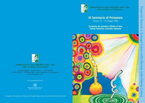
In copertina: “<strong>Ricerca</strong>trice in <strong>Primavera</strong>” <strong>di</strong> Isabella Zago, allieva Liceo Artistico Boccioni <strong>di</strong> Verona<br />
fondazione per la ricerca sulla fibrosi cistica - onlus<br />
italian cystic fibrosis research foundation<br />
III <strong>Seminario</strong> <strong>di</strong> <strong>Primavera</strong><br />
Verona 13 – 14 maggio <strong>2005</strong><br />
Screening dei portatori, Difetto <strong>di</strong> base,<br />
Cellule staminali, Controllo Infezione<br />
Progressi Recenti e Sviluppi Futuri nella <strong>Ricerca</strong> sulla Fibrosi Cistica
INDICE<br />
<strong>Fondazione</strong> per la ricerca sulla fibrosi cistica - onlus<br />
italian cystic fibrosis research foundation<br />
III <strong>Seminario</strong> <strong>di</strong> <strong>Primavera</strong><br />
Verona 13 – 14 maggio <strong>2005</strong><br />
Progressi Recenti e Sviluppi Futuri<br />
nella <strong>Ricerca</strong> sulla Fibrosi Cistica<br />
Riassunto dei contributi<br />
Presentazione Pag. 2<br />
Lidewij Henneman: Nuove strategie ed opinioni<br />
sullo screening dei portatori CF Pag. 3<br />
William Guggino: Acquisizioni sulla macchina secretiva<br />
epiteliale e prospettive <strong>di</strong> terapia del <strong>di</strong>fetto <strong>di</strong> base CF Pag. 10<br />
Guoshun Wang: Terapia della fibrosi cistica<br />
basata su cellule staminali Pag. 12<br />
Dieter Gruenert: Commento alla pubblicazione <strong>di</strong> G. Wang<br />
su cellule staminali Pag. 18<br />
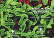
Roberto Buzzetti e Cesare Braggion: Prevenzione<br />
e controllo dell’infezione respiratoria in fibrosi cistica Pag. 22<br />
Moderatori del <strong>Seminario</strong> Pag. 27<br />
III SEMINARIO DI PRIMAVERA
2<br />
PRESENTAZIONE<br />
Lo scopo dei Seminari <strong>di</strong> <strong>Primavera</strong>, giunti quest’anno<br />
alla terza e<strong>di</strong>zione, è quello <strong>di</strong> dare evidenza<br />
ad alcuni dei problemi emergenti nel campo della<br />
fibrosi cistica, verso i quali si sta muovendo un<br />
significativo interesse <strong>di</strong> stu<strong>di</strong>o e ricerca. Viene<br />
chiesto per questo l’aiuto <strong>di</strong> persone che hanno<br />
dato <strong>di</strong> recente contributi rilevanti a queste problematiche,<br />
con l’invito a prospettare ipotesi nuove <strong>di</strong><br />
lavoro e linee possibili <strong>di</strong> sviluppo <strong>di</strong> ricerca. Il carattere<br />
<strong>di</strong> seminario include l’ampio spazio che viene<br />
dato alla comunicazione degli esperti ma anche alla<br />
<strong>di</strong>scussione che ne dovrebbe seguire.<br />
Il primo tema, affrontato dalla Dr.essa Lidewij<br />
Henneman, una stu<strong>di</strong>osa <strong>di</strong> Amsterdam attenta alle<br />
<strong>di</strong>namiche sociali e alle iniziative <strong>di</strong> prevenzione che<br />
si sviluppano intorno all’impatto delle malattie<br />
genetiche sulla popolazione, riguarda il ripensamento<br />
allo screening dei portatori del gene CFTR. I<br />
tentativi <strong>di</strong> offrire alla popolazione la possibilità <strong>di</strong><br />
identificare il loro stato <strong>di</strong> eterozigote per questo<br />
gene hanno una storia ormai <strong>di</strong> 15 anni, data dalla<br />
scoperta del gene CFTR. A tuttoggi, non pare esservi<br />
una strategia con<strong>di</strong>visa sull’opportunità e sulla<br />
fattibilità <strong>di</strong> uno screening <strong>di</strong> popolazione. La<br />
Dr.essa Henneman riferisce dell’esperienza olandese<br />
e ricapitola per noi le controversie storiche ed i<br />
pochi punti fermi cui forse si sta arrivando.<br />
Il Dr William Guggino, biologo molecolare <strong>di</strong><br />
Baltimora, uno dei più attivi stu<strong>di</strong>osi <strong>di</strong> fisiopatologia<br />
della proteina CFTR, riferisce delle nuove conoscenze<br />
acquisite sui meccanismi <strong>di</strong> maturazione e<br />
<strong>di</strong> funzionamento <strong>di</strong> questa proteina canale,<br />
soprattutto nella sua interazione con altre proteine<br />
nella complessa macchina cellulare che porta<br />
all’azione <strong>di</strong> trasporto del cloro nella membrana<br />
apicale. Abbiamo chiesto al Dr Guggino <strong>di</strong> tentare<br />
PRESENTAZIONE<br />
anche <strong>di</strong> illuminare le prospettive che si aprono<br />
oggi verso terapie del <strong>di</strong>fetto <strong>di</strong> base come sviluppo<br />
<strong>di</strong> queste nuove conoscenze.<br />
Una tematica calda che sta attraendo oggi molti<br />
gruppi <strong>di</strong> ricerca è quella delle cellule staminali,<br />
embrionali o adulte, per le potenzialità che esse<br />
hanno <strong>di</strong> essere impiegate per la terapia riparativa<br />
<strong>di</strong> parecchie malattie su base degenerativa, incluse<br />
alcune malattie genetiche. La fibrosi cistica si can<strong>di</strong>da<br />
tra queste, con l’ipotesi che cellule staminali<br />
in<strong>di</strong>fferenziate possano essere veicolate e trapiantate<br />
a livello polmonare dopo aver subito una correzione<br />
genica ed essere state indotte a moltiplicarsi<br />
e <strong>di</strong>fferenziarsi in cellule respiratorie. Siamo certamente<br />
ai primi passi per questa strategia. Al Dr<br />
Guoshun Wang <strong>di</strong> New Orleans , che con il suo<br />
gruppo ha recentemente prodotto risultati preliminari<br />
molto interessanti, specificamente mirati alla<br />
fibrosi cistica, è stato chiesto <strong>di</strong> farci il punto sulla<br />
ricerca attiva in questo promettente filone.<br />
In questo <strong>Seminario</strong> abbiamo voluto inserire anche<br />
un breve report conclusivo sullo stu<strong>di</strong>o <strong>di</strong> revisione<br />
della letteratura, condotto da un gruppo che si è<br />
attivato nell’ambito della <strong>Fondazione</strong> <strong>Ricerca</strong> Fibrosi<br />
Cistica, sulla prevenzione e il controllo dell’infezione<br />
respiratoria in fibrosi cistica. Si tratta <strong>di</strong> una vasta<br />
rassegna critica <strong>di</strong> tutto ciò che è stato prodotto<br />
negli ultimi anni su questo tema assai controverso,<br />
che coinvolge peraltro in maniera pesante i comportamenti<br />
dei centri <strong>di</strong> cura e le ansie dei malati e delle<br />
famiglie. Riferirà nella sessione scientifica il Dr<br />
Roberto Buzzetti, che è stato il coor<strong>di</strong>natore e la<br />
guida <strong>di</strong> questo gruppo e, nella sessione <strong>di</strong>vulgativa,<br />
il Dr Cesare Braggion, uno degli autori del volume<br />
che raccoglie i risultati <strong>di</strong> questa fatica e che viene<br />
<strong>di</strong>stribuito ai partecipanti del <strong>Seminario</strong>.
Lidewij Henneman:<br />
Department of Clinical Genetics<br />
and Human Genetics, VU University<br />
Me<strong>di</strong>cal Center, Amsterdam, The<br />
Netherlands<br />
La dr.ssa Lidewij Henneman, scienziata della salute,<br />
ottenne nel 2002 il titolo <strong>di</strong> PhD al VU University<br />
Me<strong>di</strong>cal Center <strong>di</strong> Amsterdam <strong>di</strong>scutendo la tesi<br />
(promossa dal Prof. LP ten Kate) su ”Preconceptional<br />
cystic fibrosis carrier screening – desirability and<br />
feasibility in The Nederlands”. Ha ricevuto il premio<br />
“Dutch Public Health Award” per questo progetto <strong>di</strong><br />
ricerca. La dr.ssa Hhenneman è ora ricercatrice<br />
senior all’Istituto <strong>di</strong> ricerca per la Me<strong>di</strong>cina Extramurale<br />
(EMGO) presso il <strong>di</strong>partimento <strong>di</strong> Salute Pubblica<br />
ed Occupazionale, <strong>di</strong> Genetica Clinica e Genetica<br />
Umana. I suoi interessi <strong>di</strong> ricerca sono nel campo<br />
della genetica <strong>di</strong> comunità, specialmente degli<br />
screening genetici. Essa supervisiona e coor<strong>di</strong>na<br />
ricerche <strong>di</strong> persone in formazione su <strong>di</strong>verse tematiche:<br />
barriere e possibilità per l’implementazione <strong>di</strong><br />
screening genetici, rischi della comunicazione nel<br />
tumore ere<strong>di</strong>tario della mammella, conoscenze pubbiche<br />
e professionali e attitu<strong>di</strong>ni verso gli sviluppi<br />
della genetica, relazione tra informazione del rischio<br />
genetico, l’immagine <strong>di</strong> sé stessi e i comportamenti<br />
<strong>di</strong> salute. Lideij Henneman è segretaria dell’associazione<br />
olandese <strong>di</strong> genetica <strong>di</strong> comunità nonché<br />
membro del gruppo <strong>di</strong> lavoro olandese sull’assistenza<br />
preconcezionale.<br />
Nuove strategie ed<br />
opinioni sullo screening<br />
dei portatori CF<br />
The purpose of CF population-based<br />
carrier screening<br />
For many autosomal recessive <strong>di</strong>seases, such as<br />
cystic fibrosis (CF), it is possible to investigate<br />
whether a person is an unaffected carrier. If both<br />
partners are carrier, each child has a 1-in-4 risk of<br />
having CF. Population carrier screening is possible<br />
because there is a suitable test available and it<br />
merits evaluation because CF is a relatively common,<br />
serious <strong>di</strong>sease, lea<strong>di</strong>ng to significant morbi<strong>di</strong>ty<br />
and early death. Furthermore, reproductive<br />
options are available for identified carrier couples.<br />
Most carriers do not know that they are a carrier,<br />
so they are not aware of their risk and, in most cases,<br />
the <strong>di</strong>agnosis of a child with CF is highly unexpected.<br />
Over 80% of the patients are born in families<br />
with no history of CF, and it has therefore been<br />
suggested that CF carrier screening in the general<br />
population is warranted (Williamson, 1993). In<br />
ad<strong>di</strong>tion, Holloway and Brock (1994) have shown<br />
that only 8%-24% of all carrier couples would be<br />
detected if testing the relatives of CF patients was<br />
restricted to the second cousin level and closer (so<br />
called “cascade testing”).<br />
Population-based carrier screening programmes<br />
aimed at the identification of CF carrier status in<br />
couples before the birth or conception of a child,<br />
make it possible to inform these couples about<br />
their risk of having a child with CF. The purpose of<br />
CF carrier screening then is to offer these couples<br />
the opportunity to anticipate on this increased risk,<br />
and to make informed reproductive decisions.<br />
Possible reproductive options for carrier couples<br />
include e.g. avoi<strong>di</strong>ng pregnancy, acceptance of the<br />
risk of having a child with CF, prenatal <strong>di</strong>agnosis<br />
and selective termination of the pregnancy, or<br />
preparation for a child with CF. Although one of<br />
the consequences of screening may be a reduction<br />
SCREENING DEI PORTATORI 3
in the number of births of children with CF, or a<br />
reduction in the costs of health care, such reductions<br />
are not a purpose of the screening. A screening<br />
programme that results in a strong reduction in<br />
the number of children born with CF, but does not<br />
enable participants to make an autonomous<br />
informed decision, fails completely.<br />
Target groups for carrier screening<br />
In most countries, population screening for CF carriers<br />
is not included in standard me<strong>di</strong>cal care, and<br />
the debate centres on the question of whether it is,<br />
indeed, desirable, although support for screening is<br />
growing. Carrier screening can theoretically take<br />
place at <strong>di</strong>fferent times throughout life. Possible<br />
target groups are pregnant women and their partners<br />
(pregnancy or prenatal screening), in<strong>di</strong>viduals<br />
or couples before pregnancy (preconceptional<br />
screening), school-age children (school screening),<br />
and newborns (neonatal screening). Each approach<br />
has its own pros and cons (Wildhagen et al., 1998).<br />
Carrier screening on newborns and school children<br />
is generally inadvisable due to the time-lapse until<br />
the information is useful and the fact that those<br />
tested cannot decide for themselves whether or<br />
not they want to know if they are a carrier<br />
(Williamson et al., 1993; Wildhagen et al., 1998).<br />
Screening all in<strong>di</strong>viduals of reproductive age is<br />
unlikely to be systematic, as many people will not<br />
consider testing to be relevant. Pregnancy screening<br />
and preconceptional couple screening seem to<br />
be the best options for targeting future parents.<br />
Pregnancy screening has a practical advantage<br />
because of existing facilities, and since most pregnant<br />
women contact their general practitioner or<br />
visit antenatal clinics, the target group is easy to<br />
reach. A <strong>di</strong>sadvantage is that this type of screening<br />
leaves limited reproductive options if both partners<br />
are carriers, and might impose a time constraint<br />
when decisions about prenatal <strong>di</strong>agnosis have to<br />
be made. Preconceptional screening provides a<br />
maximum number of reproductive options and a<br />
minimum of (time) constraint (Raeburn et al.,<br />
1994), and is worldwide considered to be the most<br />
appropriate strategy for CF carrier screening.<br />
However, one <strong>di</strong>sadvantage of preconceptional<br />
screening is the absence, in most countries, of a<br />
preconceptional consultation system, and there-<br />
4<br />
SCREENING DEI PORTATORI<br />
fore considerable effort must be made to reach the<br />
target population, i.e. those contemplating pregnancy.<br />
Given the limited resources that are available<br />
for preconceptional health care, and the fact<br />
that in most countries an antenatal infrastructure<br />
already exists, most CF carrier screening programmes<br />
aim at screening during pregnancy.<br />
In the Netherlands, where the percentage of<br />
planned pregnancies is high (85%) (De Walle et. al.,<br />
1999), preconceptional screening seems to be the<br />
most suitable option. Prenatal screening could be<br />
used as a ‘safety net’ for pregnant women who do<br />
not attend preconceptionally.<br />
Attitudes towards preconceptional<br />
carrier screening<br />
In general, the target population has positive attitudes<br />
towards preconceptional carrier screening<br />
(Poppelaars et al., 2004a; Henneman et al., 2003).<br />
Research has also shown that most parents of<br />
children with CF report that they wish they had<br />
known their risk beforehand (Henneman et al.,<br />
2001), therefore an increase in the number of<br />
prospectively identified carrier couples seems<br />
desirable. In ad<strong>di</strong>tion, several stu<strong>di</strong>es have demonstrated<br />
that most CF patients and their relatives<br />
have a positive attitude towards a routine offer of<br />
CF screening in the population (Conway et al.,<br />
1994; Watson et al., 1991).<br />
A questionnaire study among 102 potential<br />
providers of screening (representative sample of<br />
Dutch GPs) has shown that 55% had a positive<br />
attitude towards routinely offering screening, and<br />
more than 80% was in favour of informing the target<br />
population about the possibility of having a<br />
carrier test (Poppelaars et al., 2004b). Other stu<strong>di</strong>es<br />
confirm these results (Boulton et al., 1996; Watson<br />
et al., 1991).<br />
Ethical considerations<br />
An important criterion that is applied to genetic<br />
screening programmes is that they should only be<br />
considered if the beneficial aspects outweigh the<br />
harmful aspects in screening. The introduction of<br />
genetic carrier screening programmes in the general<br />
population raises many psychosocial, ethical,<br />
legal and economic issues and concerns. The possi-
le adverse effects of screening include anxiety<br />
caused by information which cannot be used to<br />
make positive personal choices; undue pressure on<br />
in<strong>di</strong>vidual choice; social stigmatisation of in<strong>di</strong>viduals<br />
who might decline an offer of screening; misuse<br />
of the information, and <strong>di</strong>scrimination based<br />
on test-results after <strong>di</strong>sclosure to third parties such<br />
as insurers or employers. Screening also has implications<br />
for families, since it not only involves the<br />
in<strong>di</strong>vidual being tested, but also other family<br />
members who have not consented to testing.<br />
Carrier detection has also raised the question of<br />
whether the option of terminating an affected<br />
pregnancy devalues the lives of CF patients or<br />
impedes the search for a cure (Haddow et al.,<br />
1999). Furthermore, questions related to the wide<br />
clinical and ethnic variability, and the changing<br />
prognosis of the <strong>di</strong>sease have been raised, inclu<strong>di</strong>ng<br />
a huge anticipated burden that it would<br />
impose on genetic counselling resources (Grody et<br />
al., 2001).<br />
One of the main technical problems with CF carrier<br />
screening is that the test to identify carriers is<br />
not 100% sensitive, because not all mutations are<br />
known (in most populations that are of European<br />
origin, approximately 90-95% of CF mutations can<br />
be detected). This means that for couples in which<br />
a mutation is detected in one of the partners, while<br />
the other is negative tested, there will still be a<br />
chance that they are a carrier couple.<br />
Consequently, these so-called +/– couples might be<br />
more anxious because they will have a residual risk<br />
of having a child with CF while they cannot be<br />
offered clear options, such as prenatal <strong>di</strong>agnosis.<br />
Special attention must be paid to this imperfect<br />
test-sensitivity in the education and counselling<br />
provided when screening is offered.<br />
Results of a research study on<br />
preconceptional screening<br />
Preconceptional CF carrier screening of couples has<br />
been investigated in a large research study in the<br />
Netherlands, between 1997-2000 (Henneman et<br />
al., 2003). Over 38,000 persons, aged 20-35 years,<br />
were invited by mail either by the Municipal Health<br />
Services (MHS) or by their general practitioner (GP)<br />
to participate in a screening programme with their<br />
partner if they were planning a pregnancy. Pre-test<br />
counseling was provided either during a group session<br />
or during a GP consultation. Of those who<br />
received an invitation, 20% had a partner with<br />
whom they were planning children. Participation<br />
varied accor<strong>di</strong>ng to the pre-test setting, with the<br />
primary care setting producing the highest rate of<br />
attendance (25%). Whether letters were sent by<br />
MHS or GP had no influence on participation. 559<br />
couples (96%) consented to testing after education.<br />
The main reason given for not respon<strong>di</strong>ng to<br />
the invitation was ‘lack of time to attend’ (48%).<br />
Very few couples reported that that they had participated<br />
only because they felt they could not<br />
refuse. In total 69% of non-participants and 89%<br />
of participants believed that screening should be<br />
offered to all couples that are planning a pregnancy.<br />
Eighteen CF carriers were identified; their partners<br />
tested negative. Eight participants reported<br />
feeling worried, and seven carriers reported, when<br />
asked, that they felt less healthy (Henneman et al.,<br />
2002). Nevertheless the results suggest that these<br />
drawbacks are tolerable, since the same subjects<br />
would decide to have the test again. Overall, 88%<br />
of participants would recommend testing to others.<br />
This study has shown that in the absence of<br />
established preconceptional care services, preconceptional<br />
CF carrier screening is feasible, both in<br />
terms of practical achievements and target group<br />
accessibility.<br />
Further steps<br />
A more extensive screening study is needed to<br />
answer the question whether implementation of<br />
preconceptional CF carrier screening on a larger<br />
scale is also possible in the Netherlands, and to find<br />
the best way of introducing a national programme<br />
for cystic fibrosis carrier screening. Moreover, preference<br />
has been given to placing pre-test counselling<br />
for CF carrier screening into a broader preconception<br />
care programme inclu<strong>di</strong>ng information<br />
on risk factors in pregnancy (e.g. alcohol, smoking),<br />
and health promoting activities and <strong>di</strong>scussing<br />
their family history.<br />
The Health Council of the Netherlands has currently<br />
established a committee to advice the Minister<br />
of Health Welfare and Sports whether and how<br />
preconception care, inclu<strong>di</strong>ng CF carrier screening,<br />
is desirable and feasible.<br />
SCREENING DEI PORTATORI 5
References<br />
Boulton M, Cummings C, Williamson R. The views of general practitioners on community carrier screening for cystic fibrosis.<br />
Br J Gen Pract 1996;46:299-301.<br />
Conway SP, Allenby K, Pond MN. Patient and parental attitudes toward genetic screening and its implications at an adult cystic<br />
fibrosis centre. Clin Genet 1994;45:308-12.<br />
De Walle HE, Jong-van den Berg LT, Cornel MC. Periconceptional folic acid intake in the Northern Netherlands. Lancet<br />
1999;353:1187.<br />
Grody WW, Cutting GR, Klinger KW, Richards CS, Watson MS, Desnick RJ. Laboratory standards and guidelines for populationbased<br />
cystic fibrosis carrier screening. Genet Med 2001;3:149-54.<br />
Haddow JE, Bradley LA, Palomaki GE, Doherty RA, et al. Issues in implementing prenatal screening for cystic fibrosis: results<br />
of a working conference. J Med Screen 1999;6:60-6.<br />
Henneman L, Bramsen I, Van Os ThAM, Reuling IEW, Heyerman HGM, Van der Laag J, Van der Ploeg HM, Ten Kate LP. Attitudes<br />
towards reproductive issues and carrier testing among adult patients and parents of children with cystic fibrosis (CF).<br />
Prenat Diagn 2001;21:1-9<br />
Henneman L, Bramsen I, van der Ploeg HM, ten Kate LP. Preconception cystic fibrosis carrier couple screening: impact, understan<strong>di</strong>ng,<br />
and satisfaction. Genet Test 2002;6:195-202.<br />
Henneman L, Bramsen I, van Kempen L, van Acker MB, Pals G, van der Horst HE, Ader HJ, van der Ploeg HM, ten Kate LP.<br />
Offering preconceptional cystic fibrosis carrier couple screening in the absence of established preconceptional care services.<br />
Community Genet 2003;6:5-13<br />
Holloway S, Brock DJ. Cascade testing for the identification of carriers of cystic fibrosis. J Med Screen 1994;1:159-64.<br />
Poppelaars FAM, Henneman L, Ader HJ, Cornel MC, Hermens RP, van der Wal G, ten Kate LP. Preconceptional cystic fibrosis<br />
carrier screening: attitudes and intentions of the target population. Genet Test. 2004a;8:80-9.<br />
Poppelaars FA, Ader HJ, Cornel MC, Henneman L, Hermens RP, van der Wal G, ten Kate LP. Attitudes of potential providers<br />
toward preconceptual cystic fibrosis carrier screening. J Genet Couns. 2004b;13:31-44.<br />
Raeburn JA. Screening for carriers of cystic fibrosis. Screening before pregnancy is needed. BMJ 1994;309:1428-9.<br />
Watson EK, Williamson R, Chapple J. Attitudes to carrier screening for cystic fibrosis: a survey of health care professionals, relatives<br />
of sufferers and other members of the public. Br J Gen Pract 1991;41:237-40.<br />
Wildhagen MF, Ten Kate LP, Habbema JD. Screening for cystic fibrosis and its evaluation. Br Med Bull 1998;54:857-75.<br />
Williamson R. Universal community carrier screening for cystic fibrosis? Nature Genetics 1993;3:195-201.<br />
Gli obiettivi dello screening<br />
dei portatori nella popolazione<br />
Per molte malattie autosomiche recessive, quale la<br />
fibrosi cistica (CF), è possibile cercare se una persona<br />
sia un portatore sano. Se entrambi i partners<br />
sono portatori, ogni bambino ha un rischio 1 su 4<br />
<strong>di</strong> avere la fibrosi cistica. Lo screening <strong>di</strong> popolazione<br />
dei portatori è possibile poiché è <strong>di</strong>sponibile<br />
un test affidabile ed esso merita una valorizzazione<br />
perché la fibrosi cistica è relativamente <strong>di</strong>ffusa,<br />
è una malattia seria e porta ad una significativa<br />
morbi<strong>di</strong>tà ed anche a morte precoce. Inoltre, per le<br />
coppie <strong>di</strong> portatori identificate sono <strong>di</strong>sponibili<br />
delle opzioni riproduttive.<br />
La maggior parte dei portatori non sa che sono<br />
portatori, cosicchè essi non sono consapevoli del<br />
6<br />
Traduzione Italiana<br />
SCREENING DEI PORTATORI<br />
loro rischio e, nella maggior parte dei casi, la <strong>di</strong>agnosi<br />
<strong>di</strong> un bambino con CF rimane del tutto inattesa.<br />
Oltre l’ 80% dei pazienti nascono in famiglie<br />
che non hanno storie <strong>di</strong> CF, ed è stato pertanto<br />
suggerito che sia assicurato per la popolazione<br />
generale lo screening dei portatori CF (Williamson,<br />
1993) inoltre, Holloway e Brock (1994) hanno<br />
<strong>di</strong>mostrato che solo l’ 8% - 24% <strong>di</strong> tutte le coppie<br />
<strong>di</strong> portatori sarebbero identificate se l’esame dei<br />
parenti <strong>di</strong> pazienti CF fosse ristretto al livello dei<br />
secon<strong>di</strong> cugini e dei parenti più stretti (cosiddetto<br />
“testing a cascata”).<br />
I programmi <strong>di</strong> screening <strong>di</strong> popolazione dei portatori<br />
orientati all’identificazione dello stato <strong>di</strong> portatore<br />
CF nelle coppie, prima della nascita o del<br />
concepimento <strong>di</strong> un bambino, rendono possibile<br />
informare queste coppie circa il loro rischio <strong>di</strong> avere<br />
un bambino con CF. L’intento dello screening dei
portatori CF è quin<strong>di</strong> quello <strong>di</strong> offrire a queste<br />
coppie l’opportunità <strong>di</strong> renderle edotte in anticipo<br />
su questo aumentato rischio e <strong>di</strong> prendere quin<strong>di</strong><br />
decisioni riproduttive informate. Le possibili<br />
opzioni riproduttive per coppie <strong>di</strong> portatori includono<br />
ad esempio: l’evitare una gravidanza, l’accettare<br />
il rischio <strong>di</strong> avere un bambino CF, la <strong>di</strong>agnosi<br />
prenatale e l’interruzione selettiva <strong>di</strong> gravidanza,<br />
o la preparazione per un bambino con CF.<br />
Benchè una delle conseguenze dello screening<br />
possa essere una riduzione nel numero <strong>di</strong> nascite<br />
<strong>di</strong> bambini con CF o una riduzione nei costi dell’assistenza,<br />
tali riduzioni non sono un obiettivo<br />
dello screening. Un programma <strong>di</strong> screening che<br />
risultasse in una forte riduzione nel numero <strong>di</strong><br />
bambini nati con CF ma che non mettesse i partecipanti<br />
al programma in una con<strong>di</strong>zione <strong>di</strong> prendere<br />
una decisione autonoma informata fallirebbe<br />
completamente.<br />
Gruppi sui quali si può applicare lo<br />
screening dei portatori<br />
Nella maggior parte dei paesi lo screening <strong>di</strong><br />
popolazione per il portatore CF non è incluso negli<br />
standard <strong>di</strong> assistenza me<strong>di</strong>ca e il <strong>di</strong>battito si<br />
concentra infatti sulla questione se esso sia desiderabile,<br />
nonostante stia aumentando il sostegno<br />
allo screening. Lo screening del portatore può teoricamente<br />
avvenire in momenti <strong>di</strong>fferenti della<br />
vita. Possibili gruppi interessati sono le donne in<br />
gravidanza ed i loro partners (screening in gravidanza<br />
o pre-natale), in<strong>di</strong>vidui o coppie prima della<br />
gravidanza (screening pre-concezionale), bambini<br />
in età scolare (screening scolare), e neonati<br />
(screening neo-natale). Ogni approccio ha i suoi<br />
pro e contro (Willdhagen ed altri, 1998). Lo screening<br />
dei portatori nei neonati o nei bambini in età<br />
scolare è generalmente non consigliabile a causa<br />
del lungo lasso <strong>di</strong> tempo che intercorre fino al<br />
momento in cui l’informazione <strong>di</strong>venta utile ed a<br />
causa del fatto che i soggetti testati non possono<br />
decidere da sè stessi se desiderano o no conoscere<br />
se essi sono portatori ( Williamson ed al. 1993;<br />
Willdhagen ed al. 1998). Lo screening <strong>di</strong> tutti gli<br />
in<strong>di</strong>vidui in età riproduttiva è improbabile che<br />
possa essere sistematico, poichè molte persone<br />
non considerano che sia rilevante un tale esame.<br />
Gli screening delle coppie in gravidanza o in fase<br />
pre-concezionale sembrano essere le opzioni<br />
migliori per interessare i futuri genitori. Lo screening<br />
in gravidanza ha un vantaggio pratico in virtù<br />
delle facilitazioni oggi esistenti e, poichè la<br />
maggior parte delle donne in gravidanza contattano<br />
il loro me<strong>di</strong>co generico o si fanno visitare<br />
negli ambulatori pre-natali, questo gruppo è facile<br />
da raggiungere. Uno svantaggio è che questo<br />
tipo <strong>di</strong> screening lascia limitate opzioni riproduttive<br />
nel caso che entrambi i partners siano portatori<br />
e può imporre un vincolo <strong>di</strong> tempo allorché si<br />
debbono prendere decisioni circa la <strong>di</strong>agnosi prenatale.<br />
Lo screening pre-concezionale consente<br />
un numero massimo <strong>di</strong> opzioni riproduttive ed un<br />
minimo <strong>di</strong> vincoli <strong>di</strong> tempo (Raeburn ed al., 1994)<br />
ed è considerato in tutto il mondo essere la più<br />
appropriata strategia per lo screening del portatore<br />
CF. Tuttavia, uno svantaggio dello screening<br />
pre-concezionale è l’assenza, nella maggior parte<br />
dei paesi, <strong>di</strong> un sistema <strong>di</strong> consultazione pre-concezionale<br />
e pertanto deve essere messo in atto<br />
uno sforzo considerevole per raggiungere la popolazione<br />
interessata, cioè coloro che prendono in<br />
considerazione una gravidanza. Date le limitate<br />
risorse che sono <strong>di</strong>sponibili per una assistenza<br />
pre-concezionale e stante che nella maggior parte<br />
dei paesi esiste già qualche infrastruttura prenatale,<br />
la maggior parte dei programmi <strong>di</strong> screening<br />
del portatore si orientano allo screening<br />
durante la gravidanza.<br />
In Olanda, dove la percentuale delle gravidanze<br />
programmate è alta (85%) (Dewalle ed al. 1999), lo<br />
screening pre-concezionale sembra essere l’opzione<br />
più adatta. Lo screening pre-natale potrebbe<br />
essere usato come una “rete <strong>di</strong> salvataggio” per<br />
donne in gravidanza che non si sono occupate del<br />
problema in fase pre-concezionale.<br />
Attitu<strong>di</strong>ni verso lo screening<br />
preconcezionale del portatore<br />
In generale, la popolazione target ha attitu<strong>di</strong>ni<br />
positive verso lo screening preconcezionale del<br />
portatore (Poppelaars et al, 2004a; Henneman et<br />
al, 2003). Alcuni stu<strong>di</strong> hanno anche <strong>di</strong>mostrato<br />
che la maggior parte dei genitori <strong>di</strong> bambini con<br />
CF riportano che essi avrebbero desiderato conoscere<br />
anticipatamente il loro stato <strong>di</strong> rischio<br />
SCREENING DEI PORTATORI 7
(Henneman et al, 2001), e pertanto l’incremento<br />
nel numero <strong>di</strong> coppie <strong>di</strong> portatori identificate prospettivamente<br />
sembrerebbe auspicabile. Inoltre,<br />
alcuni stu<strong>di</strong> hanno <strong>di</strong>mostrato che la maggior parte<br />
dei pazienti CF e loro parenti hanno una positiva<br />
attitu<strong>di</strong>ne nei confronti <strong>di</strong> uno screening CF<br />
routinario nella popolazione (Conway et al, 1994;<br />
Watson et al, 1991).<br />
Uno stu<strong>di</strong>o basato su questionario condotto presso<br />
102 potenziali providers <strong>di</strong> screening (campione<br />
rappresentativo dei me<strong>di</strong>ci generici olandesi) ha<br />
mostrato che il 55% aveva una attitu<strong>di</strong>ne positiva<br />
nei confronti <strong>di</strong> uno screening offerto come routine,<br />
e più dell’80% era favorevole ad informare la<br />
popolazione target circa la possibilità <strong>di</strong> avere un<br />
test del portatore (Poppelaars et al, 2004b). Altri<br />
stu<strong>di</strong> confermano questi risultati (Boulton et al,<br />
1996; Watson et al, 1991).<br />
Considerazioni <strong>di</strong> or<strong>di</strong>ne etico<br />
Un importante criterio che viene applicato ai programmi<br />
<strong>di</strong> screening genetico è che essi dovrebbero<br />
essere presi in considerazione se gli aspetti <strong>di</strong><br />
beneficio superano gli aspetti potenzialmente<br />
nocivi dello screening. L’introduzione <strong>di</strong> programmi<br />
<strong>di</strong> screening genetico dei portatori solleva<br />
parecchi problemi e preoccupazioni <strong>di</strong> or<strong>di</strong>ne psicosociale,<br />
etico, legale ed economico. I possibili<br />
effetti avversi dello screening includono: l’ansia<br />
causata da informazioni che non possono essere<br />
usate per fare positive scelte personali; la stigmatizzazione<br />
sociale <strong>di</strong> alcuni in<strong>di</strong>vidui che potrebbero<br />
rifiutare l’offerta <strong>di</strong> screening; cattivo uso<br />
dell’informazione e <strong>di</strong>scriminazione basata sui<br />
risultati del test una volta che essi fossero rivelati<br />
a terzi, come ad assicuratori o datori <strong>di</strong> lavoro.<br />
Lo screening ha anche implicazioni per le famiglie,<br />
poiché esso non coinvolge soltanto il soggetto<br />
che intende essere testato ma anche altri membri<br />
della famiglia che non hanno acconsentito a farsi<br />
testare. L’identificazione del portatore ha anche<br />
sollevato la questione se l’opzione <strong>di</strong> interrompere<br />
una gravidanza affetta non tolga valore alla<br />
vita dei pazienti CF o non impe<strong>di</strong>sca la ricerca <strong>di</strong><br />
nuove cure (Haddow et al, 1999). Inoltre, sono<br />
stati sollevati problemi relativi alla grande variabilità<br />
clinica ed etnica della malattia, incluso il<br />
8<br />
SCREENING DEI PORTATORI<br />
preve<strong>di</strong>bile enorme carico che verrebbe imposto<br />
alle risorse per la consulenza genetica (Grody et<br />
al, 2001).<br />
Uno dei principali problemi tecnici che si hanno<br />
con lo screening del portatore CF è che il test per<br />
identificare i portatori non è sensibile al 100%,<br />
poiché non tutte le mutazioni sono conosciute:<br />
nella maggior parte delle popolazioni <strong>di</strong> origine<br />
europea approssimativamente il 90-95% delle<br />
mutazioni CF può essere riconosciuto con i test<br />
correnti (In Italia e negli altri paesi me<strong>di</strong>terranei<br />
anche molto meno, ndr). Questo significa che per<br />
le coppie in cui una mutazione sia identificata in<br />
uno dei partners, mentre l’altro risultasse negativo<br />
al test, rimarrebbe ancora una certa probabilità<br />
che essi siano una coppia <strong>di</strong> portatori. Di conseguenza,<br />
queste cosiddette “coppie +/-“ potrebbero<br />
entrare più in ansia dal momento che esse<br />
avranno un rischio residuo <strong>di</strong> avere un bambino<br />
con CF, mentre ad esse non potrebbero essere<br />
offerte chiare opzioni , quali la <strong>di</strong>agnosi prenatale.<br />
Una particolare attenzione deve essere posta a<br />
questa imperfetta sensibilità del test nella informazione<br />
e nella consulenza fornita quando viene<br />
offerto lo screening.<br />
Risultati <strong>di</strong> uno stu<strong>di</strong>o<br />
sullo screening preconcezionale<br />
Lo screening preconcezionale del portatore CF è<br />
stato sottoposto a stu<strong>di</strong>o in una larga ricerca in<br />
Olanda, condotta tra il 1997 e il 2000 (Henneman<br />
et al, 2003). Oltre 38.000 persone, in età tra i 20 e<br />
i 35 anni, furono invitate per via postale, sia dai<br />
Servizi Municipali per la Salute (MHS) che dal loro<br />
me<strong>di</strong>co generico (GP) a partecipare ad un programma<br />
<strong>di</strong> screening, assieme ai loro partners, nel<br />
caso che essi stessero programmando una gravidanza.<br />
Veniva fornita una consulenza pre-test sia<br />
durante una seduta in gruppo che in occasione <strong>di</strong><br />
una consultazione con il loro me<strong>di</strong>co curante. Tra<br />
quelli che ricevettero l’invito, il 20% avevano un<br />
partner con cui intendevano avere bambini. La<br />
partecipazione variava in base al tipo <strong>di</strong> consulenza<br />
pre-test: la consulenza con il me<strong>di</strong>co curante<br />
produsse il più alto tasso <strong>di</strong> partecipazione<br />
(25%). Non vi fu invece influenza sul grado <strong>di</strong> partecipazione<br />
in rapporto all’origine delle lettere <strong>di</strong>
invito, MSH o GP. 559 coppie (96%) acconsentirono<br />
<strong>di</strong> essere testate dopo la seduta <strong>di</strong> informazione.<br />
La principale ragione addotta per non aver<br />
risposto all’invito fu la “mancanza <strong>di</strong> tempo per<br />
partecipare” (48%). Assai poche coppie riferirono<br />
che esse avevano partecipato perché avevano la<br />
sensazione che non avrebbero potuto rifiutare. In<br />
totale il 69% dei non partecipanti e l’89% dei partecipanti<br />
credevano che lo screening avrebbe<br />
dovuto essere offerto a tutte le coppie che stavano<br />
pianificando una gravidanza.<br />
Furono identificati 18 portatori CF; i loro partner<br />
risultarono negativi al test. Otto partecipanti riferirono<br />
<strong>di</strong> sentirsi preoccupati e 7 portatori riferirono,<br />
su richiesta specifica, che essi si sentivano<br />
meno sani (Henneman et al, 2002). Tuttavia i risultati<br />
dello stu<strong>di</strong>o suggeriscono che questi inconvenienti<br />
sono tollerabili, poiché gli stessi soggetti si<br />
sentirebbero <strong>di</strong> decidere ancora per il test.<br />
Complessivamente, l’88% dei partecipanti raccomanderebbero<br />
ad altre persone <strong>di</strong> farsi testare.<br />
Questo stu<strong>di</strong>o ha <strong>di</strong>mostrato che, in assenza <strong>di</strong><br />
servizi istituzionali <strong>di</strong> assistenza preconcezionale,<br />
lo screening preconcezionale dei portatori CF è<br />
fattibile, sia in termini <strong>di</strong> risultati pratici che <strong>di</strong><br />
accessibilità al gruppo interessato.<br />
Ulteriori passi<br />
E necessario uno stu<strong>di</strong>o più esteso per rispondere<br />
alla questione se l’implementazione <strong>di</strong> uno screening<br />
preconcezionale del portatore CF su larga<br />
scala sia possibile anche in Olanda e per trovare<br />
quale sia la modalità migliore per introdurre un<br />
programma nazionale per tale screening. Inoltre, è<br />
stata data preferenza a collocare la consulenza<br />
pre-test per lo screening del portatore CF in un più<br />
vasto programma <strong>di</strong> assistenza preconcezionale,<br />
che includa l’informazione sui fattori <strong>di</strong> rischio per<br />
la gravidanza ( ad esempio, alcool, fumo, etc), attività<br />
mirate a promuovere lo stato <strong>di</strong> salute e <strong>di</strong>scussione<br />
sulla storia famigliare.<br />
Il Consiglio per la Salute dell’Olanda ha attualmente<br />
costituito una commissione con lo scopo <strong>di</strong><br />
fornire suggerimenti al Ministro della Salute e dello<br />
Sport sul se e come un’assistenza preconcezionale<br />
, che includa anche lo screening del portatore<br />
CF, sia desiderabile e fattibile.<br />
SCREENING DEI PORTATORI 9
William B. Guggino<br />
Johns Hopkins University School<br />
of Me<strong>di</strong>cine, Baltimore, USA<br />
Il Dr William Guggino è professore <strong>di</strong> Fisiologia e<br />
Pe<strong>di</strong>atria presso la Johns Hopkins School of<br />
Me<strong>di</strong>cine <strong>di</strong> Baltimora. Egli ha rivestito e riveste<br />
parecchi incarichi <strong>di</strong> <strong>di</strong>rezione <strong>di</strong> ricerca, tra cui:<br />
Centro <strong>di</strong> Terapia Genica della Fibrosi Cistica presso<br />
l’NIH, Centro Specializzato <strong>di</strong> <strong>Ricerca</strong> per la Fibrosi<br />
Cistica sempre al NIH, vice <strong>di</strong>rettore <strong>di</strong> Ricerche in<br />
Pe<strong>di</strong>atria presso la J. Hopkins University School <strong>di</strong><br />
Baltimora.Grande esperto nelle tecniche <strong>di</strong> biologia<br />
molecolare, i suoi interessi <strong>di</strong> ricerca più recenti<br />
riguardano: <strong>di</strong>agnosi, patogenesi e trattamento delle<br />
malattie genetiche nei bambini; ruolo della CFTR<br />
nello sviluppo dei polmoni e funzionamento dei<br />
canali del cloro; meccanismi <strong>di</strong> trasporto tubulare<br />
renale, con particolare interesse per le interazioni<br />
proteiche nella secrezione <strong>di</strong> flui<strong>di</strong>; interazione tra<br />
proteina CFTR ed altre proteine nella maturazione e<br />
funzionamento <strong>di</strong> CFTR; nuovi approcci farmacologici<br />
per mo<strong>di</strong>ficare l’espressione e la maturazione <strong>di</strong><br />
CFTR; biologia molecolare della CF in generale; trial<br />
clinico con duramicina.<br />
10<br />
DIFETTO DI BASE<br />
Acquisizioni sulla macchina<br />
secretiva epiteliale<br />
e prospettive <strong>di</strong> terapia<br />
del <strong>di</strong>fetto base CF<br />
CFTR, a Cl- channel that is regulated by phosphorylation<br />
and ATP hydrolysis, is localized at the apical<br />
membrane in secretory epithelia such as the<br />
conducting airways and in the apical and basolateral<br />
membrane of the sweat duct. CFTR me<strong>di</strong>ates<br />
ion and water transport across the epithelial barrier<br />
1. A number of ion channels work in concert<br />
with CFTR. These include outwardly rectifying<br />
chloride channels (ORCC), epithelial so<strong>di</strong>um channels<br />
(ENaC), and inwardly rectifying potassium<br />
channels (ROMK2) (see 2 for a review). CFTR is also<br />
functionally associated with signal transduction<br />
enzymes 1. The functional interaction of CFTR with<br />
other proteins suggests that CFTR may be physically<br />
associated with these proteins.<br />
Ion channels in neuronal tissues are not <strong>di</strong>ffusely<br />
<strong>di</strong>stributed throughout the neurons but are rather<br />
localized and clustered at specialized subcellular<br />
site such as presynaptic and postsynaptic membranes.<br />
Scaffol<strong>di</strong>ng proteins that contain PDZ<br />
domains typically bind to the C-terminus of ion<br />
channels and organize them in three-<strong>di</strong>mension<br />
complexes at these locations 3. PDZ domain is a<br />
modular protein interaction domain consisting of<br />
80-90 amino acids. It was originally identified in<br />
post-synaptic density protein PSD95, drosophila<br />
tumor suppressor Dlg and epithelial tight junction<br />
protein ZO-1 3. In ad<strong>di</strong>tion to targeting channels<br />
and receptors to the specialized membrane, the<br />
action of scaffol<strong>di</strong>ng proteins organizes the related<br />
signaling molecules into macromolecular complexes.<br />
For example, the PDZ protein InaD localizes<br />
the transient receptor potential (TRP) channel to<br />
the rhabdomere, and assembles TRP into a functional<br />
protein complex with signal transduction<br />
molecules 4.
Many proteins participate in the processing of<br />
CFTR from the endoplasmic reticulum (ER) to the<br />
plasma membrane and in organizing CFTR in the<br />
plasma membrane. The presentation will highlight<br />
the role of the PDZ domain proteins in regulating<br />
the trafficking and processing of CFTR<br />
and point to a role for this domain in secretory<br />
<strong>di</strong>arrhea and CF.<br />
Traduzione Italiana<br />
La proteina CFTR, un canale del cloro che è regolato<br />
attraverso la fosforilazione e l’idrolisi <strong>di</strong> ATP,<br />
è localizzata sulla membrana apicale negli epiteli<br />
secretori quali le vie aeree e le membrane apicale<br />
e baso- laterale del condotto sudoriparo. La CFTR<br />
me<strong>di</strong>a il trasporto <strong>di</strong> ioni e <strong>di</strong> acqua attraverso la<br />
barriera epiteliale. Parecchi canali ionici lavorano<br />
<strong>di</strong> concerto con il canale CFTR. Questi includono i<br />
“canali <strong>di</strong> rettifica esterna del cloro” (ORCC), i<br />
“canali epiteliali del so<strong>di</strong>o” (ENaC), e i “canali <strong>di</strong><br />
rettifica interna del potassio” (ROMK2) (vedere 2.<br />
per una rassegna sistematica). Il CFTR è anche<br />
funzionalmente associato con enzimi transduttori<br />
<strong>di</strong> segnale (1). L’interazione funzionale <strong>di</strong> CFTR<br />
con altre proteine suggerisce che la CFTR può<br />
essere fisicamente associata con queste proteine.<br />
I canali ionici nei tessuti neuronali non sono <strong>di</strong>ffusamente<br />
<strong>di</strong>stribuiti lungo i neuroni ma sono<br />
piuttosto localizzati e concentrati presso siti<br />
subcellulari specializzati quali le membrane presinaptiche<br />
e post-sinaptiche. Le proteine <strong>di</strong><br />
sostegno che contengono domini PDZ sono legate<br />
tipicamente al punto <strong>di</strong> testa c (C-terminus)<br />
dei canali ionici e li organizzano in complessi tri<strong>di</strong>mensionali<br />
in queste se<strong>di</strong> (3). Il dominio PDZ è<br />
un dominio <strong>di</strong> interazione proteica modulare<br />
References<br />
1. Fuller, C. M. and D. J. Benos. 1992. CFTR. Am.J.Physiol.Cell<br />
Physiol. 263:C267-C286.<br />
2. Devidas, S. and W. B. Guggino. 1997. The cystic fibrosis<br />
transmembrane conductance regulator and ATP.<br />
Curr.Opin.Cell Biol. 9:547-552.<br />
3. Sheng, M. and E. Kim. 1996. Ion channel associated proteins.<br />
Curr.Opin.Neurobiol. 6:602-608.<br />
4. Montell, C. 1998. TRP trapped in fly signaling web.<br />
Curr.Opin.Neurobiol. 8:389-397.<br />
costituito <strong>di</strong> 80-90 aminoaci<strong>di</strong>. Esso fu originariamente<br />
identificato nella proteina <strong>di</strong> densità<br />
post-sinaptica PSD95, nel soppressore Dlg del<br />
tumore della drosofila, e nella proteina ZO-1 delle<br />
“tight junction” epiteliali (3). Oltre ad interessare<br />
canali e recettori sulle membrane specializzate,<br />
l’azione delle proteine <strong>di</strong> sostegno organizza<br />
le relative molecole <strong>di</strong> segnale in complessi<br />
macro-molecolari. Per esempio, la proteina PDZ<br />
InaD localizza il canale recettore transitorio <strong>di</strong><br />
potenziale (TRP) a livello <strong>di</strong> rabdomero, e assembla<br />
il TRP in un complesso proteico funzionale<br />
con molecole <strong>di</strong> transduzione <strong>di</strong> segnale (4).<br />
Molte proteine partecipano all’elaborazione della<br />
proteina CFTR a livello del reticolo endoplasmico,<br />
per farla pervenire sulla membrana plasmatica,<br />
ed all’organizzazione della CFTR stessa su tale<br />
membrana.<br />
La presentazione in questo seminario illustrerà il<br />
ruolo delle proteine del dominio PDZ nel regolare<br />
il movimento e la maturazione <strong>di</strong> CFTR e focalizzerà<br />
il ruolo <strong>di</strong> questo dominio nella <strong>di</strong>arrea<br />
secretiva e nella fibrosi cistica, con possibili<br />
implicazioni per lo sviluppo <strong>di</strong> possibili strategie<br />
terapeutiche.<br />
DIFETTO DI BASE 11
Guoshun Wang<br />
Department of Me<strong>di</strong>cine and Genetics, Gene Therapy<br />
program, Louisiana State University Health Sciences<br />
Center, New Orleans, USA<br />
Il Dr Guoshun Wang, con lauree in biologia cellulare<br />
e molecolare e in me<strong>di</strong>cina veterinaria, insegna<br />
me<strong>di</strong>cina molecolare presso il <strong>di</strong>partimento <strong>di</strong><br />
Me<strong>di</strong>cina e Genetica dell’Università <strong>di</strong> Louisiana a<br />
New Orleans. E’ stato ricercatore presso il <strong>di</strong>partimento<br />
<strong>di</strong> Pe<strong>di</strong>atria nell’Università <strong>di</strong> Iowa. E’ ora<br />
attivo nel Programma <strong>di</strong> Terapia Genica presso<br />
l’Università <strong>di</strong> Louisiana. I suoi interessi <strong>di</strong> ricerca<br />
sono rivolti soprattutto alle terapie <strong>di</strong> trasferimento<br />
genico con vettori virali. Oggi è largamente impegnato<br />
nello stu<strong>di</strong>are le potenzialità <strong>di</strong> terapia della<br />
fibrosi cistica me<strong>di</strong>ante cellule staminali trattate con<br />
correzione genica.<br />
12<br />
CELLULE STAMINALI<br />
Terapia della Fibrosi Cistica<br />
basata su cellule staminali<br />
Cystic fibrosis (CF)<br />
CF is the most prevalent, fatal genetic <strong>di</strong>sorder in<br />
the Caucasian population, affecting 1 in ~2,500<br />
live births each year. Although CF affects multiple<br />
epithelia-lined organs, the most severe and lifethreatening<br />
pathology occurs in the lung. The primary<br />
physiological defect in the CF lung has been<br />
identified as an abnormality in cAMP-me<strong>di</strong>ated<br />
transepithelial Cl- transport and osmotically coupled<br />
fluid transport. Clinical manifestations include<br />
abnormally viscous mucus, poor mucociliary clearance,<br />
chronic bacterial infections, bronchiectasis<br />
and, eventually, pulmonary failure lea<strong>di</strong>ng to<br />
death. Stu<strong>di</strong>es have linked the CFTR gene defect to<br />
alterations in airway defenses and mucociliary<br />
clearance. Because of the inability to era<strong>di</strong>cate<br />
infections, bacteria colonize airways, establishing a<br />
persistent infection. The large number of neutrophils<br />
recruited to the lung release elastase and<br />
other factors causing severe airway damage.<br />
Current therapeutic approaches include a combination<br />
of postural drainage, chest percussion with<br />
antibiotics and broncho<strong>di</strong>lators, inhaling recombinant<br />
DNase, and anti-inflammatory agents. Several<br />
new approaches are under testing, such as antiproteases,<br />
mucolytic agents, and drugs for correction<br />
of so<strong>di</strong>um transport abnormalities. Although these<br />
approaches treat the symptoms, none represent a<br />
cure for the CF respiratory manifestations.<br />
Gene Therapy for CF and the<br />
encountered problems<br />
Since the isolation of the CFTR gene in 1989, introduction<br />
of a functional CFTR gene into CF airway<br />
epithelia has been thought to provide the most<br />
<strong>di</strong>rect cure for CF lung <strong>di</strong>sease. Adenovirus vectors<br />
were first exploited to test the hypothesis based on<br />
the fact that adenovirus is a common respiratory<br />
virus in clinics. It was hoped that adenovirus-
derived vectors would transduce lung epithelia<br />
with high efficiencies. Nine clinical trials using<br />
such vectors have been published. All the data<br />
demonstrated a low efficiency of gene delivery and<br />
high inflammatory responses. Adeno-associated<br />
vectors and non-viral liposomes have also been<br />
tested in CF patients, demonstrating a <strong>di</strong>sappointing<br />
efficacy as well. None of the clinical trials<br />
demonstrated any alteration of so<strong>di</strong>um hyperabsorption<br />
associated with CF. The pulmonary<br />
epithelium has evolved to prevent the invasion of<br />
the host by microbes and the same strategies act as<br />
barriers impe<strong>di</strong>ng efficient gene delivery. The barriers<br />
include mucus, lack of receptors, DNA degradation,<br />
inefficient nuclear import, lack of vector<br />
integration, and inability to target stem cells.<br />
Given these formidable obstacles to vector-based<br />
gene delivery, other alternative approaches need to<br />
be explored and developed. Using adult stem cells<br />
for CF therapy represents a novel strategy, which<br />
may overcome the aforementioned barriers.<br />
Marrow stem cells<br />
Stem cells are special populations of cells that are<br />
pluripotent and have the capacity for self-renewal.<br />
Bone marrow contains at lease two types of<br />
stem cells: hematopoietic stem cells (HSCs) and<br />
mesenchymal stem cells (MSCs). HSCs tra<strong>di</strong>tionally<br />
<strong>di</strong>fferentiate into blood lineages. They have also<br />
shown surprising plasticity to <strong>di</strong>fferentiate into<br />
epithelial cells of the liver, gut, lung, and skin<br />
(Krause et al., 2001; Jiang et al., 2002). MSCs tra<strong>di</strong>tionally<br />
<strong>di</strong>fferentiate into osteoblasts, chondroblasts,<br />
a<strong>di</strong>pocytes and hematopoietic supporting<br />
stroma. Recent reports suggest that MSCs can also<br />
<strong>di</strong>fferentiate into nonstromal tissues (Prockop et<br />
al., 2003). Even though stem cell research is still at<br />
the early stage of development, promising results<br />
have been reported with the use of HSCs and MSCs<br />
in a number of animal models for human <strong>di</strong>seases.<br />
Molecular base for adult stem cell<br />
<strong>di</strong>fferentiation<br />
All cells in one in<strong>di</strong>vidual come from a single fertilized<br />
egg. Therefore, cells comprising various tissues<br />
or organs have identical genetic information.<br />
Different <strong>di</strong>fferentiation statuses are determined<br />
by transcriptional and translational programs,<br />
which are predominantly affected by micro-environments.<br />
This speculation has been proved true by<br />
Dolly sheep’s birth (Wilmut et al., 1997). The success<br />
of cloning animals suggests that cell <strong>di</strong>fferentiation<br />
does not involve any irreversible genetic<br />
mo<strong>di</strong>fication. Therefore, adult stem cells should be<br />
able to induce to <strong>di</strong>fferentiate into tissues <strong>di</strong>stinct<br />
from those naturally predetermined, if suitable<br />
con<strong>di</strong>tions are provided. Besides <strong>di</strong>fferentiation or<br />
trans<strong>di</strong>fferentiation, other mechanisms like cell<br />
fusion have been reported, which are still beneficial<br />
therapeutically as long as functional phenotype<br />
occurs.<br />
The general appeal of mesenchymal<br />
stem cells (MSCs) and the potential<br />
for CF application<br />
My laboratory is focusing on mesenchymal stem<br />
cells for CF therapy. I would like to <strong>di</strong>scuss MSCs<br />
into some more details. MSCs have attracted<br />
increasing attention for their potential use in cellbased<br />
therapy because they have several appealing<br />
features (for reviews, see Prockop, 1997; Pittenger<br />
et al., 1999): (a) they are rea<strong>di</strong>ly isolated from a<br />
patient by simple bone marrow aspiration under<br />
local anesthesia. (b) They can be rea<strong>di</strong>ly expanded<br />
in culture up to a billion fold in 8 weeks. (c)<br />
Although the cells can be expanded rapidly, they<br />
are not immortal. Therefore they do not pose any<br />
danger of producing tumors, as seen with embryonic<br />
stem cells and with most immortal cell lines;<br />
(c) MSCs can <strong>di</strong>fferentiate ex vivo and in vivo into<br />
multiple cell lineages, inclu<strong>di</strong>ng osteoblasts, chondrocytes,<br />
apidocytes, myocytes, pneumocytes,<br />
epithelial cells, and early neural precursors; (e) They<br />
can be rea<strong>di</strong>ly transduced with genes with the use<br />
of viral and nonviral vectors; (f) The cells have the<br />
remarkable property that they home to sites of tissue<br />
injury and repair the tissue either by <strong>di</strong>fferentiating<br />
into tissue-specific cell phenotypes or by<br />
creating a milieu that increases the capacity of the<br />
endogenous cells to repair the tissue.<br />
In an early study of MSC transplantation and<br />
engraftment, Pereira and colleagues (1998) injected<br />
collagen-I mo<strong>di</strong>fied MSCs into the tail vein of<br />
sublethally irra<strong>di</strong>ated mice and determined that<br />
CELLULE STAMINALI 13
MSCs could be localized to the lung, bone, and<br />
cartilage for as long as five months post-transplant.<br />
Jiang et al. (2002) demonstrated that adult<br />
MSCs <strong>di</strong>fferentiate into all three germ layers and<br />
tissue-specific cell types inclu<strong>di</strong>ng airway epithelium,<br />
when injected into an early blastocyst. It has<br />
also been documented that adult mouse MSCs <strong>di</strong>fferentiated<br />
into type I pneumocytes in bleomycin<br />
injured lungs (Kotton et al. 2001). In a more recent<br />
study, Ortiz and colleagues demonstrated that<br />
MSCs traffic to the lung in response to injury with<br />
bleomycin, adopt an epithelium cell-like morphology,<br />
and promote the reduction of bleomycininduced<br />
inflammation and collagen deposition<br />
(Ortiz et al., 2003). Recently, human MSCs have<br />
identified to <strong>di</strong>fferentiate into cells that morphologically<br />
resemble airway epithelial cells and to<br />
express epithelial-specific genes as they <strong>di</strong>fferentiate<br />
(Spees et al., 2003). In a <strong>di</strong>fferent setting,<br />
Grove et al (2002) transplanted retrovirus-transduced<br />
bone marrow stem cells into sublethally<br />
irra<strong>di</strong>ated mice. They found that the donor stem<br />
cells repopulated in the lung and <strong>di</strong>fferentiated<br />
into lung epithelium. These results suggest the<br />
pluripotency of mesenchymal stem cells, in<strong>di</strong>cating<br />
that adult MSCs have the potential for therapy<br />
of lung <strong>di</strong>seases.<br />
In my laboratory, we tested the hypothesis that<br />
MSCs from CF patients can be isolated, expanded<br />
and gene-corrected, and the gene-corrected CF-<br />
MSCs have the ability to correct the CF-associated<br />
Cl- secretion defect by functionally transporting<br />
this anion to the apical side (Wang et al., <strong>2005</strong>). We<br />
first co-cultured human MSCs with primary human<br />
airway epithelial cells at the air-liquid interface.<br />
Expression of epithelia-specific genes, such as<br />
14<br />
CELLULE STAMINALI<br />
cytokeratin 18 and occlu<strong>di</strong>n, was induced, suggesting<br />
that under such a co-culture con<strong>di</strong>tion, MSCs<br />
<strong>di</strong>fferentiated into epithelium. Furthermore, we<br />
isolated MSCs from several CF patients. The CF-<br />
MSCs were amenable to CFTR gene correction. The<br />
CFTR-corrected CF-MSCs still retain their potential<br />
of <strong>di</strong>fferentiating into osteoblasts, a<strong>di</strong>pocytes and<br />
chondroitcytes. Importantly, the gene-corrected<br />
CF-MSCs can contribute to the apical chloride<br />
secretion in response to cAMP stimulation. These in<br />
vitro results proved the principle of using MSCs for<br />
the treatment of CF.<br />
Stem cell for CF therapy: challenges<br />
and future perspectives<br />
Even though the in vitro results look promising and<br />
marrow stem cells have the potential, this<br />
approach is still at the very early research stage.<br />
Three major issues would have to be resolved<br />
before any clinical trial attempts: 1) how to efficiently<br />
recruit CFTR-gene-corrected stem cells to<br />
the lung? 2) how to efficiently induce the genecorrected<br />
stem cells to <strong>di</strong>fferentiate into lung<br />
epithelial cells to achieve therapeutic effects in<br />
vivo? 2) how to safely gene-correct stem cells from<br />
CF patients? Basic research on stem cell biology<br />
and stem cell engineering is critical to improve our<br />
understan<strong>di</strong>ng of the underlying cellular mechanisms<br />
for stem cell <strong>di</strong>fferentiation and to achieve<br />
<strong>di</strong>rectional stem cell homing and engraftment to<br />
CF lungs. Methods to functionally test stem cell<br />
<strong>di</strong>fferentiation in CF lungs need to be developed. A<br />
suitable animal model is also needed to test the<br />
stem cell-based approach.<br />
References<br />
Grove, J. E. et al. Marrow-derived cells as vehicles for delivery of gene therapy to pulmonary epithelium. Am J Respir Cell Mol<br />
Biol 27, 645-51 (2002).<br />
Jiang, Y. et al. Pluripotency of mesenchymal stem cells derived from adult marrow. Nature 418, 41-9 (2002).<br />
Kotton, D. N. et al. Bone marrow-derived cells as progenitors of lung alveolar epithelium. Development 128, 5181-8 (2001).<br />
Krause, D. S. et al. Multi-organ, multi-lineage engraftment by a single bone marrow-derived stem cell. Cell 105, 369-77<br />
(2001).<br />
Ortiz, L. A. et al. Mesenchymal stem cell engraftment in lung is enhanced in response to bleomycin exposure and ameliorates<br />
its fibrotic effects. Proc Natl Acad Sci U S A 100, 8407-11 (2003).<br />
Pereira, R. F. et al. Marrow stromal cells as a source of progenitor cells for nonhematopoietic tissues in transgenic mice with<br />
a phenotype of osteogenesis imperfecta. Proc Natl Acad Sci U S A 95, 1142-7 (1998).<br />
Pittenger, M. F. et al. Multilineage potential of adult human mesenchymal stem cells. Science 284, 143-7 (1999).
Prockop, D. J. Marrow stromal cells as stem cells for nonhematopoietic tissues. Science 276, 71-4 (1997).<br />
Prockop, D. J., Gregory, C. A. & Spees, J. L. One strategy for cell and gene therapy: harnessing the power of adult stem cells to<br />
repair tissues. Proc Natl Acad Sci U S A 100 Suppl 1, 11917-23 (2003).<br />
Spees, J. L. et al. Differentiation, cell fusion, and nuclear fusion during ex vivo repair of epithelium by human adult stem cells<br />
from bone marrow stroma. Proc Natl Acad Sci U S A 100, 2397-402 (2003).<br />
Wang, G. et al. Adult stem cells from bone marrow stroma <strong>di</strong>fferentiate into airway epithelial cells: potential therapy for cystic<br />
fibrosis. Proc Natl Acad Sci U S A 102, 186-91 (<strong>2005</strong>).<br />
Wilmut, I., Schnieke, A. E., McWhir, J., Kind, A. J. & Campbell, K. H. Viable offspring derived from fetal and adult mammalian<br />
cells [see comments] [published erratum appears in Nature 1997 Mar 13;386(6621):200]. Nature 385, 810-3 (1997).<br />
Traduzione Italiana<br />
La fibrosi cistica (CF)<br />
La fibrosi cistica è la più <strong>di</strong>ffusa malattia genetica<br />
a prognosi grave nella popolazione caucasica:<br />
essa colpisce circa 1 su 2500 nati vivi ogni anno.<br />
Benché la CF interessi molteplici organi rivestiti<br />
da epitelio, la patologia più grave e che compromette<br />
la vita riguarda i polmoni. Il <strong>di</strong>fetto fisiologico<br />
primario nel polmone è stato identificato in<br />
una anomalia del trasporto transepiteliale <strong>di</strong> ione<br />
cloro, me<strong>di</strong>ato da AMP ciclico, e <strong>di</strong> trasporto, ad<br />
esso osmoticamente accoppiato, <strong>di</strong> flui<strong>di</strong>. Le<br />
manifestazioni cliniche comprendono un muco<br />
abnormemente viscoso, una ridotta clearance<br />
mucociliare (detersione delle vie aeree, ndr), infezioni<br />
batteriche croniche, bronchiettasie e con il<br />
tempo insufficienza respiratoria. La ricerca ha<br />
legato il <strong>di</strong>fetto del gene CFTR alle alterazioni delle<br />
<strong>di</strong>fese e della clearance mucociliare. A causa<br />
della inettitu<strong>di</strong>ne dell’organismo ad era<strong>di</strong>care le<br />
infezioni, i batteri colonizzano le vie aeree, stabilendo<br />
così una infezione persistente. Il grande<br />
numero <strong>di</strong> globuli bianchi neutrofili reclutato nel<br />
polmone libera elastasi ed altri fattori che causano<br />
un grave danno alle vie aeree. Gli attuali<br />
approcci terapeutici includono una combinazione<br />
<strong>di</strong> fisioterapia respiratoria, antibiotici e bronco<strong>di</strong>latatori,<br />
DNasi per via inalatoria, e agenti antinfiammatori.<br />
Alcuni nuovi approcci sono in corso <strong>di</strong><br />
sperimentazione, come le antiproteasi, alcuni<br />
agenti mucolitici, e farmaci per la correzione delle<br />
anomalie <strong>di</strong> trasporto del so<strong>di</strong>o. Benché queste<br />
terapie siano in grado <strong>di</strong> curare i sintomi, nessuna<br />
<strong>di</strong> esse rappresenta una terapia ra<strong>di</strong>cale per le<br />
manifestazioni respiratorie.<br />
La terapia genica della fibrosi cistica<br />
e i problemi incontrati<br />
Da quando fu isolato il gene CFTR nel 1989, si è<br />
pensato che l’introduzione <strong>di</strong> un gene CFTR funzionante<br />
negli epiteli delle vie aeree CF potesse fornire<br />
la cura ra<strong>di</strong>cale più <strong>di</strong>retta per la malattia polmonare<br />
CF. Furono utilizzati dapprima come vettori del<br />
gene gli Adenovirus, con l’intento <strong>di</strong> testare l’ipotesi<br />
basata sul fatto che l’adenovirus è un comune<br />
virus respiratorio <strong>di</strong> interesse clinico. Si sperò che i<br />
vettori derivati dall’adenovirus avrebbero trasferito<br />
il gene agli epiteli polmonari con grande efficienza.<br />
Sono stati pubblicati 9 stu<strong>di</strong> clinici basati sull’impiego<br />
<strong>di</strong> tali vettori. Tutti i dati hanno <strong>di</strong>mostrato<br />
una bassa efficienza nella capacità <strong>di</strong> trasferire il<br />
gene nonché risposte infiammatorie come effetto<br />
collaterale. Sono stati testati in pazienti CF anche<br />
virus adeno-associati e vettori liposomici ma essi<br />
hanno mostrato la stessa deludente efficacia. Nessuno<br />
degli stu<strong>di</strong> clinici ha <strong>di</strong>mostrato alcuna mo<strong>di</strong>fica<br />
dell’eccesso <strong>di</strong> assorbimento <strong>di</strong> so<strong>di</strong>o tipicamente<br />
associato a CF. L’epitelio polmonare si evolve<br />
in maniera tale da prevenire l’invasione dell’ospite<br />
da parte dei microbi ed esso mette in atto le stesse<br />
strategie <strong>di</strong>fensive offrendo barriere per impe<strong>di</strong>re un<br />
efficiente trasferimento del gene. Tali barriere includono:<br />
il muco, la mancanza <strong>di</strong> recettori, la degradazione<br />
del DNA, l’inefficiente importazione del DNA<br />
genico nel nucleo, la mancanza <strong>di</strong> integrazione del<br />
vettore con il genoma dell’ospite nonché la inettitu<strong>di</strong>ne<br />
ad interessare le cellule staminali. Stanti questi<br />
formidabili ostacoli nei confronti del trasferimento<br />
<strong>di</strong> gene basato sui vettori, necessita esplorare<br />
e sviluppare approcci alternativi.<br />
CELLULE STAMINALI 15
L’uso <strong>di</strong> cellule staminali adulte per la terapia della<br />
fibrosi cistica rappresenta una strategia innovativa<br />
che potrebbe superare le barriere sopra<br />
menzionate.<br />
Le cellule staminali midollari<br />
Le cellule staminali sono una speciale popolazione<br />
<strong>di</strong> cellule che sono pluripotenti ed hanno la capacità<br />
<strong>di</strong> autorinnovarsi. Il midollo osseo contiene<br />
almeno due tipi <strong>di</strong> cellule staminali: le cellule staminali<br />
ematopoietiche (HSCs,) e le cellule staminali<br />
mesenchimali (MSCs,). Le cellule ematopoietiche<br />
si <strong>di</strong>fferenziano in linee cellulari del sangue. Esse<br />
hanno peraltro <strong>di</strong>mostrato una sorprendente plasticità,<br />
nel senso <strong>di</strong> essere in grado <strong>di</strong> <strong>di</strong>fferenziarsi<br />
anche in cellule epiteliali del fegato, dell’intestino,<br />
dei polmoni e della pelle (Krause et al, 2001;<br />
Jiang et al, 2002). Le staminali mesenchimali abitualmente<br />
si <strong>di</strong>fferenziano in osteoblasti (cellule<br />
dell’osso), condroblasti (cellule della cartilagine)<br />
a<strong>di</strong>pociti (cellule <strong>di</strong> deposito <strong>di</strong> grasso) e cellule<br />
dello stroma ematopoietico (tessuto <strong>di</strong> sostegno<br />
del midollo). Segnalazioni recenti suggeriscono<br />
che le staminali mesenchimali possono anche <strong>di</strong>fferenziarsi<br />
in cellule <strong>di</strong> tessuti non stromali<br />
(Prockop et al, 2003). Benché la ricerca sulle cellule<br />
staminali sia ancora al suo primo sta<strong>di</strong>o <strong>di</strong> sviluppo,<br />
sono stati riportati risulati promettenti con<br />
l’uso <strong>di</strong> cellule staminali sia ematopoietiche che<br />
mesenchimali in un certo numero <strong>di</strong> modelli animali<br />
per le malattie umane.<br />
Basi molecolari per la<br />
<strong>di</strong>fferenziazione<br />
<strong>di</strong> cellule staminali adulte<br />
Tutte le cellule <strong>di</strong> un in<strong>di</strong>viduo derivano da una<br />
singola cellula uovo fecondata. Pertanto, le cellule<br />
che formano i vari tessuti ed organi hanno l’identica<br />
informazione genetica. Le <strong>di</strong>versità nei<br />
livelli e tipi <strong>di</strong> <strong>di</strong>fferenziazione sono determinate<br />
da cosiddetti programmi <strong>di</strong> trascrizione (il passaggio<br />
dell’informazione da DNA a mRNA, ndr) e <strong>di</strong><br />
translazione (dall’RNA alla sintesi della proteina,<br />
ndr), che sono in maniera preponderante influenzati<br />
dai micro-ambienti (tessuti e organi <strong>di</strong>versi,<br />
ndr). Questa speculazione è stato provato essere<br />
vera con la nascita della pecora Dolly (Wilmut et<br />
16<br />
CELLULE STAMINALI<br />
al, 1997). Il successo ottenuto nella clonazione <strong>di</strong><br />
animali suggerisce che la <strong>di</strong>fferenziazione cellulare<br />
non comporta alcuna mo<strong>di</strong>ficazione genetica<br />
irreversibile. Pertanto, le cellule staminali adulte<br />
dovrebbero essere in grado <strong>di</strong> venire indotte a <strong>di</strong>fferenziarsi<br />
in tessuti <strong>di</strong>stinti da quelli naturalmente<br />
predeterminati, se si provvede a stabilire determinate<br />
con<strong>di</strong>zioni. Accanto alla <strong>di</strong>fferenziazione o<br />
trans<strong>di</strong>fferenziazione, sono stati riportati altri<br />
meccanismi, come la fusione cellulare, che conservano<br />
un potenziale beneficio terapeutico per tutto<br />
il tempo in cui il fenotipo funzionale è in gioco.<br />
La generale attrazione per le cellule<br />
staminali mesenchimali (MSCs)<br />
e le potenziali applicazioni<br />
nella fibrosi cistica<br />
Nel mio laboratorio ci si sta focalizzando sulle cellule<br />
staminali mesenchimali per la terapia CF. Mi<br />
piacerebbe <strong>di</strong>scutere <strong>di</strong> queste cellule con qualche<br />
ulteriore dettaglio. Le MSCs hanno attirato in<br />
misura crescente l’attenzione per il loro potenziale<br />
impiego nella terapia cellulare perché esse hanno<br />
alcune caratteristiche attraenti (per alcune<br />
rassegne vedere Prockop, 1997; Pittenger et al,<br />
1999). (a) Esse vengono facilmente isolate da un<br />
paziente per mezzo <strong>di</strong> una semplice aspirazione <strong>di</strong><br />
midollo osseo in anestesia locale. (b) Esse possono<br />
essere facilmente moltiplicate in coltura fino a un<br />
miliardo <strong>di</strong> volte in 8 settimane. (c) Benché le cellule<br />
possano moltiplicarsi rapidamente, esse<br />
peraltro non sono immortali. Pertanto, esse non<br />
presentano alcun rischio <strong>di</strong> produrre tumori, come<br />
si è visto invece con le cellule staminali <strong>di</strong> origine<br />
embrionale e con la maggior parte delle linee cellulari<br />
immortalizzate. (d) Le MSCs possono <strong>di</strong>fferenziarsi<br />
ex vivo e in vivo in molteplici linee cellulari,<br />
inclusi osteoblasti, condrociti, a<strong>di</strong>pociti, miociti,<br />
pneumociti, cellule epiteliali e primi precursori<br />
neurali. (e) Esse possono essere agevolmente<br />
transdotte (trattate con materiale genico, ndr)<br />
me<strong>di</strong>ante l’impiego <strong>di</strong> vettori virali e non virali. (f)<br />
Tali cellule hanno la rilevante proprietà <strong>di</strong> prendere<br />
posto su siti <strong>di</strong> danno tissutale e <strong>di</strong> riparare il<br />
tessuto sia <strong>di</strong>fferenziandosi in cellule con il fenotipo<br />
specifico <strong>di</strong> quel tessuto oppure creando un<br />
ambiente che aumenti la capacità delle cellule originarie<br />
a riparare il tessuto.
In uno stu<strong>di</strong>o preliminare <strong>di</strong> trapianto e innesto ,<br />
Pereira e coll. (1998) iniettarono cellule staminali<br />
mesenchimali mo<strong>di</strong>ficate in cellule del collageno-I<br />
in una vena della coda <strong>di</strong> un topo irra<strong>di</strong>ato in<br />
misura subletale, stabilendo che le MSCs potevano<br />
essere localizzate a livello <strong>di</strong> polmoni, osso e<br />
cartilagine per un periodo <strong>di</strong> almeno 5 mesi dopo<br />
il trapianto. Jiang e coll. (2002) <strong>di</strong>mostrarono che<br />
le MSCs adulte sono capaci <strong>di</strong> <strong>di</strong>fferenziarsi in tutti<br />
e tre i tipi <strong>di</strong> foglietti germinali (ectoderma,<br />
mesoderma, endoderma, ndr) e in vari tipi <strong>di</strong> cellule<br />
tessuto-specifiche, tra cui cellule epiteliali delle<br />
vie aeree, quando iniettate in una blastocisti (sta<strong>di</strong>o<br />
precoce <strong>di</strong> sviluppo dell’embrione nella fase<br />
cosiddetta <strong>di</strong> blastula, ndr). E’ stato anche documentato<br />
che le MSCs <strong>di</strong> un topo adulto si <strong>di</strong>fferenziano<br />
in pneumociti <strong>di</strong> tipo I (particolari cellule<br />
degli alveoli polmonari, ndr) quando siano state<br />
danneggiate da bleomicina (Kotton et al, 2001). In<br />
uno stu<strong>di</strong>o più recente, Ortiz e coll. <strong>di</strong>mostrarono<br />
che il movimento <strong>di</strong> MSCs verso il polmone, in<br />
risposta ad un danno provocato con bleomicina,<br />
adotta una morfologia <strong>di</strong> tipo epiteliale e promuove<br />
la riduzione dell’infiammazione e della deposizione<br />
<strong>di</strong> collageno indotta da bleomicina (Ortiz et<br />
al, 2003). Recentemente, si è visto che MSCs umane<br />
si <strong>di</strong>fferenziano in cellule che morfologicamente<br />
assomigliano a cellule epiteliali delle vie aeree<br />
ed esprimono geni epitelio-specifici mano a mano<br />
che esse si <strong>di</strong>fferenziano (Spees et al, 2003). In un<br />
ambiente <strong>di</strong>verso, Grove e coll. (2002) trapiantarono<br />
cellule staminali midollari transdotte me<strong>di</strong>ante<br />
retrovirus in topi irra<strong>di</strong>ati in misura subletale. Essi<br />
trovarono che le cellule staminali del donatore<br />
ripopolavano il polmone e si <strong>di</strong>fferenziavano in<br />
cellule dell’epitelio polmonare. Questi risultati<br />
suggeriscono la pluripotenzialità delle cellule staminali<br />
mesenchimali , in<strong>di</strong>cando che le MSCs adulte<br />
hanno il potenziale per una terapia <strong>di</strong> malattie<br />
polmonari.<br />
Nel mio laboratorio noi abbiamo testato l’ipotesi<br />
che cellule staminali mesenchimali <strong>di</strong> pazienti CF<br />
possano essere isolate, moltiplicate e corrette<br />
genicamente e che le MSCs <strong>di</strong> origine CF corrette<br />
per il gene CFTR abbiano l’attitu<strong>di</strong>ne a correggere<br />
il <strong>di</strong>fetto <strong>di</strong> secrezione del cloro associato a CF<br />
me<strong>di</strong>ante il trasporto <strong>di</strong> questo anione a livello<br />
dell’apice cellulare (Wang et al, <strong>2005</strong>). Dapprima<br />
noi abbiamo coltivato insieme MSCs umane e cellule<br />
epiteliali umane primarie delle vie aeree a<br />
livello <strong>di</strong> una interfaccia aria-liquido. Fu indotta<br />
quin<strong>di</strong> l’espressione <strong>di</strong> geni specifici dell’epitelio,<br />
quali i geni della cheratina 18 e dell’occlu<strong>di</strong>na,<br />
ricavandone l’in<strong>di</strong>cazione che in tali con<strong>di</strong>zioni <strong>di</strong><br />
coltura le MSCs si <strong>di</strong>fferenziavano in epitelio.<br />
Inoltre, isolammo MSCs da alcuni pazienti CF. Le<br />
cellule staminali mesenchimali CF venivano quin<strong>di</strong><br />
sottoposte a correzione del gene CFTR. Le cellule<br />
staminali CF corrette per il gene CFTR conservavano<br />
ancora il loro potenziale <strong>di</strong> <strong>di</strong>fferenziarsi in<br />
osteoblasti, a<strong>di</strong>pociti e condrociti. E soprattutto<br />
importante, le MSCs CF geneticamente corrette<br />
potevano contribuire alla secrezione <strong>di</strong> cloro in<br />
risposta alla stimolazione con AMP ciclico. Questi<br />
risultati ottenuti in vitro provavano il principio che<br />
si potevano usare MSCs per il trattamento della<br />
fibrosi cistica.<br />
Le cellule staminali per la terapia CF:<br />
sfide e prospettive future<br />
Benché i risultati in vitro si siano mostrati promettenti<br />
e le cellule staminali midollari abbiano un<br />
sicuro potenziale terapeutico, questo approccio è<br />
ancora ad uno sta<strong>di</strong>o <strong>di</strong> ricerca molto iniziale. Tre<br />
punti fondamentali dovrebbero essere risolti prima<br />
<strong>di</strong> ogni tentativo <strong>di</strong> stu<strong>di</strong>o clinico: 1) come convogliare<br />
efficientemente al polmone cellule staminali<br />
corrette per il gene CFTR? 2) come indurre efficientemente<br />
le cellule staminali geneticamente corrette<br />
a <strong>di</strong>fferenziarsi in cellule epiteliali per raggiungere<br />
effetti terapeutici in vivo? 3) come correggere<br />
geneticamente con sicurezza per il paziente cellule<br />
staminali provenienti da pazienti CF?<br />
La ricerca <strong>di</strong> base sulla biologia delle cellule staminali<br />
e l’ingegneria genetica delle cellule staminali<br />
sono cruciali per migliorare le nostre conoscenze<br />
sul meccanismo cellulare che sta alla base<br />
della <strong>di</strong>fferenziazione delle cellule staminali e per<br />
ottenere l’orientamento delle cellule staminali a<br />
collocarsi e a trapiantarsi a livello del polmone.<br />
Necessita sviluppare meto<strong>di</strong> per testare funzionalmente<br />
la <strong>di</strong>fferenziazione <strong>di</strong> cellule staminali nel<br />
polmone. È necessario anche ottenere un modello<br />
animale adeguato per testare l’approccio terapeutico<br />
basato sulle cellule staminali.<br />
CELLULE STAMINALI 17
The recent developments in stem cell research<br />
have opened a new arena for potential therapies<br />
and organ regeneration. The fin<strong>di</strong>ngs in<strong>di</strong>cating<br />
the pluripotent capacity not only of embryonic<br />
stems (ES) cells, but also of bone marrow derived<br />
stem cells (BMSC) is a breakthrough in our understan<strong>di</strong>ng<br />
of the nature of stem cells and their origins<br />
(1-5). The fin<strong>di</strong>ng that BMSC, in particular the<br />
marrow stromal stem cells (SMC) component of<br />
the BMSC, are able to engraft in various organs<br />
and trans<strong>di</strong>fferentiate into cells that comprise the<br />
organ of engraftment and has clear implications<br />
for the repair of organ damage notably in the<br />
lungs(6-8). It is not a significant leap in imagination<br />
to see that these cells might also be effective<br />
for the repair of organs damaged as a resulted of<br />
an inherited <strong>di</strong>sease. In a recent paper by Wang et<br />
al., published in the Procee<strong>di</strong>ngs of the National<br />
Academy of Science, USA (9), the authors elegantly<br />
demonstrate the pluripotent nature of MSC by<br />
showing that a portion of the MSC take on the<br />
characteristics of the airway epithelial cells with<br />
18<br />
Dieter Gruenert:<br />
COMMENTO CELLULE STAMINALI<br />
Commento alla recente<br />
pubblicazione <strong>di</strong> G. Wang<br />
et Al. su cellule staminali e<br />
Fibrosi Cistica<br />
Professore <strong>di</strong> Me<strong>di</strong>cina <strong>di</strong> Laboratorio all’Università <strong>di</strong><br />
California e Scienziato Senior al California Pacific<br />
Me<strong>di</strong>cal Center, San Francisco, USA<br />
Riteniamo utile riportare un commento alla recente<br />
pubblicazione <strong>di</strong> G Wang et al.(Proc Natl Acad Sci<br />
USA. <strong>2005</strong>;102:181-91), redatto per noi dal prof.<br />
Dieter Gruenert, che su questa tematica già fece un<br />
autorevole intervento nella II Convention d’Autunno<br />
dei <strong>Ricerca</strong>tori Italiani CF a Verona il 20 novembre<br />
2004.<br />
which they are co-cultured. Since these airway<br />
epithelial cells are <strong>di</strong>rectly relevant to cystic fibrosis<br />
(CF) and are the primary site of the pathology<br />
associated with CF morbi<strong>di</strong>ty and mortality, MSCme<strong>di</strong>ated<br />
repair of CF-associate airway damage<br />
may have therapeutic value.<br />
The authors first showed that MSC from human<br />
subjects <strong>di</strong>fferentiate into cells that <strong>di</strong>splay morphological<br />
and antigenic characteristics of airway<br />
epithelial cells when they mixed with immortalized<br />
CF airway epithelial cells and grown with an airliquid<br />
interface to potentiate the <strong>di</strong>fferentiation of<br />
the epithelial cells. A proportion of the MSC<br />
showed a cuboidal, epithelial-like morphology and<br />
expressed a Green Fluorescent Protein (GFP) marker<br />
gene introduced into the MSC to <strong>di</strong>stinguish<br />
them from cultured airway epithelial cells. The<br />
GFP-expressing MSC-derived columnar cells also<br />
showed the presence of the airway epithelial-specific<br />
cytokeratin-18 (CK-18). When the MSC were<br />
mixed with primary airway epithelial cells and
grown with an air-liquid interface, they showed<br />
the presence of occlu<strong>di</strong>n (a tight junction protein);<br />
as many as 10% of the MSCs in the population<br />
were occlu<strong>di</strong>n positive. In ad<strong>di</strong>tion, the MSCs cocultured<br />
with the airway epithelial cells and<br />
showed occlu<strong>di</strong>n expression, were also positive for<br />
CFTR expression. The MSC grown under normal<br />
culture con<strong>di</strong>tions, i.e, submerged, showed only a<br />
typical MSC fibroblastic-type morphology with no<br />
keratin, occlu<strong>di</strong>n, or CFTR expression.<br />
When evaluating the potential for wild-type CFTR<br />
complemented MSCs from CF patients to correct<br />
the cAMP-dependent Cl transport defect associated<br />
with CF, the authors observe an increase in the<br />
IBMX and forskolin stimulated Cl efflux. In these<br />
experiments, the CF MSCs carrying a wild-type<br />
CFTR were co-cultured with the immortalized CF<br />
airway epithelial cells. To obtain the complemented<br />
MSCs, the authors took MSCs isolated from a CF<br />
patient and complemented the defective CFTR with<br />
wild-type CFTR contained in a Moloney murine<br />
leukemia virus expression vector. The complemented<br />
CF-MSCs were then mixed at various ratios with<br />
the immortalized CF airway epithelial cells. An<br />
increase in the IBMX/forskolin induced current was<br />
observed at all co-culture ratios of wild-type CFTR<br />
complemented MSC/CF airway epithelial cell (5%,<br />
10%, 20% MSC). The authors also detected an<br />
increase in IBMX/forskolin current when the CF<br />
MSCs in the co-culture were not complemented<br />
with wild-type CFTR. This may, in part, be due to<br />
secondary affects of the high concentrations of<br />
IBMX and forskolin used to stimulate cAMPdependent<br />
Cl efflux of the cells. Because of the<br />
way the Cl efflux assay was carried out, changes in<br />
the “intactness” (i.e., the tight junction integrity)<br />
of the cell monolayer could result in the perception<br />
that there was an increase in transmembrane<br />
transport of Cl. Using this assay, one cannot<br />
exclude the possibility that the Cl might be transversing<br />
the monolayer by an extracellular pathway<br />
due to leaky tight junctions. Alternatively, it is also<br />
known that high concentrations of both IBMX and<br />
forskolin will stimulate other Cl transport pathways.<br />
One or several of these alternative Cl transport<br />
pathways might also be activated. The Cl<br />
efflux assay is <strong>di</strong>fficult to perform using an air-liquid<br />
interface culture system and this measurement<br />
of Cl ion transport in the co culture system will<br />
need further study. However, there does appear to<br />
be a greater increase in cAMP-dependent Cl efflux<br />
in the co-cultures containing CF MSCs complemented<br />
with wild-type CFTR when compared to<br />
co-cultures where the CF-MSC expressed only the<br />
native, mutant CFTR. The observation that the cells<br />
show very little fusion is also significant in that it<br />
appears as if there are con<strong>di</strong>tions under which<br />
fusion can be minimized. Furthermore, the significance<br />
of fusion as applied to stem cell therapy has<br />
still not been clarified, and the degree of cell<br />
fusion (if any) that can undermine the therapeutic<br />
potential of stem cells will require elaboration.<br />
The authors demonstrate that there are cell-cell<br />
interactions that can influence the <strong>di</strong>fferentiated<br />
state of adjoining cells. While not every MSC will<br />
trans<strong>di</strong>fferentiate, there appear to be significant<br />
numbers of the MSCs that will become cells of the<br />
type surroun<strong>di</strong>ng them. This may, in part, be due<br />
to the pulipotency of the in<strong>di</strong>vidual MSCs and the<br />
extent to which an MSC has committed along a<br />
particular <strong>di</strong>fferentiated pathway. In this study,<br />
the authors also provide some insight into the<br />
potential of CF MSCs expressing wild-type CFTR to<br />
correct the ion transport defect associated with<br />
CF. These investigations will require further study<br />
and will be strengthened by Ussing chamber<br />
analysis measuring transepithelial ion transport,<br />
especially given that the co-cultures show significant<br />
transepithelial resistance.<br />
There are still numerous questions that will need<br />
to be answered before such a therapy can become<br />
viable. Most notable is the engraftment potential<br />
of MSCs in the airways of CF patients. In ad<strong>di</strong>tion,<br />
the “homing” of the MSCs to specific organ destinations<br />
will need to be elucidated. In the case of<br />
CF it will also be important to determine whether<br />
it will be sufficient to correct the airway epithelium<br />
since there also appears to be an immune cell<br />
component involve in the progression of the <strong>di</strong>sease.<br />
While this study is a first step in the evaluation<br />
of the therapeutic potential of MSCs for CF,<br />
the authors provide data in<strong>di</strong>cates a possible role<br />
MSCs in the treatment of CF and provide insight<br />
into factors that may be important in potentiating<br />
trans<strong>di</strong>fferentiation.<br />
COMMENTO CELLULE STAMINALI 19
References<br />
1. Hadjantonakis, A., and V. Papaioannou. 2001. The stem cells of early embryos. Differentiation 68(4-5):159-66.<br />
2. Wagers, A. J., J. L. Christensen, and I. L. Weissman. 2002. Cell fate determination from stem cells. Gene Ther 9(10):606-12.<br />
3. Prockop, D. J. 2003. Further proof of the plasticity of adult stem cells and their role in tissue repair. J Cell Biol<br />
160(6):807-9.<br />
4. Grove, J. E., E. Bruscia, and D. S. Krause. 2004. Plasticity of bone marrow-derived stem cells. Stem Cells 22(4):487-500.<br />
5. Krause, D. S. 2002. BM-derived stem cells for the treatment of nonhematopoietic <strong>di</strong>seases. Cytotherapy 4(6):503-6.<br />
6. Kotton, D. N., B. Y. Ma, W. V. Cardoso, E. A. Sanderson, R. S. Summer, M. C. Williams, and A. Fine. 2001. Bone marrow-derived<br />
cells as progenitors of lung alveolar epithelium. Development 128(24):5181-8.<br />
7. Krause, D. S., N. D. Theise, M. I. Collector, O. Henegariu, S. Hwang, R. Gardner, S. Neutzel, and S. J. Sharkis. 2001. Multiorgan,<br />
multi-lineage engraftment by a single bone marrow-derived stem cell. Cell 105(3):369-77.<br />
8. Pereira, R. F., K. W. Halford, M. D. O'Hara, D. B. Leeper, B. P. Sokolov, M. D. Pollard, O. Bagasra, and D. J. Prockop. 1995.<br />
Cultured adherent cells from marrow can serve as long-lasting precursor cells for bone, cartilage, and lung in irra<strong>di</strong>ated<br />
mice. Proc Natl Acad Sci U S A 92(11):4857-61.<br />
9. Wang, G., B. A. Bunnell, R. G. Painter, B. C. Quiniones, S. Tom, N. A. Lanson, Jr., J. L. Spees, D. Bertucci, A. Peister, D. J. Weiss,<br />
V. G. Valentine, D. J. Prockop, and J. K. Kolls. <strong>2005</strong>. Adult stem cells from bone marrow stroma <strong>di</strong>fferentiate into airway<br />
epithelial cells: potential therapy for cystic fibrosis. Proc Natl Acad Sci U S A 102(1):186-91.<br />
I recenti sviluppi nella ricerca sulle cellule staminali<br />
hanno aperto una nuova arena per potenziali<br />
terapie e rigenerazione <strong>di</strong> organi. I dati che in<strong>di</strong>cano<br />
la capacità pluripotente non solo delle cellule<br />
staminali embrionali (derivate da embrioni), ma<br />
anche <strong>di</strong> quelle derivate dal midollo osseo sono un<br />
passo significativo nella nostra comprensione della<br />
natura <strong>di</strong> queste cellule e della loro origine (1 –<br />
5). Il dato che le cellule staminali midollari, in particolare<br />
quelle dello stroma o mesenchima midollare<br />
(stroma o mesenchima è il tessuto connettivo<br />
entro cui stanno le cellule progenitrici <strong>di</strong> globuli<br />
rossi, globuli bianchi e piastrine del sangue, ndr),<br />
siano in grado <strong>di</strong> trapiantarsi in vari organi e <strong>di</strong><br />
<strong>di</strong>fferenziarsi in cellule che compongono l’organo<br />
che riceve il trapianto ha chiare implicazioni per la<br />
riparazione del danno <strong>di</strong> quell’organo, incluso il<br />
polmone (6 – 8). Non è <strong>di</strong>fficile immaginare che<br />
queste cellule possano essere efficaci anche per la<br />
riparazione <strong>di</strong> organi danneggiati a causa <strong>di</strong> una<br />
malattia ere<strong>di</strong>taria.<br />
In un lavoro recente, pubblicato da Wang e collaboratori<br />
nella rivista Procee<strong>di</strong>ngs of the National<br />
Academy of Sciences USA (9), gli autori <strong>di</strong>mostrano<br />
elegantemente la natura pluripotente delle cellule<br />
staminali midollari, evidenziando che una porzione<br />
20<br />
Traduzione Italiana<br />
COMMENTO CELLULE STAMINALI<br />
<strong>di</strong> esse assumono le caratteristiche delle cellule<br />
epiteliali delle vie aeree con cui esse vengono assieme<br />
coltivate. Poiché queste cellule epiteliali sono<br />
<strong>di</strong>rettamente rilevanti per la fibrosi cistica e sono il<br />
sito primario della patologia associata con l’elevata<br />
morbilità e mortalità della malattia, la riparazione<br />
del danno delle vie aeree per mezzo <strong>di</strong> cellule<br />
staminali potrebbe avere un valore terapeutico.<br />
Per prima cosa gli autori hanno <strong>di</strong>mostrato che le<br />
cellule staminali midollari prelevate da soggetti<br />
umani si <strong>di</strong>fferenziavano in cellule che mostravano<br />
le stesse caratteristiche morfologiche e antigeniche<br />
delle cellule epiteliali delle vie aeree quando<br />
essi le coltivavano insieme con cellule epiteliali<br />
delle vie aeree CF rese immortali e le facevano crescere<br />
in un ambiente <strong>di</strong> liquido ed aria per stimolare<br />
la <strong>di</strong>fferenziazione delle cellule staminali. Una<br />
quota <strong>di</strong> cellule staminali mostravano una morfologia<br />
cuboidale, simil-epiteliale, ed esprimevano<br />
un particolare gene (GFP) che essi avevano introdotto<br />
nelle cellule staminali per riconoscerle e<br />
<strong>di</strong>stinguerle da quelle delle vie aeree coltivate. Le<br />
cellule epiteliali derivate così dalle cellule staminali<br />
mostravano anche la presenza <strong>di</strong> una proteina,<br />
detta citocheratina -18, specifica dell’epitelio<br />
respiratorio. Quando le cellule staminali erano fatte<br />
crescere assieme a cellule epiteliali respiratorie
in un ambiente <strong>di</strong> aria e liquido, esse mostravano<br />
anche la presenza <strong>di</strong> un’altra proteina, chiamata<br />
“occlu<strong>di</strong>na”, che è la proteina che costituisce i<br />
ponti che legano tra loro le cellule dell’epitelio<br />
respiratorio: almeno il 10% delle cellule staminali<br />
erano positive per la presenza <strong>di</strong> occlu<strong>di</strong>na. Inoltre,<br />
le cellule staminali coltivate assieme alle cellule<br />
epiteliali delle vie aeree e caratterizzate dalla presenza<br />
<strong>di</strong> occlu<strong>di</strong>na erano anche positive per la presenza<br />
<strong>di</strong> proteina CFTR (la proteina mancante o<br />
<strong>di</strong>fettosa nella fibrosi cistica, ndr). Le cellule staminali<br />
fatte crescere in con<strong>di</strong>zioni normali <strong>di</strong> cultura,<br />
cioè immerse nel liquido <strong>di</strong> cultura, mostravano<br />
solamente la tipica morfologia dei fibroblasti<br />
(cioè delle cellule del tessuto connettivo <strong>di</strong> cui<br />
le cellule staminali mesenchimali sono abitualmente<br />
le progenitrici, ndr), senza la presenza <strong>di</strong><br />
cheratina, occlu<strong>di</strong>na e CFTR.<br />
Si è poi valutata la potenzialità <strong>di</strong> correggere il<br />
<strong>di</strong>fetto <strong>di</strong> trasporto <strong>di</strong> cloro, caratteristico della<br />
fibrosi cistica, da parte <strong>di</strong> cellule staminali <strong>di</strong>fferenziate<br />
provenienti da pazienti CF, nelle quali si<br />
era indotta la sintesi <strong>di</strong> proteina CFTR normale:<br />
effettivamente in queste cellule si è riuscito ad<br />
ottenere un incremento <strong>di</strong> flusso <strong>di</strong> cloro in seguito<br />
ad opportuna stimolazione. In questi esperimenti,<br />
le cellule staminali CF che portavano la proteina<br />
CFTR normale erano coltivate insieme a cellule<br />
respiratorie CF immortalizzate. Per ottenere le<br />
cellule staminali CF “complementate” da una CFTR<br />
normale sono state prese cellule staminali isolate<br />
da un paziente CF ed in queste si è indotta la produzione<br />
<strong>di</strong> CFTR normale con un trattamento <strong>di</strong><br />
terapia genica per mezzo <strong>di</strong> un vettore virale (un<br />
virus della leucemia dei topi) recante nel suo contesto<br />
il gene CFTR normale. Un certo incremento <strong>di</strong><br />
flusso <strong>di</strong> cloro è stato ottenuto anche in esperimenti<br />
con cellule staminali <strong>di</strong>fferenziate ma non<br />
trattate per la CFTR normale, ma questo può essere<br />
dovuto a stimolazione <strong>di</strong> canali del cloro <strong>di</strong>versi<br />
dal CFTR. Ma certamente il flusso <strong>di</strong> cloro era<br />
assai più elevato nelle cellule “normalizzate” per la<br />
CFTR rispetto alle altre. È anche interessante<br />
osservare che, nelle con<strong>di</strong>zioni <strong>di</strong> esperimento<br />
attuate, la fusione tra cellule, che potrebbe minare<br />
l’efficacia terapeutica potenziale delle cellule<br />
staminali, era ridotta al minimo.<br />
Lo stu<strong>di</strong>o ha anche <strong>di</strong>mostrato che ci sono interazioni<br />
tra le cellule, che possono influenzare lo stato<br />
<strong>di</strong> <strong>di</strong>fferenziazione <strong>di</strong> cellule contigue. Mentre non<br />
tutte le cellule staminali si <strong>di</strong>fferenziano in cellule<br />
respiratorie, appare tuttavia esservi un numero<br />
significativo <strong>di</strong> cellule staminali che <strong>di</strong>ventano cellule<br />
del tipo <strong>di</strong> quelle che le circondano. Questo può<br />
essere in parte dovuto alla pluripotenzialità della<br />
singola cellula staminale e al grado raggiunto <strong>di</strong> un<br />
suo particolare percorso <strong>di</strong> <strong>di</strong>fferenziazione. In questo<br />
stu<strong>di</strong>o, gli Autori forniscono anche alcuni<br />
approfon<strong>di</strong>menti sulla potenzialità delle cellule staminali<br />
che esprimono la CFTR normale nel correggere<br />
il <strong>di</strong>fetto <strong>di</strong> trasporto ionico associato con la<br />
fibrosi cistica, ma questo richiede ulteriori stu<strong>di</strong><br />
condotti anche con tecniche alternative.<br />
Ci sono ancora numerose questioni cui necessita<br />
rispondere prima che una tale terapia possa <strong>di</strong>ventare<br />
praticabile. Soprattutto importante è conoscere<br />
la potenzialità delle cellule staminali <strong>di</strong> trapiantarsi<br />
nelle vie aeree dei pazienti CF. Inoltre,<br />
l’annidamento delle cellule staminali secondo<br />
destinazioni <strong>di</strong> specifici organi necessita <strong>di</strong> essere<br />
chiarito. Nel caso della fibrosi cistica sarà importante<br />
determinare se sarà sufficiente correggere<br />
l’epitelio delle vie aeree, dal momento che appare<br />
esservi anche un coinvolgimento importante <strong>di</strong> cellule<br />
dell’immunità nella progressione della malattia.<br />
Se da un lato questo stu<strong>di</strong>o rappresenta solo<br />
un primo gra<strong>di</strong>no nella valutazione della potenzialità<br />
terapeutica delle cellule staminali midollari per<br />
la fibrosi cistica, d’altro canto gli autori dello stu<strong>di</strong>o<br />
forniscono dati che in<strong>di</strong>cano sicuramente un<br />
possibile ruolo <strong>di</strong> queste cellule nel trattamento<br />
della malattia e provvedono conoscenze sui fattori<br />
che possono essere importanti nel potenziare la<br />
loro <strong>di</strong>fferenziazione in cellule respiratorie.<br />
COMMENTO CELLULE STAMINALI 21
Gruppo <strong>di</strong> Lavoro “Infection Control”, <strong>Fondazione</strong> per<br />
la <strong>Ricerca</strong> sulla Fibrosi Cistica (Verona)<br />
Me<strong>di</strong>co, Specialista in Statistica Me<strong>di</strong>ca, ha svolto<br />
in passato attività come pe<strong>di</strong>atra ospedaliero e <strong>di</strong><br />
famiglia. Successivamente si è de<strong>di</strong>cato all'Epidemiologia<br />
Clinica ed all'organizzazione dei Servizi<br />
Sanitari, in particolare in ambito infantile. Ha <strong>di</strong>retto<br />
l'Ufficio Epidemiologico dell'ASL <strong>di</strong> Bergamo.<br />
Lavora come consulente presso varie agenzie, tra cui<br />
il CeVEAS <strong>di</strong> Modena (Centro per la Valutazione dell'Efficacia<br />
dell'Assistenza Sanitaria), dove si occupa<br />
<strong>di</strong> EBM e linee guida sia a livello <strong>di</strong>dattico che <strong>di</strong><br />
ricerca. Docente in svariati corsi <strong>di</strong> rilievo nazionale<br />
su argomenti <strong>di</strong> statistica, epidemiologia clinica,<br />
me<strong>di</strong>cina basata sulle prove e autore <strong>di</strong> un’ottantina<br />
<strong>di</strong> lavori scientifici, ha, tra i principali interessi<br />
scientifici, la teoria dei test <strong>di</strong>agnostici, la farmacoepidemiologia,<br />
la metodologia clinica. Lavora presso<br />
l’Università d Modena e Reggio Emilia, come cultore<br />
della materia <strong>di</strong> Statistica me<strong>di</strong>ca ed Epidemiologia.<br />
È stato conduttore nel 2002-2003 del Percorso Formativo<br />
“Dai problemi dei pazienti alla ricerca <strong>di</strong><br />
soluzioni, tra analisi organizzativa e ricerca clinica”,<br />
organizzato dalla <strong>Fondazione</strong> FFC. Di seguito è stato<br />
il coor<strong>di</strong>natore del Gruppo <strong>di</strong> Lavoro ”Infection Control”<br />
che ha prodotto i contenuti da cui deriva il presente<br />
report.<br />
22<br />
Roberto Buzzetti<br />
INFEZIONE RESPIRATORIA<br />
Prevenzione e controllo<br />
dell’infezione respiratoria<br />
in Fibrosi Cistica<br />
Cesare Braggion<br />
Gruppo <strong>di</strong> Lavoro “Infection Control”, <strong>Fondazione</strong><br />
<strong>Ricerca</strong> Fibrosi Cistica (Verona)<br />
Il Dr Cesare Braggion è <strong>di</strong>rigente me<strong>di</strong>co presso il<br />
Centro FC <strong>di</strong> Verona, presso cui opera da circa 25<br />
anni. Egli è presso tale Centro responsabile del servizio<br />
<strong>di</strong> Broncologia e Fisioterapia Respiratoria.<br />
Esperto nella <strong>di</strong>sciplina broncopneumologica, ha<br />
creato e coor<strong>di</strong>nato un qualificato laboratorio <strong>di</strong><br />
funzionalità respiratoria, presso il quale si sono formati<br />
parecchi me<strong>di</strong>ci e tecnici. Si occupa in particolare<br />
dell’assistenza a malati severi in insufficienza<br />
respiratoria, tra cui quelli che vengono preparati al<br />
trapianto polmonare. Svolge funzioni <strong>di</strong>dattiche<br />
soprattutto in campo <strong>di</strong> riabilitazione respiratoria. I<br />
suoi interessi <strong>di</strong> ricerca sono principalmente rivolti a<br />
tematiche <strong>di</strong> fisiopatologia respiratoria, monitoraggio<br />
del paziente in insufficienza respiratoria, aerosolterapia,<br />
fisioterapia e riabilitazione respiratoria. È<br />
attualmente presidente della Società Italiana Fibrosi<br />
Cistica (SIFC) ed ha fatto parte del gruppo <strong>di</strong> lavoro<br />
“Infection Control”
La colonizzazione cronica da parte <strong>di</strong> alcuni<br />
microrganismi è considerato uno dei punti critici<br />
della vita del paziente fibrocistico, forse il più<br />
temuto. È infatti convinzione <strong>di</strong>ffusa, e probabilmente<br />
fondata, che essa possa portare come conseguenza<br />
un peggioramento della prognosi. Una<br />
serie <strong>di</strong> misure “preventive” sono attualmente messe<br />
in atto nel tentativo <strong>di</strong> prevenire l’instaurarsi<br />
dell’evento infettivo: tra queste una serie <strong>di</strong> misure<br />
igieniche orientate a evitare la trasmissione <strong>di</strong><br />
germi patogeni da paziente a paziente, specie negli<br />
ambienti <strong>di</strong> cura. Non sempre tuttavia tali misure,<br />
alcune anche molto impegnative, come la segregazione<br />
dei malati, appaiono sorrette da prove solide<br />
della loro efficacia, tant’è che il comportamento<br />
dei <strong>di</strong>versi Centri (in Italia e all’estero) è parecchio<br />
variabile nei mo<strong>di</strong> e nei tempi <strong>di</strong> attuazione delle<br />
misure preventive.<br />
Questo lavoro è stato pertanto mosso dall’obiettivo<br />
<strong>di</strong> condurre una revisione critica della letteratura<br />
scientifica (1) <strong>di</strong>sponibile sui principali aspetti<br />
del controllo dell’infezione, quali:<br />
A. La relazione tra infezioni<br />
ed esiti clinici (prognosi)<br />
B. La trasmissibilità dei vari germi<br />
C. I fattori <strong>di</strong> rischio<br />
per l'acquisizione dell’infezione<br />
D. Il ruolo delle pratiche igieniche<br />
e dell'ambiente <strong>di</strong> cura<br />
nel prevenire la <strong>di</strong>ffusione dell’infezione<br />
Per ognuno dei quesiti, sono state esaminate per<br />
un’analisi comparata le linee guida più importanti<br />
a livello internazionale (USA, Regno Unito, Francia,<br />
Europa, CDC) (2-8); ci si è inoltre chiesti quali in<strong>di</strong>cazioni<br />
fossero contenute nella letteratura scientifica<br />
(revisioni sistematiche, stu<strong>di</strong> sperimentali e<br />
stu<strong>di</strong> <strong>di</strong> osservazione).<br />
Risultati<br />
La lettura comparata <strong>di</strong> ben 7 linee guida internazionali<br />
e <strong>di</strong> circa 400 articoli tratti dalla letteratura<br />
ha permesso <strong>di</strong> trarre le seguenti conclusioni:<br />
A) Relazione tra infezioni ed esiti clinici/prognosi<br />
• Sembra essere presente relazione causale fra<br />
prima infezione e infezione cronica da<br />
Pseudomonas aeruginosa (Pa) ovvero la transizione<br />
da Pa non mucoide a Pa mucoide.<br />
• Le infezioni croniche da Pa e Burkholderia cepacia<br />
complex (Bcc) sembrano associarsi in modo<br />
rilevante a un peggioramento dell’andamento<br />
clinico e della prognosi nei pazienti FC.<br />
• L’impatto prognostico <strong>di</strong> Staphylococcus aureus<br />
(Sa) rimane da valutare data la scarsità <strong>di</strong> dati<br />
<strong>di</strong>sponibili. Lo stesso <strong>di</strong>casi per i cosiddetti germi<br />
emergenti: Stenotrophomonas maltophilia (Sm),<br />
Alcaligenes xsilosoxidans (Ax), Micobatteri non<br />
tubercolari (NTMB).<br />
B) Trasmissione e trasmissibilità<br />
C) Fattori <strong>di</strong> rischio per (o protettivi verso)<br />
l'acquisizione dei germi<br />
• Il sesso femminile si associa ad una acquisizione<br />
più precoce <strong>di</strong> Pa mucoide cronico.<br />
• La più precoce età <strong>di</strong> prima comparsa <strong>di</strong> Pa o <strong>di</strong><br />
colonizzazione da Pa, lo stato <strong>di</strong> infezione da<br />
parte <strong>di</strong> Pa mucoide o un rischio <strong>di</strong> acquisire Pa<br />
sono stati associati ad un genotipo con mutazioni<br />
cosiddette “severe” (classe I, II, III).<br />
• Lo screening neonatale non si associerebbe a un<br />
ritardo <strong>di</strong> acquisizione <strong>di</strong> Pa rispetto ad una <strong>di</strong>agnosi<br />
per sintomi.<br />
• Stu<strong>di</strong> <strong>di</strong> epidemiologia molecolare documentano<br />
la trasmissione <strong>di</strong> cloni <strong>di</strong> Staphylococcus<br />
aureus (Sa), Staphylococcus aureus multiresistente<br />
(Mrsa), Pa, Burkholderia cepacia complex<br />
(Bcc), Sm e Ax fra pazienti.<br />
• È documentata la trasmissione dei principali<br />
patogeni attraverso il contatto <strong>di</strong>retto o in<strong>di</strong>retto<br />
con le secrezioni.<br />
• La trasmissione per via aerogena dei patogeni<br />
classici della FC non è documentata e la trasmissione<br />
avviene principalmente attraverso le<br />
goccioline o per contatto <strong>di</strong>retto.<br />
• I contatti stretti fra pazienti e gli ambienti<br />
affollati facilitano la <strong>di</strong>ffusione <strong>di</strong> ceppi.<br />
• Il ruolo dell’ambiente esterno e dell’ambiente<br />
domestico nell’acquisizione dei patogeni classici<br />
della FC, soprattutto per i Gram negativi non<br />
fermentanti, deve essere definito con certezza.<br />
La sorveglianza microbiologica non può riguardare<br />
esclusivamente l’ambiente <strong>di</strong> cura.<br />
INFEZIONE RESPIRATORIA 23
• Le vasche e gli ambienti umi<strong>di</strong>, costituiscono un<br />
ambiente in cui i gram negativi possono facilmente<br />
sopravvivere.<br />
• Tutti gli oggetti connessi con l’ambiente <strong>di</strong> cura<br />
esposti alle secrezioni dei pazienti possono essere<br />
contaminati e possono costituire fonte <strong>di</strong><br />
infezione. Il germe si può ritrovare nei lavan<strong>di</strong>ni,<br />
sulle mani <strong>di</strong> pazienti e del personale, nella strumentazione<br />
per l’aerosol-terapia e per la spirometria,<br />
nell’aria dopo una seduta <strong>di</strong> fisioterapia.<br />
• Per Pa. Le conoscenze più consolidate sulla trasmissione<br />
<strong>di</strong> Pa sono relative a:<br />
- Trasmissione <strong>di</strong> ceppi in pazienti ricoverati<br />
all’interno <strong>di</strong> strutture ospedaliere<br />
- Trasmissione nei campi <strong>di</strong> vacanza per<br />
pazienti o trasmissione <strong>di</strong> ceppi attraverso i<br />
contatti sociali<br />
- Superinfezione da parte <strong>di</strong> ceppi altamente<br />
trasmissibili<br />
- Ceppi altamente trasmissibili descritti frequentemente<br />
come ceppi multiresistenti<br />
- Generale necessità <strong>di</strong> segregare e evitare la<br />
socializzazione per ridurre il rischio <strong>di</strong> infezioni<br />
crociate<br />
- Incremento <strong>di</strong> morbilità associato a trasmissione<br />
<strong>di</strong> ceppi epidemici<br />
• Per Bcc. Le conoscenze più consolidate sulla trasmissione<br />
<strong>di</strong> Bcc sono relative a:<br />
- Trasmissione <strong>di</strong> ceppi in pazienti ricoverati<br />
all’interno <strong>di</strong> strutture ospedaliere<br />
- Trasmissione <strong>di</strong> ceppi nei campi estivi o trasmissione<br />
attraverso i contatti sociali<br />
- Superinfezione con ceppi appartenenti a<br />
<strong>di</strong>versi genomovars<br />
- Generale necessità <strong>di</strong> segregare e evitare la<br />
socializzazione per ridurre il rischio <strong>di</strong> infezioni<br />
crociate<br />
- Epidemie su larga scala<br />
D) Ruolo delle pratiche igieniche e dell'ambiente<br />
<strong>di</strong> cura nel prevenire la <strong>di</strong>ffusione<br />
• Colpisce la <strong>di</strong>sparità <strong>di</strong> giu<strong>di</strong>zio tra le <strong>di</strong>verse<br />
linee guida a proposito <strong>di</strong> <strong>di</strong>sinfezione routinaria<br />
delle superfici non critiche dell'ambulatorio<br />
FC e degli oggetti con cui i pazienti vengono a<br />
contatto. Non sono stati trovati stu<strong>di</strong> condotti<br />
in ambito FC che abbiano indagato l'efficacia<br />
dei <strong>di</strong>versi <strong>di</strong>sinfettanti verso i <strong>di</strong>versi patogeni<br />
respiratori.<br />
24<br />
INFEZIONE RESPIRATORIA<br />
• C'è consenso sulla necessità <strong>di</strong> sottoporre i <strong>di</strong>spositivi<br />
personali a <strong>di</strong>sinfezione perio<strong>di</strong>ca ma i<br />
protocolli <strong>di</strong> <strong>di</strong>sinfezione proposti <strong>di</strong>fferiscono<br />
notevolmente.<br />
• Per quanto riguarda gli scarichi dei lavan<strong>di</strong>ni dei<br />
centri FC, noti per essere reservoir <strong>di</strong> Pa, le linee<br />
guida raccomandano <strong>di</strong> attuare una <strong>di</strong>sinfezione<br />
regolare.<br />
• L'effetto della partecipazione a campi estivi<br />
specifici per ragazzi con FC, caratterizzati da<br />
rapporti sociali prolungati e ravvicinati, senza<br />
alcuna limitazione, sembra da evitare, pur in<br />
presenza <strong>di</strong> pareri contrastanti.<br />
• L’applicazione dell’isolamento dei pazienti<br />
durante l’ospedalizzazione ha ridotto l’incidenza<br />
<strong>di</strong> nuove infezioni, anche se non l’ha completamente<br />
abolita. Tutte le linee guida concordano<br />
sulla necessità <strong>di</strong> segregare (o separare, isolare)<br />
i pazienti con Bcc tra <strong>di</strong> loro e da tutti gli<br />
altri, sia in regime ambulatoriale che in regime<br />
<strong>di</strong> ricovero.<br />
• Viene spesso associata l'introduzione <strong>di</strong> politiche<br />
complessive <strong>di</strong> controllo delle infezioni<br />
(comprendenti la segregazione dei colonizzati),<br />
adottate in due <strong>di</strong>versi centri FC, ad una riduzione<br />
<strong>di</strong> incidenza e prevalenza <strong>di</strong> Bcc e Pa.<br />
• La segregazione viene generalmente intesa dalle<br />
linee guida come la separazione in giorni <strong>di</strong><br />
ambulatorio <strong>di</strong>versi ma anche, specie per Bcc, in<br />
locali <strong>di</strong>versi.<br />
• Per i pazienti in regime <strong>di</strong> ricovero, è riconosciuta<br />
da tutte le linee guida la necessità <strong>di</strong> collocare<br />
i pazienti in stanze singole con servizi<br />
propri per prevenire l’infezione crociata con<br />
altri pazienti FC.<br />
• Evitare l'affollamento <strong>di</strong> pazienti in attesa e<br />
ridurre il più possibile i tempi <strong>di</strong> attesa nella sala<br />
d'aspetto è raccomandato da alcune LG per prevenire<br />
l’infezione crociata.<br />
• Nelle linee guida, c'è generale consenso sul ruolo<br />
delle mani degli operatori nell’infezione crociata.<br />
Tutte le LG raccomandano infatti un'accurata<br />
igiene delle mani degli operatori.<br />
• Per quanto riguarda le misure che i pazienti<br />
possono adottare per prevenire le infezioni, le<br />
linee guida sono dunque concor<strong>di</strong> nel raccomandare:<br />
- un'accurata igiene delle mani, specialmente<br />
in occasione dei contatti con i centri <strong>di</strong> cura;
- il contenimento delle secrezioni emesse con<br />
la tosse e lo starnuto in presenza <strong>di</strong> altri<br />
pazienti e in occasione dei contatti con i<br />
centri <strong>di</strong> cura;<br />
- il mantenimento della <strong>di</strong>stanza <strong>di</strong> un metro<br />
da tutti gli altri pz FC<br />
- il non con<strong>di</strong>videre con altre persone FC utensili<br />
e <strong>di</strong>spositivi che entrano in contatto con<br />
la mucosa orale e nasale.<br />
- l'evitare situazioni <strong>di</strong> contatto ravvicinato o<br />
prolungato in ambienti chiusi con altre persone<br />
FC e l’evitare il sovraffollamento in<br />
occasioni in cui è probabile la presenza <strong>di</strong><br />
persone FC (ad es. congressi, convegni)<br />
• Non esiste consenso sull'efficacia dell'uso della<br />
mascherina da parte dei pz in occasione della<br />
loro presenza nei centri <strong>di</strong> cura.<br />
Dal punto <strong>di</strong> vista del metodo<br />
Il livello generale degli stu<strong>di</strong> non è molto elevato.<br />
Sono piuttosto rari gli stu<strong>di</strong> <strong>di</strong> coorte prospettici.<br />
Talvolta manca anche un vero gruppo <strong>di</strong> controllo,<br />
selezionato come tale all’inizio dello stu<strong>di</strong>o. La<br />
numerosità dei pazienti stu<strong>di</strong>ati è generalmente<br />
piccola, specie negli stu<strong>di</strong> prospettici.<br />
Anche per l’area D (ruolo delle pratiche igieniche e<br />
dell'ambiente <strong>di</strong> cura nel prevenire la <strong>di</strong>ffusione)<br />
sono pochi gli stu<strong>di</strong> <strong>di</strong> buon <strong>di</strong>segno che indagano<br />
l'efficacia <strong>di</strong> misure <strong>di</strong> prevenzione delle infezioni<br />
da patogeni respiratori nello specifico ambito FC.<br />
Molte delle misure storicamente adottate nei<br />
centri FC per prevenire la trasmissione dei patogeni<br />
(ad esempio la segregazione), sono state<br />
adottate in situazioni <strong>di</strong> "emergenza" costituite<br />
dal verificarsi <strong>di</strong> epidemie che hanno provocato<br />
bruschi innalzamenti <strong>di</strong> morbilità e mortalità tra i<br />
pazienti FC.<br />
I dati raccolti in ambito specifico FC a sostegno<br />
dell'impiego <strong>di</strong> queste misure provengono prevalentemente<br />
da stu<strong>di</strong> retrospettivamente condotti<br />
su dati storici, che hanno indagato sulla presenza<br />
<strong>di</strong> fattori <strong>di</strong> rischio, associati nelle persone FC<br />
all'acquisizione <strong>di</strong> determinati patogeni, oppure<br />
dalla documentazione <strong>di</strong> particolari vie <strong>di</strong> trasmissione,<br />
oppure dalla in<strong>di</strong>viduazione <strong>di</strong> sorgenti <strong>di</strong><br />
Il Gruppo <strong>di</strong> Lavoro FFC che ha partecipato in gran parte all’analisi della letteratura.<br />
INFEZIONE RESPIRATORIA 25
infezione nei luoghi <strong>di</strong> cura, con conseguente adozione<br />
<strong>di</strong> misure empiriche <strong>di</strong> prevenzione, basate<br />
quin<strong>di</strong> sul razionale teorico della presenza <strong>di</strong> un<br />
dato fattore <strong>di</strong> rischio o della possibilità <strong>di</strong> una via<br />
<strong>di</strong> trasmissione.<br />
L'efficacia <strong>di</strong> queste misure rimane nella gran<br />
parte dei casi non sperimentata attraverso la<br />
conduzione <strong>di</strong> stu<strong>di</strong> controllati, e quin<strong>di</strong> non provata.<br />
La possibilità <strong>di</strong> sottoporle a sperimentazione<br />
è controversa e solleva fondati dubbi <strong>di</strong> carattere<br />
etico.<br />
Abbreviazioni<br />
Le linee guida esistenti basano le loro raccomandazioni<br />
principalmente su stu<strong>di</strong> o su altre linee guida<br />
non FC-specifiche. In numerosi casi le raccomandazioni<br />
sono state formulate sulla base <strong>di</strong> esperienze<br />
cliniche e pareri <strong>di</strong> esperti; in molti altri casi,<br />
raccomandazioni <strong>di</strong> forza anche elevata sono fondate<br />
su prove <strong>di</strong> efficacia raccolte in campi <strong>di</strong>versi<br />
dalla FC o sulla identificazione (retrospettiva) <strong>di</strong><br />
fattori <strong>di</strong> rischio per la trasmissione. Solo in un<br />
numero relativamente basso <strong>di</strong> casi esse sono basate<br />
su vere prove <strong>di</strong> efficacia.<br />
Pa: Pseudomonas aeruginosa<br />
Bcc: Burkholderia cepacia complex<br />
Sa: Staphylococcus aureus (MSSa: Sa meticillino sensibile; MRSa: Sa meticillino resistente)<br />
Ax: Alcaligenes xylosoxidans<br />
Sm: Stenotrophomonas maltophilia<br />
NTMB: Micobatteri non tubercolari<br />
Bibliografia<br />
1. Braggion C, Buzzetti R, Festini F, Mastella G, Salvatore D, Taccetti G, Baldo E, Battistini F, Bertasi S,<br />
Braccini G, Campana S, Cappelletti L, Cariani L, Costantini D, Fiscarelli E, Furnari ML, Gagliar<strong>di</strong>ni R,<br />
Grosso B, Mangiantini F, Melotti P, Neri AS, Pizzamiglio G, Provenzano E, Ricci L, Romano L, Scambi C,<br />
Spaggiari C, Zuffo S, Bassi C. Il controllo e la prevenzione delle infezioni respiratorie nel paziente affetto<br />
da fibrosi cistica. Ed. <strong>Fondazione</strong> <strong>Ricerca</strong> FC, <strong>2005</strong>, Verona.<br />
2. Saiman L, Siegel J, Infection control recommendations for patients with cystic fibrosis: Microbiology,<br />
important pathogens, and infection control practices to prevent patient-to-patient transmission. Am J<br />
Infect Control 2003; 31 (3): S1-S62.<br />
3. CF Trust Infection Control Group. Pseudomans aeruginosa infection in people with CF: suggestions for<br />
prevention and infection control. Cystic Fibrosis Trust, 2001, Bromley.<br />
4. CF Trust Infection Control Group. A statement on Burkholderia cepacia. Cystic Fibrosis Trust, 1999,<br />
Bromley.<br />
5. Branger B, Ravilly S, Houzard S. Recommendations pour la prevention de l'acquisition et de la transmission<br />
des germes respiratoires dans la mucoviscidose. Ed. Association Française “Vaincre la<br />
Mucoviscidose” 2004, Paris.<br />
6. Doering G, Hoiby N. Early intervention and prevention of lung <strong>di</strong>sease in cystic fibrosis:<br />
a European consensus. J Cyst Fibros 2004; 3: 67– 91.<br />
7. Schulster LM, Chinn RYW, Arduino MJ, Carpenter J, Donlan R, Ashford D, Besser R,Fields B, McNeil MM,<br />
Whitney C, Wong S, Juranek D, Cleveland J. Guidelines for environmental infection control in healthcare<br />
facilities. Recommendations from CDC and the Healthcare Infection Control Practices Advisory<br />
Committee (HICPAC). Chicago IL; American Society for Healthcare Engineering/American Hospital<br />
Association; 2004.<br />
8. Guideline for Hand Hygiene in Health-Care Settings. Recommendations of the Healthcare Infection<br />
Control Practices Advisory Committee and the HICPAC/SHEA/APIC/IDSA Hand Hygiene Task Force.<br />
Morbi<strong>di</strong>ty and Mortality Weekly Report, October 25, 2002 / Vol. 51 / No. RR-16.<br />
26<br />
INFEZIONE RESPIRATORIA
MODERATORI DEL SEMINARIO<br />
Dr Faustina Lalatta, consulente genetica<br />
(<strong>Fondazione</strong> Istituti Clinici<br />
<strong>di</strong> Perfezionamento, Milano)<br />
Dr Luis Galietta, biologo molecolare<br />
(Istituto G. Gaslini, Genova)<br />
Dr. Graziella Borgo, Me<strong>di</strong>co genetista<br />
(<strong>Fondazione</strong> <strong>Ricerca</strong> Fibrosi Cistica, Verona)<br />
Prof. Giuseppe Novelli, biologo molecolare<br />
genetista (Università Tor Vergata, Roma)<br />
Prof. Giuseppe Magazzù, Professore <strong>di</strong> Pe<strong>di</strong>atria<br />
(Università <strong>di</strong> Messina, Servizio <strong>di</strong> Gastroenterologia<br />
pe<strong>di</strong>atrica e Centro Fibrosi Cistica)<br />
I MODERATORI<br />
27
III SEMINARIO DI PRIMAVERA