…going one step further - 3B Scientific
…going one step further - 3B Scientific
…going one step further - 3B Scientific

Create successful ePaper yourself
Turn your PDF publications into a flip-book with our unique Google optimized e-Paper software.
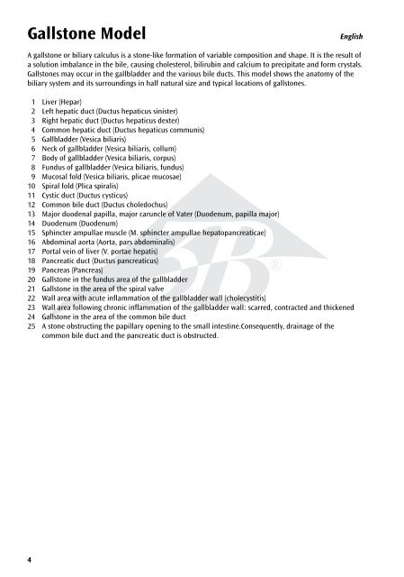
Gallst<strong>one</strong> Model<br />
4<br />
English<br />
A gallst<strong>one</strong> or biliary calculus is a st<strong>one</strong>-like formation of variable composition and shape. It is the result of<br />
a solution imbalance in the bile, causing cholesterol, bilirubin and calcium to precipitate and form crystals.<br />
Gallst<strong>one</strong>s may occur in the gallbladder and the various bile ducts. This model shows the anatomy of the<br />
biliary system and its surroundings in half natural size and typical locations of gallst<strong>one</strong>s.<br />
1 Liver (Hepar)<br />
2 Left hepatic duct (Ductus hepaticus sinister)<br />
3 Right hepatic duct (Ductus hepaticus dexter)<br />
4 Common hepatic duct (Ductus hepaticus communis)<br />
5 Gallbladder (Vesica biliaris)<br />
6 Neck of gallbladder (Vesica biliaris, collum)<br />
7 Body of gallbladder (Vesica biliaris, corpus)<br />
8 Fundus of gallbladder (Vesica biliaris, fundus)<br />
9 Mucosal fold (Vesica biliaris, plicae mucosae)<br />
10 Spiral fold (Plica spiralis)<br />
11 Cystic duct (Ductus cysticus)<br />
12 Common bile duct (Ductus choledochus)<br />
13 Major duodenal papilla, major caruncle of Vater (Duodenum, papilla major)<br />
14 Duodenum (Duodenum)<br />
15 Sphincter ampullae muscle (M. sphincter ampullae hepatopancreaticae)<br />
16 Abdominal aorta (Aorta, pars abdominalis)<br />
17<br />
18<br />
19<br />
Portal vein of liver (V. portae hepatis)<br />
Pancreatic duct (Ductus pancreaticus)<br />
Pancreas (Pancreas)<br />
®<br />
20 Gallst<strong>one</strong> in the fundus area of the gallbladder<br />
21 Gallst<strong>one</strong> in the area of the spiral valve<br />
22 Wall area with acute inflammation of the gallbladder wall (cholecystitis)<br />
23 Wall area following chronic inflammation of the gallbladder wall: scarred, contracted and thickened<br />
24 Gallst<strong>one</strong> in the area of the common bile duct<br />
25 A st<strong>one</strong> obstructing the papillary opening to the small intestine.Consequently, drainage of the<br />
common bile duct and the pancreatic duct is obstructed.