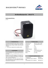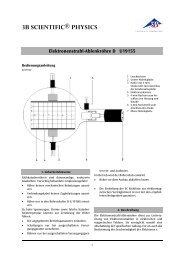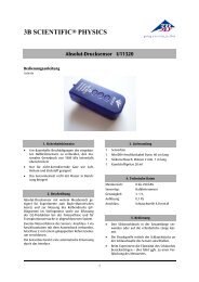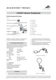
Product manual - 3B Scientific
Product manual - 3B Scientific
Product manual - 3B Scientific

Create successful ePaper yourself
Turn your PDF publications into a flip-book with our unique Google optimized e-Paper software.
L01
Latin<br />
1 Uterus<br />
2 Cavitas uteri<br />
3 Endometrium<br />
4 Myometrium<br />
5 Vagina<br />
6 Corpus luteum<br />
7 Corpus albicans<br />
8 Folliculus ovaricus primordialis<br />
9 Folliculus ovaricus primarius<br />
10 Folliculus ovaricus secundarius<br />
11 Folliculi ovarici vesiculosi<br />
12 Ovarium<br />
13 Folliculus ovaricus vesiculosus<br />
14 Ovulatio<br />
15 Impregnatio<br />
16 Spermatozoon<br />
17 Ovum cum pronuclei<br />
18 Duo blastomeri<br />
19 Quattuor blastomeri<br />
20 Tuba uterina<br />
21 Morula<br />
22 Bastocystis<br />
23 Implantatio<br />
24 Oocytus secundarius<br />
25 Corona radiata<br />
26 Zona pellucida<br />
27 Ovum<br />
28 Polocyti<br />
29 Blastomeri<br />
30 Trophoblastus<br />
31 Blastocelia<br />
32 Embryoblastus<br />
33 Decidua capsularis<br />
34 Saccus vitellinus<br />
35 Cavitas amniotica<br />
36 Mesoderma<br />
37 Coelom<br />
®
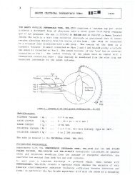
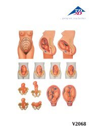
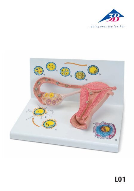
Stages of fertilisation and blastogenesis<br />
®<br />
English<br />
The model provides a schematic representation to illustrate the maturation of ova, ovulation, fertilisation<br />
and blastogenesis up until the embryo is implanted in the wall of the womb. The stages of development<br />
can be seen in large-scale models inside the ovary, the fallopian tubes and the womb. In some cases, even<br />
larger scale versions can be seen on the base.<br />
Inside the ovary, primordial, primary, secondary and tertiary follicles can all be seen as well as a split tertiary<br />
follicle and a yellow body (corpus luteum).<br />
In the fallopian tube near the ovary there is a recently split ovum with pellucid zone and corona radiata<br />
(part of the follicular epithelium) (Fig. 1).<br />
Further along in the amplulla of the uterine tube (ampulla tuba uterina), a sperm is penetrating an ova<br />
(fertilisation) (Fig. 2)<br />
Further along the fallopian tube a fertilised ovum (zygote) is shown with both a male and a female pronucleus<br />
(Fig. 3).<br />
The following stages of cleavage are illustrated:<br />
• Two-cell stage (Fig. 4)<br />
• Four-cell stage (Fig. 5)<br />
• Segmentation spheres (morula) (Fig. 6)<br />
In the wonb (cavitas uteri) is a four-day-old blastocyst (Fig. 7) and an embryo of about 12 days old, that is<br />
now fully implanted into the mucous membrane of the womb (Fig. 8).<br />
1 Uterus<br />
2 Uterine cavity<br />
3 Endometrium<br />
4 Myometrium<br />
5 Vagina<br />
6 Corpus luteum<br />
7 Corpus albicans<br />
8 Primordial ovarian follicle<br />
9 Primary ovarian follicle<br />
10 Secondary ovarian follicle<br />
11 Graafian follicles<br />
12 Ovary<br />
13 Graafian follicle<br />
14 Ovulation<br />
15 Fertilisation<br />
16 Spermatazoa<br />
17 Ovum with pronuclei<br />
18 Two-cell stage<br />
19 Four-cell stage<br />
20 Uterine tube<br />
21 Morula<br />
22 Blastocyst<br />
23 Implanted embryo<br />
24 Split ovum<br />
25 Corona radiata<br />
26 Pellucid zone<br />
27 Ovum<br />
28 Polar bodies<br />
29 Blastomere (segmentation sphere,<br />
cleavage cell)<br />
30 Trophoblast<br />
31 Cleavage, segmentation or subgerminal cavity<br />
32 Embryoblast (inner cell mass)<br />
33 Reflex decidua<br />
34 Yolk sack<br />
35 Amniotic cavity<br />
36 Extraembryonic mesoderm<br />
37 Coelom
Deutsch<br />
Das Modell veranschaulicht als schematische Darstellung die Reifung der Eizelle, des Eisprungs, die<br />
Befruchtung und die Keimesentwicklung bis hin zum eingenisteten Keim. Die Entwicklungsstadien sind<br />
zum einen vergrößert im Eierstock, Eileiter und in der Gebärmutter und zum anderen teilweise in einer<br />
weiteren Vergrößerung auf dem Sockel zu sehen.<br />
Im Eierstock sind Primordial-, Primär-, Sekundär- und Tertiärfollikel sowie ein gesprungener Tertiärfollikel<br />
und ein Gelbkörper (Corpus luteum) sichtbar.<br />
Im Eileiter nahe dem Eierstock zeigt sich eine frisch gesprungene Eizelle mit Zona pellucida und Corona<br />
radiata (Teil des Follikelepithels) (Abb. 1).<br />
Weiter aufwärts, in der Ausbuchtung des Eileiters (Ampulla tubae uterina), dringt ein Spermium in die<br />
Eizelle ein (Imprägnation) (Abb. 2).<br />
Im weiteren Verlauf des Eileiters ist eine befruchtete Eizelle (Zygote) mit einem männlichen und einem<br />
weiblichen Vorkern abgebildet (Abb. 3).<br />
Folgende Furchungsstadien sind zu sehen:<br />
• Zweizellstadium (Abb. 4)<br />
• Vierzellstadium (Abb. 5)<br />
• Maulbeere (Morula) (Abb. 6)<br />
In der Gebärmutterhöhle (Cavitas uteri) sind eine 4 Tage alte Blastozyste (Abb. 7) und ein ca. 12 Tage alter<br />
Keim, der vollständig in die Gebärmutterschleimhaut implantiert ist (Abb. 8), dargestellt.<br />
1 Gebärmutter<br />
2 Gebärmutterhöhle<br />
3 Schleimhaut<br />
4 Muskelschicht<br />
5 Scheide<br />
6 Gelbkörper<br />
7 Umgewandelter Gelbkörper<br />
8 Primordialfollikel<br />
9 Primärfollikel<br />
10 Sekundärfollikel<br />
11 Frühe Tertiärfollikel<br />
12 Eierstock<br />
13 Reifer Tertiärfollikel (Graaf‘scher Follikel)<br />
14 Eisprung<br />
15 Befruchtung<br />
16 Spermien<br />
17 Befruchtete Eizelle mit weiblichem und<br />
männlichem Vorkern<br />
18 Zweizellstadium<br />
19 Vierzellstadium<br />
Stadien der Befruchtung und Keimesentwicklung<br />
20 Eileiter<br />
21 Maulbeere<br />
22 Blastozyste ®<br />
23 Implantierter Keim<br />
24 Gesprungene Eizelle<br />
25 Corona radiata<br />
26 Zona pellucida<br />
27 Ei<br />
28 Polkörperchen<br />
29 Blastomere<br />
30 Trophoblast<br />
31 Blastozystenhöhle<br />
32 Embryoblast<br />
33 Teil der Gebärmutterschleimhaut<br />
34 Dottersack<br />
35 Amnionhöhle<br />
36 Extraembryonales Mesoderm<br />
37 Chorionhöhle
16<br />
17<br />
18<br />
19<br />
15 14 13 12<br />
1<br />
6 7<br />
20<br />
21<br />
8<br />
9<br />
10<br />
11<br />
22<br />
23<br />
1<br />
24<br />
25<br />
26<br />
2<br />
3<br />
4<br />
5
2<br />
3<br />
28 27<br />
28<br />
17<br />
15<br />
25<br />
26<br />
16
4<br />
5<br />
6<br />
29<br />
18<br />
19<br />
29<br />
21<br />
29
7<br />
8<br />
30<br />
31<br />
32<br />
22<br />
23<br />
33<br />
34<br />
35<br />
36<br />
37<br />
30
Etapas de la fertilización y formación del embrión<br />
Español<br />
El modelo muestra una representación esquemática del óvulo (huevo maduro), la ovulación, su fertilización<br />
y desarrollo hasta llegar a ser un embrión anidado. En la base se pueden observar las etapas de<br />
desarrollo, con un aumento, en parte, en el ovario, la tuba uterina y el útero (matriz) y con otros aumentos<br />
más. En el ovario se pueden ver los folículos: primordial, primario, secundario, terciario y vesicular asi<br />
como el cuerpo lúteo. En la trompa uterina (de Falopio) próxima al ovario se muestra un ovocito secundario<br />
recientemente ovulado, con su zona pelúcida y su corona radiada (parte del epitelio folicular)(Imagen<br />
1.). Más adelante en la ampolla de la trompa uterina, un espermatozoide penetra un ovocito secundario<br />
(Imagen 2.). En el transcurso sucesivo en la tuba uterina, se ha formado un cigoto (óvulo fertilizado) con<br />
un pronúcleo masculino y un pronúcleo femenino (Imagen 3). Se pueden observar las siguientes etapas de<br />
fertilización:<br />
• Primera segmentación (dos células)(Imagen 4),<br />
• Segunda segmentación (cuatro células) (Imagen 5),<br />
• Mórula (Imagen 6).<br />
Se han representado en la cavidad uterina un blastocisto de cuatro días de edad (Imagen 7) y un embrión<br />
adulto de aproximadamente doce días, que ya ha sido implantado en la túnica mucosa del útero (endometrio)<br />
(Imagen 8).<br />
1 Útero<br />
2 Cavidad uterina<br />
3 Túnica mucosa, endometrio<br />
4 Túnica muscular, miometrio<br />
5 Vagina<br />
6 Cuerpo lúteo<br />
7 Cuerpo albugineo<br />
8 Folículo ovárico primordial<br />
9 Folículo ovárico primario<br />
10 Folículo ovárico secundario<br />
11 Folículo ovárico terciario<br />
12 Ovario<br />
13 Folículo ovárico vesicular<br />
14 Ovulación<br />
15 Fertilización<br />
16 Espermatozoide<br />
17 Cigoto<br />
18 Primera segmentación (dos células)<br />
19 Segunda segmentación (cuatro células)<br />
20 Trompa uterina (de Falopio)<br />
21 Mórula<br />
22 Blastocisto<br />
23 Embrión implantado<br />
24 Ovocito secundario<br />
25 Corona radiada<br />
26 Zona pelúcida ®<br />
27 Huevo, Óvulo<br />
28 Cuerpo polar<br />
29 Blastomero<br />
30 Trofoblasto<br />
31 Blastocele<br />
32 Embrioblasto<br />
33 Parte del endometrio uterino<br />
34 Saco vitelino<br />
35 Cavidad amniótica<br />
36 Mesodermo extraembrional<br />
37 Cavidad cornea
Français<br />
Stades de la fécondation et développement embryonnaire<br />
Le modèle illustre, sous forme de représentation schématique, la maturation de l‘ovule, l‘ovulation, la<br />
fécondation et le développement embryonnaire jusqu‘à la nidation de l‘embryon. Les stades de développement<br />
sont d‘une part agrandis dans l‘ovaire, les oviductes et dans l‘utérus et, d‘autre part, représentés sur<br />
le socle, en partie dans un agrandissement supplémentaire.<br />
Dans l‘ovaire, on distingue les follicules primordiaux, primaires, secondaires et tertiaires ainsi qu‘un follicule<br />
tertiaire libéré et un corps jaune (corpus luteum).<br />
Un ovule fraîchement libéré est représenté dans l‘oviducte, à proximité de l‘ovaire, avec la zone pellucide<br />
et la corona radiata (épithélium périovulaire) (fig. 1).<br />
Un plus haut, dans l‘excavation de l‘oviducte (ampulla tubae uterina ou ampoule de la trompe utérine), un<br />
spermatozoïde pénètre dans l‘ovule (imprégnation) (fig. 2).<br />
Un peu plus loin dans l‘oviducte, un ovule fécondé (zygote) est illustré avec un pronucleus mâle et un pronucleus<br />
femelle (fig. 3).<br />
On peut observer les stades de segmentation suivants :<br />
• Stade bicellulaire (fig. 4)<br />
• Stade quadricellulaire (fig. 5)<br />
• Morula (fig. 6)<br />
Dans la cavité utérine (cavitas uteri) sont représentés des blastocystes de 4 jours (fig. 7) et un embryon<br />
d‘environ 12 jours, complètement implanté dans la muqueuse utérine (fig. 8).<br />
1 Utérus<br />
2 Cavité utérine<br />
3 Muqueuse<br />
4 Myomètre<br />
5 Vagin<br />
6 Corps jaune<br />
7 Corpus albicans<br />
8 Follicule primordial<br />
9 Follicule primaire<br />
10 Follicule secondaire<br />
11 Follicule tertiaire jeune<br />
12 Ovaire<br />
13 Follicule tertiaire mûr (follicule de De Graaf)<br />
14 Ovulation<br />
15 Fécondation<br />
16 Sperme<br />
17 Ovule fécondé avec pronucleus mâle et femelle<br />
18 Stade bicellulaire<br />
19 Stade quadricellulaire<br />
20 Oviducte<br />
21 Morula<br />
22 Blastocyste ®<br />
23 Embryon implanté<br />
24 Ovule libéré<br />
25 Corona radiata<br />
26 Zone pellucide<br />
27 Oeuf<br />
28 Globule polaire<br />
29 Blastomères<br />
30 Trophoblaste<br />
31 Cavité du blastocyste<br />
32 Embryoblaste<br />
33 Caduque capsulaire<br />
34 Sac vitellin<br />
35 Cavité amniotique<br />
36 Mésoderme extra-embryonnaire<br />
37 Cavité choriale
Estágios da fecundação e desenvolvimento da célula-ovo<br />
®<br />
Português<br />
O modelo apresenta uma representação esquemática do amadurecimento do óvulo, da ovulação, da<br />
fecundação e do desenvolvimento da célula-ovo até a sua nidação. Os estágios de desenvolvimento são<br />
visíveis, por um lado, ampliados no ovário, na tuba uterina e no útero, por outro lado, parcialmente<br />
visíveis numa outra ampliação na base do modelo.<br />
No ovário são visíveis os folículos primordiais, primários, secundários e vesiculares, assim como folículo<br />
vesicular livre e um corpo lúteo.<br />
No canal uterino perto do ovário encontra-se uma célula-ovo recém liberada com zona pelúcida e coroa<br />
radiada (parte do epitélio folicular) (fig. 1).<br />
Mais adiante, na ampola da tuba uterina, um espermatozóide penetra no óvulo (impregnação) (fig. 2).<br />
Na continuação da tuba uterina encontra-se representada uma célula-ovo fecundada (zigoto) com um procarionte<br />
masculino e um femenino (fig. 3).<br />
Os seguintes estágios estão representados:<br />
• blastômero de duas células (fig. 4)<br />
• blastômero de quatro células (fig. 5)<br />
• Mórula (fig. 6)<br />
Na cavidade uterina estão representados blastocitos de 4 dias (fig. 7) e uma célula-ovo de aproximadamente<br />
12 dias, que se encontra completamente implantada na mucosa do útero (fig. 8).<br />
1 Útero<br />
2 Cavidade uterina<br />
3 Endométrio<br />
4 Miométrio<br />
5 Vagina<br />
6 Corpo lúteo<br />
7 Corpo albicans<br />
8 Folículo primordial ovariano<br />
9 Folículo primário ovariano<br />
10 Folículo secundário ovariano<br />
11 Folículos vesiculares ovarianos<br />
12 Ovários<br />
13 Folículo de Graaf<br />
14 Ovulação<br />
15 Fecundação<br />
16 Espermatozóide<br />
17 Óvulo fecundado com procariontes feminino<br />
e masculino<br />
18 Blastômero de duas células<br />
19 Blastômero de quatro células<br />
20 Tuba uterina<br />
21 Mórula<br />
22 Blastocisto<br />
23 Célula-ovo implantada<br />
24 Oócito secundário<br />
25 Coroa radiada<br />
26 Zona pelúcida<br />
27 Óvulo<br />
28 Corpos polares<br />
29 Blastômero<br />
30 Trofoblasto<br />
31 Cavidade dos blastocitos<br />
32 Embrioblasto<br />
33 Parte da mucosa do útero<br />
34 Bolsa vitelina<br />
35 Cavidade amniótica<br />
36 Mesoderma extra-embrionário<br />
37 Celoma
Italiano<br />
Stadi della fecondazione e sviluppo germinale<br />
Il modello illustra attraverso una rappresentazione schematica la maturazione dell‘ovulo, l‘ovulazione, la<br />
fecondazione e lo sviluppo germinale fino all‘impianto dell‘embrione. Gli stadi dello sviluppo sono ingranditi<br />
nell‘ovaio, nella tuba uterina e nell‘utero e sono parzialmente visibili da un ulteriore ingrandimento<br />
sul basamento.<br />
Nell‘ovaio sono visibili i follicoli primordiali, primari e secondari, oltre al follicolo terziario rotto e al corpo<br />
luteo. Nella tuba uterina in prossimità dell‘ovaio si evidenzia un ovulo di recente maturazione con zona<br />
pellucida e corona radiata (parte dell‘epitelio follicolare) (Fig. 1).<br />
Più in alto, nella concavità della tuba uterina (Ampulla tubae uterina), uno spermatozoo penetra nell‘ovulo<br />
(impregnazione) (Fig. 2).<br />
Nel successivo percorso della tuba è raffigurato un ovulo fecondato (zigote) con un pronucleo maschile e<br />
uno femminile (Fig. 3).<br />
Si osservano quindi i seguenti stadi cellulari:<br />
• stadio a due cellule (Fig. 4)<br />
• stadio a quattro cellule (Fig. 5)<br />
• morula (Fig. 6)<br />
Nella cavità uterina sono rappresentati un blastocisto di 4 giorni (Fig. 7) e un embrione di 12 giorni<br />
completamente impiantato nella mucosa uterina (Fig. 8).<br />
1 Utero<br />
2 Cavità uterina<br />
3 Mucosa<br />
4 Strato muscolare<br />
5 Vagina<br />
6 Corpo luteo<br />
7 Corpus albicans<br />
8 Follicolo ovarico primordiale<br />
9 Follicolo ovarico primario<br />
10 Follicolo ovarico secondario<br />
11 Follicoli terziari precoci<br />
12 Ovaio<br />
13 Follicolo terziario maturo (Follicolo di Graaf)<br />
14 Ovulazione<br />
15 Fecondazione<br />
16 Spermatozoi<br />
17 Ovulo fecondato con pronucleo maschile<br />
e femminile<br />
18 Stadio a due cellule<br />
19 Stadio a quattro cellule<br />
20 Tuba uterina<br />
21 Morula<br />
22 Blastocisto<br />
®<br />
23 Embrione impiantato<br />
24 Ovocito secondario<br />
25 Corona radiata<br />
26 Zona pellucida<br />
27 Uovo<br />
28 Polociti<br />
29 Blastomeri<br />
30 Trofoblasto<br />
31 Blastocele<br />
32 Embrioblasto<br />
33 Parte della mucosa uterina<br />
34 Sacco vitellino<br />
35 Cavità amniotica<br />
36 Mesoderma extraembrionale<br />
37 Cavità coriale
<strong>3B</strong> SCIENTIFIC ®<br />
PRODUC TS<br />
<strong>3B</strong> <strong>Scientific</strong> GmbH<br />
Rudorffweg 8 • 21031 Hamburg • Germany<br />
Tel.: + 49-40-73966-0 • Fax: + 49-40-73966-100<br />
www.3bscientific.com • 3b@3bscientific.com<br />
© Copyright 2005 for instruction <strong>manual</strong> and design of product:<br />
<strong>3B</strong> <strong>Scientific</strong> GmbH, Germany<br />
L01-09/05-1