Malformação urológica em criança com síndrome de Sotos: relato ...
Malformação urológica em criança com síndrome de Sotos: relato ...
Malformação urológica em criança com síndrome de Sotos: relato ...

Create successful ePaper yourself
Turn your PDF publications into a flip-book with our unique Google optimized e-Paper software.
370 Jornal <strong>de</strong> Pediatria - Vol. 75, Nº5, 1999<br />
0021-7557/99/75-05/370<br />
Jornal <strong>de</strong> Pediatria<br />
Copyright © 1999 by Socieda<strong>de</strong> Brasileira <strong>de</strong> Pediatria<br />
Resumo<br />
RELATO DE CASO<br />
<strong>Malformação</strong> <strong>urológica</strong> <strong>em</strong> <strong>criança</strong><br />
<strong>com</strong> <strong>síndrome</strong> <strong>de</strong> <strong>Sotos</strong>: <strong>relato</strong> <strong>de</strong> caso<br />
Urological anomaly in an infant with <strong>Sotos</strong>’ syndrome: report of a case<br />
Horácio M. Scigliano 1 , Marco A. Pereira 2 , Romeu F. Daroda 3 , Fabio Marczykosky 4<br />
Objetivo: abordar o diagnóstico e as consi<strong>de</strong>rações etiológicas<br />
<strong>de</strong> malformação <strong>urológica</strong> bilateral <strong>em</strong> um caso <strong>de</strong> <strong>síndrome</strong> <strong>de</strong><br />
<strong>Sotos</strong> (gigantismo cerebral). Alertar os pediatras e urologistas da<br />
existência <strong>de</strong>ssa associação lesional.<br />
Resultados: <strong>criança</strong> <strong>de</strong> 3 meses <strong>de</strong> ida<strong>de</strong> nascida <strong>com</strong> 35<br />
s<strong>em</strong>anas através <strong>de</strong> cesariana <strong>de</strong> mãe hígida, <strong>com</strong> as características<br />
fenotípicas, radiológicas e perfil metacarpofalângica <strong>de</strong> <strong>síndrome</strong><br />
<strong>de</strong> <strong>Sotos</strong>, apresentou taquipnéia, taquicardia e episódios recorrentes<br />
<strong>de</strong> febre (até 36ºC). A urocultura mostrou 500.000 col/ml. A<br />
uretrocistografia mostrou megaureteres e dilatação calicial <strong>de</strong><br />
ambos os rins. Com diagnóstico <strong>de</strong> refluxo vésico-ureteral grau IV<br />
bilateral, foi tratada <strong>com</strong> terapia antimicrobiana. A evolução foi a<br />
melhoria progressiva e obteve alta hospitalar no momento <strong>em</strong> que<br />
se apresentou assintomática, recebendo nitrofurantoína <strong>com</strong>o profilaxia.<br />
Conclusões: assim <strong>com</strong>o <strong>em</strong> outros casos <strong>de</strong> infecção do trato<br />
urinário, os pediatras <strong>de</strong>v<strong>em</strong> suspeitar <strong>de</strong> anomalias <strong>urológica</strong>s <strong>em</strong><br />
todas as <strong>criança</strong>s <strong>com</strong> gigantismo cerebral que apresentar<strong>em</strong> quadro<br />
clínico <strong>de</strong> <strong>síndrome</strong> <strong>de</strong> <strong>Sotos</strong>. Deficiência <strong>em</strong>briológica da inervação<br />
do sist<strong>em</strong>a nervoso autônomo na musculatura dos ureteres<br />
po<strong>de</strong>riam ser uma outra causa.<br />
J. pediatr. (Rio J.). 1999; 75(5): 370-372: gigantismo, gigantismo<br />
cerebral, malformação <strong>urológica</strong>, <strong>síndrome</strong> <strong>de</strong> <strong>Sotos</strong>.<br />
Introdução<br />
Segundo De Myer1 , a <strong>síndrome</strong> <strong>de</strong> <strong>Sotos</strong> ou gigantismo<br />
cerebral é uma megaloencefalia anatômica congênita,<br />
caracterizada por um rápido crescimento ósseo até os<br />
quatro anos, gran<strong>de</strong> tamanho no nascimento, macrocefalia<br />
<strong>com</strong> dolicocefalia. O crânio cresce rapidamente até o<br />
segundo ano <strong>de</strong> vida, a<strong>com</strong>panhado <strong>de</strong> dilatação mo<strong>de</strong>rada<br />
dos ventrículos laterais cerebrais. São el<strong>em</strong>entos típicos<br />
1. Prof. Visitante do Departamento <strong>de</strong> Ciências Morfobiológicas - Fundação<br />
Universida<strong>de</strong> Fe<strong>de</strong>ral do Rio Gran<strong>de</strong> (FURG). Mestre <strong>em</strong> Patologia.<br />
2. Resi<strong>de</strong>nte (R1) <strong>em</strong> Pediatria do Hospital <strong>de</strong> Clínicas da Faculda<strong>de</strong> <strong>de</strong><br />
Medicina Ribeirão Preto - USP.<br />
3. Resi<strong>de</strong>nte (R1) <strong>em</strong> Cirurgia Geral do Hospital Universitário da FURG.<br />
4. Resi<strong>de</strong>nte (R1) <strong>em</strong> Radiologia do Hospital Universitário da Universida<strong>de</strong><br />
<strong>de</strong> Caxias do Sul.<br />
370<br />
Abstract<br />
Objective: To discuss the diagnosis and the etiology of a<br />
bilateral urologic anomaly in a patient with <strong>Sotos</strong>’ Syndrome<br />
(cerebral gigantism). To alert the pediatric physicians and urologists<br />
about the coexistence of these two malformations.<br />
Results: A three month old boy, born of a 35 week un<strong>com</strong>plicated<br />
first pregnancy by cesarian, with phenotypic, radiologic and<br />
metacarpophalangeal profile of <strong>Sotos</strong>’ Syndrome was admitted to<br />
the Hospital because of hyperpnea, tachycardia and 39º C recurrent<br />
fever. The culture of urine disclosed 500.000 col/ml. Cistouretrographic<br />
study showed bilateral megaloureter and hydronephrosis.<br />
Vesico-ureteral bilateral reflux was diagnosed and antibiotic therapy<br />
was administered. Patient’s follow up was excellent and was<br />
discharged taking preventive nitrofurantoin.<br />
Conclusions: As in other cases of urinary tract infection,<br />
pediatric physicians must consi<strong>de</strong>r urologic anomalies in children<br />
with cerebral gigantism who present the clinical presentation of<br />
<strong>Sotos</strong>’ Syndrome. Autonomic Nervous Syst<strong>em</strong> <strong>de</strong>velopment failure<br />
in ureteral muscles could be an alternative etiology.<br />
J. pediatr. (Rio J.). 1999; 75(5): 370-372: gigantism, cerebral<br />
gigantism, urologic anomaly, <strong>Sotos</strong>’ syndrome.<br />
<strong>de</strong>ssa <strong>síndrome</strong> mãos e pés gran<strong>de</strong>s, hipertelorismo,<br />
orientação antimongólica das fendas palpebrais, palato <strong>em</strong><br />
ogiva, alterações ne<strong>urológica</strong>s não progressivas e graus<br />
variáveis <strong>de</strong> retardo mental 2-8 . De orig<strong>em</strong> ainda <strong>de</strong>sconhecida,<br />
diferentes estudos suger<strong>em</strong> a orig<strong>em</strong> genética, <strong>com</strong><br />
transmissão autossômica dominante ou até recessiva 9-12 .<br />
Outras patologias relacionadas à doença são malformações<br />
cerebrais 6,8,13,14 , <strong>de</strong>feitos cardíacos congênitos<br />
15 , anomalias ósseas 16 , oculares 17 e doenças hamartoneoplásicas<br />
18-20 . As anomalias <strong>urológica</strong>s congênitas foram<br />
minimamente referenciadas na literatura 21,22 . Os<br />
autores relatam um caso fenotípico e radiologicamente <strong>de</strong><br />
<strong>síndrome</strong> <strong>de</strong> <strong>Sotos</strong>, associado a anomalias <strong>urológica</strong>s <strong>em</strong><br />
lactente, e discut<strong>em</strong> o diagnóstico e a etiologia.
<strong>Malformação</strong> <strong>urológica</strong> <strong>em</strong> <strong>criança</strong>... - Scigliano HM et alii<br />
Relato <strong>de</strong> Caso<br />
Criança <strong>de</strong> três meses <strong>de</strong> ida<strong>de</strong>, sexo masculino, cor<br />
branca, proce<strong>de</strong>nte <strong>de</strong> área rural, <strong>com</strong> história <strong>de</strong> crises<br />
convulsivas epilépticas tratadas, admitida na clínica pediátrica<br />
no Departamento Materno Infantil da Faculda<strong>de</strong> <strong>de</strong><br />
Medicina da Fundação Universida<strong>de</strong> <strong>de</strong> Rio Gran<strong>de</strong>, RS,<br />
g<strong>em</strong>ente, taquipnéico, taquicárdico e <strong>com</strong> episódios recorrentes<br />
<strong>de</strong> febre <strong>de</strong> até 39 graus. Nascida <strong>de</strong> 35 s<strong>em</strong>anas<br />
através <strong>de</strong> cesariana, <strong>com</strong> peso <strong>de</strong> 3.100 gr, altura 57 cm,<br />
<strong>de</strong> mãe hígida <strong>com</strong> 35 anos <strong>de</strong> ida<strong>de</strong> e pré-natal s<strong>em</strong><br />
intercorrências. Ao exame clínico, a <strong>criança</strong> apresentava<br />
as características fenotípicas e radiológicas da <strong>síndrome</strong><br />
<strong>de</strong> <strong>Sotos</strong>. O padrão <strong>de</strong> perfil metacarpofalângica (PPMF)<br />
<strong>de</strong> ambas as mãos foram diagnósticas nessa doença,<br />
segundo os critérios estabelecidos na literatura23,24 .<br />
O raio-x <strong>de</strong> tórax não apresentou alterações; a ultrasonografia<br />
abdominal não mostrou malformações. Os<br />
exames sorológicos foram normais. H<strong>em</strong>atócrito <strong>de</strong> 35%;<br />
h<strong>em</strong>oglobina 11,6 g/dl; leucograma e plaquetas <strong>de</strong> forma<br />
e quantida<strong>de</strong> normais. H<strong>em</strong>ocultura negativa.O exame <strong>de</strong><br />
urina tipo 1 colhido por PSP foi forte indicativo <strong>de</strong><br />
bacteriúria mo<strong>de</strong>rada. A urocultura mostrou 500.000 col/<br />
ml confirmando a infecção urinária. Após tratamento <strong>com</strong><br />
antimicrobiano e posterior urina tipo1 <strong>com</strong> urina estéril,<br />
foi realizado uretrocistografia retrógrada <strong>com</strong>o método<br />
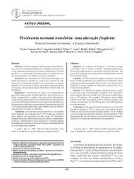
Figura 1 - Uretrocistografia retrógrada. É observado<br />
refluxo vésico-ureteral grau IV bilateral,<br />
presença <strong>de</strong> megaureteres e hidronefrose<br />
Jornal <strong>de</strong> Pediatria - Vol. 75, Nº5, 1999 371<br />
investigatório, e esta mostrou megaureteres e dilatação<br />
calicial nos dois rins, que levam ao diagnóstico <strong>de</strong> refluxo<br />
vésico-ureteral grau IV (Figura 1).<br />
A <strong>criança</strong> evoluiu <strong>com</strong> melhora progressiva, recebendo<br />
alta hospitalar no momento <strong>em</strong> que se apresentou<br />
assintomática, usando nitrofurantoína <strong>com</strong>o profilaxia.<br />
Discussão<br />
Reconhecer os distintos tipos <strong>de</strong> malformações <strong>em</strong><br />
<strong>criança</strong>s tornou-se fato imprescindível, pois a reparação<br />
cirúrgica ou seu controle médico permitirá, na maioria dos<br />
casos, uma boa qualida<strong>de</strong> <strong>de</strong> vida.<br />
O gigantismo cerebral não endócrino, <strong>de</strong>scrito inicialmente<br />
por <strong>Sotos</strong> e col. <strong>em</strong> 19672 , <strong>com</strong> 220 <strong>relato</strong>s isolados<br />
na literatura, é uma das <strong>síndrome</strong>s <strong>de</strong> malformações<br />
congênitas mais <strong>com</strong>plexas. O diagnóstico é s<strong>em</strong>pre clínico25<br />
, <strong>com</strong> um fenótipo característico: gigantismo somático<br />
<strong>com</strong> dolicocefalia, microftalmia, hipertelorismo, palato<br />
<strong>em</strong> ogiva, mento pontiagudo, mãos e pés gran<strong>de</strong>s e<br />
maior ida<strong>de</strong> óssea4 . Na radiologia geral do corpo <strong>de</strong>stacase<br />
<strong>de</strong>sproporção crâneo-facial, às vezes <strong>com</strong> formação <strong>de</strong><br />
osso na fontanela anterior13,26 , presença <strong>de</strong> vértebras<br />
planas13 , disostose periférica16 e ossos longos das mãos<br />
e pés <strong>com</strong> <strong>de</strong>sproporção <strong>de</strong> crescimento. Esta última<br />
alteração po<strong>de</strong> constatar-se através do padrão <strong>de</strong> perfil<br />
metacarpofalângica (PPMF), estudo <strong>de</strong>senvolvido por<br />
Poznanski e col. <strong>em</strong> 197227 . Trata-se da representação<br />
gráfica das principais medidas dos19 ossos longos do<br />
metacarpo e das falanges, sendo muito útil no diagnóstico<br />
<strong>de</strong> doenças congênitas23,24 . Em nosso paciente o valor foi<br />
coinci<strong>de</strong>nte <strong>com</strong> os dados da literatura.<br />
As anomalias <strong>urológica</strong>s congênitas relacionadas à<br />
<strong>síndrome</strong> e referidas na literatura são escassas. Moriyama<br />
e col. 21 , <strong>em</strong> 1984, <strong>de</strong>screveram a coexistência, <strong>em</strong> uma<br />
<strong>criança</strong> japonesa <strong>de</strong> 4 anos <strong>de</strong> ida<strong>de</strong>, das <strong>com</strong>plicações do<br />
refluxo vésico-ureteral e da <strong>síndrome</strong> <strong>de</strong> <strong>Sotos</strong>. Hamma<strong>de</strong>h<br />
e col. 22 , <strong>em</strong> 1995, relataram um caso <strong>de</strong> divertículo<br />
vesical congênito <strong>em</strong> adulto <strong>com</strong> fenótipo <strong>de</strong> <strong>síndrome</strong> <strong>de</strong><br />
<strong>Sotos</strong>. Mas os megaureteres e a dilatação calicial renal, <strong>em</strong><br />
conseqüência do refluxo vésico-ureteral, que nós achamos<br />
<strong>de</strong> causa congênita, foram <strong>de</strong>scritos por Moriyama e<br />
col. 21 . Um dado importante é que a ultra-sonografia foi<br />
normal e a uretrocistografia mostrou o refluxo. Isto<br />
ocorreu porque, na ocasião <strong>em</strong> que foi realizado o exame,<br />
provavelmente existia pouca urina na bexiga e, conseqüent<strong>em</strong>ente,<br />
a hidronefrose <strong>de</strong>corrente do refluxo não foi<br />
observada. As causas congênitas mais freqüentes <strong>de</strong>ssa<br />
anomalia são obstrução do colo da bexiga, presença <strong>de</strong><br />
valvas uretrais posteriores, obstrução da junção vésicoureteral,<br />
hipertrofia distal do músculo ureteral e atresia<br />
ureteral bilateral28,29 . Nenhuma <strong>de</strong>ssas alterações foi<br />
encontrada <strong>em</strong> nosso paciente.<br />
Se a coexistência da <strong>síndrome</strong> <strong>de</strong> <strong>Sotos</strong> e a anormalida<strong>de</strong><br />
<strong>urológica</strong> bilateral <strong>de</strong>scrita é só coincidência, ou se<br />
ambas têm relação etiológica <strong>com</strong> uma causa subjacente <strong>de</strong><br />
crescimento anômalo, ainda é duvidoso. Não existe uma
372 Jornal <strong>de</strong> Pediatria - Vol. 75, Nº5, 1999<br />
explicação satisfatória para o crescimento <strong>em</strong> excesso<br />
constatado principalmente nos ossos. S<strong>em</strong> dúvida, o(s)<br />
fator(es) responsável(veis) <strong>de</strong>ve(m) agir no útero no<br />
momento da concepção, e não na hora do nascimento, pois<br />
os pacientes <strong>com</strong> <strong>síndrome</strong> <strong>de</strong> <strong>Sotos</strong> apresentam também<br />
<strong>de</strong>rmatóglifos raros, cujo padrão <strong>em</strong>briológico <strong>com</strong>pletase<br />
perto da 18ª s<strong>em</strong>ana <strong>de</strong> gestação e permanece s<strong>em</strong><br />
modificações o resto da vida, fato <strong>de</strong>scrito por Miller e<br />
Giroux <strong>em</strong> 1966 30 .<br />
Entretanto, as evidências que t<strong>em</strong>os é <strong>de</strong> que a anormalida<strong>de</strong><br />
<strong>urológica</strong> <strong>em</strong> nosso paciente po<strong>de</strong> ser conseqüência<br />
do refluxo vésico-ureteral, porém uma causa na própria<br />
pare<strong>de</strong> dos ureteres não po<strong>de</strong> ser <strong>de</strong>scartada. É provável<br />
que os megaureteres nos pacientes <strong>com</strong> <strong>síndrome</strong> <strong>de</strong> <strong>Sotos</strong><br />
represent<strong>em</strong> uma alteração <strong>em</strong>briológica da inervação da<br />
musculatura ureteral. Similares <strong>de</strong>ficiências da inervação<br />
do sist<strong>em</strong>a nervoso autônomo <strong>em</strong> outros órgãos a<strong>com</strong>panhados<br />
<strong>de</strong> febre foram <strong>de</strong>scritos por Appenzeller e Sny<strong>de</strong>r,<br />
<strong>em</strong> 1969 14 .<br />
A associação da <strong>síndrome</strong> <strong>de</strong> <strong>Sotos</strong> <strong>com</strong> outros processos<br />
incontroláveis <strong>de</strong> crescimento, <strong>com</strong>o as neoplasias<br />
malignas, ainda é especulação 19,20 . Ruvalcaba e col. 18<br />
também <strong>de</strong>screveram a coexistência da <strong>síndrome</strong> <strong>com</strong><br />
lesões hamartomatosas (pólipos intestinais e mudanças na<br />
pigmentação dos genitais).<br />
Apesar <strong>de</strong> o refluxo vésico-ureteral estar presente <strong>em</strong><br />
20-40% das <strong>criança</strong>s <strong>com</strong> infecção do trato urinário e as<br />
anomalias dos ureteres constituír<strong>em</strong> um achado freqüente<br />
nas autópsias ou nas pielografias, e às vezes s<strong>em</strong> importância<br />
clínica, este, sendo bilateral e grau IV, <strong>de</strong>ve ser<br />
consi<strong>de</strong>rado <strong>em</strong> pacientes <strong>com</strong> <strong>síndrome</strong> <strong>de</strong> <strong>Sotos</strong> e<br />
infecção do trato urinário.<br />
Referências bibliográficas<br />
1. De Myer W. Megalencephaly in children. Neurology 1972; 22:<br />
634-43.<br />
2. <strong>Sotos</strong> JF, Dodge PR, Muirhead D. Cerebral gigantism in<br />
childhood: a syndrome of excessively rapid growth with acromegalic<br />
features and a nonprogressive neurologic disor<strong>de</strong>r. New<br />
Engl J Med 1964; 271: 109-16.<br />
3. Abraham JM, Snodgrass GJ. <strong>Sotos</strong>’ syndrome of cerebral<br />
gigantism. Arch Dis Child 1969; 44: 203-10.<br />
4. Jaeker J, van <strong>de</strong>r Schueren-Lo<strong>de</strong>weyckx M, Ecckels R. Cerebral<br />
gigantism syndrome. A report of four cases and review of the<br />
literature. Z Kin<strong>de</strong>rheilk 1972; 112: 332-46.<br />
5. <strong>Sotos</strong> JF, Cutler EA, Dodre P. Cerebral gigantism. Am J Dis<br />
Child 1977; 131:625-7.<br />
6. Wit JM, Be<strong>em</strong>er FA, Barth PG, Oorthumys JW, Dijkstra PF,<br />
van <strong>de</strong>r Bran<strong>de</strong> JL et al. Cerebral gigantism (<strong>Sotos</strong>’ syndrome).<br />
Compiled data of 22 cases. Analysis of clinical features, growth,<br />
and plasma somatomedin. Eur J Pediatr 1985; 144: 131-40.<br />
7. Reimao RN, Marques Dias MJ. Síndrome <strong>de</strong> <strong>Sotos</strong>. Registro <strong>de</strong><br />
dois casos. Arq Neuropsiquiatr 1980; 38: 150-9.<br />
8. Moretti Ferreira D, Koiffmann CP, Wajntal A, Diament AJ, De<br />
Mendonça BB, Mattieli J,et al. Macrossomia, macrocrania e<br />
incoordinação motora da infancia. Síndrome <strong>de</strong> <strong>Sotos</strong> (Mc.<br />
Kusick 11755): estudo <strong>de</strong> 7 casos e revisão <strong>de</strong> aspectos clínicos<br />
<strong>de</strong> 198 casos publicados. Arq Neuropsiquiatr 1991; 49: 164-71.<br />
<strong>Malformação</strong> <strong>urológica</strong> <strong>em</strong> <strong>criança</strong>... - Scigliano HM et alii<br />
9. Nevo S, Zeltzer M, Ben<strong>de</strong>rly A. Evi<strong>de</strong>nce for autosomal<br />
recessive inheritance of cerebral gigantism. J Med Genet 1974;<br />
11: 158-65.<br />
10. Zonana J, <strong>Sotos</strong> JF, Romshe CA, Fisher DA, El<strong>de</strong>rs MJ, Rimoiu<br />
DL. Dominant inheritance of cerebral gigantism. J Pediatr<br />
1977; 91: 251-6.<br />
11. Smith A, Farrar JR, Silink M, Judzewitsch R. Investigations in<br />
dominant <strong>Sotos</strong>’ syndrome. Ann Genet 1981; 24: 226-8.<br />
12. Winship IM. <strong>Sotos</strong> syndrome. Autosomal dominant inheritence<br />
substantiated. Clin Genet 1985; 28: 243-6.<br />
13. Poznanski AK, Stephenson JM. Radiographic findings in hypothalamic<br />
acceleration of growth associated with cerebral atrophy<br />
and mental retardation (cerebral gigantism). Radiology 1967;<br />
88: 446-56.<br />
14. Appenzeller O, Sny<strong>de</strong>r RD. Autonomic failure with persistent<br />
fever in cerebral gigantism. J Neurol Neurosurg Psychiatry<br />
1969; 32: 123-8.<br />
15. Kaneko K, Tsukahara M, Tachibaina H, Izurashige H, Kuwano<br />
A, Kajii T. Congenital Heart <strong>de</strong>fects in <strong>Sotos</strong> sequence. Am J<br />
Med Genet 1987; 26: 569-76.<br />
16. Evans PR. <strong>Sotos</strong>’ syndrome (cerebral gigantism) with peripheral<br />
dysostosis. Arch Dis Child 1971; 46: 199-202.<br />
17. Koenekoop RK, Rosenbaum KN, Traboulsi EI. Ocular findings<br />
in a family with <strong>Sotos</strong>’ syndrome (cerebral gigantism). Am J<br />
Ophthalmol 1995; 119: 657-8.<br />
18. Ruvalvaba RH, Myhre S, Smith DW. <strong>Sotos</strong>’ Syndrome with<br />
intestinal polyposis and pigmentary changes of the genitalia.<br />
Clin Genet 1980; 18: 413-6.<br />
19. Sugarman GI, Heuser ET, Reed WB. A case of cerebral<br />
gigantism and hepatic carcinoma. Am J Dis Child 1977; 131:<br />
631-3.<br />
20. Nance MA, Neglia JP, Talwar D, Berry SA. Neuroblastoma in<br />
a patient with <strong>Sotos</strong>’ syndrome. J Med Genet 1990; 27: 130-2.<br />
21. Moriyama M, Terasshima K, Fukushima Y, Kuroki Y. Urological<br />
anomalies in patients with <strong>Sotos</strong>’ syndrome. Nippon Hinyokika<br />
Gakkai Zasshi 1984; 75: 591-3.<br />
22. Hamma<strong>de</strong>h MY, Dutta SN, Cornaby AJ, Morgan RJ. Congenital<br />
urological anomalies in <strong>Sotos</strong>’ syndrome. Br J Urol 1995; 76:<br />
133-5.<br />
23. Butler MG, Meaney FJ, Kittur S, Hersh JH, Hornstein L.<br />
Metacarpophalangeal pattern profile analysis in <strong>Sotos</strong>’ syndrome.<br />
Am J Med Genet 1985; 20: 625-9.<br />
24. Butler MG, Dijkstra PF, Meaney FJ, Gale DD. Metacarpophalangeal<br />
pattern profile analysis in <strong>Sotos</strong>’ syndrome: a follow-up<br />
report on 34 subjects. Am J Med Genet 1988; 29: 143-7.<br />
25. Cole TR, Hughes HE. <strong>Sotos</strong>’ Syndrome: a study of the diagnostic<br />
criteria and natural history. J Med Genet 1994; 31: 20-32.<br />
26. Caffey J. Anterior fontanel bone. Report of a case. In: Gellis SS<br />
ed. Year Book of Pediatrics 1962-1963. Chicago. Year Book<br />
Medical Publ. 1963. p. 401-2.<br />
27. Butler MG, Meaney FJ, Kaler SG. Metacarpophalangeal pattern<br />
profile analysis in clinical genetics: an applied anthropometric<br />
method. Am J Phys Anthropol 1986; 70: 195-201.<br />
28. Potter EL. Pathology of the Fetus and the Newborn. Chicago.<br />
The Year Book Publ; 1952.<br />
29. Wigglesworth JS. Perinatal Pathology. Vol. 15. Major Probl<strong>em</strong>s<br />
in Pathology. Phila<strong>de</strong>lphia: WB Saun<strong>de</strong>rs; 1984<br />
30. Miller JR, Giroux J. Dermatoglyphics in pediatric practice. J<br />
Pediatr 1966; 69: 302-12.<br />
En<strong>de</strong>reço para correspondência:<br />
Dr. Horácio M. Scigliano<br />
Depto. <strong>de</strong> Ciências Morfobiológicas - FURG<br />
Rio Gran<strong>de</strong> - RS - Brasil - CEP 96203-270<br />
Fone: (532) 311.222 - ramal 168<br />
Fone/Fax: (532) 31.5914