

1 Úvod:
1 Úvod:
1 Úvod:

Create successful ePaper yourself
Turn your PDF publications into a flip-book with our unique Google optimized e-Paper software.
ightness knob" actually changes the size of the spot on the sample; changing it from<br />
a wide dispersed spot to a pinpoint beam.<br />
3. The beam is restricted by the condenser aperture (usually user selectable), knocking<br />
out high angle electrons (those far from the optic axis, the dotted line down the center)<br />
4. The beam strikes the specimen and parts of it are transmitted<br />
5. This transmitted portion is focused by the objective lens into an image<br />
6. Optional Objective and Selected Area metal apertures can restrict the beam; the<br />
Objective aperture enhancing contrast by blocking out high-angle diffracted electrons,<br />
the Selected Area aperture enabling the user to examine the periodic diffraction of<br />
electrons by ordered arrangements of atoms in the sample<br />
7. The image is passed down the column through the intermediate and projector lenses,<br />
being enlarged all the way<br />
8. The image strikes the phosphor image screen and light is generated, allowing the user<br />
to see the image. The darker areas of the image represent those areas of the sample<br />
that fewer electrons were transmitted through (they are thicker or denser). The lighter<br />
areas of the image represent those areas of the sample that more electrons were<br />
transmitted through (they are thinner or less dense)<br />
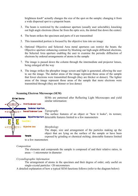
Scanning Electron Microscope (SEM)<br />
SEMs are patterned after Reflecting Light Microscopes and yield<br />
similar information:<br />
Topography<br />
The surface features of an object or "how it looks", its texture;<br />
detectable features limited to a few manometers<br />
Morphology<br />
The shape, size and arrangement of the particles making up the<br />
object that are lying on the surface of the sample or have been<br />
exposed by grinding or chemical etching; detectable features limited<br />
to a few manometers<br />
Composition<br />
The elements and compounds the sample is composed of and their relative ratios, in<br />
areas ~ 1 micrometer in diameter<br />
Crystallographic Information<br />
The arrangement of atoms in the specimen and their degree of order; only useful on<br />
single-crystal particles >20 micrometers<br />
A detailed explanation of how a typical SEM functions follows (refer to the diagram below):


