Report On Long final version 22.4.10

Create successful ePaper yourself
Turn your PDF publications into a flip-book with our unique Google optimized e-Paper software.
<strong>Report</strong> <strong>On</strong> <strong>Long</strong>-Term Dialysis Via The PakuMed<br />
Titan-Port D ® Dialysis Port System<br />
Dr. med. Jens Benders and Prof. Dr. med. Markus Hollenbeck<br />
Case report<br />
We wish to report on a patient with<br />
terminal renal failure caused by a vascular<br />
nephropathy secondary to long-standing<br />
arterial hypertension and generalized<br />
arteriosclerosis. Dialysis dependence<br />
began in March 2007 after several<br />
hydropic decompensation episodes in the<br />
form of a cardiorenal syndrome secondary<br />
to a dilitative form of ischemic<br />
cardiomyopathy. Apart from severe heart<br />
failure and the high grade aortic, mitral,<br />
and tricuspid valve insufficiency, atrial<br />
fibrillation was present as well. The patient<br />
was on oral phenprocoumon<br />
anticoagulation since implantation of a<br />
VVI-Pacemaker several years ago.<br />
Moreover the patient had a colon<br />
segmental resection for carcinoma with a<br />
temporary diverting colostomy several<br />
decades ago as well as a left<br />
hemicolectomy for a carcinoma recurrence<br />
in 2006. Severe intraabdominal adhesions<br />
were present at that time.<br />
At the time of her first dialysis the 84-yearold<br />
patient was in satisfactory condition<br />
with respect to her age.<br />
Dialysis Port Systems Description<br />
Dialysis port systems can be implanted for<br />
performing hemodialysis when other<br />
therapeutic options, such as surgically<br />
constructed shunts, are considered risky or<br />
contraindicated for anatomical or medical<br />
reasons (cardiologic problems in<br />
particular).<br />
The port systems are fully implantable<br />
subcutaneously. The port is made of<br />
titanium and is biocompatible,<br />
hypoallergenic, and does not interfere with<br />
MRI studies.<br />
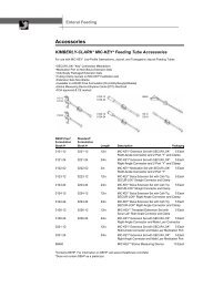
Fig.1: shows a double-lumen port system<br />
Due to preexisting intraabdominal<br />
adhesions secondary to multiple bowel<br />
surgeries, peritoneal dialysis was<br />
considered contraindicated.<br />
Due to the patient’s heart failure an avshunt<br />
was also not considered as an option.<br />
We decided to implant a dialysis port<br />
system as the treatment of choice.<br />
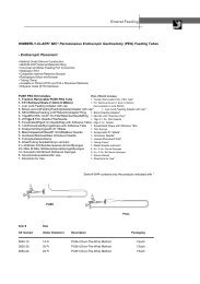
Fig. 2 shows a special dialysis port puncture needle
Implantation of port systems<br />
Implantation of the port system via the<br />
right subclavian vein would prove to be<br />
technically difficult due to prior placement<br />
of a cardiac pacemaker. Moreover the left<br />
jugular vein was chronically occluded. The<br />
catheter lines were passed easily via the<br />
right internal jugular vein (fig. 3, 4, 5).<br />
After implantation hemodialysis could<br />
proceed and continue without problems<br />
(fig. 6).<br />
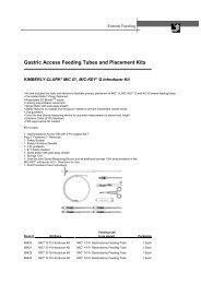
Fig. 3: Surgical field of right internal jugular vein<br />
Fig. 6: Depiction of an implanted port system<br />
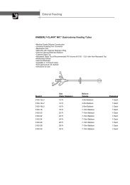
Fig. 4: Operative field port system<br />
After disinfection and sterile draping of the<br />
puncture sites each port chamber is<br />
accessed with the special port needles. The<br />
dialysis nurse wears sterile gloves and a<br />
mask. The portal lock solution is aspirated.<br />
The system is then flushed with<br />
physiologic saline. Upon termination of<br />
dialysis 46% citrate solution is once again<br />
injected. 3-point rotation of the membrane<br />
is recommended to ensure even<br />
distribution of puncture points to prevent<br />
excessive wear of the membrane material.<br />
Fig. 5: Post-op x-ray of port system<br />
Conducting hemodialysis via port<br />
system
Fig. 7: Rotation principle of puncture sites<br />
Fig. 9: Implanted port system in use (2,5 years after<br />
implantation, post pacemaker battery revision)<br />
Dialysis blood flow rates of 300 ml / min<br />
were achieved. Excellent dialysis<br />
efficiency values measured as Kt/V (urea)<br />
were observed averaging 1,4 .<br />
Fig.8 illustrates the port system with in situ<br />
dialysis needles at 5 months post<br />
implantation<br />
During this 2,5 year period there were no<br />
thrombotic or infection complications.<br />
<strong>Long</strong>-Term Course<br />
After a three year course of hemodialysis<br />
the patient was in very good clinical<br />
condition. Heart failure had improved and<br />
the patient was fully compensated.<br />
Because her cardiac status had improved<br />
we decided to proceed with a shunt<br />
procedure on her left forearm and<br />
performed the surgery along with a<br />
pacemaker battery replacement. Upon<br />
uneventful initiation of hemodialysis via<br />
shunt the port system was later removed.<br />
Fig. 8: Port system in use at 5 months<br />
Risks and Problems<br />
Due to a slowly progressive skin defect on<br />
one of the puncture sites, a new puncture<br />
site proved necessary. Fig. 9 illustrates the<br />
anatomic situation 2,5 years after<br />
beginning hemodialysis.<br />
Knappschaftkrankenhaus Bottrop<br />
Osterfelder Str. 157<br />
46242 Bottrop<br />
Klinik für Nephrologie und Rheumatologie<br />
Prof. Dr. med. M. Hollenbeck (Ltd. Arzt)<br />
Dr. med. J. Benders (Oberarzt)<br />
E-mail: hollenbeck@kk-bottrop.de<br />
jens.benders@kk-bottrop.de