Trinidad-and-Tabago-Congerss-Abstract-Book

You also want an ePaper? Increase the reach of your titles
YUMPU automatically turns print PDFs into web optimized ePapers that Google loves.
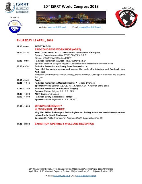
20 th ISRRT World Congress 2018<br />
Hosted by<br />
Website: www.isrrt2018.org.tt<br />
Email: queries@isrrt2018.org.tt<br />
THURSDAY 12 APRIL, 2018<br />
07:00 – 8:00 REGISTRATION<br />
PRE-CONGRESS WORKSHOP (ASRT)<br />
08:00 – 8:30 Bonn Call to Action 2017 – ISRRT Global Assessment of Progress<br />
Speaker: Donna Newman B.A. RT (R) CNMT F.A.S.R.T.<br />
Director of Professional Practice ISRRT<br />
08:30 – 9:00 Radiation Protection in Africa - The Journey So Far<br />
Speaker: Elizabeth Balogun, Regional Coordinator for Professional Practice in Africa<br />
09:00 – 9:30 Radiation Protection <strong>and</strong> Safety Panel Discussion on<br />
Bonn Call for Action assessment around the world (Participation <strong>and</strong> Feedback from<br />
Audience)<br />
Moderator <strong>and</strong> Panellists: Stewart Whitley, Donna Newman, Christopher Steelman <strong>and</strong> Elizabeth<br />
Balogun<br />
09:30 – 9:45 Break<br />
09:45 – 10:45 Radiation Protection in Medical Imaging: A Holistic Overview<br />
Speaker: Michael Latimer M.S.R.S., R.T., FASRT, ASRT Chairman of the Board<br />
10:45 – 11:45 Radiation Protection for Paediatric Imaging<br />
Speaker: Michael Odgren B.S., R.T., RPA<br />
11:45 – 13:00 ASRT Sponsored Lunch<br />
13:00 – 14:00 Radiation Safety in Radiation Therapy<br />
Speaker: S<strong>and</strong>ra Hayden M.A., R.T., FASRT<br />
15:00 – 16:00 OPENING CEREMONY<br />
HUTCHINSON LECTURE<br />
Why Well Skilled Radiological Technologists <strong>and</strong> Radiographers are needed more than ever<br />
to face Public Health Challenges<br />
Speaker: Dr. Pablo Jimenez, Pan American Health Organization (PAHO)<br />
17:00 – 20:00 EXHIBITION OPENING & WELCOME RECEPTION<br />
20 th International Society of Radiographers <strong>and</strong> Radiological Technologist, World Congress<br />
April 12 – 15, 2018 • Hyatt Regency <strong>Trinidad</strong>, Wrightson Road, Port of Spain, <strong>Trinidad</strong>, W.I.<br />
Website: www.isrrt2018.org.tt Email: queries@isrrt2018.org.tt
20 th ISRRT World Congress 2018<br />
Hosted by<br />
Website: www.isrrt2018.org.tt<br />
Email: queries@isrrt2018.org.tt<br />
FRIDAY 13 APRIL 2018<br />
V10 4.2.18<br />
08:15 – 9:00 REGISTRATION<br />
08:30 – 9:30 PLENARY SESSION<br />
What Makes an Effective Leader<br />
Dr. Melissa Jackowski R.T. (R) (M), ASRT President Elect<br />
9:30 – 10:30 BREAK<br />
10:30 - 12:30 EDUCATION SEMINAR RM.1<br />
Chair: Gabrielle Baptiste<br />
10:30 The Development of Critical Thinking in Diagnostic Radiography ED1-1<br />
Aarthi Ramlaul<br />
University of Hertfordshire, UK<br />
10:50 The Use of Digital Storytelling to Teach Evidence Based Breast Imaging to<br />
Radiography Students: Translating Reality into Best Practice: A Case Study ED1-2<br />
Cynthia Cowling<br />
Monash University, Australia<br />
11:10 Implementing the Flipped Classroom in Radiation Safety ED1-3<br />
Dr. Timmeri Cohen<br />
Virginia Commonwealth University, Richmond, Virginia<br />
11:30 Promotion of Improved St<strong>and</strong>ards of Radiography Education through Transition<br />
Management<br />
ED1-4<br />
Karen Finlay<br />
Central Queensl<strong>and</strong> University, Australia<br />
11:50 Perceived Benefits of Near Peer Teaching <strong>and</strong> Learning with 2 nd Year Radiography<br />
Students at Monash University<br />
ED1-5<br />
Lori Boyd<br />
Monash University, Australia<br />
12:30 – 13:30 LUNCH<br />
13:30 – 15:00 QUALITY MANAGEMENT RM.1<br />
Chair: Anzar Nasrudeen<br />
13:30 Establishing a Radiology Outpatient Clinic for Renal Tumor Biopsy Patients QM1-1<br />
Bo Mussmann<br />
Odense University Hospital, Denmark<br />
13:50 Using “Timeouts” to Promote Quality <strong>and</strong> Efficiency during Vascular Interventional<br />
Radiography Procedures<br />
QM1-2<br />
Craig St. George<br />
American Society of Radiologic Technologists, USA<br />
20 th International Society of Radiographers <strong>and</strong> Radiological Technologist, World Congress<br />
April 12 – 15, 2018 • Hyatt Regency <strong>Trinidad</strong>, Wrightson Road, Port of Spain, <strong>Trinidad</strong>, W.I.<br />
Website: www.isrrt2018.org.tt Email: queries@isrrt2018.org.tt
20 th ISRRT World Congress 2018<br />
Hosted by<br />
Website: www.isrrt2018.org.tt<br />
Email: queries@isrrt2018.org.tt<br />
Apr 13 th Cont’d<br />
13:30 – 15:00 QUALITY MANAGEMENT RM.1<br />
Chair: Anzar Nasrudeen<br />
14:10 Transforming Radiology: Applying the LEADS Framework as a Change<br />
Management Tool to Achieve Results.<br />
Nicole Dhanraj<br />
Guam Memorial Hospital, United States<br />
QM1-3<br />
15:00 – 15:30 BREAK<br />
15:30 – 17:00 HEALTH & SAFETY RM.1<br />
Chair: Jade Tucker<br />
15:50 Patient as Observer: Practical Steps to Launching a H<strong>and</strong> Hygiene Quality Assurance<br />
Program in the Medical Imaging Outpatient Setting<br />
HS1-1<br />
Jean Nash<br />
University Health Network, Canada<br />
Institute of Medical Education & Research, India<br />
16:10 How does a Radiographer’s Job Satisfaction Influence their Body Posture to cause<br />
Back Pain<br />
HS1-2<br />
Adrian Sampath<br />
COSTAATT, <strong>Trinidad</strong> & Tobago<br />
10:30 -12:30 COMPUTED TOMOGRAPHY RM.2<br />
Chair: Kavita Seedarnee<br />
10:30 National Survey of Computed Tomography Radiation Doses in Nigeria CT1-1<br />
Idris Garba<br />
Nigeria<br />
10:50 Effective Dose <strong>and</strong> Cancer Risk Estimates from Diagnostic Computed Tomography<br />
Procedures in Jamaica<br />
CT1-2<br />
Barrington Brevitt<br />
Apex Radiology, Jamaica<br />
11:50 STORIES FROM THE FRONTLINE: Identifying Challenges <strong>and</strong> Solutions for<br />
Improper CT Colonography Patient Preparation Using a Case Study Approach CT1-3<br />
Tracy Wakeford<br />
Mount Sinai Hospital, Canada<br />
12:10 The Use of MIYABI Angio-CT for Diagnosis <strong>and</strong> Treatment of Lower Gastrointestinal<br />
Bleeding Commonly Seen in Emergency<br />
CT1-4<br />
Wei-YaoKao<br />
Veterans General Hospital Taipei, Taiwan<br />
12:30 – 13:30 LUNCH<br />
20 th International Society of Radiographers <strong>and</strong> Radiological Technologist, World Congress<br />
April 12 – 15, 2018 • Hyatt Regency <strong>Trinidad</strong>, Wrightson Road, Port of Spain, <strong>Trinidad</strong>, W.I.<br />
Website: www.isrrt2018.org.tt Email: queries@isrrt2018.org.tt
20 th ISRRT World Congress 2018<br />
Hosted by<br />
Website: www.isrrt2018.org.tt<br />
Email: queries@isrrt2018.org.tt<br />
Apr 13 th Cont’d<br />
13:30 -15:00 WE CARE RM.2<br />
Chair: Sheila Legall<br />
13:30 Underst<strong>and</strong>ing the role of Perception in Communication in a clinical environment:<br />
Grounded Theory<br />
WC1-1<br />
Niekeisha Garrette<br />
COSTAATT, <strong>Trinidad</strong> & Tobago<br />
13:50 Leadership WC1-2<br />
Anushka Kattick-Mahabirsingh<br />
Infeemed Solutions & Supplies Ltd., <strong>Trinidad</strong> & Tobago<br />
14:10 Radiographer Perceptions of Professionalism WC1-3<br />
Mrs Tiina Kukkes<br />
Tartu Health Care College, Estonia<br />
15:00 – 15:30 BREAK<br />
15:30 – 17:00 GENERAL IMAGING RM.2<br />
Chair: Alex<strong>and</strong>er “S<strong>and</strong>y” Yule<br />
15:30 Balancing the Spine GI1-1<br />
Dr. Kimani White<br />
EWMSC, <strong>Trinidad</strong><br />
10:30 -12:30 ULTRASOUND RM.3<br />
Chair: Nikeisha La Croix-Simon<br />
10:50 A Case Study: Diagnosis of Heterotopic Pregnancy in an Emergency Situation US1-1<br />
Yonella Demars<br />
Virginia Commonwealth University, Richmond, Virginia<br />
11:10 Ultrasound of an Adult Meningocele US1-2<br />
Denise Choong Ai Wen<br />
National University Hospital, Singapore<br />
11:30 Ultrasound Safety, Mechanisms of Harm <strong>and</strong> Possible Side Effects US1-3<br />
Peters Ehiwe<br />
Scarborough General Hospital, <strong>Trinidad</strong> <strong>and</strong> Tobago<br />
12:30 – 13:30 LUNCH<br />
20 th International Society of Radiographers <strong>and</strong> Radiological Technologist, World Congress<br />
April 12 – 15, 2018 • Hyatt Regency <strong>Trinidad</strong>, Wrightson Road, Port of Spain, <strong>Trinidad</strong>, W.I.<br />
Website: www.isrrt2018.org.tt Email: queries@isrrt2018.org.tt
20 th ISRRT World Congress 2018<br />
Hosted by<br />
Website: www.isrrt2018.org.tt<br />
Email: queries@isrrt2018.org.tt<br />
Apr 13 th Cont’d<br />
13:30 -15:00 RADIATION PROTECTION RM.3<br />
Chair: Keya Crichlow<br />
13:30 Assessment of Occupational Radiation Doses for Medical Workers Based on<br />
Job Categories in United Arab Emirates<br />
Dimitris Katsifarakis presenting for Wiam Elshami<br />
University of Sharjah, UAE<br />
13:50 Measurements of Radiation Exposure of Radiography Students during their<br />
Clinical Training using Thermoluminescent Dosimetry<br />
Christopher Steelman presenting for Mohamed Abuzaid<br />
University of Sharjah,UAE<br />
14:10 The Use of Local Alternative Materials as Structural Shielding for Diagnostic<br />
Radiological Facilities<br />
Schimze Sagon<br />
University of Guyana, Guyana<br />
RP1-1<br />
RP1-2<br />
RP1-3<br />
15:00 – 15:30 BREAK<br />
15:30 -17:00 PROFESSIONAL DEVELOPMENT RM.3<br />
Chair: Niquesha La Croix-Simon<br />
15:30 Social Media: The Future of Formal Professional Development for Medical Radiation<br />
Practitioners in Canada <strong>and</strong> Australia<br />
PD1-1<br />
Lori Boyd <strong>and</strong> Dr. Celeste Lawson<br />
Monash University Clayton Campus, Australia<br />
15:50 Emotional Intelligence: A Literature Review of the Need to Increase Radiology<br />
Professionals’ Emotional Intelligence as a Method to Cope with<br />
Workplace Adversity<br />
PD1-2<br />
Nicole Dhanraj<br />
Guam Memorial Hospital, Guam<br />
16:10 Core Competencies of Radiographers Working in Rural hospitals of Kwazulu-Natal<br />
South Africa<br />
PD1-3<br />
Mung'omba Bernard<br />
Mosvold Hospital, South Africa<br />
20 th International Society of Radiographers <strong>and</strong> Radiological Technologist, World Congress<br />
April 12 – 15, 2018 • Hyatt Regency <strong>Trinidad</strong>, Wrightson Road, Port of Spain, <strong>Trinidad</strong>, W.I.<br />
Website: www.isrrt2018.org.tt Email: queries@isrrt2018.org.tt
20 th ISRRT World Congress 2018<br />
Hosted by<br />
Website: www.isrrt2018.org.tt<br />
Email: queries@isrrt2018.org.tt<br />
Apr 13 th Cont’d<br />
10:30 -12:30 RADIOTHERAPY RM.4<br />
Chair: Vernessa Gaines-Cuffy<br />
10:30 Inter-Professional Work in Early Detection of Breast Cancer:<br />
An Integrative Review<br />
RT1-1<br />
Bergliot Strøm<br />
Western Norway University of Applied Sciences, Norway<br />
10:50 Interdisciplinary Collaboration in Radiation Oncology RT1-2<br />
LeShell Palmer Jones<br />
Gr<strong>and</strong> Valley State University, United States<br />
11:10 Australian <strong>and</strong> New Zeal<strong>and</strong> Medical Radiations Research Network:<br />
Fostering Collaboration <strong>and</strong> Research across a Rapidly Evolving Workforce RT1-3<br />
Nigel J Anderson<br />
Principal Research Radiation Therapist, Australia<br />
11:30 Professionalism in Radiation Therapy: What Should It Look Like? RT1-4<br />
Kristin Berry<br />
Juravinski Cancer Centre - Radiation Therapy Department, Canada<br />
11:50 The Alberta, Canada Ocular Brachytherapy Program.<br />
Utilizing Patient Feedback to Guide Improvement.<br />
RT1-5<br />
Wendy Read<br />
Cross Cancer Institute Edmonton, Canada<br />
12:30 – 13:30 LUNCH<br />
13:30 -15:00 ADVANCED PRACTICE RM.4<br />
Chair: Aleth Bruce<br />
13:30 A New Model for Image Interpretation Training? Early Outcomes from<br />
an Academy Pilot<br />
AP1-1<br />
Bev Snaith<br />
University of Bradford, UK<br />
13:50 Evidence Based Practice: A Survey to Establish Factors that Influence<br />
its use within Radiographers’ Professional Practice in Ug<strong>and</strong>a<br />
AP1-2<br />
Dorothy Irene Nalweyiso<br />
Mulago National Referral Hospital, Ug<strong>and</strong>a<br />
14:10 Status <strong>and</strong> Development of Advanced Practice for Radiographers in Norway AP1-3<br />
Haakon Hjemly<br />
The Norwegian Society of Radiographers, Norway<br />
15:00 – 15:30 BREAK<br />
20 th International Society of Radiographers <strong>and</strong> Radiological Technologist, World Congress<br />
April 12 – 15, 2018 • Hyatt Regency <strong>Trinidad</strong>, Wrightson Road, Port of Spain, <strong>Trinidad</strong>, W.I.<br />
Website: www.isrrt2018.org.tt Email: queries@isrrt2018.org.tt
20 th ISRRT World Congress 2018<br />
Hosted by<br />
Website: www.isrrt2018.org.tt<br />
Email: queries@isrrt2018.org.tt<br />
Apr 13 th Cont’d<br />
15:30 -17:00 RADIOTHERAPY RM.4<br />
Chair: Devi Jankie<br />
15:30 Differentiation of Types of Breast Cancer on Mammogram using<br />
Artificial Neural Network (ANN)<br />
RT2-1<br />
Sundaran Kada<br />
Western Norway University of Applied Sciences, Norway<br />
15:50 Application of an External Interception Device to Enhance Radiation Therapy Beam<br />
Delivery to Target Sites<br />
RT2-2<br />
Kushnanan Harnarine<br />
Anamayah Memorial Hospital, Guyana<br />
16:10 An Evaluation into the Effectiveness of the Structural Radiation Shielding Barriers<br />
of a Radiation Therapy Facility in Guyana<br />
RT2-3<br />
Parmeshwarie Seodat<br />
Cancer Institute of Guyana, Guyana<br />
10:30 -12:30 MRI RM.5<br />
Chair: Aleitha Bruce<br />
10:30 Eight Year Interim Results of a 20- Year Observational Study of Transrectally Delivered,<br />
MRI-Guided Laser Interstitial Thermal Therapy of Prostate Cancer in an<br />
Outpatient Setting<br />
MRI1-1<br />
Bernadette Greenwood<br />
Desert Medical Imaging, USA<br />
10:50 Genomics MRI1-2<br />
Bernadette Greenwood<br />
Desert Medical Imaging, USA<br />
11:10 Axumin MRI1-3<br />
Bernadette Greenwood<br />
Desert Medical Imaging, USA<br />
11:30 Normal Patterns of Left Ventricular Longitudinal Strain of Young Adults on MRI MRI1-4<br />
Xiaojing Zhang for Menglu Li<br />
Chinese PLA General Hospital, China<br />
11:50 How to deal with MRI artefacts MRI1-5<br />
Catherine Muchuki<br />
Kenyatta National Hospital<br />
12:30 – 13:30 LUNCH<br />
20 th International Society of Radiographers <strong>and</strong> Radiological Technologist, World Congress<br />
April 12 – 15, 2018 • Hyatt Regency <strong>Trinidad</strong>, Wrightson Road, Port of Spain, <strong>Trinidad</strong>, W.I.<br />
Website: www.isrrt2018.org.tt Email: queries@isrrt2018.org.tt
20 th ISRRT World Congress 2018<br />
Hosted by<br />
Website: www.isrrt2018.org.tt<br />
Email: queries@isrrt2018.org.tt<br />
Apr 13 th Cont’d<br />
13:30 -15:00 DENTAL RADIOGRAPHY RM.5<br />
Chair: Naresa Mohammed<br />
13:30 Enforced Conversion, Pioneers the Way for Digital Imaging into a Dental School<br />
New Clinical Services Build<br />
DR1-1<br />
Diane Campbell<br />
University of Otago, New Zeal<strong>and</strong><br />
13:50 Patients’ Perception of Dental Radiation in <strong>Trinidad</strong> And Tobago DR1-2<br />
Dr. Arlana Bissoon<br />
School of Dentistry, The University of the West Indies, <strong>Trinidad</strong> <strong>and</strong> Tobago<br />
15:00 – 15:30 BREAK<br />
10:30 -12:30 MULTIMODALITY RM.6<br />
Chair: Fauzia Khan<br />
14:30 Engage <strong>and</strong> Participate: A Practice Council for Medical Imaging Professions MM1-1<br />
Jean Nash presenting Harinder Grewal<br />
Mount Sinai Hospital, Canada<br />
10:50 Interesting Cath Lab Case- Permanent IVC Filter Removal MM1-2<br />
David Richards<br />
Caribbean Heart Care, <strong>Trinidad</strong> <strong>and</strong> Tobago<br />
11:10 Percutaneous Drainage of a Large Volume Deep Neck Space Abscess MM1-3<br />
Dr. Robbie Rampersad<br />
Department of Radiology, Eric Williams Medical Sciences Complex, <strong>Trinidad</strong> & Tobago<br />
11:50 Spontaneous / Catamenial Pneumothorax due to Thoracic Endometriosis Syndrome:<br />
A Case Series<br />
MM1-4<br />
Dr. Fidel Rampersad<br />
UWI Med Sci DM Radiology, <strong>Trinidad</strong> & Tobago<br />
12:10 IS THAT IT – Techniques to Ensure the Post-MRI Breast Ultrasound Finding<br />
Accurately Correlates with the Area of MRI Enhancement<br />
MM1-5<br />
Aruna Mahabir<br />
University Health Network- Princess Margaret Cancer Centre, Canada<br />
12:30 – 13:30 LUNCH<br />
20 th International Society of Radiographers <strong>and</strong> Radiological Technologist, World Congress<br />
April 12 – 15, 2018 • Hyatt Regency <strong>Trinidad</strong>, Wrightson Road, Port of Spain, <strong>Trinidad</strong>, W.I.<br />
Website: www.isrrt2018.org.tt Email: queries@isrrt2018.org.tt
20 th ISRRT World Congress 2018<br />
Hosted by<br />
Website: www.isrrt2018.org.tt<br />
Email: queries@isrrt2018.org.tt<br />
Apr 13 th Cont’d<br />
13:30 – 15:00 RADIATION THERAPY SAFETY<br />
Chair: Jael Cudjoe<br />
13:30 Strengthening the Safety Culture in Radiotherapy through the use of Incident<br />
Learning Systems from the IAEA perspective<br />
Maria Law<br />
Director of Education, ISRRT<br />
14:15 Role of Radiation Therapists in Creating a Patient Safe System in<br />
Brachytherapy Delivery<br />
Chek Wee Tan<br />
Board Member, ISRRT<br />
RM.6<br />
RTS1-1<br />
RTS1-2<br />
15:00 – 15:30 BREAK<br />
15:30 -17:00 COMPUTED TOMOGRAPHY RM.6<br />
Chair: Rebecca Sahadeo<br />
15:30 Digital Training Platform for Chest Image Interpretation: An RCT CT2-1<br />
Dr. Sonyia McFadden presenting for Laura McLaughlin<br />
Ulster University, Irel<strong>and</strong><br />
15:50 Enhancing Radiographer Threshold CT Competencies<br />
Through Clinical Simulation<br />
CT2-2<br />
Maryann Hardy<br />
University of Bradford, UK<br />
16:30 Drop-In CT for Intensive Care Patients CT2-3<br />
Kim Storm Rasmussen<br />
Odense University Hospital, Denmark<br />
20 th International Society of Radiographers <strong>and</strong> Radiological Technologist, World Congress<br />
April 12 – 15, 2018 • Hyatt Regency <strong>Trinidad</strong>, Wrightson Road, Port of Spain, <strong>Trinidad</strong>, W.I.<br />
Website: www.isrrt2018.org.tt Email: queries@isrrt2018.org.tt
20 th ISRRT World Congress 2018<br />
Hosted by<br />
Website: www.isrrt2018.org.tt<br />
Email: queries@isrrt2018.org.tt<br />
SATURDAY 14 APRIL 2018<br />
8:15 – 9:00 REGISTRATION<br />
9:00– 10:30 PLENARY SESSIONS<br />
Chair: Maria Law<br />
9:00 – 9:30 From Radiographer to Teacher: Brains, Courage <strong>and</strong> Heart in Professional Borderl<strong>and</strong>s<br />
Professor Bobby Harreveld PhD, MEd, BEd, Dip Tch, DipT&AS, CQ University<br />
9:30 – 10:00 Panel Discussions from Various Countries: From Radiographer to Teacher<br />
Professor Bobby Harreveld, Ms. Wilma Collins, Christopher Steelman, Karen Finlay,<br />
Napapong Pongnapan<br />
10:00 - 10:30 BREAK<br />
10:30 -12:30 GENERAL IMAGING RM.1<br />
Chair: Rebecca Sahadeo, <strong>Trinidad</strong> & Tobago<br />
10:30 Advances In Radiation in Guyana: Academic <strong>and</strong> Regulatory Perspectives GI2-1<br />
Petal Surujpaul<br />
Georgetown Public Hospital Corporation, Guyana<br />
11:10 Overview of Education, Licensing <strong>and</strong> Practice of Imaging Sciences in<br />
Latin America<br />
GI2-2<br />
Jose Rafael Moscoso-Alvarez<br />
Universidad Central del Caribe, Puerto Rico<br />
11:30 The “Doves” Among Radiography Examiners in the Inaugural Clinical Radiography<br />
Examination in Singapore<br />
GI2-3<br />
Chong Chun Meng<br />
Tan Tock Seng Hospital, Singapore<br />
12:30 – 13:30 LUNCH<br />
13:30 -15:00 WE CARE RM.1<br />
Chair: Kushnanan Harnarine, Guyana<br />
13:30 Because We Care WC2-1<br />
Cheryl Turner<br />
ZDi Solutions, LLC, USA<br />
20 th International Society of Radiographers <strong>and</strong> Radiological Technologist, World Congress<br />
April 12 – 15, 2018 • Hyatt Regency <strong>Trinidad</strong>, Wrightson Road, Port of Spain, <strong>Trinidad</strong>, W.I.<br />
Website: www.isrrt2018.org.tt Email: queries@isrrt2018.org.tt
20 th ISRRT World Congress 2018<br />
Hosted by<br />
Website: www.isrrt2018.org.tt<br />
Email: queries@isrrt2018.org.tt<br />
Apr 14 th Cont’d<br />
13:30 -15:00 WE CARE RM.1<br />
Chair: Kushnanan Harnarine, Guyana<br />
13:50 Ethics Presentations at CPD Events: Do We Care about the Patient? WC2-2<br />
Hesta Friedrich-Nel<br />
Central University of Technology, South Africa<br />
14:10 Accreditation - Because 'We Care' WC2-3<br />
Venice Gill<br />
Queen Elizabeth Hospital, Barbados<br />
15:00 – 15:30 BREAK<br />
15:30 -17:00 WORKSHOP<br />
Chair: Alex<strong>and</strong>er “S<strong>and</strong>y” Yule<br />
15:30 The Paradox of the Radiographer: Who, What <strong>and</strong> Where are We? WS1-1<br />
Cynthia Cowling, Susan Ward<br />
Monash University, Australia<br />
10:30 -12:30 QUALITY MANAGEMENT RM.2<br />
Chair: Jenny Lind Ulerie, <strong>Trinidad</strong> & Tobago<br />
10:30 Using the 5S Methodology to Improve Quality <strong>and</strong> Efficiency in the<br />
Vascular Interventional Radiography Department<br />
QM2-1<br />
Craig St. George<br />
American Society of Radiologic Technologists, USA<br />
10:50 Analysis of Radiology Examination Request Forms from four Hospitals in<br />
Dar es Salaam, Tanzania<br />
QM2-2<br />
Stephen Samson Mkoloma<br />
Ocean Road Cancer Institute, Tanzania<br />
11:10 Study of Factors Affecting Service Quality of Main Radiology Department of the<br />
National Hospital of Sri Lanka<br />
QM2-3<br />
Vitharana Gamage Wimalasena<br />
National Hospital of Sri Lanka, Sri Lanka<br />
11:30 Delay Of Reporting Of Film in the Radiology Department QM2-4<br />
Jalila Keens Douglas<br />
COSTAAT, <strong>Trinidad</strong> & Tobago<br />
11:50 Musculoskeletal Injuries among Radiographers in <strong>Trinidad</strong> <strong>and</strong> Tobago QM2-5<br />
Jendayi Tull<br />
COSTAAT, <strong>Trinidad</strong> & Tobago<br />
12:30 – 13:30 LUNCH<br />
20 th International Society of Radiographers <strong>and</strong> Radiological Technologist, World Congress<br />
April 12 – 15, 2018 • Hyatt Regency <strong>Trinidad</strong>, Wrightson Road, Port of Spain, <strong>Trinidad</strong>, W.I.<br />
Website: www.isrrt2018.org.tt Email: queries@isrrt2018.org.tt
20 th ISRRT World Congress 2018<br />
Hosted by<br />
Website: www.isrrt2018.org.tt<br />
Email: queries@isrrt2018.org.tt<br />
Apr 14 th Cont’d<br />
13:30 -15:00 PROFESSIONAL DEVELOPMENT RM.2<br />
Chair: Keya Crichlow<br />
13:30 Factors that Drive Continuing Professional Development in<br />
Radiographers of <strong>Trinidad</strong> &Tobago<br />
PD2-1<br />
Anushka Kattick-Mahabirsingh<br />
Infeemed Solutions & Supplies Ltd., <strong>Trinidad</strong> & Tobago<br />
13:50 We Care: We are RTs PD2-2<br />
Stewart Whitley<br />
Treasurer, ISRRT<br />
14:10 Reflective Practice PD2-3<br />
Justin Mahabirsingh<br />
Infeemed Solutions & Supplies Ltd., <strong>Trinidad</strong> & Tobago<br />
15:00 – 15:30 BREAK<br />
15:30 -17:00 COMPUTED TOMOGRAPHY RM.2<br />
Chair: Anzar Nasrudeen, Guyana<br />
15:30 Optimisation in Abdominal CT: Comparison of Image Quality between Filtered Back<br />
Projection <strong>and</strong> a Model-Based Iterative Reconstruction.<br />
CT3-1<br />
Bharti Kataria<br />
Department of Medical & Health Sciences, Linköping University, Sweden<br />
15:50 The Study on the Personalized Scanning Protocol of Low-Dose Contrast Media<br />
with the Third-Generation Dual-Source CT for Coronary Computed<br />
Tomogaphy Angiography<br />
CT3-2<br />
Jie Liu<br />
The First Affiliated Hospital of Zhengzhou University, China<br />
16:10 Comparing the Contrast Enhancement of Head & Neck CT Angiogram<br />
from the Left <strong>and</strong> Right Elbow Intravenous Contrast Injection.<br />
CT3-3<br />
Edward Chan<br />
The University of Hong Kong Shenzhen Hospital, Hong Kong<br />
10:30 -12:30 ADVANCED PRACTICE RM.3<br />
Chair: Ramona Ch<strong>and</strong>erballi, Guyana<br />
10:30 Implementing Advanced Practice: An Exploration of the Clinical Enablers AP2-1<br />
Maryann Hardy<br />
University of Bradford, UK<br />
10:50 Introduction of Reporting Radiographers in a Danish Department of Radiology -<br />
Professional Role Development, Management <strong>and</strong> Perspectives<br />
AP2-2<br />
Pica Andersen<br />
Hospital Little Belt, Kolding, Denmark<br />
11:10 Advanced Practice Coordinator- Emerging Practice in Radiology AP2-3<br />
Sean Richardson<br />
William Osler Health System; Humber River Hospital & University of Liverpool,Canada<br />
20 th International Society of Radiographers <strong>and</strong> Radiological Technologist, World Congress<br />
April 12 – 15, 2018 • Hyatt Regency <strong>Trinidad</strong>, Wrightson Road, Port of Spain, <strong>Trinidad</strong>, W.I.<br />
Website: www.isrrt2018.org.tt Email: queries@isrrt2018.org.tt
20 th ISRRT World Congress 2018<br />
Hosted by<br />
Website: www.isrrt2018.org.tt<br />
Email: queries@isrrt2018.org.tt<br />
Apr 14 th Cont’d<br />
10:30 -12:30 ADVANCED PRACTICE RM.3<br />
Chair: Ramona Ch<strong>and</strong>erballi, Guyana<br />
11:30 Awareness <strong>and</strong> Use of Diagnostic Reference Levels in Radiography:<br />
A Snapshot of Practice across Europe<br />
AP2-4<br />
Sonyia McFadden<br />
Ulster University, Irel<strong>and</strong><br />
11:50 An Analysis of the Role of the Medical Dosimetry Educator AP2-5<br />
LeShell Palmer Jones<br />
Gr<strong>and</strong> Valley State University, USA<br />
12:30 – 13:30 LUNCH<br />
13:30 -15:00 RADIATION PROTECTION RM.3<br />
Chair: Kavita Seedarnee<br />
13:30 Awareness <strong>and</strong> Knowledge of Radiation Dose <strong>and</strong> Associated Risks Among<br />
Final Year Medical Students in Norway<br />
Sundaran Kada<br />
Western Norway University of Applied Sciences, Norway<br />
13:50 Ward Invasion: An Investigation into the Fears of Radiation Exposure by<br />
Non-Radiological Staff<br />
Akayla Khadija Springer<br />
COSTAATT, <strong>Trinidad</strong> & Tobago<br />
14:10 The Relationship of Radiographic Techniques <strong>and</strong> Digital Radiography<br />
Exposure Index<br />
Zhen Ong<br />
Singapore General Hospital, Singapore<br />
RP2-1<br />
RP2-2<br />
RP2-3<br />
15:00 – 15:30 BREAK<br />
15:30 -17:00 RISK ASSESSMENT RM.3<br />
Chair: Jyoti Deonarine, <strong>Trinidad</strong> & Tobago<br />
15:30 Risk Assessment - An Introduction RA1-1<br />
Justin Mahabirsingh<br />
Infeemed Solutions & Supplies Ltd., <strong>Trinidad</strong> & Tobago<br />
15:50 Medical Physicist Risk Assessment in Diagnostic Radiology RA1-2<br />
Rosanna Beharry<br />
University of the West Indies, <strong>Trinidad</strong> & Tobago<br />
16:10 Medical Physicist Risk Assessment in Radiation Therapy RA1-3<br />
Nadira N<strong>and</strong>lal<br />
Naparima Boys’ College, <strong>Trinidad</strong> & Tobago<br />
20 th International Society of Radiographers <strong>and</strong> Radiological Technologist, World Congress<br />
April 12 – 15, 2018 • Hyatt Regency <strong>Trinidad</strong>, Wrightson Road, Port of Spain, <strong>Trinidad</strong>, W.I.<br />
Website: www.isrrt2018.org.tt Email: queries@isrrt2018.org.tt
20 th ISRRT World Congress 2018<br />
Hosted by<br />
Website: www.isrrt2018.org.tt<br />
Email: queries@isrrt2018.org.tt<br />
Apr 14 th Cont’d<br />
10:30 -12:30 RADIOTHERAPY RM.4<br />
Chair: Karene Martin, <strong>Trinidad</strong> & Tobago<br />
10:30 Evaluating Secondary Thyroid Dose in Total Breast Irradiation RT3-1<br />
Melanie Dempsey<br />
Virginia Commonwealth University, USA<br />
10:50 Dosimetric Comparison of Left-Sided Breast Cancer Radiotherapy between Self-Held<br />
Respiration Monitoring Device (SHRMD) For Deep Inspiration Breath Hold (DIBH)<br />
<strong>and</strong> Free Breathing (FB)<br />
RT3-2<br />
Yin Ping Ng<br />
Hong Kong Sanatorium & Hospital, Hong Kong<br />
11:10 The Quest for Excellence in Radiation Therapy Education:<br />
The Effectiveness of Patient Education in a Classroom Setting<br />
RT3-3<br />
Marcia Smoke<br />
Juravinski Cancer Centre - Radiation Therapy Department, Canada<br />
11:30 Augmenting a Radiation Therapist Research Culture across a<br />
Multi-Campus Service<br />
RT3-4<br />
Nigel J Anderson<br />
Principal Research Radiation Therapist, Australia<br />
11:50 Barriers in Voluntary Error Reporting in Radiotherapy:<br />
A Case of an Oncology Centre in Botswana<br />
RT3-5<br />
Sindiso Nleya<br />
National University of Science <strong>and</strong> Technology, Zimbabwe<br />
12:30 – 13:30 LUNCH<br />
13:30 -15:00 GENERAL IMAGING RM.4<br />
Chair: Lynette Laloo, <strong>Trinidad</strong> & Tobago<br />
13:30 Position of Radiographers in Quality Control Programmes in Tanzania GI3-1<br />
Stephen Samson Mkoloma<br />
Ocean Road Cancer Institute, Tanzania<br />
13:50 From an Osteoarthritic Knee <strong>and</strong> Hip, to the Conformis CT Protocol<br />
to the Customized Knee Implant<br />
GI3-2<br />
Didier Nussbaumer<br />
ConforMIS, Inc, USA<br />
14:10 Bauman’s Angle: A Relevant Tool in the Radiological Assessment of Bone<br />
Alignment in Reduced Supracondylar Fracture of the Humerus in Children GI3-3<br />
Elizabeth Balogun<br />
National Orthopedic Hospital, Nigeria<br />
15:00 – 15:30 BREAK<br />
20 th International Society of Radiographers <strong>and</strong> Radiological Technologist, World Congress<br />
April 12 – 15, 2018 • Hyatt Regency <strong>Trinidad</strong>, Wrightson Road, Port of Spain, <strong>Trinidad</strong>, W.I.<br />
Website: www.isrrt2018.org.tt Email: queries@isrrt2018.org.tt
20 th ISRRT World Congress 2018<br />
Hosted by<br />
Website: www.isrrt2018.org.tt<br />
Email: queries@isrrt2018.org.tt<br />
Apr 14 th Cont’d<br />
15:30 -17:00 RADIOTHERAPY RM.4<br />
Chair: Jael Cudjoe, <strong>Trinidad</strong> & Tobago<br />
15:30 Predicting Feeding Tube Needs in Head <strong>and</strong> Neck Radiotherapy Patients:<br />
Independently Validating a Feeding Tube Prognostic Tool<br />
RT4-1<br />
Nigel J Anderson<br />
Principal Research Radiation Therapist, Australia<br />
15:50 Quantification of Inter- And Intra-Fraction Positioning Errors in Patients Of<br />
Hepato Cellular Carcinoma with Portal Vein Tumor Thrombosis Treated with<br />
Linac Based SBRT using Active Breath Coordinator System<br />
RT4-2<br />
Teekendra Singh, Dr. T. Kataria, Dr. K. Narang<br />
Medanta- The Medicity, India<br />
16:10 Optimization of Radiation Exposure to the Eye Lens in Stereotactic Radiosurgery RT4-3<br />
Kohei Kawasaki<br />
Chiba Cerebral <strong>and</strong> Cardiovascular Center, Japan<br />
10:30 -12:30 GENERAL IMAGING RM.5<br />
Chair: C<strong>and</strong>ice Parris, Barbados<br />
10:30 Chiropractic Radiography - Essential Expectations GI4-1<br />
Brendan Corr<br />
Canadian Memorial Chiropractic College, Canada<br />
10:50 Digital Imaging & Social Media "What Happens in the Hospital Doesn't Always<br />
Stay in the Hospital"<br />
GI4-2<br />
Tosca Bridges Taylor<br />
John Muir Health, USA<br />
11:10 Smartphone Use in Healthcare Facilities:<br />
A Review of the Literature on Benefits <strong>and</strong> Associated Risks<br />
GI4-3<br />
Lori Boyd <strong>and</strong> Dr. Celeste Lawson<br />
Monash University, Australia<br />
11:30 Technological Innovation - Friend or Foe? What if we did nothing at all? GI4-4<br />
Elen Moyo<br />
University Health Network, Canada<br />
11:50 Health Care Technology Today: A Living <strong>and</strong> Breathing Ecosystem GI4-5<br />
Stacy de Gale<br />
Petrotrin, Pointe-a-Pierre, <strong>Trinidad</strong> & Tobago<br />
12:30 – 13:30 LUNCH<br />
20 th International Society of Radiographers <strong>and</strong> Radiological Technologist, World Congress<br />
April 12 – 15, 2018 • Hyatt Regency <strong>Trinidad</strong>, Wrightson Road, Port of Spain, <strong>Trinidad</strong>, W.I.<br />
Website: www.isrrt2018.org.tt Email: queries@isrrt2018.org.tt
20 th ISRRT World Congress 2018<br />
Hosted by<br />
Website: www.isrrt2018.org.tt<br />
Email: queries@isrrt2018.org.tt<br />
13:30 -15:00 MRI RM.5<br />
Chair: Aleth Bruce, <strong>Trinidad</strong> & Tobago<br />
14:10 A case study: Agenesis of the Corpus Callosum in a 24-year-old male patient. MRI2-1<br />
Joseph Mosca presenting for Nadia Johnson<br />
COSTAATT, <strong>Trinidad</strong> & Tobago<br />
15:00 – 15:30 BREAK<br />
15:30 -17:00 IMAGING INFORMATICS RM.5<br />
Chair: Akayla Springer, <strong>Trinidad</strong> & Tobago<br />
15:30 From Analogy Imaging to Improvised Teleradiology II1-1<br />
Adam Francis Zuberi<br />
Muhimbili University of Health <strong>and</strong> Allied Science, Tanzania<br />
15:50 Anomalous Coronary Arteries – An Incidental Find II1-2<br />
David Richards<br />
<strong>Trinidad</strong> & Tobago<br />
16:10 How Can Cloud-Based RIS / PACS Revolutionise Patient Care? II1-3<br />
Dr. Jamaal Brown<br />
Consultant Radiologist, VEPRO, <strong>Trinidad</strong> & Tobago<br />
10:30 -12:30 COMPUTED TOMOGRAPHY RM.6<br />
Chair: Kushnanan Harnarine, Guyana<br />
10:50 Shielding in CT – An Update for 2018 CT4-1<br />
Daniel DeMaio<br />
University of Hartford, USA<br />
11:10 Diagnostic Reference Levels in Routine Adult <strong>and</strong> Paediatric Computed Tomography<br />
Examinations: A Systematic Review<br />
CT4-2<br />
Idris Garba<br />
Bayero University Kano State, Nigeria<br />
11:30 Interest of MRI in the Diagnosis of Osteo-Articular Pathologies at General Hospital<br />
DOUALA<br />
MRI2-1<br />
Nwedjiwe Nana Narcisse<br />
Hospital General de Douala, Cameroon<br />
11:50 Contrast Medium Optimization for Low Dose by Prospective ECG-Triggering<br />
Coronary CT Angiography using 256-MDCT<br />
CT4-3<br />
Jie Liu<br />
The First Affiliated Hospital of Zhengzhou University, China<br />
20 th International Society of Radiographers <strong>and</strong> Radiological Technologist, World Congress<br />
April 12 – 15, 2018 • Hyatt Regency <strong>Trinidad</strong>, Wrightson Road, Port of Spain, <strong>Trinidad</strong>, W.I.<br />
Website: www.isrrt2018.org.tt Email: queries@isrrt2018.org.tt
20 th ISRRT World Congress 2018<br />
Hosted by<br />
Website: www.isrrt2018.org.tt<br />
Email: queries@isrrt2018.org.tt<br />
Apr 14 th Cont’d<br />
12:30 – 13:30 LUNCH<br />
13:30 -15:00 ULTRASOUND RM.6<br />
Chair: Jenny Lind Ulerie<br />
13:30 The Role of Duplex Doppler Ultrasound in the Assessment of Patients with<br />
Abdominal Pain<br />
US2-1<br />
Peters Ehiwe<br />
Scarborough General Hospital, <strong>Trinidad</strong> & Tobago<br />
13:50 Renal resistive index values among patients with hydronephrosis in Zimbabwe US2-2<br />
Bigboy Tendai Rakata<br />
National University of Science <strong>and</strong> Technology, Zimbabwe<br />
MAMMOGRAPHY<br />
Chair: Jenny Lind Ulerie<br />
RM.6<br />
13:50 Solving the Challenges of Technically Difficult Stereotactic Image-Guided Breast Biopsies:<br />
Lessons from the Frontline<br />
MM1-1<br />
Aruna Mahabir<br />
University Health Network- Princess Margaret Cancer Centre, Canada<br />
15:00 – 15:30 BREAK<br />
15:30 -17:00 MRI RM.6<br />
Chair: Br<strong>and</strong>on Pierre, <strong>Trinidad</strong> & Tobago<br />
15:30 Consistency Study of 3D-ASL <strong>and</strong> DSC-PWI in Assessment of Hemodynamics of<br />
Patients with Moyamoya Disease<br />
Jinge Zhang<br />
West China Hospital, China<br />
MRI3-1<br />
20 th International Society of Radiographers <strong>and</strong> Radiological Technologist, World Congress<br />
April 12 – 15, 2018 • Hyatt Regency <strong>Trinidad</strong>, Wrightson Road, Port of Spain, <strong>Trinidad</strong>, W.I.<br />
Website: www.isrrt2018.org.tt Email: queries@isrrt2018.org.tt
20 th ISRRT World Congress 2018<br />
Hosted by<br />
Website: www.isrrt2018.org.tt<br />
Email: queries@isrrt2018.org.tt<br />
SUNDAY 15 APRIL 2018<br />
8:15 – 9:00 REGISTRATION<br />
PLENARY SESSION<br />
Chair:<br />
9:00 – 9:30 Imaging in Radiation Therapy: A blend of Science <strong>and</strong> Art<br />
Dr. Dylan Narinesingh MBBS, MMed, FCRadOnc, Executive Medical Director NWRHA, <strong>Trinidad</strong><br />
9:30 - 10:00 The MR-Linac explained: the challenges <strong>and</strong> clinical experiences.<br />
Andrew W. Beavis<br />
PhD, BSc, FIPEM, FBIR, MInstP, MioD Head of Radiation/ Medical Physics<br />
10:00 - 10:30 BREAK<br />
10:30 -12:30 MAMMOGRAPHY RM.1<br />
Chair: Gabrielle Baptiste, <strong>Trinidad</strong> & Tobago<br />
10:30 Perceptions of Breast Cancer <strong>and</strong> Utilization of Mammography in Ghana M2-1<br />
Terry Konn-Karangwa<br />
Brookdale College, USA<br />
10:50 Image Quality <strong>and</strong> Patient Radiation Dose in Mammography at a<br />
Major Nepali Hospital M2-2<br />
Ganesh Bahadur Pokharel<br />
National Academy of Medical Sciences, Nepal<br />
11:10 Challenges in Mammography Education <strong>and</strong> Training Today:<br />
The Perspectives of Radiography Teachers/Mentors <strong>and</strong><br />
Students in Five European Countries M2-3<br />
Bergliot Strøm<br />
Western Norway University of Applied Sciences, Norway<br />
11:30 A Self-Directed Learning Intervention for Radiographer Rating Mammography<br />
Breast Density M2-4<br />
Evelyn Wasike<br />
Kenyatta National Hospital, Kenya<br />
12:30 – 13:30 LUNCH<br />
20 th International Society of Radiographers <strong>and</strong> Radiological Technologist, World Congress<br />
April 12 – 15, 2018 • Hyatt Regency <strong>Trinidad</strong>, Wrightson Road, Port of Spain, <strong>Trinidad</strong>, W.I.<br />
Website: www.isrrt2018.org.tt Email: queries@isrrt2018.org.tt
20 th ISRRT World Congress 2018<br />
Hosted by<br />
Website: www.isrrt2018.org.tt<br />
Email: queries@isrrt2018.org.tt<br />
Apr 15 th Cont’d<br />
10:30 -12:30 COMPUTED TOMOGRAPHY RM.2<br />
Chair: Kavita Seedarnee, <strong>Trinidad</strong> & Tobago<br />
10:30 Hemodynamic Changes in Patients with Ecmo Demonstrated by Contrast Enhanced CT-<br />
Implication for Image Acquisition Technique<br />
CT5-1<br />
Nithiya Jayamani<br />
Singapore General Hospital, Signapore<br />
11:10 Variations in Renal Vasculature:<br />
A CT Angiography Case Study at a Private Centre<br />
CT5-2<br />
Simbarashe Gashirai<br />
National University of Science <strong>and</strong> Technology, Zimbabwe<br />
12:30 – 13:30 LUNCH<br />
10:30 -12:30 ADVANCED PRACTICE RM.3<br />
Chair: Jenny Lind Ulerie, <strong>Trinidad</strong> & Tobago<br />
10:30 Developing Evidence Based Practice: Experiences from the SEPRAIDD Project AP3-1<br />
Bev Snaith<br />
University of Bradford, UK<br />
10:50 Diagnostic Imaging <strong>and</strong> Physical Therapy AP3-2<br />
Stacy de Gale<br />
Petrotrin, Pointe-a-Pierre, <strong>Trinidad</strong> & Tobago<br />
11:10 Interprofessional Collaboration about Patient Safety in<br />
Medical Imgaing-Preliminary Results<br />
AP3-3<br />
Lise-Lott Lundvall<br />
University Hospital Linkoping, Sweden<br />
12:30 – 13:30 LUNCH<br />
20 th International Society of Radiographers <strong>and</strong> Radiological Technologist, World Congress<br />
April 12 – 15, 2018 • Hyatt Regency <strong>Trinidad</strong>, Wrightson Road, Port of Spain, <strong>Trinidad</strong>, W.I.<br />
Website: www.isrrt2018.org.tt Email: queries@isrrt2018.org.tt
20 th ISRRT World Congress 2018<br />
Hosted by<br />
Website: www.isrrt2018.org.tt<br />
Email: queries@isrrt2018.org.tt<br />
Apr 15 th Cont’d<br />
10:30 -12:30 RADIOTHERAPY RM4<br />
Chair: Karene Martin<br />
10:30 Skin Care for Radiation Therapy Patients RT5-1<br />
Angela Cashell<br />
University of Toronto, Canada<br />
10:50 Evaluating the Effect of Linens on Surface Skin Dose <strong>and</strong> Patient Perspectives on<br />
Respect <strong>and</strong> Dignity in Radiation Therapy<br />
RT5-2<br />
Marcia Smoke<br />
Juravinski Cancer Centre - Radiation Therapy Department, Canada<br />
11:10 Stereotactic Body Radiotherapy as an Alternative to Brachytherapy in<br />
Gynaecologic Cancer<br />
RT5-3<br />
Ebison Chinherende<br />
The Cancer Centre Eastern Caribbean, Antigua<br />
11:30 Assessment of Personnel Dosimetry in Radiotherapy at Cancer Institute Guyana RT5-4<br />
Basmattie Sawh<br />
University of Guyana, Turkeyen Campus, Guyana<br />
11:50 Presentation Trends of Cervical Cancer Patients:<br />
A Case Study of a Radiotherapy Centre in Zimbabwe<br />
RT5-5<br />
Polite Mukwada<br />
National University of Science <strong>and</strong> Technology, Zimbabwe<br />
12:30 – 13:30 LUNCH<br />
13:30 CLOSING CEREMONY<br />
20 th International Society of Radiographers <strong>and</strong> Radiological Technologist, World Congress<br />
April 12 – 15, 2018 • Hyatt Regency <strong>Trinidad</strong>, Wrightson Road, Port of Spain, <strong>Trinidad</strong>, W.I.<br />
Website: www.isrrt2018.org.tt Email: queries@isrrt2018.org.tt
20 th ISRRT World Congress 2018<br />
Hosted by<br />
Website: www.isrrt2018.org.tt<br />
Email: queries@isrrt2018.org.tt<br />
COMPUTED TOMOGRAPHY<br />
P1<br />
P2<br />
The effect of heart rate on the pulmonary artery angiography with low contrast agent CT<br />
Weiguo Zhang<br />
Department of Radiology,Beijing Chaoyang Hospital, China<br />
Low-Dose CT Thorax: A Comparison of X-CARE, an Organ-Based Tube-Current Modulation<br />
<strong>and</strong> Bismuth Shielding.<br />
Mikael Oseberg<br />
Akershus University Hospital, Norway<br />
EDUCATION, MANAGEMENT, PROFESSIONALISM<br />
P3<br />
P4<br />
P5<br />
P6<br />
P7<br />
P8<br />
P9<br />
P10<br />
Study on Cerebral Blood Flow of Youth Auditory Cortex under different frequent sound<br />
stimulation using Arterial Spin Labeling Perfusion Weighted Imaging.<br />
Xiaojing Zhang<br />
Chinese PLA General Hospital, China<br />
Factors contributing to lack of occupational radiation monitoring in Southern Malawi<br />
Cowles Chilingulo<br />
Malawi<br />
Aggressive pseudotumor tissue reactions, long term complications of total hip replacement<br />
Mabel Zelicovich<br />
Tel Aviv Sourasky Medical Center, Isreal<br />
The Challenges of Relying on Patient-reported Medical History in a Breast Imaging Program<br />
Sheena Chung<br />
Joint Department of Medical Imaging - Women's College Hospital, Canada<br />
A comparative analysis of diagnostic radiographers’ emotional intelligence across subspecialties<br />
<strong>and</strong> against other Allied Health Professions at a tertiary teaching hospital in Asia<br />
Shiu Suwn Yeo<br />
Singapore General Hospital, Singapore<br />
Educating Globally in Medical Imaging using eLearning <strong>and</strong> INTEREST-PBL<br />
Carmen Saunders-Russell<br />
California State University Northridge, United States<br />
Educating Globally in Medical Imaging using eLearning <strong>and</strong> INTEREST-PBL<br />
Carmen Saunders-Russell<br />
California State University Northridge, United States<br />
Approaching Development of a new education programme in Diagnostic Radiography<br />
Alex<strong>and</strong>ra Partner, University of Derby, UK<br />
20 th International Society of Radiographers <strong>and</strong> Radiological Technologist, World Congress<br />
April 12 – 15, 2018 • Hyatt Regency <strong>Trinidad</strong>, Wrightson Road, Port of Spain, <strong>Trinidad</strong>, W.I.<br />
Website: www.isrrt2018.org.tt Email: queries@isrrt2018.org.tt
20 th ISRRT World Congress 2018<br />
Hosted by<br />
Website: www.isrrt2018.org.tt<br />
Email: queries@isrrt2018.org.tt<br />
P11<br />
Back to basic: With modern technology, personal touch is the brilliance of human<br />
connection<br />
Jacqueline Umali<br />
National Healthcare Group Diagnostics Singapore, Singapore<br />
GENERAL RADIOGRAPHY, RADIATION PROTECTION<br />
P12<br />
P13<br />
P14<br />
Dental Radiological Practice in <strong>Trinidad</strong> <strong>and</strong> Tobago: A Pilot Project<br />
Dr. Arlana Bissoon<br />
School of Dentistry, University of the West Indies, <strong>Trinidad</strong> <strong>and</strong> Tobago<br />
Quality improvement on plain radiographs by obligatory internal course held by reporting<br />
radiographers<br />
Marie Midtgaard<br />
Kolding Hospital, Denmark<br />
External exposure dose of F-18 FDG PET assessed by using a semiconductor personnel<br />
dosimeter to protect workers from radiation<br />
Yasuyuki Takahashi<br />
Hirosaki University, Japan<br />
RADIOTHERAPHY, RADIATION ONCOLOGY<br />
P15<br />
P16<br />
P17<br />
Pattern of geometric changes of parotid gl<strong>and</strong> in conventional <strong>and</strong> intensity modulated<br />
radiotherapy in nasopharyngeal cancer patients<br />
Dr. Vincent WC Wu<br />
Hong Kong Polytechnic University, China<br />
Evidence or tradition? A Canadian review of the management of radiation induced skin<br />
reactions<br />
Angela Cashell<br />
Princess Margaret Cancer Centre, Canada<br />
Role of examination request form in Radiation Dose Reduction<br />
Charles Omondi Okello<br />
Ministry of Health Headquarters, Kenya<br />
ULTRASOUND<br />
P18<br />
The effectiveness of ultrasound-guided intra-abdominal drainage of abscesses <strong>and</strong><br />
abnormal fluid collections at the Georgetown Public Hospital, Guyana.<br />
Ramona Ch<strong>and</strong>erballi<br />
Guyana<br />
20 th International Society of Radiographers <strong>and</strong> Radiological Technologist, World Congress<br />
April 12 – 15, 2018 • Hyatt Regency <strong>Trinidad</strong>, Wrightson Road, Port of Spain, <strong>Trinidad</strong>, W.I.<br />
Website: www.isrrt2018.org.tt Email: queries@isrrt2018.org.tt
20 th ISRRT World Congress 2018<br />
Hosted by<br />
Website: www.isrrt2018.org.tt<br />
Email: queries@isrrt2018.org.tt<br />
EDUCATION SEMINAR<br />
The Development of Critical Thinking in Diagnostic Radiography<br />
ED1-1<br />
Aarthi Ramlaul<br />
Principal Lecturer <strong>and</strong> Programme Leader, Diagnostic Radiography <strong>and</strong> Imaging, School of Health <strong>and</strong> Social Work,<br />
University of Hertfordshire, United Kingdom<br />
Purpose: Critical thinking, including rational evaluation, is essential to student radiographers’ success in meeting<br />
the ever-changing dem<strong>and</strong>s of clinical practice. The vital role of critical thinking skills is reflected in competency<br />
frameworks across radiography <strong>and</strong> the health professions. However little is known about how critical thinking develops<br />
within radiography Higher Education. The aim of this study was to explore students’ <strong>and</strong> tutors’ perceptions of how<br />
critical thinking develops during an undergraduate radiography university degree programme.<br />
Methods: The study was undertaken via a qualitative methodological design employing semi-structured face-to-face<br />
interviews. Data was analysed using thematic <strong>and</strong> conceptual analysis of core emergent themes.<br />
Findings: Participants perceived that written assignments <strong>and</strong> problem-based learning tasks helped to develop the<br />
higher order cognitive skills of analysis <strong>and</strong> evaluation at university. Clinical placement learning, however, was found<br />
to play the greater part in the development of critical thinking skills. Progression of learning from year 1 to year 3<br />
fostered opportunities for the development of critical thinking skills. However, the learning trajectory was found to be a<br />
non-linear process. Both students <strong>and</strong> tutors acknowledged challenges, which hindered the development of critical<br />
thinking skills.<br />
Conclusion: Although clinical placement learning was profound, it was evident that students’ generally lacked the<br />
facility to develop a critical thinking attitude. The largely instruction-led nature of practice coupled with protocol driven<br />
examinations created impediments to developing critical thinking skills. Challenges to developing critical thinking, both<br />
in university <strong>and</strong> in clinical placement, have implications for pedagogy <strong>and</strong> practice, <strong>and</strong> require further exploration.<br />
Key words: critical thinking, decision-making, evaluation, judgment, reasoning.<br />
The Use of Digital Storytelling to Teach Evidence Based Breast Imaging to Radiography Students:<br />
Translating Reality into Best Practice: A Case Study<br />
Cynthia Cowling 1 , Susan Bower 2<br />
1 Senior Lecturer, Monash University, Australia; 2 Executive Producer, Bowerbird Productions<br />
ED1-2<br />
Purpose: The sensitive <strong>and</strong> emotive environment of breast imaging involves an integrative approach encompassing<br />
the technical <strong>and</strong> psycho-social, which is challenging to replicate for teachers of student radiographers, whilst<br />
maintaining an appropriate level of pedagogy. Audio/visual teaching resources are available that share the personal,<br />
emotive experiences of breast cancer patients or practical radiographic techniques, anatomy <strong>and</strong> pathology, but none<br />
than combine all. The challenge was to engage both male <strong>and</strong> female students within an academic environment at a<br />
more complex level, incorporating the highest level of technical knowledge <strong>and</strong> ability with the real life emotions<br />
frequently encountered in this discipline.<br />
Method: A high quality video production featuring a woman undergoing breast imaging was produced in a digital<br />
storytelling format, for presentation to third year Medical Imaging students. It included her anxieties, experiences,<br />
interactions with medical <strong>and</strong> health staff, <strong>and</strong> relationships with family <strong>and</strong> friends. The video was complemented <strong>and</strong><br />
integrated with short online visual lectures which incorporated anatomy, pathology, technique, communication, special<br />
procedures, biopsies <strong>and</strong> latest advances, self-review opportunities for students, practical h<strong>and</strong>s-on sonography labs<br />
using breast phantoms <strong>and</strong> a final exam.<br />
Results: The video illustrated how tensions, pressures <strong>and</strong> anxieties of the patient can be ameliorated through best<br />
practice techniques. Students were surveyed. A strong preference for module was revealed. 70% enjoyed the module<br />
<strong>and</strong> were engaged by the story-line. Only 5% found it a distraction. There was 100% pass rate for this component of<br />
the course. Interest was sustained in fourth year by three students pursuing research projects in Breast Imaging<br />
Conclusion: Although there is no shortage of general radiographers currently, there has been a sustained lack of<br />
interest, particularly among younger professionals in Mammography. As well as meeting the requirements for practical<br />
experience in the clinical workplace for student radiographers, this innovative pedagogic approach integrating<br />
knowledge, behaviour <strong>and</strong> attitudes, created significant interest in the field<br />
20 th International Society of Radiographers <strong>and</strong> Radiological Technologist, World Congress<br />
April 12 – 15, 2018 • Hyatt Regency <strong>Trinidad</strong>, Wrightson Road, Port of Spain, <strong>Trinidad</strong>, W.I.<br />
Website: www.isrrt2018.org.tt Email: queries@isrrt2018.org.tt
20 th ISRRT World Congress 2018<br />
Hosted by<br />
Website: www.isrrt2018.org.tt<br />
Email: queries@isrrt2018.org.tt<br />
Implementing the Flipped Classroom in Radiation Safety<br />
Dr. Timmeri Cohen<br />
Department of Radiation Sciences, Virginia Commonwealth University, Richmond, USA<br />
ED1-3<br />
Purpose: The purpose of this endeavor was to engage <strong>and</strong> empower radiation science in the subject of radiation<br />
safety.<br />
Methods: For over twenty years’ radiation safety at Virginia Commonwealth University has been taught in a traditional<br />
lecture style classroom. In an effort to promote radiation safety <strong>and</strong> student engagement instructors adopted a flipped<br />
classroom for radiography, radiation therapy, <strong>and</strong> nuclear medicine students. The flipped classroom provided students<br />
with lecture materials prior to classroom time. Classroom time is devoted to in-class discussions, worksheets, <strong>and</strong><br />
project presentations. The goal of the flipped classroom was to encourage radiation science students to practically<br />
apply the knowledge they obtained prior to in person sessions with the instructor.<br />
Results <strong>and</strong> Conclusions: The creation of a dynamic innovative classroom allowed students to practically apply<br />
radiation safety principles to their clinical environments. Classroom projects included student led presentations<br />
exploring radiation safety initiatives such as Image Gently (pediatrics), Image Wisely (adults), <strong>and</strong> Choose Wisely<br />
(avoiding unnecessary imaging exams).<br />
Key words: flipped classroom, radiologic sciences, clinical education, clinical preparedness<br />
Promotion of Improved St<strong>and</strong>ards of Radiography Education Through Transition Management<br />
Karen Finlay 1 , Julie Fleming, Bobby Harreveld<br />
1 Senior Lecturer, Central Queensl<strong>and</strong> University, Australia<br />
ED1-4<br />
Purpose: How do radiographers navigate the space between the clinical role <strong>and</strong> the educational role to become<br />
comfortable in the teaching environment? Exploration of this question is crucial if the profession is to realise its aim of<br />
achieving improved st<strong>and</strong>ards of education for future generations of radiographers <strong>and</strong> radiological technologists. Once<br />
employed in a higher education institution, healthcare practitioners begin a transition from their clinical role to becoming<br />
healthcare educators <strong>and</strong> navigation of this transition can be problematic<br />
Methods: This presentation considers initial findings from qualitative research exploring the experiences of healthcare<br />
practitioners on their transition to becoming healthcare educators, specifically in the discipline of radiography. Data<br />
were collected from literature that specifically considered the transition of radiographers to radiography educators. Such<br />
data were found to be sparse. Thematic analysis investigated the facilitators <strong>and</strong> barriers of transition. Themes related<br />
to the transition to teaching in higher education institutions were developed.<br />
Results: A brief history of radiography educator training provides the context for the traditional transition of<br />
radiographers to radiography educators. Further, the qualifications required to teach radiography in Australia <strong>and</strong> the<br />
United Kingdom offers an insight into the support required to manage this transition. A number of barriers were found.<br />
They include identity, work role, confidence <strong>and</strong> self-perceived competence.<br />
Conclusion: Findings demonstrate that further education, support <strong>and</strong> mentorship, <strong>and</strong> tapered workloads allow time<br />
for professional learning to manage the transition.<br />
Perceived Benefits of Near Peer Teaching <strong>and</strong> Learning with 2 nd Year Radiography Students at Monash<br />
University<br />
ED1-5<br />
Lori Boyd 1 , Dr. Celeste Lawson, Natalie Nguyen<br />
1 Senior Lecturer, Monash University, Australia<br />
Introduction: The combination of increased accessibility <strong>and</strong> technological advancement has encouraged the<br />
prevalence of smartphone usage amongst the medical community, which in turn, has changed the l<strong>and</strong>scape of how<br />
healthcare is practiced. A review of the literature was conducted to evaluate the influence of smartphone adoption on<br />
the delivery of care, <strong>and</strong> in particular, to identify the benefits <strong>and</strong> associated risks with smartphone use. An analysis of<br />
potential risk mitigation strategies was also performed.<br />
20 th International Society of Radiographers <strong>and</strong> Radiological Technologist, World Congress<br />
April 12 – 15, 2018 • Hyatt Regency <strong>Trinidad</strong>, Wrightson Road, Port of Spain, <strong>Trinidad</strong>, W.I.<br />
Website: www.isrrt2018.org.tt Email: queries@isrrt2018.org.tt
20 th ISRRT World Congress 2018<br />
Hosted by<br />
Website: www.isrrt2018.org.tt<br />
Email: queries@isrrt2018.org.tt<br />
Methods: A systematic review of literature published after 2012 was undertaken using keywords on Scopus <strong>and</strong> Ovid<br />
MEDLINE databases. Applying exclusion criteria resulted in 33 relevant articles that were reviewed <strong>and</strong> analysed for<br />
the purpose of this review.<br />
Results: Smartphone apps provide healthcare practitioners improved access to medical resources <strong>and</strong> more options<br />
for efficient communication compared to traditional pagers. There are risks associated with smartphone use in clinical<br />
settings, such as cross-transmission of nosocomial pathogens or as a source of distraction, although risks can be<br />
managed through regulation <strong>and</strong> guidelines.<br />
Conclusion: Smartphone use in a clinical setting provides benefits for patients, provided risks are adequately<br />
managed. This systematic review revealed that there are no studies that assess the direct implication of these on<br />
patient outcomes. Future studies should therefore endeavour to quantitatively correlate smartphone use with patientrelated<br />
clinical outcomes.<br />
COMPUTED TOMOGRAPHY<br />
National Survey of Computed Tomography Radiation Doses in Nigeria<br />
Idris Garba 1 , F. Zarb, M.F. McEntee, S.G. Fabri<br />
1 Radiographer (Lecturer), Department of Radiography, Bayero University Kano State, Nigeria<br />
CT1-1<br />
Introduction: Radiation dose surveys help recognise variations of radiation doses from different Computed<br />
Tomography (CT) centres where the same examination is carried out, justifying the need for optimisation of CT<br />
protocols.<br />
Methods: A CT radiation dose survey was carried out on 23 CT facilities across Nigeria. Permissions were sought <strong>and</strong><br />
obtained from the National Research Ethics Committee of Nigeria <strong>and</strong> Research ethics committee of the University of<br />
Malta.<br />
Results: The study established diagnostic reference levels (DRLs) in terms of CT dose index (CTDI) <strong>and</strong> dose length<br />
product (DLP) values for adult patients at the 75th percentile as 69 mGy <strong>and</strong> 1827 mGy*cm for head CT; 16 mGy <strong>and</strong><br />
850 mGy*cm for chest CT <strong>and</strong> 20 mGy <strong>and</strong> 1592 mGy*cm for abdomen CT. Paediatric head DRLs were also<br />
established as 42 mGy <strong>and</strong> 1220 mGy*cm for
20 th ISRRT World Congress 2018<br />
Hosted by<br />
Website: www.isrrt2018.org.tt<br />
Email: queries@isrrt2018.org.tt<br />
Results: There were variations of effective dose among facilities conducting CT examinations of similar anatomic areas<br />
ranging from 8.03 mSv to 23.2 mSv. In excess of 50% of the cases reviewed reported normal radiological findings. This<br />
raises the issue of diagnostic efficacy, was there a need for a CT scan to be done?<br />
Conclusion: There is a need to manage <strong>and</strong> document effective dose delivered to patients during CT procedures as<br />
accumulated radiation exposure increase the risks for cancers <strong>and</strong> other genetic anomalies.<br />
STORIES FROM THE FRONTLINE: Identifying Challenges <strong>and</strong> Solutions for Improper CT<br />
Colonography Patient Preparation Using a Case Study Approach<br />
Tracy Wakeford 1 , Jaqueline Razik, Feng Chen<br />
1 CT Technologist, Mount Sinai Hospital, Canada<br />
CT1-3<br />
Purpose: A CT colonography (CTC) is a viable alternative for those patients who cannot complete a traditional<br />
colonoscopy. CTC requires intensive <strong>and</strong> very specific patient preparation that differs significantly from colonoscopy.<br />
Large numbers of patients attend for CTC improperly prepped. This results in frequently cancelled appointments, <strong>and</strong><br />
extends the number of days of “clear fluid” diet for patients. The aim of this abstract is to describe the most common<br />
reasons for, <strong>and</strong> potential solutions to, inadequate CTC patient prep using a case study approach.<br />
Methods: The CTC practice for a busy urban, multi-cultural hospital was reviewed over 4 weeks. Patients who were<br />
inadequately prepped were identified, <strong>and</strong> their clinical history, images <strong>and</strong> the course of communication between<br />
healthcare professionals was reviewed. Four case studies were identified that were most illustrative of the spectrum of<br />
potential causes of inadequate patient prep.<br />
Results: Case 1: Out-patient attending for a routine CTC. Technologist screened <strong>and</strong> consented the patient, who stated<br />
prep was followed as directed. Upon completing primary imaging, it became apparent that the patient had not<br />
understood or followed the prep. The patient was re-instructed about the prep, <strong>and</strong> remained on a clear liquid diet for<br />
an additional day.<br />
Case 2: CTC booked following a failed colonoscopy. The technologist is told by the medical team that the patient is<br />
prepped, <strong>and</strong> should have the CTC that afternoon as they were still fasting. The medical team is not aware of need for<br />
specialized prep for CTC <strong>and</strong> became angry with the technologist for not completing the study with the patient as is.<br />
Case 3: Patient with long st<strong>and</strong>ing history of constipation arrives for CTC. This information is not noted on the<br />
requisition. Patient asks the technologist if the laxative preparation was correct, as they have yet to have a bowel<br />
movement in the last day. A scout image is completed <strong>and</strong> determines that the patient’s bowels are not clean. The<br />
patient must endure an additional day of clear liquid diet.<br />
Case 4: An online request was created for an inpatient to have CTC. After several days of phone calls between the<br />
multiple members of the patient’s medical team, nursing floor, radiologists, <strong>and</strong> technologists, the patient had yet to<br />
receive appropriate preparation. During this time, the patient was kept on a restricted clear fluid diet.<br />
Conclusions: Improper patient prep for CTC causes problems for patient care, <strong>and</strong> inefficiencies within the healthcare<br />
system. The most frequent causes of improper prep were: 1) patients not underst<strong>and</strong>ing the instructions; 2) patients<br />
receiving prep for the wrong examination; 3) the medical need for non-st<strong>and</strong>ard prep; 4) miscommunication between<br />
healthcare professionals. It is possible to solve many of these challenges through improvements in communication <strong>and</strong><br />
information availability.<br />
The Use of MIYABI Angio-CT for Diagnosis <strong>and</strong> Treatment of Lower Gastrointestinal<br />
Bleeding Commonly Seen in Emergency<br />
Wei-Yao Kao 1 , Chieu-An Liu<br />
1 Radiographer, Department of Radiology Veterans General Hospital Taipei, Taiwan<br />
CT1-4<br />
Purpose:The diagnosis or treatment of lower gastrointestinal bleeding is one of the common situation by emergency<br />
angiography. How to improve the diagnosis positive rate, in order to avoid the situation that after several times of<br />
intermittent bleeding still cannot find the bleeding location.<br />
20 th International Society of Radiographers <strong>and</strong> Radiological Technologist, World Congress<br />
April 12 – 15, 2018 • Hyatt Regency <strong>Trinidad</strong>, Wrightson Road, Port of Spain, <strong>Trinidad</strong>, W.I.<br />
Website: www.isrrt2018.org.tt Email: queries@isrrt2018.org.tt
20 th ISRRT World Congress 2018<br />
Hosted by<br />
Website: www.isrrt2018.org.tt<br />
Email: queries@isrrt2018.org.tt<br />
Methods: Using MIYABI Angio-CT to examine patients with lower gastrointestinal bleeding, <strong>and</strong> using the contrast<br />
injection to improve the diagnosis of bleeding <strong>and</strong> its location. This procedure can also help using microcatheter to<br />
catch the blood vessels <strong>and</strong> thus improve treatment rate <strong>and</strong> reduce operation time.<br />
Results: Use several cases of lower gastrointestinal bleeding patients to confirm the use of MIYABI Angio-CT helps<br />
finding bleeding location <strong>and</strong> then treating it. It is recommended to use this method for cases that are not able to find<br />
bleeding locations after repeated intermittent bleeding.<br />
Conclusion: With the advancement in technology, equipment is also more advanced. The use of MIYABI Angio-CT to<br />
examine lower gastrointestinal bleeding patients can improve diagnosis rate, but also provide more comprehensive<br />
treatment plans. Although the amount of radiation may rise, for such critical situation is still a viable recommendation.<br />
ULTRASOUND<br />
A Case Study: Diagnosis of Heterotopic Pregnancy in an Emergency Situation<br />
US1-1<br />
Yonella Demars<br />
Assistant Professor, Department of Radiation Sciences, Virginia Commonwealth University, Richmond, USA<br />
Purpose: The purpose of this case study is to inform sonographers on the diagnosis, treatment, ultrasound<br />
appearance, <strong>and</strong> outcomes of heterotopic pregnancies.<br />
Methods: Using both the transabdominal <strong>and</strong> transvaginal approaches to exclude ectopic pregnancy in a woman over<br />
30 years of age, the rare diagnosis of heterotopic pregnancy was established. The role of the sonographer, when<br />
excluding ectopic pregnancy, cannot be suboptimal when an intrauterine pregnancy (IUP) is confirmed. Further in depth<br />
evaluation of the adnexas is m<strong>and</strong>atory to exclude a heterotopic/ectopic pregnancy.<br />
Results: After the diagnosis of a heterotopic pregnancy, the patient underwent a diagnostic laparoscopy with right<br />
salpingectomy. The 8-week intrauterine pregnancy was carried to term.<br />
Conclusions: Heterotopic pregnancy is a rare occurrence, accounting for 1/30,000 spontaneous pregnancies. The<br />
role of sonographer in excluding an ectopic pregnancy cannot be limited when an IUP is identified. A thorough<br />
examination of the adnexas during the transvaginal ultrasound allows for timely intervention on the patient’s behalf.<br />
Key words: ectopic pregnancy, heterotopic pregnancy, ultrasound<br />
Ultrasound of an Adult Meningocele<br />
US1-2<br />
Choong, Denise Ai Wen 1,2,3 , Sheehan, Colette 2 , Stanton, Marie. 1 , Herlihy, Therese. 1<br />
1 Radiography <strong>and</strong> Diagnostic Imaging, School of Medicine, University College Dublin, Dublin 4; 2 Radiology Department,<br />
Connolly Hospital Blanchardstown, Dublin 15; 3 National University Hospital, Singapore<br />
Introduction: Meningoceles are herniations of meninges, through an embryological defect in the neural arches of the<br />
vertebrae. Classified as spina bifida cystica, meningoceles are a rare type of closed neural tube defect, which is covered<br />
by skin. This case study demonstrates the possible ultrasound findings of an adult meningocele. Methods <strong>and</strong> results:<br />
A 39 year old male presented at the emergency department with a palpable, tender mass on his left lateral chest wall.<br />
He stated that this was a new swelling <strong>and</strong> complained of unexplained weight loss over the last 6 months. He has a<br />
history of spina bifida. Ultrasonography revealed a complex cystic mass with internal septations <strong>and</strong> soft tissue<br />
components. Colour Doppler interrogation demonstrated minimal vascularity within the soft tissue components <strong>and</strong><br />
pulsation artefacts within cystic portions of the mass. Additionally a solid fingerlike projection was identified at the<br />
inferior aspect of the mass. Due to posterior shadowing from the spine, the deep extension of the mass could not be<br />
fully evaluated.<br />
Discussion: A previous renal computed tomography scan reported an incidental finding of a meningocele 5 years<br />
prior. Most meningoceles are detected <strong>and</strong> surgically treated in the antenatal or perinatal stages. Adult meningoceles<br />
are much rarer <strong>and</strong> patients often present with pain or neurological symptoms. Ultrasound is useful for assessing cyst<br />
contents which may include fibrous b<strong>and</strong>s, aberrant nerve roots or glial nodules. Sonographers should be aware of<br />
normal pulsations of cerebrospinal fluid which can differentiate meningoceles from other complex cystic lesions.<br />
20 th International Society of Radiographers <strong>and</strong> Radiological Technologist, World Congress<br />
April 12 – 15, 2018 • Hyatt Regency <strong>Trinidad</strong>, Wrightson Road, Port of Spain, <strong>Trinidad</strong>, W.I.<br />
Website: www.isrrt2018.org.tt Email: queries@isrrt2018.org.tt
20 th ISRRT World Congress 2018<br />
Hosted by<br />
Website: www.isrrt2018.org.tt<br />
Email: queries@isrrt2018.org.tt<br />
Conclusion: While ultrasound can be useful here, it is difficult to assess the entire extent of the mass. In addition to<br />
plain spine radiographs, magnetic resonance imaging is often recommended for its superior anatomical visualization<br />
<strong>and</strong> would contribute to a more definitive diagnosis. Surgical treatment for adults is considered when there are severe<br />
symptoms of paraparesis or bladder dysfunction.<br />
Keywords: Ultrasound, meningocele, spina bifida<br />
Ultrasound Safety, Mechanisms of Harm <strong>and</strong> Possible Side Effects<br />
Peters Ehiwe<br />
Medical Imaging Scientist, Scarborough General Hospital, <strong>Trinidad</strong> <strong>and</strong> Tobago<br />
US1-3<br />
Purpose: This paper discusses the main mechanisms for harm associated with the use of ultrasound, <strong>and</strong> their<br />
consequences.<br />
Background: The term "ultrasound" applies to all sound waves with a frequency above the audible range of normal<br />
human hearing which is about 20 kHz. The frequencies used in diagnostic ultrasound are typically between 2 <strong>and</strong><br />
18 MHz. Waves of this high frequency can cause some damage to the human body, <strong>and</strong> perhaps even more to a<br />
foetus.<br />
Ultrasound therapy is used for treating many conditions such as cancers, tumours, dental conditions, etc. In most of<br />
these, ultrasound is used physically to establish diagnosis providing an image of certain internal organs or parts of the<br />
human body, making it easy to track down abnormalities or possible illnesses. Ultrasound can also be used in fighting<br />
tumours though these cases are far less frequent.<br />
With ultrasound imaging we transmit energy into the body via the transducer (probe). With increasing power output<br />
there is a potential of inflicting harm.<br />
Conclusion: At the end of this presentation participants will be sensitized in line with the thermal effects (e.g. cavitation,<br />
gas body effect <strong>and</strong> streaming) <strong>and</strong> biological effects (e.g. teratogen, intestinal petechiae, in vitro degradation of the<br />
DNA) of ultrasound. The ramifications <strong>and</strong> recommendations with respect to thermal index <strong>and</strong> mechanical index on<br />
ultrasound scanners will be reflected upon.<br />
RADIOTHERAPY<br />
Inter-Professional Work in Early Detection of Breast Cancer:<br />
An Integrative Review<br />
Bergliot Strøm 1 , J.A. Pires Jorge, N. Richli Meystre, E. Metsälä, T. Kukkes <strong>and</strong> B.N.R. Hafslund<br />
1<br />
Assistant Professor, Western Norway University of Applied Sciences, Norway<br />
RT1-1<br />
Purpose: To identify how inter-professional roles of health care staff, <strong>and</strong> each role-taking part in the detection <strong>and</strong><br />
diagnosis of breast cancer have been described.<br />
Methods: A comprehensive search was performed to identify relevant studies focused on the different roles health<br />
care professionals play in early detection <strong>and</strong> diagnosis of breast cancer. To structure the literature search <strong>and</strong> support<br />
the inclusion <strong>and</strong> exclusion criteria, the Population Intervention Context Outcome (PICO) strategy was used. A list of<br />
Medical Subject Headings (MeSH terms) was developed <strong>and</strong> supplied with a broad set of keywords. The following<br />
databases were searched: CINAHL, MEDLINE, Embase, Web of Science, ERIC <strong>and</strong> The Cochrane Library. A<br />
shortened version of STROBE checklist ensured evaluation of all the types of studies in this integrative review. The<br />
different design of studies allowed the use of thematic analysis to present the results.<br />
Results: The 21 included studies resulted in three main categories describing the role of health care professionals; (1)<br />
Communication such as health promotion, information <strong>and</strong> support; (2) The Professional’s tasks such as role expansion<br />
<strong>and</strong> qualifications; (3) Inter-Professional Teamwork such as efficacy for the professions, individuals, health care team,<br />
<strong>and</strong> patient.<br />
20 th International Society of Radiographers <strong>and</strong> Radiological Technologist, World Congress<br />
April 12 – 15, 2018 • Hyatt Regency <strong>Trinidad</strong>, Wrightson Road, Port of Spain, <strong>Trinidad</strong>, W.I.<br />
Website: www.isrrt2018.org.tt Email: queries@isrrt2018.org.tt
20 th ISRRT World Congress 2018<br />
Hosted by<br />
Website: www.isrrt2018.org.tt<br />
Email: queries@isrrt2018.org.tt<br />
Conclusion: Results <strong>and</strong> conclusions will be available at the end of 2017. However, the preliminary conclusion seem<br />
to show the need of more extensive integrative reviews <strong>and</strong> the improvement of inter-professional collaboration<br />
regarding the overlapping of some described tasks.<br />
Interdisciplinary Collaboration in Radiation Oncology<br />
LeShell Palmer Jones<br />
Gr<strong>and</strong> Valley State University, United States<br />
RT1-2<br />
Purpose: The purpose of this information is to provide participants with a better underst<strong>and</strong>ing of the Radiation<br />
Oncology process. Radiation Oncology is a highly technological, multidisciplinary process. Many healthcare<br />
professionals are involved with a cancer patient’s treatment. Now, Radiation Oncology treatments have become more<br />
complex, the field has become even more interdisciplinary.<br />
Objectives: -Have a better underst<strong>and</strong>ing of the Radiation Therapy process<br />
-Know the key professionals that are involved in the Radiation Therapy process<br />
-Be familiar with the steps of Radiation Therapy simulation <strong>and</strong> treatment<br />
-Be familiar with the Generational differences experienced in Radiation Therapy clinics<br />
-Have a better underst<strong>and</strong>ing of how other imaging modalities are incorporated <strong>and</strong> utilized in Radiation Oncology<br />
Methods/Results: This presentation is not a research project. The information to be presented is about professional<br />
interactions in Radiation Oncology.<br />
Conclusion: The field of Radiation Oncology has evolved <strong>and</strong> advanced so much over the last few years <strong>and</strong> will<br />
continue to do so due to the technological advances <strong>and</strong> will continue to be one of the most interdisciplinary fields in<br />
healthcare.<br />
Australian <strong>and</strong> New Zeal<strong>and</strong> Medical Radiations Research Network:<br />
Fostering Collaboration <strong>and</strong> Research across a Rapidly Evolving Workforce<br />
Nigel J Anderson, Australian <strong>and</strong> New Zeal<strong>and</strong> Medical Radiations Research Network Steering Group<br />
Principal Research Radiation Therapist, Peter MacCallum Cancer Centre, Australia<br />
RT1-3<br />
Purpose: Medical radiations led research has long been embedded in the practice of Australian <strong>and</strong> New Zeal<strong>and</strong><br />
(ANZ) professionals. Research engagement <strong>and</strong> participation continues to transcend formalised PhD <strong>and</strong> Masters<br />
Programs for suitably qualified professionals, through to innovative clinical research initiatives driving improved patient<br />
care for our patients <strong>and</strong> professional growth for our teams. In 2012, the Australia <strong>and</strong> New Zeal<strong>and</strong> Medical Radiations<br />
Research Network (ANZMRRN) was established following a recognised appetite from ANZ Medical Radiations<br />
professionals. Initially driven by a steering group of radiation therapists, ANZMRRN membership has grown rapidly to<br />
in excess of 280 professionals- capturing membership (in addition to radiation therapists) from medical imaging,<br />
academia (educators <strong>and</strong> students), radiation oncology medical physics alike. Members are spread far <strong>and</strong> wide across<br />
the six states of Australia <strong>and</strong> both the north <strong>and</strong> south isl<strong>and</strong>s of New Zeal<strong>and</strong>, encapsulating a vast yet complementary<br />
skill set.<br />
The network aims to support <strong>and</strong> promote research within the medical radiations professions across ANZ, through<br />
facilitating collaboration across the many disciplines. A freely available network, the ANZMRRN objectives include:<br />
i) Improving the quality of research activities<br />
ii) Establishing multi-centre research projects<br />
iii) Improving patient care <strong>and</strong> treatment outcomes.<br />
The diverse ANZMRRN membership creates a platform for knowledge transfer, international networking <strong>and</strong><br />
mentorship to provide resources- both material <strong>and</strong> human- to translate an idea into outcomes. Sharing of conference<br />
presentations, member profiles <strong>and</strong> an online journal club facilitate ongoing interaction.<br />
The aim of this presentation is to share the ANZ experience with the global medical radiations community, to help derive<br />
further collaborative links amongst the wider international workforce, creating exciting, meaningful opportunities for<br />
continued professional development to enable optimal care for our patients.<br />
20 th International Society of Radiographers <strong>and</strong> Radiological Technologist, World Congress<br />
April 12 – 15, 2018 • Hyatt Regency <strong>Trinidad</strong>, Wrightson Road, Port of Spain, <strong>Trinidad</strong>, W.I.<br />
Website: www.isrrt2018.org.tt Email: queries@isrrt2018.org.tt
20 th ISRRT World Congress 2018<br />
Hosted by<br />
Website: www.isrrt2018.org.tt<br />
Email: queries@isrrt2018.org.tt<br />
Professionalism in Radiation Therapy: What Should It Look Like?<br />
RT1-4<br />
Kristin Berry, Marcia Smoke MSc, MRT(T), RTT, ACT, Emily Ho BMRSc, MRT(T), RT(T), Salman Arif BHSc, BMRSc,<br />
MRT(T) & Joseph E. Hayward PhD, MCCPM<br />
Juravinski Cancer Centre, McMaster University & Mohawk College Medical Radiation Sciences Program, Canada<br />
Purpose: The aim of this study was to compare perceptions of professionalism among radiation therapists <strong>and</strong> patients<br />
at a large, academic, Canadian Cancer Centre. The study included patients undergoing radical radiation treatment as<br />
well as radiation therapists with differing levels of experience.<br />
Methods: A quantitative survey was developed after conducting a comprehensive literature review. Using a five-point<br />
Likert scale, participants were asked to rate the importance of 22 traits <strong>and</strong> behaviours as they relate to the<br />
professionalism of a radiation therapist. Participants were also asked to indicate which behaviours they felt were most<br />
important <strong>and</strong> which were least important. Participation was on a volunteer basis with 28 radiation therapists <strong>and</strong> 73<br />
patient participants. Data were analyzed using the Mann-Whitney <strong>and</strong> Kruskal-Wallis tests with a significance level<br />
α=0.05 to determine if there were any significant differences in the median responses between the two participant<br />
groups as well as among varying demographic traits.<br />
Results: The results indicated that as compared to radiation therapists, patients attributed a higher level of importance<br />
to traits surrounding empathy <strong>and</strong> compassion, communication, positive work atmosphere <strong>and</strong> confidentiality. Both<br />
radiation therapists <strong>and</strong> patients agreed that competence was the most important behaviour <strong>and</strong> that appearance of<br />
the radiation therapist was the least important trait to professionalism. There were no significant differences among<br />
varying demographics except for patient gender. Female patients valued behaviours related to communication <strong>and</strong><br />
respect for authority more than male patients.<br />
Conclusion: This study suggests that for quality patient care, professionalism policies should place more emphasis on<br />
radiation therapist competency <strong>and</strong> should include communication traits. Future research could benefit from a<br />
qualitative approach to gather the unique perspective of patients <strong>and</strong> radiation therapists allowing for thematic analysis.<br />
Qualitative research could address why there is a difference between patients’ <strong>and</strong> radiation therapists’ perceptions of<br />
professionalism.<br />
The Alberta, Canada Ocular Brachytherapy Program. Utilizing Patient Feedback to Guide<br />
Improvement<br />
RT1-5<br />
Wendy Read MRT(T), ACT, FCAMRT 1 ; R. Trafford Crump Ph.D.; Ezekiel Weis MD, MPH, FRCSC, Diplomate ABO<br />
1 Brachytherapist, Cross Cancer Institute Edmonton, Canada<br />
Purpose: Uveal melanoma is the most common primary adult ocular tumor (iris, ciliary body <strong>and</strong> choroid). It accounts<br />
for just 5% of all melanomas. Patients face a poor prognosis, with a serious threat of visual loss <strong>and</strong> the consequences<br />
associated with that impaired function.Historically, treatment was by enucleation, providing a histological diagnosis <strong>and</strong><br />
effective tumor treatment. These benefits however, were obtained at the cost of immediate complete visual loss. In an<br />
effort to preserve both vision <strong>and</strong> globe, episcleral plaque brachytherapy has become an effective treatment<br />
alternative.The objective of this project was to measure, evaluate, <strong>and</strong> report on the care experiences of patients who<br />
were treated at the Alberta Ocular Brachytherapy Program. A knowledge gap currently exists with patient experience<br />
<strong>and</strong> satisfaction during treatment. By objectively measuring these experiences, the clinical team can improve their<br />
delivery of patient-centered care.<br />
Methods: The validated Consumer Assessment for Healthcare Providers <strong>and</strong> Systems (CAHPS) Adult Surgical Survey<br />
was modified to include questions assessing brachytherapy care. Patients were chosen from the program's clinical<br />
database. Two mail outs were sent 4-15 months post-surgery. Data was collected using the RedCap (Research<br />
electronic data Capture) website.<br />
Results: 25 surveys were used for analysis. Response rate 62.5%. Overall patients were satisfied with the quality of<br />
care. Ratings were lowest for: communication regarding the post-operative course while the plaque was in place <strong>and</strong><br />
post-operative pain management. Functioning ability during treatment was explored. Vision was affected in 52% of<br />
respondents, mood-28%, sleep-36%, driving ability-62.5%, reading ability-60% <strong>and</strong> ability to perform daily care-20.8%.<br />
20 th International Society of Radiographers <strong>and</strong> Radiological Technologist, World Congress<br />
April 12 – 15, 2018 • Hyatt Regency <strong>Trinidad</strong>, Wrightson Road, Port of Spain, <strong>Trinidad</strong>, W.I.<br />
Website: www.isrrt2018.org.tt Email: queries@isrrt2018.org.tt
20 th ISRRT World Congress 2018<br />
Hosted by<br />
Website: www.isrrt2018.org.tt<br />
Email: queries@isrrt2018.org.tt<br />
Conclusion: The survey showed that overall patients were satisfied with the care they received. The responses<br />
highlighted two areas for improvement: communication regarding the post-operative course while the plaque is in place<br />
<strong>and</strong> post-operative pain management.<br />
MRI<br />
Eight Year Interim Results of a 20- Year Observational Study of Transrectally Delivered,<br />
MRI-Guided Laser Interstitial Thermal Therapy of Prostate Cancer in an Outpatient Setting<br />
Bernadette M. Greenwood 1 , John F. Feller, MD, Wes Jones, RT<br />
1 Chief Research Officer, Desert Medical Imaging, USA<br />
MRI1-1<br />
Purpose: In the United States alone, new prostate cancer cases for 2016 were estimated at 180,890 <strong>and</strong> deaths at<br />
26,1201. Focal therapies for low risk <strong>and</strong> intermediate risk localized prostate cancer are increasingly being explored.<br />
Additionally, new treatments for patients in a salvage setting are being studied. Our objective is to investigate the<br />
efficacy of using MR-guided laser focal therapy for MR visible prostate cancer utilizing a transrectal approach for laser<br />
applicator placement <strong>and</strong> therapy delivery in an outpatient setting. Lasers have been used for soft tissue necrotization<br />
for decades2. A commercially available MR-guided biopsy system accommodates insertion of a 980nm laser fiber for<br />
insertion into biopsy proven cancerous lesions facilitating ablation of MR-visible tumor.<br />
Methods: All MRI-guided therapy was delivered using a 1.5 Tesla Philips Achieva XR system (Philips Healthcare, Best,<br />
The Netherl<strong>and</strong>s) for both image acquisition <strong>and</strong> real-time thermometry. DynaCAD <strong>and</strong> DynaLOC (Invivo, Orl<strong>and</strong>o, FL,<br />
USA) software were used for image analysis <strong>and</strong> laser fiber placementl planning. Laser focal therapy was delivered<br />
using a Visualase (Medtronic, Minneapolis, MN, USA) 15W, 980 nm diode laser applicator introduced transrectally<br />
using the DynaTRIM (Invivo, Orl<strong>and</strong>o, FL, USA). MR imaging was used to monitor energy deposition <strong>and</strong> coagulation<br />
necrosis.<br />
Results: Under IRB-approved, HIPAA-compliant protocol, 98 men were treated. 138 cancer foci were treated. Total<br />
procedure time was between 1.5 <strong>and</strong> four hours MRI volume of coagulation necrosis ranged from 0.6 to 38 cc (average<br />
7.7cc). No serious adverse events or morbidity were reported. Of the 30 positive 6 mo. Biopsies, 23 were of the<br />
treatment regions, consistent with residual or recurrent cancer in 23% of biopsies performed of the treatment site at 6<br />
mos. post therapy. We observed a 45% decrease in mean PSA at 12 months post therapy <strong>and</strong> no statistically significant<br />
change in IPSS <strong>and</strong> SHIM scores.<br />
Conclusion: Our data indicate that outpatient, transrectally delivered MRI-guided laser focal therapy for prostate<br />
cancer is both safe <strong>and</strong> feasible. In the current climate of cost-reduction <strong>and</strong> emphasis on minimally-invasive treatment<br />
of cancer, focal treatment of prostate cancer may be an attractive option. The precision <strong>and</strong> controllability achieved<br />
under MRI-guidance may have favourable results for cost effectiveness <strong>and</strong> quality of life without eliminating the<br />
possibility of whole-gl<strong>and</strong> treatment in the patient’s future. We will continue to follow these men for twenty years as part<br />
of an IRB-approved clinical trial (NCT# 02243033)3.<br />
Genomics<br />
Bernadette M. Greenwood 1<br />
1 Chief Research Officer, Desert Medical Imaging, USA<br />
MRI1-2<br />
Axumin<br />
Bernadette M. Greenwood 1<br />
1 Chief Research Officer, Desert Medical Imaging, USA<br />
MRI1-3<br />
20 th International Society of Radiographers <strong>and</strong> Radiological Technologist, World Congress<br />
April 12 – 15, 2018 • Hyatt Regency <strong>Trinidad</strong>, Wrightson Road, Port of Spain, <strong>Trinidad</strong>, W.I.<br />
Website: www.isrrt2018.org.tt Email: queries@isrrt2018.org.tt
20 th ISRRT World Congress 2018<br />
Hosted by<br />
Website: www.isrrt2018.org.tt<br />
Email: queries@isrrt2018.org.tt<br />
Normal Patterns of Left Ventricular Longitudinal Strain of Young Adults on MRI<br />
MRI1-4<br />
Menglu Li, Junchao LI, Liuquan CHENG, Yundai CHEN, Wei DONG, Guang ZHI, Xiaoxu XI, Xiaojing Zhang<br />
Radiology department technologist, Chinese PLA General Hospital, China<br />
Purpose: The left ventricular(LV) longitudinal <strong>and</strong> transverse strains of the young adults on different segments <strong>and</strong><br />
planes using feature tracking technology (FT) on cardiac MR cine imaging were measured to setup the normal patterns<br />
of strain peak <strong>and</strong> its time-to-peak (TTP).<br />
Methods: Twenty-nine young healthy volunteers aged 24~34 years were enrolled in the study. On Philips MRI scanner<br />
Multiva 1.5T, the cine imaging of three LV long-axis planes, including 2-chamber, 3-chamber <strong>and</strong> 4-chamber views,<br />
were acquired. The peaks <strong>and</strong> TTPs of the LV strain <strong>and</strong> displacement were measured using TomTec software based<br />
on feature tracking technology. The patterns of strain <strong>and</strong> displacement on apical, middle <strong>and</strong> basal levels (by 16-AHA<br />
segments) <strong>and</strong> three long-axis planes were investigated.<br />
Results: The LV longitudinal <strong>and</strong> transvers displacements increased from apical to middle to basal levels, which was<br />
consistent with visual inspection. Compared to the displacements, the longitudinal strain were homogeneous in 2-<br />
chamber, 3-chamber <strong>and</strong> 4-chamber views, the global longitudinal strain was (-22.44±3.48), F=1.441,P=0.243, while<br />
the transvers strain was not. The strains <strong>and</strong> displacements had the same TTPs on different levels or planes, (9.83 +<br />
0.94) TU (time unit: 1/24RR interval).<br />
Conclusion: The LV global longitudinal strain had homogeneous peak <strong>and</strong> TTP on different planes for reference.<br />
How to deal with MRI artefacts<br />
Catherine Muchuki<br />
Kenyatta National Hospital<br />
MRI1-5<br />
Purpose: Many different artefacts can occur during magnetic resonance imaging (MRI), some affecting the<br />
diagnostic quality, while others may be confused with pathology. An artefact is a feature appearing in an image that is<br />
not present in the original object. Artefacts can be classified as patient related, signal processing-dependent <strong>and</strong><br />
hardware (machine) related. Artifacts include signal loss, pile-up artifacts, geometric distortion, <strong>and</strong> failure of fat<br />
suppression<br />
Materials & Methods: Information was collected on patients undergoing MRI examinations on 1.5 T Philip machine<br />
recording any kind of artefact during examination. The consent form was also used as a measure of the accuracy of<br />
information the patients record. Some of the questions asked on the consent form is the presence of any dentures,<br />
orthopedic device any other metallic objects.<br />
Results: Most of the artifacts noted were motion artifacts, metallic foreign bodies, orthodontic braces <strong>and</strong> orthopedic<br />
artifacts. Various methods were applied of eliminating them such as increasing the field of view (FOV), use spin echo<br />
sequences (SE) with a short echo time (TE) <strong>and</strong> use of oversampling techniques to reduce aliasing amongst others.<br />
Conclusion: Knowledge of different types of artifacts <strong>and</strong> their origin, <strong>and</strong> of possible foreign bodies is necessary to<br />
eliminate them or to reduce their negative influence on MR images by adjusting acquisition parameters. It is also<br />
necessary to take them into consideration when interpreting the images.<br />
20 th International Society of Radiographers <strong>and</strong> Radiological Technologist, World Congress<br />
April 12 – 15, 2018 • Hyatt Regency <strong>Trinidad</strong>, Wrightson Road, Port of Spain, <strong>Trinidad</strong>, W.I.<br />
Website: www.isrrt2018.org.tt Email: queries@isrrt2018.org.tt
20 th ISRRT World Congress 2018<br />
Hosted by<br />
Website: www.isrrt2018.org.tt<br />
Email: queries@isrrt2018.org.tt<br />
MULTIMODALITY<br />
Engage <strong>and</strong> Participate: A Practice Council for Medical Imaging Professions<br />
MM1-1<br />
Harinder Grewal MRT(N), Jean Nash MRT(R)(MR); Gladys Mokaya RN; Paul Cornacchione MRT(N); Catherine<br />
Wang<br />
Clinical Instructor, Mount Sinai Hospital- Nuclear Medicine, Canada<br />
Purpose: Working in a large medical imaging department sectioned by specialty <strong>and</strong> by location often generates<br />
inconsistencies in clinical practice. Variation in clinical practice can be detrimental to the quality of patient care. To<br />
address this challenge, a practice council was created to improve practice consistency <strong>and</strong> provide a forum to discuss<br />
professional issues that transcend modality specialization. The goal was to create a forum where staff could engage,<br />
participate <strong>and</strong> represent each imaging specialties across all locations to advance practice, education, research<br />
development <strong>and</strong> quality. The purpose of this abstract is to highlight the challenges <strong>and</strong> opportunities of developing an<br />
inter-professional Practice Council for medical imaging professionals.<br />
Methods: The 20 member Practice Council has met regularly since June 2015, consisting of technologists,<br />
sonographers <strong>and</strong> nurses from all imaging specialties <strong>and</strong> across all locations of our large, multi-site department. They<br />
are primarily responsible for defining consistent practice criteria but also for envisioning, developing <strong>and</strong> communicating<br />
practice changes.<br />
Results: In the first year, the Practice Council focused on two main areas identified as important by staff: h<strong>and</strong> hygiene<br />
<strong>and</strong> technologist image quality peer review. Discussions were rich <strong>and</strong> dynamic, resulting in innovative yet practical<br />
ideas for improvement. In its second year, the council focused on eliminating patient identification incidents <strong>and</strong> the<br />
implementation of modality-specific journal clubs, all while sustaining the initiatives from Year 1. Although there have<br />
been challenges gathering frontline practitioners together <strong>and</strong> maintaining the momentum of initiatives already<br />
implemented, the council has been supported by the management team.<br />
Conclusions: The practice council has already made significant impact on the consistency of clinical practice across<br />
a department sectioned by modality <strong>and</strong> location. It has also provided a unique forum that empowers imaging<br />
professionals to engage <strong>and</strong> participate in the provision of excellent patient care in a complex healthcare environment.<br />
Interesting Cath Lab Case- Permanent IVC Filter Removal<br />
David Richards, Dr. Risshi D. Rampersad, Dr. Dale Maharaj, Dr. Carlos Calderas<br />
Caribbean Heart Care, <strong>Trinidad</strong> <strong>and</strong> Tobago<br />
MM1-2<br />
Purpose: The Cath Lab is a very versatile environment which lends itself easily for a wide range of procedures,<br />
although the most frequently done procedures are cardiac catheterisation. Procedures that can be done range from<br />
but not limited to, Perm Cath placement, Pacemaker/ ICD/ CRTD implants, EVAR, peripheral angiograms <strong>and</strong><br />
angioplasties, cerebral angiograms <strong>and</strong> coiling. During these various studies interesting cases are encountered.<br />
Methods: We present interesting cases encountered in the Cath Lab at Caribbean Heart Care Medcorp Ltd, St Clair<br />
Medical, St Clair, <strong>Trinidad</strong>. Incidental finding <strong>and</strong> two attempts to repair of a giant aneurysm <strong>and</strong> Permanent IVC filter<br />
removal.<br />
Results: Giant aneurysm: Its repair using a flow diverter was unsuccessful <strong>and</strong> the aneurysm had to be coiled. IVC<br />
Filter was successfully removed.<br />
Conclusion: The Cath Lab is a very versatile environment where a broad spectrum of medical diagnostic <strong>and</strong><br />
therapeutic procedures can be done. Some are very serious <strong>and</strong> life threatening but are done via minimally invasive<br />
procedures <strong>and</strong> significantly reduces the risks when compared with conventional surgical approaches.<br />
Objectives:<br />
1. Report our findings.<br />
2. Awareness that these procedures are available locally<br />
3. Education<br />
20 th International Society of Radiographers <strong>and</strong> Radiological Technologist, World Congress<br />
April 12 – 15, 2018 • Hyatt Regency <strong>Trinidad</strong>, Wrightson Road, Port of Spain, <strong>Trinidad</strong>, W.I.<br />
Website: www.isrrt2018.org.tt Email: queries@isrrt2018.org.tt
20 th ISRRT World Congress 2018<br />
Hosted by<br />
Website: www.isrrt2018.org.tt<br />
Email: queries@isrrt2018.org.tt<br />
Percutaneous drainage of a large volume deep neck space abscess<br />
MM1-3<br />
Solaiman Juman MBBS FRCS 1,2 , Robbie Rampersad MBBS DM 3 , Rickhi Ramoutar MBBS 1,2<br />
1 Department of Otolaryngology, Eric Williams Medical Sciences Complex, <strong>Trinidad</strong> & Tobago<br />
2 DM Otorhinolaryngology Programme, Department of Clinical Surgical Sciences, Faculty of Medical Sciences,<br />
University of the West Indies, St Augustine, <strong>Trinidad</strong> & Tobago<br />
3 Department of Radiology, Eric Williams Medical Sciences Complex, <strong>Trinidad</strong> & Tobago<br />
Introduction: Deep neck space abscesses represent a clinical scenario of infection of the fascial spaces of the neck.<br />
They are regarded as head <strong>and</strong> neck emergencies <strong>and</strong> can result in significant morbidity <strong>and</strong> potential mortality if not<br />
treated appropriately. The removal of pus can be done by intraoral or extraoral apronaches <strong>and</strong> each is fraught with its<br />
individual risks. Contemporary technology now affords the option of percutaneous drainage of the infected material,<br />
with proven decreased risk when compared to open approaches. This approach has been used to drain unilocular,<br />
small abscesses. We present a case where drainage was done for a significantly larger abscess than has been<br />
previously noted.<br />
Care Report: A previously well twenty (20) years old male, presented with a 5 days complaint of diffuse neck swelling.<br />
He had fallen off a bicycle one (1) week before presentation <strong>and</strong> struck his neck <strong>and</strong> chest after which he began<br />
experiencing neck swelling two (2) days after his fall with associated dysphagia to both solids <strong>and</strong> liquids, low grade<br />
fevers <strong>and</strong> decreased range of movement of the neck. There was no odynophagia.<br />
Examination showed a young man in no respiratory distress <strong>and</strong> there was diffuse swelling in the anterior aspect of the<br />
neck, that was mildly tender <strong>and</strong> erythematous. Oral cavity examination showed pharyngeal oedema but no obvious<br />
bulging, <strong>and</strong> normal appearance of the the tonsils <strong>and</strong> dentition.<br />
His white blood cell count was 17,000 with an 87.7% neutrophilia. He was prescribed intravenous antibiotics,<br />
intramuscular pain medication <strong>and</strong> a CT scan of the head <strong>and</strong> neck, that revealed hypo-densities in the anterior neck<br />
0.8 cm (AP) x 7.6 cm (TS), involving both carotid, parapharyngeal <strong>and</strong> retropharyngeal spaces, extending from the<br />
fourth cervical vertebra body to the the right anterior mediastinum, measuring 12.6 cm (CC). There was also a 0.7 cm<br />
(AP) x 1.2 cm (TS) X 3.3 cm (CC) right retropharyngeal collection, with normal appearances of the thyroid gl<strong>and</strong> <strong>and</strong><br />
bony structures. These findings were suggestive of abscess collections. The patient had ultrasound guided placement<br />
of a size ten French (10F) drainage catheter under local anaesthetic with twenty-five millilitres (25 mLs) of purulent<br />
material aspirated. Intravenous antibiotic administration, consisting of co-amoxiclav <strong>and</strong> metronidazole continued, <strong>and</strong><br />
daily charting of the drain output showed a total of three hundred <strong>and</strong> fifty-two millilitres (352 mLs) of purulent material<br />
was collected over a five day period with sequential decrease in output for each day.<br />
His white blood cell count decreased to 13,500 two (2) days after placement of the drain, with clinical improvement<br />
noted on day one (1) post drain placement - he had improved range of movement of the neck <strong>and</strong> no problems with his<br />
oral tolerance. He was subsequently discharged from hospital on oral antibiotics six (6) days after placement of the<br />
drain <strong>and</strong> followed in the outpatient clinic, where he had an uneventful recovery.<br />
Discussion: Deep neck space infection is a term used to encompass infections within the potential spaces or fascial<br />
planes of the neck, with resultant cellulitis or abscess.1 They are usually bacterial infections that originate in the upper<br />
aero-digestive tract <strong>and</strong> spread to anatomically contiguous areas.2 In some cases, often rare ones, an infected neck<br />
space represents the presence of malignancy <strong>and</strong> open incision <strong>and</strong> drainage can produce spillage of tumour cells with<br />
the possibility of seeding.3 Percutaneous drainage of deep neck space abscesses was shown to be a safe alternative<br />
to open drainage with level Ib evidence by Biron et al, in 2013.4 The algorithm developed in that study advocated<br />
ultrasound guided drainage in patients who presented without airway compromise, such as with our patient. Thus far,<br />
the largest volume drained percutaneously was 35 millilitres by Niazi et al.5 Initial aspiration from our patient’s infected<br />
cavity produced 25 millilitres of pus with total drainage of 352 millilitres. Our report shows that it can be safely attempted<br />
in even larger abscess cavities than previously reported, once there is no airway compromise.<br />
20 th International Society of Radiographers <strong>and</strong> Radiological Technologist, World Congress<br />
April 12 – 15, 2018 • Hyatt Regency <strong>Trinidad</strong>, Wrightson Road, Port of Spain, <strong>Trinidad</strong>, W.I.<br />
Website: www.isrrt2018.org.tt Email: queries@isrrt2018.org.tt
20 th ISRRT World Congress 2018<br />
Hosted by<br />
Website: www.isrrt2018.org.tt<br />
Email: queries@isrrt2018.org.tt<br />
Spontaneous / Catamenial Pneumothorax Due To Thoracic Endometriosis Syndrome:<br />
A Case Series<br />
Dr. Fidel Rampersad, Carina Aguilar, Ashton Ramsundar, Aaron Baldeosingh<br />
UWI Med Sci DM Radiology, <strong>Trinidad</strong> & Tobago<br />
MM1-4<br />
Purpose: The presence of endometrial deposits in the thorax can lead to Thoracic Endometrial Syndrome. These<br />
patients can present with a spontaneous or catamenial pneumothorax, <strong>and</strong> less likely with haemoptysis.<br />
Methods: We report three cases of women presenting with spontaneous pneumothoraces (two of which presented<br />
with catamenial pneumothoraces, as they were menstruating at the time of presentation). Cross sectional imaging with<br />
CT, confirmed the presence of pneumothoraces <strong>and</strong>/or haemothoraces on the right side, with cystic or soft-tissue<br />
density pleural deposits noted on the right hemidiaphragm.<br />
Results: Diagnosis was confirmed by various methods including cytology of the pleural fluid, CT guided biopsy of the<br />
endometrial deposit <strong>and</strong> VATS.<br />
Conclusion: Thoracic Endometriosis Syndrome is an uncommon condition, but is an important consideration for<br />
women presenting with spontaneous or catamenial pneumothorax. CT chest is useful in its diagnosis, especially when<br />
faced with the typical clinical scenario<br />
IS THAT IT – Techniques to Ensure the Post-MRI Breast Ultrasound Finding Accurately<br />
Correlates with the Area of MRI Enhancement<br />
Aruna Mahabir 1 , Dr. S. Kulkarni<br />
1 Radiological Technologist, University Health Network- Princess Margaret Cancer Centre<br />
US1-5<br />
Purpose: Magnetic Resonance Imaging (MRI) is an extremely sensitive imaging modality to evaluate breast lesions,<br />
however it is limited in specificity. Areas of enhancement in breast MRI scans require further investigation <strong>and</strong> breast<br />
Ultrasound is frequently the modality chosen to correlate these MRI findings. As MRI <strong>and</strong> ultrasound are fundamentally<br />
different modalities acquired in different patient positions, considerable clinical expertise is required when performing<br />
ultrasound scans <strong>and</strong> correlating MRI areas of enhancement with sonographic findings. The purpose of this<br />
presentation is to describe some useful techniques for breast imaging technologists when performing post-MRI<br />
ultrasound examinations to determine whether sonographic findings correctly correlate with the areas of MRI<br />
enhancement.<br />
Methods: Breast MRI images will be reviewed <strong>and</strong> techniques to correlate findings on ultrasound will be discussed.<br />
The appropriate MRI sequences which should be used by technologists when performing the ultrasound examination<br />
will be identified. Specific techniques to determine the appropriate quadrant, o’clock position, distance from nipple,<br />
proximity to l<strong>and</strong>marks, <strong>and</strong> enhancement characteristics suggesting correlation, will be presented.<br />
Results: Developing spatial interpretations of MRI sequences can assist in correlating breast MRI <strong>and</strong> ultrasound<br />
findings. By viewing the axial <strong>and</strong> sagittal subtraction sequences, technologists can approximate which quadrant,<br />
o’clock position <strong>and</strong> distance from the nipple an area of enhancement corresponds to the MRI finding. Sonographic<br />
features can then be correlated to MRI images to locate the corresponding area of enhancement as well as provide<br />
information regarding the depth of the lesion <strong>and</strong> surrounding breast features.<br />
Conclusions: Breast Imaging technologists routinely view mammography <strong>and</strong> breast ultrasound images. Familiarity<br />
of the presentation of pathology on breast MRI sequences can be beneficial for second look post MRI ultrasound<br />
examinations. Information presented in this presentation discussed techniques beneficial to technologists in interpreting<br />
MRI images <strong>and</strong> provided suggestions for accurate correlation under sonographic guidance, thus improving the ability<br />
of the technologist to contribute to an accurate diagnosis for the patient.<br />
20 th International Society of Radiographers <strong>and</strong> Radiological Technologist, World Congress<br />
April 12 – 15, 2018 • Hyatt Regency <strong>Trinidad</strong>, Wrightson Road, Port of Spain, <strong>Trinidad</strong>, W.I.<br />
Website: www.isrrt2018.org.tt Email: queries@isrrt2018.org.tt
20 th ISRRT World Congress 2018<br />
Hosted by<br />
Website: www.isrrt2018.org.tt<br />
Email: queries@isrrt2018.org.tt<br />
QUALITY MANAGEMENT<br />
Establishing a Radiology Outpatient Clinic for Renal Tumor Biopsy Patients<br />
QM1-1<br />
Bo Mussmann 1 , Birthe Degn 2 , Jarl Christensen 3 , Ole Graumann 4<br />
1 Research Radiographer PhD, Odense University Hospital, Denmark; 2 Head of CT, Master of learning processes;<br />
3 Consultant, Master in Business Innovation <strong>and</strong> Concept Making; 4 MD, PhD, Associate professor<br />
Purpose: The number of renal tumor biopsies performed in the radiology department is constantly increasing. Often<br />
the radiology department is only responsible for the invasive procedure, while other aspects of the procedure are<br />
managed by the urology department. Thus, much time is spent planning, preparing <strong>and</strong> coordinating the procedure,<br />
which may increase time from primary diagnostic imaging to biopsy for tumor histopathology. At our hospital a radiology<br />
outpatient clinic will be established <strong>and</strong> patients referred for renal biopsy are the first to be included. Thus, the radiology<br />
department will take responsibility for all aspects of the procedure, i.e. planning, patient information, performing the<br />
biopsy, post-procedural observation etc. The purpose of the innovation project was to map all actions in the current<br />
<strong>and</strong> future state in a radiology outpatient clinic in order to create an improved biopsy procedure.<br />
Methods: We arranged an interdisciplinary cross organizational consensus meeting with a board of stakeholders. Each<br />
participant had detailed knowledge on different parts of the renal tumor biopsy procedure, i.e. a urologist, radiologist,<br />
clinical nurse, patient coordinator, radiology booking secretary, RIS/PACS manager, head of CT <strong>and</strong> a specialist<br />
radiographer. An innovation consultant led the process of mapping current <strong>and</strong> future state using a case-based<br />
innovative approach.<br />
Results: The innovative mapping of the biopsy procedure resulted in consensus about immediate improvements which<br />
may reduce time from primary diagnostic imaging to biopsy by at least one day. The most important improvements<br />
were: 1) Faster interdisciplinary decision on the biopsy grade, type <strong>and</strong> procedure. 2) Improved follow-up on blood<br />
samples <strong>and</strong> anticoagulation regulation. 3) At least two patient transfers can be avoided.<br />
Conclusion: Establishing a radiology outpatient clinic for renal tumor biopsy will markedly reduce time from primary<br />
diagnostic imaging to biopsy. Furthermore, major potential optimization areas can be immediately implemented in<br />
clinical practice.<br />
Using “Timeouts” To Promote Quality <strong>and</strong> Efficiency During Vascular Interventional<br />
Radiography Procedures<br />
Craig St. George<br />
Director of Education, American Society of Radiologic Technologists, USA<br />
QM1-2<br />
Purpose: The field of vascular interventional radiography is always advancing with new life saving techniques. Because<br />
of the complex nature of the VIR environment, patient safety must remain on the precipice of importance. I will discuss<br />
the simple technique, the pre-procedure timeout, which supports patient care techniques to improve individually<br />
focused care during a vascular procedure.<br />
Methods: As a previous technologist supervisor for a high output provider I aim to provide evidence that adequately<br />
performing a thorough timeout requires very little time <strong>and</strong> can help to avoid wrong person, wrong procedure, <strong>and</strong><br />
wrong site incidents. Videos were recorded before actual vascular procedures although; the videos cannot be shared<br />
during this presentation due to facility protected health information protocol. Photography will be used to demonstrate<br />
the appearance of the process <strong>and</strong> examples of a tool used to complete a proper timeout.<br />
Results: Thorough time outs improve the overall accuracy of performing the correct procedure on the correct patient.<br />
There are very few requirements of information for each time out procedure of which can be extended. Including more<br />
information than is required in a time out only further improves the safety <strong>and</strong> efficiency of the procedure, <strong>and</strong> the<br />
procedure team, by eliminating the need to verify patient data intra-procedurally for uncovered topics, such as doublechecking<br />
the patient’s chart for allergy details before administering a medication. The information is readily available<br />
on the time out board in clear view <strong>and</strong> on average the process takes only 37 seconds.<br />
Conclusion: The key point is that it can take only a few extra seconds to stop adverse events in the health care setting.<br />
Taking the extra time to perform a thorough timeout can eliminate many errors. Additionally, making pertinent patient<br />
20 th International Society of Radiographers <strong>and</strong> Radiological Technologist, World Congress<br />
April 12 – 15, 2018 • Hyatt Regency <strong>Trinidad</strong>, Wrightson Road, Port of Spain, <strong>Trinidad</strong>, W.I.<br />
Website: www.isrrt2018.org.tt Email: queries@isrrt2018.org.tt
20 th ISRRT World Congress 2018<br />
Hosted by<br />
Website: www.isrrt2018.org.tt<br />
Email: queries@isrrt2018.org.tt<br />
information available before the procedure can save time during the procedure making the whole patient experience<br />
more positive.<br />
Transforming Radiology: Applying the LEADS Framework as a Change Management Tool to<br />
Achieve Results.<br />
Nicole Dhanraj<br />
Radiology Administrator, Guam Memorial Hospital, Guam<br />
QM1-3<br />
Purpose: Radiology is facing the need to do more with less. The current radiology environment is plagued with<br />
numerous issues to include the achieving improved quality outcomes, patient <strong>and</strong> staff satisfaction while operating with<br />
reduced budgets.<br />
Methods: LEADS is a framework that has been used in the Canadian Health System to achieve results <strong>and</strong> transform<br />
systems. In this session, participants will underst<strong>and</strong> what LEADS is, <strong>and</strong> its applicability to the US healthcare industry.<br />
In the session, participants will learn the components of the LEADS framework <strong>and</strong> how it can be used to promote<br />
behavioral changes to achieve results. Participants will also underst<strong>and</strong> how the framework can be used to lead <strong>and</strong><br />
develop oneself to be an effective leader in the whole system <strong>and</strong> as an individual within radiology. Participants will<br />
learn strategies to create <strong>and</strong> develop trust amongst the radiology team. They will also underst<strong>and</strong> how LEADS can<br />
help support collaborative teams that can help support radiology initiatives as well as transform healthcare system as<br />
a whole.<br />
Results: Learning Objective #1 Participants will learn the underpinnings of the LEADS framework <strong>and</strong> how to use it as<br />
an assessment tool<br />
Learning Objective #2 Participants will underst<strong>and</strong> the LEADS concept <strong>and</strong> to create trust <strong>and</strong> build coalitions to achieve<br />
employee <strong>and</strong> department results<br />
Learning Objective #3 Participants will learn how to use LEADS to engage employees <strong>and</strong> promote an empowered<br />
work environment<br />
Conclusion: By the end of this session, radiology leaders will be equipped with a tool that can serve as their guide to<br />
go beyond being good leaders. The framework pushes leaders to think, plan <strong>and</strong> map the change initiatives needed to<br />
achieve results <strong>and</strong> ultimately system transformation.<br />
WE CARE<br />
Underst<strong>and</strong>ing the Role of Perception in Communication in a Clinical Environment:<br />
Grounded Theory<br />
WC1-1<br />
Niekeisha Garrette<br />
COSTAATT, <strong>Trinidad</strong> & Tobago<br />
The association between misperception in the clinical environment <strong>and</strong> continuous complaints made by both students<br />
<strong>and</strong> radiographers has been suspected but not confirmed. Additionally, the excessive backlogs of clinical competencies<br />
<strong>and</strong> assessments which prevents students from successfully completing their undergraduate degree in Radiography<br />
within the stipulated time, over the years although addressed, hasn’t been resolved. The purpose of this study provides<br />
an explanation for students <strong>and</strong> radiographers of each other’s expectations <strong>and</strong> perceptions of each other. Therefore,<br />
interviews <strong>and</strong> observations will be used to investigate the role of perception in communication between students <strong>and</strong><br />
radiographers alike. The data collected will then be analyzed through comparison of incidents, interactions, actions <strong>and</strong><br />
consequences using the grounded theory methodology, then used to develop a theory to further facilitate learning. The<br />
theory formed will then be assessed on a new, smaller sample.<br />
Keywords: communication, perception, grounded theory, clinical environment, radiography, students<br />
Leadership in Care<br />
Anushka Kattick-Mahabirsingh<br />
Director, Infeemed Solutions & Supplies Ltd., <strong>Trinidad</strong> & Tobago<br />
WC1-2<br />
20 th International Society of Radiographers <strong>and</strong> Radiological Technologist, World Congress<br />
April 12 – 15, 2018 • Hyatt Regency <strong>Trinidad</strong>, Wrightson Road, Port of Spain, <strong>Trinidad</strong>, W.I.<br />
Website: www.isrrt2018.org.tt Email: queries@isrrt2018.org.tt
20 th ISRRT World Congress 2018<br />
Hosted by<br />
Website: www.isrrt2018.org.tt<br />
Email: queries@isrrt2018.org.tt<br />
Radiographer Perceptions of Professionalism<br />
Tiina Kukkes, Kevin Aas, Valerie Challen, Zinaida Läänelaid<br />
Radiographer, Tartu Health Care College, Estonia<br />
WC1-3<br />
Introduction: Knowledge, skills <strong>and</strong> principles of regulating documents, professional st<strong>and</strong>ards, codes of ethics are<br />
obtained <strong>and</strong> implemented in daily work in case they have been internalised as personal underst<strong>and</strong>ings. Radiographer<br />
perceptions of professionalism are influenced by theoretical studies, work experience <strong>and</strong> roles models but also by the<br />
personal characteristics of professionals.<br />
Purpose: To explore perceptions of professionalism <strong>and</strong> the factors influencing those perceptions amongst Estonian<br />
radiographers. Sample <strong>and</strong> methods: Qualitative research design involving focus group interviews with radiographers:<br />
3 focus group interviews (6 to 10 participants per group) in different hospitals with the duration of each interview about<br />
one hour. A semi-structured interview scheme including four main questions: 1. How do you underst<strong>and</strong> the term<br />
professionalism? 2. What influences the development of professionalism? 3. How do you underst<strong>and</strong> the term<br />
unprofessionalism? 4. What skills/qualities do you need to develop to become a professional? Thematic analysis was<br />
used for data analysis.<br />
Results: Four main areas of professionalism were explored, involving a number of themes <strong>and</strong> sub-themes: 1)<br />
characteristic features of professionalism were related to specialist skills (e.g. patient safety, good image quality etc.),<br />
general skills (e.g. communication <strong>and</strong> teamwork skills, etc.) <strong>and</strong> personal features (e.g. empathy, creativity, courage<br />
etc.); 2) factors influencing the development of professionalism involved practical work experience, education <strong>and</strong><br />
training <strong>and</strong> personal features; 3) common manifestations of unprofessional conduct were related to specialist skills,<br />
general skills <strong>and</strong> personal characteristics; 4) developmental needs for the achievement of professionalism were also<br />
related to specialist skills, general skills <strong>and</strong> personal characteristics.<br />
Conclusions: Radiographer perceptions of professionalism correspond to the requirements set in the Professional<br />
St<strong>and</strong>ard <strong>and</strong> the Code of Ethics. Perceptions of professionalism develop in the course of time <strong>and</strong> they are influenced<br />
by education, training, professional experience, including the contextual factors, <strong>and</strong> role models as well as personal<br />
features of the particular professional.<br />
RADIATION PROTECTION<br />
Assessment of Occupational Radiation Doses for Medical Workers based on Job Categories in<br />
United Arab Emirates<br />
Dimitris Katsifarakis presenting for Wiam Elshami 1 , MM. Abuzaid 1 O. Mira 2 , M. Hameed 2<br />
1 University of Sharjah, Medical Diagnostic Imaging department, UAE<br />
2 Ministry of Health <strong>and</strong> Prevention, National Radiation Protection Center, UAE<br />
RP1-1<br />
Introduction: Long-term exposure to low doses of ionizing radiation constitutes a major risk to medical workers in the<br />
workplace. Monitoring of occupational radiation doses received by medical personnel should be given serious<br />
consideration. Thus, analysis of occupational radiation dose <strong>and</strong> radiation safety is vital to examine changes, trends<br />
<strong>and</strong> establish protective measures.<br />
Purpose: This study aims to evaluate the occupational radiation dose based on job categories for medical workers<br />
exposed to ionizing radiation in Kuwait <strong>and</strong> Al-Qassimi hospitals, Sharjah, UAE.<br />
Methods: A cross-sectional study of 317 medical personnel exposed to ionizing radiation for a period of five years<br />
(2012-2016). Dose records were extracted from the National Radiation Protection Centre in the Ministry of Health, UAE.<br />
Dose records document radiation dose for each worker throughout his career. The average annual effective dose was<br />
calculated <strong>and</strong> analyzed based on jobs category <strong>and</strong> the accumulated dose over five years (2012-2016) calculated.<br />
The doses were measured using Thermo-Luminescence Dosimeters (TLD-100 (LiF:Mg,Ti)) placed over the lead apron<br />
at the chest level in all types of workers except for Cath’s lab in which the TLD placed at the thyroid protective collar.<br />
For nuclear medicine, a h<strong>and</strong> dosimeter was used to measure the h<strong>and</strong> dose distribution.<br />
20 th International Society of Radiographers <strong>and</strong> Radiological Technologist, World Congress<br />
April 12 – 15, 2018 • Hyatt Regency <strong>Trinidad</strong>, Wrightson Road, Port of Spain, <strong>Trinidad</strong>, W.I.<br />
Website: www.isrrt2018.org.tt Email: queries@isrrt2018.org.tt
20 th ISRRT World Congress 2018<br />
Hosted by<br />
Website: www.isrrt2018.org.tt<br />
Email: queries@isrrt2018.org.tt<br />
Results: Annual average effective dose <strong>and</strong> accumulated dose over five years (2012-2016) for medical workers were<br />
found to be below the international recommended dose limit of 20 mSv. However, nuclear medicine <strong>and</strong> operating<br />
theater are receiving higher doses compared to other job categories.<br />
Measurements of Radiation Exposure of Radiography Students during their<br />
Clinical Training using Thermoluminescent Dosimetry<br />
RP1-2<br />
MM. Abuzaid 1 , Wiam Elshami 1 , Christopher Steelman 2 .<br />
1 Medical Diagnostic Imaging Department, College of Health Sciences University of Sharjah, United Arab Emirates.<br />
2 International Society of Radiographer & Radiological Technologist (ISRRT) - Regional Coordinator of Professional<br />
Practice for the Americas<br />
Purpose: The assessment of the radiation dose received by the radiography students during their academic clinical<br />
training is an important component of their practice. This practice fosters the development of their knowledge <strong>and</strong><br />
provides the opportunity for students to develop their approach to personal radiation safety. Radiography student’s<br />
present special safety concerns among existing staff due to their inexperience, therefore, quantifying their radiation<br />
exposure is crucial. The objective of this study is to assess the effective radiation dose incurred by the undergraduate<br />
students during their clinical training.<br />
Methods: 312 students were monitored using thermoluminescent dosimetry (TLD) during the years of 2009-2015. The<br />
TLDs were analyzed using a Harshaw TLD reader after a 16-week working period to evaluate the whole-body doses<br />
HP (10) <strong>and</strong> skin dose HP (07).<br />
Results: The results establish that the values of radiation dose received by the students are well below the annual<br />
average dose recommended by national <strong>and</strong> international authorities.<br />
Conclusions: The occupational exposure dose to students did not reach the value of 1 mSv. This study demonstrates<br />
that current radiation protection measures are acceptable <strong>and</strong> there was no risk of overexposure.<br />
Keywords: Thermoluminescent dosimetry, Occupational exposure, Occupational Dose, Clinical Training,<br />
Radiation, Effective dose<br />
The Use of Local Alternative Materials as Structural Shielding for Diagnostic Radiological Facilities RP1-3<br />
Schimze Sagon 1 , Petal Surujpaul<br />
1 Student, University of Guyana, Guyana<br />
Purpose: X-ray radiation shielding is based on the principle of attenuation, which is the ability to reduce the initial<br />
intensity of radiation through a barrier material. The research aimed to obtain the most efficient shielding material to<br />
attenuate ionizing radiation in the diagnostic range (0-150 kV) using locally available clay soil, lateritic soil <strong>and</strong> white<br />
s<strong>and</strong> combined with Portl<strong>and</strong> limestone cement.<br />
Methods: A mix design was used to determine the mix ratio that would attain the highest density <strong>and</strong> compressive<br />
strength, hence more suitable for radiation protection. Sample blocks of dimensions 20cm x 15cm <strong>and</strong> thicknesses<br />
4cm, 8cm, 12cm, 16cm <strong>and</strong> 20cm were prepared. Using a RayMax Medical Corp conventional x-ray machine <strong>and</strong><br />
RaySafe X2 dosimeter, in narrow beam geometry, kerma readings were obtained at energy potentials of 50kVp, 70kVp,<br />
90kVp, 110kVp, 130kVp <strong>and</strong> 145kVp without the presence of any block, then with blocks in the path of the beam. The<br />
percent transmission <strong>and</strong> attenuation coefficient for each block type <strong>and</strong> thickness were calculated. Half value layer<br />
(HVL) <strong>and</strong> tenth value layer (TVL) were extracted from a graph which plotted percent transmission against block<br />
thickness.<br />
Results: Blocks that preserved a lesser cement to aggregate ratio attained a higher density <strong>and</strong> compressive strength.<br />
Percentage transmission was reduced to below 10 % at the tested x-ray energies for all mixture types <strong>and</strong> block<br />
thicknesses. As the block thickness <strong>and</strong> x-ray energy increased, the linear attenuation coefficient decreased. HVL<br />
ranged between 1.7 cm <strong>and</strong> 2 cm, while TVL ranged between 3.3 cm <strong>and</strong> 4 cm.<br />
Conclusion: Blocks composed of cement <strong>and</strong> white s<strong>and</strong> <strong>and</strong> cement <strong>and</strong> clay soil, with mix ratios of 1:3 for the former<br />
<strong>and</strong> 1:3 <strong>and</strong> 1:4 for the latter can be used as structural shielding. Of the mixtures, cement <strong>and</strong> white s<strong>and</strong> attained the<br />
20 th International Society of Radiographers <strong>and</strong> Radiological Technologist, World Congress<br />
April 12 – 15, 2018 • Hyatt Regency <strong>Trinidad</strong>, Wrightson Road, Port of Spain, <strong>Trinidad</strong>, W.I.<br />
Website: www.isrrt2018.org.tt Email: queries@isrrt2018.org.tt
20 th ISRRT World Congress 2018<br />
Hosted by<br />
Website: www.isrrt2018.org.tt<br />
Email: queries@isrrt2018.org.tt<br />
highest density <strong>and</strong> compressive strength, however, cement <strong>and</strong> clay soil proved to be most stable, both in its<br />
engineering properties <strong>and</strong> attenuating ability<br />
ADVANCED PRACTICE<br />
A New Model for Image Interpretation Training? Early Outcomes from an Academy Pilot<br />
Bev Snaith 1 ; J Harcus; N Spencer<br />
1 Clinical Professor of Radiography University of Bradford/Mid Yorkshire Hospitals, UK<br />
AP1-1<br />
Purpose: Radiographers have been extending their scope of practice in image interpretation internationally, with some<br />
embedding definitive reporting in new roles. Training has traditionally been clinically based underpinned by formal<br />
academic education programmes at masters level. Clinical workload pressures <strong>and</strong> workforce shortages have been<br />
identified as challenges to the development of staff. Further, inconsistencies in mentorship <strong>and</strong> support result in<br />
variation in scope of practice <strong>and</strong> report st<strong>and</strong>ards. In 2017 a collaborative training environment (Academy) for the<br />
development of advanced radiographer practitioners was developed in the UK to provide intensive accelerated training.<br />
This presentation outlines the development <strong>and</strong> its evaluation to date.<br />
Method: Trainees attend the academy part time, with transition to the clinical workplace training after 4 months.<br />
Training is provided by experienced clinical educators, with input from radiologists <strong>and</strong> other specialists. A baseline<br />
electronic survey of trainees <strong>and</strong> clinical sites was undertaken. Initial <strong>and</strong> midpoint formative image interpretation<br />
assessments evaluated reporting skills development. Final end-point image bank test, trainee focus groups <strong>and</strong><br />
interviews with mentors are planned.<br />
Results: The trainees (n=8) experience varies (1-14 yrs; mean 6yrs). Initial confidence was higher for image<br />
interpretation than report writing. The expectations from trainees focus on the peer support offered by training in a<br />
group, with a more structured programme. Opportunities for report st<strong>and</strong>ardisation, growth in reporting capacity <strong>and</strong><br />
trainee support were identified by mentors <strong>and</strong> managers.<br />
Conclusion: Early outcomes suggest this is a transferrable model of clinical training which releases capacity in the<br />
workplace <strong>and</strong> provides st<strong>and</strong>ardisation in skill development.<br />
Evidence Based Practice: A survey to establish factors that influence its use within Radiographers’<br />
Professional Practice in Ug<strong>and</strong>a<br />
Dorothy Irene Nalweyiso<br />
Senior Radiographer, Mulago National Referral Hospital, Ug<strong>and</strong>a<br />
AP1-2<br />
Purpose: Evidence Based Practice (EBP) has led to the evolution of provision of clinical care from basing on opinions<br />
of established order to guide practice to use of evidence derived from robust researches (Wilkinson, et al., 2012). The<br />
main aim of the study was to establish factors that influence the use of EBP within radiographers’ practice with a view<br />
to generate information to contribute to the body of knowledge to inform policy in Ug<strong>and</strong>a.<br />
Methods: A cross-sectional survey was conducted among 83 radiographers who were conveniently selected to<br />
participate in the study. Data was collected using a modified version of information literacy for evidence based nursing<br />
questionnaire which was provided to the participants through an email. The response rate in this study was 32%.<br />
Results: There were more males (69.5%) than females in this study <strong>and</strong> majority (84%) were aged below 40 years.<br />
Most of the radiographers (73.5%) had not worked for more than 10 years <strong>and</strong> majority had a diploma. At bivariate<br />
analysis, radiographers with postgraduate education were about twice more likely to use EBP in their practice than the<br />
undergraduates (Crude OR, 95%CI: 2.25, 1.12-9.03). Those who attend to many clients per day (>15 clients per day)<br />
were about 20% less likely to report use of EB. Radiographers who reported themselves to have high knowledge of<br />
EBP were 10 times more likely to use EBP than those with low knowledge. With binary logistics regression analysis,<br />
knowledge emerged as the only factor that was significantly associated with use of EBP among radiographers in<br />
Ug<strong>and</strong>a, (Adjusted OR, 95%CI: 9.89:3.54, 27.64).<br />
20 th International Society of Radiographers <strong>and</strong> Radiological Technologist, World Congress<br />
April 12 – 15, 2018 • Hyatt Regency <strong>Trinidad</strong>, Wrightson Road, Port of Spain, <strong>Trinidad</strong>, W.I.<br />
Website: www.isrrt2018.org.tt Email: queries@isrrt2018.org.tt
20 th ISRRT World Congress 2018<br />
Hosted by<br />
Website: www.isrrt2018.org.tt<br />
Email: queries@isrrt2018.org.tt<br />
Conclusion: Overall this study found high knowledge levels of EBP <strong>and</strong> high use of EBP among radiographers in<br />
Ug<strong>and</strong>a. However, majority of radiographers had negative attitude towards use of evidence based practice.<br />
Status <strong>and</strong> Development of Advanced Practice for Radiographers in Norway<br />
Haakon Hjemly<br />
The Norwegian Society of Radiographers, EFRS Treasurer, Norway<br />
AP1-3<br />
Purpose: Development of the professional role of radiographers in Norway has been substantial since the profession<br />
was established in the early 70s. In parallel with the development <strong>and</strong> academising of radiography education, there has<br />
been a gradual change in the tasks previously performed by other groups, as well as continuous supply of new tasks<br />
as a result of the great advances in computing <strong>and</strong> technology in diagnostic imaging <strong>and</strong> radiation therapy. My<br />
presentation will focus on current status of the scope of practise for radiographers in Norway, ongoing education <strong>and</strong><br />
projects <strong>and</strong> near future developments. What are the drivers <strong>and</strong> barriers for radiographers to take greater<br />
responsibilities in diagnostic imaging? What does the Government want? What does the radiographers <strong>and</strong> the<br />
radiologists think about the issue? Results from my MSc study on role development for radiographers in Norway will<br />
be presented.<br />
DENTAL RADIOGRAPHY<br />
Enforced Conversion, Pioneers the Way for Digital Imaging into a Dental School<br />
New Clinical Services Build<br />
Diane Campbell<br />
Charge Medical Radiation Technologist, University of Otago, New Zeal<strong>and</strong><br />
DR1-1<br />
Purpose: A new Clinical Service Building CSB under construction at the Dental School, University of Otago, New<br />
Zeal<strong>and</strong> is due for completion September 2018 offering a full range of dentomaxillofacial digital x-ray systems, housing<br />
35 x-ray imaging machines, tended by a sole vendor. The central Radiography location will house 8 machines, the rest<br />
will be distributed throughout various clinic locations in the CSB. Premature failure of non-digital x-ray equipment in the<br />
existing building, necessitated installation of digital equipment into an unprepared Radiography, lacking infrastructure<br />
to support the process.<br />
Methods: The transition to enterprise-wide extraoral <strong>and</strong> intraoral digital imaging is planned for in the new CSB build,<br />
due to go live January 2019. Another separate ancillary self-contained clinical environment, is due to commence<br />
operating with digital intraoral equipment in December 2017. However the unplanned, unorthodox, introduction of digital<br />
extra oral imaging into the existing Dental School has identified more by chance than by planning, many issues <strong>and</strong><br />
challenges. These include: IT, technical support, data storage, back-ups, viewing, training clinicians <strong>and</strong> students, as<br />
well as acceptance <strong>and</strong> confidence in using digital systems. This paper will discuss issues <strong>and</strong> challenges of clinical<br />
implementation of digital imaging into this unique environment, as a precursor to implementation into a planned<br />
environment.<br />
Results: Identified barriers to acceptance by clinicians <strong>and</strong> students requesting digital x-rays, were mainly due to<br />
inadequate viewing facilities <strong>and</strong> unfamiliarity with these systems. The phased intraoral digital rollout includes training<br />
for all users promoting adoption testing the ability to view, retrieve <strong>and</strong> archive images remote from this location.<br />
Conclusion: This unconventional process of introducing digital imaging has proved a huge learning curve presenting<br />
many challenges <strong>and</strong> has identified many obstacles to overcome towards an enterprise-wide rollout of intraoral <strong>and</strong><br />
extraoral digital imaging in the new CSB Dental School.<br />
20 th International Society of Radiographers <strong>and</strong> Radiological Technologist, World Congress<br />
April 12 – 15, 2018 • Hyatt Regency <strong>Trinidad</strong>, Wrightson Road, Port of Spain, <strong>Trinidad</strong>, W.I.<br />
Website: www.isrrt2018.org.tt Email: queries@isrrt2018.org.tt
20 th ISRRT World Congress 2018<br />
Hosted by<br />
Website: www.isrrt2018.org.tt<br />
Email: queries@isrrt2018.org.tt<br />
Patients’ Perception of Dental Radiation in <strong>Trinidad</strong> <strong>and</strong> Tobago<br />
Dr. Arlana Bissoon 1, 2 , D Harrynarine 2 , N Dasent 2<br />
1 Lecturer Oral Radiology, School of Dentistry, the University of the West Indies, <strong>Trinidad</strong> <strong>and</strong> Tobago.<br />
2 Unit of Oral Diseases, School of Dentistry, the University of the West Indies, <strong>Trinidad</strong> <strong>and</strong> Tobago.<br />
DR1-2<br />
Purpose: The aim of this study was to identify the current st<strong>and</strong>ards of radiological practice in <strong>Trinidad</strong> in relation to<br />
international ionising radiation regulations with respect to dentistry.<br />
Methods: A 32 point questionnaire that was previously tested was administered to 55 dentists who practice in different<br />
regions of <strong>Trinidad</strong>. The survey aimed to gather information on demographic factors, types of radiographic equipment,<br />
techniques <strong>and</strong> processing <strong>and</strong> radiation protection measures.<br />
Results: The majority of dentists (61.8%) were not aware of the technical details of their equipment. Thirty-three<br />
dentists did not know the kVp of their machines <strong>and</strong> 17 dentists were not aware of the intraoral film speed that they<br />
utilize. Of the respondents, 85.5% used rounded collimators. The most common technique for periapical radiography<br />
was the paralleling technique (64%). Many respondents own panoramic X-ray units, but the majority of them (90.9%)<br />
did not take panoramic radiographs on all patients regardless of the dentist’s working environment. This was proven to<br />
be statistically significant (p
20 th ISRRT World Congress 2018<br />
Hosted by<br />
Website: www.isrrt2018.org.tt<br />
Email: queries@isrrt2018.org.tt<br />
Role of Radiation Therapists in Creating a Patient Safe System in Brachytherapy Delivery<br />
Chek Wee Tan<br />
ISRRT, Board Member<br />
RTS1-2<br />
Purpose: Brachytherapy is a type of radiotherapy where sealed radioactive source is placed inside the patient for<br />
treatment. It is a treatment procedure which can be used for different body sites especially for cervical, breast, prostate<br />
<strong>and</strong> skin cancer. Brachytherapy enables higher doses of radiation to be delivered to more specific areas of the body<br />
compared to conventional radiotherapy. With the recent advances in radiotherapy technology, brachytherapy has also<br />
advanced from 2D brachytherapy using orthogonal x-ray films to more sophisticated treatment application using more<br />
sophisticated applicators <strong>and</strong> CT <strong>and</strong> MRI images for treatment planning to allow for better accuracy in targeting<br />
malignancies,<br />
Although the roles of radiation therapists vary from countries to countries in brachytherapy, radiation therapists are<br />
however the ultimate gatekeeper in the delivery of radiation dose to patients. Radiation Therapists need to be vigilant<br />
<strong>and</strong> careful during delivery <strong>and</strong> need to underst<strong>and</strong> their role in the safety <strong>and</strong> emergency procedures in brachytherapy.<br />
It is also important for radiation therapists to be equipped with training, continuing education development to be able to<br />
perform <strong>and</strong> managed the change in treatment techniques <strong>and</strong> the sophisticated equipment effectively.<br />
HEALTH & SAFTEY<br />
Patient as Observer: Practical Steps to Launching a H<strong>and</strong> Hygiene Quality<br />
Assurance Program in the Medical Imaging Outpatient Setting<br />
Jean Nash 1 , Paul Cornacchione, Harinder Grewal, Catherine Wang<br />
1 Practice Leader, Medical Imaging, University Health Network, Canada<br />
HS1-1<br />
Purpose: As a multi-hospital medical imaging department with 500+ staff performing 450,000+ imaging exams<br />
annually, we are faced with the daily challenge of ensuring compliance with h<strong>and</strong> hygiene policies. To quantify<br />
compliance, the “Patient As Observer” approach to auditing staff h<strong>and</strong> hygiene was selected based on its cost<br />
effectiveness, easy translatability to clinical practice <strong>and</strong> the opportunity to engage <strong>and</strong> empower our patients.<br />
Methods: The “Patient As Observer” program was designed based on recently published literature <strong>and</strong> aligned with<br />
an existing program at one of our five hospitals. Departmental executive <strong>and</strong> practice leaders were tasked to create a<br />
single program inclusive of our technologists, nurses, physicians, students <strong>and</strong> support staff. A target of 90%<br />
compliance was set to meet st<strong>and</strong>ards published by the World Health Organization. Processes to facilitate h<strong>and</strong><br />
hygiene were implemented once best practice st<strong>and</strong>ards were defined. The program was created involving design of<br />
the patient audit form, development of a volunteer training package <strong>and</strong> communication leading up to <strong>and</strong> post launch.<br />
Results: While the “Patient As Observer” program was developed with input from all the hospitals involved, the launch<br />
was phased by location over an 8 month period of time. During the audit time period February 2015 to June 2017, 1384<br />
patient audit forms were collected. Overall, patients told us our h<strong>and</strong> hygiene compliance rate was 75.6%. That this<br />
number varied significantly by hospital <strong>and</strong> by specialty. Challenges encountered with the program included difficulty<br />
retaining volunteers, the workload associated with collecting <strong>and</strong> transcribing paper surveys, <strong>and</strong> difficulty sustaining<br />
patient engagement.<br />
Conclusion: The “Patient As Observer” method has provided a cost effective method to audit staff h<strong>and</strong> hygiene<br />
practices in a large department, spread over five hospitals. Although there have been challenges maintaining<br />
commitment to the program, strategies are being developed to re-energize the program.<br />
20 th International Society of Radiographers <strong>and</strong> Radiological Technologist, World Congress<br />
April 12 – 15, 2018 • Hyatt Regency <strong>Trinidad</strong>, Wrightson Road, Port of Spain, <strong>Trinidad</strong>, W.I.<br />
Website: www.isrrt2018.org.tt Email: queries@isrrt2018.org.tt
20 th ISRRT World Congress 2018<br />
Hosted by<br />
Website: www.isrrt2018.org.tt<br />
Email: queries@isrrt2018.org.tt<br />
How does a Radiographer’s Job Satisfaction Influence their Body Posture to cause Back Pain<br />
Adrian Sampath<br />
Student Radiographer, COSTAATT, <strong>Trinidad</strong> & Tobago<br />
HS1-2<br />
Introduction: During my experience as a student radiographer, I have notice a constant outcry of back pain with<br />
radiographers I trained with. My research was designed to identify the causes of the outcry, help health care<br />
professionals become aware of the possible outcomes of poor body posture <strong>and</strong> help remedy with a simple exercise<br />
routine.<br />
Methods: A check list from the Clinical Human Factor Groups (CHFG) was modified to the scope of this research.<br />
Using observational methods to obtain how radiographers interact with general x-ray equipment to obtain high quality<br />
x-ray images, the findings were compared to what the recommended body postures are on the checklist. In addition,<br />
questionnaires were used to help identify if radiographers have any unforeseen reasons or could justify themselves for<br />
practicing poor body posture.<br />
Results: A simple exercise routine was developed based on the findings. Recommendations was also included with<br />
respect to a radiographer’s attire, posture <strong>and</strong> interactions with general x-ray equipment.<br />
Conclusion: Causes as to why radiographers practice poor body posture was identified <strong>and</strong> a exercise routine<br />
developed with sufficient recommendations that could aid in the reduction of the likelihood of poor ergonomics which<br />
contributes to back pain.<br />
GENERAL IMAGING<br />
Balancing the Spine<br />
Dr. Kimani White<br />
Orthopaedic Consultant, EWMSC, <strong>Trinidad</strong> & Tobago<br />
GI1-1<br />
Purpose: Sagittal balance describes the alignment of those static <strong>and</strong> dynamic factors in the body designed to maintain<br />
an efficient, stable stance <strong>and</strong> gait. Since its recognition, it has become an increasingly important topic in any pathology<br />
that has the potential to disrupt balance, but especially in thoracolumbar spinal surgery, <strong>and</strong> particularly in the<br />
management of adult spinal deformity. Pelvic morphology is a critical component, influencing regional, segmental <strong>and</strong><br />
global balance <strong>and</strong> serving as the coupling link between the weight bearing, mobile lower limbs <strong>and</strong> the dependent<br />
spine.<br />
Methods: A variety of techniques exist for the evaluation of balance, including force-plate analysis <strong>and</strong> radiographic<br />
analysis. While the former is perhaps more accurate, the latter is far more practical, useful <strong>and</strong> hence common. From<br />
these analyses emerge an ever-increasing multitude of diverse parameters, quantifying aspects of balance. These<br />
include linear <strong>and</strong> angular relationships between different anatomic structures. While each parameter has its own merit,<br />
recent studies have identified the most clinically significant parameters by virtue of their impact on st<strong>and</strong>ardized quality<br />
of life measures.<br />
Results: Using these parameters, normal spines can be classified, pathological <strong>and</strong> compensatory patterns identified<br />
<strong>and</strong> the potential for realignment assessed. Values such as the Pelvic Incidence, Pelvic Tilt, <strong>and</strong> Sagittal Vertical Axis<br />
have emerged as pivotal, <strong>and</strong> hence these values are used for diagnosis <strong>and</strong> as a guide for rehabilitation <strong>and</strong> surgical<br />
intervention. The values have also been used in underst<strong>and</strong>ing older pathologies such as spondylolisthesis <strong>and</strong> the<br />
evolution of the degenerative cascade in the transformation of the aging spine.<br />
Conclusion: An underst<strong>and</strong>ing of the principles of Sagittal balance is thus critical in the management of spinal<br />
pathology. Application of these principles, customization to the patient variables, a multidisciplinary approach, can<br />
produce an optimal outcome.<br />
20 th International Society of Radiographers <strong>and</strong> Radiological Technologist, World Congress<br />
April 12 – 15, 2018 • Hyatt Regency <strong>Trinidad</strong>, Wrightson Road, Port of Spain, <strong>Trinidad</strong>, W.I.<br />
Website: www.isrrt2018.org.tt Email: queries@isrrt2018.org.tt
20 th ISRRT World Congress 2018<br />
Hosted by<br />
Website: www.isrrt2018.org.tt<br />
Email: queries@isrrt2018.org.tt<br />
PROFESSIONAL DEVELOPMENT<br />
Social Media: The Future of Formal Professional Development for Medical Radiation Practitioners in<br />
Canada <strong>and</strong> Australia<br />
Lori Boyd 1 , Dr. Celeste Lawson, Natalie Nguyen<br />
1 Senior Lecturer, Monash University Clayton Campus, Australia<br />
PD1-1<br />
Purpose: At the ISRRT World Congress in Seoul, South Korea in 2016 we presented preliminary data into online<br />
professional development (PD) by medical radiation practitioners in Canada <strong>and</strong> Australia, which indicated a trend<br />
towards online self-directed learning. This presentation will present the findings in full. Significantly, our research<br />
established that social media provides a cost-effective opportunity to incorporate formal PD for radiographers,<br />
radiation therapists <strong>and</strong> nuclear medicine technologists in Australia <strong>and</strong> Canada.<br />
Method: In 2016, members of the Canadian Association of Medical Radiation Technologists (CAMRT) <strong>and</strong> the<br />
Australian Society of Medical Imaging <strong>and</strong> Radiation Therapy (ASMIRT) were invited to complete an online survey<br />
regarding their personal use of online platforms for PD. The survey collected data about current use, satisfaction, <strong>and</strong><br />
future use of online professional development tools. There were 239 responses.<br />
Results: All respondents used online tools to access PD, mostly the internet <strong>and</strong> intranet. This use, however, was often<br />
not initiated in a time relevant manner, with some PD requiring participants to schedule their attendance at certain<br />
times. The methods of choice were those that could be conducted at a time that suited the individual rather than PD<br />
that required lead-in time. Radiation therapists, in particular, were receptive to the idea of formal PD using social media<br />
such as blogs, LinkedIn, Twitter, apps <strong>and</strong> online journal clubs. The platforms preferred offer speed <strong>and</strong> ease of access<br />
regardless of timing, geographical location or occupation. Respondents acknowledged the risks associated with online<br />
professional development <strong>and</strong> the use of social media, <strong>and</strong> felt well equipped to manage those risks.<br />
Conclusion: This research is now exploring the types of professional development that could be modified <strong>and</strong><br />
accessed via these platforms in a manner that affords appropriate evidence for recognition by registration bodies <strong>and</strong><br />
professional associations.<br />
Emotional Intelligence: A Literature Review of the need to increase Radiology Professionals’<br />
Emotional Intelligence as a method to cope with Workplace Adversity<br />
Nicole Dhanraj<br />
Radiology Administrator, Guam Memorial Hospital, Guam<br />
PD1-2<br />
Introduction: Radiology Professionals are faced with many stressors in their daily work that impact their ability to<br />
bounce back or cope successfully. These issues include caring or dealing with terminal patients, very ill pediatric<br />
patients, emotional <strong>and</strong> distraught families that need to make critical decisions, severe personality differences amongst<br />
our colleagues, working with varying leadership styles from management <strong>and</strong> managing the impact of the stresses from<br />
our home/personal life. These high stress situations impact a professional’s ability to cope <strong>and</strong> maintain a caring,<br />
empathetic demeanor. Radiology professionals are bombarded to improve their clinical skills constantly. Continuing<br />
Education is often m<strong>and</strong>atory. However, most continuing education focus on the professional’s clinical skills, <strong>and</strong> little<br />
to none focus on developing the professional personally. Consequently, these professionals are left to h<strong>and</strong>le their<br />
emotions on their own. Intense emotions that arise from these situations can significantly impact an employee's<br />
effectiveness, lead to termination or contribute to a major sentinel event.<br />
Methods: This literature review provides radiology professionals evidence-based strategies to help recognize <strong>and</strong><br />
underst<strong>and</strong> emotions, how to react <strong>and</strong> manage emotions in the workplace, how to harness emotions to maintain<br />
professional stability <strong>and</strong> be an effective employee <strong>and</strong> prevent burnout.<br />
Results: Emotional intelligence is a popular concept as psychologists believe that anything that impact a person's<br />
mind, also impacts the organization they work for, therefore impacting retention. The results of the paper discusses the<br />
benefits of increasing one's emotional intelligence as well as the importance establishing a support system to increase<br />
one's emotional intelligence that will ultimately result in more successful <strong>and</strong> resilient Radiology professionals that are<br />
better equipped to cope with the dem<strong>and</strong>s inherent to the profession.<br />
20 th International Society of Radiographers <strong>and</strong> Radiological Technologist, World Congress<br />
April 12 – 15, 2018 • Hyatt Regency <strong>Trinidad</strong>, Wrightson Road, Port of Spain, <strong>Trinidad</strong>, W.I.<br />
Website: www.isrrt2018.org.tt Email: queries@isrrt2018.org.tt
20 th ISRRT World Congress 2018<br />
Hosted by<br />
Website: www.isrrt2018.org.tt<br />
Email: queries@isrrt2018.org.tt<br />
Conclusion: It is imperative that the radiology professional seek methods to increase their emotional intelligence to<br />
cope with the stressors faced within the workplace so that they can maintain empathy <strong>and</strong> underst<strong>and</strong>ing especially<br />
toward patients. Increasing one’s emotional intelligence improves resiliency <strong>and</strong> consequently increases an employee’s<br />
effectiveness. Consequently, it is of equal importance to develop not only the professional’s clinical skills but personal<br />
skills.<br />
Core Competencies of Radiographers Working in Rural Hospitals of Kwazulu-Natal, South Africa<br />
Dr. Mung'omba Bernard 1 , Prof. Annali DH Botha<br />
1 Assistant Director: Radiography Mosvold Hospital, South Africa<br />
PD1-3<br />
Introduction: The contribution of general diagnostic imaging in the diagnosis <strong>and</strong> clinical management of patients in<br />
rural hospitals of South Africa is enormous. Rural radiographers require, over <strong>and</strong> above traditional radiographic<br />
expertise, additional competencies which to a certain degree are unique however not limited to rural practice. Previous<br />
studies however have focused more attention primarily on other rural health professionals such as doctors <strong>and</strong> nurses<br />
leaving a research need in this field. This paper focuses on the additional competencies that may be required for rural<br />
radiographers.<br />
Purpose: of the study was to investigate <strong>and</strong> identify additional core competencies required by radiographers working<br />
in rural hospitals of KwaZulu-Natal in order to propose a CPD strategy aimed at rural radiographers.<br />
Methods: An exploratory sequential design was utilised with qualitative (Phase I) <strong>and</strong> quantitative (Phase II) str<strong>and</strong>s<br />
involving 7 participants <strong>and</strong> 109 respondents respectively. Only radiographers working in rural KZN hospitals were<br />
included in the study. The four major themes <strong>and</strong> categories identified in Phase I was used to develop data collection<br />
instrument for Phase II of the study.<br />
Results: Collectively, the results revealed that there were a number of additional core competencies such as, but not<br />
limited to, teamwork, ability to do basic obstetric ultrasound scans, leadership, management <strong>and</strong> reporting on plain<br />
radiographs, all of which are required by rural radiographers. In 2014 when these competencies were checked against<br />
a single curriculum it was found that majority of them were either partially covered or not at all covered.<br />
Conclusion: The study provides additional information on context-specific core competencies <strong>and</strong>, therefore may act<br />
as a catalyst to influence the future of radiographers working in rural areas of South Africa.<br />
RADIOTHERAPY<br />
Differentiation of types of Breast Cancer on Mammogram using Artificial Neural Network (ANN)<br />
Sundaran Kada 1 , Hay Tang<br />
1 Associate professor, Western Norway University of Applied Sciences, Norway<br />
RT2-1<br />
Purpose: The most common invasive breast cancers include invasive ductal carcinoma (IDL) <strong>and</strong> invasive lobular<br />
carcinoma (ICL). About eight of ten invasive breast cancers are invasive (or infiltrating) ductal carcinomas (IDC) <strong>and</strong><br />
about one invasive breast cancer in ten is an ILC. As for carcinoma in situ, ductal carcinoma in situ (DCIS) is the most<br />
common non-invasive type taking up 80-90% of carcinoma in situ. Mammography is the common non-invasive<br />
screening method of breast cancer. Mammography has a high false negative <strong>and</strong> false positive rate. Computer aided<br />
diagnosis (CAD) systems have been commercialized to help in micro-calcification detection <strong>and</strong> malignancy<br />
differentiation. Yet, little has been explored in differentiating breast cancers with artificial neural network (ANN), one<br />
example of CAD systems. The aim of this study is to develop a CAD system to differentiate different types of breast<br />
cancer <strong>and</strong> to evaluate the feasibility of CAD application in clinical practice.<br />
Methods: 160 mammograms were collected (including IDC, ILC <strong>and</strong> DCIS were of equal number (each type of cancer<br />
40images)) plus 40 control images. All cancers were screened by the mammography unit <strong>and</strong> further proven by biopsy<br />
histologically between November 2012 <strong>and</strong> November 2015. Mammograms were analysed with a CAD system <strong>and</strong><br />
Image Feature Assessment Program. CAD system determines the possible regions of interest (ROI) which are then<br />
used for feature extraction.<br />
20 th International Society of Radiographers <strong>and</strong> Radiological Technologist, World Congress<br />
April 12 – 15, 2018 • Hyatt Regency <strong>Trinidad</strong>, Wrightson Road, Port of Spain, <strong>Trinidad</strong>, W.I.<br />
Website: www.isrrt2018.org.tt Email: queries@isrrt2018.org.tt
20 th ISRRT World Congress 2018<br />
Hosted by<br />
Website: www.isrrt2018.org.tt<br />
Email: queries@isrrt2018.org.tt<br />
Results: The accuracy for detection of IDC against normal is 97.5% (N=40, normal 20, abnormal 20), for ILC against<br />
normal is; 97.5 %( N=40, normal=20, abnormal =20); for DCIS against normal is 76.9 %( N=39, normal =20,<br />
abnormal=19) respectively. One image for DCIS is omitted due to the image quality did not meet our program criteria.<br />
Conclusion: Our study indicated that using image feature detections of IDC <strong>and</strong> ILC are excellent while for DCIS it is<br />
very good.<br />
Application of an External Interception Device to Enhance Radiation Therapy Beam Delivery to<br />
Target Sites<br />
RT2-2<br />
Kushnanan Harnarine 1 , Petal Padmini Surujpaul<br />
1 Radiographer, Anamayah Memorial Hospital, Guyana<br />
Purpose: Radiation therapy has been one of the major methods for the treatment of cancer patients with over 50%<br />
patients receiving radiation therapy during their course of illness. Significant improvements have been made to the first<br />
linear accelerator <strong>and</strong> computer technology, however, there are still some gaps that could be filled as it relates to the<br />
conventional linear accelerators. The Linac model of interest is a Siemens Mid Energy Primus, since this is the only<br />
Linac available to treat patients in Guyana. The main objective of this project is to optimize the dose of radiation<br />
delivered to patients during treatment by reducing the occurrences of hot spots <strong>and</strong> promoting organ sparing.<br />
Methods: This paper is a theoretical proposition which proffers a mathematical model for optimizing the doses delivered<br />
to the target site. Radiotherapy was used as the main method of exposure where Elekta <strong>and</strong> Varian Linacs were<br />
simulated for dose verification using a computer based software PRIMO. The dose profiles were obtained from the trial<br />
simulations <strong>and</strong> were analyzed.<br />
Results: The proposed shape for the device will take the morphology of a frustum, which has two openings at both<br />
opposite ends, therefore, allowing the radiation beam to enter <strong>and</strong> exit the device. This device will be lined with selected<br />
nanoparticles that possesses the properties to attenuate the scattered photons. It was proven by experiment that<br />
materials besides lead, in their nanoparticle state are more effective than that of a lead equivalent thickness for various<br />
energy range.<br />
Conclusion: Optimizing the dosage of the radiation delivered to patient can be done by incorporating composite<br />
nanoparticles, which will reduce the occurrences of hot spots <strong>and</strong> directly promote organ sparing outside of the<br />
treatment field.<br />
An Evaluation into the Effectiveness of the Structural Radiation Shielding Barriers of a<br />
Radiation Therapy Facility in Guyana<br />
Parmeshwarie Seodat 1 , Petal P Surujpaul, Devis Errada<br />
1 Intern Radiation Therapist, Cancer Institute of Guyana, Guyana<br />
RT2-3<br />
Purpose: This project thoroughly investigates the integrity of the structural radiation shielding barriers, built in 2006, of<br />
the Cancer Institute of Guyana (CIG) which houses a 6 MV Linear Accelerator (LINAC). A retractable beam stopper<br />
renders all barriers of the bunker as secondary. However, the barriers receiving the beam’s primary incidence at the<br />
gantry angles 900 <strong>and</strong> 2700, Walls A <strong>and</strong> B, are annotated as primary. A phantom was used for the other barriers to<br />
measure for scatter.<br />
Method: This study aims to validate the effectiveness of the structural radiation barriers through measurements <strong>and</strong><br />
computations of the Instantaneous, Time Averaged <strong>and</strong> Weekly Dose Rates of the barriers <strong>and</strong> the attenuation<br />
coefficients using the Tenth value layer of the concrete. A Fluke Biomedical Ionization Chamber was used to record<br />
the Instantaneous measurements. The results gathered serve as a basis for comparison to the Institute’s<br />
Commissioning Report <strong>and</strong> the International St<strong>and</strong>ards.<br />
Results: The results of the project determined the radiation shielding barriers at CIG very effective. The time averaged,<br />
weekly dose rates <strong>and</strong> the existing barriers’ instantaneous dose rates recorded by the ionization chamber were<br />
significantly lower than those calculated. Scattered radiation measured less than 0.1% of the primary beam, the<br />
international st<strong>and</strong>ard. However, calculations for barrier thicknesses required were more than existing barrier<br />
thicknesses, a result of the LINAC’s field size parameter maximum being utilized.<br />
20 th International Society of Radiographers <strong>and</strong> Radiological Technologist, World Congress<br />
April 12 – 15, 2018 • Hyatt Regency <strong>Trinidad</strong>, Wrightson Road, Port of Spain, <strong>Trinidad</strong>, W.I.<br />
Website: www.isrrt2018.org.tt Email: queries@isrrt2018.org.tt
20 th ISRRT World Congress 2018<br />
Hosted by<br />
Website: www.isrrt2018.org.tt<br />
Email: queries@isrrt2018.org.tt<br />
Conclusion: These verifications of the radiation shielding barriers’ effectiveness were within the stipulated st<strong>and</strong>ards<br />
<strong>and</strong> well below calculated outcome.<br />
COMPUTED TOMOGRAPHY<br />
Digital Training Platform for Chest Image Interpretation: An RCT<br />
CT2-1<br />
Dr. Sonyia McFadden, Ms. Laura McLaughlin, Dr. Ciara Hughes, Dr. Raymond Bond, Dr. Jonathan McConnell, Dr.<br />
Nicholas Woznitza, Mr. Andrew Cairns, Dr. Ayman Elsayed<br />
Ulster University, Northern Irel<strong>and</strong><br />
Purpose: Time delays <strong>and</strong> errors exist within image interpretation which lead to delays in patient care <strong>and</strong><br />
misdiagnosis. However, little research has tested training guides <strong>and</strong> no research has been performed on chest image<br />
interpretation training based on eye tracking technology.<br />
Methods: Reporting radiographers interpreted 20 chest images during pre <strong>and</strong> post intervention interpretation sessions<br />
(n=320). Participants within the intervention group had access to a digital training platform for nine months following<br />
their initial assessment. The training platform included a digital search strategy training tool <strong>and</strong> an educational<br />
multimedia tool to communicate the search strategies using eye tracking technology.<br />
The interpretation sessions were completed using eye tracking technology <strong>and</strong> whilst thinking aloud.<br />
An online survey gathered feedback on the platform.<br />
Results: Both groups increased true negative (TN) rates following the test period. Intervention group TN’s increased<br />
from approximately 54.5% to 65% <strong>and</strong> control group increased from 47.7% to 77.5% (p=0.015). True positive (TP) rate<br />
increased within the intervention group from 30.6% to 47.5% following the test period, whereas the TP rate of the<br />
control group decreased following the test period from 44.4% to 30.0%. False positive (FP) rate decreased for both<br />
groups, in the intervention group from 45.5% to 35.0% <strong>and</strong> in the control group also from 52.3% to 22.5% following the<br />
test period (p=0.006). Both groups were more confident in their provided diagnosis following the test period, this<br />
increased most in the intervention group, increasing from a mean of 4.4/10 to 5.4/10. Participants found the training<br />
platform useful however they commented on the difficulty in finding time to use it.<br />
Conclusion: Confidence increased most in the intervention group. Some improvements were noted in participant<br />
performance following access to the tool, however the training platform needs to be tested with a larger group of<br />
participants.<br />
Enhancing Radiographer Threshold CT Competencies through Clinical Simulation<br />
Maryann Hardy 1 , Martine Harris<br />
1 Professor, University of Bradford, UK<br />
CT2-2<br />
Purpose: The last decade has seen extensive growth of Computed Tomography (CT) place greater expectations on<br />
radiographers to participate in CT image acquisition early in their careers. Pre-qualification technical skills development<br />
is achieved through the integration of academic <strong>and</strong> clinical learning but pressures on health service capacity limit<br />
opportunities for students to obtain practical experience. Practice simulation has been shown to be beneficial to learning<br />
but limited opportunities exist in relation to CT skills development. This proof of concept study explore whether a<br />
bespoke CT simulation web platform could increase student confidence <strong>and</strong> competence in non-contrast head<br />
acquisition.<br />
Methods: Supported by funding from Health Education Engl<strong>and</strong>, an integrated CT simulation programme distributed<br />
through the internet was developed. This programme reflected current clinical CT practice taking the student from<br />
patient registration through to discharge requiring decision making on image acquisition <strong>and</strong> reconstruction processes.<br />
Both practice <strong>and</strong> assessment modes were incorporated into the programme for monitoring student CT skills<br />
development <strong>and</strong> errors in decision-making.<br />
20 th International Society of Radiographers <strong>and</strong> Radiological Technologist, World Congress<br />
April 12 – 15, 2018 • Hyatt Regency <strong>Trinidad</strong>, Wrightson Road, Port of Spain, <strong>Trinidad</strong>, W.I.<br />
Website: www.isrrt2018.org.tt Email: queries@isrrt2018.org.tt
20 th ISRRT World Congress 2018<br />
Hosted by<br />
Website: www.isrrt2018.org.tt<br />
Email: queries@isrrt2018.org.tt<br />
Results: The CT simulation programme enabled students to practice their clinical skills <strong>and</strong> see the consequences of<br />
errors in parameter choice on image quality. Students reported greater confidence in CT processes prior to clinical<br />
placement <strong>and</strong> ability to questions clinical radiographers from a wider knowledge base.<br />
Conclusions: CT simulation technologies that enable errors in judgement to be made, <strong>and</strong> consequences appreciated,<br />
increase fundamental knowledge <strong>and</strong> underst<strong>and</strong>ing of the technology. Expansion of the CT simulation programme to<br />
include chest imaging is being explored.<br />
Drop-In CT for Intensive Care Patients<br />
Kim Storm Rasmussen 1 , Birthe Degn 2 , Bo Mussmann 3<br />
1 Specialist Radiographer, 2 Head of CT, Master of learning processes, 3 Research radiographer, PhD<br />
Odense University Hospital, Denmark<br />
CT2-3<br />
Purpose: CT examinations in critically ill patients dem<strong>and</strong> coordination between the intensive care units (ICU) <strong>and</strong> the<br />
radiology department <strong>and</strong> often the ICU will interrupt ongoing medical treatment to allow the patient to be scanned<br />
when a time slot is open at the radiology department. Furthermore, the scanner will be blocked while the patient is<br />
being transported to the radiology department.Thus, the scanner capacity is not used effectively.The primary purpose<br />
of the study was to establish a procedure where the CTexamination would not interfere negatively with the treatment<br />
plan. The secondary purpose was to evaluate if the patients were scanned without waiting time <strong>and</strong> to evaluate the use<br />
of the scanner capacity.<br />
Methods: We introduced a drop-in system where the ICUs prepare the patient when appropriate with regard to the<br />
treatment plan. Without prior notice the ICU calls the CT-booking office <strong>and</strong> announces that the patient arrives in CT<br />
within 10 minutes. The CT-team will then finish the current examination <strong>and</strong> prepare for the ICU scan.<br />
Results: During 5 months we scanned a total of 91 ICU patients (39 CT-head, 39 chest/abdomen <strong>and</strong> 13<br />
miscellaneous) none of which had any waiting time. 2 Time spent on one patient dropped from approximately 40 to 20<br />
minutes on average. The radiology department spent very little time coordinating, except when the ICU staff were<br />
unfamiliar with the new procedure <strong>and</strong> called in advance to arrange specific time slots for CT. In a few cases two<br />
patients arrived at the same time, but they were still scanned immediately because it was possible to clear another<br />
scanner.<br />
Conclusion: Drop-in CT works well for ICU-patients <strong>and</strong> the procedure meets the needs of both ICU <strong>and</strong> radiology<br />
departments. ICU-patients can be scanned without waiting time <strong>and</strong> the scanner capacity can be more effectively used.<br />
GENERAL IMAGING<br />
Advances in Radiation in Guyana: Academic <strong>and</strong> Regulatory Perspectives<br />
Petal Surujpaul<br />
Medical Physicist, Georgetown Public Hospital Corporation, Guyana<br />
GI2-1<br />
Purpose: A four-year intense journey through the various advancements <strong>and</strong> challenges faced in Guyana as we<br />
transcend from initiating academic emphasis on a Bachelors of Science in Medical Imaging, towards obtaining<br />
membership from the IAEA, <strong>and</strong> now developing regulatory requirements for both medical <strong>and</strong> industrial sectors. Newer<br />
more solicited aspects include extensive aspects of diverse research, along with the student’s <strong>and</strong> collaborative<br />
institutional inputs.<br />
Regulatory requirements are not yet formally in existence for Industrial radiography <strong>and</strong> radioactive sources use, but<br />
have been recently implemented in radiotherapy for the health sector. The mining areas are predominant users of<br />
radioactive sources which have now pushed the government to place some much needed emphasis on regulatory<br />
requirements for transport, h<strong>and</strong>ling, storage <strong>and</strong> use of same. IAEA’s strategic input has been a guidance tool in<br />
implementing radiation protective measures by first starting with the radioactive source inventory, detection methods,<br />
radiation emergency preparation <strong>and</strong> capacity building for port health <strong>and</strong> occupationally exposed workers.<br />
20 th International Society of Radiographers <strong>and</strong> Radiological Technologist, World Congress<br />
April 12 – 15, 2018 • Hyatt Regency <strong>Trinidad</strong>, Wrightson Road, Port of Spain, <strong>Trinidad</strong>, W.I.<br />
Website: www.isrrt2018.org.tt Email: queries@isrrt2018.org.tt
20 th ISRRT World Congress 2018<br />
Hosted by<br />
Website: www.isrrt2018.org.tt<br />
Email: queries@isrrt2018.org.tt<br />
There are currently two local committees that address the short comings of radiation safety <strong>and</strong> emergencies in<br />
Guyana, i.e. via the International Health Regulations (IHR) <strong>and</strong> the National Committee on the Management of<br />
Radiation. The former is guided by Pan-American Health Organization (PAHO), whilst the latter is guided by the<br />
government of Guyana through the Ministry of the Presidency, formerly Ministry of Natural Resources.<br />
Method: A non-technical oriented verbal presentation aimed at promoting <strong>and</strong> developing focus on radiation protection<br />
<strong>and</strong> knowledge in Guyana, which is new at moving forward with these aspects. Since this is not a Research based<br />
presentation, there are no direct results to present.<br />
Conclusion: Suggestions on the way forward, <strong>and</strong> development of collaborative efforts with both the ISRRT <strong>and</strong> SRTT.<br />
Overview of Education, Licensing <strong>and</strong> Practice of Imaging Sciences in Latin America<br />
GI2-2<br />
Jose Rafael Moscoso-Alvarez 1 , Cecilia Muñoz Barabino<br />
1 Program Director, Universidad Central del Caribe, Puerto Rico<br />
Purpose: This presentation describes the levels of technology in practice <strong>and</strong> radiography education offered in Central<br />
<strong>and</strong> South America. Latin America is comprised of some 20 independent countries whit the commonality of speaking<br />
in Spanish <strong>and</strong> Portuguese.<br />
Methods: As part of the exploration, a description of the differences in curricula <strong>and</strong> academic requirements is<br />
summarized for each country. The credentials <strong>and</strong> licenses in use, if any, are also shared as well as comparing the<br />
levels <strong>and</strong> differences in perspective among professional colleagues.<br />
Conclusion: We conclude describing the professional experience in Peru, a country judged by some to be more<br />
advanced in practice in medical imaging.<br />
The “Doves” among Radiography Examiners in the Inaugural Clinical Radiography Examination in<br />
Singapore<br />
Chong Chun Meng 1 , Tham Kum Ying<br />
1 Senior Radiographer, Tan Tock Seng Hospital, Singapore<br />
GI2-3<br />
Purpose: The Bachelor in Diagnostic Radiography by the Singapore Institute of Technology (SIT) is new <strong>and</strong> the first<br />
undergraduate radiography programme in Singapore. A 2-part summative clinical examination was organised to assess<br />
Year 1 students’ clinical <strong>and</strong> reasoning skills: Part 1 assesses the student’s ability to perform a radiographic examination<br />
for a real patient <strong>and</strong> Part 2 is a structured oral examination conducted by a clinical examiner. The test-centric<br />
Angoff method is used to set the pass-mark. After the exam the examinee-centric Borderline Regression method (BRM)<br />
is also used to calculate the pass-mark as a quality assurance measure. This study aims to determine the correlation<br />
between the pass-marks determined by each of the methods <strong>and</strong> student performance.<br />
Methods <strong>and</strong> Results: Eight senior radiographers with many years of teaching non-bachelor radiography students are<br />
appointed as clinical examiners. They complete an examiner training <strong>and</strong> calibration session before using the Angoff<br />
method. One week before the exam, the senior radiographers determined a pass-mark of X% for the clinical<br />
examination <strong>and</strong> Y% for structured oral examination using Angoff method. The students performed much better than<br />
expected, scoring a mean of X+51.5% (st<strong>and</strong>ard deviation SD 5.3%) for clinical examination <strong>and</strong> Y+33.6% (SD 10.8%)<br />
for oral examination i.e. all of them passed based on the Angoff pass-marks. Using BRM the pass-mark was X+49%<br />
<strong>and</strong> Y+25% for clinical <strong>and</strong> oral examination respectively. The BRM pass-marks would have led to 23.6% <strong>and</strong> 14.5%<br />
failure rate for clinical <strong>and</strong> oral examination respectively.<br />
Conclusion: An interesting phenomenon of radiography examiners being too lenient with st<strong>and</strong>ard setting has been<br />
identified. The examiners were inexperienced with the examination format <strong>and</strong> with the first cohort of bachelor students<br />
<strong>and</strong> hence may have underestimated the students’ capabilities. The BRM pass-marks are useful as quality assurance<br />
measures to help fine-tune the examiners. As far as the question of hawks versus doves is concerned, the radiography<br />
examiners are definitely doves!<br />
20 th International Society of Radiographers <strong>and</strong> Radiological Technologist, World Congress<br />
April 12 – 15, 2018 • Hyatt Regency <strong>Trinidad</strong>, Wrightson Road, Port of Spain, <strong>Trinidad</strong>, W.I.<br />
Website: www.isrrt2018.org.tt Email: queries@isrrt2018.org.tt
20 th ISRRT World Congress 2018<br />
Hosted by<br />
Website: www.isrrt2018.org.tt<br />
Email: queries@isrrt2018.org.tt<br />
QUALITY MANAGEMENT<br />
Using The 5S Methodology to Improve Quality <strong>and</strong> Efficiency in the Vascular Interventional<br />
Radiography Department<br />
Craig St. George<br />
Director of Education, American Society of Radiologic Technologists, USA<br />
QM2-1<br />
Purpose: Vascular interventional radiography is a technologically advanced field that consistently offers new life saving<br />
techniques which often leads to advances in equipment <strong>and</strong> inventory technology. Because of the complex <strong>and</strong> fast<br />
nature of the VIR environment, patient safety is essential where any means of eliminating waste <strong>and</strong> errors should be<br />
considered. I will discuss a Six Sigma Methodology that is used to organize the physical exam room <strong>and</strong> the providers<br />
working in the exam room to coordinate better, more accurate, <strong>and</strong> individually focused care before <strong>and</strong> during a<br />
vascular procedure.<br />
Methods: As a previous technologist supervisor for a high output health care provider I will share research <strong>and</strong> six<br />
sigma methodology studies that improved efficiency <strong>and</strong> therefore the quality of care in VIR. The 5S program was<br />
implemented in each exam room to improve consistency <strong>and</strong> accuracy for storing inventory.<br />
Results: Keeping exam rooms organized improves procedural efficiency. Inventory is easy to locate <strong>and</strong> postprocedure<br />
cleanup is conducted quickly because everything has a place. The 5S projects do tend to invoke some<br />
irritation by those who are not part of the project team. Change <strong>and</strong> learning a new system is not always accepted<br />
openly which makes sustaining changes more difficult <strong>and</strong> therefore much more important. A surprising amount of<br />
clutter was eliminated by the project team which led to an equally surprising increase in job satisfaction among the<br />
majority of VIR personnel. Most individuals were enthusiastic about the newly cleaned <strong>and</strong> organized exam rooms.<br />
Conclusion: Implementing quality initiatives, such as a 5S program, in the department also can improve efficiency by<br />
limiting time spent searching for inventory items or running around trying to turn over the room for the next patient.<br />
When wheels-in <strong>and</strong> wheels-out time improves, department throughput improves leading to increased employee<br />
satisfaction <strong>and</strong> more importantly patient satisfaction.<br />
Analysis of Radiology Examination Request Forms from Four Hospitals in Dar es Salaam, Tanzania QM2-2<br />
Stephen Samson Mkoloma 1 , Hellen Kundasai Njau<br />
1 Graduate Radiographer, Ocean Road Cancer Institute, Tanzania<br />
Purpose: To evaluate the adequacy <strong>and</strong> relevance of patient data <strong>and</strong> clinical information transmitted to the radiology<br />
<strong>and</strong> imaging section by the referring doctors in view of the justification dem<strong>and</strong>s according to International Atomic<br />
Energy Agency st<strong>and</strong>ards.<br />
Methods: A retrospective quantitative study was employed where rating scale was developed. 100 request forms from<br />
each hospital accessed from patients’ files were evaluated to see if the proper completion of the form was adhered by<br />
referring clinicians. A total of 400 patients’ form were involved. Statistical analysis was made using SPSS version 20.<br />
Results: It is observed that 99.5% of forms had the name of patient documented, 83.3% documented the age <strong>and</strong><br />
96.5% documented gender of the patient. Also, 53.3% of forms had satisfactory clinical history, 35.7% were<br />
unsatisfactory while 11% didn’t have clinical history. Diagnosis was documented satisfactorily in 70.5% of the forms,<br />
2.8% were unsatisfactory <strong>and</strong> 26.8% had no provisional or confirmed diagnosis. In terms of previous radiation<br />
examination information, 88% not documented <strong>and</strong> 12% the information was not applicable, <strong>and</strong> only 2.5% documented<br />
of LMP while 49% didn’t have information <strong>and</strong> 48.5% were male patients hence the criteria was not applicable.<br />
Conclusion: properly filled request form is vital if we want to acquire precision in radiological reports, however, the<br />
inadequate presentation of patient information such as history, LMP <strong>and</strong> age on request form can easily mimic proper<br />
justification of exposure dose to patients by radiographers. Researcher recommends that clinician must insist in<br />
complete compliance to needed information for proper radiology reports <strong>and</strong> radiation safety.<br />
Key Words: Radiation safety, justification, request form, diagnosis, x-ray examination, ultrasound, referring doctor,<br />
Exposure.<br />
20 th International Society of Radiographers <strong>and</strong> Radiological Technologist, World Congress<br />
April 12 – 15, 2018 • Hyatt Regency <strong>Trinidad</strong>, Wrightson Road, Port of Spain, <strong>Trinidad</strong>, W.I.<br />
Website: www.isrrt2018.org.tt Email: queries@isrrt2018.org.tt
20 th ISRRT World Congress 2018<br />
Hosted by<br />
Website: www.isrrt2018.org.tt<br />
Email: queries@isrrt2018.org.tt<br />
Study of Factors Affecting Service Quality of Main Radiology Department of The National Hospital of<br />
Sri Lanka<br />
QM2-3<br />
Vitharana Gamage Wimalasena<br />
Principal, Sri Lanka School of Radiography, Sri Lanka<br />
Purpose: The National Hospital of Sri Lanka (NHSL) is the major government hospital situated in Colombo. It has a<br />
bed capacity of 3294. The NHSL is the national referral hospital to which the patients can be referred from any hospital<br />
within the isl<strong>and</strong>. The patients from different clinics <strong>and</strong> wards are referred for radiology examinations to the Main<br />
radiology department. Patient satisfaction is a criterian for determining quality of a service. It is an important <strong>and</strong> useful<br />
quality improvement tool for healthcare services. In this study radiography service of Main radiology department of the<br />
National Hospital of sri lanka (NHSL) was condsidered. The research was desinged to identify the factors affecting<br />
patient satisfaction <strong>and</strong> therfore the service quality of the main radiology department of National Hospital of Sri lanka<br />
(NHSL). The study was a descriptive cross sectional study. The quality influencing factors as described by Lee et all<br />
(2016) was selected. They are: Infrastructure (space, cleanliness, toilet, environment), people(timeliness, willing to<br />
listen <strong>and</strong> help) <strong>and</strong> accessibility (access to the department, appointment system, ), empathy (politness, explanations,<br />
listening, responsivenes, knowledge), tangibles (cleanliness, convenience, advancement of equipment <strong>and</strong> knowledge,<br />
staff uniform), safety(safety <strong>and</strong> comfort of the department, accuracy <strong>and</strong> proficiency of staff, free of infection),<br />
efficiency(waiting time for imaging procedure <strong>and</strong> receiving report), improvement of care(accuracy of diagnosis, disease<br />
prevention meaures) were considered.<br />
Method: A sample of Clinic patients (40) were selected for the study. Data was collected through a questionire. Data<br />
was analyzed with a 5 point Likert scale of SPSS version 16, Cronbach’s alfa, descriptive statistics, correlation <strong>and</strong><br />
regression models <strong>and</strong> ANOVA was applied for data analysis.<br />
Result & Conclusion: Infrastructure (waiting hall space, cleanliness, toilet, environment) was at disagree level.<br />
Therfore waiting hall space, cleanliness, toilet, environment are seems at unsatisfactory level. Responses with regards<br />
to the other factors are in nuetral level. Even though there is a slightly improvent in improvement of care, all the other<br />
factors are having negative response.<br />
Delay of Reporting of Film in the Radiology Department<br />
Jalila Keens Douglas<br />
Student Radiographer, COSTAATT, <strong>Trinidad</strong> & Tobago<br />
QM2-4<br />
Purpose: A radiographic examination is an integral part of clinical management <strong>and</strong> care of a patient in a hospital.<br />
Improving patient care has become a priority for all health care providers with the overall objective of achieving a high<br />
degree of patient satisfaction. The purpose of this study was to investigate the delay in reporting of film in the radiology<br />
department.<br />
Methods: This research was carried out for four months. Data collection began on January 11th <strong>and</strong> data collection<br />
was finished by late April. The distribution of questioners was carried out on Monday, Wednesday <strong>and</strong> Friday where<br />
observation of the radiographers’ daily routines <strong>and</strong> how they h<strong>and</strong>le patients <strong>and</strong> equipment in the department can be<br />
recorded. Data from the patient suggestion boxes would also be obtained on these days <strong>and</strong> visual observations<br />
conducted.<br />
Results: The measurement of turnaround times is the most commonly adopted performance metric to gauge the<br />
success of radiology service today. While TAT is a very important part of the equation, it should not be the only measure<br />
since it is does not appropriately account for report accuracy. When measuring report TATs, it is critical that all hospitals<br />
across a health system track performance against a common set of measurement criteria to ensure they are delivering<br />
a consistent level of service. Radiology providers should therefore be held accountable to these service levels as part<br />
of their contract. The Radiology Quality Institute recommends the following st<strong>and</strong>ards for final report TATs: At the Eric<br />
Williams Medical Sciences Complex a monthly <strong>and</strong> quarterly reports for the system will be created in order to gauge<br />
the overall performance of their radiology enterprise <strong>and</strong> identify areas that need improvement. End-to-end turnaround<br />
times involve many stages including ordering the scan, sending the scan, validation <strong>and</strong> reading. The turnaround times<br />
shown below are defined as “Validate to Read” which is the time from validation to the radiologist performing the read.<br />
20 th International Society of Radiographers <strong>and</strong> Radiological Technologist, World Congress<br />
April 12 – 15, 2018 • Hyatt Regency <strong>Trinidad</strong>, Wrightson Road, Port of Spain, <strong>Trinidad</strong>, W.I.<br />
Website: www.isrrt2018.org.tt Email: queries@isrrt2018.org.tt
20 th ISRRT World Congress 2018<br />
Hosted by<br />
Website: www.isrrt2018.org.tt<br />
Email: queries@isrrt2018.org.tt<br />
Conclusion: In conclusion, there is a delay in reporting as shown in the table above. From the collected data it is<br />
evident that there exists a major delay in turnaround time throughout all modalities at the department, especially that<br />
of MRI. When interviewed radiographers themselves had stated that patients often complain about the length of time<br />
they are made to wait to receive MRI reports. This is evidence that the problem does indeed exist.<br />
As mentioned before, the research was conducted by means of a checklist of fourteen questions which was distributed<br />
amongst fifteen radiographers <strong>and</strong> five radiologists at the Eric Williams Medical Sciences Complex.<br />
Musculoskeletal Injuries among Radiographers in <strong>Trinidad</strong> <strong>and</strong> Tobago<br />
Jendayi Tull<br />
Student Radiographer, COSTAATT, <strong>Trinidad</strong> & Tobago<br />
QM2-5<br />
Purpose: Musculoskeletal injuries among radiographers in <strong>Trinidad</strong> <strong>and</strong> Tobago pose as a concern to both the<br />
individual <strong>and</strong> health institution. In the radiology department, there is an increase in the number of injuries among<br />
radiographers due to manual labour <strong>and</strong> repetitive motion. These injuries can become intense <strong>and</strong> may lead to a<br />
reduction in productivity among workers in the department. It may also cause an increase in the amount of sick days<br />
taken by radiographers <strong>and</strong> may even cause them to retire earlier than expected. This is not beneficial to the<br />
radiographer <strong>and</strong> poses as a problem to any health institution.<br />
Method: A 20-item, structured questionnaire was used for this study. The questionnaire used was formatted with close<br />
ended <strong>and</strong> open ended questions to assess the link between radiographers <strong>and</strong> musculoskeletal injuries. Items covered<br />
included possible symptoms that may be experienced by radiographers in the course of their job performance. A second<br />
method of choice was the use of a checklist. The checklist was utilized to evaluate <strong>and</strong> prioritise the hazards within the<br />
department, <strong>and</strong> to decipher whether the risk of musculoskeletal injuries do exist.<br />
Results: In collecting the data, it was shown that radiographers are at risk <strong>and</strong> do suffer from musculoskeletal injuries.<br />
The study reveals serious problems faced by radiographers such as heavy psychological strain in the work place,<br />
physical workload, poor workplace ergonomics, <strong>and</strong> the lack of ergonomic awareness. The most commonly affected<br />
area was the lower back, with a prevalence of 35%. The study also displayed that 70% of the radiographers responded<br />
that time was taken off work due to either pain or injury.<br />
Conclusion: Musculoskeletal injuries do occur among radiographers in <strong>Trinidad</strong> <strong>and</strong> Tobago. Musculoskeletal injuries<br />
varied based on years working in the profession <strong>and</strong> the modality practiced.<br />
ADVANCED PRACTICE<br />
Implementing Advanced Practice: An Exploration of the Clinical Enablers<br />
Maryann Hardy<br />
Professor, University of Bradford, UK<br />
AP2-1<br />
Purpose: Advanced radiographer practice is well established in the UK but its implementation has been inconsistent.<br />
Published studies have identified barriers to the adoption of advanced practice <strong>and</strong> skills mix in clinical practice but no<br />
study has explored the enablers of advanced practice adoption.<br />
Methods: Using a grounded theory approach, this study explores the factors necessary for the enablement of advanced<br />
radiographic practice. Data from NHS benchmarking, supplemented by data from a previous study exploring advanced<br />
practice across the UK, was used to identify positive deviant clinical sites (those where advanced radiographic practice<br />
was established <strong>and</strong> widespread). Key individuals responsible for service design within these organisations (e.g. lead<br />
radiologist; service manager) were invited to participate in the study. Consent to participate was recorded as part of<br />
interview process <strong>and</strong> all participants were provided with an audio copy of interview <strong>and</strong> full transcription.<br />
Results: Data analysis is ongoing but key factors are beginning to emerge as potentially essential factors for enabling<br />
the adoption of advanced practice.<br />
Conclusions: While much work has been undertaken comparing radiographers with other professions <strong>and</strong> identifying<br />
the barriers to the clinical implementation of advanced radiographic practice, no identified study has explored the key<br />
20 th International Society of Radiographers <strong>and</strong> Radiological Technologist, World Congress<br />
April 12 – 15, 2018 • Hyatt Regency <strong>Trinidad</strong>, Wrightson Road, Port of Spain, <strong>Trinidad</strong>, W.I.<br />
Website: www.isrrt2018.org.tt Email: queries@isrrt2018.org.tt
20 th ISRRT World Congress 2018<br />
Hosted by<br />
Website: www.isrrt2018.org.tt<br />
Email: queries@isrrt2018.org.tt<br />
factors necessary for successful advanced radiographic practice adoption. The findings of this study will provide key<br />
criteria necessary for the successful adoption of advanced practice that can be applied to clinical practice environments.<br />
Introduction of Reporting Radiographers in a Danish Department of Radiology -<br />
Professional Role Development, Management <strong>and</strong> Perspectives<br />
Pica Andersen<br />
Head Radiographer, Hospital Little Belt, Kolding, Denmark<br />
AP2-2<br />
Purpose: Due to a shortage of Radiologists in Sc<strong>and</strong>inavia, many Departments of Radiology already have, or are<br />
considering educating, Radiographers to report plain musculoskeletal radiographs. The Society of Radiologists has<br />
shown resistance towards the concept <strong>and</strong> this resistance has caused conflicts locally. The number of plain<br />
radiographs, CT <strong>and</strong> MRI scans is constantly increasing at Kolding Emergency Hospital, making it is necessary for to<br />
educate Reporting Radiographers to meet the dem<strong>and</strong>s for rapid reporting of examinations.<br />
Methods: We began educating our Reporting Radiographers in 2009 at Birmingham University <strong>and</strong> at The University<br />
of Southern Denmark. Initially we experienced resistance prior to <strong>and</strong> during the introduction. We also had varying<br />
difficulties during implementation that are often seen when introducing new concepts like professional role<br />
development.<br />
Five years after introduction of Reporting Radiographers we performed interviews with the musculoskeletal Radiologists<br />
at our department to uncover their views on Radiographers reporting. To gain information with greater detail <strong>and</strong> depth<br />
unstructured interviews with open ended questions was performed.<br />
Results: The department now employs 4 Reporting Radiographers. The interviews with the Radiologists showed that<br />
the Reporting Radiographers are of great value to the department <strong>and</strong> that their work is appreciated. They report<br />
radiographs <strong>and</strong> acts as a link between Radiologists <strong>and</strong> Radiographers bringing a greater underst<strong>and</strong>ing of each<br />
other´s work. Consequently communication <strong>and</strong> collaboration is enhanced which improves image quality beneficial to<br />
the patients. Furthermore the Radiologists now have more time to report <strong>and</strong> focus on MRI <strong>and</strong> CT scans.<br />
Conclusion: Kolding Department of Radiology has experienced ups <strong>and</strong> downs in the process of educating Reporting<br />
Radiographers <strong>and</strong> now experience success. We wish to share our experiences <strong>and</strong> the “do´s <strong>and</strong> don’ts” from a<br />
management perspective to other departments whom are considering implementing reporting radiographers, <strong>and</strong><br />
perhaps help them get an easier start.<br />
Advanced Practice Coordinator- Emerging Practice in Radiology<br />
AP2-3<br />
Sean Richardson<br />
Medical Radiation Technologist, William Osler Health System; Humber River Hospital & University of Liverpool, Canada<br />
Purpose: The increased dem<strong>and</strong>s for medical services concomitant with the shortage of healthcare personnel has<br />
forced a change in the delivery of healthcare services. From a radiological perspective there are a number of<br />
approaches that have been instituted to address the changing healthcare system. One such approach is the emerging<br />
practice of Advanced Practice Coordinator (APC), successfully instituted at three healthcare institutions in Ontario<br />
Canada.<br />
The Advanced Practice Coordinator role was designed to optimize imaging services by delegating certain radiological<br />
responsibilities once performed by radiologists to specially trained imaging technologists. The original responsibilities<br />
included, but are not limited to, working with inpatient programs <strong>and</strong> other team members to resolve issues, triage <strong>and</strong><br />
scheduling inpatient referrals <strong>and</strong> expediting patient transportation. However, this role continues to evolve.<br />
The APC plays a pivotal role in the provision of appropriate patient preparation for requested imaging procedures by<br />
using clinical expertise, leadership <strong>and</strong> underst<strong>and</strong>ing of the organization, medical directives <strong>and</strong> decision-making to<br />
improve access <strong>and</strong> quality of care. Applicants to this position (APC) must possess a professional designation in one<br />
of the imaging professions along with a minimum of 5 years recent relevant clinical experience in Diagnostic imaging,<br />
ability to implement <strong>and</strong> evaluate planned changes, ability to define specific areas of practice, ability to supervise others<br />
20 th International Society of Radiographers <strong>and</strong> Radiological Technologist, World Congress<br />
April 12 – 15, 2018 • Hyatt Regency <strong>Trinidad</strong>, Wrightson Road, Port of Spain, <strong>Trinidad</strong>, W.I.<br />
Website: www.isrrt2018.org.tt Email: queries@isrrt2018.org.tt
20 th ISRRT World Congress 2018<br />
Hosted by<br />
Website: www.isrrt2018.org.tt<br />
Email: queries@isrrt2018.org.tt<br />
<strong>and</strong> evaluate the needs of the department as they relate to the role, ability to lead a team <strong>and</strong> work within a team, ability<br />
to be self-directed, interact <strong>and</strong> adapt effectively with other professionals in complex, dynamic situations, <strong>and</strong> ability to<br />
transfer knowledge, coach <strong>and</strong> mentor others. Additionally, the practitioner should demonstrate clinical leadership,<br />
advanced judgment <strong>and</strong> a high level of knowledge, skills, ability <strong>and</strong> personal attributes as well as professional<br />
autonomy <strong>and</strong> responsibility to ensure effective <strong>and</strong> efficient patient throughput <strong>and</strong> care.<br />
Awareness <strong>and</strong> Use of Diagnostic Reference Levels in Radiography:<br />
A Snapshot of Practice Across Europe<br />
Sonyia Mc Fadden 1 , Thom Roding, Geert de Vries, Martin Benwell, H Bijwaard, Jelle Scheurleer<br />
1 Lecturer, Ulster University, Irel<strong>and</strong><br />
AP2-4<br />
Purpose: Use of Diagnostic reference levels (DRL) has been shown to reduce the overall radiation dose <strong>and</strong> the range<br />
of doses observed in clinical practice however the ways DRL are being developed varies. Data from 2014 highlights<br />
that DRL for adult x-ray examinations have been established in 72% of the 36 European countries, whilst only 39% of<br />
the countries have established DRL for paediatric x-ray examinations. For adult DRL, 77 % are based on national dose<br />
surveys in Europe while the rest are based on published values or recommendations e.g. EC recommendations. The<br />
aim of the study was to investigate current knowledge <strong>and</strong> use of DRL across Europe through the use of a<br />
questionnaire.<br />
Methods: A r<strong>and</strong>om selection of 50 % of countries (n=17) which were members of the European Federation of<br />
Radiographer Societies (EFRS) were selected from sealed envelopes. The educational institutions in each country<br />
were contacted via their contact details supplied on the EFRS website. Each educational institute identified clinical<br />
radiographers to complete a survey via SurveyMonkey. Descriptive statistics were performed with Excel <strong>and</strong> SPSS<br />
version 21.<br />
Results: Completed questionnaires were returned from 12 out of the 17 different countries selected. The use <strong>and</strong><br />
awareness of DRL was encouraging, with 74 % of respondents using National DRL <strong>and</strong> a further 13 % using LDRL,<br />
this equated to 87 % using DRL. An additional 13 % of respondents were not aware of DRLs in use.<br />
Conclusion: Staff awareness of DRL needs to be increased to ensure DRL are adhered to both locally, nationally <strong>and</strong><br />
internationally. Smaller countries without DRL can compare their typical patient dose quantities in present practice to<br />
DRL already established in other countries.<br />
An Analysis of the Role of the Medical Dosimetry Educator<br />
LeShell Palmer Jones<br />
Gr<strong>and</strong> Valley State University, USA<br />
AP2-5<br />
Purpose: Medical Dosimetry is a relatively new field in the allied professions that has gained significant recognition in<br />
radiation oncology. Medical Dosimetrists work very closely with other medical professionals in the management of<br />
cancer. Individuals desiring to enter the profession have several options for education. Medical Dosimetry educators<br />
have the responsibility of teaching students the skills they need to function as entry level dosimetrists.<br />
Methods: Design: -The study was conducted using survey methodology to analyze the role of the medical dosimetry<br />
educator<br />
Project Sample: -Consisted of 19 JRCERT accredited medical dosimetry programs. There were 13 respondents, which<br />
yielded a 68% response rate.<br />
Survey Questionnaire: -The survey was divided into 5 areas: Program Information, Medical Dosimetry Educator<br />
Information, Roles & Responsibilities of Medical Dosimetry educators & recommendations for preparation,<br />
Recommendations for Future Medical Dosimetry educator preparation <strong>and</strong> Perceptions of new educational<br />
requirements<br />
Project Steps: -An e-mail was sent to the medical dosimetry educators explaining the purpose of the study <strong>and</strong> asking<br />
them to complete the survey. The survey was administered on SurveyMonkey. Approximately 2 weeks after the initial<br />
e-mail was sent, a follow-up e-mail was sent.<br />
20 th International Society of Radiographers <strong>and</strong> Radiological Technologist, World Congress<br />
April 12 – 15, 2018 • Hyatt Regency <strong>Trinidad</strong>, Wrightson Road, Port of Spain, <strong>Trinidad</strong>, W.I.<br />
Website: www.isrrt2018.org.tt Email: queries@isrrt2018.org.tt
20 th ISRRT World Congress 2018<br />
Hosted by<br />
Website: www.isrrt2018.org.tt<br />
Email: queries@isrrt2018.org.tt<br />
Results: The survey tool analyzed the data <strong>and</strong> gave percentages for the questions, with the exception of the openended<br />
questions <strong>and</strong> responses<br />
Results: The Medical Dosimetry Educators reported they have experienced higher student quality <strong>and</strong> better student<br />
outcomes as a result of the new educational m<strong>and</strong>ate. The educators also reported that they received the majority of<br />
the training to do their jobs while on the job but suggested that future educators obtain formal training.<br />
Conclusion: While the educational requirements for medical dosimetrists have increased due to educational programs<br />
shifting from on the job training/certificate programs to university programs, the job dem<strong>and</strong>s for medical dosimetry<br />
educators have increased as well. As a result of this, educators reported having faculty roles at their sponsoring<br />
institutions. Lastly, medical dosimetry educators have stated that they feel that more formal training is needed for future<br />
medical dosimetry educators<br />
RADIOTHERAPY<br />
Evaluating Secondary Thyroid Dose in Total Breast Irradiation<br />
Melanie Dempsey 1 , Timmerie Cohen<br />
1 Program Director Radiation Therapy, Virginia Commonwealth University, USA<br />
RT3-1<br />
Purpose: The aim of this research was to determine if thyroid dose during total supine breast irradiation can be reduced<br />
by using specific set-up methods. Research shows that there is indirect radiation dose to the thyroid gl<strong>and</strong> during total<br />
breast treatments. The thyroid gl<strong>and</strong> is relatively radiosensitive <strong>and</strong> exploring the indirect radiation dose to the gl<strong>and</strong><br />
with the use of common supine breast set-up methods could inform therapists how to decrease scatter dose to the<br />
thyroid gl<strong>and</strong>.<br />
Methods: Using an adult anthropomorphic phantom with breast attachments six common supine breast techniques<br />
were simulated, planned, <strong>and</strong> treated using 3-D conformal plans. Each treatment technique was performed on a 6MV<br />
linear accelerator with thermoluminscent dosimeters (TLDs) placed in the thyroid location of the phantom. This was<br />
repeated seven times for each treatment method. The TLD readings to the thyroid area were compared between each<br />
type of treatment method (Half Beam Blocked, Pin <strong>and</strong> Shift, Single Isocenter, Field in a Field, Half Beam Blocked with<br />
a supraclavicular field, Pin <strong>and</strong> Shift with a supraclavicular field, <strong>and</strong> Three Field Single Isocenter).<br />
Results: When comparing total breast irradiation methods without the supraclavicular field, the Field in a Field plan<br />
had the lowest scatter reading to the thyroid area of the phantom. The highest scatter reading was with the Pin <strong>and</strong><br />
Shift method. When plans were compared that included a supraclavicular field, the Pin <strong>and</strong> Shift method again had the<br />
highest scatter reading to the thyroid area. The lowest scatter reading when comparing three field breast methods was<br />
the Half Beam Blocked method.<br />
Conclusion: When considering specific treatment methods for breast cancer, avoiding the Pin <strong>and</strong> Shift method for<br />
tangents-only treatments <strong>and</strong> three field methods which include the supraclavicular nodes, may be a viable way to<br />
reduce scatter dose to the thyroid gl<strong>and</strong>. Using the Field in a Field method for breast tangents-only is a feasible way to<br />
reduce scatter dose to the thyroid. The Half Beam Blocked method demonstrated lower thyroid scatter readings when<br />
compared to the other three field methods. More research is needed concerning scatter dose to the thyroid in breast<br />
cancer patients. This study hopefully will add practical knowledge about reduction of scatter dose to the thyroid gl<strong>and</strong><br />
breast cancer patients.<br />
.<br />
Dosimetric Comparison of Left Sided Breast Cancer Radiotherapy between Self-Held<br />
Respiration Monitoring Device (SHRMD) for Deep Inspiration Breath Hold (DIBH) <strong>and</strong><br />
Free Breathing (FB)<br />
RT3-2<br />
Yin Ping Ng 1 , Ku Ki Man, Kong Sze Wan, Chan Yuen Yan, Wong Sze Ming, Tony Kong, George Chiu, Ben Yu<br />
1 Acting Senior Dosimetrist, Hong Kong Sanatorium & Hospital, Hong Kong<br />
Purpose: Reduction of cardio-pulmonary radiation doses can be achieved using DIBH in radiotherapy for patients with<br />
conserving surgery of left breast cancer. Our team in the Hong Kong Sanatorium &Hospital has designed a monitoring<br />
device which can trace the respiration motions of patient during radiotherapy. This SHRMD provides light signal with<br />
20 th International Society of Radiographers <strong>and</strong> Radiological Technologist, World Congress<br />
April 12 – 15, 2018 • Hyatt Regency <strong>Trinidad</strong>, Wrightson Road, Port of Spain, <strong>Trinidad</strong>, W.I.<br />
Website: www.isrrt2018.org.tt Email: queries@isrrt2018.org.tt
20 th ISRRT World Congress 2018<br />
Hosted by<br />
Website: www.isrrt2018.org.tt<br />
Email: queries@isrrt2018.org.tt<br />
sound notification to guide the patient during their breathing cycle, it assists both patients <strong>and</strong> therapists by notifying<br />
that the breathing threshold is within the optimal range. This study is to evaluate the potential dosimetric benefits by<br />
using the SHRMD for DIBH compare with FB.<br />
Methods: 16 Left breast cancer patients who underwent breast conservative surgery <strong>and</strong> planned for irradiation of<br />
chest wall with tangential beams only were selected. Two sets of planning CT scan were acquired with FB <strong>and</strong> DIBH<br />
respectively. Tangential opposed treatment plans with FB <strong>and</strong> DIBH were generated for each patient. Dosimetric<br />
parameters including Dmax, Dmean <strong>and</strong> D50 of the heart <strong>and</strong> left anterior descending coronary artery (LAD), Dmean<br />
<strong>and</strong> D50 of the left lung were compared. Non -parametric singed rank test was used to estimate statistical significance<br />
of the differences between dosimetric values of the above OARs for the two breathing techniques.<br />
Results: DIBH showed that there was:<br />
i) reduction of heart Dmax (29.9%, SD 15.6%, p=0.00024), Dmean (16.6%, SD 9.9%, p=0.00024) <strong>and</strong> D50 (7.3%, SD<br />
4.2%, p=0.00012)<br />
ii) reduction of LAD Dmax (41.6%, SD 18.3%, p=0.00024), Dmean (31.9%, SD 32.9%, p=0.0081) <strong>and</strong> D50 (27.5%, SD<br />
31.2%, p=0.001)<br />
iii) no difference in Dmean <strong>and</strong> D50 of lung between DIBH <strong>and</strong> FB<br />
Conclusion: This study demonstrated the potential for DIBH to reduce the risk of cardiac mortality for the common<br />
technique of adjuvant irradiation after conservative surgery for breast cancer.<br />
The Quest for Excellence in Radiation Therapy Education: The Effectiveness of Patient Education in a<br />
Classroom Setting<br />
RT3-3<br />
Marcia Smoke 1 , Jasmina Kucic, Salman Arif, Thomas J. Farrell<br />
1 Head of Radiation Therapy/Manager of Radiation TherapyClinical Education Director Juravinski Cancer Centre<br />
ISRRT Council Member for Canada.<br />
Purpose: Patients actively seek information prior to radiation therapy. An effective patient educational program should<br />
incorporate different teaching strategies in order to accommodate a variety of learning preferences such as written<br />
material, audio-visual aids <strong>and</strong> facilitation of group discussion during teaching sessions. The purpose of this study was<br />
to determine the effectiveness of prostate patient teaching sessions in a classroom setting.<br />
Methods: A 5-point Likert scale survey was designed <strong>and</strong> distributed to prostate cancer patients undergoing curative<br />
radiation therapy. Twenty-four prostate cancer patients participated in the study. Patients received two surveys to<br />
complete; one before <strong>and</strong> one after the group teaching session. The pre-teaching survey contained 5 questions to<br />
measure a baseline of patients’ knowledge of radiation therapy <strong>and</strong> their psychological well-being. The post-teaching<br />
survey contained 13 questions that assessed patient satisfaction of the group teaching session. Survey results were<br />
tested for statistical significance using a Sign-test (p value
20 th ISRRT World Congress 2018<br />
Hosted by<br />
Website: www.isrrt2018.org.tt<br />
Email: queries@isrrt2018.org.tt<br />
Purpose: Peter MacCallum Cancer Centre (Peter Mac) is Australia’s largest public radiation therapy provider, operating<br />
five campuses across metropolitan Melbourne <strong>and</strong> the state of Victoria. Radiation therapy technician-driven research<br />
is critical to drive innovation, professional development <strong>and</strong> improve patient outcomes. However, providing an<br />
integrated research approach to a diverse Peter Mac workforce of 200+ radiation therapy technicians poses many<br />
challenges. The sheer weight of staff numbers, geography <strong>and</strong> differing priorities was fragmenting effective research<br />
pathways.<br />
In late 2015, a new research model was introduced to respond to these challenges. The vision for radiotherapy driven<br />
research within the new model was to develop an equitable, integrated <strong>and</strong> transparent environment to enable multilevel<br />
radiation therapy technician engagement across the organisation, regardless of previous research experience.<br />
Methods: Numerous initiatives have resulted from the research model, including: i) An overarching, ethics-approved<br />
retrospective treatment planning study protocol, to facilitate expedited evidence based technique development.<br />
ii) A st<strong>and</strong>ardised research project initiation pathway with a central project repository, enabling accountability <strong>and</strong> crosscampus<br />
transparency.<br />
iii) An automated publication bulletin of current literature to facilitate ongoing professional development <strong>and</strong> currency<br />
of practice.<br />
Results: In the 2 years since the introduction of the new research model, research outputs have increased significantly.<br />
Currently, 19 radiation therapy technician-driven projects are active, with many enjoying cross-campus collaboration.<br />
Externally delivered presentations have increased from 6 in 2016 to 20 in 2017, with a further increase forecast for<br />
2018. Furthermore, many are on track for publication translation.<br />
Conclusion: This aim of this presentation is to detail Peter Mac’s newly-formed research structure, to provide insight<br />
into potential application in the wider radiation therapy community, <strong>and</strong> scope for international collaborative links across<br />
our rapidly evolving profession.<br />
Barriers in Voluntary Error Reporting in Radiotherapy: A Case of an Oncology Centre in Botswana<br />
Sindiso Nleya 1 ; Linnet Nhari; Godfrey Azangwe; Polite Mukwada <strong>and</strong> Lidion Sib<strong>and</strong>a<br />
1 National University of Science <strong>and</strong> Technology, Zimbabwe<br />
RT3-5<br />
Purpose: Voluntary error reporting is an important cyclic audit tool used in radiation oncology to promote a radiation<br />
safety culture. Identification, evaluation <strong>and</strong> ratification of barriers to voluntary reporting forms the three pillars of the<br />
process. Although there was an error reporting policy framework at the research site anecdotal evidence showed that<br />
errors were under reported. Tentatively, the aim of this study was to explore barriers to voluntary error reporting in order<br />
to evaluate circumstances leading to their occurrence <strong>and</strong> their subsequent h<strong>and</strong>ling.<br />
Methods: Data was collected using both passive <strong>and</strong> active data sources through a prospective naturalistic observation<br />
<strong>and</strong> a survey method. Observation of practice during external beam therapy <strong>and</strong> brachytherapy were conducted to elicit<br />
any errors in the treatment application processes <strong>and</strong> the reporting practices. A semi-structured questionnaire was<br />
used to solicit the opinions of the radiographers pertaining to errors in reporting.<br />
Results: There was overwhelming statistical evidence (p=0.07) that errors were underreported. Identified barriers<br />
included extra work involved in generating a report (88%), staff shortage (84%), interrogative investigation (78%), fear<br />
of disciplinary actions (50%), lack of objectivity in the assessments of error reports (72%), lack of positive feedback on<br />
reported errors (84%) <strong>and</strong> lack of motivation to report errors (50%) <strong>and</strong> lack of team spirit (50%).<br />
Conclusion: It can be concluded that main causes of underreporting errors can be addressed within the research site<br />
by socialising radiographers towards self-introspection <strong>and</strong> policy makers towards increased dialogue <strong>and</strong><br />
implementation of policies supporting team spirit <strong>and</strong> professionalism.<br />
20 th International Society of Radiographers <strong>and</strong> Radiological Technologist, World Congress<br />
April 12 – 15, 2018 • Hyatt Regency <strong>Trinidad</strong>, Wrightson Road, Port of Spain, <strong>Trinidad</strong>, W.I.<br />
Website: www.isrrt2018.org.tt Email: queries@isrrt2018.org.tt
20 th ISRRT World Congress 2018<br />
Hosted by<br />
Website: www.isrrt2018.org.tt<br />
Email: queries@isrrt2018.org.tt<br />
GENERAL IMAGING<br />
Chiropractic Radiography - Essential Expectations<br />
Brendan Corr<br />
Faculty Radiographer, Canadian Memorial Chiropractic College, Canada<br />
GI4-1<br />
Purpose: As Faculty Radiographer at CMCC, it is my responsibility to prepare students in Years 1 <strong>and</strong> 3 to underst<strong>and</strong><br />
the effective <strong>and</strong> legitimate use of radiography in diagnosis <strong>and</strong> treatment by chiropractors. I work very closely with<br />
chiropractic radiologists <strong>and</strong> we integrate our programs as closely as possible <strong>and</strong> remove "vertical silos" within the<br />
courses.<br />
Methods: We started out by coordination subject content in year 1 by planning with Diagnostic Imaging <strong>and</strong> Anatomy<br />
to ensure students received fundamental instruction before challenging both radiographic performance <strong>and</strong><br />
interpretation skills. This process is continued in Year 3 with detailed instruction in radiographic techniques, including<br />
practical assessment before students enter Year 4 <strong>and</strong> clinical experience.<br />
Results: Chiropractors in Canada are authorised to request <strong>and</strong> perform radiographic examinations to aid in diagnosis<br />
<strong>and</strong> treatment of conditions which rely on spinal alignment <strong>and</strong> therapeutic manipulation to improve quality of life for<br />
the patient. These examinations are crucial to the chiropractic process but must be exercised cautiously to reduce<br />
patient exposure unnecessarily. By creating this awareness <strong>and</strong> knowledge-based practice, the needs of both patients<br />
<strong>and</strong> practitioners are being met.<br />
Conclusion: As more chiropractors choose to request examinations through external imaging clinics, it is vital that<br />
those facilities underst<strong>and</strong> chiropractic expectations in examination <strong>and</strong> interpretation.<br />
Digital Imaging & Social Media "What happens in the hospital doesn't always stay in the hospital"<br />
Tosca Bridges Taylor<br />
Education Coordinator/ Radiology Instructor/ Radiologic Technologist, John Muir Health, USA<br />
GI4-2<br />
Purpose: Digital imaging <strong>and</strong> social media have forever changed the l<strong>and</strong>scape of how we communicate <strong>and</strong> transfer<br />
digital images <strong>and</strong> information. With the incorporation of the electronic medical record it has streamlined the process<br />
of sharing medical records <strong>and</strong> getting accurate diagnosis.<br />
Methods: The methods of processing this information more expeditiously has come at a cost of increased patient<br />
privacy <strong>and</strong> confidentiality breaches.<br />
Results: The results of the information highway have blurred the lines between what is medically <strong>and</strong> ethically<br />
acceptable for posting on social media <strong>and</strong> what is a violation of governmental laws set in place to protect the patients’<br />
medical record.<br />
Conclusion: To avoid consequences such as having your radiology license repealed, ruining an organizations<br />
reputation, or losing you job! We must stay diligent <strong>and</strong> responsible when posting images online.<br />
Smartphone Use in Healthcare Facilities: A Review of the Literature on Benefits <strong>and</strong><br />
Associated Risks<br />
Lori Boyd 1 <strong>and</strong> Dr. Celeste Lawson, Natalie Nguyen<br />
1 Senior Lecturer, Monash University, Australia<br />
GI4-3<br />
Purpose: The combination of increased accessibility <strong>and</strong> technological advancement has encouraged the prevalence<br />
of smartphone usage amongst the medical community, which in turn, has changed the l<strong>and</strong>scape of how healthcare is<br />
practiced. A review of the literature was conducted to evaluate the influence of smartphone adoption on the delivery of<br />
care, <strong>and</strong> in particular, to identify the benefits <strong>and</strong> associated risks with smartphone use. An analysis of potential risk<br />
mitigation strategies was also performed.<br />
20 th International Society of Radiographers <strong>and</strong> Radiological Technologist, World Congress<br />
April 12 – 15, 2018 • Hyatt Regency <strong>Trinidad</strong>, Wrightson Road, Port of Spain, <strong>Trinidad</strong>, W.I.<br />
Website: www.isrrt2018.org.tt Email: queries@isrrt2018.org.tt
20 th ISRRT World Congress 2018<br />
Hosted by<br />
Website: www.isrrt2018.org.tt<br />
Email: queries@isrrt2018.org.tt<br />
Methods: A systematic review of literature published after 2012 was undertaken using keywords on Scopus <strong>and</strong> Ovid<br />
MEDLINE databases. Applying exclusion criteria resulted in 33 relevant articles that were reviewed <strong>and</strong> analysed for<br />
the purpose of this review.<br />
Results: Smartphone apps provide healthcare practitioners improved access to medical resources <strong>and</strong> more options<br />
for efficient communication compared to traditional pagers. There are risks associated with smartphone use in clinical<br />
settings, such as cross-transmission of nosocomial pathogens or as a source of distraction, although risks can be<br />
managed through regulation <strong>and</strong> guidelines.<br />
Conclusion: Smartphone use in a clinical setting provides benefits for patients, provided risks are adequately<br />
managed. This systematic review revealed that there are no studies that assess the direct implication of these on<br />
patient outcomes. Future studies should therefore endeavour to quantitatively correlate smartphone use with patientrelated<br />
clinical outcomes.<br />
Technological Innovation - Friend or Foe? What if we did nothing at all?<br />
Elen Moyo<br />
Director of Radiation Therapy, University Health Network, Canada<br />
GI4-4<br />
Purpose: The rate of change is speeding up exponentially across technologically-driven professions such as Medical<br />
Radiation Technologies. Some have argued that automation will transform healthcare professions just as powerfully as<br />
machine computing did at NASA. That organizations <strong>and</strong> professions struggle to cope with technological innovation is<br />
well documented in change literature. For Medical Radiation Technology professions, stakes are high for maneuvering<br />
quickly to add value to patient care. However, when professionals do not see the benefit of changing or prospects of<br />
success, they will resist adapting to technological innovation. To motivate radiation therapists at a large, North American<br />
cancer center to actively engage in adapting to technological innovation, small focus groups were conducted.<br />
Methods: Focus group participants included management <strong>and</strong> radiation therapists who work in treatment planning,<br />
education, treatment delivery <strong>and</strong> advanced practice. Participants considered the question of ‘what if we did nothing at<br />
all about the changes brought by technological innovation?’ Four other questions were posed first in order to frame the<br />
main question. (What does all this mean to me? What excites me about all this? What concerns me if anything? What<br />
could I possibly do about this?) At each of the nine sessions, small group responses were taken up in a large group<br />
<strong>and</strong> are to be analyzed for themes before being communicated back to all radiation therapists within a month of the<br />
last focus group.<br />
Results: The majority of the preliminary results reflect a measured excitement <strong>and</strong> anticipation of technological<br />
innovation as a friend, provided radiation therapists actively engage in shaping the change. A few responses report<br />
apprehensions about skill enhancement requirements, management support <strong>and</strong> potential displacement.<br />
Conclusion: Engaging radiation therapists in considering technological innovation seems to create a healthy sense of<br />
urgency around the need to adapt.<br />
Health Care Technology Today: A Living <strong>and</strong> Breathing Ecosystem<br />
Stacy De Gale<br />
Physical Therapist, Petrotrin, Pointe-a-Pierre, <strong>Trinidad</strong> & Tobago<br />
GI4-5<br />
Purpose: As all professions continue to exp<strong>and</strong> their roles <strong>and</strong> functions; <strong>and</strong> patients/clients continue to dem<strong>and</strong><br />
faster <strong>and</strong> improved services; <strong>and</strong> employers dem<strong>and</strong> increased accountability while expecting increased productivity<br />
in less time so as to satisfy the financial bottom line; discussion of improving st<strong>and</strong>ards in healthcare is necessary. With<br />
all these dem<strong>and</strong>s, we are expected to become more efficient with our time <strong>and</strong> at the same time, generate income.<br />
How can we realistically achieve this? Technology is the solution. We are sometimes afraid to utilize technology<br />
because of fear, resistance to change or because we think our work will become harder. However, technology is an<br />
ecosystem which we should not be afraid, but rather embrace the multiple numbers of possibilities it can provide.<br />
20 th International Society of Radiographers <strong>and</strong> Radiological Technologist, World Congress<br />
April 12 – 15, 2018 • Hyatt Regency <strong>Trinidad</strong>, Wrightson Road, Port of Spain, <strong>Trinidad</strong>, W.I.<br />
Website: www.isrrt2018.org.tt Email: queries@isrrt2018.org.tt
20 th ISRRT World Congress 2018<br />
Hosted by<br />
Website: www.isrrt2018.org.tt<br />
Email: queries@isrrt2018.org.tt<br />
COMPUTED TOMOGRAPHY<br />
Shielding in CT – An Update for 2018<br />
Daniel DeMaio<br />
Radiography Program Director, University of Hartford, USA<br />
CT4-1<br />
Purpose: This lecture begins with an overview of the current state of overall dose reduction initiatives in computed<br />
tomography (CT).<br />
Methods: The current controversies regarding CT dose will be outlined. The primary focus will be the use of shielding<br />
during CT to reduce patient dose. The benefits <strong>and</strong> proper utilization of lead-equivalent shielding will be reviewed. The<br />
ongoing controversy regarding the proper use of bismuth in-plane shielding will also be discussed, including literaturesupported<br />
best practices for the continued use of bismuth shielding.<br />
Results/Conclusion: Participants will learn how to improve their utilization of shielding for appropriate CT examinations<br />
in their own practice.<br />
Diagnostic Reference Levels in Routine Adult <strong>and</strong> Paediatric Computed<br />
Tomography Examinations: A Systematic Review<br />
Idris Garba 1 , F. Zarb, M.F. McEntee, S.G. Fabri<br />
1 Radiographer (Lecturer), Bayero University Kano State, Nigeria<br />
CT4-2<br />
Purpose: Different methods of establishing diagnostic reference levels (DRLs) exist. This review aimed to evaluate<br />
published studies establishing DRLs for the most common adult <strong>and</strong> paediatric computed tomography (CT)<br />
examinations worldwide <strong>and</strong> identify the methodologies applied to establish them.<br />
Methods: Five publication databases (CINAHL, Medline, ScienceDirect, PubMed <strong>and</strong> Academic Search Complete)<br />
were searched in addition to grey literature manually searched using Google Scholar.<br />
Results: Forty (40) articles were selected for final review. The main methodologies used to establish DRLs were based<br />
on either phantom or human measurements. DRLs established using human data are higher than those obtained using<br />
phantoms. Descriptive <strong>and</strong> inferential statistical analyses were performed on adult <strong>and</strong> paediatric DRLs indicating<br />
variations in DRLs which were only statistically significant for adult brain CT.<br />
Conclusion: Further research is required to determine the cause of variations in DRLs between centres <strong>and</strong> countries<br />
for the same examination.<br />
Keywords: CT; DRLs; adult; paediatrics<br />
Interest of MRI in the Diagnosis of Osteo-Articular Pathologies at General Hospital DOUALA<br />
Nweddjiwe Nana Narcisse, Tongouva Gilbert, Neossi Mathurin<br />
Senior Radiographer, Hospital General de Douala, Cameroon<br />
MRI2-1<br />
Purpose: Osteo-articular pathology have more benefited from the all medical technique imaging in the particular from<br />
MRI wich have any advantage in osteo-articular exploration. This method of imagery is recent in our country <strong>and</strong> its<br />
establishment is still rare there. Objective: evaluate the interest of MRI in the diagnosis of osteo-articular pathologies.<br />
Method: cross-type descriptive study during four months about 34 patients. It was carried out in the Services of<br />
Radiology of the HGD <strong>and</strong> the CIMD. The acquisition of the images was done on two apparatuses IRM of Hitachi mark<br />
of 0,4T of intensity. The collected data were based on the analysis of a data sheet.<br />
Results: Thus, 340 patients were retained for our study, 230 men <strong>and</strong> 110 women. The sex ratio was 2. The mean<br />
age was 37.6 with extremes between 6 <strong>and</strong> 61 years. The social strata were dominated by company executives<br />
(26.5%). Trauma <strong>and</strong> pain were the most frequent indications. The lower limb (85.3%) <strong>and</strong> particularly the knee (58.8)<br />
were more realized than the upper limb (14.7%). The used protocol was specific to each segment <strong>and</strong> was based on<br />
SE (100%), FSE (82.4%), EG (47.1%), FS (55.9%) <strong>and</strong> IR (44.1%) sequences T1 (100%), T2 (100%), DP (67.7%) <strong>and</strong><br />
T2 * (47.1%). 97% of IRM pathological <strong>and</strong> were dominated by traumatic pathologies (38,2%) <strong>and</strong> degenerative<br />
(20,6%), from where the agreement IRM- clinic was 97%.<br />
20 th International Society of Radiographers <strong>and</strong> Radiological Technologist, World Congress<br />
April 12 – 15, 2018 • Hyatt Regency <strong>Trinidad</strong>, Wrightson Road, Port of Spain, <strong>Trinidad</strong>, W.I.<br />
Website: www.isrrt2018.org.tt Email: queries@isrrt2018.org.tt
20 th ISRRT World Congress 2018<br />
Hosted by<br />
Website: www.isrrt2018.org.tt<br />
Email: queries@isrrt2018.org.tt<br />
Conclusion: MRI explores during the same exam all the osteo-articular system. It permitted to detect 97% of<br />
pathologies wich affect this system. However, it availability is limited in our country.<br />
Keywords: MRI, osteo-articular pathologies, Douala<br />
Contrast Medium Optimization for Low Dose by Prospective ECG-Triggering<br />
Coronary CT Angiography using 256-MDCT<br />
Jie Liu<br />
Radiographer, the First Affiliated Hospital of Zhengzhou University, China<br />
CT4-3<br />
Purpose: To investigate the feasibility of reducing contrast medium (CM) for prospective ECG-triggering coronary CT<br />
angiography.<br />
Methods: Ninety patients undergoing 100 kV Axial-mode prospective ECG-triggering cCTA were enrolled. The patients<br />
were included, if they had heart rate ≤ 100 bpm, cardiothoracic ratio ≤ 0.5, ejection fraction ≥ 55%. The patients were<br />
r<strong>and</strong>omly assigned to 3 CM injection protocols: group A (n=30) 0.7 ml CM (Ultravist 370) per kg patient weight; group<br />
B (n=30) 0.6 ml/kg; group C (n=30) 0.5 ml/kg. In all groups, 50 ml saline was injected as chaser <strong>and</strong> injection speed of<br />
CM <strong>and</strong> saline was 5 – 6 ml/s. The CT values of superior vena cava (SVC), pulmonary artery (PA), ascending aorta<br />
(AA), left ventricle (LV), left main artery (LM) <strong>and</strong> proximal segment of right coronary artery (RCA) were measured. The<br />
image quality of coronary artery was evaluated with a 16-segment scheme on a 4-point scale (1-excellent, 4-nondiagnosis).<br />
Results: The body weight of 3 groups was not significantly different (75± 12, 75± 9 <strong>and</strong> 76 ± 13 kg, p = 0.819). The CT<br />
value of SVC was significantly different (145.8 ± 74.8, 103.2 ± 39.4, <strong>and</strong> 83.2 ± 27.6 HU, p < 0.001). The CT value of<br />
PA was significantly different (197.9 ± 104.6, 127.7 ± 50.4, <strong>and</strong> 91.6 ± 31.3 HU, p < 0.001). The CT value of LV was<br />
significantly different (445.7 ± 76.2, 428.2 ± 70.6, <strong>and</strong> 387.1 ± 82.1 HU, p = 0.018). However, The CT value was not<br />
significantly different on AA (456.1 ± 61.6, 451.9 ± 64.1, <strong>and</strong> 443.5 ± 78.2 HU, p = 0.738), LM (466.2 ± 82.2, 476.3 ±<br />
75.7, <strong>and</strong> 455.0 ± 83.9 HU, p = 0.767), <strong>and</strong> proximal segment of RCA (453.8 ± 68.2, 453.6 ± 76, <strong>and</strong> 451.4 ± 75.2 HU,<br />
p = 0.981). The mean image quality score was 1.47 ± 0.01, 1.45 ± 0.02 <strong>and</strong> 1.44 ± 0.01 (p = 0.936). The volume of CM<br />
was 53 ± 8, 45 ± 8 <strong>and</strong> 38 ± 6 ml for 3 groups (p < 0.001).<br />
Conclusion: CM can reduce to 0.5 ml/kg (185 mg Iodine/kg) for prospective ECG-triggering cCTA using 256-MDCT<br />
without compromise diagnostic image quality in selected patients. Clinical Relevance: Due to less than 1 s scan time<br />
of cCTA, CM can be reduced to 0.5 ml/kg, which can alleviate the burden of contrast induced nephrology.<br />
WE CARE<br />
Because We Care<br />
Cheryl Turner 1 , Adams, R., Brown, K., Jackowski, M.<br />
1 Chief Learning Officer, ZDi Solutions, LLC, USA<br />
WC2-1<br />
Purpose: Current research demonstrates that radiation therapists oftentimes demonstrate signs of significant stress<br />
due to their working environments, citing incivility, poor team relationships, mismanagement, emotional trauma, <strong>and</strong><br />
employee bullying. It was found that despite the overwhelming stressors associated with cancer care, patients served<br />
as motivating factors for radiation therapists’ continuance in the profession. Radiation therapists claimed that the<br />
emotional connections to their patients outweighed the burdens of associated stressors.<br />
Methods: A phenomenological qualitative method was used to explore the experiences of radiation therapists in select<br />
regional cancer centers. The data collection process included individual interviews, online focus groups, <strong>and</strong> personal<br />
journaling by the participants, whom were chosen through purposeful sampling. Data analysis was conducted via a<br />
hermeneutic interpretive approach following a systematic analytical guide as detailed by previous qualitative<br />
researchers.<br />
Results: Findings suggested that the radiation therapists experienced substantial stress in their work situations, which<br />
they most often attributed to uncontrollable job situations stemming from various sources; <strong>and</strong>, they struggled with<br />
methods of coping, likely choosing to continuing working through stressful situations in lieu of demonstrable coping.<br />
20 th International Society of Radiographers <strong>and</strong> Radiological Technologist, World Congress<br />
April 12 – 15, 2018 • Hyatt Regency <strong>Trinidad</strong>, Wrightson Road, Port of Spain, <strong>Trinidad</strong>, W.I.<br />
Website: www.isrrt2018.org.tt Email: queries@isrrt2018.org.tt
20 th ISRRT World Congress 2018<br />
Hosted by<br />
Website: www.isrrt2018.org.tt<br />
Email: queries@isrrt2018.org.tt<br />
However, the radiation therapists attributed their professional inspiration to be the caring relationships between<br />
themselves <strong>and</strong> their patients.<br />
Conclusion: Some of the results were consistent with previous stress <strong>and</strong> burnout research; however, participants did<br />
not exhibit signs of depersonalization when interacting with patients. Instead, patients proved to be the greatest sources<br />
of motivation for RTTs returning to work each day.<br />
Ethics Presentations at CPD Events: Do we care about the patient?<br />
Hesta Friedrich-Nel 1 , L Munro<br />
1 Head of Department, Central University of Technology, South Africa<br />
WC2-2<br />
Purpose: The continuing professional development (CPD) guidelines of the Health Professions Council of South Africa<br />
(HPCSA) include ethics, human rights <strong>and</strong> medical law. Practitioners must obtain a specified number of ethics<br />
continuing education units (CEUs). Over the decades the Society of Radiographers of South Africa (SORSA) has<br />
offered on-going CPD activities for radiographers to obtain their m<strong>and</strong>atory ethics CEUs. To date no studies have been<br />
undertaken in South Africa to determine whether the content of CPD activities meets the HPCSA ethics guidelines in<br />
terms of whether radiographers do apply knowledge gained in their interactions with patients. In view of this gap in the<br />
literature this study aimed to determine whether South African radiographers are of the opinion that the content of ethics<br />
CPD activities enabled them to apply the information to benefit patients, indicating the ‘we care’ attitude.<br />
Methods: A questionnaire was used to conduct a quantitative, descriptive, <strong>and</strong> exploratory survey. Invitations to<br />
participate in the online survey were placed on various regional branches’ SORSA Facebook pages, <strong>and</strong> the SORSA<br />
website. The questionnaire had three sections: demographic information, ethics as offered at CPD activities to meet<br />
the HPCSA guidelines, <strong>and</strong> the definition of ethics. The questionnaire was pilot tested before implementation.<br />
Results: There was a 41% response rate. The majority (86%) of respondents stated they were familiar with the HPCSA<br />
CPD guidelines. Forty-six percent (46%) stated they were familiar with the topic of ethics (moral philosophy) being<br />
concerned with human character <strong>and</strong> conduct; 46 % stated they could apply the principles in their work place; <strong>and</strong> 27%<br />
were of the opinion they needed to learn more about the topic.<br />
Conclusion: Gaps were identified for future CPD events so that we can attain the ‘we care for our patients' goal.<br />
Accreditation - Because 'We Care'<br />
Venice Gill<br />
Queen Elizabeth Hospital, Barbados<br />
WC2-3<br />
Purpose: The external evaluation <strong>and</strong> management of hospital quality are two intimately related aspects of the same<br />
phenomenon: the need to improve the quality of health services <strong>and</strong> provide confidence of this quality to all stakeholders<br />
Hospitals today are faced with a variety of challenges which include an increased dem<strong>and</strong> for services, decreasing<br />
budgets, <strong>and</strong> a lack of available beds. Accreditation is a way to support the development of a sustainable culture of<br />
safety <strong>and</strong> quality improvement that becomes core to an organization. This drives quality improvement <strong>and</strong> patient<br />
safety to the benefit of patients, families, staff, stakeholders, <strong>and</strong> the community.<br />
Methods: Accreditation is one of the most important tools health care <strong>and</strong> service organizations have in their toolbox<br />
to help providers save <strong>and</strong> improve lives. The Queen Elizabeth Hospital, Barbados has embarked on a comprehensive<br />
program to assess a broad range of services, such as governance, leadership, infection control <strong>and</strong> prevention,<br />
inpatient services, diagnostic imaging <strong>and</strong> more. In addition to improving our processes <strong>and</strong> systems, accreditation<br />
sends a strong signal to our community <strong>and</strong> marketplace that we take quality seriously.<br />
Results: Staff engagement <strong>and</strong> communication across the organization has improved as a culture of quality <strong>and</strong> safety<br />
is developed. Quality improvement activities have been prioritized to improve patient safety. The process shows our<br />
commitment to a safe, patient-focused environment which decreases risks <strong>and</strong> associated liability costs. In an<br />
increasingly competitive environment, it differentiates our services from those of our competitors.<br />
20 th International Society of Radiographers <strong>and</strong> Radiological Technologist, World Congress<br />
April 12 – 15, 2018 • Hyatt Regency <strong>Trinidad</strong>, Wrightson Road, Port of Spain, <strong>Trinidad</strong>, W.I.<br />
Website: www.isrrt2018.org.tt Email: queries@isrrt2018.org.tt
20 th ISRRT World Congress 2018<br />
Hosted by<br />
Website: www.isrrt2018.org.tt<br />
Email: queries@isrrt2018.org.tt<br />
PROFESSIONAL DEVELOPMENT<br />
Factors that Drive Continuing Professional Development in Radiographers of <strong>Trinidad</strong> & Tobago<br />
Anushka Kattick-Mahabirsingh<br />
Director, Infeemed Solutions & Supplies Ltd., <strong>Trinidad</strong> & Tobago<br />
PD1-1<br />
We Care – We are RTs<br />
Stewart Whitley<br />
Honorary Treasurer, ISRRT<br />
PD1-2<br />
Radiographers <strong>and</strong> Radiological technologists play an important <strong>and</strong> pivotal role in the diagnosis <strong>and</strong> treatment of<br />
disease ad conditions.<br />
This short presentation focusses on some of the professional aspects which Radiographers <strong>and</strong> Radiological<br />
technologists are committed to as well as exploring the challenges, privileges, responsibilities <strong>and</strong> the personal <strong>and</strong><br />
team roles <strong>and</strong> qualities required of our profession.<br />
Reflective Practice<br />
Justin Mahabirsingh<br />
Sales Director, Infeemed Solutions & Supplies Ltd., <strong>Trinidad</strong> & Tobago<br />
PD1-3<br />
Introduction: The capacity to reflect on action so as to engage in the process of continuous learning. The principles<br />
<strong>and</strong> approaches to reflective practice will be discussed together with the overall benefits of being a reflective<br />
practitioner.<br />
Methods: An overview of the different approaches that can be used as a reflective practitioner is discussed. Each<br />
approach is reviewed from a theoretical <strong>and</strong> practical aspect. The importance of documentation to success as a<br />
reflective practitioner is also discussed.<br />
Results: A sensitization of different approaches to reflective practice. Additionally, an underst<strong>and</strong>ing of practical<br />
methods for each approach will allow the reader to underst<strong>and</strong> <strong>and</strong> apply in their own lives either professionally or<br />
personally.<br />
Conclusion: Participants will have a better underst<strong>and</strong>ing of reflective practice <strong>and</strong> being a reflective practitioner.<br />
RADIATION PROTECTION<br />
Awareness <strong>and</strong> Knowledge of Radiation Dose <strong>and</strong> Associated Risks among Final Year<br />
Medical Students in Norway<br />
Sundaran Kada<br />
Associate professor, Western Norway University of Applied Sciences, Norway<br />
RP3-1<br />
Purpose: The number of medical imaging examinations that use ionizing radiation in Norway is increasing. For<br />
example, between 1993 <strong>and</strong> 2002 there was an overall increase of 15% . The number of computed tomography (CT)<br />
examinations doubled (11-21%) between 2002- 2008 <strong>and</strong> the total radiation dose from CT examinations was 79% in<br />
the year 2008 compared to 66% in 2002.The objective was to assess the knowledge of radiation dose <strong>and</strong> the<br />
associated risks with ionizing imaging examinations of medical students in their final year of training prior to graduating.<br />
Methods: An online questionnaire was sent to all final year medical students from two universities. The questionnaire<br />
consisted of radiation dose <strong>and</strong> risk related questions, with multiple choices, only one of these choices was the correct<br />
answer. A ‘correct’ answer was given one mark <strong>and</strong> no mark was given for ‘incorrect/don’t know’ answers. The total<br />
mean score ranged from 0 to 11, with higher scores representing greater knowledge about radiation doses <strong>and</strong> the<br />
associated risks.<br />
20 th International Society of Radiographers <strong>and</strong> Radiological Technologist, World Congress<br />
April 12 – 15, 2018 • Hyatt Regency <strong>Trinidad</strong>, Wrightson Road, Port of Spain, <strong>Trinidad</strong>, W.I.<br />
Website: www.isrrt2018.org.tt Email: queries@isrrt2018.org.tt
20 th ISRRT World Congress 2018<br />
Hosted by<br />
Website: www.isrrt2018.org.tt<br />
Email: queries@isrrt2018.org.tt<br />
Results: Ninety-nine students completed <strong>and</strong> returned the questionnaire yielding a response rate of 45%. The total<br />
mean score was 3.91 out of possible 11. Only eighteen students scored more than five points (50%). Students who<br />
reported moderate confidence in their knowledge about radiation dose <strong>and</strong> risks, scored significantly higher than<br />
students who reported no confidence (p= 0.003).There was a moderate positive correlation between students that<br />
reported moderate confidence <strong>and</strong> radiation knowledge scores (rho= .301, p=.002).<br />
Conclusion: Overall medical students’ knowledge of radiation dose <strong>and</strong> the risks associated with ionizing imaging<br />
examinations was reported to be low<br />
Ward Invasion: An Investigation into the Fears of Radiation Exposure by Non-Radiological Staff<br />
Akayla Khadija Springer<br />
COSTAATT, <strong>Trinidad</strong> & Tobago<br />
RP3-2<br />
Mobile x-ray units are specifically designed to be used in situations where patient that is to be examined cannot be<br />
transported to the hospital’s Radiology Department’s stationary x-ray equipment. There are special areas within the<br />
hospital (intensive care unit, recovery room <strong>and</strong> high dependency units) where the arrival of the mobile x-ray machine<br />
will be quite common, <strong>and</strong> for obvious reasons. The question is; are these x-ray units welcomed by staff in the<br />
aforementioned areas? On these wards, or as previously called ‘special areas’, the nursing personnel are often<br />
confronted <strong>and</strong> barraged with the sudden warning cry that signals the use of the x-ray machine, the cries often heard<br />
are of “x-ray”, “exposure” or a less clamorous cry of “get out of the way” by the radiographer.<br />
There is no doubt about the benefits of x-rays for the patient, nor the need for it; however, nursing personnel, <strong>and</strong> other<br />
axillary staff, whom work on these wards have a lot to worry about. The added stress created by the intrusion of the<br />
mobile x-ray unit is just “another nuisance”, as stated by a nurse. The worry then comes, about any risk or danger<br />
posed to him or her from these radiation emitting devices. The purpose of this research was to highlight any fears<br />
associated with mobile x-rays <strong>and</strong> derive a solution to those fears. Persons who are employed with healthcare<br />
institution, should be educated on the carrying methods in which to protect themselves from radiation, for there not to<br />
be fearful <strong>and</strong> render efficient care to their charges. To address the issue, an educational program or presentation was<br />
carried out at the Eric Williams Medical Sciences Complex to include all staff <strong>and</strong> civilians to eradicate the fears<br />
associated with mobile x-rays. The seminar encompassed the methods of radiation protection, the biological effects of<br />
radiation, as well as the long term <strong>and</strong> short term effects of radiation sickness.<br />
The Relationship of Radiographic Techniques <strong>and</strong> Digital Radiography Exposure Index<br />
Zhen Ong 1 , Sim, W.Y. & Tan, X.W.<br />
1 Radiographer, Singapore General Hospital, Singapore<br />
RP3-3<br />
Purpose: Exposure index (EI) is useful in providing radiographers with an estimated exposure value received by the<br />
flat panel detector. However, EI is often under-utilised as no relationship between EI <strong>and</strong> radiographic technical factors<br />
has been established. Radiographers are unaware of how to utilise EI to benefit patients <strong>and</strong> optimise practice.<br />
Therefore, the study aims to investigate the relationship of various radiographic technical factors (kVp, mAs, collimation<br />
size, centring point, source-image distance (SID) <strong>and</strong> additional beam filtration) with EI.<br />
Methods: The Alderson R<strong>and</strong>o phantom <strong>and</strong> Siemens Axiom Aristos <strong>and</strong> Ysio units with the Trixell detectors were<br />
used. Exposures were made on the phantom, <strong>and</strong> each factor was varied. Graphical relationships were plotted, <strong>and</strong><br />
correlation analysis was performed on the data.<br />
Result: Positive correlations between kVp <strong>and</strong> EI (r=0.988, p
20 th ISRRT World Congress 2018<br />
Hosted by<br />
Website: www.isrrt2018.org.tt<br />
Email: queries@isrrt2018.org.tt<br />
GENERAL IMAGING<br />
Position of Radiographers in Quality Control Programmes in Tanzania<br />
Stephen Samson Mkoloma 1 , Hellen Kundasai Njau<br />
1 Graduate Radiographer, Ocean Road Cancer Institute, Tanzania<br />
GI3-1<br />
Purpose: To establish the position <strong>and</strong> role of a radiographer in quality control programmes. No previous research was<br />
found in relation to Tanzania but researcher’s experience which is supported by various literature review reveal that<br />
radiographers are insufficiently used for quality control as well as quality assurance activities.<br />
Methods: A quantitative cross-sectional design using a descriptive approach is used where a structured questionnaire<br />
developed by researchers using previous studies obtained through a literature review <strong>and</strong> in consultation with a<br />
statistician was sent to radiographers electronically via radiographers’ forums <strong>and</strong> e-mails. Radiographers who after<br />
being asked filled the questionnaire meant to automatically gave the consent, however information leaflet was attached<br />
with the question thereon. Statistical analysis was done using SPSS version 20 <strong>and</strong> Microsoft Excel 2003<br />
Results: Majority of radiographers (78%) don’t participate in quality control activities in their respective hospitals,<br />
however 93% do only basic activities such as machine warm ups <strong>and</strong> dusting. 96% revealed that they don’t do it<br />
because of lack of equipment for the task <strong>and</strong> 72% need trainings.<br />
Conclusion: Results show that radiographer have minimal role in Quality Control <strong>and</strong> Quality Assurance despite that<br />
they have that knowledge from their university studies. Also they reflected that they have zeal for it but supply of QA<br />
<strong>and</strong> QC equipment is challenging. We recommend provision of need equipment <strong>and</strong> trainings for radiographers. Without<br />
proper quality control measures, delivery of eminent <strong>and</strong> safe radiography may be compromised.<br />
Key Words: Medical radiation, quality control, quality assurance, safe radiation, radiographers.<br />
From an Osteoarthritic Knee <strong>and</strong> Hip, to the Conformis CT Protocol to the Customized Knee Implant GI3-2<br />
Didier Nussbaumer 1 , Carolyn Bonaceto<br />
1 Imaging Support Technologist, ConforMIS, Inc, USA<br />
Purpose: Knee <strong>and</strong> Hip Anatomy, Knee Imaging, Knee Pathology.<br />
Methods: Surgical Treatments, Conformis CT Protocol, Customized Knee Implant, CAD (computer-aided design).<br />
Conclusion: Post-Surgical Radiographic Assessment, Patient Experience<br />
Bauman’s Angle: A Relevant Tool in the Radiological Assessment of Bone Alignment in Reduced<br />
Supracondylar Fracture of the Humerus in Children<br />
Mrs. Elizabeth Balogun 1 , Dr. B.O Balogun, Dr.G.O Awosanya. Dr.G.T Adebule.<br />
1 Radiographer National Orthopedic Hospital, Nigeria<br />
GI3-3<br />
Purpose: Supracondylar fracture of the humerus is the most common fracture of the elbow in children. Most are<br />
complete fractures <strong>and</strong> the diagnosis is usually obvious. A major problem in the post reduction management of these<br />
fractures especially after application of plaster, is the radiographic assessment of rotational <strong>and</strong> varus – valgus<br />
alignment. This study aims at comparing the Baumann’s angle between the injured <strong>and</strong> the uninjured side to assess<br />
the accuracy of reduction.<br />
Methods: Radiographs of the elbow in 40 children receiving treatment for displaced supracondylar fractures of the<br />
humerus, 90% of which were due to falls from st<strong>and</strong>ing heights were analyzed at the fracture clinic of the National<br />
Orthopaedic Hospital, Lagos, Nigeria.<br />
Result: 62.5% were males while 37.5% were females with mean age of 7.04 years. 95% of cases are extension<br />
fractures while 5% are flexion. The Baumann angle in the normal arm of the patients ranged between 60 to 76 degrees<br />
with a mean of 71.8.The Baumann angle at post-reduction best correlated well with that at follow-up greater than 5<br />
degrees. Although excellent results were obtained in 60%, an overall satisfactory cosmetic outlook was obtained in a<br />
total of 97.5%.<br />
20 th International Society of Radiographers <strong>and</strong> Radiological Technologist, World Congress<br />
April 12 – 15, 2018 • Hyatt Regency <strong>Trinidad</strong>, Wrightson Road, Port of Spain, <strong>Trinidad</strong>, W.I.<br />
Website: www.isrrt2018.org.tt Email: queries@isrrt2018.org.tt
20 th ISRRT World Congress 2018<br />
Hosted by<br />
Website: www.isrrt2018.org.tt<br />
Email: queries@isrrt2018.org.tt<br />
Conclusion: Baumann’s angle is hereby recommended in displaced supracondylar fracture of the humerus.<br />
Comparative radiograph with the normal elbow is the best for a true Baumann’s angle.<br />
MRI<br />
A case study: Agenesis of the Corpus Callosum in a 24-year-old male patient.<br />
Joseph Mosca presenting for Nadia Johnson<br />
Student Radiographer, COSTAATT, <strong>Trinidad</strong> & Tobago<br />
MRI3-1<br />
This case study researches Agenesis of the Corpus Callosum in a 24-year-old male patient. This condition is considered<br />
to be partial or total absence of the Corpus Callosum in the brain. The patient had been involved a Motor Vehicular<br />
Accident <strong>and</strong> arrived at a hospital. The doctors requested a Brain MRI with regular protocols to examine for any brain<br />
injuries. The condition was found accidentally, <strong>and</strong> it was astounding how the patient was functioning as normal before<br />
the MVA.<br />
ULTRASOUND<br />
The Role of Duplex Doppler Ultrasound in the Assessment of Patients with Abdominal Pain<br />
Peters Ehiwe<br />
Medical Imaging Scientist, Scarborough General Hospital, <strong>Trinidad</strong> & Tobago<br />
US2-1<br />
Purpose: This paper discusses the potential role of ultrasound in assessing mesenteric compression <strong>and</strong> mesenteric<br />
ischaemia (used collectively to describe stenosis of the superior mesenteric artery, coeliac axis, <strong>and</strong> assume stenosis<br />
or occlusion of the inferior mesenteric artery) <strong>and</strong> their relation to abdominal pain in patients.<br />
Background: Abdominal pain is a common condition <strong>and</strong> almost all patients that present for abdominal ultrasound<br />
with the clinical diagnosis: ”abdominal pain, ?cause”, end up with ultrasound reports which read “normal study”. Despite<br />
the fact that chronic abdominal pain is a common complaint, ‘intestinal angina’ is diagnosed only infrequently.<br />
It is agreed that at least two of three splanchic vessels need to show haemodynamically significant or complete<br />
occlusion for symptoms to occur, <strong>and</strong> Doppler ultrasound has a role in selecting patients for more invasive<br />
investigations such as angiography.<br />
Method: Using a pictorial essay I shall review the duplex Doppler findings of the mesenteric arteries, discuss the<br />
variations, the causes of these variations <strong>and</strong> the implications thereof.<br />
Outcome: A great percentage of ultrasound scans performed on patients with abdominal pains are inconclusive. Most<br />
of the time the reports show no obvious visceral abnormality. At the end of this presentation participants will be<br />
sensitized in line with the causes of abdominal pain (intestinal ischaemia <strong>and</strong> angina, median arcuate ligament<br />
syndrome-MALS) when all abdominal viscera appear ‘normal’ on ultrasound. Participants will sensitized to porta hepatis<br />
haemodynamics, aortic flow haemodynmics, coeliac axis <strong>and</strong> superior mesenteric artery flow dynamics <strong>and</strong> the<br />
technical factors that affect the velocities in these vessels.<br />
Keywords: Mesenteric ischaemia, intestinal angina, MALS (median arcuate ligament syndrome), angiography.<br />
Haemodynamics.<br />
Renal Resistive Index Values among Patients with Hydronephrosis in Zimbabwe<br />
Bigboy Tendai Rakata 1 , Howard Mabari<br />
1 Radiographer/Lecturer, National University of Science <strong>and</strong> Technology, Zimbabwe<br />
US2-2<br />
Purpose: Hydronephrosis, a condition that occurs when one kidney swells due to the failure of normal drainage of<br />
urine, can be of obstructive or non-obstructive origin. Hydronephrosis is one of the conditions diagnosed using<br />
ultrasonography. An effective diagnosis that differentiates obstructive from non-obstructive hydronephrosis must be<br />
done early to avoid a myriad of complications which can be caused by obstructive hydronephrosis. Grey scale<br />
20 th International Society of Radiographers <strong>and</strong> Radiological Technologist, World Congress<br />
April 12 – 15, 2018 • Hyatt Regency <strong>Trinidad</strong>, Wrightson Road, Port of Spain, <strong>Trinidad</strong>, W.I.<br />
Website: www.isrrt2018.org.tt Email: queries@isrrt2018.org.tt
20 th ISRRT World Congress 2018<br />
Hosted by<br />
Website: www.isrrt2018.org.tt<br />
Email: queries@isrrt2018.org.tt<br />
ultrasound is inconclusive in the evaluation of hydronephrosis. The renal resistive index (RRI) can accurately<br />
differentiate non-obstructive from obstructive hydronephrosis. RRI values vary according to race <strong>and</strong> ethnicity,<br />
however, no RRI had been established for the Zimbabwean population. In this study we determined the RRI values for<br />
a cohort of Zimbabwean patients.<br />
Methods: A cross sectional study with quantitative data collection methods was done. A total of 89 patients, 57 with<br />
hydronephrosis <strong>and</strong> 32 with normal kidneys, were conveniently sampled. The determination of RRI values was done<br />
using colour Doppler. Spectral Doppler was used to demonstrate the arterial spectral waveform which was insonated<br />
using a 3mm Doppler gate. 3 RRI measurements were done in each kidney <strong>and</strong> the mean calculated to minimise<br />
operator error.<br />
Results: The mean RRI values established were; 0.61±0.03 for normal kidneys, 0.79±0.05 for obstructive<br />
hydronephrosis <strong>and</strong> 0.63±0.03 for non- obstructive hydronephrosis. There were no significant differences between RRI<br />
values of the Zimbabwean cohort <strong>and</strong> that published for the American population. However, there were differences<br />
between the RRI for the Zimbabwean cohort <strong>and</strong> that published for the Korean population.<br />
Conclusion: The findings of this cohort of Zimbabwean patients seem to agree with those of American population,<br />
further studies however, need to be done to generalise the findings to the entire Zimbabwean population.<br />
Solving the Challenges of Technically Difficult Stereotactic Image-Guided Breast Biopsies:<br />
Lessons from the Frontline<br />
Aruna Mahabir 1 , Dr. S. Kulkarni<br />
1 Radiological Technologist, University Health Network- Princess Margaret Cancer Centre, Canada<br />
MM1-1<br />
Purpose: New or developing micro-calcifications on mammogram are typically given a Breast Imaging Reporting <strong>and</strong><br />
Data System classification of 4, <strong>and</strong> require further investigation. An effective method of obtaining histological samples<br />
is with the use of a stereotactic, image-guided biopsy. In clinical practice, several challenges occur when attempting to<br />
perform this complex examination, such as (i) difficulty positioning patients, (ii) appropriately positioning the microcalcifications<br />
within the field of view, <strong>and</strong> (iii) troubleshooting inconsistencies between the clinical presentation of the<br />
micro-calcifications <strong>and</strong> the calculated target points. The aim of this presentation is to describe <strong>and</strong> illustrate various<br />
techniques to overcome some commonly seen obstacles using challenging clinical scenarios.<br />
Methods: Adjustments to patient positioning, as well as various radiological techniques using a prone stereotactic<br />
device will be presented <strong>and</strong> illustrated using clinical case studies. This includes approaching the target from nonst<strong>and</strong>ard<br />
tube angles, as well as several innovative x, y <strong>and</strong> z axis error trouble shooting techniques.<br />
Results: Developing a spatial underst<strong>and</strong>ing of the complex interplay between patient positioning <strong>and</strong> the position of<br />
the micro-calcifications relative to the stereotactic biopsy device, can assist in the positioning of the target area<br />
accurately within the imaging field of view. The case studies presented will illustrate commonly seen challenges, key<br />
points in the decision-making process, <strong>and</strong> associated mathematical as well as axes adjustments necessary to<br />
successfully obtain useful samples.<br />
Conclusions: Stereotactic, image-guided biopsy is a vital component of an accurate diagnosis for many breast<br />
patients. This is however, a technically complex <strong>and</strong> dem<strong>and</strong>ing procedure that relies on the positioning skills <strong>and</strong><br />
experience of the breast imaging technologist. This presentation used a case study approach to illustrate various<br />
techniques to overcome some common obstacles to successfully obtaining a viable histological sample.<br />
20 th International Society of Radiographers <strong>and</strong> Radiological Technologist, World Congress<br />
April 12 – 15, 2018 • Hyatt Regency <strong>Trinidad</strong>, Wrightson Road, Port of Spain, <strong>Trinidad</strong>, W.I.<br />
Website: www.isrrt2018.org.tt Email: queries@isrrt2018.org.tt
20 th ISRRT World Congress 2018<br />
Hosted by<br />
Website: www.isrrt2018.org.tt<br />
Email: queries@isrrt2018.org.tt<br />
WORKSHOP<br />
The Paradox of the Radiographer: Who, What <strong>and</strong> Where are We?<br />
WS1-1<br />
Cynthia Cowling 1 , Susan Ward 2<br />
1 Senior Lecturer, Monash University, Australia; 2 Freelance Coach / Facilitator / Strategic Advisor Former Senior<br />
Director, Learning & Development –Clevel<strong>and</strong> Clinic Abu Dhabi<br />
Purpose: At the completion of this workshop participants will:<br />
• Explore their own work <strong>and</strong> psycho-social environment to determine <strong>and</strong> implement change<br />
• Underst<strong>and</strong> the factors that impact employee engagement<br />
• Recognise the relationship between patient experience <strong>and</strong> radiography pride <strong>and</strong> self-esteem<br />
• Identify strategies to improve inter-professional communications<br />
Radiographers are essential to the effective provision of health care by supporting important diagnostic procedures that<br />
provide information for decision-making. This information can determine an entire course of treatment for a<br />
patient. They encounter patients at a time when they are vulnerable, often in pain <strong>and</strong> quite frequently worried as to<br />
what the imaging study will reveal. Given this context, radiographers should have intense pride about the care they<br />
provide <strong>and</strong> self-esteem about their profession.<br />
Research would suggest that they do not always have high levels of self-esteem <strong>and</strong> feel under appreciated by their<br />
fellow healthcare associates. For example, they often work in a time managed environment which gives little time for<br />
the critical psycho-social aspect of their work <strong>and</strong> compromises interaction with the patient.<br />
This workshop will explore the following topics in an interactive method:<br />
- What can managers <strong>and</strong> radiographers learn from a recent cross-cultural examination of issues related to the<br />
sociological practice of radiographers<br />
- How do elements of pride <strong>and</strong> self-esteem impact employee engagement? <strong>and</strong> what does that mean to the patient<br />
experience?<br />
- How is employee engagement linked to quality patient care?<br />
- How can health care organizations <strong>and</strong> Imaging Managers recognize radiographers to acknowledge their<br />
contribution <strong>and</strong> foster pride <strong>and</strong> engagement amongst the profession? What works—what doesn’t <strong>and</strong> how will you<br />
know?<br />
- How can radiographers better inform other health care professionals about their knowledge, abilities, <strong>and</strong> role.<br />
COMPUTED TOMOGRAPHY<br />
Optimisation in Abdominal CT: Comparison of Image Quality between Filtered Back<br />
Projection <strong>and</strong> a Model-Based Iterative Reconstruction.<br />
Bharti Kataria<br />
Department of Medical & Health Sciences, Linköping University, Sweden<br />
CT3-1<br />
Introduction: Collective effective dose from Multi Detector Computed Tomography (MDCT) is 70% although CT<br />
accounts for approximately 10-15% of all medical imaging procedures. CT of the abdomen delivers one of the highest<br />
radiation doses therefore compliance with radiation protection <strong>and</strong> patient safety principles is necessary. Iterative<br />
reconstruction (IR) may facilitate dose reduction without any impact on image quality.<br />
Purpose: To estimate the dose reduction potential of IR by comparing visual image quality in an abdominal CT<br />
examination with filtered back projection (FBP) <strong>and</strong> IR strengths 3 <strong>and</strong> 5.<br />
Methods: A selection of 50 patients undergoing a clinical abdominal CT examination were recruited. Siemens<br />
Somatom Force was used to obtain 3 data sets at 30%, 70% <strong>and</strong> 100% dose levels for each patient. Image quality was<br />
assessed using 4 image criteria from the European Guidelines for Image Criteria for CT as well as overall noise <strong>and</strong><br />
20 th International Society of Radiographers <strong>and</strong> Radiological Technologist, World Congress<br />
April 12 – 15, 2018 • Hyatt Regency <strong>Trinidad</strong>, Wrightson Road, Port of Spain, <strong>Trinidad</strong>, W.I.<br />
Website: www.isrrt2018.org.tt Email: queries@isrrt2018.org.tt
20 th ISRRT World Congress 2018<br />
Hosted by<br />
Website: www.isrrt2018.org.tt<br />
Email: queries@isrrt2018.org.tt<br />
image quality. Five radiologists with varying experience scored the images on a 5 point Likert- type ordinal scale. Visual<br />
Grading Regression (VGR) was used to determine the potential dose reduction <strong>and</strong> weighted kappa (Kw) to calculate<br />
inter-observer <strong>and</strong> intra-observer agreements. Significance level was set at p = 0.05.<br />
Results: Log mAs values showed a significant strong effect of the iterative reconstruction algorithm with dose reduction<br />
potential of 22-47 % at strength 3 <strong>and</strong> 34-74% at strength 5 compared to FBP (p< 0.001). Inter-observer reliability<br />
showed an agreement of 71-76% with a (Kw) ranging from 0.201 to 0.286. Intra-observer reliability ranged from 0.525<br />
– 0.783 (Kw) with percentage agreement of 82-96%.<br />
Conclusion: Improved image quality is obtained using model based iterative reconstruction algorithm showing an<br />
increase in potential dose reduction with increasing strength compared to FBP for almost all criteria assessed.<br />
Key Words: Dose, Computed Tomography, Iterative Reconstruction, Abdomen, FBP<br />
Study on the Personalized Scanning Protocol of Low-Dose Contrast Media with the Third-Generation<br />
Dual-Source CT for Coronary Computed Tomogaphy Angiography<br />
CT3-2<br />
Jie Liu 1 , Jianbo Gao<br />
1 Technologist, Radiography Department, the First Affiliated Hospital of Zhengzhou University, China<br />
Purpose: To evaluate the personalized scanning protocol of low radiation dose <strong>and</strong> low-dose contrast media by using<br />
third-generation dual-source CT scanner for coronary artery CT angiography(CCTA) with a prospective Turbo Flash<br />
scan sequence <strong>and</strong> comparing the different dosage of contrast media.<br />
Methods: The DSCT with a prospective Turbo Flash scan sequence was performed in 60 patients whose heart rate<br />
were less than 80 bpm <strong>and</strong> BMI was unlimited. All the patients were r<strong>and</strong>omized into two groups. The dosage of contrast<br />
media in group A was per kilogram of body weight multiplied by 0.5ml/kg <strong>and</strong> the flow rate was 5 mL/s (350 mgI/mL).<br />
In group B, the dosage was per kilogram of body weight multiplied by 0.6ml/kg, <strong>and</strong> the flow rate was the same.<br />
According to the Care Dose4D technology of DSCT, the optimal voltage of each patient was selected automatically,<br />
<strong>and</strong> the raw data were reconstructed by ADMIRE iterative algorithms technique. The differences of age, BMI <strong>and</strong> heart<br />
rate of patients were compared, <strong>and</strong> the background noise, signal-to-noise ratio (SNR), radiation dose, CT value <strong>and</strong><br />
contrast-to-noise ratio (CNR) of the aortic root <strong>and</strong> each segment of the coronary artery were measured <strong>and</strong> compared.<br />
The image quality of coronary artery in two groups was compared by the 4 points method (1~4 points, excellent ~ to<br />
nonassessable) segment-based.<br />
Results: The image quality of two groups can meet the requirements of clinical diagnosis. There was no significant<br />
difference in the gender, age, BMI <strong>and</strong> heart rate of the patients in two groups. Similarly, there was no significant<br />
difference in the image noise, SNR, radiation dose <strong>and</strong> CNR of coronary artery in each segment in two groups (p>0.05).<br />
The mean effective radiation dose (ED) was 0.78±0.24mSv, the average dose-length product (DLP) was<br />
56.01±17.70mGy*cm, the CT dose index (CTDI) was 3.40±1.02mGy, <strong>and</strong> there was a significant difference in the<br />
average dosage of contrast media in two groups (p>0.05; Group A: 36.2±6.1ml; Group B: 42.7±8.4ml). There was no<br />
significant difference in the CT values from the 3 main coronary artery <strong>and</strong> the proximal <strong>and</strong> distal segment in two<br />
groups (p>0.05), while the CT values from left atrial <strong>and</strong> left ventricular had significant difference (p
20 th ISRRT World Congress 2018<br />
Hosted by<br />
Website: www.isrrt2018.org.tt<br />
Email: queries@isrrt2018.org.tt<br />
Comparing the Contrast Enhancement of Head & Neck CT Angiogram from the Left <strong>and</strong> Right Elbow<br />
Intravenous Contrast Injection.<br />
Edward Chan 1 ; HE, Chang; CHENG, Lin; WANG, Wei<br />
1 Senior Radiographer, the University of Hong Kong Shenzhen Hospital, Hong Kong<br />
CT3-3<br />
Purpose: The aim of this study is comparing the contrast enhancement of the head <strong>and</strong> neck CT angiogram that<br />
contrast injection is giving from either left or right antecubital vein, in order to evaluate which injection site is the best.<br />
Methods: 40 cases of the head <strong>and</strong> neck CTA examination since 2016 were collected. Based on the location of<br />
intravenous injection, 20 left elbow cases <strong>and</strong> 20 right elbow cases were grouped. The measurements of contrast<br />
enhancement (CT number) in aorta arch, both common carotid arteries <strong>and</strong> both middle cerebral arteries were obtained.<br />
Contrast caused artifacts surrounding superior vena cava, brachiocephalic trunk, left common carotid artery <strong>and</strong> left<br />
subclavian artery were observed. The reflux length of the contrast medium in common jugular vein of the injection side<br />
was measured. The above data were used for statistical analysis to see which injection site was the best.<br />
Result: The CT numbers of left common carotid artery of right antecubital vein group was higher than the left group<br />
(531.7 86.3 HU vs 462.0±100.3HU; P < 0.05), the similar results were observed in right common cerebral artery<br />
(471.7±54.2 HU vs 416.8±97.7HU; P < 0.05). The contrast enhancements of the other vessels were no significantly<br />
difference. In the number of contrast caused artifacts, right antecubital vein group was less than the other group (2.55<br />
vs 2 1.45; P < 0.05). In the contrast of jugular vein reflux length, right antecubital vein group was shorter than the left<br />
elbow group (5.3±4.1 mm vs 17.6±11.4 mm; P < 0.05).<br />
Conclusion: The intravenous contrast medium injection at right antecubital vein could give the best vessels<br />
enhancement when compared with the left-side intravenous injection in this study. It also displayed lesser artifacts to<br />
the surrounding anatomy, as well as shorter contrast agent reflux in the jugular vein than the left-side injection.<br />
Therefore, intravenous contrast injection at right antecubital vein could provide superior contrast enhancement for the<br />
Head & Neck CT angiogram.<br />
RISK ASSESSMENT<br />
Risk Assessment - An Introduction<br />
Justin Mahabirsingh<br />
Sales Director, Infeemed Solutions & Supplies Ltd., <strong>Trinidad</strong> & Tobago<br />
RA1-1<br />
Introduction: Risk Assessment is the determination of the likelihood of an event taking place <strong>and</strong> the severity of<br />
outcome. It utilises five (5) key stages together with a risk matrix to help in analyse outcomes <strong>and</strong> controls.<br />
Methods: A review of risk versus hazard will be reviewed followed by the stages of risk assessment. Part of this review<br />
will outline the hazard categories <strong>and</strong> hazard scenario. Following this will be a review of hierarchy of controls <strong>and</strong> how<br />
using the risk matrix helps is determination of areas of focus, identification of controls <strong>and</strong> the potential outcomes of<br />
the control measures.<br />
Results: A sensitization of hazards <strong>and</strong> risks will be clear together with the steps involved in risk assessment that can<br />
be used in any areas of work.<br />
Conclusion: Participants will have a better underst<strong>and</strong>ing of risk <strong>and</strong> hazards as well as the ability to perform a risk<br />
assessment in any job scope.<br />
Medical Physicist Risk Assessment in Diagnostic Radiology<br />
Rosanna Beharry<br />
University of the West Indies, <strong>Trinidad</strong> & Tobago<br />
RA1-2<br />
Introduction: One of the main role <strong>and</strong> responsibility of a medical physicist is to protect <strong>and</strong> safe guard the patients,<br />
staff <strong>and</strong> public. One of the methods in which this can be done in a diagnostic radiology department is to develop a risk<br />
assessment for workers. By identifying hazards, safety procedures <strong>and</strong> protection techniques can be implemented.<br />
20 th International Society of Radiographers <strong>and</strong> Radiological Technologist, World Congress<br />
April 12 – 15, 2018 • Hyatt Regency <strong>Trinidad</strong>, Wrightson Road, Port of Spain, <strong>Trinidad</strong>, W.I.<br />
Website: www.isrrt2018.org.tt Email: queries@isrrt2018.org.tt
20 th ISRRT World Congress 2018<br />
Hosted by<br />
Website: www.isrrt2018.org.tt<br />
Email: queries@isrrt2018.org.tt<br />
Methods: A medical physicist perceptive was taken when examining the roles <strong>and</strong> responsibilities of workers in<br />
diagnostic radiology, <strong>and</strong> various hazards were identified.<br />
These hazards were ranked as high-risk factors, medium-risk factors <strong>and</strong> low-risk factors based on severity <strong>and</strong><br />
likelihood of occurrence. Finally, control measures were identified together with recommended mitigation measures.<br />
Results: From the risk matrix, 41% of hazards were rated as high risk, 51% medium risk <strong>and</strong> 8% low risk. Hazards<br />
rated high risk included: ergonomics, ionising radiation, manual h<strong>and</strong>ling, work stress, mechanical, explosion <strong>and</strong> lead<br />
poisoning. Medium risk included: slips, trips <strong>and</strong> falls, manual h<strong>and</strong>ling, mechanical, burns, ventilation, occupational<br />
violence, air quality, ionising radiation exposure, cuts/abrasions <strong>and</strong> biological hazards, while, low risk factors found to<br />
be non-ionising radiation <strong>and</strong> temperature. Hierarchy of controls including elimination, substitution, isolation,<br />
engineering controls, administrative controls <strong>and</strong> personal protective equipment (PPE) were pinpointed for each hazard<br />
<strong>and</strong> associated solutions were proposed.<br />
Conclusion: Highest risk factors found within a diagnostic radiology department were: ergonomics, manual h<strong>and</strong>ling<br />
<strong>and</strong> slips, trips <strong>and</strong> falls.<br />
Medical Physicist Risk Assessment in Radiation Therapy<br />
Nadira N<strong>and</strong>lal<br />
Naparima Boys’ College, <strong>Trinidad</strong> & Tobago<br />
RA1-3<br />
Introduction: A Risk Assessment was done for the Medical Physicists job scope in Radiotherapy.<br />
Methods: A risk matrix was used to identify the various hazard scenarios <strong>and</strong> frequencies to which a Medical Physicist<br />
is exposed to in the Radiotherapy Department. Suggestions of controls for the hazards identified were made <strong>and</strong><br />
potential control after implementation<br />
Results: Computer related hazards comprised eighty-three activities which had an associated risk of six to twenty. The<br />
most hazardous were electrical shocks, burns <strong>and</strong> bodily harm due to explosions. There were twenty-eight electrical<br />
hazards ranging from explosions which include corroded batteries, electrical shocks, electrical burns <strong>and</strong> fires. The<br />
associated risks for these ranged from sixteen to twenty <strong>and</strong> accounted for the largest risks in Radiotherapy.<br />
There were thirty-four measurement <strong>and</strong> field work hazards with associated risks ranging from six to sixteen. The most<br />
detrimental include: injury to head from falling debris, injury from falling from a ladder, hernia <strong>and</strong> back injury from lifting<br />
phantoms <strong>and</strong> slips, trips <strong>and</strong> falls.<br />
Hazards of stress from workers or confrontational individuals are possible in five tasks <strong>and</strong> both have a risk factor of<br />
nine. Activities which include receiving Brachytherapy <strong>and</strong> Radiotherapy sources, spills <strong>and</strong> leaking sources account<br />
for nine potential hazard occurrences. These all have a risk factor of fifteen.<br />
Conclusion: In Radiotherapy, risks were identified from electrical hazards, exposure to ionizing radiation, computer<br />
related duties, measurements <strong>and</strong> field work.<br />
RADIOTHERAPY<br />
Predicting Feeding Tube Needs in Head <strong>and</strong> Neck Radiotherapy Patients:<br />
Independently Validating a Feeding Tube Prognostic Tool<br />
Nigel J Anderson 1 , JE Jackson, M Wada, M Schneider, M Rolfo, M Fah<strong>and</strong>ej, V Khoo.<br />
1 Principal Research Radiation Therapist, Peter MacCallum Cancer Centre, Australia<br />
RT4-1<br />
Purpose: Head <strong>and</strong> neck cancer (HNC) <strong>and</strong> its treatment with radiotherapy (RT), are associated with dysphagia,<br />
malnutrition <strong>and</strong> weight loss. Enteral feeding via a feeding tube (FT) is a common method of patient nutrition<br />
supplementation during/immediately following RT. Advanced T-stage <strong>and</strong> level-2 nodal involvement (L2) have been<br />
internally associated with prolonged FT use. We have developed an easy-to-use prognostic tool to enable individualised<br />
FT prognostication based on these key clinical indicators. The aim of this study was to validate the prognostic value of<br />
this tool in predicting FT use.<br />
Methods: Fifty-four patients (N=54) treated with definitive RT (+/-chemotherapy) were retrospectively analysed. T-<br />
stage, L2, prophylactic-FT insertion (Yes/No), FT-utilisation (weeks), <strong>and</strong> RT-inclusive percentage weight change were<br />
20 th International Society of Radiographers <strong>and</strong> Radiological Technologist, World Congress<br />
April 12 – 15, 2018 • Hyatt Regency <strong>Trinidad</strong>, Wrightson Road, Port of Spain, <strong>Trinidad</strong>, W.I.<br />
Website: www.isrrt2018.org.tt Email: queries@isrrt2018.org.tt
20 th ISRRT World Congress 2018<br />
Hosted by<br />
Website: www.isrrt2018.org.tt<br />
Email: queries@isrrt2018.org.tt<br />
recorded. Patients were dichotomised into FT-indicated (FTI) vs FT-not-indicated (FTNI) based upon median expected<br />
FT dependence. T-Stage ≤2 patients, without L2 involvement, were classified as FTNI. All other patients were FTI.<br />
Frequency distributions between groups were analysed.<br />
Results: Patients with high-risk FT prognostic tool indication were more likely to have FTs inserted (T-Stage≥3, p
20 th ISRRT World Congress 2018<br />
Hosted by<br />
Website: www.isrrt2018.org.tt<br />
Email: queries@isrrt2018.org.tt<br />
Optimization of Radiation Exposure to the Eye Lens in Stereotactic Radiosurgery<br />
Kohei Kawasaki 1 , Kyoko Aoyagi, Osamu Nagano, Takahiro Kageyama<br />
1 Radiological Technologist, Chiba Cerebral <strong>and</strong> Cardiovascular Center, Japan<br />
RT4-3<br />
Purpose: In 2012, International Commission on Radiological Protection (ICRP) published a threshold dose for cataract<br />
as 0.5 Gy. However, little evidence is available on determination of radiation dose to the eye lens in gamma knife<br />
radiosurgery (GKS). The purpose of this study was to confirm the fact of radiation exposure to the eye lens <strong>and</strong> produce<br />
an appropriate treatment plan for GKS.<br />
Methods: From January 2015 to December 2016, 57 patients (39 women; age 27-84 years) with single lesion of<br />
meningioma were enrolled in this study. We retrospectively measured dose to the eye lens in the treatment plans that<br />
have been performed for these patients. To investigate whether a reasonable dose distribution could be designed we<br />
re-created dose plan regarding the eye lens as at-risk organ in the cases that lens dose exceeded 0.5 Gy. Then we<br />
compared treatment parameters between plans before <strong>and</strong> after re-planning. Statistical analysis was performed using<br />
Wilcoxon signed rank test. P values < 0.05 were considered to indicate significant differences.<br />
Results: The maximum dose to the eye lens reached the threshold level for cataract in 13 cases. In re-created<br />
treatment plans for all of these cases, the maximum dose to the eye lens was reduced to less than 0.5 Gy although<br />
treatment time was prolonged by 11.6 minutes on average. There was no significant difference in dose covering 95%<br />
of the lesion volume (D95).<br />
Conclusion: In 23% of patients who underwent GKS, the eye lenses were exposed beyond the ICRP threshold dose<br />
for cataract. Ophthalmological follow-up might be required in these cases. In addition, we could produce optimal<br />
treatment plan with reduced lens dose to less than the threshold dose for cataract while keeping D95 with acceptable<br />
prolongation of treatment time.<br />
IMAGING INFORMATICS<br />
From Analogy Imaging to Improvised Teleradiology<br />
Adam Francis Zuberi<br />
Student, Muhimbili University of Health <strong>and</strong> Allied Science, Tanzania<br />
II-1<br />
Introduction: According to the Tanzania Radiology Society (TARASO), Chairman, Dr. Ramadhan Kazena, Tanzania<br />
possesses only 60 registered radiologists, with a population of approximately 50 million people, local experts are<br />
extremely overwhelmed. This was revealed during the second day of the 9th Pan African Congress of Radiology <strong>and</strong><br />
Imaging (PACORI) done in 2017.However the hypothesis of the study done at ocean road cancer institute , Benjamin<br />
Mkapa <strong>and</strong> Muhimbili Nation Hospital also revealed that,1.subspecialist radiologists can provide consultation<br />
effectively to general radiologist as part of routine clinical operations.2.Teleradiology will improve the quality of the final<br />
radiological report in both urban <strong>and</strong> remote areas hence improve the care process , save money <strong>and</strong> patients life.<br />
Methods: In 2016/2017 consecutive computed tomographic, magnetic resonance imaging <strong>and</strong> x ray studies, the initial<br />
interpretation provided by physicians were subsequently reviewed by specialist with final consensus report available,<br />
”Reality” was established by final consensus reports. To control for potential bias, 250 adult MR Imaging <strong>and</strong> 350<br />
pediatric radiologic studies were interpreted initially by specialist <strong>and</strong> then generalists. Again, the reality was established<br />
by final consensus report.<br />
Result: There was disagreement between generalist <strong>and</strong> specialist radiologist interpretations in 250 (31.3Percent) of<br />
the cases reviewed. These disagreements are stratified further by independent specialists, who graded them as<br />
important, very important. Differences were considered very important in 98Percent of the case reviewed.<br />
Conclusion: Consultations by subspecialist radiologists improved the quality of the radiology reports studies <strong>and</strong> at<br />
least in some cases, improved the process of care by eliminating unnecessary procedures or suggesting more specific<br />
follow up examinations. Teleradiology service can be provided cost effectively from the payers’ perspective <strong>and</strong> may<br />
save additional cost when unnecessary procedure can be eliminated, also allows trained specialist to be available 24/7.<br />
20 th International Society of Radiographers <strong>and</strong> Radiological Technologist, World Congress<br />
April 12 – 15, 2018 • Hyatt Regency <strong>Trinidad</strong>, Wrightson Road, Port of Spain, <strong>Trinidad</strong>, W.I.<br />
Website: www.isrrt2018.org.tt Email: queries@isrrt2018.org.tt
20 th ISRRT World Congress 2018<br />
Hosted by<br />
Website: www.isrrt2018.org.tt<br />
Email: queries@isrrt2018.org.tt<br />
Anomalous Coronary Arteries – An Incidental Find<br />
David Richards<br />
Head Radiographer, Caribbean Heart Care, Med Corp. Ltd., <strong>Trinidad</strong> & Tobago<br />
II-2<br />
Purpose: Coronary Angiography is the goal st<strong>and</strong>ard in imaging the coronary arteries, which in t<strong>and</strong>em with other<br />
medical examinations, aid to determine further patient management. Normal coronary artery anatomy covers a wide<br />
range of subtle variations, ranging from caliber, length <strong>and</strong> trajectory of the individual vessels to number, size,<br />
distribution of the septal <strong>and</strong> diagonal branches, therefore no two angiograms are exactly the same. Coronary Artery<br />
anomalies however represent a significant deviation from the normal spectrum. These anomalies are usually<br />
discovered by autopsy or as incidental findings in medical imaging.<br />
Methods: We present two case studies carried out on young men at Cath Lab at Caribbean Heart Care Medcorp Ltd.,<br />
St Clair Medical, St Clair <strong>Trinidad</strong>. Both men exhibited symptoms in the absence of known risk factors for Coronary<br />
Artery Disease <strong>and</strong> a Coronary Angiogram was ordered in both cases. showing anomalous origins of the LCA from the<br />
Right Valsalva <strong>and</strong> one of the RCA from the Left Valsalva.<br />
Results: One patient had a Left Coronary Artery arise from the Right Valsalva <strong>and</strong> the other had the Right Coronary<br />
Artery arise from the Left Valsalva. Both patients were referred for further imaging to determine the trajectory of the<br />
arteries to assess their risk of sudden cardiac death <strong>and</strong> possible treatment.<br />
Conclusion: The importance of Angiogram <strong>and</strong> CT scan for investigation <strong>and</strong> risk analysis of each patient with<br />
coronary artery anomalies.<br />
How can Cloud-Based RIS / PACS Revolutionise Patient Care?<br />
Dr. Jamaal Brown<br />
Customer Relationship Manager, Radiology Consultant, VEPRO, <strong>Trinidad</strong> & Tobago<br />
II-3<br />
Purpose: Diagnostic imaging is of paramount importance in the early detection, diagnosis <strong>and</strong> treatment of diseases.<br />
Consequently, medical imaging procedures are continuously increasing, creating a need for collaboration of healthcare<br />
professionals, <strong>and</strong> consolidating patient medical records from multiple locations <strong>and</strong> devices. Outdated Teleradiology<br />
solutions involving constant data tourism, <strong>and</strong> traditional RIS / PACS fall behind in terms of networking of locally<br />
available information with all healthcare stakeholders, <strong>and</strong> availability of multimedia patient data at any time, <strong>and</strong><br />
anywhere. Internet <strong>and</strong> cloud-based Healthcare IT solutions can answer these emerging needs <strong>and</strong> enable improved<br />
patient outcomes with a timely diagnosis <strong>and</strong> at the same time significantly reduce costs.<br />
Methods: The presentation will introduce the real-life use cases of Cloud-based RIS / PACS solutions, illustrating their<br />
role for shortening patient waiting times, sharing of tasks among healthcare professionals, <strong>and</strong> financial advantages.<br />
Results / Conclusions: Cloud-based RIS / PACS brings together all healthcare stakeholders from different physical<br />
locations as one team <strong>and</strong> one workflow in real-time. With the increased engagement of stakeholders, patients have<br />
enhanced knowledge <strong>and</strong> underst<strong>and</strong>ing of their own health condition, leading them to take part in the health providers'<br />
decision-making process, ultimately results with improved care <strong>and</strong> communication. From the financial point of view,<br />
care providers are able to unlock these benefits without large capital investments since Cloud-based solutions offered<br />
as EaaS (Everything as a Service) model with monthly rental payments. While technology management is in safe<br />
h<strong>and</strong>s, healthcare professionals have one main focus. The patient!<br />
20 th International Society of Radiographers <strong>and</strong> Radiological Technologist, World Congress<br />
April 12 – 15, 2018 • Hyatt Regency <strong>Trinidad</strong>, Wrightson Road, Port of Spain, <strong>Trinidad</strong>, W.I.<br />
Website: www.isrrt2018.org.tt Email: queries@isrrt2018.org.tt
20 th ISRRT World Congress 2018<br />
Hosted by<br />
Website: www.isrrt2018.org.tt<br />
Email: queries@isrrt2018.org.tt<br />
MRI<br />
Consistency Study Of 3D-ASL <strong>and</strong> DSC-PWI in Assessment of Hemodynamics of Patients with<br />
Moyamoya Disease<br />
Jinge Zhang 1 , Chunchao Xia,Zhenlin Li<br />
1 Radiographer, West China Hospital, China<br />
MRI3-3<br />
Purpose: Our study explored the consistency of 3D arterial spin labeling(3D-ASL) <strong>and</strong> dynamic susceptibility<br />
contrast-enhanced perfusion weighted imaging(DSC-PWI) in assessment of hemodynamics of patients with<br />
moyamoya disease(MMD).<br />
Methods: 24 patients with MMD diagnosed by DSA who underwent vascular reconstruction operation were selected<br />
as MMD group. Images of 3D-ASL <strong>and</strong> DSC-PWI in the week before operation were retrospectively analyzed. Perfusion<br />
parameter were measured in CBF maps at operative side within middle cerebral artery territory <strong>and</strong> cerebellar<br />
hemisphere. Relative perfusion parameter(rCBF) was calculated. 17 patients without hypoperfusion <strong>and</strong><br />
cerebrovascular disease were selected as the control group. rCBF of MMD group <strong>and</strong> control group were compared<br />
using independent sample t test. Paired t test <strong>and</strong> Pearson correlation analysis were performed between rCBF-PWI<br />
<strong>and</strong> rCBF-ASL in MMD group.<br />
Results: Both rCBF-PWI <strong>and</strong> rCBF-ASL were significantly different between MMD group <strong>and</strong> control group(P
20 th ISRRT World Congress 2018<br />
Hosted by<br />
Website: www.isrrt2018.org.tt<br />
Email: queries@isrrt2018.org.tt<br />
Designs for an integrated MR-Linac were proposed as early as 2000, however treatment units have only just started to<br />
be installed <strong>and</strong> used clinically. Given the complexity of the engineering problems of combining an MRI system with a<br />
Linac development has been arguably slow. In 2014 the first MR-guided treatments were performed using on an MR-<br />
Cobalt system produced by ViewRay Inc., who developed the technology to gain momentum <strong>and</strong> clinical experience in<br />
MR-guided radiotherapy. In 2017 the first MR-Linac patient was treated as a ‘one off’ research protocol on an Elekta<br />
prototype system <strong>and</strong> quickly followed by the implementation of a clinical service using a ViewRay MR-Linac system.<br />
This presentation will introduce the rationale for the clinical use of MR-guided radiotherapy <strong>and</strong> give examples of clinical<br />
cases from the early adopters to illustrate the benefit it offers. The challenges of building an MR-Linac will be discussed<br />
<strong>and</strong> the engineering solutions will be presented without the use of complex equations! Attending this lecture will give<br />
the audience an excellent overview <strong>and</strong> appreciation of this new technology, how we expect it to benefit patients <strong>and</strong><br />
its role in achieving daily on-table adaptive treatments <strong>and</strong> contributing to moving us towards a personalised<br />
radiotherapy approach.<br />
MAMMOGRAPHY<br />
Perceptions of Breast Cancer <strong>and</strong> Utilization of Mammography in Ghana M1-1<br />
Terry Konn-Karangwa<br />
Professor; Educational Consultant, Brookdale College, USA<br />
Purpose: The presentation will share results of a recent study that examined the knowledge <strong>and</strong> perceptions creating<br />
opportunities <strong>and</strong> potential barriers to breast cancer education <strong>and</strong> initiation of earlier patient referrals to mammography<br />
<strong>and</strong> breast cancer services in Ghana. Introduction: Breast cancer is one of the most common cancers in Ghana,<br />
however preventative measures <strong>and</strong> diagnostic testing is not used regularly by Ghanaian women resulting in delays<br />
<strong>and</strong> late stage diagnosis, poor quality of life after diagnosis <strong>and</strong> lower survival rates.<br />
Methods: More than 200 structured interviews with female patients, <strong>and</strong> nurses, physicians, <strong>and</strong> other health care<br />
providers responsible for direct patient care at local health centers were conducted to identify potential barriers with<br />
health education efforts <strong>and</strong> timely patient referrals to mammography <strong>and</strong> treatment.<br />
Results: Results provide an insight into the influence health care providers have on patient decisions to seek care in<br />
a timely manner <strong>and</strong> the strengths <strong>and</strong> weaknesses of current health education practices.<br />
Conclusion: Ghanaian women are not knowledgeable about breast cancer <strong>and</strong> methods of early detection <strong>and</strong><br />
treatment <strong>and</strong> health care professionals lack training, educational resources <strong>and</strong> dedicated time to educate <strong>and</strong> counsel<br />
patients effectively. Radiologic Technologists must play a more active role in patient education.<br />
Image Quality <strong>and</strong> Patient Radiation Dose in Mammography at a Major Nepali Hospital M1-2<br />
Ganesh Bahadur Pokharel, Anders Widmark 1,2,3,4 Jorunn Widmark 1,2,5 <strong>and</strong> Ganesh Bahadur Pokharel 1,6,7<br />
1 Tribhuvan University Teaching Hospital, 2 University Hospital of North Norway, 3 Norwegian Radiation Protection<br />
Authority, 4 Gjøvik University College, 5 Oslo University Hospital, 6 National Academy of Medical Sciences, 7 Nepal<br />
Radiological Society, Kathm<strong>and</strong>u, Nepal<br />
Purpose: Good quality images are key in mammography to find small lesions at early stage which are crucial to cure<br />
a patient. Skilled radiographer <strong>and</strong> regular quality checks of dedicated radiographic equipment <strong>and</strong> chemicals used in<br />
developing process are important to ensure consistently superior images. The dose projected should be also taken<br />
care of since mammary gl<strong>and</strong>s are considered highly radiosensitive (ICRP 2007).<br />
Methods: Since mammography has one of the most difficult projection technique, an experienced <strong>and</strong> a moderately<br />
experienced radiographer were employed <strong>and</strong> given daily instructions. Images of 4.5cm tissue equivalent phantom with<br />
common exposure settings (AEC) were taken two times a day. Under controlled temperature, images were exposed<br />
<strong>and</strong> a sensitometer strip was developed. These actions were performed for two weeks (fourteen days) starting with the<br />
change of chemicals in the first day. The contrast values obtained were compared with those that were recommended<br />
by Norwegian National Mammography Screening Program.<br />
20 th International Society of Radiographers <strong>and</strong> Radiological Technologist, World Congress<br />
April 12 – 15, 2018 • Hyatt Regency <strong>Trinidad</strong>, Wrightson Road, Port of Spain, <strong>Trinidad</strong>, W.I.<br />
Website: www.isrrt2018.org.tt Email: queries@isrrt2018.org.tt
20 th ISRRT World Congress 2018<br />
Hosted by<br />
Website: www.isrrt2018.org.tt<br />
Email: queries@isrrt2018.org.tt<br />
Results: We perceived significant decrease in speed <strong>and</strong> contrast. The difference in estimated Mean Gl<strong>and</strong>ular Dose<br />
administered to the patient was >4times higher in end than the beginning of the 14days period. The contrast values<br />
were higher than the recommended value on the 1st day, below the recommended value by the 7th day <strong>and</strong> decreased<br />
more below the that by the end of the fourteen-day cycle. The Optical Density immediately after chemical change was<br />
found to be 1.79 which decreased to 0.51 by the end of the cycle.<br />
Conclusion: We realized that the fluctuations in the OD in the images which are caused by developer process <strong>and</strong><br />
lack of a constant developer chemical concentration need to be addressed. If resources allow, the best solution would<br />
be to replace the analog technique with advanced Computed Radiography(CR), Direct Digital Radiography(DDR)<br />
mammography detectors or even three-dimensional Tomosynthesis Mammography.<br />
Challenges in Mammography Education <strong>and</strong> Training Today: The Perspectives of Radiography<br />
Teachers/Mentors <strong>and</strong> Students in Five European Countries M1-3<br />
Bergliot Strøm 1 , J.A. Pires Jorge, N. Richli Meystre, A. Henner, T. Kukkes, E. Metsälä, C. Sá dos Reis.<br />
1 Assistant Professor, Western Norway University of Applied Sciences, Norway<br />
Purpose: This study aims to explore current challenges in mammography education from the perspectives of<br />
radiography teachers, mentors <strong>and</strong> students.<br />
Methods: A qualitative study including two focus groups interviews, with radiography teachers/mentors (n=5) <strong>and</strong><br />
student radiographers (n=5) exploring their perspectives on challenges in mammography education today. The<br />
content analysis methodology proposed by Graneheim <strong>and</strong> Lundeman was applied to the interviews.<br />
Results: Three main categories were identified, each with subcategories identified as: (1) Building Bridges; Applying<br />
Theoretical knowledge in Practice, Performing Mammograms, Communication <strong>and</strong> Quality Assessment (2) State of<br />
the Art in Mammography; Personal Attitudes <strong>and</strong> Skills, Quality Awareness <strong>and</strong> Patient Care (3) Exploring the<br />
Curriculum; Time Constraints, Capacity in Clinical Placement, Multidisciplinary Field <strong>and</strong> Elective Course.<br />
Conclusion: The short study period allocated to this discipline <strong>and</strong> lack of material resources were considered the<br />
main limitations in mammography education, both impacting on the development of students’ skills. Breast<br />
positioning, patient communication <strong>and</strong> quality control were considered key factors affecting mammography<br />
performance, patient experience <strong>and</strong> diagnostic outcome <strong>and</strong> should therefore be the core focus in mammography<br />
education.<br />
A Self-Directed Learning Intervention for Radiographer Rating Mammography Breast Density M1-4<br />
Evelyn Wasike 1 , Ernest Ekpo, Mark Mckentee, Peter Hogg<br />
1 Senior Therapy Radiographer, Kenyatta National Hospital, Kenya<br />
Purpose: Subjective methods of mammographic breast density (MBD) assessment are prone to inter-reader variability.<br />
This work aims to assess the impact of a short self-directed learning intervention on radiographers’ reproducibility of<br />
MBD assessment.<br />
Method: The study used two sets of images (test <strong>and</strong> intervention) containing a left craniocaudal <strong>and</strong> left mediolateral<br />
oblique views. The test set had MBD ratings from VolparaTM <strong>and</strong> radiologists using the fourth edition Breast Imaging<br />
<strong>and</strong> Data Systems (BI-RADS®). Seven radiographers rated the MBD of the test set before <strong>and</strong> after a self-directed<br />
learning intervention using the percentage descriptors in fourth edition BI-RADS® Atlas. The inter-reader agreement,<br />
agreement between radiographers <strong>and</strong> VolparaTM as well as radiologist was assessed using a Weighted Kappa (кw).<br />
Results: Overall, radiographers’ inter-reader agreement (кw) was substantial (0.79; 95%CI: 0.70–0.87) before the<br />
intervention <strong>and</strong> almost perfect (0.84; 95%CI: 0.77–0.90) after the intervention. Before the intervention, radiographers<br />
demonstrated fair agreement with radiologists (0.24; 95%CI:-0.46–0.61) <strong>and</strong> VolparaTM (0.24; 95% CI: -0.41–0.59). A<br />
fair but slightly improved agreement was also observed between radiographers <strong>and</strong> radiologists (0.31; 95% CI: -0.33 -<br />
0.64) as well as VolparaTM (0.28; 95% CI: -0.34- 0.61) after the intervention.<br />
Conclusion: Findings demonstrate that a short duration self-directed learning intervention reduces inter-reader<br />
differences in MBD classification, but has a negligible impact on improving the agreement between inexperienced <strong>and</strong><br />
expert readers.<br />
Keywords: Training; Mammography; BI-RADS®; Volumetric assessment; VolparaTM; Inter-reader variability<br />
20 th International Society of Radiographers <strong>and</strong> Radiological Technologist, World Congress<br />
April 12 – 15, 2018 • Hyatt Regency <strong>Trinidad</strong>, Wrightson Road, Port of Spain, <strong>Trinidad</strong>, W.I.<br />
Website: www.isrrt2018.org.tt Email: queries@isrrt2018.org.tt
20 th ISRRT World Congress 2018<br />
Hosted by<br />
Website: www.isrrt2018.org.tt<br />
Email: queries@isrrt2018.org.tt<br />
COMPUTED TOMOGRAPHY<br />
Hemodynamic Changes in Patients with ECMO demonstrated by Contrast Enhanced CT-<br />
Implication for Image Acquisition Technique<br />
Nithiya Jayamani 1 , Dr. Narayan Lath<br />
1 Senior Radiographer, Singapore General Hospital, Singapore<br />
CT5-1<br />
Purpose: Extracorporeal membrane oxygenation (ECMO) is an established tool for circulatory <strong>and</strong> respiratory support.<br />
ECMO patients often require computed tomography (CT) imaging to continuously monitor their condition. Contrast<br />
enhanced CT imaging for such patients’ pose much challenges. A thorough underst<strong>and</strong>ing of the different types of<br />
ECMO circuits <strong>and</strong> specific hemodynamic changes is essential for optimal image acquisition. However, specific<br />
hemodynamic changes associated with patients on ECMO determine the contrast-enhanced patterns. In CT, altered<br />
hemodynamics in ECMO patients requires special considerations <strong>and</strong> h<strong>and</strong>ling in contrast injection <strong>and</strong> its timing.<br />
Therefore, this study is carried out to demonstrate the changes in hemodynamics in ECMO patients captured on<br />
contrast enhanced CT examinations. In this article, we determine the strategies for contrast injection in relation to the<br />
ECMO flow, cardiac function <strong>and</strong> the placement of ECMO cannulas.<br />
Method: A retrospective review of studies related to CT imaging techniques used for ECMO patients in relation to<br />
physiological interactions of extracorporeal <strong>and</strong> spontaneous circulations was done. Several studies will be analyzed<br />
to evaluate the implication of this new technique in our current workflow.<br />
Results: Based on literature review, several factors influence the distribution of contrast material <strong>and</strong> its delivery to the<br />
target structure. They include placement of ECMO cannula, ECMO flow rate, cardiac function <strong>and</strong> the route of the<br />
contrast injection.<br />
Conclusion: It is recommended to take note of the following factors which will aid in obtaining satisfactory images for<br />
ECMO’s patients. They include temporary reduction of ECMO flow, selection of injection sites <strong>and</strong> increasing volume<br />
of the contrast medium are required. Since ECMO can be arranged in many configurations, there is no specific guideline<br />
in performing such CT examinations. A multidisciplinary team approach is needed to make an individual optimized <strong>and</strong><br />
safe scan strategy for every ECMO patient.<br />
Variations in Renal Vasculature: A CT Angiography Case Study at a Private Centre<br />
Simbarashe Gashirai 1 , Ndumiso Ndlovu <strong>and</strong> Ellen F Maguranyanga<br />
1 Radiography Lecturer, National University of Science <strong>and</strong> Technology, Zimbabwe<br />
CT5-2<br />
Purpose: Kidney vascular variations are important parameters in planning for surgical procedures such as<br />
nephrectomy <strong>and</strong> renal transplants. Variations in kidney vasculature have been reported to show social, ethnic <strong>and</strong><br />
racial differences (Ozkan et al., 2013) hence the need to generate knowledge of anatomical variants for each population.<br />
Population based research on variations in renal vasculature enriches knowledge <strong>and</strong> provides referents in teaching<br />
<strong>and</strong> clinical practice. The aim of this study was to establish the variations in renal vasculature in patients that presented<br />
for CT Angiography (CTA) at a private radiology centre.<br />
Method: The study was an exploratory <strong>and</strong> descriptive study that adopted both retrospective <strong>and</strong> prospective methods<br />
of data collection. 115 CTA examinations were reviewed from the PACS system. Axial sections, maximum intensity<br />
projection (MIP) <strong>and</strong> 3D volume rendered images were analysed for each patient. A structured form was used to capture<br />
information pertaining to level of origin, number of renal arteries <strong>and</strong> side of occurrence of arteries. Merklin <strong>and</strong> Michels<br />
classifications of kidney vasculature were used for the study. Descriptive statistics were computed using Microsoft excel<br />
2010. Paired samples t test was used to compare prevalence based on demographic data.<br />
Results: The majority of renal arteries (57.4%) originated in the region of L1 – L2. Accessory renal arteries were present<br />
in 32 (27.8%) of the patients <strong>and</strong> were almost equally distributed between the left (14.7%) <strong>and</strong> the right (13%). Only 2<br />
patients (1.8%) had bilateral multiple accessory renal arteries. Pre-caval arteries were observed in 3 patients (2.6%).<br />
Accessory renal arteries were likely to be present in men than in females (p= 0.07).<br />
Conclusion: Accessory renal arteries were observed in 27.8% with an almost equal distribution on the left <strong>and</strong> right<br />
20 th International Society of Radiographers <strong>and</strong> Radiological Technologist, World Congress<br />
April 12 – 15, 2018 • Hyatt Regency <strong>Trinidad</strong>, Wrightson Road, Port of Spain, <strong>Trinidad</strong>, W.I.<br />
Website: www.isrrt2018.org.tt Email: queries@isrrt2018.org.tt
20 th ISRRT World Congress 2018<br />
Hosted by<br />
Website: www.isrrt2018.org.tt<br />
Email: queries@isrrt2018.org.tt<br />
sides. Prevalence of accessory renal arteries in this cohort of Zimbabwean patients is generally comparable to those<br />
in literature.<br />
ADVANCED PRACTICE<br />
Developing Evidence Based Practice: Experiences from the SEPRAIDD Project<br />
Bev Snaith 1 , K Flintham, M Harris, K Alzyoud, A Engl<strong>and</strong>, P Hogg<br />
1 Clinical Professor of Radiography University of Bradford, UK<br />
AP3-1<br />
Purpose: Technology has advanced significantly over the last century, but many techniques have remained relatively<br />
unchanged since their inception. Referrer pressure to alter image acquisition parameters is often experienced, with<br />
limited evaluation of dose or technical factors. One example is the erect pelvis radiograph, which is gaining popularity<br />
amongst orthopaedic surgeons. This project aimed to develop a protocol for image acquisition of such radiographs,<br />
establishing the impact on image quality <strong>and</strong> dose. This presentation outlines the stages of development of a new<br />
evidence based technique <strong>and</strong> identifies the roles, criteria <strong>and</strong> research requirements.<br />
Method: Following initial literature review <strong>and</strong> anthropomorphic phantom experimentation the research took place over<br />
3 distinct phases. In phase 1 patients, stratified by body mass index, attending for a pelvis radiograph were recruited<br />
to undergo measurements of their trunk whilst supine <strong>and</strong> erect. This enabled modelling of anatomical soft tissue<br />
changes between the orientations. Phase 2 comprised a further phantom experiment to optimise patient specific image<br />
acquisition parameters. Phase 3 required patients to consent to an erect radiograph in addition to the st<strong>and</strong>ard supine<br />
examination.<br />
Results: Variation in anatomical measures was noted with patient orientation changes, requiring adjustment of<br />
traditional exposures <strong>and</strong> techniques. This project has demonstrated a systematic approach to developing the evidence<br />
base for image acquisition technique.<br />
Conclusion: Radiography requires a research-based approach to practice; clinical-academic collaboration supports<br />
changes in technique which require dissemination <strong>and</strong> embedding into change. Further research into the impact on<br />
clinical decision making <strong>and</strong> diagnosis is required for erect pelvis radiography.<br />
Diagnostic Imaging <strong>and</strong> Physical Therapy<br />
Stacy de Gale<br />
Physical Therapist, Petrotrin, Pointe-a-Pierre, <strong>Trinidad</strong> & Tobago<br />
AP3-2<br />
Purpose: The provision of healthcare <strong>and</strong> health care payments are evolving with a greater need to be more consumer<br />
centered <strong>and</strong> value centric. Physiotherapy/physical therapy (PT) must therefore also evolve to meet a myriad of societal<br />
needs through more efficient <strong>and</strong> cost sensitive means, while still achieving optimal outcomes. The scope of PT<br />
practice has greatly evolved with many jurisdictions, states <strong>and</strong> countries having some form of autonomous practice or<br />
direct access. As a direct result of this access, there has been demonstrable enhanced quality of patient care, higher<br />
levels of patient satisfaction, lower wait times <strong>and</strong> reduced costs. A sometimes overlooked but vital function of the firstcontact<br />
practitioner is referral to other clinicians in other areas of specialty, including IMAGING SERVICES.<br />
Imaging instructional content is now an integral part of at least 98% of all PT programmes internationally, thereby<br />
allowing basic competencies in imaging use <strong>and</strong> decision-making. This presentation serves to provide the history of<br />
the evolution of Imaging in PT practice <strong>and</strong> evidence to support the need for greater incorporation of imaging by PTs<br />
with facility for PTs to order images when deemed necessary.<br />
20 th International Society of Radiographers <strong>and</strong> Radiological Technologist, World Congress<br />
April 12 – 15, 2018 • Hyatt Regency <strong>Trinidad</strong>, Wrightson Road, Port of Spain, <strong>Trinidad</strong>, W.I.<br />
Website: www.isrrt2018.org.tt Email: queries@isrrt2018.org.tt
20 th ISRRT World Congress 2018<br />
Hosted by<br />
Website: www.isrrt2018.org.tt<br />
Email: queries@isrrt2018.org.tt<br />
Interprofessional Collaboration about Patient Safety in Medical Imaging-Preliminary Results<br />
Lise-Lott Lundvall<br />
Radiographer, PhD-student, University Hospital Linkoping, Sweden<br />
AP3-3<br />
Purpose: Medical imaging incorporates patient safety risks with the examinations. Interprofessional collaboration is<br />
put forward as important for improving patient safety within health care. This paper presents preliminary results from<br />
an ongoing research. The aim of this study was to examine how different professional practices involved in medical<br />
imaging collaborate <strong>and</strong> ensure patient safety in practical work.<br />
Method: Qualitative design with data collection by 14 open interviews. The interviewees were radiologists (N=5),<br />
radiographers (N=5) <strong>and</strong> physicists (N=4). All interviews were recorded <strong>and</strong> transcribed verbatim. Firstly an inductive<br />
analysis was made for each profession separately. Data concerning collaborative work was interpreted from a practice<br />
theory perspective.<br />
Results: The three studied professions collaborated about issues related to image quality <strong>and</strong> prevention of risks for<br />
the patient with the radiological examination. It was both planned <strong>and</strong> non-planned activities. The planned activities<br />
involved all three studied professions. The non-planned activities mainly occurred in the workflow with imaging. The<br />
communication about issues related to patient safety was written communication through the IT-systems <strong>and</strong> verbal<br />
communication by telephone calls or physical meetings.<br />
Conclusions: Interprofessional collaboration for securing patient safety in this context are activities for securing image<br />
quality, no patient harm or risk with the examination <strong>and</strong> diagnostic certainty with the examination.<br />
RADIOTHERAPY<br />
Skin Care for Radiation Therapy Patients<br />
RT5-1<br />
Angela Cashell<br />
Princess Margaret Cancer Centre; Clinical Educator, Radiation Medicine Program; Instructor, University of Toronto,<br />
Canada<br />
Purpose: Skin reactions from radiation therapy are one of the most common side effects <strong>and</strong> can affect a patient’s<br />
quality of life. In some treatments, a severe radiation induced skin reaction (RISR) may even limit the delivered dose,<br />
<strong>and</strong> potentially compromise treatment outcomes. There are some established best practices in this area but approaches<br />
to skin care <strong>and</strong> advice to patients appears to have changed little over the years <strong>and</strong> are often based on tradition rather<br />
than scientific evidence.<br />
Methods: In this presentation, I will focus on the recent evidence that is available in the literature to support changes<br />
to our practice for pretreatment, treatment <strong>and</strong> post treatment care of the skin. I will discuss the variation in management<br />
by focusing on recent studies completed in the UK <strong>and</strong> Canada. I will also share some data from a qualitative study of<br />
the patient’s perspective <strong>and</strong> the impact of a skin reaction.<br />
Results: Evidence on skin care management is incredibly diverse, <strong>and</strong> this presentation will look at what is currently<br />
available <strong>and</strong> compare that to what is currently happening in practice. I will identity gaps in the literature <strong>and</strong> suggest<br />
ways to address the changes in practice. New <strong>and</strong> innovative products will also be highlighted.<br />
Conclusion: The overall goal of this presentation will be to review the evolution of skin care practice <strong>and</strong> to discuss<br />
the body of literature that supports change. The information presented will be useful to all practitioners involved in<br />
Radiation Therapy <strong>and</strong> of course, to the patients we treat. The goal will be to provide information that is evidence-based<br />
<strong>and</strong> ultimately serves to provide the patient with choices to ensure self-care <strong>and</strong> comfort during <strong>and</strong> beyond their<br />
treatment.<br />
20 th International Society of Radiographers <strong>and</strong> Radiological Technologist, World Congress<br />
April 12 – 15, 2018 • Hyatt Regency <strong>Trinidad</strong>, Wrightson Road, Port of Spain, <strong>Trinidad</strong>, W.I.<br />
Website: www.isrrt2018.org.tt Email: queries@isrrt2018.org.tt
20 th ISRRT World Congress 2018<br />
Hosted by<br />
Website: www.isrrt2018.org.tt<br />
Email: queries@isrrt2018.org.tt<br />
Evaluating the Effect of Linens on Surface Skin Dose <strong>and</strong> Patient Perspectives on Respect <strong>and</strong><br />
Dignity in Radiation Therapy<br />
Marcia Smoke<br />
Head of Radiation Therapy/Manager of Radiation Therapy, Juravinski Cancer Centre - Radiation Therapy<br />
Department, Canada; ISRRT Council Member for Canada<br />
RT5-2<br />
Purpose: This study investigates the effects of linens on surface skin dose. The study included a survey asking patients<br />
their perspectives on respect <strong>and</strong> dignity in radiation therapy (RT). The study site covers patients during treatment with<br />
a linen sheet but the actual treatment port is not covered.<br />
Methods: Dose measurements were acquired using GafchromicTM EBTXD film <strong>and</strong> measured skin dose changes in<br />
the treatment port with towels, draw sheets, hospital gowns, <strong>and</strong> flannel blankets. Irradiations were performed on<br />
polystyrene phantoms <strong>and</strong> a RANDO® phantom. The level of significance for the physics data was determined using<br />
uncertainty calculations. A quantitative <strong>and</strong> qualitative patient survey was developed by the team of investigators. The<br />
patient survey asked about the patient’s perspective on the dignity <strong>and</strong> respect they received during treatment. A<br />
qualitative question asked patients for feedback on how radiation therapists could improve on patient care.<br />
Results: Physics data showed that there is an increase in surface skin dose when a linen is used to cover the field.<br />
The greatest increase in surface dose was observed at lower megavoltage (MV) energies, <strong>and</strong> when covering with a<br />
towel. The physics data highlights the importance of considering surface skin dose when covering patients with linens.<br />
A total of 69 patients completed the survey <strong>and</strong> the data indicated that all patients felt they were treated with respect<br />
<strong>and</strong> dignity during RT treatments. The qualitative question revealed that patients felt that the radiation therapists<br />
provided exceptional patient care. No differences in responses were seen between age <strong>and</strong> gender groups.<br />
Conclusion: Surface skin dose is increased when materials are placed as a cover to the treatment port.<br />
The study indicated that patients are comfortable with the current practice at the study centre in not covering the<br />
treatment port during radiation therapy treatment.<br />
Stereotactic Body Radiotherapy as an Alternative to Brachytherapy in Gynaecologic Cancer RT5-3<br />
Ebison Chinherende<br />
The Cancer Centre Eastern Caribbean, The Medical Pavilion P.O Box W599 Michael’s Mount, Queen Elizabeth<br />
Highway, St John’s, Antigua, Antigua <strong>and</strong> Barbuda.<br />
Purpose:The st<strong>and</strong>ard treatment for locally advanced cervical <strong>and</strong> endometrial cancer patients combines<br />
chemotherapy (CTX) along with conventional external beam radiation therapy <strong>and</strong> brachytherapy boost (BB). BB is a<br />
valuable treatment option because it allows for a high dose to the tumour while maintaining a steep dose gradient<br />
sparing the nearby structures. To treat to a tumoricidal dose using EBRT alone would lead to significant dose to nearby<br />
normal structures (mainly rectum, small bowel <strong>and</strong> the bladder) which would entail a high likelihood of acute <strong>and</strong> late<br />
toxicity. However, some patients are unable to tolerate brachytherapy due to medical or other reasons. Stereotactic<br />
body radiotherapy (SBRT) offers an alternative form of treatment.<br />
Methods: Patients were identified <strong>and</strong> retrospectively reviewed using departmental MOSAIQ Version 2.6, <strong>and</strong> Monaco<br />
TPS 5.10 databases. Eligibility criteria for the review included gynaecologic cancer patients who received an SBRT<br />
boost instead of a BB. All patients that were initially evaluated <strong>and</strong> had been recommended to undergo a BB, <strong>and</strong> for<br />
medical or other reasons, they were not able to have BB treatment <strong>and</strong> were thus treated with SBRT. A total of 5<br />
gynaecologic patients who could not have brachytherapy received SBRT as an alternative for the treatment of their<br />
malignancies. Three patients required t<strong>and</strong>em <strong>and</strong> ovoid brachytherapy <strong>and</strong> two required a cylinder. Median SBRT<br />
dose was 20Gy in four fractions.<br />
Results: For SBRT boost plans, the largest CTV volume was 126.1cm³ (range: 44-126.1cm³). Median PTV coverage<br />
was 99% (range: 97-100%). Median conformity index was 0.99 (range: 0.97-0.99). Maximum rectal point dose ranged<br />
from 18.626.7Gy (median: 21Gy); median rectal dose to 1cm³ was 20Gy (range: 17-26Gy) <strong>and</strong> median rectal dose to<br />
2cm³ was 20Gy (range: 20-26Gy). Maximum bladder point dose ranged from 18.4-25.9Gy (median: 26Gy); median<br />
bladder dose to 1cm³ was 20Gy (range: 18-25Gy); <strong>and</strong> median bladder dose to 2cm³ was 20Gy (range: 17.8<br />
24.6Gy). None of the patients included in this study experienced acute toxicity during SBRT. No grade 3 toxicity was<br />
reported on follow-up.<br />
20 th International Society of Radiographers <strong>and</strong> Radiological Technologist, World Congress<br />
April 12 – 15, 2018 • Hyatt Regency <strong>Trinidad</strong>, Wrightson Road, Port of Spain, <strong>Trinidad</strong>, W.I.<br />
Website: www.isrrt2018.org.tt Email: queries@isrrt2018.org.tt
20 th ISRRT World Congress 2018<br />
Hosted by<br />
Website: www.isrrt2018.org.tt<br />
Email: queries@isrrt2018.org.tt<br />
Conclusions: This study show acceptable SBRT dose distribution, <strong>and</strong> dose to organs at risk (OARs). This is also<br />
reflected by the fact that, none of the patients included in this study experienced acute toxicity during SBRT. Only one<br />
patient complained of bowel toxicity (grade 2) on 6 weeks follow-up.<br />
Assessment of Personnel Dosimetry in Radiotherapy at Cancer Institute Guyana<br />
RT5-4<br />
Basmattie Sawh 1 , Deivis Errada 2 , Petal. P. Surujpaul 3<br />
1 Final Year Student, University of Guyana, Turkeyen Campus, Guyana; 2 Medical Physicist, Cancer Institute Guyana;<br />
3 Medical Physicist, Cancer Institute Guyana<br />
Purpose: The only radiotherapy facility in Guyana, located at Cancer Institute Guyana (CIG) has a linear accelerator,<br />
potential to produce photons of 6 Megavolts <strong>and</strong> 6 electrons energies (5, 7, 8, 10, 12 <strong>and</strong> 14 MeV). This research aims<br />
to monitor the amount of ionizing radiation retrospectively received by the OEP in the Radiotherapy facility at CIG for 5<br />
consecutive years (2010 – 2014) <strong>and</strong> to assess the findings to the National <strong>and</strong> International Dose limits (20mSv/year),<br />
for both safety <strong>and</strong> regulatory purposes to avoid biological effects. The OEP are routinely monitored by the quarterly<br />
Genesis Ultra TLD dosimeter authorized by Sierra Radiation Dosimeter Service. Even though the results were well<br />
below the National <strong>and</strong> International dose limit, the results varied with the minimum mean Annual Dose being 0.264mSv<br />
<strong>and</strong> the maximum mean Annual dose being 2.353mSv. Each OEP dose readings throughout the years was below<br />
5mSv/year. These results allows the researcher to conclude even though the Dose is well below 20mSv, the need for<br />
optimization still applies. Background radiation, cannot be removed completely, but continuous monitoring should be<br />
done to help keep allows for radiation dose As Low As Reasonable Achievable.<br />
Keywords: Radiation Therapy, TLD, Ionizing Radiation, Dose, OEP<br />
Presentation Trends of Cervical Cancer Patients: A Case Study of a Radiotherapy Centre in<br />
Zimbabwe<br />
Polite Mukwada 1 , Lydia Tizora Mapfumo; Sindiso Nleya, Lidion Sib<strong>and</strong>a<br />
1 Lecturer-Diagnostic Radiographer, National University of Science <strong>and</strong> Technology, Zimbabwe<br />
RT5-5<br />
Purpose: Zimbabwe is among countries with the highest incidence of cervical cancer in the world. According to the<br />
Ministry of Health <strong>and</strong> Child Care National Cancer Prevention <strong>and</strong> Control Strategy 2014-2018, cervical cancer is the<br />
commonest gynaecological cancer locally <strong>and</strong> constitutes 13% of all the cancers in Zimbabwe. The 5-year survival rate<br />
for stage II cervical cancer is about 60% while for stage III is about 35%. Elsewhere, literature on presentation trends<br />
of cervical cancer patients has been identified yet the important link into policy formulation <strong>and</strong> implementation has<br />
remained evasive. The aim of this study was to establish the central tendency <strong>and</strong> the reasons thereof regarding the<br />
stage at which cervical cancer patients present for treatment in order to make inferences on the recommended focal<br />
point for policy formulation.<br />
Methods: Data was collected using both passive <strong>and</strong> active data sources through a documentary review method <strong>and</strong><br />
structured interviews.<br />
Results: The age range of participants was 30-80years with the peak at 51-60years. The most prevalent histological<br />
type was the squamous cell carcinoma, (80%). There was enough statistical evidence that patients present with late<br />
disease (stage III-IV: 87%). The main contributory factors to late presentation were cited as seeking alternative<br />
intervention before conventional treatment (73%); financial challenges (54%) <strong>and</strong> limited health education on cancer<br />
causes (100%), screening (90%) <strong>and</strong> available treatment options (96%).<br />
Conclusion: It can be concluded that the observed late presentation is consistent with literature evidence of<br />
compromised chances of survival reported for low income settings. Strengthening of health education together with<br />
increased funding for cancer programs to improve cancer diagnosis <strong>and</strong> management strategies is recommended.<br />
20 th International Society of Radiographers <strong>and</strong> Radiological Technologist, World Congress<br />
April 12 – 15, 2018 • Hyatt Regency <strong>Trinidad</strong>, Wrightson Road, Port of Spain, <strong>Trinidad</strong>, W.I.<br />
Website: www.isrrt2018.org.tt Email: queries@isrrt2018.org.tt
20 th ISRRT World Congress 2018<br />
Hosted by<br />
Website: www.isrrt2018.org.tt<br />
Email: queries@isrrt2018.org.tt<br />
POSTERS<br />
COMPUTED TOMOGRAPHY<br />
The effect of heart rate on the pulmonary artery angiography with low contrast agent CT<br />
Weiguo Zhang<br />
Department of Radiology,Beijing Chaoyang Hospital, China<br />
P1<br />
Objective: To investigate the effect of different heart rate on pulmonary artery angiography (CT) in pulmonary artery<br />
angiography with low dose contrast agent.<br />
Method: According to the different heart rate of 64 patients who underwent CTPA were divided into two groups. group<br />
A for low heart rate group, the heart rate less than 75 beats / min, group B for high heart rate group, heart rate more<br />
than 75 beats / min, using the same scanning method of CTPA scans, measured separately the two groups of data of<br />
the pulmonary artery trunk <strong>and</strong> left <strong>and</strong> right pulmonary artery <strong>and</strong> leaf artery the value of CT <strong>and</strong> seek average. The<br />
two groups <strong>and</strong> the main pulmonary artery <strong>and</strong> the left <strong>and</strong> right pulmonary artery <strong>and</strong> each lobe artery CT values were<br />
measured <strong>and</strong> calculated the mean CT value. Comparison between the two groups image quality score, the average<br />
CT value, signal to noise ratio (SNR), contrast to noise ratio (CNR), evaluation the influence of superior vena cava<br />
contrast agent residues to developing pulmonary artery <strong>and</strong> pulmonary embolism diagnosis positive rate difference.<br />
Results: On the same scanning <strong>and</strong> drug injection methods premise. The average CT value of pulmonary artery in<br />
patients with A <strong>and</strong> B two groups were (327.62 + 94.65) Hu, (221.01 + 47.24) Hu, A group was significantly higher than<br />
that of B group t=5.27, P
20 th ISRRT World Congress 2018<br />
Hosted by<br />
Website: www.isrrt2018.org.tt<br />
Email: queries@isrrt2018.org.tt<br />
Conclusion: The ESD was significantly reduced during the low-dose CT examination with bismuth shielding, but it<br />
notably increased the image noise. X-CARE reduced the ESD minimally compared to the CT low-dose scans, <strong>and</strong> it<br />
also increased the image noise. The most efficient way to achieve an optimal thoracic CT examination is to use a<br />
st<strong>and</strong>ard low-dose protocol.<br />
EDUCATION, MANAGEMENT, PROFESSIONALISM<br />
Study on Cerebral Blood Flow of Youth Auditory Cortex under different frequent sound<br />
stimulation using Arterial Spin Labeling Perfusion Weighted Imaging.<br />
Xiaojing Zhang<br />
Chinese PLA General Hospital, China<br />
P3<br />
Introduction: Auditory center is an important structure of brain, the perfusion of auditory center is related to auditory<br />
function. The purpose is to study the changes of cerebral blood flow (CBF) of auditory cortex under stimulation of<br />
different frequency of sound using arterial spin labeling (ASL) perfusion weighted imaging.<br />
Methods: 13 volunteers were examined using the GE Discovery 750 3.0Tesla MR system with 32 channels head coil.<br />
The whole brains were scanned with 3D FSPGR. Non-stimulation ASL, under low, middle <strong>and</strong> high frequency sound<br />
to bilateral ears were acquired respectively. All the data were transferred to adw4.6 work station. CBF values of auditory<br />
cortex were measured. Paired t-test was performed with SPSS17.0 statistical software.<br />
Results: The CBF value of right temporal lobe was 73.31±10.99ml/min/100g with non-stimulation. The left was<br />
73.10±10.72 ml/min/100g. There was no significant difference (p>0.05). The CBF values of left temporal lobe<br />
were74.33 ±11.76, 76.03±10.14 <strong>and</strong> 73.17±11.85 ml/min/100g corresponding to stimulating right ear with low, middle<br />
<strong>and</strong> high frequency sound. The CBF of right temporal lobe were70.46±11.52, 70.92±11.03 <strong>and</strong> 67.71±10.60<br />
ml/min/100g corresponding to stimulating right ear with low, middle <strong>and</strong> high frequency sound. The CBF of left temporal<br />
with stimulation to right ear were higher than those of right (p
20 th ISRRT World Congress 2018<br />
Hosted by<br />
Website: www.isrrt2018.org.tt<br />
Email: queries@isrrt2018.org.tt<br />
regulatory body to enforce the requirements are the challenges leading to non-compliance. Radiation dose rates for<br />
occupational exposure are within normal limits in 75% of the x-ray facilities while dose rates for public exposure are<br />
exceeding normal limits in 56.3% of the x-ray facilities. All the radiographers showed positive attitude towards<br />
occupational radiation exposure monitoring.<br />
Conclusion: There is a lack of organization <strong>and</strong> implementation of a national regulatory infrastructure governing<br />
protection against ionizing radiation. An effective radiation safety infrastructure which includes laws <strong>and</strong> regulations is<br />
required in the country.<br />
Aggressive pseudotumor tissue reactions, long term complications of total hip replacement<br />
Mabel Zelicovich<br />
Tel Aviv Sourasky Medical Center, Isreal<br />
P5<br />
Introduction: Aggressive pseudotumor is a condition of localized bone reabsorption in contact with prosthetic material.<br />
It is speculated to be a consequence of metal hypersensitivity or inflammatory reaction to excessive wear. Our case is<br />
about an 85 years old woman with bilateral total hip replacement. On the right side she passed twice: the first one was<br />
in 1993 <strong>and</strong> the second was in 2005.<br />
Methods: She was hospitalized on September 2017 because of high fever <strong>and</strong> strong pain in her right hip. It was<br />
performed a pelvis x-ray, a CT scan of lower extremities, that found a fluid collection surrounded the RT hip. It was<br />
performed an ultrasound scan of the hip, <strong>and</strong> decided to make aspiration of the collection. It was taken 30 ml of fluid.<br />
The material was sent to pathology for analysis. Results yet to come.<br />
Conclusion: We want to show through this case that a combine of imaging modes can be a tool of solve problems <strong>and</strong><br />
give the exact diagnosis for this kind of pathologies.<br />
The Challenges of Relying on Patient-reported Medical History in a Breast Imaging Program<br />
Sheena Chung<br />
Joint Department of Medical Imaging - Women's College Hospital, Canada<br />
P6<br />
Introduction: Every year, millions of Canadian women undergo breast screening imaging as part of their routine health<br />
care. The patients’ medical history is important in personalizing the approach to this care. This study evaluated the<br />
accuracy of patients’ self-reported medical history <strong>and</strong> the patient experience with two iterations of the screening form<br />
used to collect that information.<br />
Methods: This study used a single institution, two-phase design. Data accuracy was assessed by comparing patientreported<br />
information (via blank screening forms) to information automatically captured in the electronic medical records<br />
of a consecutive cohort of 60 patients attending for a repeat visit. Then, study questionnaires were used to collect data<br />
on the patients’ experience with a blank form versus a pre-filled form (65 consecutive new <strong>and</strong> returning patients).<br />
Results: 40% of patients (n=60) could accurately recall when <strong>and</strong> where their last mammogram was performed.<br />
Significantly fewer patients could accurately recall the date of their last breast US (n=43) <strong>and</strong> breast MRI (n=20) (14%<br />
<strong>and</strong> 10%, respectively; p
20 th ISRRT World Congress 2018<br />
Hosted by<br />
Website: www.isrrt2018.org.tt<br />
Email: queries@isrrt2018.org.tt<br />
EDUCATION, MANAGEMENT, PROFESSIONALISM<br />
A comparative analysis of diagnostic radiographers’ emotional intelligence across<br />
sub-specialties <strong>and</strong> against other Allied Health Professions at a tertiary teaching hospital in Asia<br />
Shiu Suwn Yeo<br />
Singapore General Hospital, Singapore<br />
P7<br />
Objective: This study aims to assess diagnostic radiographers’ Trait Emotional Intelligence (EI) across seven subspecialties<br />
(General Radiography, Ultrasound, Breast Imaging, CT, MRI, Interventional Radiology <strong>and</strong> Nuclear<br />
Medicine) <strong>and</strong> against three other Allied Health Professions (Occupational Therapists-OT, Physiotherapists-PT, <strong>and</strong><br />
Radiation Therapists-RT) at a tertiary teaching hospital in Asia.<br />
Method: A validated, self-reporting Trait EI Questionnaire (TEIQue-SF) by Petrides (London Psychometric Laboratory<br />
at University College London) measuring participants’ Trait EI in five categories, which include Global (general emotion<br />
functions) <strong>and</strong> four factor scores (Well-being, Self-control, Emotionality <strong>and</strong> Sociability), was distributed to all<br />
radiographers (n=240), <strong>and</strong> three other Allied Health Professions (n=258). Additional demographic data was gathered.<br />
Additional demographic data was gathered. Analysis was generated using ANOVA.<br />
Results: Response rate was around 70%. There was no statistically significant difference in Global EI <strong>and</strong> its four<br />
factor scores amongst the seven sub-specialties in radiography, OT, PT <strong>and</strong> RT. A significant correlation was found<br />
between global EI <strong>and</strong> all Allied Health professions with more than 10 years of working experiences when compared<br />
with 1-3 years (mean difference 0.45, 95% CI (0.18, 0.72), p = 0.001) <strong>and</strong> 4-6 years (mean difference 0.39, 95% CI<br />
(0.09, 0.68), p = 0.01) of experiences. Global EI was significantly <strong>and</strong> positively related to married status (mean<br />
difference 0.27, 95% CI (0.07, 0.47), p = 0.007). Males had higher sociability scores than females (mean difference<br />
0.32, 95% CI (0.05, 0.58), p = 0.02).<br />
Conclusion: Findings of study revealed that specialty choice had no significant impact on EI. On the contrary, marital<br />
status <strong>and</strong> years of working experiences could greatly influence one’s Global EI level. Gender played a part, but only<br />
in sociability category where males scored significantly higher than females.<br />
Educating Globally in Medical Imaging using eLearning <strong>and</strong> INTEREST-PBL<br />
Carmen Saunders-Russell<br />
California State University Northridge, United States<br />
P8&9<br />
Professional development courses that focus on increasing knowledge <strong>and</strong> improving webinars to bridge the gap in<br />
professional development courses can be a successful undertaking for any agency if the program has a means for<br />
identifying areas in which the quality of the program content, implementation, <strong>and</strong> delivery could improve. The<br />
conceptual framework model, interest-problem-based learning (INTEREST-PBL) model, <strong>and</strong> Malcolm Knowles’s theory<br />
of adult learning can be used to ground as well as assess the effectiveness of an eLearning Webinar educational<br />
project. By utilizing research questions that are focused on the implementation of webinar courses, including an<br />
examination of, how effective the implementation was, <strong>and</strong> the identification of areas of strengths <strong>and</strong> weaknesses an<br />
eLearning webinar educational project can help to address the gap in medical imaging services availability as it currently<br />
exists. The World Health Organization (World Health Organization [WHO], 2011), has identified that approximately twothirds<br />
of the population of the world have little to no access to life-saving radiology services. In developing <strong>and</strong><br />
underprivileged communities, serious health disparities exist in the availability of medical imaging care, <strong>and</strong> services<br />
<strong>and</strong> the shortage of personnel to perform the procedures. Populations in remote or under-resourced settings separated<br />
from modern technology bear an increased burden of morbidity <strong>and</strong> mortality. The absence of access to medical<br />
imaging services is a major global problem. Cultural beliefs regarding medical care are strong within certain underresourced<br />
communities. Governmental support for training programs is important, however, courses geared toward<br />
developing <strong>and</strong> cultivating knowledge in the field of medical imaging is important. Access to medical imaging services<br />
has a direct impact on international public health outreach programs. INTEREST-PBL is a method that can be employed<br />
to help bridge the educational gap as it relates to access <strong>and</strong> evaluate the educational programs effectiveness.<br />
20 th International Society of Radiographers <strong>and</strong> Radiological Technologist, World Congress<br />
April 12 – 15, 2018 • Hyatt Regency <strong>Trinidad</strong>, Wrightson Road, Port of Spain, <strong>Trinidad</strong>, W.I.<br />
Website: www.isrrt2018.org.tt Email: queries@isrrt2018.org.tt
20 th ISRRT World Congress 2018<br />
Hosted by<br />
Website: www.isrrt2018.org.tt<br />
Email: queries@isrrt2018.org.tt<br />
Approaching Development of a new education programme in Diagnostic Radiography<br />
Alex<strong>and</strong>ra Partner<br />
University of Derby, UK<br />
P10<br />
Back to basic: With modern technology, personal touch is the brilliance of human connection<br />
Jacqueline Umali<br />
National Healthcare Group Diagnostics Singapore, Singapore<br />
P11<br />
Introduction: Missed health care appointments is a caused of significant concern for healthcare providers as these<br />
impact upon the health of the patient <strong>and</strong> treatment outcome. This quality improvement project aimed is to reduce the<br />
wasted ultrasound appointment in National Healthcare Group Diagnostics, Ang Mo Kio Polyclinic in Singapore. The<br />
international bench-mark for primary care clinics is 5-10%.<br />
Method: This study applied a systematic method design based on the platform of CPIP (Clinical Practice Improvement)<br />
programme. The team crafted the mission statement which is to reduce the wasted ultrasound slots from 14% to 5% in<br />
six months’ time. The team drew a process map of how patients are given their appointments. A quantitative survey<br />
was conducted to patients, family members <strong>and</strong> care givers on the possible reason why patient missed their<br />
appointment <strong>and</strong> for not being prepared for the procedure, n=55. A qualitative component includes in-depth interviews<br />
<strong>and</strong> discussion with Patient Service Assistant <strong>and</strong> referring Clinicians. A fishbone was also drawn to analyse the root<br />
causes <strong>and</strong> 5 interventions were identified as suitable for PDSA (Plan, Do, Study, Act) which are: call patients 5 days<br />
before their appointment time, offer cancelled patients suitable rebooking schedule, increase one slot ,removed<br />
cancelled patients from the appointment system <strong>and</strong> replacing cancelled slots.<br />
Result: After the 5th PDSA cycle, wasted ultrasound slots dropped to 8.75% in the post intervention period. The<br />
appointment waiting time reduced from 21 days to next day <strong>and</strong> all patients turned up well prepared for their procedure.<br />
Conclusion: We conclude that an improved strategy for communications with patients is still the best bet so far.<br />
GENERAL RADIOGRAPHY, RADIATION PROTECTION<br />
Dental Radiological Practice in <strong>Trinidad</strong> <strong>and</strong> Tobago: A Pilot Project<br />
Dr. Arlana Bissoon<br />
School of Dentistry, University of the West Indies, <strong>Trinidad</strong> <strong>and</strong> Tobago<br />
P12<br />
Introduction/ Objective: The aim of this study was to identify the current st<strong>and</strong>ards of radiological practice in <strong>Trinidad</strong><br />
in relation to international ionising radiation regulations with respect to dentistry.<br />
Methods: A 32 point questionnaire that was previously tested was administered to 55 dentists who practice in different<br />
regions of <strong>Trinidad</strong>. The survey aimed to gather information on demographic factors, types of radiographic equipment,<br />
techniques <strong>and</strong> processing <strong>and</strong> radiation protection measures.<br />
Results: The majority of dentists (61.8%) were not aware of the technical details of their equipment. Thirty-three<br />
dentists did not know the kVp of their machines <strong>and</strong> 17 dentists were not aware of the intraoral film speed that they<br />
utilize. Of the respondents, 85.5% used rounded collimators. The most common technique for periapical radiography<br />
was the paralleling technique (64%). Many respondents own panoramic X-ray units, but the majority of them (90.9%)<br />
did not take panoramic radiographs on all patients regardless of the dentist’s working environment. This was proven to<br />
be statistically significant (p
20 th ISRRT World Congress 2018<br />
Hosted by<br />
Website: www.isrrt2018.org.tt<br />
Email: queries@isrrt2018.org.tt<br />
Quality improvement on plain radiographs by obligatory internal course held by reporting<br />
Radiographers<br />
Marie Midtgaard<br />
Kolding Hospital, Denmark<br />
P13<br />
External exposure dose of F-18 FDG PET assessed by using a semiconductor personnel<br />
dosimeter to protect workers from radiation<br />
Yasuyuki Takahashi<br />
Hirosaki University, Japan<br />
P14<br />
RADIOTHERAPHY, RADIATION ONCOLOGY<br />
Pattern of geometric changes of parotid gl<strong>and</strong> in conventional <strong>and</strong> intensity modulated<br />
radiotherapy in nasopharyngeal cancer patients<br />
Dr Vincent WC Wu<br />
Hong Kong Polytechnic University, China<br />
P15<br />
Introduction: This study aimed to compare the patterns of radiation induced parotid gl<strong>and</strong> changes between<br />
conventional (ConRT) <strong>and</strong> intensity modulated radiotherapy (IMRT) in nasopharyngeal carcinoma (NPC) patients<br />
during <strong>and</strong> after a radiotherapy course.<br />
Methods: A total of 56 adult NPC patients treated with IMRT (n = 28) or conventional radiotherapy (n = 28) were<br />
recruited. Kilovoltage CT scans of head <strong>and</strong> neck regions were acquired before radiotherapy, at 10th, 20th <strong>and</strong> 30th<br />
fractions, <strong>and</strong> 3 months after treatment. Parotid gl<strong>and</strong> was delineated in the corresponding CT slices <strong>and</strong> its mean dose<br />
was calculated from the treatment planning system. The volumetric <strong>and</strong> geometric changes of the parotid gl<strong>and</strong> at<br />
various time intervals were studied by comparing with the pre-treatment structure set. The pattern of changes was<br />
compared between the two techniques.<br />
Results: The mean parotid gl<strong>and</strong> dose of IMRT plans (37.5±9.5 Gy) was significantly lower than the ConRT plan<br />
(49.1±7.4 Gy).The parotid gl<strong>and</strong> volume, DICE similarity coefficient <strong>and</strong> lateral dimension of patient head gradually<br />
decreased during the radiotherapy course <strong>and</strong> partially recovered in 3 months post-treatment. The extents of<br />
geometrical changes of parotid gl<strong>and</strong> in the ConRT group were greater than that of the IMRT group. However the<br />
differences were not significant until at 3 month after treatment, where the IMRT group showed significantly better<br />
volume recovery.<br />
Conclusion: Similar parotid gl<strong>and</strong> size <strong>and</strong> location changes were observed during the treatment course in both ConRT<br />
<strong>and</strong> IMRT. However IMRT demonstrated better parotid volume recovery after treatment.<br />
Evidence or tradition? A Canadian review of the management of radiation induced skin reactions<br />
Angela Cashell<br />
Princess Margaret Cancer Centre, Canada<br />
P16<br />
Introduction: Skin reactions from radiation therapy are one of the most common side-effects <strong>and</strong> can cause distress<br />
to some patients. In some cases, a severe radiation induced skin reaction (RISR) may even limit the delivered dose<br />
<strong>and</strong> potentially compromise treatment outcomes. Despite some established best practices, approaches to skin care<br />
<strong>and</strong> advice to patients appears to have changed little over the years <strong>and</strong> are often based on tradition rather than<br />
evidence. Canadian departments use different products <strong>and</strong> approaches with respect to the prevention <strong>and</strong><br />
management of RISR, although there has been little previous work to examine national practice patterns.<br />
Methods: This study used a 42-item electronic skin care survey based on work by the UK Society <strong>and</strong> College of<br />
Radiographers. The categories were pre-treatment (assessment <strong>and</strong> prophylactic skin care), treatment (erythema, dry<br />
desquamation, moist desquamation), post-treatment <strong>and</strong> also other related practice issues (e.g. ongoing research).<br />
20 th International Society of Radiographers <strong>and</strong> Radiological Technologist, World Congress<br />
April 12 – 15, 2018 • Hyatt Regency <strong>Trinidad</strong>, Wrightson Road, Port of Spain, <strong>Trinidad</strong>, W.I.<br />
Website: www.isrrt2018.org.tt Email: queries@isrrt2018.org.tt
20 th ISRRT World Congress 2018<br />
Hosted by<br />
Website: www.isrrt2018.org.tt<br />
Email: queries@isrrt2018.org.tt<br />
The survey was distributed to an identified radiation therapist-registered nurse dyad at each radiation therapy<br />
department in Canada (n=44). The respondent dyad was selectively sampled to obtain a representative picture of interprofessional<br />
practice at each centre, <strong>and</strong> to capture potential practice differences between the two professional groups.<br />
Results: 33 departments responded (75%). Results suggest a variation in management across the country. A number<br />
of departments continue to advise patients not to wash or use deodorant/antiperspirant despite evidence in the literature<br />
that supports its use. Additionally there seem to be unnecessary restrictions on how much product to apply <strong>and</strong> when,<br />
as well as conflicting post-radiotherapy instructions regarding sun exposure. New products that promote patient comfort<br />
or prolong the onset of reactions, were also identified at certain centres.<br />
Conclusions: The results of the study will provide information to departments on how they compare to available best<br />
evidence, a baseline picture of Canadian practice <strong>and</strong> an impetus for change if necessary. This survey demonstrated<br />
that even when evidence exists there is still a wide variety of practice. Where there is little evidence, informed patient<br />
preference should be encouraged to minimise disruption to established routines.<br />
Role of examination request form in Radiation Dose Reduction<br />
Charles Omondi Okello<br />
Ministry of Health Headquarters, Kenya<br />
P17<br />
Methods: poorly filled request forms were isolated based on: i) Request not specific to area of interest in keeping with<br />
radiographic anatomy ii) Inadequate or no clinical information provided iii) Use of non-conventional terms iv) Failure to<br />
use prescribed order form v) Failure to include critical information vi) Irrelevant request not indicated for that<br />
examination modality vi) Forms that do not comply with st<strong>and</strong>ard requests were set apart. The reason why the nature<br />
of the request would lead to unnecessary irradiation of patients was identified.<br />
Results: 10% of the requests were not specific to the area of interest 35% of the forms had incomplete information<br />
12% of the forms had unconventional terms 50% of the forms excluded critical information such as last menstrual<br />
period. 20% of the requests were not on prescribed forms 5% of the requests were not indicated for the chosen modality<br />
7% of the requests were difficult to interpret<br />
Conclusion: A large proportion of the requests were poorly filled out leading to unnecessary irradiation of patients. To<br />
eliminate the problem, it is proposed that requesting clinicians be trained on the importance of radiation safety <strong>and</strong><br />
justification for examination requests. A printable electronic request form be designed with a software that introduces<br />
prerequisites while filling the form. The clinician should not be able to fill the next field before a critical field is properly<br />
filled.<br />
ULTRASOUND<br />
The effectiveness of ultrasound-guided intra-abdominal drainage of abscesses <strong>and</strong> abnormal<br />
fluid collections at the Georgetown Public Hospital, Guyana.<br />
Ramona Ch<strong>and</strong>erballi<br />
Guyana<br />
P18<br />
20 th International Society of Radiographers <strong>and</strong> Radiological Technologist, World Congress<br />
April 12 – 15, 2018 • Hyatt Regency <strong>Trinidad</strong>, Wrightson Road, Port of Spain, <strong>Trinidad</strong>, W.I.<br />
Website: www.isrrt2018.org.tt Email: queries@isrrt2018.org.tt
20 th ISRRT World Congress 2018<br />
Hosted by<br />
Website: www.isrrt2018.org.tt<br />
Email: queries@isrrt2018.org.tt<br />
A<br />
A Engl<strong>and</strong> 80<br />
A. Henner 78<br />
Aaron Baldeosingh 35<br />
Aarthi Ramlaul 2, 23<br />
Adam Francis Zuberi 16, 74<br />
Adrian Sampath 3, 44<br />
Akayla Khadija Springer 13, 65<br />
Alex<strong>and</strong>ra Partner 21, 88<br />
Anders Widmark 77<br />
Andrew Cairns 48<br />
Andrew W. Beavis 18, 76<br />
Angela Cashell 20, 22, 81, 89<br />
Annali DH Botha 46<br />
Anushka Kattick-Mahabirsingh 4, 12, 37, 64<br />
Arlana Bissoon 8, 22, 42, 88<br />
Aruna Mahabir 8, 17<br />
Ashton Ramsundar 35<br />
Ayman Elsayed 48<br />
B<br />
B.N.R. Hafslund 28<br />
B.O Balogun 66<br />
Barrington Brevitt 3<br />
Basmattie Sawh 20<br />
Ben Yu 56<br />
Bergliot Strøm 6, 18<br />
Bernadette Greenwood 7<br />
Bev Snaith 6, 19<br />
Bharti Kataria 12, 69<br />
Bigboy Tendai Rakata 17, 67<br />
Birthe Degn 36<br />
Bo Mussmann 2<br />
Bobby Harreveld 10, 24<br />
Brendan Corr 15, 59<br />
C<br />
C. Sá dos Reis 78<br />
Carina Aguilar 35<br />
Carlos Calderas 33<br />
Carmen Saunders-Russell 21, 87<br />
Carolyn Bonaceto 66<br />
Catherine Muchuki 7, 32<br />
Catherine Wang 33, 43<br />
Cecilia Muñoz Barabino 50<br />
Celeste Lawson 5, 15, 24, 45, 59<br />
Chan Yuen Yan 56<br />
Chang He 71<br />
Charles Omondi Okello 22, 90<br />
Chek Wee Tan 9, 43<br />
Cheryl Turner 10<br />
Chieu-An Liu 26<br />
Chong Chun Meng 10, 50<br />
Christopher Steelman 1, 5, 10, 39<br />
Chunchao Xia 76<br />
Ciara Hughes 48<br />
Cowles Chilingulo 21, 85<br />
Craig St. George 2, 11, 36, 51<br />
Cynthia Cowling 2, 11<br />
D<br />
Dale Maharaj 33<br />
Daniel DeMaio 16, 61<br />
David Richards 8, 16, 33, 75<br />
Denise Choong Ai Wen 4<br />
Devis Errada 47<br />
Diane Campbell 8, 41<br />
Didier Nussbaumer 14, 66<br />
Dimitris Katsifarakis 5, 38<br />
Donna Newman 1<br />
Dorothy Irene Nalweyiso 6, 40<br />
Dylan Narinesingh 18, 76<br />
E<br />
E. Metsälä 28, 78<br />
Ebison Chinherende 20, 82<br />
Edward Chan 12<br />
Elen Moyo 15, 60<br />
Elizabeth Balogun 1, 14<br />
Ellen F Maguranyanga 79<br />
Emily Ho 30<br />
Ernest Ekpo 78<br />
Evelyn Wasike 18<br />
Ezekiel Weis 30<br />
20 th International Society of Radiographers <strong>and</strong> Radiological Technologist, World Congress<br />
April 12 – 15, 2018 • Hyatt Regency <strong>Trinidad</strong>, Wrightson Road, Port of Spain, <strong>Trinidad</strong>, W.I.<br />
Website: www.isrrt2018.org.tt Email: queries@isrrt2018.org.tt
20 th ISRRT World Congress 2018<br />
Hosted by<br />
Website: www.isrrt2018.org.tt<br />
Email: queries@isrrt2018.org.tt<br />
F<br />
F. Zarb 25, 61<br />
Feng Chen 26<br />
Fidel Rampersad 8, 35<br />
G<br />
G.O Awosanya 66<br />
G.T Adebule 66<br />
Ganesh Bahadur Pokharel 18, 77<br />
Geert de Vries 55<br />
George Chiu 56<br />
Gladys Mokaya 33<br />
Godfrey Azangwe 58<br />
Guang ZHI 32<br />
H<br />
H Bijwaard 55<br />
Haakon Hjemly 6, 41<br />
Harinder Grewal 8, 33, 43<br />
Hay Tang 46<br />
Hellen Kundasai Njau 51, 66<br />
Herlihy, Therese 27<br />
Hesta Friedrich-Nel 11<br />
Howard Mabari 67<br />
I<br />
Idris Garba 3, 16<br />
J<br />
J Harcus 40<br />
J.A. Pires Jorge 28, 78<br />
Jacqueline Umali 22, 88<br />
Jalila Keens Douglas 11, 52<br />
Jamaal Brown 16, 75<br />
Jaqueline Razik 26<br />
Jarl Christensen 36<br />
Jasmina Kucic 57<br />
JE Jackson 72<br />
Jean Nash 3, 8, 33, 43<br />
Jelle Scheurleer 55<br />
Jendayi Tull 11, 53<br />
Jianbo Gao 70<br />
Jie Liu 12, 16, 62<br />
Jinge Zhang 17<br />
John F. Feller 31<br />
Jonathan McConnell 48<br />
Jorunn Widmark 77<br />
Jose Rafael Moscoso-Alvarez 10, 50<br />
Joseph E. Hayward 30<br />
Joseph Mosca 16, 67<br />
Julie Fleming 24<br />
Junchao LI 32<br />
Justin Mahabirsingh 12, 13, 64, 71<br />
K<br />
K Alzyoud 80<br />
K Flintham 80<br />
K. Brown 62<br />
K. Narang 15, 73<br />
Karen Finlay 2, 10<br />
Kevin Aas 38<br />
Kim Storm Rasmussen 9<br />
Kimani White 4, 44<br />
Kohei Kawasaki 15<br />
Kong Sze Wan 56<br />
Kristin Berry 6, 30<br />
Ku Ki Man 56<br />
Kushnanan Harnarine 7, 10, 11, 16, 47<br />
Kyoko Aoyagi 74<br />
L<br />
L Munro 63<br />
Laura McLaughlin 9, 48<br />
LeShell Palmer Jones 6, 13, 29, 55<br />
Lidion Sib<strong>and</strong>a 58, 83<br />
Lin Cheng 71<br />
Linnet Nhari 58<br />
Lise-Lott Lundvall 19, 81<br />
Liuquan CHENG 32<br />
Lori Boyd 2, 5, 15<br />
Lydia Tizora Mapfumo 83<br />
M<br />
M Fah<strong>and</strong>ej 72<br />
M Harris 80<br />
20 th International Society of Radiographers <strong>and</strong> Radiological Technologist, World Congress<br />
April 12 – 15, 2018 • Hyatt Regency <strong>Trinidad</strong>, Wrightson Road, Port of Spain, <strong>Trinidad</strong>, W.I.<br />
Website: www.isrrt2018.org.tt Email: queries@isrrt2018.org.tt
20 th ISRRT World Congress 2018<br />
Hosted by<br />
Website: www.isrrt2018.org.tt<br />
Email: queries@isrrt2018.org.tt<br />
M Rolfo 72<br />
M Schneider 72<br />
M Wada 72<br />
M. Hameed 38<br />
M. Jackowski 62<br />
M.F. McEntee 25, 61<br />
Mabel Zelicovich 21, 86<br />
Marcia Smoke 14, 20, 30, 82<br />
Maria Law 9, 10, 42<br />
Marie Midtgaard 22, 89<br />
Mark Mckentee 78<br />
Martin Benwell 55<br />
Martine Harris 48<br />
Maryann Hardy 9, 12, 48, 53<br />
Melanie Dempsey 14<br />
Melissa Jackowski 2<br />
Menglu Li 7, 32<br />
Michael Latimer 1<br />
Michael Odgren 1<br />
Mikael Oseberg 21, 84<br />
MM. Abuzaid 38<br />
Mohamed Abuzaid 5<br />
Mung'omba Bernard 5<br />
N<br />
N Spencer 40<br />
N. Richli Meystre 28, 78<br />
Nadia Johnson 16, 67<br />
Nadira N<strong>and</strong>lal 13, 72<br />
Napapong Pongnapan 10<br />
Narayan Lath 79<br />
Natalie Nguyen 24, 45, 59<br />
Ndumiso Ndlovu 79<br />
Nicholas Woznitza 48<br />
Nicole Dhanraj 3, 5, 37, 45<br />
Niekeisha Garrette 4, 37<br />
Nigel J Anderson 6, 14, 15, 29, 57, 72<br />
Nithiya Jayamani 19<br />
Nwedjiwe Nana Narcisse 16<br />
O<br />
O. Mira 38<br />
Ole Graumann 36<br />
Osamu Nagano 74<br />
P<br />
P Hogg 80<br />
Pablo Jimenez 1<br />
Parmeshwarie Seodat 7, 47<br />
Paul Cornacchione 33, 43<br />
Petal Surujpaul 10, 39, 47, 49<br />
Peter Hogg 78<br />
Peters Ehiwe 4, 17, 28, 67<br />
Pica Andersen 12, 54<br />
Polite Mukwada 20, 58<br />
R<br />
R. Adams 62<br />
R. Trafford Crump 30<br />
Ramona Ch<strong>and</strong>erballi 12, 13, 22, 90<br />
Rickhi Ramoutar 34<br />
Risshi D. Rampersad 33<br />
Robbie Rampersad 8, 34<br />
Rosanna Beharry 13, 71<br />
S<br />
S. Kulkarni 35, 68<br />
S.G. Fabri 25, 61<br />
Salman Arif 30, 57<br />
S<strong>and</strong>ra Hayden 1<br />
Schimze Sagon 5<br />
Sean Richardson 12, 54<br />
Sheena Chung 21, 86<br />
Shiu Suwn Yeo 21, 87<br />
Simbarashe Gashirai 19<br />
Sindiso Nleya 14, 83<br />
Solaiman Juman 34<br />
Sonyia McFadden 9, 13, 48<br />
Stacy de Gale 15, 19, 80<br />
Stanton, Marie 27<br />
Stephen Samson Mkoloma 11, 14<br />
Stewart Whitley 1, 12, 64<br />
Sundaran Kada 7, 13, 64<br />
Susan Ward 11<br />
T<br />
T. Kataria 15, 73<br />
Takahiro Kageyama 74<br />
20 th International Society of Radiographers <strong>and</strong> Radiological Technologist, World Congress<br />
April 12 – 15, 2018 • Hyatt Regency <strong>Trinidad</strong>, Wrightson Road, Port of Spain, <strong>Trinidad</strong>, W.I.<br />
Website: www.isrrt2018.org.tt Email: queries@isrrt2018.org.tt
20 th ISRRT World Congress 2018<br />
Hosted by<br />
Website: www.isrrt2018.org.tt<br />
Email: queries@isrrt2018.org.tt<br />
Teekendra Singh 15<br />
Terry Konn-Karangwa 18, 77<br />
Tham Kum Ying 50<br />
Thom Roding 55<br />
Thomas J. Farrell 57<br />
Tiina Kukkes 4, 28, 38, 78<br />
Timmeri Cohen 2, 24<br />
Tony Kong 56<br />
Tosca Bridges Taylor 15, 59<br />
Tracy Wakeford 3<br />
V<br />
V Khoo 72<br />
Valerie Challen 38<br />
Venice Gill 11, 63<br />
Vincent WC Wu 22, 89<br />
Vitharana Gamage Wimalasena 11, 52<br />
W<br />
W.Y. Sim 65<br />
Wei DONG 32<br />
Wei Wang 71<br />
Weiguo Zhang 21, 84<br />
Wei-YaoKao 3<br />
Wendy Read 6, 30<br />
Wes Jones 31<br />
Wiam Elshami 5, 38, 39<br />
Wilma Collins 10<br />
Wong Sze Ming 56<br />
X<br />
X.W. Tan 65<br />
Xiaojing Zhang 7, 21, 32, 85<br />
Xiaoxu XI 32<br />
Y<br />
Yasuyuki Takahashi 22, 89<br />
Yin Ping Ng 14<br />
Yonella Demars 4, 27<br />
Yundai CHEN 32<br />
Z<br />
Zhen Ong 13<br />
Zhenlin Li 76<br />
Zinaida Läänelaid 38<br />
20 th International Society of Radiographers <strong>and</strong> Radiological Technologist, World Congress<br />
April 12 – 15, 2018 • Hyatt Regency <strong>Trinidad</strong>, Wrightson Road, Port of Spain, <strong>Trinidad</strong>, W.I.<br />
Website: www.isrrt2018.org.tt Email: queries@isrrt2018.org.tt