WRD_2022_special edition_WEB

Create successful ePaper yourself
Turn your PDF publications into a flip-book with our unique Google optimized e-Paper software.
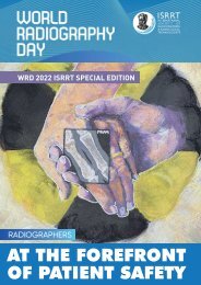
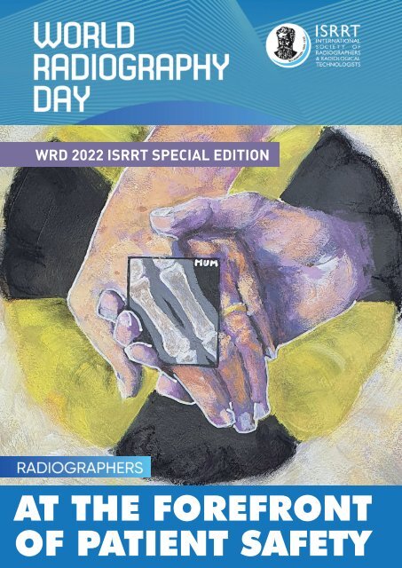
<strong>WRD</strong> <strong>2022</strong> ISRRT SPECIAL EDITION<br />
AT THE FOREFRONT<br />
OF PATIENT SAFETY
Cover artist - Leslie Robinson<br />
Leslie has again created a <strong>special</strong><br />
commission for the cover of this <strong>special</strong><br />
<strong>edition</strong> to celebrate World Radiography<br />
Day <strong>2022</strong> on November 8.<br />
Leslie trained to be a radiographer in the<br />
UK in the early 1980s and then worked as<br />
a radiographer in the UK and Saudi Arabia,<br />
eventually <strong>special</strong>ising in CT and MRI.<br />
She went on to become a radiography lecturer and researcher at the<br />
University of Salford in the UK.<br />
She has presented all over the world, has published widely and<br />
achieved a Doctorate in Education. She was recognised for her work<br />
by the UK Society of Radiographers, receiving the Radiographer of the<br />
Year prize (regional and national) twice. This year she was awarded the<br />
prestigious Fellowship of the College of Radiographers.<br />
Leslie always enjoyed the visual element of radiography and, on<br />
retirement in 2018, she dusted off her paint brushes and started to<br />
paint again. Her love of painting the human form has been enhanced<br />
by many years of looking at and understanding radiographic anatomy.<br />
She has integrated this knowledge into her paintings to show both the<br />
abstract and objective sides of the human form.<br />
2
CONTENTS<br />
Message from the ISRRT President 5<br />
Message from the ISRRT Regional Director Europe 6<br />
Thematic approach – practicing radiographer’s view:<br />
Anne Dorte Blankholm, Denmark 11<br />
Beth Weber, USA 15<br />
William D. Bryan, USA 16<br />
Denise Chong, Singapore 18<br />
Debbie Gilley, USA 20<br />
Gill Harrison, United Kingdom 22<br />
Manuel José Cruz Duarte Lobo, Portugal 25<br />
Brandon Hirsch, USA 28<br />
Kitiwat Khamwan, Thailand 30<br />
Mark Chukwudi Okeji, Nigeria 34<br />
Anastasia Sarchosoglou, Greece 36<br />
Ellian Schlattmann, Germany 38<br />
Boniface Yao, Côte d’Ivoire 42<br />
Edward Wong, Hong Kong 50<br />
ISRRT Societies – member views and initiatives on Artificial Intelligence:<br />
Australia 54<br />
India 57<br />
Indonesia 58<br />
Greece 62<br />
Portugal 64<br />
Taiwan 66<br />
Zimbabwe 69<br />
Contributing stakeholders views:<br />
Michael Fuchsjäger, ESR 72<br />
Jesse Clarijs-de Jong, Ricardo Khine, EFRS 74<br />
In this Special Edition, articles and reports by authors/societies<br />
do not necessarily reflect or represent the opinion<br />
and thesis of the ISRRT.<br />
ISRRT SPECIAL EDITION ON WORLD RADIOGRAPHY DAY <strong>2022</strong><br />
3
THEMATIC APPROACH – PRACTICING RADIOGRAPHER’S VIEW<br />
4 ISRRT SPECIAL EDITION ON WORLD RADIOGRAPHY DAY <strong>2022</strong>
Message from the<br />
ISRRT President<br />
By Donna Newman, ISRRT President<br />
Dear ISRRT Members and Radiography<br />
Professional Stakeholders,<br />
World Radiography Day will be celebrated<br />
around the world by radiographers/<br />
radiological technologists on November<br />
8, <strong>2022</strong>. This day has not only become an<br />
important time to celebrate our profession,<br />
but also to increase public awareness of<br />
the vital role that diagnostic imaging and<br />
radiation therapy plays in healthcare. This<br />
year’s theme was chosen to raise awareness,<br />
help educate and celebrate radiographers/<br />
radiological technologists in their daily<br />
practices highlighting how they are key to<br />
patient safety not only relating to radiation<br />
safety but other aspects of patients’ safety.<br />
I’m excited to share this <strong>special</strong> addition<br />
of World Radiography Day with the theme,<br />
“Radiographers at the Forefront of<br />
Patient Safety”, highlighting the role that<br />
radiographers/ radiological technologists<br />
play in promoting and maintaining radiation<br />
safety and general patient safety in all<br />
aspects of the patient journey.<br />
Part of the key success of the ISRRT is its<br />
member experts from around the world<br />
and we are delighted to share some of the<br />
daily best practices. As a global organization<br />
we hope that our members and health<br />
professionals around the world will read<br />
this book and incorporate what they learn<br />
from our authors and help drive the ISRRT<br />
key messages of creating, influencing and<br />
impacting change within their countries and<br />
daily practices.<br />
The World Health Authority (WHO) is leading<br />
a global initiative on radiation safety in<br />
strengthening medical Imaging in an effort<br />
to have Universal Health Care Coverage in all<br />
countries around the world. In this process,<br />
the Radiation and Health Unit of the WHO<br />
works to improve the quality and safety of the<br />
use of radiation for medical imaging which is<br />
essential for the diagnosis and management<br />
of non-communicable diseases (cancer,<br />
heart disease, stroke, chronic disease the<br />
obstructive pulmonary disease).<br />
Additionally, the Global Action Plan on Patient<br />
Safety 2020-2030, towards eliminating<br />
avoidable harm in Health Care has been<br />
developed as part of this strategy and<br />
adopted by the WHO Assembly in May<br />
of 2021. The WHO also decided to have<br />
Director-General report back on progress<br />
in the implementation of the global patient<br />
safety action plan 2021–2030 to the Seventysixth<br />
World Health Assembly in 2023. This<br />
important global document focuses on the<br />
WHO’s Global action plan on patient safety<br />
and provides a Framework of suggested<br />
actions for governments, non-State<br />
actors, international organizations and<br />
intergovernmental organizations. These<br />
actions have been grouped under the<br />
following seven strategic objectives (SOs).<br />
SO1: Make zero avoidable harm to patients a<br />
state of mind and a rule of engagement<br />
in the planning and delivery of health<br />
care everywhere.<br />
SO2: Build high reliability health systems<br />
and health organizations that protect<br />
patients daily from harm.<br />
SO3: Assure the safety of every clinical<br />
process.<br />
SO4: Engage and empower patients and<br />
families to help and support the<br />
journey to safer health care. SO5:<br />
Inspire, educate, skill and protect<br />
every health worker to contribute to<br />
the design and delivery of safe care<br />
systems.<br />
SO6: Ensure a constant flow of information<br />
and knowledge to drive mitigation of<br />
risk, a reduction in levels of avoidable<br />
harm and improvements in the safety<br />
of care.<br />
SO7: Develop and sustain multisectoral and<br />
multinational synergy, solidarity and<br />
partnerships to improve patient safety<br />
and quality of care.<br />
The ISRRT has contributed to this WHO<br />
strategic agenda representing your global<br />
voice around the world in the development of<br />
this plan. As a global organization we are also<br />
committed to disseminating and contributing<br />
to the success of this plan. We continue to<br />
do this by developing educational materials<br />
which includes radiation safety and safety for<br />
our patients through our ISRRT Facebook live<br />
session, the ISRRT E-learning platform and<br />
our ISRRT Position statements. Continuing<br />
with the ISRRT strategy for elevating safety,<br />
the World Radiography Day Special Edition<br />
is a perfect forum to showcase some best<br />
practices from our member experts around<br />
the world.<br />
The ISRRT collaborated with ISRRT Member<br />
Experts, ISRRT Member Societies and<br />
Regional Stakeholders to create this <strong>special</strong><br />
addition highlighting the Radiographers/<br />
Radiological Technologist’s crucial role in<br />
their protocols and procedures ensuring<br />
safe practice not only in radiation but also<br />
other areas of our engagement with patients.<br />
You will see these strategies demonstrated<br />
throughout this issue as radiographers/<br />
radiological technologist share their best<br />
practices toward radiation safety and general<br />
safety in their daily roles and practices.<br />
This educational publication will demonstrate<br />
the high educational and professional<br />
standards radiographers/radiological<br />
technologists live by each day and will help<br />
strengthen radiation safety and the general<br />
safety of patient care worldwide.<br />
Enjoy this issue and the ISRRT hopes that<br />
its ISRRT members and Radiography<br />
Stakeholders will find relevant resources,<br />
educational tips and ideas that will help<br />
radiographers /radiological technologists<br />
influence, impact and create change in their<br />
daily workplaces in countries across the<br />
world. Just think if each of us incorporates<br />
one idea from these articles within our daily<br />
practice, we will have contributed to elevating<br />
patient safety globally.<br />
ISRRT SPECIAL EDITION ON WORLD RADIOGRAPHY DAY <strong>2022</strong><br />
5
MESSAGE FROM THE ISRRT REGIONAL DIRECTOR EUROPE<br />
Patient safety beyond<br />
radiation protection in CT<br />
By E. Agadakos, ISRRT Regional Director Europe<br />
Introduction<br />
PATIENT safety according to the WHO<br />
is the absence of preventable harm to a<br />
patient during the process of health care<br />
and reduction of risk of unnecessary<br />
harm associated with health care to an<br />
acceptable minimum. Whereas patient harm<br />
is defined as, “an injury that was caused<br />
by medical management (rather than the<br />
underlying disease) and that prolonged the<br />
hospitalization, produced a disability at the<br />
time of discharge, or both.” (Harvard Medical<br />
Practice Study)<br />
Purpose<br />
The article aims to highlight the significance<br />
of our professional role in patient safety<br />
beyond radiation protection and in upholding<br />
the functionality of healthcare systems<br />
during this pandemic<br />
Background<br />
WHO data:<br />
• The occurrence of adverse events due to<br />
unsafe care is likely one of the 10 leading<br />
causes of death and disability in the<br />
world 1 .<br />
• Each year, 134 million adverse events<br />
occur in hospitals in low- and middleincome<br />
countries (LMICs), due to unsafe<br />
care, resulting in 2.6 million deaths 2 .<br />
• In high-income countries, it is estimated<br />
that one in every 10 patients is harmed<br />
while receiving hospital care 3 . The harm<br />
can be caused by a range of adverse<br />
events, with nearly 50% of them being<br />
preventable 4 .<br />
In computed tomography (CT), patient<br />
safety is traditionally linked to radiation<br />
protection and contrast medium (CM) issues.<br />
Although CT examinations are performed<br />
based on the three principles of radiation<br />
protection (i.e. justification, optimization<br />
and dose restrictions - ALARA), the method<br />
remains a high radiation dose modality.<br />
While authoritative bodies, healthcare<br />
professionals, equipment vendors are<br />
troubled with the direct risks and continue<br />
to promote and develop dose reduction<br />
strategies, CT radiographers are often faced<br />
with a wide range of patient safety concerns.<br />
According to the Joint Commission<br />
International, there are 6 International<br />
Patient Safety Goals (IPSGs), and CT<br />
radiographers need to be aware of the<br />
actions to attain them. 5<br />
1. Identify patients correctly<br />
Patient identification is a cause of error for<br />
several adverse events. Radiographers must<br />
use at least two steps to identify patients<br />
prior to a CT examination. Ask patients to<br />
tell you their name. Repeat the question if<br />
necessary and ask for their D.O.B. Never<br />
state the patient’s name. Allow time for the<br />
patient to tell you their name. Simply ask:<br />
“What is your name?” Always cross check<br />
patient name against their ID bracelet. The<br />
ward and bed number is an inappropriate<br />
method to identify patients. 6<br />
2. Improve effective communication<br />
Obtaining patient history and conveying<br />
instructions prior and after the CT<br />
examination is always important for patient<br />
cooperation and patient consent as well as<br />
for guidance following IV of CM.<br />
Verbal errors, and ineffective communication,<br />
are a common cause of medical errors,<br />
risking patient safety. Moreover, they can<br />
minimize patient cooperation affecting the CT<br />
Figure 1: Facts on Patient Safety (Annual data from WHO& JCI).<br />
6 ISRRT SPECIAL EDITION ON WORLD RADIOGRAPHY DAY <strong>2022</strong>
Figure 2: International Patient Safety Goals (Joint Commission International,JCI).<br />
scanning procedure causing motion artifacts<br />
due to breathing and patient movement. In<br />
addition they may result in adverse IV CM<br />
events. Always ensure that the patient has<br />
understood your questions and instructions.<br />
The 5Cs are an excellent tool leading to<br />
effective communication 7 .<br />
Poor communication risk factors include:<br />
1. Disruptive behavior including rude<br />
language or verbal abuse.<br />
2. Environmental noise issues such as cell<br />
phones, pagers, and phones.<br />
3. Cultural differences among patients and<br />
providers.<br />
The 5 Cs to effective communication<br />
• Clear: Words used should be simple<br />
and the easy to understand.<br />
• Concise: Message should be kept<br />
as concise as possible.<br />
• Complete: The information provided<br />
should be complete. Therefore,<br />
it should answer the basic Why?<br />
When? Where? How? questions<br />
related to the subject matter.<br />
• Courteous: Delivered in a calm tone<br />
and positive manner and respectful<br />
to needs and preferences of the<br />
receiver. Body language and nonverbal<br />
cues should portray respect<br />
and empathy.<br />
• Consistent: The message and<br />
information given should be<br />
the same, irrespective of who<br />
is providing or receiving that<br />
information.<br />
4. Hierarchy issues.<br />
5. Providers acting as autonomous agents.<br />
6. Personality differences.<br />
7. Language barriers.<br />
8. Lack of working as a team.<br />
9. Multiple conversations are occurring<br />
simultaneously.<br />
10. Socioeconomic variables, such as<br />
education and literacy.<br />
3. Improve safety of high alert medication<br />
Adverse drug reaction is the most common<br />
safety incident in CT, with medication and<br />
IV safety is the second most common safety<br />
incident in CT. according to a study of multiyear<br />
experience in incident reporting in CT 8<br />
For instance a medication error may occur<br />
following the administration of high dose<br />
epinephrine to manage a severe allergic<br />
reaction to IV CM or the use of CM in patients<br />
with contraindications causing nephrotoxicity<br />
and eventually renal failure. Such adverse<br />
incidents are avoided with comprehensive<br />
patient history, electronic patient files and<br />
adherence to ESUR guidelines on IV CM<br />
administration based on GFR levels. 9 It is<br />
significant that patients are differentiated<br />
correctly depending on their previous history<br />
related to CM allergies in order to undergo<br />
the indicated procedure with or without IV<br />
CM. Furthermore radiographers need to<br />
be alert and recognize an allergic reaction<br />
to CM early using the Ring and Messmer<br />
classification of anaphylaxis severity 10 .<br />
In general the CM administered is decided<br />
prior to the examination while the iodine concentration<br />
and flow rate depend on patient<br />
body weight and diagnostic requirements.<br />
The procedure is performed with automatic<br />
injectors and IV pumps which need to function<br />
correctly and as intended to minimize<br />
the risk of:<br />
i) Extravasation<br />
ii) air embolism<br />
iii) nephrotoxicity<br />
iv) lactic acidosis<br />
v) Iodine related reactions- CM allergy<br />
In addition it is expected that air is removed<br />
from patient tubing prior to the injection, vein<br />
integrity is maintained and CM is heated.<br />
4. Ensure correct site, correct procedure,<br />
correct side<br />
Radiographers must confirm that the correct<br />
patient undergoes the correct procedure<br />
including the correct region and when<br />
applicable the correct side. Ask the patient<br />
using simple language. For eg “What exam<br />
are you having today?” .<br />
• Always cross check corresponding<br />
referral or scheduled exam on PACS-RIS<br />
• Avoid the use of mirroring and or other<br />
post processing correction tools<br />
• Apply correct side markers when<br />
applicable and patient position directly on<br />
your scan data and topogram by assigning<br />
correct patient position and table<br />
direction eg. supine head first<br />
• Always ensure equipment is maintained<br />
and functions according to vendors<br />
specification 11<br />
5. Reduce the risk of healthcare infections<br />
Generally, radiographers are aware of<br />
the basic infection control principles and<br />
CT departments seem well prepared to<br />
cope with highly communicable infectious<br />
diseases. Unfortunately these expectations<br />
ISRRT SPECIAL EDITION ON WORLD RADIOGRAPHY DAY <strong>2022</strong><br />
7
THEMATIC APPROACH – PRACTICING RADIOGRAPHER’S VIEW<br />
Figure 4: ISRRT e Learning platform on CT, Position Statement on patient safety (ISRRT).<br />
were quickly dismissed with the first wave<br />
of the COVID-19 pandemic in early 2020. The<br />
healthcare systems were too weak and staff<br />
were not prepared to detain the impact of the<br />
pandemic Social distancing was inevitable,<br />
clinical radiographers in hospitals and<br />
institutions complained of limited access to<br />
PPE, inadequate staff training, shortage of<br />
PPE, reduced understanding of PPE use and<br />
confused PPE guidance. 12<br />
The past two years radiographers had to<br />
overcome an airborne disease with an<br />
amazingly high transmission rate. Their<br />
anxiety in managing patients at the front line<br />
while protecting themselves and other staff<br />
from infection was affecting their practice<br />
and their well being. Many were infected<br />
leading to staff shortage in CT departments.<br />
The appeal for working guidelines became<br />
apparent. However the initial working<br />
guidelines assumed that healthcare would<br />
be delivered at a safe social distance of<br />
1.5 m and when inevitable, healthcare<br />
professionals were instructed to wear<br />
Personal Protection Equipment, PPE (WHO,<br />
ECDC, CDC National Infection Control<br />
Committees). Admittedly, CT imaging<br />
cannot be performed at 1.5m distance<br />
from the patient and PPE was not readily<br />
available for radiographers. Either, due to<br />
the lack of supplies or due to a minority of<br />
government officials who failed to recognize<br />
radiographers as front line professionals.<br />
This unjust perception was resolved by the<br />
quick response of professional societies.<br />
Moreover, aimed guidelines were necessary<br />
to strengthen their professional capacity and<br />
self confidence to handle patients in need<br />
of CT examinations, particularly when CT<br />
was initially considered the examination of<br />
choice for COVID-19 detection and follow<br />
up. The Panhellenic Society of Radiological<br />
Technologists in Greece response was<br />
to develop and to disseminate focused<br />
guidelines for radiographers in all modalities<br />
via free webinars by the end of March 2020. 13<br />
These instructions reshaped the general<br />
guidelines published by WHO, ECDC,<br />
CDC and the National Infection Control<br />
Committee to support our professionals who<br />
had no previous knowledge and sufficient<br />
experience in similar situations. The<br />
Figure 3: Managing asymptomatic patients in CT (Panhellenic Society of RTs 22/03/2020).<br />
8 ISRRT SPECIAL EDITION ON WORLD RADIOGRAPHY DAY <strong>2022</strong>
Figure 5: Use of chest imaging in COVID-19. A rapid advice (WHO, 11/6/2020).<br />
feedback from participants was encouraging<br />
and an e - conference on clinical practice<br />
during the pandemic followed including<br />
academics and radiography students. This<br />
significant work was later incorporated in the<br />
ISRRT e-learning platform and courses, the<br />
ISRRT COVID-19 practical guidelines, ISRRT<br />
rapid guide, ISRRT Position Statement and<br />
WHO publication titled “Use of chest imaging<br />
in COVID-19. A rapid advice” 14,15<br />
Once radiographers visit the aforementioned<br />
web pages they will become familiar and<br />
are expected to implement the following<br />
five fundamental principles to minimize<br />
contagion:<br />
1. Separate red (contaminated) and blue<br />
(clean) zones,<br />
2. Exercise donning and doffing procedures<br />
of PPE correctly.<br />
3. Apply the WHO five moments hand<br />
hygiene effectively<br />
4. Ensure appropriate & frequent room and<br />
equipment decontamination/disinfection<br />
and ventilation according to local<br />
regulations and vendor specifications<br />
5. Practice with transparency i.e. record<br />
suspected and confirmed cases and<br />
report relevant incidents<br />
6. Reduce of the risk of patient harm from<br />
fall<br />
Hundreds of thousands of patients fall every<br />
year and of those 30-35% sustain an injury.<br />
According to JCI it is ranked among the top<br />
10 incidents that caused or created potential<br />
for serious patient injury or death 16 . In a<br />
survey of 415 radiology-related lawsuits in<br />
U.S., radiologic technologists appeared to be<br />
called to court most often in cases of patient<br />
falls. 17 Falls have been identified as an event<br />
that is preventable and should never occur.<br />
Frequently, radiographers support porters<br />
during lifting and moving patients to and<br />
from the CT table, consequently they should:<br />
• practice safe patient handling techniques,<br />
• communicate with patient and porters,<br />
• be aware of policies<br />
• use appropriate restraints and transfer<br />
equipment.<br />
Preventing adverse events in a medical<br />
imaging department can be challenging<br />
due to the number of patients seen, the<br />
amount of equipment in use, and the rapid<br />
pace of care delivery in this setting. Keep in<br />
mind that most medical errors occur before<br />
and after the procedure. Therefore it is<br />
essential that radiographers practice in a CT<br />
department which focuses on three actions:<br />
measure, intervene, and prevent.<br />
The healthcare system must first, identify<br />
and describe (measure) a safety issue, act to<br />
help the patient (intervene), and then avoid<br />
similar events in the future (prevent).<br />
Today most healthcare institutions exercise<br />
a proactive patient safety methodology<br />
shifting from a blame culture to Safety<br />
culture. They implement a sociotechnical<br />
approach to measure the wellness of a<br />
health system combining culture, processes<br />
and technology.<br />
Conclusion<br />
Despite all measures and safety precautions<br />
radiographers/radiological technologists<br />
remain at the point of service and thus<br />
maintaining a physical distance of 1.5 mm<br />
from their patient is inevitable. Although<br />
we are customarily committed to patient<br />
radiation protection our role extends further.<br />
Patient safety requires that radiographers<br />
practice encompassing the six IPSGs, aiming<br />
for enhanced patient safety and to safeguard<br />
the National Healthcare System during and<br />
after the coronavirus pandemic.<br />
We can only guarantee this by adhering to<br />
the working guidelines developed by our<br />
national society and the ISRRT Position<br />
Statement of the Radiographer/Radiological<br />
Technologist’s Role in Patient Care and<br />
Patient Safety August 2021<br />
References<br />
1. Jha AK. Presentation at the “Patient<br />
Safety – A Grand Challenge for<br />
Healthcare Professionals and<br />
Policymakers Alike” a Roundtable at the<br />
Grand Challenges Meeting of the Bill &<br />
ISRRT SPECIAL EDITION ON WORLD RADIOGRAPHY DAY <strong>2022</strong><br />
9
THEMATIC APPROACH – PRACTICING RADIOGRAPHER’S VIEW<br />
Take home message<br />
Answer these questions:<br />
1. Is my CT department safe enough<br />
for patients?<br />
2. Do I know enough about patient<br />
safety?<br />
3. Have I done enough to improve<br />
patient safety?<br />
4. Should my patient advocacy extend<br />
beyond<br />
Melinda Gates Foundation, 18 October<br />
2018 (https://globalhealth.harvard.edu/<br />
qualitypowerpoint, accessed 23 July<br />
2019).<br />
2.. National Academies of Sciences,<br />
Engineering, and Medicine. Crossing the<br />
global quality chasm: Improving health<br />
care worldwide. Washington (DC): The<br />
National Academies Press; 2018<br />
www.nap.edu/catalog/25152/crossingthe-global-quality-chasm-improvinghealth-care-worldwide,<br />
(accessed 01<br />
July <strong>2022</strong>).<br />
3.. Slawomirski L, Auraaen A, Klazinga<br />
N. The economics of patient safety:<br />
strengthening a value-based approach<br />
to reducing patient harm at national<br />
level. Paris: OECD; 2017<br />
www.oecd.org/els/health-systems/<br />
The-economics-of-patient-safety-<br />
March-2017.pdf, (ac-cessed 1 July<br />
2019).<br />
4. de Vries EN, Ramrattan MA,<br />
Smorenburg SM, Gouma DJ,<br />
Boermeester MA. The incidence and<br />
nature of in-hospital adverse events:<br />
a systematic review. Qual Saf Health<br />
Care. 2008;17(3):216–23. http://doi.<br />
org/10.1136/qshc.2007.023622<br />
www.ncbi.nlm.nih.gov/<br />
pubmed/18519629<br />
5. www.jointcommissioninternational.org/<br />
standards/international-patient-safetygoals/<br />
6. Patient Safety in Medical Imaging: a<br />
joint paper of the European Society<br />
of Radiology (ESR) and the European<br />
Federation of Radiographer Societies<br />
(EFRS). European Society of Radiology<br />
(ESR) Insights into Imaging (2019) 10:45<br />
(https://doi.org/10.1186/s13244-019-<br />
0721-y<br />
7. Migliorini M., Portelli J.L., Effective<br />
communication by radiographers: at the<br />
heart of patient safety. ISRRT Special<br />
<strong>edition</strong> on World Radiography Day<br />
(2019); 30-33<br />
8. Mansouri, M., Aran, S., Shaqdan,<br />
K.W. et al. How often are Patients<br />
Harmed When They Visit the Computed<br />
Tomography Suite? A Multi-year<br />
Experience, in Incident Reporting, in a<br />
Large Academic Medical Center. Eur<br />
Radiol 26, 2064–2072 (2016). https://doi.<br />
org/10.1007/s00330-015-4061-0<br />
9. ESUR Guidelines on Contrast Agents<br />
10.0. European Society of Urogenital<br />
Radiology. www.esur.org/wp-content/<br />
uploads/<strong>2022</strong>/03/ESUR-Guidelines-<br />
10_0-Final-Version.pdf<br />
10. Poziomkowska-Gesicka I, Kurek M.<br />
Clinical Manifestations and Causes<br />
of Anaphylaxis. Analysis of 382 Cases<br />
from the Anaphylaxis Registry in<br />
West Pomerania Province in Poland.<br />
Int J Environ Res Public Health.<br />
2020 Apr 17;17(8):2787. doi: 10.3390/<br />
ijerph17082787. PMID: 32316622;<br />
PMCID: PMC7215547.<br />
11. ISRRT Guidance Document on QA/QC in<br />
CT, ISRRT July 2019<br />
12. Wang, J.; Zhou, M.; Liu, F. (1 May<br />
2020). Reasons for healthcare<br />
workers becoming infected with novel<br />
coronavirus disease 2019 (COVID-19)<br />
in China. Journal of Hospital Infection.<br />
105 (1): 100–101. doi:10.1016/j.<br />
jhin.2020.03.002. ISSN 0195-6701. PMC<br />
7134479. PMID 32147406. (accessed 01<br />
July <strong>2022</strong>)<br />
13. https://pasyta.gr/pandimia/%ce%b5<br />
%ce%be%ce%ad%cf%84%ce%b1%<br />
cf%83%ce%b7-%ce%bf%cf%80%ce<br />
%bf%ce%b9%ce%bf%cf%85%ce%b<br />
4%ce%b7%cf%80%ce%bf%cf%84%<br />
ce%b5-%ce%b1%cf%81%cf%81%c-<br />
f%8e%cf%83%cf%84%ce%bf%cf%85-<br />
%cf%83%cf%84%ce%bf%ce%bd-<br />
%cf%85-2 (accessed 01 July <strong>2022</strong>)<br />
14. ISRRT’s Position Statement of<br />
the Radiographer/Radiological<br />
Technologist’s Role in Patient Care and<br />
Patient Safety August 2021<br />
15. COVID-19- Use of chest imaging. A rapid<br />
advice WHO, 11/6/2020<br />
16. www.centerfortransforminghealthcare.<br />
org/improvement-topics/preventingfalls/<br />
(accessed 01 July <strong>2022</strong>)<br />
17. Lynn, Sandra D. 1999 Ethics and Law for<br />
the Radiologic Technologist. The Free<br />
Library (January, 1),<br />
www.thefreelibrary.com/Ethics<br />
and Law for the Radiologic<br />
Technologist.-a057642995 (accessed 01<br />
July <strong>2022</strong>)<br />
10 ISRRT SPECIAL EDITION ON WORLD RADIOGRAPHY DAY <strong>2022</strong>
Magnetic Resonance safety from<br />
a Danish perspective<br />
By Anne Dorte Blankholm, Denmark<br />
Anne Dorte Blankholm<br />
Anne Dorte was educated at the School<br />
of Radiography in Aarhus, Denmark in<br />
1987. After working in neuroradiology<br />
for two years at Aarhus University<br />
Hospital, in 1989 she went on to<br />
Magnetic Resonance Imaging. Later, a<br />
large part of her time became dedicated<br />
to cardiovascular MRI research and<br />
teaching MRI to radiographers, doctors<br />
and PhD students. She then worked as<br />
a research radiographer before being<br />
appointed as charge radiographer at the<br />
hospital’s MR Centre. She did a Master<br />
of Science in Magnetic Resonance<br />
Imaging at St. Martins College in<br />
Lancaster, UK, then returned to a<br />
research position at Aarhus University<br />
Hospital, while also working as a<br />
consultant for Philips Healthcare. In this<br />
position she did her PhD in medicine,<br />
entitled Non-contrast magnetic<br />
resonance angiography in renal<br />
transplantation and renal donation. She<br />
is now responsible for research in the<br />
profession of radiographers at Aarhus.<br />
Anne Dorte Blankholm is a Past<br />
President of the International Society<br />
for Magnetic resonance Radiographers<br />
and Technologists) and the Vice Chair of<br />
the Nordic Chapter of the International<br />
Society for Magnetic Resonance in<br />
Medicine, appointed MR expert by the<br />
European Federation of Radiographers<br />
Societies and Vice Chair of the<br />
Education Committee for the education<br />
of Radiographers, University College<br />
Nordjylland, DK.<br />
Introduction<br />
BEING a radiographer and working with<br />
Magnetic Resonance (MR) for more than<br />
30 years, MR safety and patient safety has<br />
always been on my agenda.<br />
When I started in MR in 1989, MR safety<br />
and patient safety was important. MR was<br />
a new imaging modality and MR education<br />
was scarce. Therefore, I was involved in<br />
starting MR education in Denmark in the<br />
early 90s. Very few implants had been<br />
tested back then and it was not uncommon<br />
that we had to decline to scan patients<br />
with implants.<br />
Things have developed over time, but<br />
we still have considerable work on the<br />
safety agenda. This article provides<br />
a brief summary of the history of MR<br />
safety, information about directives, MR<br />
safety guidelines and consensus papers.<br />
Education, teamwork and responsibility<br />
will be discussed and finally, I will mention<br />
some of the organizational and political<br />
work I am involved in.<br />
History<br />
In Denmark, we have strict government<br />
regulations for the use of x-rays. This<br />
is not the case for MR, which I believe<br />
is quite a riddle. Exposure to radiation<br />
in x-ray imaging may kill people over<br />
time due to the carcinogenic effect and<br />
potential development of cancer, which is<br />
increasing with increasing x-ray dose. In<br />
MR, we may kill people in seconds if they<br />
are hit by heavy metallic objects attracted<br />
by the strong magnetic field. The first<br />
deadly accident related to MR was reported<br />
in 2001, when the six-year-old Michael<br />
Colombini tragically died after being hit by<br />
an oxygen tank at a hospital in New York 1 .<br />
After more than 20 years (+30 years for<br />
MR), one should expect that we had learned<br />
to avoid such accidents, but unfortunately,<br />
they seem to continue to happen. In 2018,<br />
there was a deadly accident in India, in<br />
2019 two serious accidents in Sweden and<br />
in October 2021 another deadly accident in<br />
India 2-5 . All of these accidents could have<br />
been prevented.<br />
It is a concern in the MR community<br />
that MR-related accidents and incidents<br />
(adverse events) are underreported but<br />
documentation is beginning to be published<br />
in the literature 6-8 . Different databases<br />
have been established for the reporting of<br />
adverse events in the healthcare sector<br />
and in some countries, also in Denmark, it<br />
is mandatory to report adverse events to a<br />
national database. Most of the databases<br />
are not constructed specifically for MRrelated<br />
adverse events, which can make<br />
it difficult when searching for MR-related<br />
adverse events. A list of international<br />
databases and related guidance for<br />
reporting can be found at: www.ismrm.<br />
org/mr-safety-links/incident-reportingguidance/<br />
It is extremely important that<br />
adverse events are reported because it<br />
is the only way we can learn from other<br />
people’s mistakes and thereby prevent<br />
future incidents and accidents.<br />
The status of MR safety is not static. The<br />
MR discipline develops continuously with<br />
added higher and lower field strengths,<br />
stronger and faster gradients, and new<br />
sequences etc. – not to forget all the new<br />
types of implants, also including nonmedical<br />
implants. With an aging population<br />
and the increasing number of available<br />
implants, the need for a user-friendly<br />
database including all implants and<br />
devices and the associated MR labelling<br />
is becoming more and more urgent, both<br />
to ensure patient safety but also to save<br />
money and time resources. We need to<br />
update MR safety education regularly as<br />
recommended in several guidelines and<br />
consensus papers 9-13 . MR safety is very<br />
different now compared to the early years<br />
of MR.<br />
Directives, guidelines, and consensus<br />
papers<br />
There are no common European guidelines<br />
on MR safety. There is a Directive from the<br />
ISRRT SPECIAL EDITION ON WORLD RADIOGRAPHY DAY <strong>2022</strong><br />
11
THEMATIC APPROACH – PRACTICING RADIOGRAPHER’S VIEW<br />
European Parliament, Directive 2013/35/<br />
EU of the European parliament and the<br />
Council, on the minimum health and safety<br />
requirements regarding the exposure of<br />
workers to the risks arising from physical<br />
agents (electromagnetic fields). As the title<br />
indicates, this directive concerns the safety<br />
of workers and not patients 14 . However,<br />
some points in this directive also impact on<br />
patient safety, such as the demand for risk<br />
assessment as in Chapter II, Obligations Of<br />
Employers, Article 4, Assessment of risks<br />
and determination of exposure, 7, stating<br />
the following “The employer shall be in<br />
possession of an assessment of the risks in<br />
accordance with Article 9(1)(a) of Directive<br />
89/391/EEC and shall identify which<br />
measures must be taken in accordance<br />
with Article 5 of this Directive. The risk<br />
assessment may include the reasons why<br />
the employer considers that the nature<br />
and the extent of the risks related to<br />
electromagnetic fields make a further<br />
detailed risk assessment unnecessary.<br />
The risk assessment shall be updated on<br />
a regular basis, particularly if there have<br />
been significant changes which could<br />
render it out of date, or if the results of the<br />
health surveillance referred to in Article 8<br />
show this to be necessary.” 14<br />
Consensus papers are available<br />
regarding recommended responsibilities<br />
for management of MR safety 9 and for<br />
recommended minimum requirements for<br />
performing MRI in humans in a research<br />
setting 10 .<br />
The most well known and relatively recently<br />
updated MR safety guidelines are from the<br />
United Kingdom, Australia and the USA 11-13 .<br />
Some countries in Europe have their own<br />
MR safety guidelines but this, however,<br />
is not the case in Denmark or the other<br />
Scandinavian countries.<br />
Useful guidelines are available, but<br />
the question is if we follow them. In my<br />
experience, some departments in Denmark<br />
do not follow a guideline to the point<br />
because we do not have a guideline in<br />
Danish. Hence, sometimes the department<br />
leaders have never read a guideline and<br />
just rely on local regulations and homemade<br />
guidelines, which, in principle, may<br />
be outdated or insufficient. However, the<br />
MR community has always had a high<br />
degree of self-regulation, and we are<br />
highly dependent on local policies and<br />
procedures. This means that some MR<br />
departments, facilities and clinics have a<br />
high degree of MR safety, but we have to<br />
bear in mind that MR safety is never better<br />
than the extent to which staff and leaders<br />
are educated.<br />
Education<br />
Some of the guidelines define educational<br />
levels: 2. a person with supervision rights<br />
and educated to handle patients, perform<br />
the MR examination, be responsible for<br />
patient and staff safety, 1. a person at<br />
a lower level responsible only for own<br />
safety only in the MR environment and<br />
without supervision rights. A minimum<br />
of two levels is absolutely necessary<br />
because guests in the MR environment<br />
such as anesthesia staff, hospital porters<br />
and cleaning staff need a certain level of<br />
education in MR safety to contribute to a<br />
safe MR environment for patients and staff.<br />
It has been shown that guests in the MR<br />
environment constitute a safety risk 6 .<br />
Updates on MR safety education is needed<br />
on a regular basis both because techniques<br />
develop and because we need refresher<br />
courses just as in other areas such as<br />
in cardiopulmonary resuscitation (CPR).<br />
Updates are recommended in several<br />
guidelines and consensus papers 9-13 .<br />
Consensus papers and MR safety<br />
guidelines recommend a teamwork<br />
approach where a Magnetic Resonance<br />
Safety Medical Director (MRMD)/ Magnetic<br />
Resonance Safety Research Director<br />
(MRRD), a Magnetic Resonance Safety<br />
Officer (MRSO) and a Magnetic Resonance<br />
Safety Expert (MRSE) are defined 10 .<br />
The MRMD/MRDR is the person who<br />
is operationally responsible for the MR<br />
facility. This title is in clinical departments<br />
held by a medical doctor, whereas in<br />
research departments the MRRD can be<br />
held by a person with another professional<br />
background. The MRSO is typically held<br />
by a radiographer or a MR technologist<br />
responsible for MR safety in daily practice.<br />
The MRSO has a key role in the day-to day<br />
safe operation of the MR unit. The MRSE<br />
can be held by a MR physicist or another<br />
professional with higher level of technical<br />
expertise in MR (could be found locally<br />
for larger clinical sites or externally for<br />
smaller ones). In practice, we have been<br />
working with a model like this for many<br />
years at Danish university hospitals. With<br />
the consensus papers and guidelines<br />
recommending this model, which has been<br />
widely endorsed by several professional<br />
bodies in Europe, USA, and Australia, we<br />
are obliged to work with a model like this 9-<br />
13<br />
. This may sound easy, and departments/<br />
facility leaders appoint people for these<br />
positions, but we must bear in mind that<br />
titles are not enough - education must<br />
be an integrated part. A radiologist is not<br />
necessarily well educated in MR safety, a<br />
physicist or an engineer is not necessarily a<br />
<strong>special</strong>ist in MR safety and a radiographer<br />
or MR technologist is not by default<br />
educated in MR safety at a level of expertise<br />
to fulfil a role of an MRSO.<br />
Therefore, it must be ensured that the level<br />
and extent of education for these safety<br />
roles is defined, and that education at this<br />
level is available and preferably certified to<br />
match the education system in the country<br />
in question.<br />
Educational content<br />
In 2021, the European Federation<br />
of Radiographer Societies (EFRS)<br />
published an MRSO Role Descriptor:<br />
A European Qualifications Framework<br />
(EQF) benchmarking document (15). The<br />
purpose of this document was to propose<br />
a robust framework of knowledge, skills<br />
and competences required by the MRSOs<br />
and to help inform and standardize the<br />
requirements for MR safety training of MR<br />
radiographers in Europe 15 .<br />
This document describes the roles of<br />
the MRSO in different countries, the key<br />
subjects the MRSO should know about,<br />
as well as the knowledge, skills and<br />
12 ISRRT SPECIAL EDITION ON WORLD RADIOGRAPHY DAY <strong>2022</strong>
competences required for a European<br />
MRSO suggesting that the knowledge<br />
should be at EQF level 7, which is equal<br />
to master’s level 15 .<br />
Certification<br />
Currently, courses are offered in several<br />
places and countries. The only certification<br />
I know of is made by The American Board<br />
of Magnetic Resonance Safety (ABMRS).<br />
They conduct examinations for both MRMD/<br />
MRRD, MRSO and MRSE. The content of<br />
the exam (I have only taken the MRSO) is<br />
perfectly sufficient, well organized and<br />
executed. I suggest that we create our own<br />
national or European certification suited<br />
for our educational system. It should not<br />
be necessary for radiographers to travel to<br />
other countries to undertake MRSO courses<br />
or examinations or have representatives<br />
from the AMBRS to travel and organize<br />
the examinations, this should be available<br />
nationally. There are many people who need<br />
these educational courses, and traveling is<br />
expensive and not sustainable.<br />
Therefore, I currently collaborate with a<br />
Danish university to explore the possibility<br />
of setting up MRSO courses.<br />
Locally, I have taken the initiative to<br />
launch e-learning courses. Support was<br />
given by the Aarhus University Hospital<br />
management team. In collaboration with<br />
the regional center for e-learning and an<br />
MR physicist, I created basic and advanced<br />
e-learning courses recently launched in<br />
Central Denmark Region. The intention is<br />
that this course can supplement in-person<br />
training and be used as annual refresher<br />
courses hoping that we reach more<br />
people than with the in-person courses.<br />
It takes many resources to gather entire<br />
departments for in-person teaching,<br />
hence in a busy working environment,<br />
e-learning may be more flexible because<br />
it can be undertaken at any time where it<br />
suits the individual. At Aarhus University<br />
Hospital and in collaboration with The<br />
Danish Council of Radiographers, I arrange<br />
biannual Nordic MR safety meetings. These<br />
meetings have become very popular, and it<br />
is our plan to continue arranging these.<br />
Organizational, political, and<br />
strategic work<br />
As mentioned in the background, MR<br />
safety has always been high on my agenda.<br />
Therefore, I find it strange that we do not<br />
have the same strict regulations for MR<br />
as for x-rays. For several years I have<br />
been collaborating with the Danish council<br />
of radiographers (Radiograf Rådet, RR).<br />
In 2019, we arranged a conference on<br />
MR safety at the Danish parliament. We<br />
aimed to attract political attention to the<br />
importance of MR safety for the patients<br />
and advocated for the ministry of health<br />
to take the initiative to establish a Danish<br />
guideline for MR safety. The conference<br />
was well attended, and the politicians<br />
showed great interest. Then we had<br />
a referendum in Denmark and a new<br />
government, followed by the COVID-19<br />
pandemic, which stole the political focus.<br />
Therefore, I contacted the RR and together<br />
with the president of the RR, Chartlotte<br />
Graungaard, I took the initiative to form<br />
a Danish MR safety guideline. We started<br />
by contacting the Danish Society of<br />
Radiologists (Dansk Radiologisk Selskab,<br />
(DRS)), The Danish Society of Medical<br />
Physicists (Dansk Selskab for Medicinsk<br />
Fysik (DSMF)) and the Danish society<br />
for medical magnetic resonance (Dansk<br />
Selskab for Medicinsk Magnetisk Resonans<br />
(DSMMR)). In collaboration, we founded a<br />
steering committee and a working group<br />
which are currently creating a Danish<br />
national guideline on MR safety. When the<br />
guideline work is finalized, there will be<br />
a hearing including the political system,<br />
professional societies, public and private<br />
hospital leaders etc. It is our hope that the<br />
guideline will be well received, by leaders<br />
in the healthcare sector and the politicians,<br />
who we hope will endorse it.<br />
Norway and Sweden are also in the process<br />
of creating their own national guidelines<br />
for MR safety but for the time being no<br />
formal collaboration is going on to form a<br />
Scandinavian or Nordic guideline for MR<br />
safety. I acknowledge that we need MR<br />
safety guidelines in our own languages<br />
but a collaboration between the Nordic<br />
countries would be useful.<br />
The number of MR examinations and the<br />
number of patients with implants, both<br />
medical and non-medical, are increasing.<br />
Still, we are occasionally challenged to<br />
find out exactly what kind of implants<br />
the patients have. We need exactly the<br />
type, name, and number of the implant<br />
to find the MR labelling. Therefore, this<br />
information should always be listed<br />
in the patient’s medical record, and a<br />
comprehensive user-friendly implant<br />
databases is still on our wish list as<br />
radiographers and MR technologists.<br />
Research<br />
Research is performed in MR safety<br />
and the increasing academization of the<br />
radiographer profession has resulted in<br />
radiographers publishing in this area.<br />
However, more research is needed to<br />
focus attention on best practice in MR<br />
safety, the importance of reporting MRrelated<br />
adverse events, educational needs,<br />
interdisciplinary teamwork and sufficient<br />
staffing to ensure patient safety in relation<br />
to MR.<br />
Summary<br />
Over the years, MR safety has developed<br />
and changed. In Denmark we still lack a<br />
national guideline, but work is currently<br />
in progress. We need to attract the<br />
attention of leaders and authorities to<br />
the importance of teamwork, education,<br />
minimum staffing, regular MR safety<br />
education updates and general MR safety<br />
risk assessment of the MR facilities.<br />
Furthermore, it must be ensured that the<br />
necessary information regarding implants<br />
is available in the patient’s medical record<br />
and a user-friendly implant database is<br />
established including easy access to the<br />
MR labelling of the implants and devices.<br />
References<br />
1. https://abcnews.go.com/US/<br />
story?id=92745&page=1<br />
2. https://timesofindia.indiatimes.com/<br />
city/mumbai/was-2018-mri-death-atnair-hospital-probed-bombay-hc-asksbmc/articleshow/70670328.cms<br />
3. www.patientsafetysolutions.com/docs/<br />
November_5_2019_A_Near_Fatal_<br />
ISRRT SPECIAL EDITION ON WORLD RADIOGRAPHY DAY <strong>2022</strong><br />
13
THEMATIC APPROACH – PRACTICING RADIOGRAPHER’S VIEW<br />
MRI_Incident.htm<br />
4. www.itnonline.com/article/nurseinjured-mri-accident-swedish-hospital<br />
5. www.auntminnieeurope.com/index. –<br />
er&sub=def&pag=dis&ItemID=620730<br />
6. Blankholm AD, Hansson B. Incident<br />
reporting and level of MR safety<br />
education: A Danish national study.<br />
Radiography 2020;26(2):147-153.<br />
7. Hansson B, Olsrud J, Wilen J, Owman<br />
T, Hoglund P, Bjorkman-Burtscher IM.<br />
Swedish national survey on MR safety<br />
compared with CT: a false sense of<br />
security? Eur Radiol 2019;30(4):1918-<br />
1926.<br />
8. Kihlberg J, Hansson B, Hall A,<br />
Tisell A, Lundberg P. Magnetic<br />
resonance imaging incidents are<br />
severely underreported: a finding in<br />
a multicentre interview survey. Eur<br />
Radiol. <strong>2022</strong>; 32(1):477-488.<br />
9. Calamante F, Ittermann B, Kanal<br />
E, Inter-Society Working Group<br />
on MRS, Norris D. Recommended<br />
responsibilities for management of MR<br />
safety. Journal of magnetic resonance<br />
imaging : JMRI 2016;44(5):1067-1069.<br />
10. Calamante F, Faulkner WH Jr,<br />
Ittermann B, et al. MR system<br />
operator: recommended minimum<br />
requirements for performing MRI in<br />
human subjects in a research setting.<br />
Journal of Magnetic Resonance<br />
Imaging : JMRI 2015;41(4):899-902.<br />
11. MHRA Medicines and Healthcare<br />
Products Regulatory Agency. Safety<br />
Guidelines for Magnetic Resonance<br />
Imaging Equipment in Clinical Use.<br />
The Medicines and Healthcare<br />
Products Regulatory Agency; Feb 2021.<br />
12. Kanal E, Greenberg. T, Hoff MN, et<br />
al. ACR Manual on MR Safety. 1.0 ed.<br />
Volume 2020: American College of<br />
Radiology; 2020.<br />
13. Thee Royal Australien and New<br />
Zealand College of Radiologists®.<br />
MRI Safety Guidelinees. Version 3.0<br />
Published 2021.<br />
www.ranzcr.com/college/documentlibrary/ranzcr-mri-safety-guidelines<br />
14. The European Parliament and the<br />
Council. Directive 2013/35/Eu ff<br />
The European Parliament and of<br />
the Council of 26 June 2013 on<br />
the minimum health and safety<br />
requirements regarding the exposure<br />
of workers to the risks arising from<br />
physical agents (electromagnetic<br />
fields) (20th individual Directive<br />
within the meaning of Article 16(1) of<br />
Directive 89/391/EEC) and repealing<br />
Directive 2004/40/EC,. The European<br />
Parliament and of the Council.<br />
Directive2013/35/EU. 2013.<br />
15. EFRS MRSO working group.<br />
Blankholm AD, Bougais C,<br />
Malamateniuo C, McGee A, Owman<br />
T and Silva V. European Federation<br />
of Radiographer Societies (EFRS)<br />
published Magnetic Resonance Safety<br />
Officer (MRSO) Role Descriptor: An<br />
European Qualifications Framework<br />
(EQF) benchmarking document.<br />
https://api.efrs.eu/api/assets/<br />
posts/256<br />
14 ISRRT SPECIAL EDITION ON WORLD RADIOGRAPHY DAY <strong>2022</strong>
<strong>2022</strong> Radiographers at the<br />
forefront of patient safety<br />
By Beth Weber, USA<br />
Beth Weber<br />
Beth Weber, MPH, RT(R), RDMS,<br />
CRA, FASRT, has been the Director<br />
of Imaging Services and Privacy<br />
Officer for the Avera Heart Hospital<br />
in Sioux Falls, South Dakota since<br />
2000.<br />
Beth is involved in the Avera Health<br />
Systems Radiology Service Line,<br />
serving as chair of the Quality and<br />
Safety Committee.<br />
Beth received a Master’s in<br />
Public Health with a certificate in<br />
Healthcare Executive studies from<br />
the University of Minnesota in 2007.<br />
Beth’s progressive professional<br />
experience includes being a staff<br />
Diagnostic Radiologic Technologist,<br />
Sonographer, Chief Technologist,<br />
and Director of Imaging Services.<br />
Beth served four years on the Board<br />
of Directors of the American Society<br />
of Radiologic Technologists /ASRT<br />
as Vice Speaker and Speaker of the<br />
House of Delegates. Beth currently<br />
serves on the American Registry<br />
of Radiologic Technologists / ARRT<br />
Board of Trustees. Beth is part of the<br />
ARRT legislative committee.<br />
A COMMON reason that Radiology<br />
Technologists enter the profession is<br />
“to help others”. Patient safety is an<br />
essential component of the Radiographers’<br />
responsibilities in helping others.<br />
The American Society of Radiologic<br />
Technologists / ASRT Radiography<br />
curriculum 1 ; includes patient safety in<br />
the didactic courses of Patient Care and<br />
Assessment, Infection Control, Radiation<br />
Protection, Professional Liability, and<br />
students further review patient safety<br />
during clinical instructions.<br />
During patient care and assessment the<br />
radiographer verifies the order is entered<br />
appropriately for the correct patient.<br />
Patients are transported and assisted<br />
safely on and off imaging tables. The exam<br />
is performed with; age specific protocol,<br />
proper patient identification, as well as<br />
marked with applicable side indicator<br />
marker.<br />
Radiographers pay close attention to<br />
infection control measures routinely<br />
applying universal precautions for the<br />
safety of the patient and themselves.<br />
When necessary the suitable isolation<br />
requirements are applied. During the<br />
recent pandemic, the technologist<br />
performed chest exams through the clear<br />
glass window of the patient’s room with<br />
the assistance of the nurse caring for the<br />
patient, thus reducing the number of staff<br />
donning and doffing personal protective<br />
equipment.<br />
The ALARA “as low as reasonably<br />
achievable” (ICRP103) 2 principle is<br />
synonymous with the radiographers’ role<br />
in patient safety. It is the radiographers’<br />
responsibility to protect the public, patient<br />
and themselves from unnecessary radiation<br />
exposure.<br />
of the 10 leading causes of death and<br />
disability in the world.(3) Unfortunately,<br />
sometimes safe patient care is not<br />
adequate to prevent errors from occurring,<br />
“Human Error” is sometimes not the<br />
liability of the radiographers. System<br />
failures need to be considered and<br />
reviewed.<br />
Radiographers are educationally prepared<br />
and clinical competent to perform<br />
examinations ensuring patient safety. The<br />
radiographers emphasis on patient safety<br />
is knowing they too will be a patient and<br />
need someone “to help them.”<br />
References<br />
1. ASRT American Society of Radiologic<br />
Technologists; Radiography Curriculum;<br />
www.asrt.org<br />
2. International Commission on<br />
Radiological Protection. ICRP<br />
www.icrp.org<br />
3. World Health Organization/ WHO report<br />
in 2019;<br />
www.who.org<br />
In the 2019 World Health Organization<br />
report finds; “the occurrence of adverse<br />
events due to unsafe care is likely one<br />
ISRRT SPECIAL EDITION ON WORLD RADIOGRAPHY DAY <strong>2022</strong><br />
15
THEMATIC APPROACH – PRACTICING RADIOGRAPHER’S VIEW<br />
A personal account of radiation safety<br />
in interventional radiology<br />
By William D. Bryan, USA<br />
William D. Bryan<br />
William Bryan is a registered<br />
radiologist assistant working in<br />
Columbia, Missouri.<br />
He received his bachelors degree from<br />
the University of Missouri Columbia<br />
in Columbia, Missouri. After receiving<br />
his bachelors degree, he worked as<br />
both a diagnostic and interventional<br />
radiographer for six years before<br />
perusing his masters degree.<br />
He graduated with his masters degree<br />
from Midwestern Sate University in<br />
Wichita Falls, Texas.<br />
He now works as a radiologist<br />
assistant at University of Missouri<br />
Hospital and Clinic in both diagnostic<br />
and interventional radiology.<br />
William is also active with volunteer<br />
work for the American Society of<br />
Radiologic Technologists.<br />
INTERVENTIONAL radiology is an exciting<br />
field where interventional radiographers<br />
assist physicians in using a fluoroscopic<br />
machine, commuted tomography machine,<br />
or ultrasound scanner to guide operators<br />
for image guided minimally invasive<br />
procedures. Examples of procedures include<br />
removing thrombus from a clotted vessel,<br />
treating cancer with image guided therapies,<br />
stopping bleeds, and placing long term<br />
venous access and countless additional<br />
procedures. While the interventional<br />
radiographer may not be positioning a<br />
broken limb on a cassette/image receptor<br />
and choosing exposure factors to obtain<br />
an image, we are still the stewards of<br />
proper radiation techniques needed for a<br />
procedure. We must rely on our training<br />
and education as radiographers to apply<br />
principles learned from radiography school<br />
to interventional practices. Potentially,<br />
interventional procedures can be lengthy<br />
and require a high radiation dose which<br />
could potentially lead to harmful effects<br />
of radiation exposure to patients and<br />
staff involved. Knowledge of imaging<br />
characteristics, patient safety, and operator<br />
safety ensure that we continue to assist the<br />
physicians we work next to while providing<br />
optimal patient care and follow best<br />
practices in reference to radiation safety.<br />
Most modern interventional units are<br />
digital imaging systems that use a x-ray<br />
tube and an imaging intensifier system<br />
with automatic exposure controls and<br />
<strong>special</strong>ized dosing protocols (usually vendor<br />
specific) to create an image. While the<br />
interventional radiographer is not setting<br />
the kv, ma, and time of exposure needed<br />
applying knowledge of basic imaging factors<br />
can help reduce patient and operator<br />
dose. The first exposure factor I like to<br />
remember is to collimate. Collimating an<br />
image uses filters within the x-ray tube to<br />
decrease the size of tissue exposed. Propper<br />
collimation ensures proper exposure to<br />
the area of anatomy while reducing scatter<br />
radiation. The second exposure factor I<br />
like to remember is magnification factors.<br />
Interventional radiologists may need<br />
to focus on fine detail of tiny anatomic<br />
structures to complete cases. One easy way<br />
to view these is to increase magnification<br />
of the x-ray tube however, this increases<br />
the amount of radiation used to create the<br />
image. Instead of magnifying the image,<br />
adjusting the imaging intensifier can<br />
sometimes yield the same result without<br />
changing exposure factors. Remember, the<br />
farther the image intensifier is away from<br />
the anatomy exposed, the more magnified<br />
the image will appear. The final imaging<br />
factor I like to remember is to always use<br />
pulsed fluoroscopy at the lowest frame rate<br />
required for the procedure. Using pulsed<br />
fluoroscopy at low frame rates reduces<br />
dose while maintaining diagnostic quality<br />
imaging. While interventional radiographers<br />
may not use the same imaging characters<br />
to create our images as our colleagues<br />
in other modalities, it is still important to<br />
remember and apply basics of exposures to<br />
our examinations.<br />
Patient safety is one of the pillars that<br />
radiographers strive to uphold. In diagnostic<br />
imaging and in interventional imaging, we<br />
are there to help ensure that every exam<br />
completed is done in a safe environment.<br />
Just as in diagnostic imaging, patient<br />
positioning is critical to ensure a safe<br />
exam. It is imperative that interventional<br />
technologists position patient on procedural<br />
tables in the proper way for the procedure<br />
to be completed. Proper patient positioning<br />
usually leads to a more efficient exam<br />
which usually lowers radiation dose. Patient<br />
shielding is rarely used in interventional<br />
imaging however, on the rare occasions that<br />
shields are used it is important to remember<br />
where the source of radiation is and that<br />
we want to position shields between the<br />
source and the image. If shields are used<br />
incorrectly, dose will actually increase.<br />
Finally, being mindful of the amount of time<br />
fluoroscopy is use for a procedure will help<br />
decrease dose. Obviously, radiation must<br />
16 ISRRT SPECIAL EDITION ON WORLD RADIOGRAPHY DAY <strong>2022</strong>
e used to watch wires and catheters in the<br />
body however, it is not always necessary<br />
to use radiation while panning the table,<br />
prepping devices, or monitoring patient<br />
vital signs. Decreasing the amount of time<br />
radiation is used decreases dose. All of the<br />
above are examples of how providing a safe<br />
environment for the patient can lead to a<br />
more efficient procedure with the lowest<br />
amount of radiation needed.<br />
As interventional radiographers we spend<br />
the vast majority of every day assisting with<br />
procedures that require the use of ionizing<br />
radiation. This leads to higher levels of<br />
occupational exposure than some of our<br />
collogues that work in other modalities of<br />
radiography. Lowering radiation exposure to<br />
ourselves is important to prevent potential<br />
side effects of occupational dose. The<br />
first way I lower occupational exposure to<br />
myself to wear a lead apron and leaded<br />
glassed while involved with a procedure.<br />
Wearing lead works to lower occupational<br />
exposure by scatter radiation interacting<br />
with and being absorbed by lead worn by<br />
the operator instead of scatter radiation<br />
interacting with tissues of the operator.<br />
This is important to remember in regards to<br />
operator exposure to radiosensitive tissues<br />
such as the thyroid and eyes. The next way I<br />
lower my occupational exposure is by always<br />
remembering the inverse square law and<br />
applying that to my daily practice. We all<br />
know that exposure reduces by the square of<br />
the distance, therefore the further away I am<br />
able to position myself from the source of<br />
radiation, the lower exposure I will receive.<br />
Newer machines on the market are even<br />
provide robot arms to manipulate wires and<br />
catheters thus allowing human operators to<br />
be further away from the source. Another<br />
creative way to lower operator exposure<br />
is to use surgical drapes and surgical<br />
wear that is imbedded with lead. Multiple<br />
manufactures sell drapes, surgical caps,<br />
surgical gowns, and disposable lead<br />
“chuks” that can be positioned on a sterile<br />
environment to absorbed scatter radiation<br />
and decrease exposure to others involved<br />
with the procedure. Lowering occupational<br />
dose theoretically could lead to a longer<br />
and more safe career practicing as an<br />
interventional radiographer.<br />
Interventional radiology is an exciting<br />
modality that relies heavily on the use of<br />
ionizing radiation to guide procedure to help<br />
patients we see on a daily basis. We all know<br />
the practice of ALARA (as low as reasonably<br />
achievable) and above are just a few<br />
examples of how I engage with that practice<br />
on my daily work as a radiographer. As the<br />
radiographer in the room working aside<br />
physicians, we are the stewards of radiation<br />
use and can help protect the patient and<br />
ourselves to wisely use x-rays to help our<br />
patients.<br />
ISRRT SPECIAL EDITION ON WORLD RADIOGRAPHY DAY <strong>2022</strong><br />
17
THEMATIC APPROACH – PRACTICING RADIOGRAPHER’S VIEW<br />
Radiographers at the forefront<br />
of patient safety<br />
By Denise Chong, Singapore<br />
Denise Chong<br />
Denise is the President of the<br />
Singapore Society of Radiographers<br />
and the Singapore representative<br />
of the International Society for<br />
Radiographers and Radiological<br />
technologists (ISRRT).<br />
She is a senior radiographer at<br />
National University Hospital (NUH)<br />
from the Department of Diagnostic<br />
Imaging and graduated with a<br />
Masters of Science in Ultrasound<br />
from University College Dublin,<br />
Ireland and has been working at<br />
NUH for 11 years. Denise performs<br />
general, paediatric and vascular<br />
ultrasound scans and has a <strong>special</strong><br />
interest in musculoskeletal (MSK)<br />
ultrasound. She currently is the<br />
ultrasound section lead in the<br />
department and also facilitates the<br />
advanced training of juniors in MSK<br />
ultrasound.<br />
Denise also <strong>special</strong>ises in the use<br />
of the EOS low dose X-ray system at<br />
NUH for dedicated long film scoliosis<br />
and lower limb 2D and 3D imaging.<br />
She also retains a keen interest in<br />
intra-operative radiography.<br />
AS MEDICAL imaging professionals,<br />
patient safety is a key tenet of our practice<br />
no matter what modality we <strong>special</strong>ize<br />
in. Whenever patients arrive at our<br />
departments and are under our care,<br />
it is our responsibility to ensure that<br />
their physical and psychological safety<br />
is protected. With a focus on ultrasound,<br />
this piece will briefly touch on aspects of<br />
safety in ultrasound – from the biological<br />
effects of ultrasound waves, environmental<br />
safety and infection control specific to<br />
our ultrasound practice, to less tangible,<br />
psychological impact that undergoing an<br />
ultrasound procedure can have on patients.<br />
Furthermore, ultrasound is an operator<br />
dependent modality with radiographers<br />
requiring to perform some level of reporting<br />
and as such competency becomes crucial<br />
in ensuring patient safety in ultrasound<br />
practice.<br />
Among the range of modalities in medical<br />
diagnostic imaging ultrasound often seen as<br />
the safest mode of imaging due to its lack<br />
of use of radiation. This key advantage of<br />
ultrasound, in addition to being cost effective<br />
and readily available, is the reason it is<br />
considered the modality of choice to image<br />
the more ‘vulnerable’ patient population that<br />
possesses particularly delicate structures<br />
such as paediatric and obstetrics patients.<br />
Since the early application of ultrasound as a<br />
diagnostic tool (Kane et al. 2004), there have<br />
been little to no reports of the detrimental<br />
acute effects of ultrasound on the human<br />
body, leading to the widespread belief that<br />
diagnostic ultrasound is harmless. It is<br />
important to remember however that the<br />
absence of evidence of harmful effects<br />
does not exclude the potential harm that<br />
ultrasound can have on the human body.<br />
The application of ultrasound waves on the<br />
body results in the deposition of energy<br />
within biological tissues which can lead<br />
to heating or thermal effects, mechanical<br />
interactions with molecules or cause tiny<br />
bubbles to form in fluids that can lead to<br />
cavitation. (Moderiano et al, 2018; Hedrick,<br />
2013) The heating properties of ultrasound<br />
are evident as it forms the basis for<br />
therapeutic ultrasound on musculoskeletal<br />
tissues. When used safely by trained<br />
professionals, this therapy can increase<br />
circulation to promote healing in muscles,<br />
tendons and nerves. In obstetric diagnostic<br />
ultrasound examinations however, small<br />
but sustained rises in temperature can<br />
cause cell damage within foetal tissues<br />
and even cause foetal abnormalities if a<br />
2°C temperature rise is sustained over an<br />
extended 2-hour period. The bubbles that<br />
form as a bioeffect of ultrasound can react<br />
to the soundwaves, undergo large changes<br />
in size, expanding and contracting rapidly<br />
before collapsing. This violent bursting<br />
of bubbles can fragment and damage<br />
delicate membranes. In modern day<br />
instrumentation, two indices were developed<br />
as indicators of the acoustic output power of<br />
the machines and the likelihood of inducing<br />
the aforementioned bioeffects. The Thermal<br />
Index (TI) and Mechanical Index (MI) are<br />
displayed on the ultrasound display at all<br />
times so radiographers can easily see what<br />
the potential risk is to the patient with<br />
regard to generating these bioeffects. (Ter<br />
Haar, 2011) To ensure the safety of patients<br />
and minimize these effects, radiographers<br />
are trained to pay attention to these indices<br />
and apply the ALARA principle in ultrasound<br />
by ensuring that scan times are not<br />
prolonged unnecessarily, e<strong>special</strong>ly over a<br />
fixed scan area.<br />
In terms of environmental safety, long<br />
dangling ultrasound transducer cables may<br />
trip patients and personnel who may not<br />
notice them. This is particularly important<br />
in the ultrasound setting where a large<br />
number of patients may be fasting and<br />
hypoglycemic, or may experience postural<br />
hypotension when arising from laying down<br />
after the duration of the scan. This may<br />
lead to dizzy spells and an unsteady gait,<br />
thereby increasing the chance of patient<br />
falls, e<strong>special</strong>ly if these cables are not tidied.<br />
Radiographers routinely keep transducer<br />
cables tidy, away from the floor and pay<br />
attention to these patients during and<br />
e<strong>special</strong>ly after the scan is complete.<br />
18 ISRRT SPECIAL EDITION ON WORLD RADIOGRAPHY DAY <strong>2022</strong>
Good infection control and hand hygiene<br />
practices are imperative in our daily work for<br />
our patient’s safety. These include regular<br />
hand washing and using alcohol hand rub<br />
frequently at the 5 moments of hand hygiene<br />
for each patient interaction, as well as<br />
paying attention to the use of appropriate<br />
personal protective equipment depending<br />
on the type of precaution. Cleaning and<br />
disinfection of common fomites is essential.<br />
In ultrasound, a potential source of infection<br />
comes from the acoustic gel that is used<br />
for every ultrasound procedure. Ideally<br />
single use bottles are recommended.<br />
Commonly however, for economies of<br />
scale and to reduce our environmental<br />
impact with plastic waste, acoustic gel may<br />
be purchased in large format containers<br />
ranging from 1 to 5 litre bottles, and smaller<br />
gel bottles may be refilled and reused. Extra<br />
care must be taken by radiographers in<br />
this instance as it is associated with higher<br />
rates of contamination. While scanning,<br />
radiographers should ensure there is<br />
no contact between the dispensing tip of<br />
the small gel bottle and the transducer<br />
scanning surface. During the examination,<br />
gel that has been in contact with the<br />
patient’s skin remains on the transducer<br />
surface, and without careful application of<br />
additional gel, bacteria may be introduced<br />
into the bottle and can increase the risk<br />
of infection when the same gel is used<br />
on other patients. (Nyhsen et al, 2017)<br />
For immunocompromised patients, post<br />
operative patients and ultrasound guided<br />
interventional procedures, sterile gel is<br />
used. The cleaning of transducers should<br />
be performed after every procedure with<br />
appropriate cleaning agents. Through<br />
careful attention to these routine actions,<br />
radiographers are able to keep patients safe<br />
by limiting these possible routes of infection.<br />
Radiographers also play an important role<br />
in protecting each patient’s psychological<br />
safety by maintaining high standards of<br />
professional competency in ultrasound and<br />
actively practicing effective communication<br />
with clinicians and patients. As a highly<br />
operator dependent modality that relies<br />
heavily on the skill of the radiographer, it is<br />
critical that they are trained and competent<br />
to perform a thorough sonographic<br />
assessment of the patient, acquire and<br />
interpret appropriate information. (Liu et al,<br />
2017) For example, since ultrasound images<br />
are commonly not acquired in a manner that<br />
can provide a dataset for post procedure<br />
analysis, identification and recognition of<br />
abnormalities is required at the time of<br />
image acquisition. Otherwise, pathologies<br />
may be missed by the radiographer and<br />
hence the reporting radiologist. This may<br />
lead to possible misdiagnosis, reduced<br />
confidence and possible psychological stress<br />
of the patient.<br />
Currently radiographers performing all<br />
types of ultrasound undergo <strong>special</strong>ised<br />
training through post graduate qualifications<br />
or rigorous on job training and regular<br />
image quality and reporting audits to<br />
maintain high standards of sonographic<br />
assessment and high quality imaging to<br />
practice competently and safely. Through<br />
this training and experience, they are<br />
also able to recognise the limitations of<br />
ultrasound and escalate these cases to<br />
radiologists and refer for further imaging<br />
as required. In addition to honing clinical<br />
skills through practice and continuous<br />
professional development, radiographers<br />
also undergo training on how to effectively<br />
communicate with patients when the need<br />
to hold critical conversations arises. This is<br />
particularly important in obstetric scanning<br />
and possibly in rural settings where the<br />
radiographer practices independently.<br />
The mindful disclosure or non-disclosure<br />
of sensitive results, depending on each<br />
practitioner’s clinical setting, is extremely<br />
important in providing patients with not<br />
only appropriate medical care but also<br />
reassurance and psychological safety.<br />
While there are more ways in which<br />
radiographers can ensure patient safety<br />
during their ultrasound practice, the key<br />
approaches are summarized as awareness<br />
of technical machine settings and potential<br />
bioeffects of ultrasound on patients,<br />
attentiveness to the patient’s physical<br />
environment, compliance with strict<br />
infection control practices, as well as being<br />
competent clinically and communicating<br />
effectively. As we radiographers support<br />
our healthcare systems at the frontlines,<br />
we need to constantly strive to provide<br />
appropriate, competent and safe care<br />
for our patients. Patient safety cannot be<br />
undermined, no matter how busy we are or<br />
which modality we <strong>special</strong>ise in.<br />
References<br />
D. Kane, W. Grassi, R. Sturrock, P. V. Balint<br />
(2004), A brief history of musculoskeletal<br />
ultrasound: ‘From bats and ships to<br />
babies and hips’, Rheumatology, 43(7)<br />
pp.931-933, https://doi.org/10.1093/<br />
rheumatology/keh004<br />
Hedrick, W.R. (2013). Technology for<br />
Diagnostic Sonography.<br />
Liu RB, Donroe JH, McNamara RL, Forman<br />
HP, Moore CL. (2017) The practice and<br />
implications of finding fluid during pointof-care<br />
ultrasonography. JAMA Intern<br />
Med., 177, pp.1818-1825.<br />
Moderiano M, McEvoy M, Childs J,<br />
Esterman A. (2018)Safety of Ultrasound<br />
Exposure: Knowledge, Attitudes,<br />
and Practices of Australasian<br />
Sonographers. Journal of Diagnostic<br />
Medical Sonography., 34(5), pp.357-367.<br />
doi:10.1177/8756479318791512<br />
Nyhsen CM, Humphreys H, Koerner RJ,<br />
Grenier N, Brady A, Sidhu P, Nicolau<br />
C, Mostbeck G, D’Onofrio M, Gangi A,<br />
Claudon M.(2017) Infection prevention<br />
and control in ultrasound - best practice<br />
recommendations from the European<br />
Society of Radiology Ultrasound Working<br />
Group. Insights Imaging. 8(6):523-535.<br />
doi: 10.1007/s13244-017-0580-3. Epub<br />
2017 Nov 27. PMID: 29181694; PMCID:<br />
PMC5707224.<br />
Ter Haar G. (2011) Ultrasonic imaging:<br />
safety considerations. Interface<br />
Focus. 6;1(4):686-97. doi: 10.1098/<br />
rsfs.2011.0029. Epub 2011 May 25. PMID:<br />
22866238; PMCID: PMC3262273.<br />
ISRRT SPECIAL EDITION ON WORLD RADIOGRAPHY DAY <strong>2022</strong><br />
19
THEMATIC APPROACH – PRACTICING RADIOGRAPHER’S VIEW<br />
Be the Change<br />
By Debbie Gilley, USA<br />
Debbie Gilley<br />
Debbie Gilley retired in 2021 from<br />
the IAEA, as a Radiation Protection<br />
Specialist in the Radiation<br />
Protection of Patients Unit.<br />
Past professional responsibilities<br />
include Government Relations<br />
Specialist with the American<br />
Association of Physicists in<br />
Medicine (AAPM); twenty-four<br />
years of service to the Florida<br />
Bureau of Radiation Control,<br />
United States as Manager of the<br />
Training and Quality Assurance<br />
Program; and ten years of service<br />
as a Radiotherapist and Radiation<br />
Safety Officer for a community<br />
based hospital.<br />
Academically she has an<br />
undergraduate and master’s<br />
degree from Florida State<br />
University and has also attended<br />
Oak Ridge Associated Universities.<br />
IN 2021, the International Atomic Energy<br />
Agency launched a new training initiative to<br />
improve radiation safety culture in medicine.<br />
This training material is available at: www.<br />
iaea.org/resources/rpop/resources/<br />
radiation-safety-culture-in-medicine. This<br />
training material is designed for facilitated<br />
discussion based on case studies in medical<br />
applications using ionizing radiation. There<br />
are questions about the case and questions<br />
about safety culture to be discussed in an<br />
open transparent way utilizing a trained<br />
facilitator. Safety culture is defined as the<br />
assembly of characteristics and attitudes in<br />
the organization, its managers and workers,<br />
which assure that, as an overriding priority,<br />
safety issues receive the attention warranted<br />
by their significance 1 . This is further defined<br />
by identifying traits within an organization<br />
that can be measured and observed. A trait<br />
is a personal and organization presence<br />
such as a pattern of thinking, feeling and<br />
behaving that prioritizes safety. The training<br />
is composed of 10 case studies, one for each<br />
of the 10 traits of a strong safety culture.<br />
The traits are described behaviors within an<br />
organization that promotes a strong culture<br />
of safety.<br />
These traits describe the characteristics one<br />
would see in an organization that promotes a<br />
strong safety culture.<br />
They are:<br />
1. Personal accountability<br />
2. Questioning attitude<br />
3. Effective safety communications<br />
4. Leadership safety values and actions<br />
5. Decision-making<br />
6. Respectful work environment<br />
7. Continuous learning<br />
8. Problem identification and resolution<br />
9. Environment for raising concerns<br />
10. Work process<br />
Change in culture is hard! Each radiographer<br />
brings to the work place their own culture,<br />
based on their societal expectation, family<br />
environment, religion, education and other<br />
influencers. Radiographers are also asked<br />
to incorporate into an existing organizational<br />
culture where they work. Their choice<br />
of profession also has influence on their<br />
activities in the work environment. Many<br />
professional organizations have ethical<br />
standards that influence behavior in the<br />
workplace. Professional organizations<br />
are positioned to support radiographers<br />
through training to implement the traits to<br />
strengthen radiation safety culture.<br />
A change in culture takes time requiring<br />
positive energy from all individuals to<br />
effectively change the way people think and<br />
how they act. Looking at each of the traits<br />
we see how they work together to improve<br />
safety by changing behavior. Radiographers<br />
are key to making the change; they first<br />
must accept responsibilities for the culture<br />
of safety within their own organization. In<br />
fact the first trait of the 10 traits is personal<br />
accountability.<br />
Personal Accountability<br />
Personal accountability reflects the<br />
belief that both leaders and employees<br />
are individually responsible for their<br />
performance and the roles they play in<br />
radiation protection and patient safety.<br />
Individuals with strong safety culture take<br />
personal accountability to support radiation<br />
protection values and this is reflected in<br />
the activities they perform in the clinical<br />
environment. Its coning in to the area<br />
of interest when taking an x-ray of the<br />
chest, its setting parameters that reduce<br />
unnecessary exposure to the patient, its<br />
taking the time to make sure the correct<br />
patient is getting the correct examination.<br />
The incorporation of radiation safety<br />
culture into healthcare setting can help<br />
prevent injuries and deaths and help reduce<br />
unnecessary or unintended radiation dose to<br />
patients and staff.<br />
These individuals create an organizational<br />
culture to assure the safe operation of<br />
the facility, patient safety, and care for<br />
their colleagues and members of the<br />
public. Leaders have an important role to<br />
demonstrate person accountability by their<br />
20 ISRRT SPECIAL EDITION ON WORLD RADIOGRAPHY DAY <strong>2022</strong>
ehavior. They provide employees with<br />
the resources they need to safety perform<br />
their duties. This includes training and<br />
professional development, time to perform<br />
the task and any resources needed to<br />
perform the task, such as assuring there is<br />
an adequate number of lead aprons for all<br />
staff, assuring that ionizing equipment is<br />
properly maintained and when not, provide<br />
for service. Leaders establish “through<br />
doing” the commitment to a strong safety<br />
culture, these actions also reinforce the<br />
expected behavior of the employees.<br />
Everyone must take personal ownership for<br />
their own actions and be accountable for<br />
these actions. Leaders have an additional<br />
responsibility to encourage positive<br />
reinforcement of behavior that promotes a<br />
strong culture of safety.<br />
Be the change<br />
Even if you are not in a “formal leadership”<br />
role, you can be a leader in supporting a<br />
strong culture of safety. Its how you conduct<br />
yourself and this is reflected in your circle of<br />
influence. Others see your behavior, approve<br />
and mimic you. It may take a few more<br />
seconds to assure that you have the correct<br />
patient for the correct exam, it may take a<br />
few more seconds to collimate that image<br />
before making the exposure and it may take<br />
a few more seconds to set a “technique”<br />
that would optimize the exposure, but it is<br />
always a reflection of you as a professional.<br />
Others will notice, others may observe and<br />
then others may adopt. There are always<br />
naysayers in any organization, but we don’t<br />
let those individuals have us change our<br />
beliefs. Radiographers who take personal<br />
responsibility for radiation safety culture can<br />
and are the catalyst for change.<br />
collimated images will be acknowledged<br />
as good practices and equipment will be<br />
less likely to be damaged in the day-to-day<br />
operations. Managers need to make sure<br />
the environment where employee’s work<br />
is safe and those who do not ascribe to<br />
the culture may not be a good fit for the<br />
organization and management will need<br />
to work with these employees or suggest<br />
another environments that might be better<br />
for them. In these situations, management<br />
is the catalyst of change.<br />
So how do we become the “change?”<br />
There are four activities that can be<br />
performed by any radiographer to improve<br />
the radiation safety culture of the workplace.<br />
They are:<br />
• Lead by example;<br />
• Improve communication with peers and<br />
patients;<br />
• Prioritize a few critical activities; and<br />
• Involve other radiographers in becoming<br />
active in strengthening radiation safety<br />
culture.<br />
Radiographers who choose to “own” the<br />
responsibility can and will be the change<br />
to improve radiation safety culture. They<br />
will be accountable for safety culture and<br />
for the overall improvement in culture<br />
of the organization. As professionals,<br />
radiographers should embrace this<br />
responsibility and be the change to improve<br />
the culture of radiation protection and<br />
patient safety.<br />
Reference<br />
1. IAEA Safety Standards, The Management<br />
for Nuclear Installations, IAEA 2009<br />
(Vienna, Austria).<br />
Leaders too, can be the change. Walking<br />
around management is very effective for<br />
managers as well as employees. When<br />
employees feel that management “cares”<br />
they will adapt to the standards set forth<br />
by management in the organization. Those<br />
lead aprons will be returned to the rack<br />
instead of bunched on the table or floor, the<br />
ISRRT SPECIAL EDITION ON WORLD RADIOGRAPHY DAY <strong>2022</strong><br />
21
THEMATIC APPROACH – PRACTICING RADIOGRAPHER’S VIEW<br />
Radiographers at the forefront of<br />
patient safety: Ultrasound<br />
By Gill Harrison, United Kingdom<br />
Gill Harrison<br />
Gill is a radiographer and sonographer<br />
with over 30 years’ experience in<br />
clinical practice and education. She<br />
was Associate Professor, Ultrasound<br />
Programme Director and Lead for<br />
Teaching Excellence in the School of<br />
Health Sciences at City, University of<br />
London before starting work at the<br />
Society and College of Radiographers<br />
(SCoR) in March 2020, as Professional<br />
Officer for Ultrasound. She teaches on<br />
a number of ultrasound programmes,<br />
particularly professional issues,<br />
medico-legal issues, ergonomics and<br />
safe practice. Gill has also been involved<br />
in RAD-AID, teaching ultrasound to<br />
learners in Ethiopia and previously<br />
taught ultrasound in Uganda as<br />
part of a British Medical Ultrasound<br />
Society project. Gill’s involvement<br />
with ultrasound includes committee<br />
member, lead accreditor and training<br />
lead for CASE, chair of the Health<br />
Education England’s (HEE) Sonographer<br />
Training Group, external examiner,<br />
editor for the College of Radiographers<br />
and HEE’s elearning for healthcare<br />
clinical imaging modules (obstetrics),<br />
deputy editor of the journal ‘Ultrasound’<br />
and journal reviewer. Gill has been a<br />
leadership coach at City. Currently she is<br />
coaching on the Council of Deans #150<br />
Leaders programme and organises<br />
student leadership placements with<br />
the SCoR.<br />
WHEN I started ultrasound training in<br />
the United Kingdom the early 1990s, to<br />
complete the national Diploma in Medical<br />
Ultrasound I was in a lucky position<br />
to be working with a tight knit team of<br />
sonographers and radiologists who had<br />
all learnt ultrasound together. This meant<br />
that we were scanning most of the cases<br />
that came through the department. We<br />
provided independent reports and over<br />
time began to communicate findings to<br />
patients in areas other than obstetrics. At<br />
the time many sonographers were focusing<br />
their practice predominantly in obstetrics.<br />
General ultrasound was the domain of the<br />
radiologist and national level discussion<br />
was ongoing about whether radiographers/<br />
sonographers were able to safely provide<br />
ultrasound reports, without impacting on<br />
patient outcomes 1 .<br />
Developments in ultrasound practice<br />
by radiographers continued, as the<br />
debates about patient safety progressed.<br />
With appropriate education, supported<br />
training and experience in ultrasound,<br />
sonographers and radiologists began<br />
to demonstrate comparable results for<br />
independent report writing. As audits<br />
were undertaken and findings published<br />
to evidence this, a gradual change took<br />
place, partly driven by the shortage of<br />
radiologists in the NHS 1,2 but also due to<br />
an increasing evidence base suggesting<br />
high standards of patient care and<br />
outcomes. The career development of<br />
radiographers in ultrasound continued<br />
globally, although their role as independent<br />
reporting sonographers was limited in<br />
many countries, and this continues to be<br />
the case in some areas. 2–6 Radiographers/<br />
sonographers have gradually expanded<br />
their areas of clinical practice into<br />
all aspects of ultrasound. Meanwhile<br />
technological enhancements continue<br />
at a rapid pace and are providing ongoing<br />
opportunities for radiographers/<br />
sonographers to develop service provision,<br />
improve diagnosis and accuracy of<br />
reporting to enhance the quality and safety<br />
of the service to patients.<br />
Education and registration:<br />
Many ultrasound practitioners in the<br />
UK are radiographers registered with a<br />
statutory regulatory body, the Health and<br />
Care Professions Council (HCPC). There<br />
are an increasing number of sonographers<br />
who are unable to register with the HCPC<br />
or other statutory regulatory body. To try<br />
and mitigate this and ensure an element<br />
of safety for patients, the College of<br />
Radiographers administered a voluntary<br />
register of sonographers. This closed in<br />
2021 and transferred to the Register of<br />
Clinical Technologists (RCT) on the advice<br />
of the Professional Standards Authority<br />
(PSA) in their report into sonography<br />
regulation. 7 The RCT register has<br />
robust processes to protect the public.<br />
It is however still a voluntary register;<br />
sonographers do not need to apply to join<br />
this register and can practise without<br />
any registration. On-going lobbying for<br />
sonographer regulation continues in the<br />
UK and other countries such as Australia.<br />
To provide patients with safe, effective<br />
ultrasound examinations high quality<br />
education, training, assessment and<br />
on-going audit is required. In the UK the<br />
national Diploma in Medical Ultrasound<br />
was replaced by Higher Education<br />
Institution awards at master’s level (post<br />
graduate certificate, diploma or master’s<br />
e.g. MSc) in the early 1990s. To oversee the<br />
quality standards of education provision<br />
the Consortium for the Accreditation of<br />
Sonographic Education (CASE) was formed<br />
in 1993 and has always advocated that<br />
the person performing the ultrasound<br />
examination should be responsible for<br />
issuing the final report. 1 CASE continues<br />
to provide a high level of scrutiny for<br />
ultrasound programmes in the UK and<br />
Ireland, however CASE accreditation is<br />
not mandatory, so there are programmes<br />
operating outside the accredited route.<br />
22 ISRRT SPECIAL EDITION ON WORLD RADIOGRAPHY DAY <strong>2022</strong>
Due to the national shortage of<br />
sonographers 8–10 in the UK work is<br />
progressing on the development of a<br />
career framework, preceptorship and<br />
capability development framework, to<br />
increasing the number of sonographers<br />
qualifying and entering the workforce<br />
from other routes e.g. direct entry for<br />
non-health care professionals. 11 In the UK<br />
the ‘gold standard’ is for an ultrasound<br />
course to be accredited by CASE. Until<br />
2019 CASE only accredited postgraduate<br />
ultrasound education or focused courses<br />
with a narrow and clearly defined scope of<br />
practice. In response to the need to grow<br />
the workforce, CASE developed standards<br />
for sonographic education 12 to enable both<br />
undergraduate and postgraduate entry<br />
to ultrasound practice. It is essential that<br />
anyone completing a CASE accredited<br />
programme of study can perform, interpret,<br />
analyse and report the ultrasound<br />
examination. This requires intense clinical<br />
and academic training. Many education<br />
providers across the world are looking at<br />
how best to ensure standards of ultrasound<br />
training meet the needs of patients, not<br />
only in the ability to perform the scans, but<br />
also to manipulate the equipment to reduce<br />
the chance of harm, communicate with<br />
patients to explain the procedure fully and<br />
provide patient choice, discuss findings if<br />
appropriate and explain the next steps in<br />
their care pathway.<br />
Simulation has developed rapidly in<br />
ultrasound. Many education providers<br />
have integrated simulation into the early<br />
stages of ultrasound training, to take some<br />
pressure off busy clinical departments,<br />
standardise the learning experience and<br />
allow students to learn in a more relaxed<br />
setting. Colleagues in New Zealand tested<br />
out an intensive pre-course education<br />
package using simulation, phantoms<br />
and volunteer patients to increase the<br />
clinical skills of students prior to starting<br />
placements, thus ensuring a level of<br />
competence before they saw real patients.<br />
The impact on patient care was not<br />
evaluated as part of that initial study but<br />
having learners with a good understanding<br />
of the basic skills is likely to improve<br />
patient confidence and reduce the time<br />
they need to be in the hospital or clinic<br />
environment.<br />
Simulation can also be used to provide<br />
communication skills training with<br />
peer and supervisor feedback. This<br />
was introduced into the course at City,<br />
University of London many years ago as<br />
part of the flipped classroom approach to<br />
teaching, giving learners the opportunity<br />
to share multi-professional feedback. 13<br />
By using simulation for communication<br />
skills training, students are able to try out<br />
methods of communicating unexpected or<br />
complex findings. Doing this in the clinical<br />
setting when a patient is anxious or upset<br />
can be more challenging for students and<br />
patients alike.<br />
In England, Health Education England<br />
has funded several imaging academies<br />
to assist in the training of radiologists,<br />
reporting radiographers and sonographers.<br />
As part of that funding, Clinical Ultrasound<br />
Training Academies (CLUSTAs) are being<br />
developed, with a view to increasing the<br />
clinical placement capacity in England for<br />
sonographer training. As these develop<br />
further, the vision is to assist existing<br />
sonographers to develop their clinical skills<br />
into other areas of ultrasound practice, but<br />
also develop wider non-clinical skills to<br />
enhance the patient experience and safety<br />
though service development, analysis of<br />
clinical techniques and care pathways via<br />
research and teaching the next generation<br />
of health care professionals. Similar<br />
initiatives are being considered in the<br />
devolved nations of the UK and in other<br />
countries around the world.<br />
Professional body input<br />
In the UK the professional body for<br />
radiographers is the Society and College<br />
of Radiographers (SCoR). Imaging and<br />
radiography professional bodies around<br />
the world provide advice and guidance for<br />
ultrasound and other areas of imaging.<br />
The Society of Radiographers (SoR) have<br />
recently updated documents such as the<br />
SoR and BMUS ‘Guidelines for Professional<br />
Ultrasound Practice’, input into national<br />
guidance on the safe use of ultrasound<br />
gel, transducer decontamination and<br />
various clinical guidelines. Their input<br />
also includes working with governmental<br />
bodies to lobby for appropriate professional<br />
recognition, workforce planning, funding<br />
for education and career development,<br />
equipment and processes to provide safe<br />
access to ultrasound services for patients.<br />
Developments<br />
More sonographers are now engaging<br />
in the wider aspects of advanced and<br />
consultant clinical practice, including<br />
education, leadership and management,<br />
service development and research. 3 The<br />
value of developing non-clinical skills and<br />
expertise on patient safety has already<br />
been discussed, but it can also impact on<br />
sonographer job satisfaction and motivation<br />
to continue to improve the care they<br />
provide. Using the latest evidence-based<br />
research and guidance to inform practice<br />
developments, backed up by clinical<br />
audit to assess local implementation and<br />
outcomes, leads to an on-going cycle of<br />
quality enhancements in care.<br />
Sonographers are also extending their<br />
clinical role to include, for example<br />
elastography, interventional procedures<br />
such as fine need aspiration, biopsy and<br />
contrast enhanced ultrasound. Others<br />
are <strong>special</strong>ising in a clinical area such as<br />
breast, musculoskeletal or head and<br />
neck imaging, whereby they are involved<br />
in the whole patient care pathway<br />
and imaging, not only performing the<br />
ultrasound examination. Sonographers’<br />
roles in obstetrics and gynaecology might<br />
include early pregnancy assessment,<br />
ISRRT SPECIAL EDITION ON WORLD RADIOGRAPHY DAY <strong>2022</strong><br />
23
THEMATIC APPROACH – PRACTICING RADIOGRAPHER’S VIEW<br />
management and discharge or fetal<br />
medicine assessment of complex or<br />
high-risk pregnancy follow-up using high<br />
levels of expertise, communication and<br />
counselling skills.<br />
Artificial intelligence (AI) is being<br />
developed, with research studies<br />
investigating AI clinical applications of<br />
ultrasound. As machine learning and<br />
deep learning systems come to the<br />
market, radiographers/sonographers<br />
will be well placed to further enhance<br />
patient care and communication. Working<br />
with researchers, manufacturers and<br />
patient groups ultrasound practitioners<br />
will need to validate algorithms, monitor<br />
safety, determine ways to implement the<br />
technology into patient care pathways<br />
whilst ensuring the patient experience<br />
remains the same or ideally is improved.<br />
Conclusion<br />
Ultrasound practice is undergoing<br />
major developments across the globe<br />
in terms of technological innovations<br />
and radiographer/sonographer practice.<br />
Radiographers are developing knowledge,<br />
skills, and competency to progress in<br />
ultrasound practice, enhance patient care<br />
pathways utilising the latest technological<br />
developments whilst providing high levels<br />
of patient communication. All of this is<br />
underpinned by appropriate education,<br />
training, on-going lifelong learning, audit,<br />
mentoring and reviewing patient outcomes<br />
and feedback, to provide excellence in<br />
clinical care and safety.<br />
References<br />
1. Gibbs, V., Edwards, H., and Harrison,<br />
G. (2017). Independent reporting<br />
sonographers: Could other countries<br />
follow the UK’s lead? Imaging Ther.<br />
Pract., 25–29. https://openaccess.city.<br />
ac.uk/id/eprint/18310/<br />
2. Harrison, G., Kraus, B., Martins<br />
Dos Santos, R., Noij-Rijkes, S., and<br />
Pedersen, M.R.V. (2021). The role of<br />
radiographers in ultrasound: A survey<br />
of the national societies within the<br />
European Federation of Radiographer<br />
Societies (EFRS). Radiography. https://<br />
doi.org/10.1016/j.radi.2021.02.003<br />
3. Harrison, G., Martins dos Santos,<br />
R., Kraus, B., and Pedersen, M.R.V.<br />
(2021). Radiographers in ultrasound:<br />
Motivation and role expansion. A<br />
survey of European Federation of<br />
Radiographer Societies (EFRS).<br />
Radiography 27, 1185–1191. https://<br />
doi.org/10.1016/j.radi.2021.07.003<br />
4. ISRRT (2020). Ultrasound scope of<br />
Practice.<br />
www.isrrt.org/ultrasound-scopepractice.<br />
5. Pedersen, M.R.V., Kraus, B.,<br />
Santos, R., and Harrison, G. (<strong>2022</strong>).<br />
Radiographers’ individual perspectives<br />
on sonography - A survey of European<br />
Federation of Radiographer Societies<br />
(EFRS). Radiography 28, 31–38.<br />
6. Prentakis, A., Stefanoyiannis, A.,<br />
Georgiadis, K., Coleman, L., Foley,<br />
S., Herlig, D., Kollas, P., Kowalik,<br />
A., Tomczak, J., and Chatziioannou,<br />
S. (2016). Education, training, and<br />
professional issues of radiographers in<br />
six European countries: a comparative<br />
review. J. Eur. C. 5 (1). https://doi.<br />
org/10.3402/jecme.v5.31092<br />
7. Professional Standards Authority<br />
(2019). Report to Health Education<br />
England Right-touch assurance for<br />
sonographers based on risk of harm<br />
arising from practice.<br />
www.professionalstandards.org.<br />
uk/news-and-blog/latest-news/<br />
detail/2019/07/02/right-touchassurance-assessment-forsonographers<br />
8. Society of Radiographers (2019).<br />
Ultrasound Workforce UK Census<br />
2019.<br />
www.sor.org/learning-advice/<br />
professional-body-guidanceand-publications/documentsand-publications/policy-<br />
guidance-document-library/<br />
ultrasound-workforce-uk-census-2019<br />
9. Centre for Workforce Intelligence,<br />
(CfWI) (2017). Securing the future<br />
workforce supply - Sonography<br />
workforce review.<br />
www.gov.uk/government/publications/<br />
review-of-the-sonography-workforcesupply<br />
10. NHS England (2020). Diagnostics:<br />
Recovery and renewal.<br />
www.england.nhs.uk/publication/<br />
diagnostics-recovery-and-renewalreport-of-the-independent-review-ofdiagnostic-services-for-nhs-england/<br />
11. Health Education England.<br />
Sonography.<br />
www.hee.nhs.uk/our-work/sonography<br />
12. Consortium for the Accreditation<br />
of Sonographic Education (2019).<br />
Standards for Sonographic Education.<br />
Version 2.0. CASE.<br />
www.case-uk.org/information/<br />
publications/<br />
13. Harrison, G., and Harris, A. (2014).<br />
Postgraduate Medical Ultrasound<br />
Programme: Have we Flipped? .<br />
Learning at City 4, 25–38. https://<br />
openaccess.city.ac.uk/id/eprint/4894/<br />
24 ISRRT SPECIAL EDITION ON WORLD RADIOGRAPHY DAY <strong>2022</strong>
Conventional radiography on the<br />
forefront of patient safety<br />
By Manuel José Cruz Duarte Lobo, Portugal<br />
Manuel José Cruz<br />
Duarte Lobo<br />
• 2007 – Degree in Radiology at the<br />
Coimbra Health School<br />
• 2013 – Master in Health<br />
Management in the Bragança<br />
Polytechnic Institute<br />
• Several Ultrasound Post Graduate<br />
Courses since 2009<br />
• Radiographer in the Local Health<br />
Unit of the Northeast – Portugal<br />
since 2007<br />
• Lecturer of Ultrasound courses.<br />
• Board President of Medical<br />
Imaging and Radiotherapy<br />
Portuguese Association (APIMR)<br />
since 2016<br />
• Member of the International<br />
Society of Clinical Ultrasound<br />
THE classical Conventional Radiology (CR)<br />
has come a long way since Wilhelm Conrad<br />
Roentgen presented the results of his<br />
investigations at the memorable session<br />
of the Society of Physics and Medicine<br />
in Würzburg on December 29, 1895. The<br />
discovery of X-rays provided, for the first<br />
time, the possibility of visualization of the<br />
interior of the human body without the need<br />
to open it (Abrantes, 2017).<br />
In many ways, radiography has changed<br />
little from the early days of its use. We still<br />
capture a shadow image of the sample with<br />
a detector opposite the x-ray source. Film<br />
is sometimes still used with procedures<br />
and processes technologists were using in<br />
the late 1800’s.<br />
Film processing has evolved to an<br />
automated state, producing more<br />
consistent film quality by removing manual<br />
processing variables. Electronics and<br />
computers allowed the evolution towards<br />
the use of “filmless radiography” that<br />
provides a means of capturing an image,<br />
digitally enhancing, sending the image<br />
anywhere in the world, and archiving an<br />
image that won’t be deteriorated with time<br />
(Iowa State University, <strong>2022</strong>).<br />
Although in recent times the CR may<br />
be seen as a “less important exam” in<br />
the imaging panorama, it is still a very<br />
important technique that shouldn’t<br />
be ignored or substituted by other<br />
more expensive and/or more radiation<br />
techniques. It is important that all the<br />
stakeholders have this in mind so that all<br />
the potential of this imaging technique<br />
could be fully achieved, rationalizing the<br />
use of other less accessible and more<br />
expensive techniques. For instance, The<br />
International Society of Radiographers and<br />
Radiological Technologists (ISRRT) had<br />
a very interesting webinar talking about<br />
Adaptative Radiology, providing some hints<br />
and tricks about CR. This kind of events are<br />
very important so that radiographers could<br />
improve and enhance their technical skills<br />
and deliver more quality exams.<br />
Hence, new tools for conventional<br />
Radiology are becoming available such<br />
as Direct Digital Imaging and Artificial<br />
Intelligence (AI), the latter, with a rapid<br />
impulse because of the COVID-19<br />
pandemic.<br />
The AI is a hot theme regarding the future<br />
of Medical Imaging in general, always<br />
raising the question of whether these<br />
professions would be threatened by these<br />
advances. Alongside this matter, there<br />
is also the question of whether AI could<br />
help manage the huge number of medical<br />
images produced daily and help to mitigate<br />
the shortage of <strong>special</strong>ists responsible for<br />
performing and reporting these exams.<br />
They also discuss whether they could help<br />
to humanize health care more, as they<br />
leave more free time for professionals for<br />
patient care (Murphy & Liszevsky, 2019).<br />
A study by Leeuwen, Rooij, Schalekamp,<br />
Ginneken, & Rutten (2021) highlighted six<br />
essential advantages of AI systems applied<br />
to medical imaging sciences:<br />
1. Improved workflow;<br />
2. Shorter exam reading time;<br />
3. Dose and intravenous contrast<br />
reduction;<br />
4. Early detection of disease;<br />
5. Improved diagnostic accuracy;<br />
6. More personalized diagnosis.<br />
Preliminary results from the application<br />
of AI to medical sciences have been very<br />
promising, proving to be a useful tool to<br />
support and complement areas such as,<br />
Detection of pulmonary nodules (Nam et al,<br />
2019), Covid-19 patterns (Goel, Murugan,<br />
Mirjalili & Chakrabartty, 2021), Fracture<br />
detection (Rayan et al, 2019; Thian et al,<br />
2019; Kuo et al, <strong>2022</strong>) and Intracranial<br />
hemorrhages (Arbabshirani et al, 2018).<br />
In the study by Nam et al (2019), AI tools<br />
surpassed the performance of Radiologists,<br />
ISRRT SPECIAL EDITION ON WORLD RADIOGRAPHY DAY <strong>2022</strong><br />
25
THEMATIC APPROACH – PRACTICING RADIOGRAPHER’S VIEW<br />
try to reduce and optimize it.<br />
Justification, quality criteria and referrals<br />
Although technological issues are<br />
important to reduce dose, technologist<br />
work processes and criteria are also very<br />
important. Justification and optimization<br />
are the two fundamental principles of<br />
radiation protection in medical exposures,<br />
as follows:<br />
Figure 1: Types of conversion in direct radiography. Source: TWI Global (TWI, 2002) .<br />
at the same time that they contributed<br />
to all of them improving their detection<br />
skills, which proves their usefulness and<br />
complementarity. In other study by Kuo<br />
et al (<strong>2022</strong>), in a systematic review and<br />
meta-analysis of 42 studies (37 studies with<br />
radiography and five studies with CT), the<br />
pooled diagnostic performance from the<br />
use of artificial intelligence (AI) to detect<br />
fractures had a sensitivity of 92% and 91%<br />
and specificity of 91% and 91%, on internal<br />
and external validation, respectively.<br />
It should be noted that despite all this<br />
fantastic evolution in the field of artificial<br />
intelligence to detect imaging patterns, it<br />
is worthless if the image is not obtained<br />
under optimal technical conditions, a<br />
factor very much in favor of the continued<br />
importance of the radiographers.<br />
Direct Digital Solutions to reduce dose and<br />
repetition<br />
Digital radiography (DR) is an advanced<br />
form of x-ray inspection which produces<br />
a digital radiographic image instantly on<br />
a computer. This technique uses x-ray<br />
sensitive plates to capture data during<br />
object examination, which is immediately<br />
transferred to a computer without the<br />
use of an intermediate cassette. The<br />
incident x-ray radiation is converted into<br />
an equivalent electric charge and then to<br />
a digital image through a detector sensor<br />
that can be made in a direct or indirect way,<br />
as described in the image.<br />
Main advantages of Direct Digital radiology:<br />
1. Shorter exposure times;<br />
2. Use of analysis tool and defect<br />
recognition software;<br />
3. Digital image enhancement and data<br />
storage;<br />
4. Higher productivity, immediate<br />
feedback;<br />
5. Enhanced SNR and linearity.<br />
While both computed radiography<br />
(CR) and digital radiography (DR) have<br />
a wider dose range and can be post<br />
processed to eliminate noise, DR has<br />
many advantages over CR. DR improves<br />
workflow by producing higher image quality<br />
instantaneously while providing up to three<br />
times more dose efficiency than CR. With<br />
ongoing technological advancements and<br />
reduction in price, DR is fast becoming the<br />
preferred choice for non-destructive testing<br />
operators (TWI, <strong>2022</strong>).<br />
It is also important to have an appropriate<br />
use of DAP chambers incorporated in<br />
the equipment, as well as removable anti<br />
scatter grids so that we can control better<br />
the dose that is delivered to patients and<br />
Medical exposures shall be justified by<br />
weighing the expected diagnostic or<br />
therapeutic benefits against the potential<br />
radiation detriment, with account taken<br />
of the benefits and the risks of available<br />
alternative techniques that do not involve<br />
exposure to radiation. The principle of<br />
justification applies at three levels in<br />
medicine (ICRP, 2013).<br />
Table 1: Questions for referring physicians when considering imaging procedures.<br />
What the referrer should consider<br />
Has it been done already?<br />
Do I need it?<br />
Do I need it now?<br />
It is the best examination available?<br />
Have I explained well the problem?<br />
1. Do more good that harm to society and<br />
patient;<br />
2. A procedure is justified for patients<br />
showing relevant symptoms;<br />
3. If the clinical condition could be detected<br />
by those techniques.<br />
Justification of an exam must rely on<br />
professional evaluation of comprehensive<br />
patient information including: relevant<br />
clinical history, prior imaging, laboratory<br />
and treatment information.<br />
When indicated and available, imaging<br />
media that do not use ionizing radiation,<br />
e.g. ultrasonography (sound waves) or<br />
MRI (radiofrequency and electromagnetic<br />
Preventable exposures to radiation<br />
Same previous exams recently made<br />
Undertaking investigations when the<br />
results are unlikely to change patient<br />
management<br />
Investigation too early (before possible<br />
treatment)<br />
Doing the best suitable investigation to the<br />
clinical condition<br />
Failing to provide appropriate clinical<br />
information and questions that imaging are<br />
expected to answer<br />
Source: Adapted from who communication radiation risks in pediatric patients (WHO, 2016).<br />
26 ISRRT SPECIAL EDITION ON WORLD RADIOGRAPHY DAY <strong>2022</strong>
Figure 2: Digital radiology flowchart. Source: ImageGently.org<br />
waves) are preferred, e<strong>special</strong>ly in<br />
children and in pregnant women<br />
(particularly when direct fetal exposure<br />
may occur during abdominal/pelvic<br />
imaging). The final decision may also be<br />
influenced by cost, expertise, availability<br />
of resources and/or patient values. In<br />
the context of the system of radiation<br />
protection, optimization signifies keeping<br />
doses “as low as reasonably achievable”<br />
(ALARA). (ICRP, 2013).<br />
The Image Gently Alliance provide us a<br />
flowchart to give some orientation in our<br />
workflow as we can see on the following<br />
page (Image Gently Alliance, <strong>2022</strong>).<br />
Other important issue is the duplication<br />
of imaging already performed at other<br />
healthcare facilities which constitutes<br />
a significant fraction of unnecessary<br />
examinations. To prevent this repetition,<br />
previous investigations (including images<br />
and reports) should be recorded in PACS<br />
or Patient Clinical Process, in sufficient<br />
detail and be available to other healthcare<br />
providers (private and public). This<br />
would help record an individual patient’s<br />
imaging history.<br />
Imaging referral guidelines<br />
Faced with a clinical presentation, the<br />
referrer makes a decision based upon best<br />
medical practice. However, complexities<br />
and rapid advances in medical imaging<br />
make it difficult for referrers to follow<br />
changes in evidence-based standards of<br />
care. Guidance for justification of imaging<br />
is usually provided by professional societies<br />
in conjunction with national ministries of<br />
health.<br />
Referral guidelines for appropriate use<br />
of imaging provide information on which<br />
particular imaging exam is most apt<br />
to yield the most informative results<br />
for a clinical condition, and whether<br />
another lower-dose modality is equally<br />
or potentially more effective, hence more<br />
appropriate. Imaging referral guidelines<br />
should be advisory rather than compulsory,<br />
and this questions below should be always<br />
on the professionals mind. (WHO, 2016).<br />
Without any control mechanisms, this type<br />
of phenomenon will overflow the work of<br />
radiographers, increasing his burden of<br />
work and decreasing his morale in general,<br />
because them think that their jobs hasn´t<br />
been used properly. We will work harder,<br />
with no suitable clinical criteria and<br />
delivering avoidable radiation into patients.<br />
Hence it is important that Radiographers<br />
could build comprehensive bridges<br />
between multidisciplinary teams because<br />
they are still feeling very under-evaluated<br />
in their working context. They must<br />
have the ability to work with the teams,<br />
regarding their profession limits and with<br />
their own autonomy.<br />
ISRRT SPECIAL EDITION ON WORLD RADIOGRAPHY DAY <strong>2022</strong><br />
27
THEMATIC APPROACH – PRACTICING RADIOGRAPHER’S VIEW<br />
Elevating patient care with Artificial<br />
Intelligence: An educator’s point of view<br />
By Brandon Hirsch, USA<br />
Brandon Hirsch<br />
Brandon Hirsch is an assistant<br />
professor in radiologic sciences at<br />
Southern Illinois University (SIU) in<br />
Carbondale, IL. He primarily instructs<br />
in the radiation therapy <strong>special</strong>ty,<br />
but also teaches courses within<br />
the radiography and online degree<br />
programs. He enjoys participating in<br />
professional activities including ASRT<br />
state affiliate conferences and was<br />
recently appointed to the editorial<br />
review board for both ASRT journals,<br />
radiologic technology and radiation<br />
therapy.<br />
He has been employed clinically for<br />
over 10 years including roles within<br />
radiography, computed tomography<br />
and medical dosimetry. He has been<br />
an instructor/professor at SIU since<br />
2014. He enjoys teaching students<br />
at various levels in their academic<br />
careers and feels lucky to be able to<br />
help educate the next generation of<br />
medical imaging professionals.<br />
I OFTEN at times find myself during lectures<br />
giving examples of how informatics,<br />
technology and radiologic equipment<br />
function and how those functions affect<br />
image quality. For example, when speaking<br />
about pixel and matrix sizes I may ask<br />
students if they remember how computer<br />
screens looked before flatscreens came<br />
out with the 16:9 aspect ratio and how poor<br />
the resolution was. Or when discussing the<br />
dead man switch on the x-ray portable I<br />
may explain it as how telephones used to<br />
have a coiled cord connecting the receiver<br />
to the transmitter just like the deadman<br />
switch will when they get to their clinical<br />
rotations. Each year less and less of the<br />
class have recollections of such antiquated<br />
mechanisms and technologies and it<br />
amuses me when I witness the increasingly<br />
absent memories of film processing and<br />
light bulbs with filaments.<br />
Change is inherent in diagnostic imaging<br />
and radiotherapy, as we are all radiologic<br />
technologists by profession. Technological<br />
change should be fun and keep us<br />
invigorated as we learn new things and<br />
see our healthcare profession change with<br />
technological improvements. Perhaps the<br />
most impactful technological change in<br />
recent years has been and will continue<br />
to be machine learning and artificial<br />
intelligence. Moore’s law was an observation<br />
of computational power doubling about<br />
every two years made in the 1960’s and this<br />
technological trend continues. While not a<br />
literal law, it is more of an observation of the<br />
trend of computational improvement over<br />
time. Around 2012-13, I recall interning for<br />
a medical physicist who performed remote<br />
work for a radiation oncology clinic. Our<br />
tasks were mostly related to treatment<br />
planning which entailed operating a<br />
treatment planning system remotely. Long<br />
story short, there was a fair amount of lag<br />
on our screen which was displaying their<br />
computer hundreds of miles away. Fast<br />
forward to today, I perform an identical<br />
task remotely and rarely experience any lag<br />
between my computer and the host site.<br />
A more recent example of technological<br />
improvements could be video conference<br />
software performance pre-pandemic to now.<br />
Data transmission and processing<br />
capabilities seem to have conquered the<br />
requisite for more common tasks such as<br />
remote logins and teleconferencing enabling<br />
health professionals to log in to remote<br />
clinics far away. Smaller rural and isolated<br />
areas are now connected to a larger network<br />
of resources and professionals. Now that<br />
our processing power has largely met all our<br />
workplace demands, with multiple screens<br />
and applications simultaneously running,<br />
where will the next horizon be? It seems this<br />
will occur with new developments involving<br />
artificial intelligence or machine learning<br />
applications. While our processors are<br />
more capable of multitasking and largescale<br />
computations and our networks can<br />
transmit big data, the coding and algorithm<br />
development will be next in line to get major<br />
improvements.<br />
Artificial intelligence is a popular buzzword<br />
in radiology and its subfields, and a few<br />
basic definitions can help to explain the<br />
role AI may play in our daily work lives.<br />
Machine learning is commonly used as a<br />
synonym for artificial intelligence, which<br />
describes algorithm-driven computer<br />
learning that improves with an increase in<br />
repetitions. Two types of machine learning<br />
may be described as supervised versus<br />
unsupervised learning. Supervised learning<br />
is that which utilizes more human input<br />
such that a training dataset is labeled by a<br />
human which could be annotating pathology<br />
or defining a region of interest on an image<br />
data set. Unsupervised learning on the other<br />
hand does not depend upon human input<br />
or labeling in order to find a relationship<br />
amongst the data. A blend of both methods,<br />
semi supervised learning may utilize a<br />
small amount of labeled data but a larger<br />
amount of unlabeled data. In short, AI offers<br />
the potential to analyze data and make a<br />
prediction or an analysis outcome.<br />
28 ISRRT SPECIAL EDITION ON WORLD RADIOGRAPHY DAY <strong>2022</strong>
In diagnostic imaging there is potential for<br />
AI applications to analyze image datasets<br />
to detect pathology. This of course has not<br />
been without debate, as critics worry about<br />
false positives or false negatives from AI<br />
decision making. The enormous potential<br />
of AI however seems to have helped wane<br />
the trepidation of potential pitfalls and<br />
compromise may be found by establishing<br />
AI as an augmentation to human decision<br />
making. Concerning pathology detection,<br />
AI applications can help with triage and<br />
emergency situations by analyzing an image<br />
set before a radiologist is available, thereby<br />
flagging statim cases. Pathology detection<br />
applications can also give human dictation a<br />
second set of eyes.<br />
The big data era provides the nutrients<br />
needed for AI applications to thrive and<br />
seems too promising to go unanalyzed.<br />
Data scientists and health informatics<br />
have become a permanent fixture within<br />
healthcare organizations, and they are eager<br />
to show their value within the healthcare<br />
industry. The electronic medical record<br />
(EMR) includes a patient’s comprehensive<br />
medical history and information and may<br />
be used to help predict outcomes and<br />
identify unique patient characteristics.<br />
This of course extends beyond imaging and<br />
radiotherapy, and one need not look far to<br />
explore AI applications outside of your field.<br />
In radiation oncology, automated processes<br />
have been implemented such as auto<br />
segmentation whereby normal organs<br />
are contoured to identify their location on<br />
a dataset. Automated planning seeks to<br />
utilize big data from previous radiation<br />
treatments to automatically design new<br />
treatment plans. Somewhat ironically, when<br />
we ask how these automated processes<br />
are considered with respect to quality<br />
assurance, we see human verification being<br />
suggested as being necessary to affirm<br />
machine determinations in the workflow.<br />
Some suggest an independent algorithm to<br />
corroborate the findings of another.<br />
While discussing the specific details of how<br />
AI may be utilized, we need not forget that<br />
healthcare is all about caring for people. So<br />
how does all of this help our patients and<br />
improve their outcomes? Hopefully their<br />
services are improved by shorter wait times,<br />
faster results, individualized care plans and<br />
lower costs. Clinical decision support (CDS)<br />
can be augmented with big data whereby<br />
physicians can consult with patients on<br />
potential risks and benefits of treatment<br />
options. Perhaps wishful thinking would<br />
extrapolate those automated processes may<br />
result in lowered costs for patients.<br />
Patient safety concerning optimized<br />
outcomes can hopefully be improved with<br />
machine intelligence augmenting clinical<br />
decisions. Telemedicine and improved data<br />
sharing can offer remote and rural areas<br />
access to physicians and <strong>special</strong>ists not<br />
otherwise available. Adding AI to this data<br />
sharing only strengthens this position. Time<br />
and resources saved from reduced travel<br />
should also be considered.<br />
Research centered around AI in medical<br />
imaging has been growing rapidly each<br />
year and readers can engage the firehose<br />
of information with a quick google scholar<br />
search. AI has many facets and has<br />
already inserted itself into many aspects<br />
of the workflow in diagnostic imaging and<br />
radiotherapy. Radiologic science is by nature<br />
a technological field which is becoming<br />
increasingly integrated with information<br />
science. As healthcare professionals we<br />
should seek to understand the risks and<br />
benefits for our patients.<br />
ISRRT SPECIAL EDITION ON WORLD RADIOGRAPHY DAY <strong>2022</strong><br />
29
THEMATIC APPROACH – PRACTICING RADIOGRAPHER’S VIEW<br />
Patient safety in nuclear medicine:<br />
Radiological technologist perspectives<br />
By Kitiwat Khamwan, Thailand<br />
Kitiwat Khamwan<br />
Dr Kitiwat Khamwan is an Assistant<br />
Prof. & Medical Physicist at the<br />
Division of Nuclear Medicine,<br />
Department of Radiology, Faculty of<br />
Medicine, Chulalongkorn University.<br />
He currently is a program director<br />
in radiological technology program,<br />
Chulalongkorn University, and<br />
chair of radiological technology<br />
continuing education of Thailand.<br />
He’s also a committee member<br />
of the Thai Society of Radiological<br />
Technologists, Thai Medical Physicist<br />
Society, Council Member of ISRRT,<br />
including the Executive Committee<br />
of Southeast Asian Federation of<br />
Organizations for Medical Physics,<br />
Professional Relations Committee<br />
of Asia-Asia-Oceania Federation<br />
for Medical Physics. He’s been<br />
an invited speaker at local and<br />
international conferences and has<br />
published extensively in academic<br />
journals, proceedings, abstracts,<br />
book chapters, and contributes<br />
as a reviewer for several journals<br />
including Medical Physics,<br />
Radiological Physics and Technology,<br />
etc. His professional expertise<br />
is in medical physics, e<strong>special</strong>ly<br />
radiopharmaceutical dosimetry,<br />
kinetic modelling, theranostics<br />
in nuclear medicine, patient dose<br />
optimization, radiation protection,<br />
including radiation risk evaluation in<br />
pediatric nuclear medicine.<br />
PATIENT safety is a crucial element of highquality<br />
healthcare. It has been defined as<br />
the reduction of risk of unnecessary harm<br />
associated with healthcare to an acceptable<br />
minimum. However, errors are very<br />
common in healthcare centers. Radiological<br />
technologists have an important role<br />
influenced on improving patient safety<br />
and involving the frontline caregivers for<br />
a mechanism to raise concerns regarding<br />
patient safety culture in place. For more<br />
than six decades, nuclear medicine studies<br />
have been used to evaluate practically all<br />
systems within the body as well as to image<br />
many types of cancer. The use of low levels<br />
of radiation in these procedures has some<br />
possible risk but it is also recognized that if<br />
an appropriate procedure is not performed<br />
when necessary, due to fear of radiation,<br />
it can be detrimental to the patient. In<br />
accordance with the “Position statement<br />
on dose optimization for nuclear medicine<br />
and molecular Imaging Procedures” stated<br />
by the Society of Nuclear Medicine and<br />
Molecular Imaging (SNMMI), when nuclear<br />
medicine and PET imaging procedures<br />
are performed correctly on appropriate<br />
patients, the benefits of the procedure<br />
very far outweigh the potential risks. The<br />
procedure that provides the most useful<br />
clinical information is the one that should<br />
be performed.<br />
In nuclear medicine, small amounts of<br />
radioactive agents are administered to<br />
the patient to allow physicians to examine<br />
molecular processes within the body.<br />
These procedures are highly effective,<br />
safe and painless diagnostic tools that<br />
present a detailed view of what’s going on<br />
inside a patient’s body at the molecular<br />
level. Furthermore, radiation dose for<br />
all Nuclear Medicine and PET imaging<br />
procedures is optimized so that the patient<br />
receives the smallest possible amount<br />
of radiopharmaceutical that will provide<br />
the appropriate diagnostic information.<br />
As such, dealing with patient with<br />
radiopharmaceuticals, nuclear medicine<br />
technologists should be appropriately<br />
trained and certified, and should have<br />
comprehensive quality control measures in<br />
place to ensure that the patients receive the<br />
best, safest and most appropriate care.<br />
Radiopharmaceuticals are used to<br />
diagnose and treat a variety of diseases<br />
ranging from cancer to noncancerous<br />
disorders. Radiation exposure to patients<br />
is usually minimal for most diagnostic<br />
scans and slightly higher when being used<br />
as therapies. This is because diagnostic<br />
imaging uses a low-energy radiation to<br />
see the target organ on the scan, while<br />
the therapy requires a higher energy<br />
radiation to target and kill the abnormal<br />
cells. Radiopharmaceuticals can save<br />
lives and improve a patient’s quality of<br />
life by providing diagnostic information<br />
crucial for appropriate medical care or<br />
delivering a much-needed therapy. Nuclear<br />
medicine technologists should usually work<br />
following the ALARA principle (As Low As<br />
Reasonably Achievable) to carefully select<br />
the amount of radiopharmaceutical that<br />
will provide an accurate test with the least<br />
Figure 1: Patient’s vital sign monitoring during 18F-NaF PET/CT examination for chronic<br />
hemodialysis patient.<br />
30 ISRRT SPECIAL EDITION ON WORLD RADIOGRAPHY DAY <strong>2022</strong>
Figure 2: Nuclear medicine diagnostic<br />
imaging procedures performed by<br />
nuclear medicine technologist.<br />
Figure 3: A patient was intravenously<br />
injected of 177Lu-PSMA for prostate<br />
cancer treatment, and 177Lu-DOTATATE<br />
for neuroendocrine tumor treatment<br />
and then observed patient condition<br />
by nuclear medicine team consisting<br />
nuclear medicine physician, nurse, and<br />
nuclear medicine technologist.<br />
amount of radiation exposure to the patient.<br />
The actual dosage is determined by the<br />
patient’s body weight, the reason for the<br />
study and the body part being imaged. The<br />
targeted nature of radiopharmaceuticals<br />
allows them to be delivered mostly to<br />
the organ of interest while maintaining<br />
a low whole-body radiation exposure.<br />
Administration of radioactivity to patients<br />
for diagnosis or treatment of diseases<br />
raises concern for radiation exposure<br />
to family, caregivers, and the public.<br />
Therefore, radiation safety restriction<br />
instruction should be established and<br />
followed to minimize such exposure.<br />
In diagnostic imaging procedures, there<br />
are several possibilities of the occurrence<br />
of an error such as inadequate technique,<br />
failure of understanding, wrong diagnostic<br />
judgment, combination of several factors,<br />
wrongs in report and communication. In<br />
the hospital environment, possible nuclear<br />
emergencies may occur whenever the use<br />
of radioisotopes for diagnostic purposes<br />
and for therapeutic purposes. When<br />
prescribing the exam, various moments<br />
are identified such as, identification of<br />
the patient, evaluation of the opportunity<br />
to perform the service (justification),<br />
identification of appropriateness, the<br />
coexistence of any contraindications,<br />
the correct preparation of the patient. In<br />
the preparation phase of the diagnostic<br />
or therapeutic procedure it is possible<br />
to identify the moment of drafting and<br />
printing the daily program, the process<br />
of identifying the patient and the type of<br />
service, the drafting of the anamnesis<br />
and the consequent forecast of adverse<br />
reactions, the compilation of labels, the<br />
preparation of the radiopharmaceutical and<br />
its identification in relation to the patient<br />
and the service to be performed, the time of<br />
administration of the radiopharmaceutical.<br />
When the diagnostic or therapeutic<br />
procedure is performed, the possible error<br />
could be occurred and should be careful<br />
when identifying the patient, when checking<br />
the time and type of service, during the<br />
distance to and from the diagnostic section,<br />
when positioning the patient, the technical<br />
management of the service, the data<br />
processing phase, and the identification of<br />
labels - the right test with the right dose<br />
given to the right patient at the right time.<br />
The impact of wrong patient, wrong site<br />
and wrong procedure events in nuclear<br />
medicine can be profound. Unnecessary<br />
radiation exposure, delayed or incorrect<br />
treatment and patient distress make this an<br />
important safety issue.<br />
Patient safety for diagnostic<br />
nuclear medicine<br />
Patients in nuclear medicine undergo<br />
diagnostic tests with administration of<br />
short-lived radionuclides to detect diseases<br />
in different parts of the body. Since the<br />
amount of radioactivity administered is<br />
extremely small, the radiation exposure to<br />
nuclear medicine personnel and the public<br />
is minimal. So, all diagnostic patients are<br />
discharged after the procedures without<br />
any specific instruction as to radiation<br />
exposure to the family members or the<br />
public. The following is a brief outline of<br />
the essential steps in the patient safety<br />
management of patients administered with<br />
radioactivity while in the nuclear medicine<br />
section, or in hospital.<br />
• During the procedure (scanning), the<br />
technologist should not be too close<br />
to the patient and maintain an optimal<br />
distance from the patient to minimize<br />
the exposure. <br />
• For compromised or incontinent<br />
patients, care should be taken to<br />
avoid contamination of the patient’s<br />
bed or stretcher. If contaminated,<br />
decontamination should be performed.<br />
Otherwise, these items should be<br />
removed and let decay.<br />
• If the floor of the scanning room or<br />
injection room is contaminated, they<br />
must be decontaminated using general<br />
rules. <br />
Patient safety in therapies<br />
nuclear medicine<br />
Management of therapeutic patients are<br />
quite different from diagnostic patients<br />
because of the higher level of exposure<br />
involved. Unsealed radionuclides in the<br />
form of liquid are administered orally<br />
ISRRT SPECIAL EDITION ON WORLD RADIOGRAPHY DAY <strong>2022</strong><br />
31
THEMATIC APPROACH – PRACTICING RADIOGRAPHER’S VIEW<br />
Figure 4: Radiation dose measurement<br />
performed by nuclear medicine<br />
technologist to measure the<br />
radioactivty contamination on nuclear<br />
medicine staff after the theranostic<br />
treatment procedure, and to measure<br />
the radiation exposure from patient.<br />
or intravenously to treat cancers in<br />
patients. Patients are treated for cancer by<br />
intravenous administration of 153Sm, and<br />
223Ra on outpatient basis. Because of the<br />
particulate radiations, radiation exposure<br />
from the patient to the caregiver or the<br />
family is insignificant. Also, in the case of<br />
treatment of hyperthyroid patients with ≤30<br />
mCi (1 GBq) 131I, radiation exposure from<br />
the patients are not deemed hazardous with<br />
some limitations. Therefore, these patients<br />
are allowed to leave the facility following<br />
treatment, and only general radiation safety<br />
precautions are advised. On the other hand,<br />
thyroid cancer patients are treated after<br />
thyroidectomy with oral administration of<br />
hundreds of mCi (GBqs) of 131I. Radiation<br />
exposure from these patients increases by<br />
many orders of magnitude necessitating an<br />
effective measure of radiation protection.<br />
For treatment, the patient in this category<br />
is assigned a designated private room<br />
away from common traffic in the hospital<br />
because of the high radiation exposure.<br />
Release consideration of patients<br />
treated with 131 I<br />
The primary concern for the release of<br />
patients administered with radioactivity is<br />
the exposure to the public and the family<br />
members. Arrangements should be in<br />
place to manage the release of patients<br />
and consideration should be given to the<br />
respective flow of patients and visitors in<br />
the facility. The instructions must be given<br />
to the released patient if the dose to others<br />
is likely to exceed 1 mSv, which is the dose<br />
limit for the public. The recommendation<br />
for release consideration from hospital of<br />
I-131 therapy are as followings: for patient<br />
dose rate measurement at 1 m < 30 μSv/<br />
hr, moderate restrictions on family contact<br />
after release should be advised; for patient<br />
dose rate measurement at 1 m < 70 μSv/<br />
hr, high restrictions on family contact after<br />
release should be advised. To achieve this<br />
goal, the patient is recommended to adopt<br />
ALARA principles at home and around, so<br />
that the dose to others from exposure does<br />
not exceed 1 mSv. This is accomplished<br />
if the patient follows a set of instructions<br />
given to patient, which vary with the<br />
circumstances of the patient at home.<br />
Additional instructions are required to be<br />
given to breast-feeding patients who are<br />
administered with radiopharmaceuticals<br />
and released, if the dose to the breast-fed<br />
infant or child likely exceed 1 mSv on the<br />
assumption of continuous breast-feeding.<br />
The instructions include discontinuation<br />
of breast-feeding for a period of<br />
time depending on the radionuclide<br />
administered.<br />
Patient safety in theranostics<br />
Theranostics, a pairing of diagnostic<br />
biomarkers with therapeutic agents, was<br />
first introduced into nuclear medicine in<br />
1940’s to identify the relevant molecular<br />
targets in cancer for the treatment and<br />
imaging on thyroid cancer patients with<br />
radioactive iodide-131 or RAI. Among<br />
several radionuclides used for theranostics,<br />
Lutetium-177 (177Lu) has been recognized<br />
as a radioisotope that widely used for<br />
therapeutic agent in targeted radionuclide<br />
therapy. It is critical to perform dosimetry<br />
analysis as part of patient treatment to<br />
calculate absorbed dose from radionuclide<br />
administration by tumors and critical<br />
organs to evaluate the potential of<br />
treatment success. As patients undergo<br />
many treatment fractions, radiation doses<br />
received should be of concern and the<br />
appropriate administered activity for each<br />
fraction should be calculated in order to<br />
avoid toxicity to critical organs. Radiation<br />
emission after 177Lu therapy is quite low,<br />
allowing the treatment to be done in the<br />
outpatient setting. Except for the first few<br />
days after therapy, there are no specific<br />
restrictions for contact with other persons.<br />
Radiation activity persists at low levels<br />
for several weeks after therapy because<br />
of ongoing decay of the administered<br />
radionuclides. This radiation activity is not<br />
harmful to others but can be detected by<br />
sensitive radiation detectors. The therapy<br />
is most commonly given as four cycles with<br />
two months between treatments. Travelling<br />
while undergoing 177Lu treatment should<br />
be discussed with the patient by the clinical<br />
Figure 5: “Kid can do project”<br />
the pilot project dedicated for<br />
nuclear medicine pediatric<br />
patients at King Chulalongkorn<br />
Memorial Hospital to reduce the<br />
sedation rate in children during<br />
nuclear medicine examination.<br />
32 ISRRT SPECIAL EDITION ON WORLD RADIOGRAPHY DAY <strong>2022</strong>
care team and nuclear medicine team<br />
before treatment. Patients can arrange<br />
their schedules accordingly if they are well<br />
informed about this process ahead of time.<br />
Patient safety concern in children and<br />
pregnant women<br />
Nuclear medicine studies have been<br />
performed on babies and children of all<br />
ages for more than 40 years without any<br />
known adverse effects. The functional<br />
information about the body obtained from<br />
these exams and the low doses of radiation<br />
used make it a safe and effective diagnostic<br />
tool in children. Nuclear medicine<br />
procedures expose children to a very small<br />
amount of radiation that is within the lower<br />
range of what is received from routine<br />
diagnostic imaging procedures using<br />
x-rays. The specific amount of radiation<br />
exposure varies depending on the type of<br />
study. Preparation for a nuclear medicine<br />
examination depends on the type of scan is<br />
having, and child’s individual needs. Some<br />
scans require fasting for the actual imaging<br />
sequences, and others require fasting<br />
for sedation. Distraction techniques are<br />
utilized for all scans for all age patients in<br />
nuclear medicine. Some nuclear medicine<br />
scans need to be performed while a child<br />
is asleep under a general anesthetic.<br />
This is quite rare, but may be required<br />
if sedation will not be effective for the<br />
children and their scan. Most children can<br />
watch a movie while having their scans,<br />
bringing in a smartphone, tablet, or child’s<br />
favourite movie or TV show to keep a child<br />
occupied and distracted during the scan.<br />
This could be reduced the sedation rate<br />
and risk for pediatric patients. However,<br />
sedation of the pediatric patient for nuclear<br />
medicine imaging procedures requires<br />
careful planning, patient preparation,<br />
and monitoring to ensure patient safety.<br />
Furthermore, according to the guideline of<br />
SNNNMI regarding guideline for pediatric<br />
sedation in nuclear medicine, a written<br />
pediatric sedation policy is strongly<br />
recommended, and written medication<br />
protocol for sedation are also strongly<br />
recommended.<br />
pregnant and who are breastfeeding a<br />
child, a patient should inform physician<br />
or nuclear medicine technologist prior to<br />
having a nuclear medicine procedure so<br />
that medical care can be planned for both<br />
the mother and her baby. Some of the<br />
pharmaceuticals used in nuclear medicine<br />
procedures may pass into a breastfeeding<br />
mother’s milk and subsequently<br />
to the child. To avoid this possibility, it<br />
is important to inform physician and<br />
nuclear medicine technologist before the<br />
examination begins. Nuclear medicine<br />
studies in pregnant women should be<br />
considered when appropriate, and extra<br />
caution should be taken when developing<br />
protocol for pregnant females. For best<br />
practice, nuclear medicine staff should<br />
have questionnaire and pregnancy test<br />
screening protocol.<br />
Summary<br />
Nuclear medicine technologists play an<br />
important role influenced on improving<br />
patient safety and involving the frontline<br />
caregivers for a mechanism to raise<br />
concerns regarding patient safety culture<br />
in nuclear medicine procedure in a place.<br />
When nuclear medicine procedures<br />
are performed correctly on appropriate<br />
patients, the benefits of the procedure<br />
very far outweigh the potential risks.<br />
Therefore, dealing with patient with<br />
radiopharmaceuticals, nuclear medicine<br />
technologists should be appropriately<br />
trained and certified, and should have<br />
comprehensive quality control measures<br />
in place to ensure that the patients receive<br />
the best, safest and most appropriate care<br />
to avoid unnecessary radiation exposure,<br />
delayed, incorrect diagnostic or treatment.<br />
Administration of radioactivity to patients<br />
for diagnosis or treatment of diseases<br />
raises concern for radiation exposure<br />
to family, caregivers, and the public.<br />
Therefore, radiation safety restriction<br />
should be established and followed to<br />
minimize such exposure.<br />
In case women who are or who might be<br />
ISRRT SPECIAL EDITION ON WORLD RADIOGRAPHY DAY <strong>2022</strong><br />
33
THEMATIC APPROACH – PRACTICING RADIOGRAPHER’S VIEW<br />
Championing patient safety in radiography<br />
practice: The role of academic radiographers<br />
By Mark Chukwudi Okeji, Nigeria<br />
Mark Chukwudi Okeji<br />
Professor Mark Chukwudi Okeji has<br />
been the Registrar/Chief Executive of<br />
the Radiographers Registration Board<br />
of Nigeria (RRBN) from 2018.<br />
He is a Professor of Radiography<br />
and Imaging, Radiation Protection &<br />
Dosimetry at University of Nigeria,<br />
Nsukka, Enugu State, Nigeria.<br />
He is the current President of Africa<br />
Radiography Forum and a Fellow of<br />
the Association of Radiographers of<br />
Nigeria.<br />
Professor Okeji is currently a visiting<br />
Professor and External Examiner<br />
at many Universities in Nigeria and<br />
Africa.<br />
He has published widely in reputable<br />
international and national scholarly<br />
journals and presented several<br />
papers in National and International<br />
conferences.<br />
.<br />
Introduction<br />
THE discovery of X-rays in 1895 by Wilhelm<br />
Conrad Roentgen and its subsequent<br />
application in medical imaging marked<br />
the birth of radiography profession.<br />
X-rays was later to be applied in the<br />
field of oncology for treatment of various<br />
diseases. The addition of other non-ionizing<br />
imaging modalities such as ultrasound<br />
and MRI enlarged the clinical base of<br />
radiography profession. The application<br />
of radiation in diagnosis, treatment and in<br />
image-guided interventional procedures<br />
brought profound benefits to the patients.<br />
However, these benefits are not without<br />
risks to the patients. Patient safety is<br />
fundamental in the application of radiation<br />
for diagnosis, treatment and interventional<br />
procedures. This is to ensure that the<br />
benefits derivable from the procedure by<br />
the patients outweigh the risks therefrom.<br />
Radiographers are expected to complete<br />
formal education in tertiary institutions<br />
in order to be properly equipped with the<br />
requisite skills and experience to perform<br />
their duties, and thereafter be registered to<br />
practice.<br />
Academic radiographers have the<br />
primary roles of teaching, research and<br />
supervision. They are expected to adhere<br />
to the curriculum of study and improve<br />
on it from time to time, in order to impact<br />
the requisite skills and knowledge to<br />
student radiographers. Curriculum is a<br />
standards-based sequence of planned<br />
experiences where students practice<br />
and achieve proficiency in content and<br />
applied learning skills. Curriculum is the<br />
central guide for all educators as to what<br />
is essential for teaching and learning, so<br />
that every student has access to rigorous<br />
academic experiences. 1 The adherence<br />
to the standards in the curricula ensures<br />
safety of patients while rendering quality<br />
care. Patients’ safety outside technique are<br />
addressed under the following headings:<br />
Radiation protection and radiobiology<br />
The success of radiation protection<br />
program depends on the development of<br />
procedures for the safe use of radiation and<br />
radioactive material and on the education<br />
about radiation safety principles, the<br />
risks associated with radiation exposure<br />
and contamination, and the procedures<br />
for safe use. 2 The three fundamental<br />
principles of radiation protection for<br />
patients; justification, optimization and<br />
dose reference level (DRL) 3 should be<br />
expanded in the curriculum and adequately<br />
taught. For the benefits of our patients and<br />
the public, the radiographers should be<br />
taught that every radiological examination<br />
must be justified to ensure that the benefits<br />
outweigh the risks. The three levels of<br />
justification should be taught, namely,<br />
general level, specified procedure and<br />
application of a specific procedure to an<br />
individual. The optimization principle<br />
which is the conscious effort to reduce the<br />
radiation dose to the patient should be<br />
underscored. The two levels of optimization<br />
namely, the room design, appropriate<br />
equipment selection and installations,<br />
and everyday technique and measures<br />
to bring the radiation dose “As low as<br />
reasonably achievable” (ALARA) should<br />
be emphasized. It should be emphasized<br />
to the students and practitioners that the<br />
DRLs are regulatory limits used to improve<br />
regional, national, or local distribution of<br />
observed results for a general medical<br />
imaging task, by reducing the frequency of<br />
unjustified high or low values. The DRL is<br />
meant to promote attainment of a narrower<br />
and optimal range of values that represent<br />
good practice for specific imaging<br />
protocols. 4 The above three principles, in<br />
addition to distance time and shielding,<br />
must be emphasized and imbibed by the<br />
would-be practitioners. The content of the<br />
curriculum should be periodically revised<br />
and comparison made with others from<br />
various regions of the world.<br />
Safety from physical harm and nosocomial<br />
infection<br />
Safe patient handling techniques and<br />
basic infection control measure should<br />
34 ISRRT SPECIAL EDITION ON WORLD RADIOGRAPHY DAY <strong>2022</strong>
e addressed in the curriculum. Safe<br />
patient handling is defined as any<br />
activity requiring force to push, pull, lift,<br />
lower, transfer or in some way move<br />
or support a person or body part. 5 The<br />
students should be taught and exposed<br />
to their roles and responsibilities in<br />
handling patients, prevention of physical<br />
harm and prevention of transmission<br />
of infection from patient to patient or to<br />
staff. Specific attention should be given<br />
to teaching and learning how to handle<br />
children and other vulnerable persons<br />
such as the elderly and persons with<br />
disabilities. The post COVID-19 pandemic<br />
era brought some modifications in the<br />
basic infection control protocols which<br />
should be captured in the curriculum.<br />
The general principle for infection control<br />
is still relevant and should be taught.<br />
These are good hand washing technique,<br />
the use of alcohol-based solution for<br />
disinfection, application of personal<br />
protective equipment, proper disposal of<br />
medical wastes. 6 For radiographers, the<br />
policy on changing gowns should be one<br />
per use or the application of disposable<br />
changing gowns, which is not obtainable<br />
in developing countries. The International<br />
Society of Radiographers and Radiologic<br />
Technologists (ISRRT) should work towards<br />
producing a guideline on patients’ safety in<br />
radiology departments, as has been done<br />
by some societies. 7 The guideline will form<br />
the benchmark for curriculum development<br />
in developing countries.<br />
Prevention of reaction to contrast agents<br />
Contrast agents for conventional X-ray, CT,<br />
MRI and ultrasonography present different<br />
safety challenges. The curriculum should<br />
encompass the duties of a radiographer<br />
before the administration of contrast,<br />
during a reaction and after a reaction.<br />
The use of water soluble and low-osmolar<br />
contrast agents should be taught. The<br />
students should be made to appreciate<br />
the importance of documenting history of<br />
previous allergy due to contrast agents,<br />
determining the renal function prior to<br />
injection of the contrast and the hydration<br />
of high-risk patients. 8 The lecturers should<br />
emphasize relevant training and update<br />
courses in other to administer contrast and<br />
other emergency drugs by radiographers.<br />
In order to minimize adverse drug reactions<br />
to our patients, the dose of the contrast<br />
for a particular investigation should be<br />
considered against the weight of the patient<br />
and health status. The course contents for<br />
pharmacology for radiographers should be<br />
revised and updated.<br />
Consent and communication<br />
The ethical principle of Autonomy<br />
provides that the consent of patients<br />
undergoing radio-diagnostic examination<br />
or radiotherapy should be obtained and<br />
the benefits and the risks explained to<br />
them. 9,10 This ethical principle should<br />
be emphasized. It starts with adequate<br />
communication and obtaining patient’s<br />
consent. For patients who are incapable of<br />
decision making, such as infants, children<br />
and mentally retarded/incapacitated,<br />
the parents or guardian should take<br />
responsibility. The code of ethics for<br />
practitioners should clearly define<br />
the duties of practitioners to patients<br />
presented in a document for students and<br />
practitioners. In this way the students<br />
imbibe the culture of providing concise<br />
information about the examination or<br />
treatment, thereby allowing the patients<br />
to be part of the decision-making process<br />
during the course of their investigation or<br />
treatment. The information should be in the<br />
language understandable by the patients.<br />
Conclusion<br />
This paper presents an insight into the<br />
apparent role of academic radiographers<br />
in patient safety. It underscores the<br />
importance of teaching, research and<br />
supervision based on robust curriculum<br />
rich in content and scope on patient safety<br />
issues. The curriculum captures the<br />
outline for the training of undergraduate<br />
radiographers in patient safety and the<br />
academics are the drivers. The academic<br />
radiographers have critical roles in driving<br />
the culture of patient safety and the<br />
responsibilities of practitioners towards<br />
their patients. Several authors have<br />
advocated for the inclusion and expansion<br />
of patient safety in the undergraduate<br />
curricula. 11,12 It is the duty of academic<br />
radiographers to regularly update the<br />
curriculum and ensure its applicability<br />
in order to raise future radiographers<br />
with sound knowledge of patient safety<br />
in our work places. This task should be<br />
highlighted in ISRRT publications and the<br />
benchmark established as a guide for<br />
societies and regulatory bodies across<br />
the globe. The EFRS has a benchmark<br />
document for undergraduate training<br />
which highlights the topic on patient<br />
safety and the learning outcomes. 13 Other<br />
regions, such as Africa, Australasia and<br />
the Americas can adapt or improve on the<br />
European document.<br />
References<br />
1. Ride Rhode Island Department of<br />
Education. Accessed www.ride.ri.gov/<br />
instructionassessment/curriculum<br />
2. Bushberg, J.T, Seibert, J.A, Leidholdt,<br />
E.M Jr, Boone, J.M. The Essential<br />
Physics of Medical Imaging. 3rd<br />
Edition, Wolters Kluwer/Lippincott &<br />
Wilkins, 2012.<br />
3. International Commission on<br />
Radiological Protection (2007).<br />
The 2007 recommendations of<br />
the International commission<br />
on radiological protection. ICRP<br />
Publication 103 Ann. ICRP 37 (2–4).<br />
Available: www.icrp.org/publication.4.<br />
Kyung-Hyun, D. General principles<br />
of radiation protection in fields of<br />
diagnostic medical exposure. J. Korean<br />
Med Sci. 2016; 31:56-9.<br />
5. International Organization for<br />
Standardization (2012) ISO/TR<br />
12296:2012. Ergonomics - manual<br />
handing of people in the healthcare<br />
sector International Organization for<br />
continued on page 53<br />
ISRRT SPECIAL EDITION ON WORLD RADIOGRAPHY DAY <strong>2022</strong><br />
35
THEMATIC APPROACH – PRACTICING RADIOGRAPHER’S VIEW<br />
Managing patient safety in<br />
radiotherapy<br />
By Anastasia Sarchosoglou, Greece<br />
Anastasia Sarchosoglou<br />
Anastasia is the chair of the<br />
radiotherapy committee of the European<br />
Federation of Radiographers Societies<br />
and the liaison officer for European<br />
affairs of the Panhellenic Society of<br />
Radiological Technologists in Greece.<br />
She is a senior radiotherapy<br />
radiographer at the General Oncological<br />
Hospital of Kifissia ‘Agioi Anargyroi’,<br />
in Athens. Anastasia completed her<br />
Radiography – Radiotherapy studies at<br />
the Technological Institution of Athens<br />
and her clinical placement at Leiden<br />
University Medical Center and the<br />
Netherlands Cancer Institute, Antoni<br />
van Leeuwenhoek. She received an MSc<br />
in Professional Studies in Radiography<br />
(Leeds Metropolitan University, UK)<br />
after being awarded a scholarship<br />
from the State Scholarship Foundation<br />
(I.K.Y, Greece). She is curently a Ph.D.<br />
student at the University of West Attica<br />
in Athens. Anastasia has been program<br />
director, lecturer, and instructor for<br />
several educational courses and a<br />
member of the external examiners’<br />
committee for the state examinations<br />
for radiotherapy machine users.<br />
Anastasia has a <strong>special</strong> interest in<br />
patient safety, risk management<br />
and safety culture and was awarded<br />
first prize for the digital creation<br />
‘Communication in Safety Culture’ in<br />
the International Atomic Energy Agency<br />
(IAEA) competition ‘Towards a Strong<br />
Radiation Safety Culture in Medicine’.<br />
RADIOTHERAPY is one of the main<br />
treatment options for cancer management.<br />
About 50% of cancer patients will receive<br />
radiotherapy at least once during their<br />
treatment journey. 40% of cancer cures<br />
are attributed to radiotherapy alone or<br />
when combined with other modalities.<br />
Radiotherapy is also one of the safest<br />
disciples in medicine, due to regulations<br />
and robust quality assurance programs<br />
governing the use of ionizing radiation<br />
in medicine. “… yet, for some (patients),<br />
this essential treatment can bring harm,<br />
personal tragedy and even death.” 1<br />
“It was not out of the ordinary for<br />
something to stop the machine…It would<br />
often give a low dose rate in which you<br />
would turn the machine back on…I can’t<br />
remember all the reasons it would stop,<br />
but there were a lot of them.” Derived from<br />
the testimony of an operator involved in<br />
the Therac-25 accidents. These accidents<br />
were associated with massive overdoses to<br />
six patients between 1985 and 1987. Four<br />
patients died while two lived with serious<br />
injuries. The operator also testified that<br />
she had been informed during instructions<br />
that there were «so many safety systems»<br />
that it was impossible to overdose a patient.<br />
The primary cause of the accidents was<br />
associated with software programming<br />
errors while contributing factors including<br />
managerial, organizational and technical<br />
issues influenced the magnitude of the<br />
consequences. These accidents were<br />
considered the worst accidents in the<br />
history of medical accelerators. 2<br />
Following this and other serious events,<br />
professional bodies and regulatory<br />
authorities required enhanced radiation<br />
protection measures such as the<br />
implementation of quality management<br />
programs and error reporting systems in<br />
radiotherapy facilities. Clinical audits and<br />
quality improvement methods including<br />
plan-do-study-act cycles were also strongly<br />
recommended for quality and safety<br />
improvements. Although accidents rarely<br />
happen in radiotherapy, it is recognized that<br />
errors are much more common and occur<br />
at any stage of the process sometimes<br />
capable of compromising clinical<br />
outcomes. The International Commission<br />
on Radiological Protection (ICRP) rings<br />
the alarm bell particularly about new<br />
technologies as they may pose new risks<br />
influenced by human factors. 3 Therefore,<br />
one might wonder whether there are<br />
instances that beliefs and attitudes during<br />
Therac-25 accidents are still relevant today!<br />
The Global Patient Safety Action Plan 2021-<br />
2030 (WHO) 4 aims to eliminate avoidable<br />
patient harm placing the maxim of the<br />
Greek physician, Hippocrates (460–375<br />
BC), “first do no harm” on the foundations<br />
of healthcare systems and organizations.<br />
The action plan provides guidelines for<br />
concrete actions to be taken globally by<br />
governments, stakeholders, healthcare<br />
facilities and the WHO. One of the<br />
objectives is to build high-reliability health<br />
systems and organizations that prevent<br />
patient harm in daily practice. Highreliability<br />
organizations maintain an almost<br />
error-free performance in challenging and<br />
complex conditions. These organizations<br />
implement mechanisms to control the risks<br />
creating safer systems in which an error<br />
cannot disable the system’s safety.<br />
The Joint Position Statement by the<br />
International Atomic Energy Agency (IAEA)<br />
and WHO – Bonn Call for Action (2012),<br />
highlighted ten main actions to be taken<br />
by stakeholders to strengthen radiation<br />
protection including the implementation of<br />
risk management and the establishment<br />
of a strong safety culture to radiation<br />
facilities. Risk management (proactive<br />
and reactive) and safety culture are key<br />
elements of high-reliability organizations<br />
and now are included in the IAEA<br />
International Safety Standards and the<br />
European Council Directive 2013/59.<br />
Proactive risk management utilizes<br />
methods that assist organizations in<br />
proactively managing weaknesses in<br />
36 ISRRT SPECIAL EDITION ON WORLD RADIOGRAPHY DAY <strong>2022</strong>
their systems. The implementation of<br />
proactive risk management is supported<br />
by the fact that generally there is<br />
underreporting of errors 5 while it would<br />
be unethical to wait for an error to occur<br />
to implement interventions for patient<br />
safety improvements. There are several<br />
proactive risk assessment methods 6,3<br />
while the American Association of Physics<br />
in Medicine (AAPM) TG-100 7 designed<br />
a radiotherapy specific proactive risk<br />
management tool. There is extensive<br />
literature about the implementation of<br />
proactive risk management in radiotherapy<br />
centers with clear benefits in risk<br />
reduction.<br />
Reactive risk management, which is<br />
commonly applied to incident learning<br />
systems (ILS), analyses incidents, errors,<br />
or near misses aiming at preventing error<br />
recurrence. An effective ILS implements<br />
corrective actions to improve the system<br />
and provides feedback to the staff. On the<br />
other hand, barriers to error reporting<br />
include lack of communication in terms of<br />
learning from errors and a blame culture. 5<br />
SAFRON (SAFety in Radiation Oncology)<br />
and ROSEIS (Radiation Oncology Safety<br />
Education and Information System) are<br />
voluntary international ILS introduced by<br />
the IAEA and ESTRO, respectively. They can<br />
be used both by individuals or departments<br />
for error reporting and additionally<br />
provide valuable information toward safety<br />
improvements.<br />
An essential feature of risk management<br />
good practice is patient and family<br />
engagement. The WHO states that<br />
reporting systems should include and<br />
encourage patient-generated reports and<br />
patients’ voice is the best way to increase<br />
organizations’ efforts to improve patient<br />
safety. 5 The European Federation of<br />
Radiographer Societies in the statement<br />
on Patient Engagement and Inclusion in<br />
Radiotherapy 8 highlights the importance<br />
of patients being active partners with<br />
regard to their treatment and safety.<br />
The statement provides examples of<br />
good practices in patient identification,<br />
communication, collaboration during<br />
treatment, schedule management, and<br />
patient involvement in proactive and<br />
reactive risk management.<br />
Any patient safety initiative and intervention<br />
to be effective requires a strong safety<br />
culture (SC) to be established and<br />
maintained in healthcare organizations.<br />
SC is associated with the attitudes, beliefs,<br />
and values, of all the individuals at any level<br />
of an organization and their perception of<br />
safety as a priority. After analysis of many<br />
radiotherapy incidents, major organizations<br />
concluded that the lack of SC was the<br />
main contributing factor in the majority<br />
of the events. 3 The IAEA developed the<br />
“Radiation Safety: Trait Talks handbook”, 9<br />
a training material for building a strong<br />
SC in radiation medicine. It includes both<br />
organizational and individual aspects<br />
that shape a positive safety culture<br />
characterized by effective communication,<br />
psychological safety and confidence in the<br />
efficacy of safety interventions. Recently,<br />
the International Society of Radiographers<br />
& Radiological Technologists published the<br />
“ISRRT Safety Culture and General Safety<br />
Management Guide for Diagnostic Imaging<br />
and Radiation Therapy Departments”.10<br />
It is a useful tool for those wishing to<br />
establish and maintain a positive safety<br />
culture and improve the safety of patients<br />
in their facilities and e<strong>special</strong>ly for all<br />
radiographers who undoubtedly play a key<br />
role in patient safety.<br />
Radiotherapy radiographers (RTRs) or else<br />
radiation therapists’ (RTTs) role is critical<br />
throughout the radiotherapy pathway. 11<br />
They are well positioned to safeguard<br />
patient safety as they are involved in<br />
many stages of the radiotherapy process,<br />
but most importantly, they are the last<br />
line of defense in treatment delivery,<br />
where error detection is more likely to<br />
occur. Additionally, RTRs/RTTs’ daily<br />
interaction with patients results in a unique<br />
relationship between them, which enables<br />
them to encourage and optimize patient<br />
engagement in their treatment safety.<br />
SAFRON database and the vast majority<br />
of studies on error reporting indicate<br />
that RTRs/RTTs are committed to patient<br />
safety interventions. They discover and<br />
report the majority of errors at all stages<br />
of the radiotherapy process while the<br />
reporting rates of the other disciples are<br />
low. Nevertheless, there is also a sizable<br />
proportion of RTRs/RTTs who had been<br />
reprimanded and harassed when an error<br />
was occurred. 12 Even if this is a minority, it<br />
should not be overlooked. In fact, it is the<br />
safety culture of all team members that<br />
shapes patient safety.<br />
However, a culture change begins with<br />
education and professionalism. All<br />
countries should develop educational<br />
curriculums that follow formal<br />
recommendations and RTRs/RTTs’<br />
research should be supported. RTRs/RTTs<br />
must continually perform best practices<br />
within the profession’s standards and code<br />
of ethics and always seek for continuing<br />
professional development opportunities.<br />
At organizational level, managers and<br />
healthcare staff should recognize the<br />
RTRs/RTTs’ input, in order for cohesive<br />
radiotherapy teams to be built that<br />
support strong safety cultures. RTRs/RTTs’<br />
engagement in the organization’s patient<br />
safety policies and programs is important<br />
to ensure that front-line perspectives are<br />
taken into account. Similarly, RTRs/RTTs’<br />
involvement in national and international<br />
patient safety strategies in radiotherapy<br />
would provide the unique insights they gain<br />
from their daily interaction with patients.<br />
Now, it is the time for a “collaborative<br />
ecosystem” (WHO) whereby all parties<br />
involved including policy-makers and frontline<br />
staff contribute, share and learn so<br />
that “…no one is harmed in health care, and<br />
every patient receives safe and respectful<br />
care, every time, everywhere.” 4<br />
continued on page 53<br />
ISRRT SPECIAL EDITION ON WORLD RADIOGRAPHY DAY <strong>2022</strong><br />
37
THEMATIC APPROACH – PRACTICING RADIOGRAPHER’S VIEW<br />
Higher, faster, further -<br />
safety aspects in MRI<br />
By Ellian Schlattmann, Germany<br />
Ellian Schlattmann<br />
• 2011-2014: Qualified as a<br />
radiographer in Germany<br />
• Radiographer at University Clinic<br />
Münster, Germany since 2014<br />
• MRI radiographer since 2014<br />
• MRI research team and part of<br />
clinical trial center for studys<br />
since 2020<br />
• Co-leading radiographer<br />
department MRI since <strong>2022</strong><br />
MRI is often considered a safe examination<br />
method, which is basically correct - at<br />
least, as everyone knows, we do not use any<br />
ionizing radiation. That’s why many people<br />
like to say “just give it a try”.<br />
But it´s not quite that simple, danger is<br />
often underestimated. There are some<br />
risks that professionals should be aware of,<br />
assess and manage to keep patients safe.<br />
To this end, it is essential to raise<br />
awareness of the staff, to train them and to<br />
develop established work processes.<br />
Progress in Radiology is extremely rapid,<br />
a great gift for patients to get better<br />
diagnosis and therefore treatments. It is<br />
expected that the amount of MRI exams<br />
gets higher and higher, and the sequences<br />
should be faster and faster.<br />
However, this also means that the demands<br />
on radiographers are constantly increasing.<br />
How does the radiographer adapt to these<br />
fast changes in technology to ensure the<br />
safety of patients? Precisely because the<br />
MRI has its hidden dangers.<br />
The radiographer must be well trained<br />
to minimize the hazards that arise from<br />
misuse.<br />
So what are the aspects we have to be<br />
aware of?<br />
Since the physical basics would go beyond<br />
the scope of this article, I will assume that<br />
they are known in general terms.<br />
Lets have a look in detail: first on the MRI<br />
itsself and hazards arising from it, then<br />
on external factors that require <strong>special</strong><br />
awareness to avoid hazards resulting from<br />
improper use.<br />
The first thing that we think of if we hear<br />
the topic safety of patients in MRI is our<br />
inner voice: “don’t bring any ferromagnetic<br />
objects in the examination room!“<br />
But is it really as simple as that?<br />
Permanent magnet as a source of risk<br />
The MRI must be located in the safety area<br />
and must not be freely accessible to all.<br />
In the access control area itself, the stray<br />
field exceeds 0.5mT (5 Gauss). It must<br />
therefore be marked with appropriate<br />
safety- and warning symbols. Outside the<br />
0.5 mT line, physical interactions with<br />
implants (e.g., pacemakers) and magnetic<br />
attraction to objects are negligible.<br />
Consequently, ferromagnetic objects<br />
must not be brought into the examination<br />
room. The attraction and rotational force<br />
on ferromagnetic objects mean projectile<br />
hazards to the patient and staff.<br />
The radiographer is also the last instance<br />
to check whether all objects have actually<br />
been removed and at some point knows the<br />
last frequently forgotten objects such as<br />
the obligatory lucky coin or similar.<br />
Not to be forgotten is the observation of the<br />
MRI compatibility of surveillance monitors,<br />
sedation equipment and similar.<br />
It is possible that <strong>special</strong> distances to the<br />
MRI are to be observed. Unfortunately, as<br />
so often the case, no generally applicable<br />
standard rule can be given to you for this,<br />
but be aware to check it before using these<br />
items. It is important to remember, as it<br />
is often forgotten, e<strong>special</strong>ly by patients<br />
or external staff: the magnet is always on,<br />
even when the MRI is currently not used.<br />
Often difficult for laymen to understand and<br />
yet one of the most elementary factors!<br />
If an emergency situation does occur, it is<br />
important to remain calm and be aware of<br />
the procedures:<br />
Only the activation of the emergency stop<br />
switch leads within a few seconds to the<br />
loss of the magnetic force, by interruption<br />
of the helium cooling and thus loss of the<br />
superconductivity for the maintenance of<br />
the magnetic field.<br />
Quenching is therefore the emergency<br />
38 ISRRT SPECIAL EDITION ON WORLD RADIOGRAPHY DAY <strong>2022</strong>
solution in the event of, for example,<br />
objects becoming projectiles and patients<br />
and or staff becoming trapped.<br />
Additionally, not to be forgotten, are<br />
possible physiological effects due to the<br />
static magnetic field. There are possible<br />
side effects triggered by the strong static<br />
magnetic field:<br />
Nausea, dizziness, metallic taste,<br />
magnetophosphenes (light flashes).<br />
If one is aware of this, a fall e.g. can be<br />
avoided by simple means, such as moving<br />
more slowly in proximity of the tunnel.<br />
High frequency pulses as a source of risk<br />
As is known, high-frequency pulses are<br />
necessary for the measurement in the<br />
MRI. These interact with the patient. The<br />
specific absorption rate (SAR) refers to<br />
the radiofrequency power absorbed by<br />
the patient per unit mass [W/kg]. This<br />
absorption leads to heating of the patient.<br />
It is up to the radiographer to ensure that a<br />
certain level is not exceeded.<br />
They can directly influence the specific<br />
absorption rate and thus the change<br />
the effect on body core temperature by<br />
adjusting the sequence parameters or by<br />
selecting a different operating mode.<br />
Normally, we work in normal mode,<br />
which means a whole-body SAR of less<br />
than 2 W/kg.<br />
In the first control mode, level 1, we get up<br />
to a value of 4W/kg. A warming of the body<br />
temperature of 0.5°C (level0) or 1.0°C (level<br />
1) will not be exceeded. This is important to<br />
keep in mind. We as the user actively allow<br />
the first level mode by confirming it.<br />
We should therefore be aware that, for<br />
example, patients with disorders of body<br />
temperature regulation require <strong>special</strong><br />
attention, but also in the case of known<br />
febrile infections, since we must not, of<br />
course, increase the core body temperature<br />
even further.<br />
Patients or even newborns under<br />
anesthesia should be monitored<br />
appropriately, for example with MRIsuitable<br />
temperature detectors, since this<br />
group of patients is obviously unable to<br />
report any unusual warming phenomena.<br />
Special attention must also be paid to<br />
the SAR value for implants. Significant<br />
restrictions may be necessary here to<br />
minimize the risks to the patient.<br />
If the useof higher levels than level 0 is<br />
necessary, we should ensure sufficient<br />
air circulation. This is because excessive<br />
perspiration can of course also lead to<br />
unwanted formation of conductor loops and<br />
as a result to burns (more in the section<br />
improper patient positioning as a source<br />
of risk).<br />
Switched gradients strength as a<br />
source of risk<br />
Switched gradients are getting better,<br />
faster and stronger. This can be a blessing<br />
for more accurate imaging but also a<br />
curse for the patient. The fast and strong<br />
switching of the gradients can lead to<br />
peripheral nerve stimulation. Similar to the<br />
SAR values, we as users also approve the<br />
higher control levels here, we should pay<br />
<strong>special</strong> attention to the patient.<br />
A notification of a felt twitch or similar<br />
must therefore be taken seriously as an<br />
indicator of nerve stimulation.<br />
Let us now turn to the dangers and<br />
challenges in our daily work that affect the<br />
patient themselves. The patient arrives and<br />
is welcomed. A detailed anamnesis takes<br />
place. Several challenges may come to<br />
light:<br />
Implants as a source of risk<br />
Our patient comes in and has implants. We<br />
hear our inner voice “no metallic things<br />
into the examination room” again. So do<br />
we have to send them away? At first no, as<br />
there is still hope that this implant is MRI<br />
compatible.<br />
The first instance is to take a medical<br />
history and can “warn” us if implants are<br />
present. Of course, it is essential in the<br />
screening process really to become aware<br />
that the patient has metallic implants.<br />
At best, the exam requester has passed<br />
this information on to us, so that we can<br />
already prepare measurement conditions<br />
and the examination can take place without<br />
extra waiting time for the patient. The<br />
next instance is ourselves as the radiology<br />
department.In the clarifying discussion,<br />
we can uncover further dangers by specific<br />
questions.<br />
Accordingly, we should never rush and<br />
take for granted the patient interview, no<br />
matter how stressful it may be. Implants<br />
are becoming more and more diverse<br />
and reach from pacemakers to spinal<br />
instrumentation to bladder pacemakers.<br />
Even with emergency patients, attention<br />
must be paid to previous images and<br />
scars that might be caused by any past<br />
operations as an indicator for implants.<br />
We, the radiographers are again the<br />
last instance who can notice previously<br />
overseen things.<br />
It is always important to be aware that no<br />
two implants are alike and that certain<br />
combinations may no longer be MRI<br />
compatible. Even if this may be the case<br />
with individual implants. One must always<br />
remember that implants are in almost<br />
never MRI safe, but “only” MRI conditional<br />
and these conditions must be researched<br />
and fulfilled. Statements such as “we have<br />
always done it this way” or “we examined<br />
it the same way the other day” should<br />
always set alarm bells ringing. In our<br />
clinic we have the advantage of having a<br />
competent physicist team for approvals<br />
regarding MRI implants. This has already<br />
developed a standard of care for some<br />
implants, others must always be released<br />
on patient-specific. In our department, we<br />
book a physics department slot and share<br />
known data into this file physicists then<br />
examined all the documents and send a<br />
recommendation or an interdiction for<br />
the MRI exam.<br />
ISRRT SPECIAL EDITION ON WORLD RADIOGRAPHY DAY <strong>2022</strong><br />
39
THEMATIC APPROACH – PRACTICING RADIOGRAPHER’S VIEW<br />
How does such a recommendation<br />
look like?<br />
We describe which examination is planned.<br />
We are then given, for example, restrictions<br />
on coils, placements, slew rate, SAR limits,<br />
pauses after a certain active measurement<br />
time, whether the patient must be<br />
monitored separately and whether an<br />
examination under anesthesia is excluded,<br />
since the patient must be able to respond.<br />
This recommandation is “valid” for six<br />
months if the implant remains unchanged.<br />
After that, a new check takes place.<br />
Both directly at the beginning of the MRI<br />
registration, but also e<strong>special</strong>ly now after<br />
the recommandation, a renewed indication<br />
check of the radiologist is essential - is the<br />
MRI really necessary or is another type of<br />
examination sufficient? If the radiologist<br />
determines after the recommandation<br />
of the physicist team that the MRI is<br />
necessary despite an increased risk, a<br />
separate consultation with the patient<br />
takes place in order to explain risks and<br />
behavioral instructions and to be assured of<br />
his or her consent. For example, the patient<br />
should of course report any unusual heat<br />
sensation, etc.<br />
The scan itself and thus the compliance<br />
with the parameters is done by us, the<br />
radiographer. We should clarify in advance<br />
how many sequences are necessary,<br />
and never scan unnecessarily much just<br />
because you can. “as much as necessary,<br />
as little as possible” is also applicable here.<br />
The radiographer must therefore be aware<br />
of the entire procedure with the implant,<br />
as he must ultimately comply with the<br />
conditions and follow the recommandations<br />
of the physicists.<br />
Clear standard procedures simplify the<br />
process and make it safer for everyone.<br />
Contrast medium as a source of risk<br />
We are supposed to use contrast media...<br />
there is the inner voice again “anaphylactic<br />
shock, NSF...” so are we only doing native<br />
exams anymore?<br />
In the best case, we have already learned<br />
during the patient information session<br />
whether the patient has already received<br />
MRI contrast media and has tolerated them<br />
well - which is not a free pass, however,<br />
since the patient can still react allergically.<br />
Regardless of this, a contrast agent<br />
benefit-risk evaluation should always take<br />
place. Even if the cyclic contrast medium<br />
has a lower risk of being released due to<br />
the complete encapsulation of the Gd3+,<br />
it is still potentially releasable and toxic in<br />
vivo. If no contrast medium is required to<br />
answer the medical question, it should of<br />
course be not used.<br />
The risk of nephrogenic systemic fibrosis<br />
(NSF) is reduced due to high stability and<br />
rapid excretion of the MR contrast agent.<br />
Good to know: NSF is exclusive in patients<br />
with renal insufficiency. In 90% of cases,<br />
GFR was < 15 ml/min/1.73m². Thus, we<br />
radiographers are again the last safety<br />
instance to check the blood values before<br />
we inject the contrast medium.<br />
Time pressure as a source of risk<br />
E<strong>special</strong>ly at a time when everything<br />
is becoming more stressful, the MRI is<br />
becoming an indispensable imaging tool<br />
for patients and therefore lack of time<br />
rules the routine work, mistakes can easily<br />
happen, something can be overseen.<br />
Therefore, all risks must really be filtered<br />
out during the examination of the patient<br />
and carefully implemented during the<br />
examination.The common thread is again:<br />
the radiographer is the final instance to<br />
maintain the safety for the patient through<br />
the magnetic field, to remove the last<br />
ferromagnetic objects, but also to have all<br />
the necessary behaviors in mind for the<br />
examination itself.<br />
“Do I have to change certain<br />
parameters? Are <strong>special</strong> monitoring<br />
measures necessary? Do I have to give<br />
premedication? ...” Not to forget extern<br />
staff: they might be a source of risk, too.<br />
Often we have to cope with changing<br />
rotating staff, anesthesia, doctors, etc.<br />
All personnel entering the room must be<br />
aware of the hazards and have received<br />
work instructions. Standardized training<br />
and documentation for these personnel<br />
should be established. No matter how<br />
stressful it is and how many other patients<br />
are waiting for imaging, currently it counts<br />
to keep full concentration on the current<br />
patient-device constellation to avoid<br />
unnecessary mistakes and risks.<br />
Surroundings as a source of risk<br />
To avoid bringing the wrong materials into<br />
the examination room, it is advisable to<br />
adapt the environment to the conditions of<br />
the MRI. It is the small details that make<br />
it easier to handle the device and the<br />
patient safely. If there are no metallic ECG<br />
electrodes in the as already mentioned<br />
separated MRI - area, no wrong electrodes<br />
can be attached. If the MRI trolleys are<br />
already marked as MRI safe or conditional,<br />
then everyone does not have to think about<br />
which one is allowed into the examination<br />
room for puncture for example.<br />
Also a small thing, but one should be aware<br />
of: Close the door to the MRI examination<br />
room when the scanner is free. After<br />
all, how quickly does a patient leave the<br />
preparation room in search of a restroom<br />
or have a question and make his way to<br />
the MRI with his wheelchair? If the door is<br />
closed, the temptation is not so strong.<br />
So the small things can be a great relief.<br />
Inproper Patient positioning as a<br />
source of risk<br />
As mentioned, we have approvals and<br />
recommmandations from the physicist<br />
team for implants. We need to focus<br />
attention on physical approval because in<br />
addition to sequence parameters, there<br />
may be information about usable coils<br />
or positional variations for that patient.<br />
Are certain areas in the MRI tunnel not<br />
allowed to be touched, or is only prone or<br />
supine positioning allowed, but no lateral<br />
positioning? The danger of induction loop<br />
40 ISRRT SPECIAL EDITION ON WORLD RADIOGRAPHY DAY <strong>2022</strong>
formation and the resulting burns must<br />
never be ignored.<br />
Avoid skin-to-skin contact, as sweating<br />
makes the skin conductive and can cause<br />
burns at the points of contact. Particularly<br />
vulnerable areas are thighs, calves, folded<br />
hands and grease aprons. herefore, always<br />
interrupt skin contact with a towel, for<br />
example. The same applies to corpulent<br />
patients whose bare skin in the arm and<br />
shoulder area lies at the edge of the tunnel.<br />
In addition, the cables of the surface coils<br />
must not be placed on the bare skin.<br />
This is our responsibility. Therefore, we<br />
should take patients’ comments serious<br />
if they experience a feeling of warmth or<br />
heat and check the areas mentioned by<br />
the patient. So-called hotspots can always<br />
occur, since the exact distribution of the<br />
high-frequency field cannot be predicted<br />
for different patients and coils.<br />
If the patient lies correctly they must<br />
be equipped with an emergency bell, in<br />
order to be able to contact ourselves.<br />
The procedure and things they should<br />
report as unusual have of course already<br />
been explained at this stage. Now the<br />
noise protection is put on. As it is known,<br />
the MRI is very loud, the patient must<br />
be appropriately secured from hearing<br />
damage.<br />
Patient themselves as a source of risk<br />
In addition to the indicators already<br />
mentioned, psychological effects also have<br />
an influence on patient safety that should<br />
not be underestimated. The way we as<br />
radiographers respond to the patient can<br />
significantly change the patient’s path<br />
forward in terms of scans.E<strong>special</strong>ly in<br />
the case of children who are entering the<br />
machine for the first time, which may be<br />
understandably frightening for them, we<br />
can significantly ease the way for further<br />
upcoming examinations. If we respond well<br />
to the child, take our time, perhaps take a<br />
flight to the moon together with the coil as<br />
an astronaut’s helmet in the case of cranial<br />
examinations, and let the child tell us about<br />
their experiences afterwards, they may be<br />
much more likely to come to us the next<br />
time than if we had led them through the<br />
examination joking and having fun.<br />
A successful first examination sets the<br />
stage for follow-up examinations and<br />
makes it all the easier for the following<br />
radiographer.<br />
If we take enough time to explain things<br />
thoroughly and to respond sufficiently<br />
to the patient in the examination room,<br />
anesthesiological support can be dispensed<br />
with for further examinations.<br />
After all, this is also an intervention for the<br />
body. This should be a matter of routine,<br />
but it can sometimes be lost unknowingly<br />
due to a lack of time.<br />
If we look back at all the points mentioned<br />
above, we can see that:<br />
We have better and better equipment and<br />
examination methods at our disposal. The<br />
motto remains “higher, faster, further...”,<br />
always stay in progress to make better<br />
diagnoses.<br />
Nevertheless, we must keep in mind that<br />
not all measurements and methods are<br />
practicable for every patient. It is important<br />
to have trained personnel. Dangers must<br />
first be recognized, e<strong>special</strong>ly in MRI,<br />
(danger, burns, etc. are often only seen<br />
afterwards) and then dealt with sensibly.<br />
Unfortunately, there is no patent recipe<br />
that can be used for all patients, a “that will<br />
be fine, we have always done it that way”<br />
should make the radiographer sit up and<br />
take notice, as already mentioned.<br />
No matter how long we have been working<br />
in the profession: remain attentive. If<br />
attention and attentiveness are lost,<br />
mistakes can quickly occur. Nothing should<br />
be taken too much for granted.<br />
Having both regulated standard and<br />
emergency procedures in place simplifies<br />
the workflow at the unit immensely and<br />
prevents accidents.<br />
And keep in mind: we, the radiographer,<br />
are within most points, the last intance who<br />
can notice overseen things, manage them<br />
and keep the patients safety.<br />
ISRRT SPECIAL EDITION ON WORLD RADIOGRAPHY DAY <strong>2022</strong><br />
41
THEMATIC APPROACH – PRACTICING RADIOGRAPHER’S VIEW<br />
Patients safety policies in Africa<br />
By Boniface Yao, Côte d’Ivoire<br />
Boniface Yao<br />
Boniface has 10 years working<br />
experience in a clinical environment, 10<br />
years in an academic environment for<br />
education and training of radiographers<br />
with a focus on the development of<br />
the radiation protection culture. He<br />
has work experience in low-resource<br />
clinical settings and high technology<br />
equipment.<br />
Boniface is a consultant radiographer<br />
- MSc. Physics and Imaging and is a<br />
lecturer in medical imaging technology<br />
and radiation protection. He is a<br />
director of the department of health<br />
technologists and digital learning<br />
project coordinator, National Institute of<br />
Health Agents Training (INFAS) Abidjan,<br />
Côte d’Ivoire.<br />
• 1993: Certificate of proficiency in<br />
medical imaging and diagnostic<br />
radiology, University of Technology<br />
Compiègne/CHU Bordeaux (France).<br />
• 2012: Certificate of proficiency<br />
inradiation safety management<br />
for radiographers, Hogeschool<br />
University Brussels, Belgium.<br />
• 2020-<strong>2022</strong>: Master’s degree<br />
in E-health and digital project<br />
management – African college of<br />
information and communication<br />
technology, Abidjan, Côte d’Ivoire.<br />
• 2015-2016: Master’s degree,<br />
Technology & Health - Medical<br />
imaging and physics - University<br />
François Rabelais of Tours, France.<br />
Introduction<br />
It is well established that imaging<br />
procedures have notably increased in the<br />
past decade. Besides, the use of radiation<br />
in medicine is now pervasive and routine.<br />
From their crude beginnings 100 years<br />
ago, diagnostic radiology, nuclear medicine<br />
and radiation therapy have all evolved into<br />
advanced techniques, and are regarded<br />
as essential tools across all branches and<br />
<strong>special</strong>ties of medicine.<br />
This has resulted in tremendous benefit<br />
to humankind in diagnosis and treatment<br />
of disease. Remarkable technological<br />
advances have enabled precise imaging<br />
of many complex physiologies and often<br />
ensure more accurate diagnoses.<br />
Though medical imaging provides many<br />
benefits to patients, it can also cause<br />
potential harm. Ionizing radiation is<br />
potentially hazardous unless used with<br />
strict adherence to safety rules and<br />
procedures. Unlike most other such<br />
hazards, the risks of unguarded exposure<br />
to ionizing radiation includes the possibility<br />
of damage to future generations. Thus,<br />
the safety rules which govern all uses<br />
of ionizing radiation are concerned with<br />
preventing genetic damage as well as<br />
protecting the health of the exposed<br />
individual. When followed faithfully, these<br />
rules limit the exposure of persons who<br />
work with radioactive materials to levels far<br />
below those that are believed to cause any<br />
adverse effects. The rules and procedures<br />
set forth have one single straightforward<br />
purpose; to protect employees and the<br />
public against unnecessary and potentially<br />
harmful exposure to ionizing radiation. 1<br />
The use within medical practice thus<br />
involves an informed judgment regarding<br />
the risk/benefit ratio. This judgment<br />
requires not only medical knowledge, but<br />
also an understanding of radiation itself<br />
and implementation of regulations, rules<br />
and a policy.<br />
The challenge of medical imaging is to offer<br />
services of sufficiently high quality to allow<br />
effective treatment without compromising<br />
the vital prognosis of the patient by any<br />
overexposure to radiation or any other risk<br />
inherent in the practice. This presupposes<br />
the establishment of a quality management<br />
system that emphasizes patient safety.<br />
How to build a relevant strategy to ensure<br />
patient safety? What is the contribution<br />
of international organizations in the<br />
implementation of security policies?<br />
What is the situation of African countries?<br />
The objective of this paper is to provide<br />
an overview of patients safety policies<br />
implemented in African countries as<br />
regards to the practice of medical imaging .<br />
I. SAFETY POLICY AND PROCEDURE<br />
In its organizational Structure a safety<br />
policy should create two main organs<br />
described as follows. 2<br />
1. The Radiation Safety Committee (RSC)<br />
The Radiation Safety Committee (RSC) is<br />
responsible for evaluation of proposed uses<br />
and users of radiation and for the overall<br />
management of radiation safety under the<br />
broad scope license. It aims, among all at:<br />
• developping procedures and criteria for<br />
training and testing each category of<br />
worker,<br />
• establishingg methods for maintaining<br />
records of the Committee’s proceedings<br />
and radiation safety evaluations of<br />
proposed users and uses of radioactive<br />
materials.<br />
• developping radiation safety manuals<br />
as necessary to ensure proper program<br />
implementation and good health physics<br />
practices.<br />
• Monitoring the institutional program to<br />
maintain occupational doses as low as<br />
reasonably achievable (ALARA).<br />
• eviewing on the basis of safety with<br />
regard to the training and experience<br />
42 ISRRT SPECIAL EDITION ON WORLD RADIOGRAPHY DAY <strong>2022</strong>
standards for individuals as required by<br />
the regulations of the States<br />
• Review all incidents involving radioactive<br />
material with respect to cause and<br />
subsequent actions taken.<br />
2. The Radiation Safety Officer (RSO)<br />
The Radiation Safety Officer (RSO) will act<br />
as the executive agent for the RSC, and will<br />
utilize members of the Radiation Safety<br />
Staff to carry out specific responsibilities.<br />
The RSO will have the responsibility to:<br />
• Maintain surveillance of all activities<br />
involving radioactive material, including,<br />
monitoring and surveys of all areas in<br />
which radioactive material is used:<br />
• Determine compliance with rules and<br />
regulations, license conditions, and<br />
the conditions of project approvals<br />
authorized by the Safety Committee.<br />
• Attend periodic training to stay current<br />
with regulations and radiation safety<br />
practices.<br />
• Conduct training programs and<br />
otherwise instruct personnel in the<br />
proper procedures for the use of<br />
radioactive material prior to use, at<br />
periodic intervals (refresher training):<br />
• Supervise and coordinate the radioactive<br />
waste disposal program, including<br />
effluent monitoring and record keeping<br />
on waste storage and disposal records.<br />
• Hold periodic meetings with and<br />
provide reports to the Radiation Safety<br />
Committee.<br />
3. The Radiation Safety Staff<br />
Under the supervision of the RSO, the<br />
Radiation Safety Staff will perform required<br />
measurements (surveys, etc.), maintain<br />
and calibrate survey and monitoring<br />
instruments, train technical staff, and<br />
perform other duties as required on a<br />
day-to-day basis in accordance with policy<br />
Figure 1: International Policy relationships for radiation protection.<br />
and procedure as adopted by the Safety<br />
Committee.<br />
Other tasks include monitoring radiation<br />
core facility for safety compliance;<br />
attendance at Radiation Safety Committee<br />
meetings; meeting with Radiation Safety<br />
Officer, monitoring and ordering of supplies<br />
for the radiation core facility; and meeting<br />
and consulting with radiation core facility<br />
users.<br />
II. CONTRIBUTION OF INTERNATIONAL<br />
ORGANIZATIONS<br />
To contribute in the process of<br />
implementation of safety policies<br />
in medical imaging, international<br />
organizations joint their hands to ptovide<br />
required resources. 3<br />
1. UNSCEAR<br />
The objective of the United Nations<br />
Scientific Committee on the Effects of<br />
Atomic Radiation is to define precisely<br />
the present exposure of the population of<br />
the world to ionizing radiation. UNSCEAR<br />
assesses the levels and effects of exposure<br />
to ionizing radiation and its scientific<br />
findings underpin radiation risk evaluation<br />
and international protection standards.<br />
2. ICRP<br />
ICRP is an independant, international<br />
and non-governemental organization<br />
with the mission to provide protection,<br />
recommandations and guidance to<br />
radiation protection. ICRP aims to<br />
contribute to an appropriate level of<br />
protection for people and environment<br />
against the detrimental effects of<br />
radiation effects without unduly limiting<br />
the desirable human action that may be<br />
associated with such exposure.<br />
Upon the scientific findings on levels<br />
and effects of radiation exposure, ICRP<br />
elaborates recommandations on protection<br />
standards for regional and local bodies to<br />
transcript in national regulations.<br />
3. IAEA<br />
Widely known as the world’s “Atoms for<br />
Peace and Development” organization<br />
within the United Nations family, the IAEA<br />
is the international centre for cooperation<br />
in the nuclear field. The Agency works with<br />
its Member States and multiple partners<br />
worldwide to promote the safe, secure and<br />
peaceful use of nuclear technologies. In<br />
that way, IAEA assists member countries to<br />
implement patients safety policies.<br />
ISRRT SPECIAL EDITION ON WORLD RADIOGRAPHY DAY <strong>2022</strong><br />
43
THEMATIC APPROACH – PRACTICING RADIOGRAPHER’S VIEW<br />
III. BONN CALL-FOR-ACTION<br />
At the international plan, the IAEA with<br />
WHO identified and addressed issues<br />
arising in radiation protection in medicine<br />
during the International Conference on<br />
Radiation Protection in Medicine in 2012 in<br />
Bonn, Germany. The aim was to (i) indicate<br />
gaps in current approaches to radiation<br />
protection in medicine, (ii) identify tools for<br />
improving radiation protection, (iii) review<br />
advances, challenges and opportunities<br />
in the field of radiation protection in<br />
medicine and (iv) assess the impact of the<br />
International Action Plan for the Radiation<br />
Protection of Patients, in order to prepare<br />
new international recommendations, taking<br />
into account newer developments. 4<br />
The Bonn Call-for-Action highlights ten<br />
main actions, and related sub-actions, that<br />
were identified as being essential for the<br />
strengthening of radiation protection in<br />
medicine over the next decade including<br />
actions by all stakeholders. Five of the ten<br />
actions focused on protection and safety:<br />
Action 2 is dedicated to enhance the<br />
implementation of the principle of<br />
optimization of protection and safety;<br />
Action 3 focusses on strengthenning of<br />
manufacturers’ role in contributing to the<br />
overall safety regime to ensure improved<br />
safety of medical devices ;<br />
Action 7 is to improve prevention of<br />
medical radiation incidents and accidents,<br />
Implement and support educational safety<br />
reporting systems to learn from the return<br />
of experience of safety related events in<br />
medical uses of radiation and implement<br />
prospective risk analysis methods to<br />
enhance safety in clinical practice;<br />
Action 8 aims to strengthen radiation safety<br />
culture in health care so as to establish<br />
patient safety as a strategic priority in<br />
medical uses of ionizing radiation, and<br />
recognize leadership as a critical element<br />
of strengthening radiation safety culture<br />
Action 10 is designed to strengthen the<br />
implementation of safety requirements<br />
globally by developing practical guidance<br />
to provide for the implementation of the<br />
International Basic Safety Standards in<br />
health care globally.<br />
To sum up, numbers of actions carried<br />
out at international plan furthers the<br />
establishment of sufficient legislative<br />
and administrative framework for the<br />
protection of patients, workers and the<br />
public at national level. They include<br />
enforcing requirements for radiation<br />
protection education and training of<br />
health professionals, and performing<br />
on-site inspections to identify deficits in<br />
the application of the requirements of this<br />
framework.<br />
IV. AN OVERVIEW OF PATIENTS SAFETY<br />
POLICIES IN AFRICA<br />
Every patient has the right to be treated<br />
using the safest technology available in<br />
health facilities. This implies freedom from<br />
unnecessary or potential harm associated<br />
with health care.Therefore, all healthcare<br />
professionals and institutions have<br />
obligations to provide safe and quality<br />
health care and to avoid unintentional harm<br />
to patients.<br />
Medical errors could result in numerous<br />
preventable injuries and deaths. Adverse<br />
events have been estimated to occur in 4%<br />
to 16% of all hospitalized patients.<br />
More than half of these occur insurgical<br />
care, and more than half are preventable.<br />
Unsafe injections, blood and medicines are<br />
other important sources of patient harm<br />
worldwide. 5<br />
In the African Region, most countries<br />
lack national policies on safe health-care<br />
practices. Inappropriate funding and<br />
unavailability of critical support systems,<br />
including strategies, guidelines, tools and<br />
patient safety standards, remain major<br />
concerns in the region.8<br />
There is need for investment to enhance<br />
patient safety in health-care services.<br />
The high level of illiteracy and lack<br />
of mechanisms for forging strong<br />
partnerships limit the involvement<br />
of patients and civil society in the<br />
improvement of patient safety. This is due<br />
to cultural or societal norms regarding<br />
medical care as well as the complexity of<br />
some safety issues.<br />
In addition, other major health problems<br />
affect quality of care.9 Creation of “patient<br />
for patient safety” associations and<br />
involvement of civil society in improving<br />
patient safety are at initial stage.<br />
Besides, inadequate human resources for<br />
health, weak health-care delivery systems<br />
with suboptimal infrastructure, poor<br />
management capacity and under-equipped<br />
health facilities have brought about a<br />
situation where the likelihood of adverse<br />
events is high. This reflects the situation<br />
at all levels of health-care systems in the<br />
Region. Health-care systems that are not<br />
fully functional will inevitably result in error<br />
and patient harm.<br />
Moreover, availability of minimum<br />
supplies and basic treatment tools, lack of<br />
adequate working conditions in hospitals,<br />
overcrowding and limited microbiological<br />
information directly impact on the provision<br />
of safe patient care.<br />
Nevertheless, some countries in Africa<br />
can justify the existence of well designed<br />
policies for patients safety.<br />
Radiation safety policies, principles,<br />
directorates, and guidelines in South<br />
Africa<br />
In South Africa, there are a long list of<br />
policies and guidelines to govern patient<br />
safety directly and indirectly. A summary of<br />
the policies, directorates, and guidelines<br />
is provided In Table 1 below. Overall, the<br />
aim is to provide quality healthcare in<br />
44 ISRRT SPECIAL EDITION ON WORLD RADIOGRAPHY DAY <strong>2022</strong>
oth private and government hospitals. Ln<br />
addition, all healthcare workers, such as<br />
radiographers, must be registered with the<br />
Health Professions Council of South Africa<br />
(HPCSA). As such the HPCSA protects the<br />
rights of patients by making sure that all<br />
healthcare workers’ skills are current and<br />
can thus provide optimal patient centred<br />
care. Below, a few highlights are presented<br />
to summarise the content of some of the of<br />
the policies and guidelines.<br />
The Hazardous Substances Act, 1973 (Act<br />
15 of 1973) and Regulations (No R1332<br />
of 3 August 1973) govern the safe use of<br />
medical x-ray equipment in South Africa.<br />
The Code of Practice for Users of Medical<br />
X-ray Equipment sets out requirements<br />
and recommendations for radiation<br />
safety associated with the use of medical<br />
diagnostic x-ray equipment. The Act does<br />
not allow any person to use radiation<br />
equipment unless they hold a licence under<br />
the Act for that purpose.<br />
a) Basic radiation protection principles<br />
in South Africa are based on the IAEA<br />
principles:<br />
• The justification of the practice<br />
No radiation examination shall be<br />
adopted unless the benefit outweighs<br />
the associated risk.<br />
• The optimisation of protection<br />
Radiation doses from medical exposures<br />
and those received by the public and<br />
occupationally exposed persons must<br />
be kept as low as reasonable achievable<br />
(ALARA), economic and social factors<br />
taken into account.<br />
• Limitation of individual dose and risk<br />
All medical applications of ionising<br />
radiation must be managed in<br />
such a way that radiation doses to<br />
occupationally exposed person and<br />
members of the public do not exceed the<br />
specified dose limits (see par 6.1).<br />
b) The South African Health Products<br />
Regulatory Authority (SAHPRA)<br />
SAHPRA is tasked with regulating<br />
(monitoring, evaluating, investigating,<br />
inspecting and registering) all health<br />
products and clinical trials in South Africa.<br />
Radiation Control regulates all activities<br />
involving electronic generators of ionising<br />
radiation as well as radioactive sources<br />
used outside the nuclear fuel cycle.<br />
The aim is to protect patients, radiation<br />
workers, the public and the environment<br />
against over-exposure to ionising radiation,<br />
without limiting its beneficial use. 6<br />
Radiation Control receives its regulatory<br />
mandate through the Hazardous<br />
Substances Act (15 of 1973) (the Act),<br />
which classifies electronic generators of<br />
ionising radiation as Group III hazardous<br />
substances, and radioactive sources as<br />
Group IV hazardous substances. Section<br />
3 of the Act controls the sale, letting, use,<br />
operation, application, and installation of<br />
Group III substances. These are further<br />
regulated by Regulation R1332 of 1973.<br />
Section 3A of the Act controls the<br />
production, acquisition, disposal,<br />
importation, exportation, possession, use<br />
and conveyance of Group IV substances.<br />
These are further regulated by Regulations<br />
ISRRT SPECIAL EDITION ON WORLD RADIOGRAPHY DAY <strong>2022</strong><br />
45
THEMATIC APPROACH – PRACTICING RADIOGRAPHER’S VIEW<br />
R246 and Regulation R247 of 1993.Device<br />
control is mandated by the Schedule for<br />
Listed Electronic Products Regulation<br />
R1302 of 1991.<br />
Patient safety policy situational report in<br />
Tanzania<br />
Radiation safety in Tanzania is managed by<br />
two different bodies, the Tanzania Atomic<br />
Energy Commission (TAEC) and the Medical<br />
Radiology and Imaging Professionals<br />
Council (MRIPC). TAEC was established by<br />
Parliament Act in 2003.<br />
It was formerly known as the National<br />
Radiation Commission established which<br />
was established by Parliament Act of 1983<br />
with responsibilities to control the safe<br />
use of radiation in the country, to promote<br />
the safe use of nuclear technology and<br />
to conduct research and provide advice<br />
and information on Nuclear Science and<br />
Technology. After her establishment, TAEC<br />
was given the following roles;<br />
• To inspect all centres which uses<br />
radioactive sources in order to monitor<br />
the implementation of the Atomic<br />
Energy Act and its regulations?<br />
• To issue permits for the importation,<br />
ownership, transportation and use of<br />
radioactive sources.<br />
• To take sample and analyse radiation<br />
in all imported and exported foodstuff,<br />
fertilizers, animal foods and tobacco<br />
• To test environmental samples to<br />
identify radioactive contaminants in the<br />
environment.<br />
• To measure the level of radiation on<br />
telephone towers and communications<br />
radars.<br />
• To collect, transport and store<br />
radioactive material.<br />
• Provide radiation monitoring service to<br />
employees working in radioactive areas.<br />
• Conducting an air pollution testing<br />
station from the Radionuclides<br />
Monitoring Station.<br />
• To coordinate various nuclear<br />
technology projects in the country.<br />
• To provide public education on the<br />
benefits and effects of nuclear<br />
technology<br />
• To develop nuclear technology research<br />
for sustainable economic and social<br />
development.<br />
• To provide maintenance and services to<br />
all nuclear technology equipment’s such<br />
as X-Ray, CT-scan, and MRI etc.<br />
In 2007, on the other hand, MRIPC was<br />
established by the Parliament Act in<br />
2007. MRIPC created a structure that<br />
oversees and regulates radiology an<br />
imaging professional who work in Tanzania<br />
through an overseeing. The council<br />
consists of members from the Tanzania<br />
Association of Radiographers (TARA) and<br />
the Tanzania Radiology Society (TARASO),<br />
as well as other radiology professionals<br />
working around the country such as<br />
oncologists, nuclear medicine physicians,<br />
radiopharmacists and medical physicists.<br />
The Council have the sole authority<br />
for registering, enrolling and enlisting<br />
of Medical Radiology and Imaging<br />
Professionals.<br />
The aim of any radiology facility is to<br />
provide services of high quality at lowest<br />
dose possible to the patient, workers and<br />
public as well as low cost to the hospital<br />
and patient. This goal can be achieved when<br />
the main principles of radiation protection<br />
(justification, optimization and dose limits)<br />
are implemented. However, an organized<br />
approach is required to implement the<br />
radiation protection principles.<br />
To achieve this goal, establishment and<br />
implementation of radiation protection<br />
plan (RPP) is crucial if a facility is to<br />
provide quality radiology services at<br />
lowest dose possible and cost. Almost all<br />
radiology facilities in Tanzania do not have<br />
documented program on how radiation<br />
protection and safety is implemented.<br />
As a result; most of radiology staff in<br />
Tanzania are not aware or do not have<br />
knowledge on basic issues regarding<br />
radiation protection and regulatory<br />
requirements. There is no organized<br />
approach to radiation protection and safety<br />
matters in the country despite that there<br />
are few hospitals with local radiation safety<br />
program established.<br />
Some of short weakness in establishing<br />
radiation safe environment are;<br />
• Lack of clear assignment of<br />
responsibilities regarding radiation<br />
safety;<br />
• no platform to discuss, share, advise<br />
and learn expertise on radiation<br />
protection and safety issues and build<br />
competence within the facilities;<br />
• Lack of criteria to evaluate the facilities<br />
with regard to radiation protection and<br />
safety;<br />
• no clear policy on disposal of x-ray<br />
machines and other radiation sources,<br />
the sale, transfer, or discontinuance of<br />
use of any source of radiation;<br />
• lack of diagnostic reference levels<br />
(DRLs);<br />
• lack of knowledge in quality control<br />
measures<br />
Although the radiography workforce is<br />
limited (e<strong>special</strong>ly higher cadre level),<br />
Tanzania has been developing programs to<br />
make radiology and care more accessible.<br />
With the support from IMF to combat the<br />
effects of COVID 19, Tanzania is currently<br />
establishing a huge instalment of radiology<br />
and imaging equipment where more<br />
about 5 MRI, 29 CT Scan and 100 digital<br />
radiography equipment are installed. With<br />
this big investment, proper thought of<br />
safety is crucial.<br />
Radiographers need to be prepared in<br />
the area of managing modern equipment,<br />
radiation management and safety, quality<br />
assurance, pattern recognition, artificial<br />
intelligence in radiography and ethics and<br />
professionalism.<br />
It is also important that radiographers<br />
are supported to help them upskill<br />
their knowledge by increasing access to<br />
academic and professional scholarships.<br />
46 ISRRT SPECIAL EDITION ON WORLD RADIOGRAPHY DAY <strong>2022</strong>
3. Patient safety policy in Nigeria<br />
In Nigeria, the national safety policy is<br />
promoted by the registration board which<br />
defines the general duties and ethical<br />
responsibilities of actors as follow:<br />
A Radiographer shall:<br />
• be accountable for his/her work, and<br />
be able to practice safely and effectively<br />
within the scope of training, knowledge<br />
and competence.<br />
• avail himself or herself of the<br />
opportunities provided by the continuous<br />
education programmes of the Board<br />
(RRBN), and those approved by the<br />
Board for Continuous Professional<br />
Development to improve knowledge,<br />
competence and skill.<br />
• make known to the appropriate authority<br />
any conscientious objection he/she<br />
holds on any issue or matter relating to<br />
his/her professional practice.<br />
• competently carry out alternative<br />
examinations or procedures or<br />
treatment where in his/her professional<br />
judgment, the original request was<br />
inappropriate to the patient’s condition<br />
and the clinical history provided.<br />
• be free to exercise his/her professional<br />
judgement to refuse to accept a request<br />
for examination where the request is not<br />
appropriate and/or Justifiable.<br />
• be committed to keeping the radiation<br />
dose to the patient as low as reasonably<br />
practicable, consistent with the<br />
diagnostic and treatment needs of the<br />
patient.<br />
• not solicit for gift, favour, hospitality and<br />
any gratification that might influence<br />
his/her professional judgement.<br />
• not take alcohol or be under the<br />
influence of narcotic drugs when on duty<br />
or likely to be called to duty.<br />
• for all purpose comply with the laws<br />
of the country, state or local council<br />
where he/she works and shall have due<br />
regard to and respect for the culture and<br />
customs of the area.<br />
• provide written opinions on<br />
investigations and examinations carried<br />
out.<br />
• provide reports on medical ultrasound<br />
examinations, provided he/she is a<br />
Sonographer certified by the RRBN.<br />
• generally, have the right to administer<br />
intramuscular and intravenous<br />
injections, provided that he/she has<br />
been properly trained to provide that<br />
service.<br />
• contribute to the education of<br />
Radiographers (students, interns and<br />
trainees).<br />
• ensure that when rendering independent<br />
professional services, any advertising<br />
is honest, accurate, and lawful and<br />
does not misrepresent the Radiography<br />
profession.<br />
In the terms of article 3 of the policy<br />
document relevant to radiographers’ duties<br />
to the patient, the radiographer owes the<br />
patient a duty of care and confidentiality<br />
which include:<br />
•to hold in confidence any information<br />
relating to the patient’s condition or state,<br />
obtained from or during a diagnostic or<br />
therapeutic procedure.<br />
• to exercise due caution in passing<br />
information to the patient or his/her<br />
relations regarding the outcome of the<br />
procedure(s) carried out on the patient.<br />
• to not disclose such information to<br />
any third parties except with the clear<br />
consent of the patient<br />
• Or where there is a legal requirement<br />
to divulge such an information, it<br />
should only be given following a written<br />
permission from the Radiographer’s<br />
employer or any authority acting in that<br />
capacity.<br />
• To adhere strictly to Institutional/<br />
Departmental protocol, and established<br />
regulations/Helsinki Declaration, when<br />
conducting Teaching/Research, involving<br />
Patient or their data.<br />
Thus, the radiographer shall:<br />
• not abuse any privileged relationship<br />
with the patient or a privileged access<br />
to his/her property; must not coerce<br />
patients into sexual relationship or<br />
use their position to exploit them<br />
emotionally, financially and socially.<br />
• have due regard to the sociocultural and<br />
religious belief of the patient.<br />
• at all times act to promote and<br />
safeguard the well-being and interests<br />
of the patient, e<strong>special</strong>ly in terms of<br />
radiation protection, infection control<br />
and general health and safety.<br />
• ensure that the patient’s condition<br />
or safety is not placed at any risk<br />
consequent upon the Radiographer’s<br />
action or omission.<br />
• effectively communicate with the patient<br />
and ensure clarity/understanding by:<br />
– introducing himself/herself by name;<br />
– explaining the procedure to the patient<br />
and ensure the patient understands<br />
the benefit and risk involved in the<br />
procedure;<br />
– obtaining consent and confidence;<br />
– protecting patient’s privacy and<br />
provide chaperon where necessary.<br />
4. Patient safety policy Ghana<br />
In Ghana and most other countries in<br />
Africa, there’s no formal patients safety<br />
policy with respect to radiography practice.<br />
However, there are informal safety<br />
guidelines that radiographers employ in<br />
ther day to day activities during radiological<br />
procecedures. Examples of such include<br />
but not limited to the following:<br />
The application of Alara principle,<br />
collimation, avoidanceof repetition, the use<br />
of 10 day rule, and justification of request<br />
forms before any radiological examination<br />
is conducted.<br />
V. ACTIONS PROPOSED<br />
1. Develop and implement national<br />
policies for patient safety<br />
Guidance on the concepts and safe<br />
procedures for patient safety is a key<br />
intervention to help countries develop and<br />
implement their policies. A national policy<br />
should define standardized procedures<br />
for patient safety components. WHO<br />
guidelines could serve as the basis for the<br />
development of a national policy for patient<br />
safety.<br />
ISRRT SPECIAL EDITION ON WORLD RADIOGRAPHY DAY <strong>2022</strong><br />
47
THEMATIC APPROACH – PRACTICING RADIOGRAPHER’S VIEW<br />
A multidisciplinary approach is necessary<br />
to address patient safety issues within<br />
the framework of strengthening the<br />
health-care system. The way forward is to<br />
mobilize additional resources as part of<br />
the investment in patient safety as a health<br />
priority. Bodies are dedicated to promote<br />
and monitor patient safety and quality<br />
of health care. The national policy for<br />
patient safety should also include norms,<br />
standards and codes of ethics on patient<br />
safety.<br />
2. Improve knowledge and learning in<br />
patient safety<br />
The next step for implementation of<br />
patients safety policy is to carry out<br />
intensive sensitization campaigns on the<br />
prevention of adverse events should be<br />
held on a regular basis for health-care<br />
workers.<br />
In addition, <strong>special</strong> training programmes<br />
need to be developed to provide an<br />
understanding of the potential causes of<br />
errors. Patient safety should be included<br />
in the curricula of health-related training<br />
institutions.<br />
3. Raise awareness.<br />
Due to inadequate awareness of patients<br />
about their rights, countries should develop<br />
patient charters and provide them in local<br />
languages. The involvement of patients in<br />
raising awareness and campaigning for<br />
the development and implementation of<br />
safety improvement measures in healthcare<br />
settings is crucial. In addition, creating<br />
awareness by sharing information among<br />
health-care workers and the general<br />
population will help improve patient safety.<br />
4. Address the context in which health<br />
services and systems are developed<br />
Reducing adverse events and the risk of<br />
error in health care requires a significant<br />
and sustained response across all<br />
levels of the health-care system. Health<br />
systems should be reoriented to make<br />
patient safety an integral part of quality<br />
care improvement activities, including<br />
improvement of health infrastructure<br />
and provision of essential equipment and<br />
supplies for infection control.<br />
5. Minimize healthcare-associated<br />
infection<br />
The implementation of simple measures<br />
such as improved hygiene conditions,<br />
radiation safety management will minimize<br />
healthcare-associated infections and<br />
biological effects. It should be promoted as<br />
the entry point for subsequently enforcing<br />
other essential preventive measures. WHO<br />
and partners have published numerous<br />
tools and guidelines on management<br />
of health-care workers, blood safety,<br />
injections, hand hygiene and patients<br />
radiations safety; these could be adapted<br />
to national contexts and subsequently<br />
implemented.<br />
6. Ensure health-care waste management<br />
Availability in health-care settings of<br />
safe disposal systems for the secure<br />
containment and elimination of<br />
contaminated waste will improve the<br />
management of waste in health-care<br />
settings. Tools and guidelines developed by<br />
WHO and partners should be adapted to the<br />
national context and implemented in order<br />
to improve health-care waste management.<br />
Health-care workers should be trained on<br />
how to sort out medical waste according to<br />
type and nature.<br />
7. Ensure appropriate use, quality and<br />
safety of radiations 8<br />
A national multidisciplinary coordinating<br />
body to enhance implementation of policies<br />
in radiation protection should play a<br />
critical role in promoting appropriate use<br />
of radiations. Interventions should rely on<br />
clinical guidelines, nationwide DRLs based<br />
on essential radiological procedures should<br />
be developed to educate care providers.<br />
8. Promote partnerships<br />
Increased partnerships between patients,<br />
family members, health professionals<br />
and policy-makers will effect meaningful<br />
change in patient safety. The establishment<br />
of national associations of patients for<br />
patient safety will contribute to create<br />
safer health-care settings. The<br />
involvement of patients and civil societies<br />
in the discussion of the establishment<br />
of procedures related to patient safety<br />
will enhance quality of care and increase<br />
utilization of health services.<br />
9. Provide adequate funding<br />
Allocation of funds for patient safety<br />
activities by national authorities will<br />
demonstrate government commitment to<br />
improve safety in health-care settings; such<br />
commitment will encourage and enhance<br />
partner intervention in this area of public<br />
health. Increasing funding for patient safety<br />
will improve conditions in the workplace<br />
environment which may improve attitudes<br />
of health-care workers.<br />
10. Strengthen surveillance and capacity<br />
for research<br />
Establishment or improvement of basic<br />
data collection and promotion of research<br />
projects will allow countries to know<br />
the real magnitude of the patient safety<br />
problem. Research priorities should<br />
include epidemiological surveys and<br />
provide regular reporting of all adverse<br />
events occurring in all health-care<br />
facilities of adverse events, optimal and<br />
standardized procedures and patient safety<br />
solutions.<br />
Conclusion<br />
The challenges for safer patient care are<br />
numerous and serious, calling for global<br />
action to ensure that all concerned players<br />
contribute to the best of their ability.<br />
Improving patient safety requires welldesigned<br />
health-care systems to minimize<br />
risks to patients. Change is needed not<br />
only at systems level but also among<br />
individual health-care workers, teams<br />
and organizations. Of vital importance<br />
48 ISRRT SPECIAL EDITION ON WORLD RADIOGRAPHY DAY <strong>2022</strong>
are competent, conscientious and safetyconscious<br />
health-care workers in front-line<br />
services that are supported to deliver safe<br />
health care.<br />
This paper outlined the major features of<br />
the current situation on patients safety<br />
policies in some south, west and eastern<br />
African countries and the issues that it<br />
raises for policy making. It describes major<br />
production changes, the actions to carry<br />
out for new production methods.<br />
Upon this information, it is up to<br />
professional associations, expert groups<br />
and regulatory authorities, as well as the<br />
government to take appropriate action to<br />
establish safety policies for the benefit<br />
of patients in the interest of the health<br />
system.<br />
7. Simonsen L et al, Unsafe injections<br />
in the developing world and<br />
transmission of bloodborne pathogens:<br />
a review, Bulletin of the World Health<br />
Organization 77: 789–800, 1999; Sagoe-<br />
Moses C et al, Risk to health care<br />
workers in developing countries, New<br />
England Journal of Medicine 345(7):<br />
538–541, 2001.<br />
8. WHO, Guidelines for safe surgery,<br />
Geneva, World Health Organization,<br />
2008.<br />
References<br />
1. Information on health risks can be found<br />
in U.S. Nuclear Regulatory Commission<br />
Regulatory Guide 8.29, Instruction<br />
Concerning Risks from Occupational<br />
Exposure. www.nrc.gov/reading-rm/doccollections/reg-guides/occupationalhealth/active/8-29/08-029.pdf<br />
2. Radiation Safety Policy and Procedure<br />
Manual - University Environmental<br />
Health & Safety Office - August 15, 2003<br />
3. https://upload.wikimedia.org/wikipedia/<br />
commons/f/fe/International_policy_<br />
system_radiological_protection.png<br />
4. www.who.int/ionizing_radiation/about/<br />
med_exposure/en/<br />
5. Regional Committee for Africa, 58.<br />
(2008). Patient safety in african health<br />
services: issues and solution: report<br />
of the regional Director. World Health<br />
Organization. Regional Office for Africa.<br />
https://apps.who.int/iris/<br />
handle/10665/19987<br />
6. www.sahpra.org.za<br />
ISRRT SPECIAL EDITION ON WORLD RADIOGRAPHY DAY <strong>2022</strong><br />
49
THEMATIC APPROACH – PRACTICING RADIOGRAPHER’S VIEW<br />
Radiographers at the forefront<br />
of radiation safety<br />
By Edward Wong, Hong Kong<br />
Edward Wong<br />
Academic<br />
• Graduate as Diagnostic<br />
Radiographer in 1988 from the<br />
Hong Kong Polytechnic University<br />
• Earn a master’s degree in<br />
Information Technology (year 2004)<br />
and is currently studying doctor<br />
degree in Health Science<br />
Professional<br />
• More than 30 years of practice<br />
in radiology including public and<br />
private sectors in Hong Kong<br />
• Work as Clinical Consultant of<br />
Bayer HealthCare (2014-2019) and<br />
Senior Clinical Associate in the<br />
Hong Kong Polytechnic University<br />
(2020-present)<br />
• Chairman of Hong Kong<br />
Radiographers’ Association (2013-<br />
2019) and is General Secretary of<br />
the Association (since 2019)<br />
• Faculty Director of Medical<br />
Imaging and Informatics, Hong<br />
Kong College of Radiographers and<br />
Radiation Therapists (2011-2021)<br />
and is Vice-president (Education)<br />
of HKCRRT<br />
• ISRRT Regional Coordinator Public<br />
Relations Asia/Australasia (since<br />
2018)<br />
Interest<br />
• Main interest is Radiation<br />
Protection, PACS & Imaging<br />
Technology<br />
The theme of <strong>WRD</strong> this year is<br />
“Radiographers at the Forefront of Patient<br />
Safety”. As Patient Safety is a board topic,<br />
I will nail down a bit to Radiation Safety<br />
and through this article provide you some<br />
incites of what role radiographers/RTs are<br />
playing in the Asia/Australasia Region.<br />
I performed a general survey 1 (May, <strong>2022</strong>)<br />
to collect information from some countries<br />
in the region and this article will reflect<br />
their feedback.<br />
The range of countries in Asia/Australasia<br />
region in many aspects is very wide. We<br />
have huge differences in Gross Domestic<br />
Product (GDP) per capita, highest is<br />
Singapore 2 which is around 50 times<br />
of Nepal. We have also a very broad<br />
educational background in the region.<br />
Although I haven’t got a very concrete<br />
figure but a recent survey 3 from IAEA<br />
reported that the training period of<br />
radiographers range from one month to<br />
four years in Europe and Central Asia<br />
and training hours of radiation protection<br />
range from
and collaboration among professionals.<br />
Some hospitals in Korea will form a<br />
dose management team composed of<br />
radiographers to continuously look for<br />
improvement in the justification issue.<br />
II) Enhance the implementation of the<br />
principle of optimization of protection<br />
and safety<br />
Optimization of imaging protocols related<br />
to patient safety and radiation safety is<br />
a continuous process and radiographers<br />
should play an important part in this aspect<br />
as we are familiar with the machines<br />
and setting of parameters. However,<br />
from the result of the survey and from<br />
experience, this is true only in half of<br />
the cases. Some institution seldomly<br />
optimize their protocols. In some hospitals,<br />
medical physicists will work closely with<br />
radiographers during the optimization<br />
while in others, different colleagues are<br />
involved when workplaces are <strong>special</strong> for<br />
performing the examination.<br />
III) Strengthen manufacturer’s role in<br />
contributing to the overall safety regime<br />
Medical equipment can play a vital part in<br />
radiation safety. Routine Quality Control<br />
(QC) will guarantee their performance<br />
while high end machines usually produce<br />
less dose for image acquisition. In the<br />
survey, it was asked if QC is done by<br />
radiographers and whether radiographers<br />
will contribute to the specification<br />
preparation during new equipment<br />
procurement. For the first question, from<br />
most of the replies, radiographers will<br />
perform regular QC if appropriate tools<br />
are available. Some will perform Quality<br />
Assurance test and calibration as well.<br />
In some countries, radiographers will<br />
work along or collaborate with medical<br />
physicists in this area. For the second<br />
question, radiographers will not participate<br />
in some countries because of regulations.<br />
For others, radiographers work closely with<br />
medical physicists or are consulted during<br />
the procurement process.<br />
Poster showing theme related to radiation safety and conducted on<br />
Asia CPD network.<br />
IV) Strengthen radiation protection<br />
education and training of health<br />
professionals<br />
In the general survey 1 , I ask if<br />
radiographers need to participate radiation<br />
protection training program? The reply<br />
in A/A region is absolutely “YES”. In<br />
some countries like Australia, the CPD<br />
education sessions delivered by the<br />
workplace radiation safety officer need<br />
to be performed annually as to update<br />
on any changes in legislation or practice<br />
and policies. In countries like South<br />
Korea, if a radiographer is appointed<br />
as a Radiation Protection Officer, he or<br />
she must participate in <strong>special</strong> training<br />
program within one year of being appointed<br />
and once every two years after initial<br />
training. Therefore, in A/A region, training<br />
is well structured which is complemented<br />
by several CPD programs for on job<br />
radiographers.<br />
V) Shape and promote a strategic research<br />
agenda for radiation protection in<br />
medicine<br />
In the A/A region, particularly for the<br />
developing countries, the overall number<br />
of research done is relatively low. In<br />
case there is extra manpower, they will<br />
be allocated to production rather than<br />
research. We hope that the developed<br />
countries will take up the role in this aspect<br />
and invite others to join in some studies<br />
to elevate the level of strategic research<br />
agenda for radiation protection.<br />
VI) Increase availability of improved global<br />
information on medical exposures and<br />
occupational exposures in medicine<br />
ISRRT SPECIAL EDITION ON WORLD RADIOGRAPHY DAY <strong>2022</strong><br />
51
THEMATIC APPROACH – STUDENT’S VIEW<br />
Audit cycle and optimization flow chart using DRL as a tool.<br />
Patient nowadays know their benefits<br />
and is very eager to know their radiation<br />
dose e<strong>special</strong>ly after some high dose<br />
examination like CT. In Hong Kong,<br />
although it is not required by law, some<br />
hospitals start to record the dose in the<br />
radiology report. This move can help the<br />
patient to monitor the dose of the institute<br />
and may become one of the selection<br />
criteria of their own towards the imaging<br />
services. In the survey, question was asked<br />
if patient radiation doses are recorded by<br />
radiographers? And whether Diagnostic<br />
Reference Level (DRL) is used as a tool<br />
to monitor the radiation dose level of<br />
examinations. Most of the countries do<br />
not record the dose individually but will<br />
archive the dose in DICOM standard along<br />
with the medical images. Then if required,<br />
this dose information can be retrieved for<br />
dose calculation. In some countries like<br />
Thailand, a dose monitoring program may<br />
be installed to keep all the examination<br />
dose record. In other countries like<br />
South Korea, government had prepared<br />
a “Exposure dose calculation” program<br />
to help institutes to manage, keep track<br />
and reduce patient radiation dose. DRL is<br />
introduce by ICRP7 and is universally used<br />
for evaluating the dose level of an institute.<br />
It is already widely utilized in some tertiary<br />
care and university hospitals as a practical<br />
tool for monitoring. In some countries,<br />
DRL is recommended by government and<br />
institute will make effort to lower their<br />
dose below the DRL.<br />
VII) Improve prevention of medical radiation<br />
incidents and accidents<br />
To reduce the incidents and accidents, we<br />
should target to record all incidents and<br />
investigate on accidents, as to find ways to<br />
improve and avoid same risk in the future.<br />
Yet in some countries within the region,<br />
they do not have such mechanism. On<br />
the other hand, for most of the hospitals<br />
in A/A region, their countries have rules<br />
regulate the reporting mechanism. There<br />
will be a well-structured hierarchy and<br />
protocol of reporting and escalation of<br />
incidence reporting will be based on the<br />
severity of the incidence. In Hong Kong,<br />
we have an “Adverse Incidence Reporting<br />
System” which are used by all hospitals in<br />
government healthcare system network.<br />
In some countries, it is the requirement<br />
of radiation license holder to report<br />
certain types of radiation incident to the<br />
government.<br />
VIII) Strengthen radiation safety culture<br />
in health care<br />
One way to strengthen the culture is to<br />
establish a role/duty that purposely work<br />
on radiation protection. All countries in<br />
the region unanimity to have a post named<br />
Radiation Protection Officer. This position<br />
is established according to radiation safety<br />
regulation in some countries. Nevertheless,<br />
this position can be a radiographer or a<br />
medical physicist.<br />
Summary<br />
This article aims to review and suggest the<br />
current situation of radiographers’ role in<br />
enhancing radiation safety. The information<br />
was based on personal experience and the<br />
reply from the general survey launched on<br />
52 ISRRT SPECIAL EDITION ON WORLD RADIOGRAPHY DAY <strong>2022</strong>
mid-May <strong>2022</strong>. Although there is a broad<br />
spectrum of countries in the region,<br />
“Radiographers at the Forefront of Radiation<br />
Safety” had quite large consensus in<br />
managing the affairs related to Radiation<br />
Protection and Safety in A/A region.<br />
Acknowledgement<br />
Thanks to all society associations in A/A<br />
region that provide information and <strong>special</strong><br />
thanks to those who give details answers or<br />
comments on the questions, including V.G.<br />
Wimalasena (Sri Lanka), Kitiwat Khamwan<br />
(Thailand), Nelson Wan (Hong Kong), Min<br />
Ku (Australia) and Lee Young Joo (South<br />
Korea).<br />
References<br />
1. https://docs.google.com/<br />
forms/d/1xFt7nKK_P69FFS_<br />
bWcdVBT6zGjiBcJMU6s5G_anySis/edit<br />
2. www.statista.com/statistics/632340/<br />
asia-pacific-gross-domestic-productper-capita-by-country/<br />
3. www.researchgate.net/<br />
publication/353905615_Large_<br />
differences_in_education_and_training_<br />
of_radiographers_in_Europe_and_<br />
Central_Asia_Results_from_an_IAEA_<br />
coordinated_study<br />
4. https://worldpopulationreview.com/<br />
5. www.iaea.org/resources/rpop/<br />
resources/bonn-call-for-action-platform<br />
6. www.isrrt.org/bonn-call-action<br />
7. www.icrp.org/publication.<br />
asp?id=icrp%20publication%20135<br />
continued from page 35<br />
Standardization, Geneva. Available:<br />
www.iso.org/standard/51310.html<br />
6. Centers for Disease Control and<br />
Prevention (2017) Infection control<br />
basics. Centers for Disease Control<br />
and Prevention, Atlanta. Available:<br />
www.cdc.gov/infectioncontrol/basics/<br />
index.html<br />
7. England, A, Azevedo, K.B, Bezzina,<br />
P, Henner, A, McNulty J.P. Patient<br />
safety in undergraduate radiography<br />
curricula: a European perspective.<br />
Radiography. 2016; 22(1): S12–S19.<br />
8. van der Molen, A.J, Reimer P, Dekkers I.A<br />
et al,. Post-contrast acute kidney injury.<br />
Part 2: risk stratification, role of hydration<br />
and other prophylactic measures,<br />
patients taking metformin and chronic<br />
dialysis patients: recommendations for<br />
updated ESUR Contrast Medium Safety<br />
Committee guidelines. Eur Radiol. 2018;<br />
28:2856–2869<br />
9. Varkey, B. Principles of clinical ethics<br />
and their application to practice. Med<br />
Princ Pract. 2021; 30:17-28.<br />
10. World Health Organisation (2016).<br />
Communicating radiation risks in<br />
Paediatric Imaging.<br />
www.who.int/ionizing_radiation/<br />
pub_meet/ radiation-risks-paediatricimaging/en/<br />
11. Tregunno D, Ginsburg L, Clarke B,<br />
Norton P. Integrating patient safety<br />
into health professionals’ curricula: a<br />
qualitative study of medical, nursing<br />
and pharmacy faculty perspectives.<br />
BMJ Qual Saf. 2014; 23:257–264.<br />
12. King J, Anderson C.M. The Canadian<br />
interprofessional patient safety<br />
competencies: their role in healthcare<br />
professionals’ education. J Patient Saf.<br />
2012; 8:30–35.<br />
13. European Society of Radiology (ESR.<br />
“Patient safety in medical imaging: A<br />
joint paper of the European Society<br />
of Radiology (ESR) and the European<br />
Federation of Radiographer Societies<br />
(EFRS).” Radiography; 2019: 25.2.<br />
e26-e38.<br />
continued from page 37<br />
References<br />
1. World Health Organisation.<br />
Radiotherapy Risk Profile. Technical<br />
Manual, Geneva; 2008.<br />
2. Leveson NG., Turner CS. An<br />
Investigation of the Therac-25<br />
Accidents. Computer (Long Beach<br />
Calif) 1993;26(7):18–41. Doi: 10.1109/<br />
MC.1993.274940.<br />
3. Ortiz Lopez P., Cosset J., Dunscombe<br />
P., Holmberg O., Rosenwald J-C.,<br />
Ashton PL., et al. Preventing<br />
Accidental Exposures from New<br />
External Beam Radiation Therapy<br />
Technologies. ICRP Publication 112.<br />
Ann ICRP 2009;39(4):1–86.<br />
4. World Health Organization. Towards<br />
eliminating avoidable harm in health<br />
care. Draft Global Patient Safety<br />
Action Plan 2021-2030, Geneva: WHO<br />
Document Production Services,; 2021.<br />
5. World Health Organization. Patient<br />
Safety Incident Reporting and Learning<br />
Systems, Geneva: World Health<br />
Organization; 2020.<br />
6. European Commission. Radiation<br />
Protection N° 181. General guidelines<br />
on risk management in external beam<br />
radiotherapy, Luxembourg; 2015.<br />
7. Huq MS., Fraass BA., Dunscombe<br />
PB., Gibbons JP., Ibbott GS., Mundt<br />
AJ., et al. The report of Task Group<br />
100 of the AAPM: Application of<br />
risk analysis methods to radiation<br />
therapy quality management. Med<br />
Phys 2016;43(7):4209–62. Doi:<br />
10.1118/1.4947547.<br />
8. European Federation of Radiographer<br />
Societies. EFRS Statement on<br />
Patient Engagement and Inclusion in<br />
Radiotherapy, 2021.<br />
9. International Atomic Energy Agency<br />
(IAEA). Radiation Safety Culture Trait<br />
Talks Handbook 2021.<br />
10. International Society of Radiographers<br />
& Radiological Technologist. ISRRT<br />
Safety Culture and General Safety<br />
Management Guide for Diagnostic<br />
Imaging and Radiation Therapy<br />
Departments, 2021.<br />
11. European Federation of Radiographers<br />
Societies. EFRS Statement<br />
Radiographers in Radiotherapy:<br />
Practice across the Radiotherapy<br />
Pathway, 2019.<br />
12. Bolderston A., Di Prospero L., French<br />
J., Church J., Adams R. A culture of<br />
safety? An international comparison of<br />
radiation therapists’ error reporting. J<br />
Med Imaging Radiat Sci 2015;46(1):16–<br />
22. Doi: 10.1016/j.jmir.2014.10.007.<br />
ISRRT SPECIAL EDITION ON WORLD RADIOGRAPHY DAY <strong>2022</strong><br />
53
THEMATIC APPROACH – STUDEN’S VIEW<br />
Australia<br />
Improving and promoting patient safety<br />
THE Australian Society of Medical Imaging and<br />
Radiation Therapy (ASMIRT) is the peak body<br />
representing medical radiation practitioners in<br />
Australia. Our aims are to promote, encourage,<br />
cultivate, and maintain the highest principles of<br />
practice and proficiency of medical radiation science,<br />
always mindful that the welfare of the patient should be<br />
at the centre of everything we do.<br />
• Advocate for high quality and safe patient care.<br />
• Engage with key government agencies, industry<br />
stakeholders and the wider community.<br />
• Advocate for the best possible health outcomes,<br />
equity and access.<br />
• Advocate for the recognition of the role, scope<br />
and professional status of the medical radiation<br />
professional in health care.<br />
• Advocate for the wellbeing of the medical radiation<br />
professional.<br />
Medical Radiation Practitioners (MRPs) include<br />
radiographers, radiation therapists and nuclear<br />
medicine technologists. To practice in Australia, MRPs<br />
are required to be registered with the regulatory<br />
authority the Australian Health Practitioners Regulation<br />
Agency (AHPRA).<br />
Currently in Australia as of March 31, <strong>2022</strong> 1 , there<br />
are approximately 18,487 registered Medical<br />
Radiation practitioners. 78.5 percent are Diagnostic<br />
Radiographers, 14.5% are Radiation Therapists and<br />
7 percent are Nuclear Medicine Technologists. 68.5<br />
percent are female practitioners and 31.5 percent are<br />
male practitioners.<br />
Part of this advocacy lies in assisting consumers /<br />
patients with a variety of queries that they have. These<br />
can include and are not limited to:<br />
• Treatments and options<br />
• Lived experience<br />
• Education – courses<br />
• Assistance to find resources<br />
• Overseas practitioners seeking educational<br />
opportunities<br />
• General advice relating to medical imaging and<br />
radiation therapy<br />
• Information on specific imaging techniques<br />
• Research institutions, clinical trials<br />
• A friendly ear to listen to their concerns<br />
As a registered professional, the role MRPs spans<br />
a diverse range of technical expertise, complex<br />
clinical expertise, clinical patient management,<br />
clinical informatics, coupled with radiation safety<br />
considerations. MRPs are both registered and<br />
licenced practitioners which requires them to hold the<br />
equivalent of a “User Licence” in the State and some<br />
Territories in which they practice. They are aware of and<br />
comply with legislative requirements and have a culture<br />
of implementing monitoring procedures and quality<br />
assurance processes as well as incident reporting and<br />
review to ensure that the public and patients are safe.<br />
MRPs are responsible for the safe and accurate delivery<br />
of diagnostic and radiation therapy doses of radiation<br />
to patients. MRPs are trained in principles of radiation<br />
physics, anatomy, pathology and physiology, as well as<br />
general patient care issues such as infection control<br />
and workplace health and safety. Radiation safety<br />
principles and practice are also an essential part of an<br />
MRPs education.<br />
ASMIRT’s Advocacy<br />
ASMIRT’s mission is to empower medical radiation<br />
professionals provide excellent patient care through<br />
leadership in advocacy, education, professional<br />
standards, research and innovation. 2<br />
According to the Cambridge dictionary 3 advocacy has<br />
been defined as the public support for an idea, plan, or<br />
way of doing something. An advocate is someone who<br />
argues for, recommends supports and champions a<br />
cause or policy to influence change. As such, ASMIRT<br />
as the professional body is here to:<br />
ASMIRT Projects<br />
To provide many of these services to the consumer,<br />
ASMIRT have a wide range of <strong>special</strong>ist reference<br />
groups, working parties and committees who advise<br />
the Board of Directors on matters pertaining to areas<br />
of <strong>special</strong>ist expertise and make recommendations<br />
to the Board related to the particular discipline(s)/<br />
<strong>special</strong>ist area. ASMIRT currently have eighteen<br />
<strong>special</strong>ist reference groups, two working groups and<br />
four Committees.<br />
Infection control – PPE (COVID)<br />
During 2020 – 2021, as the health systems became<br />
impacted by COVID-19, it was vital that staff were<br />
equipped with the appropriate Personal Protective<br />
Equipment to provide continuous service to patients.<br />
It was crucial that these services whether affiliated<br />
with public hospitals or not, continued to receive the<br />
resources and support to ensure safe service provision.<br />
ASMIRT along with other stakeholders continued to<br />
advocate for provisions to maintain safe practice.<br />
Cultural Safety<br />
Recent regulatory changes to MRP Professional<br />
capabilities have highlighted the need for cultural<br />
safety to be a key capability and essential requirement<br />
for medical radiation practice. MRPs require a<br />
working knowledge of factors that contribute to and<br />
influence the health and wellbeing of Aboriginal and<br />
Torres Strait Islander Peoples. These factors include<br />
history, spirituality and relationship to land, and other<br />
determinants of health in Aboriginal and Torres Strait<br />
Islander communities. To support this, ASMIRT have<br />
54 ISRRT SPECIAL EDITION ON WORLD RADIOGRAPHY DAY <strong>2022</strong>
ISRRT SOCIETIES - MEMBER VIEWS<br />
partnered with Indigenous Allied Health Australia (IAHA) to provide<br />
MRPs professional development via online training, required to<br />
comply with National Aphra Registration.<br />
Spectrum Umbrella and Advocacy Series – assisting practitioners<br />
to assist patients<br />
Additional continuing professional development opportunities have<br />
been provided for all MRPs through ASMIRT’s magazine Spectrum.<br />
In <strong>2022</strong>, an “Umbrella series” was developed containing information<br />
such as:<br />
Diversity, Equity, and Inclusion - there are differences in health<br />
outcomes between those who identify as LGBTIQ+ and those<br />
who don’t. MRPs are encouraged to explore diversity, equity, and<br />
inclusion for our LGBTIQ+ community, to acknowledge and respect<br />
diversity and improve patient-centred care and health outcomes<br />
ensuring inclusive access for all.<br />
Culturally and linguistically diverse patients – MRPs are key in<br />
developing effective communication skills to facilitate a safe<br />
environment for patients that are within their care. Providing the<br />
best and safe care for patients requires cultural competence,<br />
knowledge of cultural diversity in patients of varying backgrounds.<br />
Culturally responsive communication for Aboriginal and Torres<br />
Strait Islander peoples – there are differing perspectives,<br />
approaches and priorities for health and well being in additional<br />
to communication differences between First Nations patients<br />
and care providers. MRPs can assist by learning culturally safe<br />
communication skills to improve engagement and reduce patient<br />
distress and anxiety when attending for imaging or treatment.<br />
Effective communication with the deaf community - It is important<br />
that practitioners have patience and empathy when dealing with<br />
patients or staff that are hard of hearing. Utilising a combination<br />
of Auslan (Australian Sign Language) interpreters and family<br />
members/escorts can help. Speak directly to the patient/client, not<br />
the person interpreting for them. The use of written communication<br />
can be used, along with basic body language and facial expressions<br />
to give context such as pointing to body parts, or physically<br />
demonstrate the action. Speaking slow, steady in a normal voice.<br />
This Umbrella series has also been supported by an advocacy<br />
series that provides information on the types and range of issues<br />
that are experienced by both consumers and MRPs.<br />
Other online events have comprised of events such as supportive<br />
care for patients with disability, and our LGBTQIA+ communities<br />
which have been designed to support the current clinical<br />
environment and the diverse range of patients that are presenting<br />
within imaging and radiation therapy facilities.<br />
Radiation Protection / Quality Assurance<br />
In most imaging and radiation therapy facilities in Australia, MRPs<br />
will perform quality control of their radiation equipment on a<br />
regular / daily basis. While most of the quality control outcomes<br />
are usually within the required technical parameters, any that are<br />
outside those parameters are passed on to the manufacturers’<br />
parent company to ensure they are rectified in either routine or<br />
corrective equipment maintenance which is performed by the<br />
manufacturers’ <strong>special</strong>ist engineers. This will be in conjunction with<br />
either a clinical application <strong>special</strong>ist of the manufacturers which<br />
supplies the equipment and / or with a medical radiation physicist.<br />
Radiation protection principles are key to ensuring the safety of<br />
patients and are extremely well established in work practices<br />
throughout both public and private radiation facilities throughout<br />
Australia. MRPs are a key part of the team involved in the<br />
optimization of radiation protocols related to patient safety and<br />
radiation safety. Other team members can include a medical<br />
radiation physicist and a Radiation Protection Officer (RPO) which<br />
is a well-established role within both public and private radiation<br />
facilities in Australia. It is the responsibility of the RPO to ensure<br />
that MRPs participate in yearly radiation protection training<br />
programs within their respective radiation facilities.<br />
ASMIRT work closely with the Australian Radiation Protection and<br />
Nuclear Safety Agency (ARPANSA) to regularly review both radiation<br />
incidents and Diagnostic Reference Levels (DRLs). In Australia<br />
there are DRLs for multi-detector computed tomography which<br />
are considered the “gold standard” in terms of benchmarking<br />
radiation doses to the Australian public who undergo multi-detector<br />
computed tomography examinations.<br />
In the instance that there is a radiation accident or incident,<br />
a facility “incident form” is filled out with any remedial<br />
recommendations to be implemented by the designated RPO. These<br />
incidents are classified into a tiered system from near miss, all<br />
the way to an abnormal exposure other than a justified medical<br />
exposure, exceeding 1 mSv total effective dose. 4,5<br />
A serious incident will require the RPO of the facility formally<br />
contacting the respective State Department of Health’s Radiation<br />
Safety Division. Procedurally this incident will be logged within<br />
their information database and be reviewed by the Department’s<br />
radiation safety officers, with their results being released back to<br />
the facility for further discussion and implementation.<br />
Contrast media<br />
A recent coronial inquest into the death of a patient has highlighted<br />
the need for all healthcare workers to be aware of patient safety in<br />
the area of contrast media delivery and the requirements for good<br />
documentation and communication.<br />
ASMIRT run an IV Cannulation and Radiographic Contrast Media<br />
course which is made up of several stages: including pre-requisite<br />
training, self-paced on-line learning and a practical component<br />
workshop via Zoom. This is designed to ensure that MRPs have the<br />
theoretical knowledge and practical skills to ensure safe provision<br />
of patient cannulation, and the ensuing post contrast examination<br />
care for the patient.<br />
Ergonomics/ Staff wellbeing<br />
The outbreak of the COVID-19 pandemic globally in 2020, highlights<br />
the types of occupational hazards experienced by healthcare<br />
workers daily in busy, stressful clinical environments. These<br />
hazards range from exposure to infection, fatigue, occupational<br />
burnout and physical and psychological exhaustion. 6,7,8<br />
ASMIRT are currently undertaking a project to identify the key<br />
ISRRT SPECIAL EDITION ON WORLD RADIOGRAPHY DAY <strong>2022</strong><br />
55
ISRRT SOCIETIES – MEMBER VIEWS<br />
Occupational Work Health and Safety issues affecting medical<br />
radiation practitioners within the Australian workplace. This<br />
will enable the development of a comprehensive document<br />
identifying the key issues and recommendations for improvements<br />
to Occupational Work Health and Safety practice in the MRP<br />
workplace. The results of this research will assist in providing<br />
recommendations for improvements to Occupational Work Health<br />
and Safety practice and will underpin the ASMIRT ergonomic<br />
guidelines document. Recommendations in this report will be of<br />
benefit to key MRP stakeholders.<br />
New technology<br />
ASMIRT supports the establishment and development of new<br />
technology to improve patient care and outcomes. For any new<br />
technology introduced, ASMIRT supports an evidence based,<br />
collaborative, interprofessional approach to ensure safety and<br />
quality. ASMIRT acknowledges that preparation is necessary<br />
in terms of training and education requirements, provision of<br />
leadership, and guidance on quality and safety in the delivery<br />
of service. It is essential that ASMIRT ensures the profession is<br />
represented at all forums and participates in the planning for new<br />
modalities such as the introduction of particle therapy in Australia.<br />
ASMIRT supports the development of education and training<br />
programs for Australian radiation therapists, and credentialing<br />
programs and the active involvement of radiation therapists in the<br />
planning and implementation of any new technology<br />
Education and training for clinicians<br />
ASMIRT currently offers a range of certifications to assist MRPs<br />
demonstrate their expanded knowledge and commitment to<br />
their <strong>special</strong>ities within the Medical Imaging profession. This<br />
certification is not a qualification like a Master’s degree which has<br />
no clinical requirement, but rather a program that ensures a level<br />
of knowledge that is measurable and common across Australia.<br />
Once the certification has been attained, ASMIRT encourages<br />
practitioners to maintain their certification to demonstrate their<br />
<strong>special</strong>ist professional practice standard.<br />
References<br />
1. Australian Health Practitioners Regulation Agency (Ahpra), 2021,<br />
Registration data table – 31 March <strong>2022</strong>,<br />
www.medicalradiationpracticeboard.gov.au/News/Statistics.aspx<br />
2. ASMIRT 2021 Annual report<br />
www.asmirt.org/asmirt_core/wpcontent/uploads/ASMIRT_<br />
Annual_Report_2021_DigitalShare.pdf<br />
3. <strong>2022</strong> Cambridge Dictionary,<br />
https://dictionary.cambridge.org/dictionary/english/advocating<br />
4. Australian Radiation Protection and Nuclear Safety Agency,<br />
<strong>2022</strong>. The Australian Radiation Incident Register<br />
www.arpansa.gov.au/regulation-and-licensing/safety-securitytransport/australian-radiation-incidents-register<br />
5. Victorian Health Department, <strong>2022</strong>. Incident reporting<br />
www.health.vic.gov.au/radiation/incident-reporting<br />
6. World Health Organisation (2020). Caring for those who care.<br />
National Programmes for Occupational Health for Health<br />
Workers.<br />
7. World Health Organisation (2020). WHO Director-General’s<br />
opening remarks at the media briefing on COVID-19 - 17 July.<br />
www.who.int/director-general/speeches/detail/who-director-<br />
general-s-opening-remarks-at-the-media-briefing-on-covid-19-<br />
--17-july-2020 (accessed 27/6/21)<br />
8. World Health Organisation (2020). WHO calls for healthy, safe<br />
and decent working conditions for all health workers, amidst<br />
COVID-19 pandemic.<br />
www.who.int/news/item/28-04-2020-who-calls-for-healthysafe-and-decent-working-conditions-for-all-health-workersamidst-covid-19-pandemic<br />
These certifications include MRI, CT, Angiography, Mammographic<br />
Practice and Preliminary Image Evaluation.<br />
ASMIRT Scholarships<br />
Each year a range of scholarships are offered to ASMIRT members<br />
to support them as they undertake personal and professional<br />
development activities. These include travel and research<br />
scholarships for both our early career and qualified practitioners.<br />
Research scholarships have been specifically designed to<br />
encourage researchers to develop projects which will add to the<br />
body of knowledge and also increase best practice within the<br />
Medical Radiation Science profession.<br />
ASMIRT continues to contribute to the advocacy on behalf of its<br />
members to ensure that practitioners are provided with the most<br />
current and up to date knowledge and resources required, in order<br />
to provide the best care in the safest manner possible to their<br />
patients.<br />
56 ISRRT SPECIAL EDITION ON WORLD RADIOGRAPHY DAY <strong>2022</strong>
India<br />
Radiographers at the Forefront of Patient Safety<br />
SERVICE to Man is Service God. Fortunately we have<br />
been entered into the service sector where we have<br />
been paid a salary as well as the thanks from the<br />
patients and their attendees. After a treating physician,<br />
only a radiological technologist among all the technical/<br />
paramedical staff will attend the patient directly. The<br />
behaviour, communication and service rendered by<br />
them reflect on the reputation of the department<br />
or hospital. Hence it is very important to handle the<br />
patient very delicately with a soft touch, keeping all the<br />
legal issues and customs in the mind. We must know<br />
that the medical & health services are also covered<br />
under consumer services. It is also important to provide<br />
better services utmost satisfactory of the patient to<br />
withstand in the competitive world.<br />
Rogi Devo Bhava (Sanskrit word): (A patient, must be<br />
treated as God)<br />
The father of Nation Mahatma Gandhi said “A Customer<br />
is the most important visitor on our premises. He is not<br />
dependent on us. We are dependent on him. He is not<br />
an interruption on our work. He is the purpose of it. He<br />
is not an outsider on our business. He is part of it. We<br />
are not doing him a favour by serving him. He is doing<br />
us a favour by giving us an opportunity to do so”. Here<br />
our customer is our patient. If we keep these words<br />
in mind, we can better understand how a Patient is<br />
important to us.<br />
so. Handle gently with Human touch. For example use<br />
Trans-lateral techniques instead of rotating the patient<br />
e<strong>special</strong>ly in trauma cases. Modify the technique, better<br />
equipment and reduce the difficulty for the patients.<br />
Digitization - Advantages and Disadvantages: After<br />
digitalization in radiology, it is unfortunate that<br />
selecting the parameters like KV and mAs are given<br />
less importance. The images can be manipulated/<br />
adjusted with the advance in software. For Centring<br />
and positioning also paid less attention. Fortunately<br />
the Patient doses are tremendously decreased due<br />
to Digital Radiography but Repetitions are increased.<br />
The image quality tremendously increased. Newer<br />
modalities Digital Mammography, MDCT, PET, SPECT,<br />
MRI, PETMRI, Bone Densitometry and DSA have a<br />
vital role in diagnosis and therapeutic in Diagnosis<br />
Radiology. The scan time tremendously decreased with<br />
MDCT and Patient Radiation doses also reduced. But<br />
the workload increased with increased patient input.<br />
Srinivasulu Siramdas<br />
Working General Secretary<br />
Society of Indian Radiographers<br />
Communication and Education: A Patient visits the<br />
Radiology and Imageology department for various<br />
diagnostic examinations. Some examinations may<br />
require preparation hence may need an appointment<br />
and some we can perform on the same day. And some<br />
we need to attend very urgently in trauma in acute<br />
emergency cases. Our quick attention and service helps<br />
to save the patient lives. Before any procedure it is<br />
important to educate the patient about its preparation,<br />
type of examination and duration in their local<br />
language, so that he/she can better understand and<br />
cooperates. So that better results can be achieved.<br />
Patient care and Safety during X- ray examinations: It<br />
is very important to observe shifting of the patient on<br />
to the x ray couch, patients on stretchers may not be<br />
shifted easily on to the X-ray Couch. They may have<br />
spine injuries or fractures etc., In such case, it is very<br />
essential to shift the patient on to the X-ray couch very<br />
carefully. Before positioning a patient, we must read<br />
the requisition properly what a consultant expecting<br />
or suspecting or interested. Don’t expect the patient to<br />
turn up as per your standard positions and think how<br />
best you can produce better images without or least<br />
troubling the patients. Please forget the word “Patient<br />
is not cooperated”. He is a patient, not a healthy person<br />
to cooperate as you desire. Don’t handle the patient<br />
as a tool and explain about the positioning while doing<br />
ISRRT SPECIAL EDITION ON WORLD RADIOGRAPHY DAY <strong>2022</strong><br />
57
ISRRT SOCIETIES – MEMBER VIEWS<br />
Indonesia<br />
Current Situation of Magnetic Resonance Imaging<br />
Services in Indonesia<br />
ABSTRACT<br />
Background: For the next five years the government<br />
will establish more than 500 centers for increasing<br />
services related to heart disease, stroke, cancer, and<br />
renal failure. MRI examination is one of the important<br />
diagnostic tools to rule out these diseases. This study<br />
tends to find out MRI services profile and how PARI<br />
(Indonesian Society of Radiographers) supporting the<br />
government’s program.<br />
Methods: Data was collected using an online<br />
survey that was conducted in the first semester of<br />
<strong>2022</strong> through a questionnaire filled out by the MRI<br />
radiographers, as well as representatives of the<br />
regional board of the PARI for Focus Group Discussion,<br />
and the data is analyzed qualitatively.<br />
Results: The questionnaire had 26 questions with<br />
total respondents 209 MRI radiographers. Most<br />
respondents came from the island of Java (71.3%).<br />
The educational qualifications of the respondents<br />
are Bachelor of Applied Science degree (49.8%), 3<br />
years Diploma degree (47.8%), and Master of Applied<br />
Science degree (2.4%). Most respondents have less<br />
than 10 years’ experience in MRI services (81.3%).<br />
Most of MRI machine is close magnets (86.1%) and 1.5<br />
T magnets (71%). Indonesia has ratio 1.11 MRI units<br />
per million population. The MRI service hour is mostly<br />
8 hours/day (52.2%), for around 10 patients (62.2%).<br />
Radiographers rotate daily (31.6%) and others rotation<br />
systems. The duration of MRI radiographers training<br />
experience varies, mostly is less than one week (40.7%).<br />
Further Radiographer need for training is about post<br />
processing, advance software, management of MRI<br />
examinations with/without contrast and MRI safety.<br />
Conclusion: The number of MR scanner compared to<br />
the total population of Indonesia should be considered<br />
increase to facilitate MRI examination accessibility<br />
e<strong>special</strong>ly outside of Java Island. Improving quantity<br />
and quality of radiographers who responsible for<br />
conducting MRI scanning through education and<br />
training.<br />
Key words: magnetic resonance imaging, education<br />
qualification, training<br />
Introduction<br />
The Ministry of Health of the Republic of Indonesia<br />
initiated a transformation in the health sector. There<br />
are 6 (six) types of transformation that will be carried<br />
out, namely the transformation of Primary Services,<br />
Referral Services, Health Resilience Systems, Health<br />
Financing Systems, Health Human Resources, and<br />
Health Technology 1 . The Ministry of Health has plans<br />
to develop 528 existing hospitals in Indonesia for<br />
improving services related to heart disease, stroke,<br />
cancer, and renal failure (urology), as the four diseases<br />
that cause the highest mortality rate in Indonesia.<br />
Establishing more centers throughout the nation it will<br />
improve people access to the hospital facility. This of<br />
course will require facilities, infrastructure, and human<br />
resources, including radiographers who are able to<br />
answer the needs of these services. In line with the<br />
transformation carried out by the Ministry of Health,<br />
PARI or the Indonesian Society of Radiographers<br />
considers it important to conduct a study of the imaging<br />
modality services used by radiographers in functioning<br />
the latest radiology services. One of these modalities<br />
is Magnetic Resonance Imaging (MRI). MRI is one<br />
of the important diagnostic tools to rule out of the<br />
heart diseases, strokes, cancer, and urology. Several<br />
studies on the management of MRI services have been<br />
conducted in several countries, such as in Japan 2 ,<br />
Australia, New Zealand, US, Canada, UK, Ireland 3<br />
as well as in West African countries 4 . In developing<br />
countries, the use of MR scanners with a high magnetic<br />
field needs to be balanced with an increase in the<br />
quality and quantity of radiographers as users of the<br />
equipment. This initial study is needed to determine the<br />
current condition, which will be the basis for planning<br />
and developing radiographers in the future. This initial<br />
study covers the profile of MR scanners, radiographer<br />
qualifications, competencies, and directions for<br />
developing MRI services in Indonesia.<br />
Method<br />
An online survey was conducted by asking 26 questions<br />
to MR radiographers and PARI regional boards in<br />
Indonesia. A total of 209 respondents participated in<br />
filling out the questionnaire in first semester of <strong>2022</strong>.<br />
Data processing was in the form of narratives and<br />
descriptions, followed by data analysis with literature<br />
studies and Focus Group Discussions of the PARI<br />
Central Board.<br />
Results<br />
1. Respondent profile<br />
Total respondent is 209 MRI radiographers across the<br />
country. Most of the respondents’ profiles are graduates<br />
of Bachelor of Applied Science degree (49.8%), 3 years<br />
Diploma (47.8%) and Master of Applied Science degree<br />
(2.4%) respectively. Respondents have been working in<br />
MRI services less than 10 years (81.3%), more than 10<br />
years (18.7%). Working location of respondents mostly<br />
in Java Island (71.3%), mostly came from DKI Jakarta<br />
(34.9%), and the rest from outside Java (28.7%) see<br />
figure 1.<br />
2. Hospital profile<br />
Distribution based on hospital category, from<br />
government hospitals (51.7%), the rest from BUMN/<br />
private hospitals (44.5%) and Army/Police hospitals<br />
(3.8%) (Figure 2). Based on hospital qualification<br />
according to Ministry of Health, there are type B<br />
(59.3%), type A (30.6%), the rest type C (9.1%) and type<br />
D (1%). Based on the number of bed capacities, 101-249<br />
beds (36.4%), 250-500 beds (35.4%), more than 500<br />
beds (20.6%) and 50-100 beds (7.7%).<br />
58 ISRRT SPECIAL EDITION ON WORLD RADIOGRAPHY DAY <strong>2022</strong>
Figure 1. Respondents’ Work Location.<br />
Figure 2. Respondents’ Hospital<br />
Figure 3: Respondents’ MR Scanners.<br />
Figure 4: Respondents’ MR Rotation.<br />
3. Profiles of MR Scanners and Examination<br />
Most of the hospitals that provide MRI services only have 1 unit of<br />
MRI in 1 hospital (87.1%) and the rest (12.9%) have more than 2 MRI<br />
devices. The population of the top 3 MR scanners brands are Philips<br />
(31.9%), GE (30.3%) and Siemens (26.9%). Other brands in Indonesia<br />
are Hitachi, Toshiba/Canon, Esaote, Bruker, and Casio (Figure 3).<br />
Magnet type of scanners are closed magnets (86.1%) and the rest<br />
are open magnets (13.9%).<br />
The distribution of MR scanners based on magnetic field strength<br />
is 1.5 Tesla (71%), 3 Tesla (18.7%),
ISRRT SOCIETIES – MEMBER VIEWS<br />
Figure 5: Respondents’ training duration.<br />
service patterns, radiographer education qualifications, training<br />
that supports competence and the direction of future service<br />
development that can be carried out by PARI in Indonesia. In<br />
general, the MR Scanners used in Indonesia has a magnetic field<br />
strength of 1.5 Tesla, with varied examination. The distribution<br />
of MR scanners is still dominated by 6 provinces in Java, but in<br />
provincial capitals outside Java, MRI services are also available.<br />
Approximately, there are 300 MR scanners in Indonesia. Globally,<br />
it is estimated that there are around 36,000 MRI machines in the<br />
world with a total production of 2,500 units annually 4 . Japan had<br />
the highest ratio of MRI units per million population with 55.21,<br />
followed by the United States with 40.44 units/million population.<br />
Germany has 34.71, Korea 30.08, Greece 29.35, Turkey 11.24 units/<br />
million population 5 . When referring to other Asian countries, such<br />
as Qatar which has 9.22 units/million population and Saudi Arabia<br />
0.97, Indonesia as a developing country have ratio 1.11. Health<br />
transformation lead by Ministry of Health expected to elevate the<br />
number and distribution of MR Scanners in hospitals so that it<br />
reaches cities/districts throughout Indonesia. Thus, later it will also<br />
be necessary to increase the number of competent radiographers<br />
who can operate MRI equipment.<br />
The pattern of radiographer rotation in MRI services shows the<br />
variations during on duty. Based on survey, daily rotation is the<br />
most. But there is chief of MR radiographers that still on MRI<br />
service, not involve in rotation. Important things to be considered<br />
here is the competence/ability of the radiographer on duty. The<br />
development of MRI will demand the ability of the radiographer<br />
to not only be able to produce images that show anatomical<br />
information, but also information related to physiology, such as MR<br />
spectroscopy and tractography. Advances in imaging technology<br />
require the appropriate number and qualification of radiographers.<br />
The current number of PARI members is around 19,000. Only 2,169<br />
radiographers (11.4%) from all members, that have the educational<br />
qualifications of Bachelor of Applied Science degree 6 . Almost half<br />
of the MR radiographers who were respondents in this study had<br />
this qualification. Radiographer education in Indonesia currently<br />
has three grade education level, three year Diploma, Bachelor<br />
of Applied Science and Master of Applied Science. There are five<br />
institutions for radiographer education in Bachelor grade, both<br />
public and private. In terms of number and distribution, there are<br />
still many on the island of Java. This is because the graduates with<br />
competence in operating advanced imaging modalities such as<br />
CT scans and MRIs, are closely related to the existence of these<br />
scanners. The distribution and number of these advanced imaging<br />
tools is of course also related to the clinical/cases that can be<br />
handled. The more variety of cases faced by radiographers, of<br />
course, requires updating of capabilities both in terms of equipment<br />
operating techniques, examination techniques, quality assurance<br />
and quality control as well as patient safety aspects. The ability and<br />
competence of the radiographer is very important for the patient.<br />
As one of the most advanced fields of medicine, the constantly<br />
changing and evolving field of diagnostic radiology directly affects<br />
the competence of radiographers and increases the demand for<br />
qualified personnel. Undoubtedly, the development of competence<br />
related to technical procedures is the focus in the professional<br />
activity of the radiographer 7 . In relation to this study, respondents<br />
stated that they really needed training in MRI techniques, in<br />
terms of post processing, advance software, management of MRI<br />
examinations with/without contrast and MRI safety. Specifically,<br />
Indonesian radiographers are also interested in deepening MRI<br />
techniques, such as Pediatric MRI, Neuro advance (Diffusion,<br />
perfusion, DTI, fMRI), MR Spectroscopy, MR Cardiac, MRA with/<br />
without contrast, where these topics become their frequent cases<br />
clinically. From the legal aspect, there have been regulations<br />
that regulate the authority and duties of radiographers in the<br />
Professional Standards of Radiographers which is set by Ministry<br />
of Health 8 . This provision is also strengthened in the Indonesian<br />
National Work Competency Standard (SKKNI) which regulates the<br />
details of MRI service activities carried out by radiographers in<br />
Indonesia 9 .<br />
The role of professional organizations in addressing issues and<br />
needs for developing the competence of radiographers is very<br />
important. PARI is expected to be able to direct the planning of<br />
student radiographers starting from the aspect of curriculum<br />
preparation that ensures the achievement of graduates’ abilities in<br />
operating the MR Scanners. The population of three years Diploma<br />
graduates currently still dominates radiology services in Indonesia.<br />
In the future, of course, it is hoped that the number and distribution<br />
of Bachelor of Applied Science degree to be increased, concordance<br />
with the legal aspects and health service’s needs. Radiographers<br />
in his/her profession, must have license that called Surat Tanda<br />
Registrasi (STR). STR issued by Health Workers Indonesian Council<br />
(KTKI) and valid for five years, after the student pass the national<br />
examination. To extend the license, radiographers must complete<br />
25 credits of continuing professional development. One of the<br />
activities is get involved in certified training. For radiographers<br />
who have worked in the MRI unit, they need training or knowledge<br />
updates related to MRI modalities, as well as technical aspects<br />
of examination and patient safety. PARI has been conducting<br />
webinars aimed at refreshing members’ knowledge. During the<br />
COVID-19 pandemic, quite a lot of these activities have been carried<br />
out. To improve the technical aspects of skills for radiographers,<br />
structured training is needed. This professional activity can answer<br />
the needs of the latest advance MRI competency development.<br />
Based on this survey results, PARI can design standardized training<br />
needs and achieve training objectives so that MRI radiographers<br />
have technical and managerial skills to perform quality MRI<br />
services. The training has a curriculum according to professional<br />
standards for radiographers and SKKNI, both in terms of material,<br />
practical tools, and the duration of the training.<br />
60 ISRRT SPECIAL EDITION ON WORLD RADIOGRAPHY DAY <strong>2022</strong>
Conclusion<br />
The number of MR scanner compared to the total population<br />
of Indonesia should be considered increase to facilitate MRI<br />
examination accessibility e<strong>special</strong>ly outside of Java Island.<br />
Improving quantity and quality of radiographers who responsible for<br />
conducting MRI scanning through education and training.<br />
References<br />
1 Head of Communication and Public Service Bureau, Ministry of<br />
Health RI, Deretan Transformasi Kesehatan oleh Menkes Budi,<br />
05-Jun-<strong>2022</strong>. [Online].<br />
Available at: https://sehatnegeriku.kemkes.go.id/baca/rilismedia/<strong>2022</strong>0531/5640005/deretan-transformation-kesehatanoleh-menkes-budi/.<br />
[Accessed: 04-Jul-<strong>2022</strong>]<br />
2 Azuma, M. dkk., A national survey on safety management at<br />
mr imaging facilities in Japan, Magnetic Resonance in Medical<br />
Sciences, vol. 20, no. 4, page 347–358, 2021.<br />
3 Castillo, J., Caruana, C.J., Morgan, P.S., Westbrook, C. dan Mizzi,<br />
A., An international survey of MRI qualification and certification<br />
frameworks with an emphasis on identifying elements of good<br />
practice, Radiography, vol. 23, no. 1, page. e8–e13, februari 2017.<br />
4 Ogbole, G.I., Adeyomoye, A.O., Badu-Peprah, A., Mensah, Y. dan<br />
Nzeh, D.A., Survey of magnetic resonance imaging availability in<br />
West Africa, Pan African Medical Journal, vol. 30, 2018.<br />
5 Conor Stewart, Number of magnetic resonance imaging (MRI)<br />
units in selected countries as of 2019. [Online]. Available at:<br />
www.statista.com/statistics/282401/density-of-magneticresonance-imaging-units-by-country/.<br />
[Accessed: 04-Jul-<strong>2022</strong>].<br />
6 Perhimpunan Radiografer Indonesia, Sistem Informasi Anggota<br />
dan Pembayaran PARI. [Online].<br />
Available at: siap.pari.or.id. [Accessed: 04-Jul-<strong>2022</strong>].<br />
7 Vanckaviciene, A., Macijauskiene, J., Blaževiciene, A., Basevicius,<br />
A. and Andersson, B.T., Assessment of radiographers’<br />
competences from the perspectives of radiographers and<br />
radiologists: A cross-sectional survey in Lithuania, BMC Medical<br />
Education, vol. 17, no. 1, January 2017.<br />
8 Ministry of Health of the Republic of Indonesia, Professional<br />
Standards for Radiographers. Indonesia, 2020.<br />
9 Ministry of Manpower of the Republic of Indonesia,<br />
Indonesian National Work Competency Standards (SKKNI) for<br />
Radiographers. Indonesia, 2020.<br />
Sugiyanto Sugiyanto 1*<br />
Hidayat Agus Sabarudin 1<br />
Fatimah Fatimah 2<br />
Tri Asih Budiati 3<br />
Agung Nugroho Setiawan 2<br />
Putu Adi Susanta 4<br />
1. Indonesian Society of Radiographers, Indonesia,<br />
2. Department of Radiodiagnostic and Radiotherapy Technic,<br />
Health Polytechnic of Semarang, Indonesia,<br />
3. Pertamina Central Hospital, Jakarta, Indonesia,<br />
4. Sanglah Central General Hospital, Denpasar, Indonesia<br />
ISRRT SPECIAL EDITION ON WORLD RADIOGRAPHY DAY <strong>2022</strong><br />
61
ISRRT SOCIETIES – MEMBER VIEWS<br />
Greece<br />
The Actions of the Panhellenic Society of RTs about<br />
Patient Safety<br />
RADIOGRAPHERS, as health professionals, have a<br />
crucial role in medical issues, e<strong>special</strong>ly patient safety.<br />
Radiographers’ knowledge and communication can<br />
improve practices that raise the issue of patient safety.<br />
but several factors have proven to help.<br />
“No matter how you define it, patient safety is nothing<br />
to take lightly. Every person who works in a healthcare<br />
facility has a part to play in keeping patients safe. Read<br />
up on these factors to discover what you and your<br />
healthcare facility can do to help.”<br />
Moreover, they are responsible for safety issues such as<br />
exposure to radiation, contrast use, data protection, and<br />
communication issues which are less apparent topics.<br />
On the other hand, lack of communication and<br />
knowledge, both internally and externally, can increase<br />
risks for patient safety incidents. A complex chain of<br />
activities represents risks in the radiology department.<br />
Of course, it needs to point out that it is not always the<br />
activities in the radiology department that cause the<br />
harm.<br />
According to the WHO, «Patient safety as a healthcare<br />
discipline aims to prevent and reduce risks, errors, and<br />
harm that occur to patients during provision of health<br />
care. Clear policies, data to drive safety improvements,<br />
leadership capacity, skilled care professionals, and<br />
effective involvement of patients in their care are all<br />
needed».<br />
As said, patient safety focuses on reducing the risk of<br />
those potential errors, injuries, or infections, however<br />
“Healthcare workers, medical associations, and<br />
individual hospitals may have different definitions of<br />
patient safety—but it becomes obvious what patient<br />
safety isn’t when patients find their health compromised<br />
due to ineffective hospital procedures.”<br />
Entering patient safety initiatives in various healthcare<br />
facilities and medical associations is a significant effort<br />
because someone must revise hospital procedures, and<br />
staff must train to work as a team to reduce errors and<br />
protect patients. No magic pill fixes patient safety issues,<br />
Some Factors that can help improve patient safety in<br />
hospitals are the following:<br />
• Use monitoring technology<br />
• Make sure patients understand their treatment/the<br />
examination<br />
• Verify all medical procedures<br />
• Follow proper handwashing procedures<br />
• Promote a team atmosphere<br />
The small actions staff take on the job daily can<br />
significantly impact hospital patient safety. Safety culture<br />
is a complex phenomenon that hospital leaders do not<br />
clearly understand. There are some subcultures of<br />
patient safety culture were identified:<br />
• leadership,<br />
• teamwork,<br />
• evidence-based,<br />
• communication,<br />
• learning,<br />
• patient-centered<br />
A key aspect of maintaining the safety of patients in<br />
diagnostic and interventional radiology is radiation<br />
protection. The three fundamental principles of radiation<br />
protection of patients are justification, optimization,<br />
and the application of doses, according to International<br />
Commission on Radiological Protection (ICRP), clarifying<br />
how they apply to radiation sources delivering exposure<br />
and to individuals receiving exposure.<br />
The Panhellenic Society of RTs has indicated the<br />
importance of patient safety with defined webinars<br />
because inappropriate radiation exposure is a central<br />
Figure 1: Online webinars for justification, optimization, and DRLs and examples for patient safety.<br />
62 ISRRT SPECIAL EDITION ON WORLD RADIOGRAPHY DAY <strong>2022</strong>
part of the responsibility of radiographers, and patients’ safety must<br />
always be paramount.<br />
The Panhellenic society of RTs made online webinars with<br />
educational characters. The title of one of these webinars was “The<br />
three pillars of radiation protection.” That webinar had four parts with<br />
the following subjects (Figure 1):<br />
• Justification<br />
• Optimization<br />
• Dose Limits-Dosimeter<br />
• Dose Limits-DRLs<br />
They focused on promoting the radiation protection rules and had<br />
an educational character. Many examples of patient safety were<br />
presented to students and radiographers. All of them dedicated<br />
around two hours for each session to watch and ask essential<br />
questions to the speakers. Some of the questions were about the<br />
safety culture.<br />
The Panhellenic Society of Radiological Technologists (RTs) mobilized<br />
immediately by producing and delivering safe practice guidelines<br />
for RTs during the era of pandemic and as a consequence for the<br />
patients. [Fig2] The team’s effort included a daily three-hour online<br />
meeting to collect and share information from WHO and our National<br />
Organization of Public Health (EODY) and, of course, update each<br />
other colleagues. In almost a week, our team developed by adapting<br />
and communicating instructions and rules to support radiographers<br />
on the frontline in every modality of medical imaging.<br />
After preparing relevant material for COVID-19, the Panhellenic<br />
Society of Radiological Technologists uploaded the guidelines for<br />
good practices during the pandemic for Radiological Technologists<br />
on its website (www.pasyta.gr). These guidelines concerned how<br />
a radiological technologists can protect themselves from a nonsymptomatic<br />
patient, not for a known COVID-19 patient, and how to<br />
keep one patient protected from another after an examination.<br />
With this in mind, the team of editors decided to communicate these<br />
instructions by exploiting a more dynamic approach by organizing live<br />
seminars, hiring, and using the WebEx platform. The Greek society<br />
completed all this effort by achieving four webinars per week for a<br />
whole month and participating in RTs beyond our expectations. It gave<br />
particular importance to videos presenting how to protect themselves<br />
by donning and doffing and disinfecting medical imaging and therapy<br />
systems.<br />
Radiographers, as part of a team caring for patients and specifically<br />
trained users of the imaging modalities involved, pay attention to the<br />
importance of patient safety by ensuring a safe environment for their<br />
patients.<br />
So, the radiographers are not only the persons who generally:<br />
• use x-rays to see inside a patient’s body,<br />
• to help in the diagnosis of what is wrong with them,<br />
• to be responsible for their medical equipment and how to open and<br />
keep them clean.<br />
• to ensure radiation used is safely monitored and the critical<br />
information is recorded after each examination, including all the<br />
images they have saved.<br />
But they are also undoubtedly an essential part of the healthcare<br />
system and have a crucial role in patient safety.<br />
References<br />
1. (DOI: 10.1111/jocn.14681)<br />
2. www.ncbi.nlm.nih.gov/pmc/articles/PMC6449408/<br />
3. www.rasmussen.edu/degrees/health-sciences/blog/patientsafety-in-hospitals/<br />
4. What is patient safety culture? A review of the literature<br />
Christine E Sammer 1, Kristine Lykens, Karan P Singh, Douglas<br />
A Mains, Nuha A Lackan PMID: 20618600 DOI: 10.1111/j.1547-<br />
5069.2009.01330.x<br />
5. Role of the radiographer, www.leedsth.nhs.uk/a-z-of-services/<br />
radiology/radiology-departments/radiology-theatre/meet-theteam/role-of-the-radiographer/<br />
6. ICRP Publication 103 (Users Edition)2007 Recommendations of<br />
the International Commission on Radiological Protection (Users<br />
Edition)<br />
www.icrp.org/publication.asp?id=ICRP%20Publication%20103%20<br />
(Users%20Edition)<br />
7. www.who.int/news-room/fact-sheets/detail/patient-safety<br />
8. https://pasyta.gr<br />
Figure 2: Online webinars every<br />
week with guidelines concerned<br />
how a radiological technologist<br />
can protect himself from a nonsymptomatic<br />
patient COVID-19,<br />
and how to keep one patient<br />
protected from another after an<br />
examination.<br />
ISRRT SPECIAL EDITION ON WORLD RADIOGRAPHY DAY <strong>2022</strong><br />
63
ISRRT SOCIETIES – MEMBER VIEWS<br />
Portugal<br />
Radiographers at the forefront of Patient Safety<br />
THE Radiology department has a steady stream of a<br />
wide variety of patients each day. Patients are referred<br />
from ambulatory care, from the emergency department<br />
and several dozens are inpatients.<br />
All patients that seek healthcare services, such as<br />
radiologic examinations, usually look for a diagnosis<br />
that could lead to a relief from their suffering. Patients<br />
arriving at the Radiology Department will be given<br />
the first look or the follow-up of a certain pathology,<br />
disease or condition that will define their future.<br />
It is unimaginable that the healthcare provided could be<br />
the origin of their suffering or even the cause of death,<br />
but sadly this is a reality in some cases.<br />
Most of these errors causing suffer, or even death,<br />
are preventable. These are due to a set of several<br />
contributing elements such as poor processes, poor<br />
methods, improper setting, fading equipment’s and/or<br />
poor management.<br />
To deliver healthcare without any kind of error is what<br />
can be understood as safety. Objectively, patient safety<br />
is the prevention of errors and adverse effects to<br />
patients associated with healthcare.<br />
Many of the errors that occur are honest errors. This<br />
means that a good professional, with the best intention<br />
will fail because the setting where the healthcare is<br />
provided will lead to that error.<br />
This idea leads to the error theory. In a brief version<br />
of this theory, errors are due to a set of various<br />
contributing elements, such as unsatisfactory<br />
processes, inadequate methods, inadequate<br />
configuration, aging equipment and/or inadequate<br />
management. It should also be in our mind that most<br />
errors in healthcare are preventable.<br />
The Swiss cheese model (figure 1) was created by<br />
James Reason and explains why accidents occur in a<br />
system. If we look closely to this model, we can see that<br />
it is completely applicable to Radiology Departments.<br />
In this model, defenses occupy an important position in<br />
the organization’s tactics. The purpose of the barriers<br />
that are created is to protect patients from potential<br />
risks. However, there are always weaknesses or flaws.<br />
Each barrier is meant to be a shield that protects the<br />
patient from error. However, these shields, or barriers,<br />
are not perfect and are represented as slices of Swiss<br />
cheese, with their traditional holes. These holes should<br />
be seen as opening, closing, and continually changing<br />
their location, rather than being static in the slice.<br />
Only one hole, or gap, in a barrier does not harm the<br />
patient, as the absence of holes in the other slices<br />
and in the same alignment will prevent the error from<br />
reaching the patient. However, when many holes in<br />
many barriers line up, even briefly, in such a way that<br />
they allow a straight line to pass through them all at<br />
the same time, a chance of accident arises, an error<br />
occurs, and patient harm happens.<br />
Holes or gaps in defense barriers occur for two<br />
reasons: active failures and latent conditions. This<br />
model supports the theory that the accident area is<br />
predominantly caused by “good people” who carry out<br />
their work in “bad systems” or in poorly planned health<br />
processes.<br />
Active failures are dangerous acts performed by the<br />
healthcare team, which is in close interaction with the<br />
patient. Active faults thus take a wide range of forms,<br />
such as errors or even inappropriate procedures.<br />
Latent conditions facilitate the occurrence of active<br />
faults because these are part of the system. These<br />
conditions can have several causes, ranging from<br />
the design of the facilities, the builders, or the<br />
management of the healthcare unit. The causes of<br />
Figure 1: Swiss Cheese Model created by James Reason. (Source: Adapted from Reason, 2000 1 )<br />
1. Reason, J. (2000). Human error: models and management. BMJ (Clinical Research Ed.), 320(7237), 768–770.<br />
64 ISRRT SPECIAL EDITION ON WORLD RADIOGRAPHY DAY <strong>2022</strong>
latent failures emphasize the importance of management and are<br />
responsible for creating the conditions for an error to happen, for<br />
example, keeping the number of workers below needs, putting<br />
pressure on the team to produce more or simply because the<br />
physical space was not designed the function.<br />
Despite the importance of latent failures in the occurrence of<br />
errors, these are not directly responsible for the error itself and,<br />
because of their role in the unfolding of events, they are also called<br />
blunt end.<br />
However, unlike active faults, latent conditions can be corrected<br />
when identified in time, and this correction can be made before an<br />
error occurs.<br />
The immediate causes of accidents are often identified as human<br />
error or technical failure, but research and analysis of the<br />
circumstances surrounding serious accidents involving planes<br />
or trains or even nuclear incidents show that in addition to the<br />
immediate causes, there are several issues that are related,<br />
with broader aspects. These aspects are related to the entire<br />
organization.<br />
Basic flaws in organizational structure, culture and procedures<br />
can lead to an accident. This environment is increasingly described<br />
in terms of perceptions, beliefs and behaviors that are generally<br />
shared by workers within the organization.<br />
Thus, the concept of safety culture emerged as a set of principles,<br />
attitudes, perceptions, skills, or standards, individually or in groups,<br />
that determine an organization’s commitment or style of proficiency<br />
in management and safety.<br />
As part of the solution, healthcare organizations may choose to<br />
adopt organizational models and strategies from other high-risk<br />
industries to minimize errors and reduce patient harm as much as<br />
possible, drawing on a consistent standard of improving the safety<br />
of care that pay.<br />
Radiographers are, in Imaging Department, the healthcare<br />
professionals that will receive the patient and perform the imaging<br />
procedure that will define the near future, or even the long-term<br />
future, of the patient.<br />
Some examples of errors in direct patient care were identified and<br />
are given below as also, some recommendations, are made.<br />
• Inadequate technique - Negatively affects the outcome of the<br />
procedure, leading to misdiagnosis. The recommendation is<br />
to ensure that all professionals keep an active continuous<br />
professional development.<br />
• Wrong patient - Leads to unnecessary exposure of patients to<br />
ionizing radiation and could lead to misdiagnosis of a severe<br />
pathology or even lead to an unnecessary intervention to a<br />
patient. The recommendation is to improve the accuracy of<br />
patient identification by using two patient identifiers such as the<br />
full name and the address before each procedure.<br />
• Wrong procedure or exam - Event that leads to unnecessary<br />
exposure of patients to ionizing radiation and could lead<br />
to misdiagnosis of a severe pathology or even lead to an<br />
unnecessary intervention to a patient. The recommendation is to<br />
double check the clinical indication and the correlate the clinical<br />
information with the patient signs and symptoms.<br />
• Healthcare associated infections - Transmission of infection<br />
within a healthcare setting is a major concern and can<br />
lead to high significant patient illnesses and deaths. The<br />
recommendation is to refresh the formation on hand hygiene<br />
procedures and in environmental cleanliness to all the<br />
professional groups.<br />
• Poor professional communication - Critical information must be<br />
accurately communicated between the Radiology Department<br />
staff and the staff from other departments or within the<br />
Radiology Department professionals. The recommendation<br />
is to encourage standardized open communication within<br />
the Radiology Department and with other departments<br />
such as SBAR (Situation, Background, Assessment and<br />
Recommendation).<br />
• Handoffs meeting - Between shifts changes, important or<br />
critical information can be lost, arising gaps in patient care. The<br />
recommendation is to take some time to minimize the possibility<br />
that information would fail to be conveyed or would be forgotten.<br />
• Contrast material administration - Radiology use of contrast<br />
agents can cause adverse events or side effects. The<br />
recommendation is that an extensive allergy background check<br />
must be done to ensure the safety of that administration. Always<br />
check the kidney function. Additionally, if possible, ensure that<br />
there is the minimum fasting period.<br />
• Professionals fatigue - The staff have, sometimes, extended<br />
work shifts or insufficient resting time between shifts that may<br />
affect patient safety. The recommendation is to try to adapt<br />
shifts to the circadian cycle, avoid followed night shifts, use of<br />
rest breaks, use bright lights outside the radiology or reporting<br />
rooms.<br />
• Lack of error reporting - Reporting errors is fundamental to<br />
error prevention because it will reveal hidden dangers and will<br />
contribute to institutional learning. The recommendation is to<br />
adopt error reporting mechanisms, assuring the confidentiality<br />
of the professional. Encourage error reporting and make clear<br />
that there is a non-punitive action towards the professional error.<br />
Of course, depending on the settings, some of these ideas will not<br />
fit exactly. Also, as part of problem solving, brainstorming meetings<br />
can be encouraged to find solutions for some of these errors that fit<br />
each institution.<br />
In conclusion, patient safety should be a priority for each<br />
Radiographer. Indeed, only when all feel the need to follow<br />
institutional, local, national and/or international guidelines or<br />
recommendations on patient safety, this topic becomes a cultural<br />
aspect of the organization. Only when this happens, patients will<br />
be safer.<br />
Kevin Barros Azevedo<br />
Ana Sofia Raminhos Brás<br />
Rui Pedro Pereira de Almeida<br />
ISRRT SPECIAL EDITION ON WORLD RADIOGRAPHY DAY <strong>2022</strong><br />
65
ISRRT SOCIETIES – MEMBER VIEWS<br />
Taiwan<br />
Implementation of Medical Radiation Exposure Quality<br />
Assurance to Ensure Patient Safety<br />
RADIATION protection is regarded as an important<br />
aspect of maintaining patient safety in diagnostic and<br />
interventional radiology. There are three core elements<br />
of radiation protection of patients - justification,<br />
optimization and the application of doses As low As<br />
reasonably Achievable (ALARA). Medical expo-sure<br />
quality assurance program is a management of medical<br />
radiation optimization. Quality assurance personnel<br />
is responsible for ensuring quality assurance tests<br />
that meet established standards so as to im-prove<br />
the medical radiation quality and reduce unnecessary<br />
radiation exposure for patients. Atomic Energy Council<br />
(AEC), the safety authority over safety supervision<br />
in radiation workplace, promotes quality assurance<br />
program of medical exposure in accordance with<br />
Article 17 of the Ionizing Radiation Protection Act.<br />
Quality assurance program is a crucial part of radiation<br />
safety in medical field. Its goal is to ensure equipment<br />
stability, patient safety, and to obtain the best image<br />
quality with reasonable radia-tion dose.<br />
With the advancements and high demand in medical<br />
technology, medical institutes have purchased a great<br />
amount of diagnostic medical imaging equipment; thus,<br />
radiation dose for a patient is increasing generally.<br />
Mammography equipment was first incorporated in<br />
the QA program in 2008, and computed tomography<br />
was included subsequently in 2011. When it comes to<br />
fluoroscopy X-ray machine, Atomic Energy Council has<br />
undertaken a long period research of the QA program<br />
since 2014; however, due to the fact that the application<br />
of fluoroscopy is complex in clinic, the exposure impact<br />
of cardiac catheterization and angiography X-ray<br />
machine is noticeably important in terms of radiation<br />
protection. The trials of the QA operation procedure<br />
have been implemented since 2018. Diagnostic medical<br />
imaging equipment included in QA project is as follows:<br />
Computed tomography (CT)<br />
1. System safety evaluation<br />
2. Slice positioning accuracy<br />
3. Slice thickness accuracy<br />
4. High contrast resolution<br />
5. Low contrast detectability<br />
6. Evaluation of image uniformity, noise and artifact<br />
7. CT number accuracy and linearity<br />
8. Radiation width<br />
9. Image display device evaluation<br />
10. Dosimetry<br />
Mammography<br />
1. Beam quality assessment<br />
2. Automatic exposure control (AEC) reproducibility<br />
3. Radiation output rate<br />
4. Compression<br />
5. Phantom image quality evaluation<br />
6. Average glandular dose<br />
7. Collimation assessment<br />
8. Image display device evaluation<br />
Fluoroscopy (cardiac catheterization and angiography)<br />
1. System safety evaluation<br />
2. AEC reproducibility<br />
3. Collimation assessment<br />
4. Image display device evaluation<br />
5. High contrast resolution<br />
6. Low contrast detectability<br />
7. Beam quality assessment<br />
8. Entrance exposure rate<br />
9. Integrated air kerma rate due to reference point<br />
As for our diagnostic quality assurance project which<br />
was supported by Atomic Energy Council in Tai-wan<br />
(project number: AEC10812047L), we inspected<br />
100 sets of mammography equipment, 100 sets<br />
of computed tomography scanners and 200 sets<br />
of X-ray machines used in cardiac catheterization<br />
la-boratories and angiography rooms for medical<br />
exposure quality assurance from 2020 to 2021. We<br />
con-ducted advanced researches on quality assurance<br />
projects for mammography and computed tomography<br />
equipment; meanwhile, we guided medical institutions<br />
to implement quality assurance program for X-ray<br />
machines used in cardiac catheter laboratory and<br />
angiography and proposed specific suggestions<br />
for improving medical exposure quality assurance<br />
regulations. The complete detailed description of required<br />
work which has been done from 2020 to 2021 are<br />
as follows:<br />
1. There were one expert consulting team and three<br />
professional teams of medical exposure qual-ity<br />
assurance composed of diagnostic radiographers,<br />
university professors international schol-ars, a total<br />
of four teams.<br />
2. Through the discussion of relevant foreign literature<br />
and the online conference of international experts,<br />
the relevant quality assurance regulations,<br />
items, allowable values and quality assur-ance<br />
procedures for international mammography,<br />
computed tomography, cardiac catheteriza-tion and<br />
angiography X-ray equipment were aggregated so<br />
as to explore the difference be-tween international<br />
and domestic medical exposure quality assurance<br />
program.<br />
3. Published relevant results of the plan<br />
implementation as a reference for scholars in<br />
important related fields.<br />
4. A consensus meeting of expert committee members<br />
was held to establish standard operating procedures<br />
for the hospital visit process and quality assurance<br />
management precautions to en-sure the stability of<br />
the quality from the visit results.<br />
66 ISRRT SPECIAL EDITION ON WORLD RADIOGRAPHY DAY <strong>2022</strong>
5. Completed two sessions of computed tomography scanners (Figure 1), two sessions of mam-mography<br />
equipment (Figure 2) and 4 sessions of X-ray machines used in cardiac catheteriza-tion and angiography (Figure<br />
3) for medical exposure quality assurance courses.<br />
(a)<br />
(b)<br />
Figure 1: (a)Explanation of CT quality assurance project and introduction of the implementation procedure.<br />
(b)Implementation of CT quality assurance process.<br />
(6)<br />
(a)<br />
(b)<br />
Figure 2: (a)Explanation of mammography quality assurance project and introduction of the implementation<br />
procedure. (b) Implementation of mammography quality assurance process.<br />
(a)<br />
(b)<br />
Figure 3: (a)Explanation of quality assurance project for cardiac catheterization and angiography and introduction<br />
of the implementation procedure. (b)Implementation of cardiac catheterization and angi-ography quality assurance<br />
process.<br />
8. We offered nationwide radiation safety training, and there were two PhD students and two master students who<br />
have completed the training.<br />
9. According to the 2019 and 2020 quality assurance operating procedures, there were 100 com-puted tomography<br />
scanners (Figure 4), 100 mammography equipment (Figure 5) and a total of 200 X-ray fluoroscopy systems<br />
from cardiac catheterization laboratories and angiography rooms (Figure 6) which were inspected for quality<br />
management.<br />
ISRRT SPECIAL EDITION ON WORLD RADIOGRAPHY DAY <strong>2022</strong><br />
67
ISRRT SOCIETIES – MEMBER VIEWS<br />
Figure 4: 100 CT scanners were inspected for quality management.<br />
Figure 5: 100 mammography X-ray machines were inspected for quality management.<br />
10 We collected and analyzed 100 sets of computed<br />
tomography scanners and mammography machines<br />
from field survey in 2020 and 2021, and we also<br />
conducted further analysis and discussion.<br />
11 The data of X-ray fluoroscopy used in cardiac<br />
catheterization laboratories and angiography rooms<br />
were collected from field visit. In addition to that, the<br />
data of the pilot scheme from 2019 to 2020 and 2020<br />
to 2021 were analyzed and discussed, a total of 200<br />
sets.<br />
A database is established based on the field visit<br />
results of quality assurance procedures between<br />
2020 and 2021 so as to set up an important reference<br />
material. Moreover, we have formulated three new<br />
versions of the quality assurance operating procedure<br />
teaching materials that can be used for reference<br />
with cur-rent clinical status, and proposed specific<br />
suggestions of regulations for enhancing medical<br />
exposure quality assurance. Annual meeting from last<br />
year selected this topic as the theme, and conducted<br />
online continuing education course for radiological<br />
technologists. This course was presented by prominent<br />
speakers from Thailand, Korea, America etc. With the<br />
repaid development of medical imaging equipment, the<br />
methods to perform a quality assurance procedure with<br />
accuracy and efficiency for the equipment is an issue<br />
worth focusing on. Safety updates and the availability of<br />
new equipment, supplies and techniques can enhance<br />
the diagnostic ability of X-ray and reduce dose to<br />
patients.<br />
We have investigated these diagnostic medical imaging<br />
equipment described above from 2020 to 2021 and<br />
acquired related data and experience in quality<br />
assurance. In addition, we disseminate information<br />
about quality assurance project to healthcare personnel<br />
and radiologists. In order to have adequate prep-aration<br />
in promoting quality assurance regulations, we will<br />
proceed diagnostic medical equipment field survey<br />
in medical institutes according to quality assurance<br />
procedures formulated by the Atomic Energy Council<br />
and optimize the reliability of the database.<br />
Chun-Yuan Tu, President of TAMRT<br />
Chen-Ting Chang, Member of Public Relations<br />
Committee<br />
68 ISRRT SPECIAL EDITION ON WORLD RADIOGRAPHY DAY <strong>2022</strong>
Zimbabwe<br />
Perceptions on patient safety: A survey of<br />
radiographers on the frontline in Zimbabwe<br />
Overview<br />
This paper explored the perceptions and experiences<br />
of Radiography Professionals in Zimbabwe on Patient<br />
safety during the Coronavirus disease (COVID-19)<br />
pandemic to a) understand working conditions in<br />
frontline roles b) assess the level of training and<br />
knowledge on safety practices, c) assess changes in<br />
adaptation to the pandemic and d) propose an optimal<br />
pathway for care in private and public care systems.<br />
Although there were numerous studies among frontline<br />
health workers in Zimbabwe (Moyo, et al., <strong>2022</strong>;<br />
Chingono , et al., <strong>2022</strong>; Dzinamarira et al., 2021) very<br />
few have reported the perceptions of Radiography<br />
professionals. Therefore, this investigation primarily<br />
focused on Radiographers, Sonographers and X-ray<br />
operators in Zimbabwe.<br />
Introduction<br />
The COVID-19 pandemic which has persisted for<br />
more than three years has been a scare for both<br />
health workers and patients (Chingono , et al., <strong>2022</strong>).<br />
Initially, the fear emanated from high transmission<br />
rates and associated mortality risks because little<br />
was known of the disease and most health systems<br />
were ill-prepared (Chersich et al., 2020; Mackworth-<br />
Young et al., 2021). The fear that many people had<br />
is slowly declining because the health fraternity is<br />
now more prepared than before. Although morbidity<br />
and mortality rates have decreased significantly<br />
there has been a heightened awareness of patient<br />
safety throughout the world. Moreover, questions on<br />
risk and safety took priority for civic organisations,<br />
professional associations and governments. Challenges<br />
continue to rise due to complacency, work overload<br />
and inadequate resources in most institutions (Moyo,<br />
<strong>2022</strong>). Despite these inadequacies, the responsibility<br />
for patient safety still lies with frontline health workers.<br />
Imaging professionals such as Radiographers and<br />
Sonographers are among the frontline workers where<br />
this huge responsibility lies. For years these imaging<br />
professionals have been custodians of radiation safety<br />
and are well aware that their role in all aspects of<br />
patient safety is of great importance.<br />
Although frontline workers have a role to play in<br />
patient safety, many were infected and succumbed<br />
to the disease (Moyo, et al., <strong>2022</strong>). Frontline workers<br />
were predisposed to the disease because they never<br />
took breaks during lockdowns, as a duty to serve and<br />
fight the pandemic. Others due to limited resources<br />
e<strong>special</strong>ly in low-income countries which had<br />
suboptimal personal protective equipment (PPE) in<br />
place, greatly risked spreading the disease and being<br />
infected (Moyo, <strong>2022</strong>; Kavenga et al., 2021).<br />
Even with the emergence of telemedicine, due to<br />
the nature of their work, Radiographers do not have<br />
the privilege to maintain the recommended social<br />
distances, when they are positioning patients, assuring<br />
them and spending considerable time with those<br />
receiving radiation treatment. So far, no studies had<br />
been documented on how they adapted their techniques<br />
in such dire circumstances on the war front to ensure<br />
patient safety.<br />
Research methods<br />
Experiences of Radiographers practising in Zimbabwe<br />
from January 2020 to date were captured using an<br />
online questionnaire over two weeks in June <strong>2022</strong>. The<br />
survey was administered via social media platforms<br />
using Qualtrics. The questionnaire comprised 26 mixed<br />
questions. It was designed based on the literature<br />
review of frontline health workers and focused on<br />
themes of protective equipment, work environment,<br />
workloads, and mental health.<br />
The data was analysed using SPSS version 16 and<br />
descriptive statistics were used to present the data.<br />
Results<br />
A total of 92 Radiographers participated in the study<br />
and of these only 92% consented. The Majority (72%) of<br />
these participants ranged from 26 to 45 years of age.<br />
About 65% were employed full-time during the period<br />
2020 to date while 20% were students. Although 7%<br />
were in academics, 80% were in clinical practice. The<br />
scope of practice ranged from General Radiography<br />
(34.8%), Computed Tomography (19%), Ultrasound<br />
(31.65%), Nuclear medicine (2%), Magnetic resonance<br />
imaging (6.3%), Mammography and Radiation Therapy<br />
(6.3%). About 44% worked in the private sector while<br />
38% were in the public sector. About 14% worked in<br />
both private and public institutions. The majority (68%)<br />
worked in the Metropolitan province, while a handful<br />
(8%) was at the district level and the rest (24%) worked<br />
at the provincial level.<br />
Perceptions<br />
In the fight against COVID-19 90% of the respondents<br />
believed that Radiographers are frontline workers and<br />
that other health workers including their managers<br />
and employers recognised that Radiographers are<br />
frontline workers. However, a significant number 27%<br />
felt that this role was not recognised. More than 50%<br />
agreed that when dealing with COVID-19 patients, they<br />
are alert to the safety of that patient, other patients,<br />
themselves, colleagues, and the public. The other 35%<br />
always felt COVID-19 patients should be isolated and<br />
attended in their own clinic and whoever gets to attend<br />
them should be given extra financial benefits.<br />
Personal Protective Equipment<br />
More than 55% reported having inadequate PPE being<br />
provided. And the remainder had adequate PPE. The<br />
quality of the PPE was reported by the majority to be<br />
of an average quality which conforms to the minimum<br />
ISRRT SPECIAL EDITION ON WORLD RADIOGRAPHY DAY <strong>2022</strong><br />
69
THEMATIC APPROACH – PRACTICING RADIOGRAPHER’S VIEW<br />
WHO standards. 18% reported having received poorquality<br />
PPE. 50% of the departments had a PPE policy<br />
while the other half was split into two between those<br />
who were not sure and those who had no policy. Those<br />
with the PPE policy outlined the following major<br />
components: Donning and Doffing of PPE; Type of<br />
PPE; Reasonable use of PPE; hand washing; COVID-19<br />
patient isolation; SOP for imaging COVID-19 positive<br />
patients. 30% were consulted in the development of<br />
the PPE policy. Of those who were not consulted but<br />
given one, 35% felt the policy addressed issues that<br />
made them protect themselves and patients. While<br />
28% felt the policy lacked some aspects to adequately<br />
protect themselves and patients. About 68% of the<br />
Radiographers were given training on their PPE policy.<br />
The PPE which was provided comprised gloves, masks,<br />
visors scrubs, and gowns.<br />
As for the factors, they felt compromised patient safety,<br />
limited space and overcrowding, infection control,<br />
sharing wards with COVID-19 patients, lack of patient<br />
screening, no social distancing, no dedicated X-ray<br />
machine for COVID-19 patients, and poor scheduling of<br />
patients.<br />
The participants requested the Radiography Association<br />
of Zimbabwe (RAZ) to raise awareness for patient<br />
safety, engage employers, to have standard protocols<br />
for modalities and guidelines for infection control.<br />
It was also tasked to have standard safety policies,<br />
conduct audits to assess general conditions of hospitals<br />
and train professional advocates for water, sanitation<br />
and hygiene (WASH).<br />
Over 45% tested positive for COVID-19 and 38% tested<br />
negative. The rest never had any tests done. Of those<br />
who tested positive about 33% tested positive twice.<br />
34% were fully vaccinated and had a booster, while 53%<br />
had two doses only. About 8% were not vaccinated.<br />
The majority did not have challenges with their<br />
mental health. However, about 18% were affected.<br />
Those that were affected were stigmatised, felt lonely<br />
and depressed, lack social support and other were<br />
distraught because of bereavement.<br />
About 59% reported their usual workload to have been<br />
decreased due to COVID-19.<br />
Discussion<br />
A study was performed online amongst Ghanaian<br />
radiographers for six weeks in which they were<br />
assessing the impact of Covid-19 in a low-resource<br />
setting through the use of a questionnaire (Akudjedu et<br />
al., 2021). This was a similar approach to the current<br />
study, given the global pandemic online surveys have<br />
proved to be reliable in carrying out data collection<br />
since participants and local regulations still give<br />
importance to social distancing. Their findings pointed<br />
out that the majority (75%) experienced high levels of<br />
work-related stress. In the current study, although<br />
Zimbabwe had gone through the fourth wave, less than<br />
20% had mental challenges related to the pandemic.<br />
In a narrative review done by (El-Hage et al., 2020)<br />
they cited stress during the pandemic as being caused<br />
by depletion of PPE, rapidly changing information,<br />
shortage of ventilators and intensive care unit beds and<br />
lack of access to up-to-date information, being isolated,<br />
feelings of uncertainty, social stigmatization and<br />
overwhelming workload and lack of social support.<br />
Training<br />
A key function of the health system is to ensure workers<br />
are trained to a level which allows them to handle<br />
emergencies and reduce the risk of infection. Careful<br />
donning and doffing of PPE remains a key defence but<br />
requires considerable training and supervision. In this<br />
study, the majority of the respondents indicated being<br />
adequately trained on how to face an infectious disease<br />
outbreak (Zervides et al., 2021, p. 19).<br />
Provision of PPE<br />
With regards to the provision of PPE, the findings<br />
of this study are in contrast to what was discovered<br />
amongst medical imaging professionals in a study<br />
done in the republic of Cyprus where they reported<br />
their workplaces to be sufficiently provided with PPR,<br />
clean supplies and equipment for 3 months in advance<br />
(Zervides et al., 2021). The radiology workforce in<br />
India, 8 countries in the middle east and North Africa is<br />
reported to have adequate PPE (Elshami et al., 2021).<br />
Since the study was done amongst southern African<br />
radiographers the differences in economies could be<br />
a factor that led to inadequate PPE provisions since<br />
the study site is a low-income country. In their study<br />
Zervides et al., they concluded that there were clear<br />
protocols concerning Covid-19 protective measures. In<br />
this study, only about 50% had a clear policy concerning<br />
Covid-19 measures. The reviewed studies were<br />
silent with regards to Radiographers being consulted<br />
on the Covid-19 policy. However, a study from Italy<br />
recommended the standardisation of policies from<br />
management to protect patients and workers (Martini<br />
et al., <strong>2022</strong>). The lack of clear infection control policies<br />
raises the possibility of suboptimal preventive practices<br />
that can affect patients.<br />
Workload<br />
Among Ghanaian radiographers, 50% of the<br />
respondents cited a decline in the workload due to<br />
Covid-19 cases (Akudjedu et al., 2021). Similar trends<br />
were reported in the current study, which could be<br />
owing to the regulations that emphasised social<br />
distancing hence fewer people were seeking urgent<br />
medical care.<br />
Adopted Measures<br />
The majority of radiographers (87%) were fully<br />
vaccinated heeding calls by the government to take<br />
preventive measures and put safety first. This is<br />
70 ISRRT SPECIAL EDITION ON WORLD RADIOGRAPHY DAY <strong>2022</strong>
particularly important since both health workers and<br />
patients are vulnerable to infection.<br />
Conclusion<br />
This study revealed that Radiographers in Zimbabwe<br />
are playing a major role at the front in the fight against<br />
COVID-19. Despite their increased risk of infection<br />
due to the nature of their job, they were committed to<br />
upholding high patient safety standards. Although some<br />
challenges have been addressed through training,<br />
vaccinations and good safety policies, some concerns<br />
remain regarding the provision of adequate PPE and<br />
policy making.<br />
References<br />
Akudjedu, T.N. et al. (2021) ‘Impact of the COVID-19<br />
pandemic on clinical radiography practice in low<br />
resource settings: The Ghanaian radiographers’<br />
perspective’, Radiography, 27(2), pp. 443–452.<br />
Chingono , R. M. et al., <strong>2022</strong>. Psychological distress<br />
among healthcare workers accessing occupational<br />
health services during the COVID-19 pandemic in<br />
Zimbabwe. Comprehensive Psychiatry, Volume 116.<br />
Dzinamarira, T., Mukwenha, S., Eghtessadi, R., Cuadros,<br />
D.F., Mhlanga, G. and Musuka, G., 2021. Coronavirus<br />
Disease 2019 (COVID-19) Response in Zimbabwe: A<br />
Call for Urgent Scale-up of Testing to meet National<br />
Capacity. Clinical Infectious Diseases, 72(10). https://<br />
doi.org/10.1093/cid/ciaa1301.<br />
El-Hage, W. et al. (2020) ‘Health professionals facing<br />
the coronavirus disease 2019 (COVID-19) pandemic:<br />
What are the mental health risks?’, Encephale, pp.<br />
S73–S80.<br />
Elshami, W. et al. (2021) ‘The radiology workforce’s<br />
response to the COVID-19 pandemic in the Middle<br />
East, North Africa and India’, Radiography, 27(2), pp.<br />
360–368.<br />
Mackworth-Young, C.R.S., Chingono, R., Mavodza,<br />
C., McHugh, G., Tembo, M., Chikwari, C.D., Weiss,<br />
H.A., Rusakaniko, S., Ruzario, S., Bernays, S. and<br />
Ferrand, R.A., 2021. Community perspectives on the<br />
covid-19 response, zimbabwe. Bulletin of the World<br />
Health Organization, 99(2). https://doi.org/10.2471/<br />
BLT.20.260224.<br />
Martini, C., Risoli, C., Nicolò, M., Tombolesi, A., Negri,<br />
J., Brazzo, O., Rigott, I.G., di Feo, D., Antonucci,<br />
G.W., Barbato, L., Pedrazzi, G., Presti, L. lo, Santos,<br />
J., Migliorini, M., Conversi, L., Vetti, E. and Devetti,<br />
A., <strong>2022</strong>. COVID-19 outbreak impact on health<br />
professionals: a survey on the Italian radiographer<br />
experience. Journal of Medical Imaging and<br />
Radiation Sciences. https://doi.org/10.1016/j.<br />
jmir.<strong>2022</strong>.02.006.<br />
Moyo, I., <strong>2022</strong>. Nurses’ experiences of providing care to<br />
suspected COVID-19 patients in a resource limited<br />
setting,. Cogent Public Health, 9(1), pp. 1-15.<br />
Moyo, I., Ndou-Mammbona , . A. A. & Mavhandu-<br />
Mudzusi, A. H., <strong>2022</strong>. Challenges faced by healthcare<br />
workers at a central hospital in Zimbabwe<br />
after contracting COVID-19: An interpretive<br />
phenomenological analysis study.. S Afr Fam Pract,<br />
64(1), pp. 1-9.<br />
Zervides, C. et al. (2021) ‘Impact of COVID-19 pandemic<br />
on radiographers in the Republic of Cyprus. A<br />
questionnaire survey’, Radiography, 27(2), pp.<br />
419–424.<br />
Bernard Nyirenda 1<br />
Bigboy Tendai Rakata 2<br />
Maitireazvo Lyman Khumbula 1<br />
1. University of Zimbabwe, Faculty of Medicine and Health Sciences,<br />
Mazoe Street, Harare, Zimbabwe.<br />
2. University of the West of England, School of Health and Social<br />
Wellbeing, Bristol, United Kingdom.<br />
Bernard Nyirenda is also the current RAZ President<br />
Bigboy Tendai Rakata is the ISRRT Council Member and a RAZ<br />
committee member.<br />
ISRRT SPECIAL EDITION ON WORLD RADIOGRAPHY DAY <strong>2022</strong><br />
71
CONTRIBUTING STAKEHOLDERS VIEWS<br />
Michael Fuchsjäger<br />
Michael Fuchsjäger is Professor<br />
of Radiology and Chairman of the<br />
Department of Radiology at the<br />
Medical University Graz, Austria.<br />
Prof. Fuchsjäger received his MD<br />
degree from and completed his<br />
residency at the Medical University<br />
of Vienna. He was a research fellow<br />
at the Memorial Sloan-Kettering<br />
Cancer Center, New York, USA and<br />
served as Chair of Radiology at Al<br />
Ain Hospital in Abu Dhabi, UAE.<br />
His clinical work and research<br />
is dedicated to diagnostic and<br />
interventional breast radiology<br />
with a focus on minimally<br />
invasive therapy. He serves on<br />
various committees for national<br />
and international radiology and<br />
senology societies, first and<br />
foremost the European Society<br />
of Radiology, where he is the<br />
Chairman of the Board of Directors.<br />
Furthermore, he is a member<br />
of the executive boards of the<br />
European Society of Breast<br />
Imaging, the Austrian Roentgen<br />
Society and the Austrian Society of<br />
Senology.<br />
Prof. Fuchsjäger has published<br />
more than 100 peer-reviewed<br />
scientific manuscripts, review<br />
articles and book chapters<br />
and presented more than 500<br />
educational lectures around the<br />
world.<br />
Be accepted – A step towards<br />
empowering patients, radiologists<br />
and radiographers<br />
By Michael Fuchsjäger<br />
IN A world where everything has to be<br />
bigger, faster and newer, radiology as a<br />
<strong>special</strong>ty has a comparably short history,<br />
beginning with the discovery of X-rays by<br />
Wilhelm Conrad Röntgen in 1895. Within<br />
this <strong>special</strong>ty radiologists are being<br />
increasingly challenged to adapt to new<br />
systems and stand firm as medicine’s<br />
gatekeepers between patients, referring<br />
physicians and their own personnel. The<br />
problematic perception of radiology as<br />
nothing more than a numbers game makes<br />
the need for radiologists to demonstrate<br />
the value that their discipline adds to the<br />
healthcare value-chain more imperative<br />
than ever before.<br />
Value-based healthcare (VBH) is a<br />
conceptualisation of healthcare centred on<br />
quality, rather than quantity. The focus on<br />
the patient’s outcome in terms of costs of<br />
delivery of healthcare and on the patient’s<br />
well-being has shifted significantly. On the<br />
one hand, every medical institution has<br />
to fight rising cost structures and seeks<br />
to improve chains of actions; on the other<br />
hand, it has become clear that patients<br />
need to be put at the centre of the care<br />
model. VBH is not a facultative model,<br />
healthcare institutions will be obliged to<br />
apply these principles in the future.<br />
Radiology is a vital part of modern medicine<br />
and a key positive contributor to patient<br />
diagnosis and continuing care. In recent<br />
years, radiologists have sought to increase<br />
their visibility to patients and referrers and<br />
put themselves on the forefront of patient<br />
care through various initiatives such as<br />
the International Day of Radiology (IDOR),<br />
and the establishment of Patient Advisory<br />
Groups (PAGs). The European Society of<br />
Radiology’s (ESR) Value-Based Radiology<br />
Subcommittee purposefully includes a<br />
PAG member, in order to fully embrace<br />
all perspectives that help make radiology<br />
more visible and patient outcomes more<br />
successful.<br />
Impact on patient outcomes, and therefore<br />
“value”, is delivered in all aspects of<br />
radiology, ranging from screening and<br />
disease prevention to detection, diagnosis,<br />
image-guided biopsy, staging of disease,<br />
evaluation of patient progress during<br />
treatment, the provision of high-level<br />
decision support, imaging biomarkers,<br />
radiation, interventional radiology and<br />
teleradiology. Radiology is home to the<br />
entire scope of imaging processes, which<br />
enables a direct exchange with referring<br />
physicians and clearer communication<br />
in what exactly is needed to treat their<br />
patients adequately.<br />
In 2017, the ESR published a concept paper<br />
on value-based radiology, highlighting five<br />
key principles that put patients’ wellbeing<br />
and relations with patients and referring<br />
physicians as the main focus:<br />
1. Appropriateness of an imaging request:<br />
this step ensures the appropriate use<br />
of radiation, avoiding unnecessary<br />
exposure and related risks, contributing<br />
to correct protocolling of exams.<br />
Appropriately prioritising patients<br />
enables treatment of urgent cases at<br />
an early stage, thus reducing patient<br />
burden and costs that would be incurred<br />
by diagnosis and treatment at a more<br />
advanced stage.<br />
2. Attention to radiation protection<br />
measures: all major radiological<br />
societies and organisations are aware<br />
of this necessity and have launched<br />
72 ISRRT SPECIAL EDITION ON WORLD RADIOGRAPHY DAY <strong>2022</strong>
adiation protection initiatives so that a<br />
number of metrics concerning radiation<br />
protection are ideally put in place in<br />
every radiological department. For<br />
example, the presence of diagnostic<br />
protocols, which entail the choice of<br />
non-ionising examinations whenever<br />
possible; the presence of low-dose<br />
protocols; a requirement to report all<br />
exposures to a radiation dose index<br />
registry; and training programmes on<br />
radiation protection.<br />
3. Reporting: Reporting should be correct,<br />
concise, clearly structured and easily<br />
comprehensible to the referring<br />
physician. Every radiology report should<br />
use standardised terminology, provide<br />
specific recommendations about<br />
further imaging or treatment, give full<br />
contact information and should ideally<br />
be available to the patient via an online<br />
portal.<br />
4. Relationship between patients and<br />
radiology personnel: The availability<br />
of detailed instructions for different<br />
examinations, the distribution of patient<br />
satisfaction questionnaires, as well as<br />
formal relationships between radiology<br />
departments and patient organisations<br />
are factors and possible metrics of<br />
the radiologist’s availability and thus<br />
visibility to patients. The ESR conducted<br />
a study in 2019, which made very clear<br />
that patients in various countries<br />
expressed a degree of dissatisfaction<br />
with the availability of radiologists for<br />
personal consultation, and also with<br />
how the results of their examinations<br />
were communicated to them.<br />
5. Continuous professional education,<br />
research and innovation: the<br />
radiologist’s qualification and expertise<br />
is of utmost importance, e<strong>special</strong>ly in a<br />
medical discipline that changes faster<br />
than any other discipline and that, on top<br />
of everything, uses Artificial Intelligence<br />
(AI) – which again presents a field in<br />
itself that undergoes constant changes<br />
and has very short technological lifecircles.<br />
These five points are just the main<br />
factors through which radiology may<br />
contribute to enhancing value for the<br />
patient, the referring physician and health<br />
policy makers. They also make it clear<br />
why radiology has to be highly visible<br />
within a value-based healthcare system.<br />
Radiologists make the first diagnosis<br />
and radiographers carry out the<br />
imaging process. They need to install<br />
trust and understanding, in order to guide<br />
their patients towards the next timesensitive<br />
steps.<br />
A story of trust, empowerment, support<br />
and understanding is that of ESR Patient<br />
Advisory Group Chair and founder of Be<br />
Accepted, Caroline Justich. A cancer<br />
patient herself, Caroline started this new<br />
tool to support female cancer patients and<br />
radiologists. She wants to provide filtered,<br />
scientifically proven and summarised<br />
online and printed information and<br />
support in one medium – information<br />
and support for patients, radiologists and<br />
radiographers.<br />
She considers herself lucky that her<br />
radiologist, Professor Michael Fuchsjäger,<br />
Chairman of the Department of Radiology<br />
at the Medical University of Graz,<br />
Austria, and Chairman of the ESR Board<br />
of Directors, prepared her for all the<br />
upcoming steps and helped her find the<br />
right mindset it takes. “He told me this<br />
would be a rollercoaster ride and my job<br />
was to put my focus on the end of the ride<br />
and have faith I will come out of the ride<br />
ok”, Justich says. It was this support, she<br />
states, that helped her make it through<br />
the hard times. This life-saving brace is<br />
precisely what many women lack when<br />
diagnosed with cancer. “Be accepted” was<br />
designed for female patients as a safety net<br />
during the initial shock, to support their<br />
mental and physical wellbeing by providing<br />
answers to the many questions they might<br />
have. It helps them through times of<br />
despair, giving life-hacks, coaching advice,<br />
and a feeling of community, highlighting<br />
that there are many others out there<br />
struggling, and offering a perspective to<br />
hold onto, as well as a stream of additional<br />
positive information.<br />
However helpful it will be for patients, Be<br />
accepted will also bring enormous benefits<br />
for radiologists and radiographers. The way<br />
the radiologist informs the patient about<br />
the diagnose makes all the difference in<br />
the patients’ next steps. Furthermore,<br />
radiologists and radiographers will<br />
profit from a well-informed, active and<br />
positive thinking patient, whom they can<br />
communicate with on an eye level. The<br />
level of information also makes a fast<br />
communication process possible, which not<br />
only saves time and money for institutions,<br />
but often contributes towards a better<br />
outcome in therapy. To summarise, the<br />
radiology staff acts as first mover and<br />
strengthens the central role of medical<br />
imaging in the chain of medical disciplines<br />
– a win-win situation for patients and their<br />
imaging team in a seemingly hopeless<br />
situation.<br />
Be accepted will launch on July 15 during<br />
the European Congress of Radiology<br />
in Vienna. A whole day of interesting<br />
presentations, interaction with <strong>special</strong>ists<br />
and the opportunity to take home the newly<br />
printed magazine awaits attendees. Expect<br />
something great to happen!<br />
ISRRT SPECIAL EDITION ON WORLD RADIOGRAPHY DAY <strong>2022</strong><br />
73
CONTRIBUTING STAKEHOLDERS VIEWS<br />
Jesse Clarijs – de Jong<br />
Jesse Clarijs - de Jong works as<br />
a <strong>special</strong>ised senior radiographer<br />
(<strong>special</strong>isation quality management)<br />
at Holland Proton Therapy Centre.<br />
After studying radiography,<br />
Jesse finished his Applied<br />
Sciences <strong>special</strong>isation in Quality<br />
Management. When he started<br />
working Holland PTC was still in the<br />
development phase. Jesse is driven<br />
by a passion to provide patients the<br />
best care they can. He is a member<br />
of the EFRS RT Committee and the<br />
board of the Dutch benchmarking<br />
association PRISMA-RT.<br />
Ricardo Khine<br />
Professor Ricardo Khine has over 22<br />
years’ experience as a Therapeutic<br />
Radiographer, with 13 years in<br />
academia. He has experience and<br />
certification from the UK, the US<br />
and Australia. He currently holds a<br />
strategic leadership position as Head<br />
of School: Health and Social Care<br />
Professions at Buckinghamshire<br />
New University, UK. He is also<br />
an Honorary Senior Lecturer in<br />
Radiotherapy & Oncology at City,<br />
University of London, UK. In 2017, he<br />
was awarded a PhD in Radiography<br />
from City, University of London;<br />
his research topic focused on the<br />
perceived impact of the consultant<br />
therapeutic radiographer role in<br />
clinical practice.<br />
Leading the way: The EFRS’ efforts<br />
to improve patient safety across<br />
Europe through radiographers<br />
By Jesse Clarijs-de Jong, Holland and Ricardo Khine, United Kingdom<br />
Introduction<br />
The European Federation of Radiographer<br />
Societies (EFRS) represents, promotes<br />
and develops the radiography profession<br />
within Europe and across all radiography<br />
<strong>special</strong>isations. With 45 national/<br />
professional bodies as members and<br />
66 academic institutions that deliver<br />
radiography education also as members<br />
the EFRS actively supports over 105,000<br />
radiographers and 8,500 radiography<br />
students. 4<br />
Radiographers play a crucial role within<br />
medical imaging, nuclear medicine and<br />
radiotherapy. Radiographers combine<br />
their interpersonal and technical skills<br />
to work together with patients and (ever<br />
evolving) medical technology. Central to<br />
the role is effective communication skills<br />
and an understanding of the importance of<br />
patient care and safety aspects. The EFRS<br />
publishes a number of statements, research<br />
publications and delivers webinars to<br />
further develop the profession and to build a<br />
stronger culture surrounding patient safety<br />
within the radiographers’ practice all across<br />
Europe.<br />
Patient safety and radiography professions’<br />
development<br />
Developments in medical technology for<br />
radiographers in all fields have changed<br />
the way patient care is delivered. These<br />
developments contribute to increase the<br />
efficiency in healthcare services, but also<br />
provide more effective and less invasive<br />
procedures for patients. To understand<br />
the impact of these developments’ further<br />
collaboration between professional societies<br />
and educational institutions is required.<br />
In response to the urgency the EFRS<br />
published a white paper containing a set of<br />
consensus statements exploring the future<br />
role and requirements for the radiography<br />
profession over the next 10-15 years<br />
grouped in categories education, research<br />
and practice. 4<br />
The technical (r)evolution taking place<br />
demands the educational institutions to be<br />
proactive and to adapt accordingly. Even<br />
more so, looking into the coming decade.<br />
As educational institutions are educating<br />
radiographers to play a vital role between<br />
patients and technology, the curricula<br />
will need to be developed to focus on the<br />
importance of patient care and safety.<br />
Educational institutions should provide skills<br />
for life-long learning, the use of technology<br />
and develop postgraduate education to<br />
prepare future radiographers to embrace<br />
the evolving technology. [4] Thus, preventing<br />
compromises to patient safety by enhancing<br />
adaptive learning skills and science-based<br />
practice of new radiographers.<br />
Within 10-15 years a research culture will<br />
be embedded into clinical departments with<br />
patients as partners. This partnership will<br />
ensure research is effectively developed, led<br />
by radiographers and includes the patients’<br />
perspective (for instance on priorities,<br />
projects, methods and design). The technical<br />
(r)evolution will allow radiographers to lead<br />
on vital research to assess safety aspects,<br />
quality of care of the patient and be valuable<br />
to the members of public. 4<br />
Looking ahead, continuous professional<br />
development and adaptation of skills and<br />
procedures within the radiographers’<br />
practice is of outmost importance. Each<br />
procedure must be optimised to maximise<br />
74 ISRRT SPECIAL EDITION ON WORLD RADIOGRAPHY DAY <strong>2022</strong>
enefit and minimise risk to the patient<br />
and the public. From the fundamental role<br />
radiographers play within each patient’s<br />
individual pathway it will increasingly be<br />
expected of the radiographer to holistically<br />
tailor procedures to the patient. 4<br />
Continuous Professional Development<br />
Radiographers have the responsibility<br />
for patient care during examinations and<br />
treatment and need to assure safe practice<br />
and a high level of quality of care to the<br />
patient. As the continuous change and<br />
technological advancements in radiography<br />
can potentially result in gaps between<br />
‘best practice’ and ‘actual practice’, it<br />
is crucial that healthcare professionals<br />
continuously build upon the knowledge and<br />
skills gained during their undergraduate<br />
education. 1 Knowledge and skills attained<br />
by healthcare professionals during their<br />
undergraduate education provides the<br />
foundation for their career, but it will be<br />
insufficient to fully support them throughout<br />
their career. This emphasises the need for<br />
continuous professional development (CPD),<br />
e<strong>special</strong>ly in radiography due to significant<br />
technological developments far greater than<br />
many other professions. 1<br />
The EFRS defines CPD as ‘the continuous<br />
learning process required to maintain,<br />
develop and improve one’s knowledge,<br />
skills and competences to work effectively<br />
and safely’. 1 CPD can be viewed as a<br />
holistic philosophy with a broad focus,<br />
acknowledging the multifaceted nature or<br />
professional practice and emphasizing the<br />
importance of personal and professional<br />
development.<br />
Patient safety is featured throughout the<br />
radiographer’s journeys through education,<br />
practice and research. CPD is essential to<br />
ensure patients receive safe care of high<br />
quality from competent professionals 1 today<br />
and in the future. Outputs generated by the<br />
EFRS cover topics on education, research<br />
and professional practice.<br />
The EFRS promotes the importance of<br />
continuous learning for radiographers<br />
by providing webinars and statement<br />
documents, by encouraging radiographers<br />
to lead on research projects and inspire<br />
radiographers to promote and engage in<br />
research within their profession.<br />
The EFRS webinars and documents cover a<br />
wide variety of subjects, for example:<br />
• Webinar series on incident cases,<br />
promoting the implementation of incident<br />
learning systems across European<br />
nations;<br />
• A webinar on Patient and Public<br />
Involvement (PPI) within research and<br />
innovation projects;<br />
• EFRS statement on the importance of the<br />
patient engagement and the patient voice<br />
within the radiographic practice.<br />
Importance of patient involvement in<br />
radiography<br />
The EFRS recommends the involvement of<br />
patients in education, practice and research<br />
in radiography. The patient voice and<br />
engagement is beneficial to all aspects in<br />
radiography and is important in supporting<br />
delivering a high quality health care. 1 An<br />
engaged patient does not only help chart<br />
their own health course, they also play an<br />
important role shaping decisions made at<br />
every point in the healthcare process. [2]<br />
Thus, patient involvement can significantly<br />
improve patient safety.<br />
Acknowledging the value of patient<br />
involvement in radiography, the EFRS<br />
has developed clear recommendations<br />
for patient engagement and including the<br />
patient voice within service delivery and<br />
innovation, education and radiographer-led<br />
research. 2 These recommendations are also<br />
further extended into radiotherapy-specific<br />
recommendations on patient involvement in<br />
process (re)design, decision making, safety<br />
management and radiographer education. 2<br />
Where appropriate the EFRS will also<br />
actively consider the patient voice within its<br />
strategy and activity plan. 2<br />
Patient Voice and Patient Engagement – an<br />
hot topic for the major stakeholders<br />
In line with the World Health Organisation<br />
global strategy on integrated people-centred<br />
health services 2016-2026, and consider the<br />
patient care, and moreover, the patientcentred<br />
care, as one of the major pillars in<br />
the future for the healthcare professionals,<br />
Radiographers won’t be aside from this<br />
topic.<br />
Both EFRS PPC Awards 3rd <strong>edition</strong> (as the<br />
first two <strong>edition</strong>s) and Elsevier Radiography<br />
Journal (EFRS official Journal) with a<br />
Special issue, will play a key role in the<br />
Patient Voice and Patient Engagement,<br />
enhancing the role of Radiographers, either<br />
on diagnostic and therapy.<br />
Since 2021, the EFRS patient-centred care<br />
awards has recognised, celebrated and<br />
promoted the work of radiographers in<br />
improving care through a patient-centred<br />
approach. The care improve can also include<br />
quality and safety. This year’s winner of the<br />
Patient-Centred Care Award <strong>2022</strong> was Rad<br />
Chat, the first therapeutic radiographer led<br />
oncology podcast.<br />
Entries for the Patient-Centred Care Award<br />
2023 can be submitted in the beginning<br />
of 2023. Deadlines and openings will be<br />
communicated through the EFRS website<br />
and platforms.<br />
From the point of view of Radiography’<br />
Editors “patient voice and patient<br />
experience are of growing importance to the<br />
radiography profession to ensure medical<br />
imaging, nuclear medicine, and radiotherapy<br />
services meet service user’s needs”. 5 In<br />
addition “listening to the patient voice and<br />
gathering feedback about patient experience<br />
provides valuable insights into how<br />
radiography service delivery and educational<br />
provision should be structured” 5 , leading<br />
this topic to a <strong>special</strong> issue and a specific<br />
call for nominations, which will end by<br />
December 5, <strong>2022</strong>.<br />
References<br />
1. European federation of radiographer<br />
societies (2018). Continuous Professional<br />
Development. Recommendations<br />
and guidance notes. https://efrs.eu/<br />
publications<br />
2. European federation of radiographer<br />
societies (2021). EFRS statement on the<br />
Importance of Patient Engagement and<br />
the Patient Voice within Radiographic<br />
Practice. https://efrs.eu/publications<br />
3. European federation of radiographer<br />
societies (2021). EFRS statement on<br />
Patient Engagement and Inclusion in<br />
Radiotherapy. https://efrs.eu/publications<br />
4. European federation of radiographer<br />
societies (<strong>2022</strong>). EFRS White Paper<br />
on the Future of the Profession.<br />
Radiographer Education, Research, and<br />
Practice (RERP); 2021-2031. https://efrs.<br />
eu/publications<br />
5. ELSEVIER Radiography Journal (<strong>2022</strong>).<br />
2023 Special Issue ‘Patient Voice<br />
and Patient Engagement’ Call for<br />
Submissions. www.journals.elsevier.<br />
com/radiography/call-for-papers/2023-<br />
<strong>special</strong>-issue-patient-voice-and-patientengagement-call-for-submissions<br />
ISRRT SPECIAL EDITION ON WORLD RADIOGRAPHY DAY <strong>2022</strong><br />
75
THEMATIC APPROACH – PRACTICING RADIOGRAPHER’S VIEW<br />
76 ISRRT SPECIAL EDITION ON WORLD RADIOGRAPHY DAY <strong>2022</strong>