

The Complete Blood Count and Associated Tests Purpose ... - RN.org
The Complete Blood Count and Associated Tests Purpose ... - RN.org
The Complete Blood Count and Associated Tests Purpose ... - RN.org

Create successful ePaper yourself
Turn your PDF publications into a flip-book with our unique Google optimized e-Paper software.
<strong>The</strong> <strong>Complete</strong> <strong>Blood</strong> <strong>Count</strong> <strong>and</strong> <strong>Associated</strong><br />
<strong>Tests</strong><br />
WWW.<strong>RN</strong>.ORG®<br />
Developed October, 2011, Expires October, 2013<br />
Provider Information <strong>and</strong> Specifics available on our Website<br />
Unauthorized Distribution Prohibited<br />
©2011 <strong>RN</strong>.ORG®, S.A., <strong>RN</strong>.ORG®, LLC<br />
By W<strong>and</strong>a Lockwood, <strong>RN</strong>, BA, MA<br />
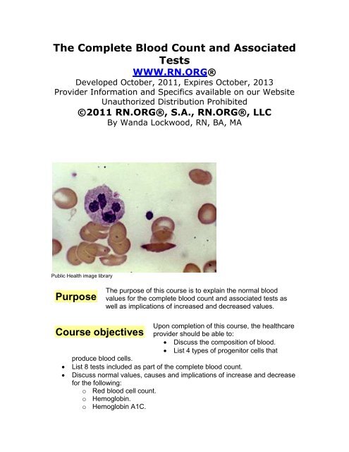
Public Health image library<br />
<strong>Purpose</strong><br />
<strong>The</strong> purpose of this course is to explain the normal blood<br />
values for the complete blood count <strong>and</strong> associated tests as<br />
well as implications of increased <strong>and</strong> decreased values.<br />
Upon completion of this course, the healthcare<br />
provider should be able to:<br />
Discuss the composition of blood.<br />
List 4 types of progenitor cells that<br />
produce blood cells.<br />
List 8 tests included as part of the complete blood count.<br />
Discuss normal values, causes <strong>and</strong> implications of increase <strong>and</strong> decrease<br />
for the following:<br />
o Red blood cell count.<br />
o Hemoglobin.<br />
o Hemoglobin A1C.<br />
Course objectives
o Hematocrit.<br />
o Erythrocyte/Red blood cell indices.<br />
o Erythrocyte sedimentation rate (ESR).<br />
o Erythropoietin (EPO).<br />
o Leukocyte/White blood cell count <strong>and</strong> differential.<br />
o Neutrophils.<br />
o Eosinophils.<br />
o Basophils.<br />
o Lymphocytes.<br />
o Monocytes.<br />
o Platelets.<br />
Explain a “shift to the left.”<br />
Explain flow cytometry.<br />
Introduction<br />
<strong>Blood</strong> is an essential living tissue that circulates throughout the body—about 5<br />
liters in the adult. <strong>Blood</strong> comprises:<br />
Liquid plasma (78%): 90% water with albumin <strong>and</strong> blood clotting factors,<br />
such as fibrinogen <strong>and</strong> globulin.<br />
Cells (22%): platelets, red blood cells, <strong>and</strong> white blood cells.<br />
<strong>Blood</strong> cells are formed in the cancellous bone of the bone marrow in the shafts of<br />
the arms, legs, ribs, sternum, <strong>and</strong> vertebrae in adults. Bone marrow is yellow in<br />
areas with many lipid cells but red in areas where formation of blood<br />
(hematopoiesis) occurs. Almost the entire marrow area is red in infants, but red<br />
marrow recedes as people mature <strong>and</strong> is replaced with yellow marrow.<br />
<strong>Blood</strong> cells are produced by stem cells, which comprise 3-5% of all marrow cells.<br />
<strong>The</strong> type of blood cells formed by progenitor stem cells is controlled by cytokines<br />
(proteins secreted by cells to signal other cells) <strong>and</strong> hormones (poietins):<br />
Interleukin-7: B <strong>and</strong> T cell lymphocytes.<br />
Erythropoietin: Erythrocytes.<br />
Thrombopoietin (with Interleukin-7): Megakaryocytes (which fragment into<br />
platelets).<br />
Granulocyte-monocyte-colony-stimulating factor (with Interleukin-3 <strong>and</strong><br />
Interleukin-5): Granulocytes <strong>and</strong> monocytes.<br />
<strong>The</strong> immature cells that are formed are called blast cells. <strong>The</strong>se blast cells<br />
continue to differentiate <strong>and</strong> develop into different types of mature cells.
US National Cancer Institute<br />
Red blood cells mature in the bone marrow before they are released into the<br />
blood, but some lymphocytes (a type of white blood cell) are immature when they<br />
leave the bone marrow <strong>and</strong> enter the bloodstream. <strong>The</strong>y travel to the thymus<br />
<strong>and</strong> other lymphoid tissue to mature. Each day the bone marrow produces huge<br />
numbers of cells (per kilogram of body weight):<br />
2.5 billion erythrocytes<br />
2.5 billion platelets<br />
1 billion granulocytes<br />
Abnormalities anywhere in this blood-producing system can effect the production<br />
of blood cells <strong>and</strong> the blood count.<br />
CBC overview<br />
<strong>The</strong> complete blood count (CBC) is one of the most frequently ordered screening<br />
laboratory tests. <strong>The</strong> CBC includes a number of different determinations,<br />
including the number, type, percentage, concentration, <strong>and</strong> quality of blood cells.<br />
In most cases, the CBC is done using an automated hematology analyzer, which<br />
can provide results in about a minute.<br />
<strong>Tests</strong> usually part of a CBC include:<br />
Red blood cell (erythrocyte) count (RBC)<br />
Hemoglobin (Hb or Hgb)<br />
Hematocrit (Hct)<br />
Red blood cell indices:(mean corpuscular volume [MCV], mean<br />
corpuscular hemoglobin (MCH), <strong>and</strong> mean corpuscular hemoglobin<br />
concentration [MCHC]<br />
White blood cell (leukocyte) count (WBC)<br />
Differential white blood cell count or "diff"
Platelet (thrombocyte) count (estimated)<br />
<strong>Blood</strong> cell morphology<br />
Norma values differ somewhat according to age <strong>and</strong> gender. <strong>The</strong> laboratory<br />
references provided in this course are meant as a guide <strong>and</strong> may vary somewhat<br />
from references used in different institutions.<br />
Laboratory tests performed on plasma are done using blood samples taken by<br />
venipuncture, usually with vacuum tubes used to collect the specimen. Tubes<br />
come in various sizes, <strong>and</strong> using the proper size is important because the tubes<br />
contain various types of anticoagulants <strong>and</strong> the volume of the specimen must be<br />
correct for the type of anticoagulant. If the ratio is incorrect, it can interfere with<br />
test results. For most hematology studies, including cell counts, blood is<br />
collected in tubes with lavender stoppers. <strong>The</strong>se tubes contain ethylene diamine<br />
tetraacetic acid (EDTA).<br />
Nursing Alert:<br />
Because the CBC count may fluctuate during the day, with regular<br />
CBC counts (such as daily or weekly), blood should be drawn at<br />
approximately the same time of day.<br />
<strong>Blood</strong> counts may be altered by hydration status. Overhydration<br />
(such as from IV fluids) increases the plasma component, <strong>and</strong> this<br />
dilutes percentages of blood elements. Dehydration, such as from<br />
inadequate fluids or NPO status, however, results in<br />
hemoconcentration, increasing the percentages of blood elements<br />
relative to plasma.<br />
Red blood cell (RBC) count<br />
Erythrocytes, commonly referred to as red blood<br />
cells (RBCs), have two primary functions:<br />
Carry oxygen from the lungs to body tissues<br />
Transfer carbon dioxide from the tissues to<br />
the lungs.<br />
<br />
As RBCs mature, they become biconcave disks <strong>and</strong><br />
produce hemoglobin, which makes up about 90% of<br />
the weight of the cell. Hemoglobin combines readily<br />
with oxygen (oxyhemoglobin) <strong>and</strong> carbon dioxide<br />
(carboxyhemoglobin). Oxyhemoglobin in arterial<br />
blood is bright red in color while the carboxyhemoglobin of venous blood appears<br />
dark red. <strong>The</strong> biconcave shape enables the maximum oxygen saturation of<br />
hemoglobin by providing more surface area for exposure of hemoglobin to
dissolved oxygen.<br />
In response to hypoxia, the hormone erythropoietin, secreted primarily by the<br />
kidneys, stimulates the bone marrow to produce red blood cells. Red blood cells<br />
are able to change shape to permit passage through small capillaries that<br />
connect arteries with veins. Normal red blood cells survive about 120 days <strong>and</strong><br />
are then ingested by phagocytic cells in the liver <strong>and</strong> kidneys.<br />
RBCs comprise about 40% of total blood volume; the RBC count is the number<br />
of red blood cells per cubic millimeter of blood. Normal red blood cells values<br />
vary according to age <strong>and</strong> gender:<br />
Age/gender RBC: Number per cubic millimeter<br />
Newborns 4.1 – 6.1 million<br />
Children (1-6) 3.9– 5.3 million<br />
Adult males: 4.5 – 6.0 million<br />
Adult females 4.2 – 5.0 million (slightlywith pregnancy)<br />
Immature erythrocytes are called reticulocytes. <strong>The</strong>y usually mature into red<br />
blood cells within 2 days after release into the blood stream. A reticulocyte<br />
count measures the percentage of reticulocytes in relation to the RBC count,<br />
<strong>and</strong> it is specifically used to monitor bone marrow function. Reticulocyte counts<br />
are normally very stable. When the RBC count increases, the reticulocyte count<br />
can help to determine the degree <strong>and</strong> rate of RBC overproduction. Normal<br />
ranges for reticulocytes include:<br />
Age/gender Reticulocytes: %-age of RBCs<br />
Newborns 3.0% - 7%<br />
Infants >12 wks 0.2% - 2.0%<br />
Adult males: 0.5% - 1.5%<br />
Adult females 0.5% - 2.5%<br />
An increased reticulocyte count indicates that the bone marrow is responding to<br />
the need for increased red blood cell production, such as may occur with acute<br />
loss of blood, iron-deficiency anemia, hemolytic anemia, megaloblastic anemia,<br />
or treatment for anemia. Thus, the reticulocyte count is often used to monitor<br />
response to treatment for anemia. In conditions in which red cell production is<br />
stimulated, a concomitant increase in reticulocytes is usually present, such as at<br />
high altitudes. Pregnant women <strong>and</strong> newborns also tend to show increased<br />
reticulocyte counts.<br />
A decreased reticulocyte count occurs with alcoholism, aplastic anemia, renal<br />
disease, folic acid deficiency, <strong>and</strong> bone marrow failure. Some drugs, such as<br />
azathioprine, dactinomycin, hydroxyurea, methotrexate, <strong>and</strong> zidovudine may
decrease reticulocyte counts. <strong>The</strong> reticulocyte count may be falsely decreased<br />
after transfusion because of the dilutional effect.<br />
Various disorders <strong>and</strong> treatments may involve an<br />
Increased RBC count<br />
increase in RBC count (polycythemia), A very high<br />
RBC mass will slow blood velocity <strong>and</strong> increase the<br />
risk of intravascular clotting.<br />
Primary polycythemia vera is abnormal increase in red blood cells with cause<br />
unknown <strong>and</strong> does not result from hypoxia or physiological need. Polycythemia<br />
vera may be treated by hydroxyurea to slow down bone marrow overproduction<br />
of red blood cells. Interferon is sometimes used to lower RBC count. Phlebotomy<br />
may be done weekly to decrease the concentration of red blood cells. Hydration<br />
is especially important as dehydration may increase viscosity of the blood.<br />
Secondary polycythemia may be caused by a variety of factors:<br />
At high altitudes, less atmospheric weight pushes air into the lungs,<br />
causing a decrease in the partial pressure of oxygen <strong>and</strong> resultant<br />
hypoxia, so the RBC count increases at high altitude increases to<br />
compensate for lower atmospheric pressure <strong>and</strong> oxygen, making more<br />
hemoglobin available to transport oxygen.<br />
Erythropoietin-secreting tumors increase production of red blood cells.<br />
Renal disorders may increase red blood cell count.<br />
Increased exertion <strong>and</strong> muscle mass related to strenuous exercises<br />
increase RBC count in order to keep pace with dem<strong>and</strong>s for oxygen.<br />
Some drugs, such as gentamicin, corticosteroids, methyldopa, may cause<br />
an increased RBC count.<br />
Hypoxia associated with smoking may trigger increase in RBC production<br />
to compensate.<br />
Chronic lung disease in adults <strong>and</strong> children with congenital heart defects<br />
characterized by cyanosis cause polycythemia.<br />
Excessive exercise, anxiety, pain, <strong>and</strong> dehydration.<br />
Nursing Alert:<br />
Maintaining adequate hydration is critical to preventing venous<br />
thrombosis in those with polycythemia vera, so long periods without<br />
fluids should be avoided. If patients with polycythemia require fluid<br />
restriction, such as those with heart failure or congenital heart<br />
defects, the physician should specify the amount of daily fluids <strong>and</strong><br />
the patients should be carefully monitored.<br />
RBC counts are affected by position, stasis, <strong>and</strong> hydration. <strong>Count</strong>s<br />
decrease slightly if a patient is recumbent <strong>and</strong> increase slightly when<br />
the patient is upright. Leaving the tourniquet on for more than 60
seconds when obtaining a blood sample may increase the RBC<br />
count. Dehydration increases RBC count <strong>and</strong> overhydration<br />
decreases it.<br />
A decreased RBC count (anemia) may result from a<br />
Decreased RBC count<br />
decrease in the number of red blood cells although it<br />
can also be caused by a decrease in hemoglobin<br />
content. A decreased RBC count is caused by:<br />
Hemolytic anemia.<br />
Chemotherapy.<br />
Hemorrhage resulting in loss of RBCs.<br />
Decreased plasma volume from severe burns, shock, severe vomiting, or<br />
intestinal obstruction.<br />
Pregnancy.<br />
Malignancies: Hodgkin’s disease, multiple myeloma, leukemia.<br />
Addison’s disease.<br />
Rheumatic fever.<br />
Nutritional deficit.<br />
Subacute endocarditis.<br />
Lack of substances necessary for the production of RBCs.<br />
Bone marrow suppression, often related to chemotherapy (amphotericin<br />
B, floxuridine, <strong>and</strong> phenylbutazone) or radiation.<br />
Hemodilution from excess IVs.<br />
.<br />
Hemoglobin<br />
<strong>The</strong> hemoglobin molecule is comprised of four subunits (alphas <strong>and</strong> betas), with<br />
each containing an iron-containing pigment (heme) <strong>and</strong> a protein (globulin). One<br />
gram of hemoglobin can carry up to 1.34 mL of oxygen. While the amount of<br />
oxygen carried on each molecule of hemoglobin may vary, in general the ability<br />
of the blood to carry oxygen directly relates to the hemoglobin concentration.<br />
More hemoglobin, more oxygen, <strong>and</strong> vice versa.<br />
Hemoglobin measures the amount of the oxygen-carrying protein (hemoglobin) in<br />
a volume of blood, providing an indication of the ability of the blood to oxygenate<br />
the tissue. Hemoglobin determination is used to screen for anemia <strong>and</strong><br />
determine response to treatment. Hemoglobin is measured in grams per deciliter<br />
<strong>and</strong> closely corresponds to RBC numbers since 90% of red blood cells are<br />
composed of hemoglobin.<br />
Age/gender Hemoglobin: grams/deciliter<br />
Newborns 14.5 – 24.5 g/dl<br />
Children (1-6 years) 9.5 – 14. 1 g/dl<br />
Adult males: 14.0 – 17.5 g/dl<br />
Adult females 12.0 – 16.0 g/dl
Hemoglobin: critical low <strong>and</strong> high values:<br />
A hemoglobin value under 6 g/dl may result in heart failure.<br />
A hemoglobin value over 18 g/dl may cause vascular occlusion because<br />
of hemoconcentration.<br />
Increased hemoglobin<br />
Heart failure.<br />
COPD.<br />
Dehydration.<br />
Erythrocytosis.<br />
Hemoconcentration.<br />
High altitudes.<br />
Polycythemia vera.<br />
Increased hemoglobin occurs with the following:<br />
Burns<br />
Because hemoglobin is carried on red blood cells,<br />
Decreased hemoglobin<br />
any decrease in circulating RBCs affects the<br />
hemoglobin level. Conditions with abnormal types<br />
of hemoglobin often result in lower total hemoglobin because the red blood cells<br />
with abnormal hemoglobin are readily damaged. Hemoglobin electrophoresis is<br />
used to distinguish among the different types of abnormal hemoglobin.<br />
Specific disorders that result in decreased hemoglobin include:<br />
Thalassemia is an autosomal recessive hemoglobinopathy resulting in<br />
insufficient globulin in either the alpha or beta subunits.<br />
Sickle cell disease causes an abnormally-shaped hemoglobin known as<br />
sickle hemoglobin (hgbS). <strong>The</strong>se misshapen RBCs tend to clump <strong>and</strong><br />
form vessel occlusions.<br />
Iron deficiency anemia (hypochromic anemia) is a condition in which red<br />
blood cells have less hemoglobin than normal because of inadequate iron,<br />
so the cells are able to transport less oxygen. This results in a normal<br />
RBC count but low hemoglobin.<br />
Physiological anemia of pregnancy: During pregnancy, the plasma<br />
proportion of the blood exp<strong>and</strong>s, so the hemoglobin falls because of the<br />
increased volume.<br />
Serial blood draws: Neonates <strong>and</strong> premature infants are especially<br />
vulnerable to anemia from repeated blood draws.<br />
Hemolytic disorders.<br />
Malignancies, such as leukemia, carcinoma, lymphomas.<br />
Hodgkin’s disease.<br />
Pregnancy.<br />
Nutritional deficit.<br />
Fluid retention, <strong>and</strong> IV overload.
Hemoglobin A1C<br />
Although not part of the st<strong>and</strong>ard complete blood count, an additional test<br />
associated with hemoglobin, hemoglobin A1C is used to determine an average<br />
plasma concentration of glucose over a 3-month period—compared to a st<strong>and</strong>ard<br />
fasting blood glucose that reflects blood glucose during a one-time fasting state.<br />
<strong>The</strong> fasting glucose level may fluctuate according to diet <strong>and</strong> other factors, so it<br />
is less reliable as an indicator of actual glucose levels than hemoglobin A1C.<br />
Erythrocytes survive about 120 days, <strong>and</strong> during that time the erythrocytes are<br />
exposed to glucose circulating in the plasma. <strong>The</strong> glucose molecules join with<br />
the hemoglobin forming glycated (glycosylated) hemoglobin. <strong>The</strong> percentage of<br />
glucose in the cells indicates the average glucose level that the cell was exposed<br />
to. Different laboratory procedures may result in slightly different readings, but<br />
normal readings are usually in the 4-6% range, <strong>and</strong> diabetics are usually advised<br />
to maintain readings
Adult females 36% - 48%<br />
Hematocrit: critical low <strong>and</strong> high values:<br />
A hematocrit of less than 18% can result in cardiac failure.<br />
A hematocrit of over 54% may result in spontaneous blood clotting.<br />
If the RBC count <strong>and</strong> the hemoglobin are both within normal limits, one can<br />
estimate the hematocrit as approximately three times the hemoglobin. For<br />
example, a person whose hemoglobin is 14 would have a hematocrit of about<br />
42%. <strong>The</strong> heart rate usually increases with hematocrit
Other causes of decreased hematocrit include:<br />
Anemia.<br />
Bone marrow hyperplasia.<br />
Hemolytic disorders.<br />
Fluid retention.<br />
Pregnancy.<br />
Splenomegaly.<br />
Nutritional deficit.<br />
Medications may induce hemolysis in sensitive individuals.<br />
Patients who tend to have chronically low hemoglobin, such as those receiving<br />
renal dialysis, may have few symptoms as their bodies have adjusted to the low<br />
level; however, those with a sudden drop, such as from hemorrhage, may<br />
develop indications of shock, with pallor, hypotension, <strong>and</strong> hypoxia.<br />
Nursing Alert:<br />
Activity level is a concern with low hemoglobin. If low hematocrit<br />
is caused by hemorrhage, the BP may stay stable initially as the<br />
plasma volume increases from interstitial fluid although the BP<br />
will fall with fluid deficit. <strong>The</strong> heart is often a better test to<br />
determine activity tolerance.<br />
Tilt test: Take blood pressure supine <strong>and</strong> after sitting. If the<br />
pulse rate increases from the act of sitting, tolerance for activity<br />
may be impaired. Patients may need slow, steady exercise or<br />
activities with rest periods.<br />
Low hematocrit requires increased production of red blood cells,<br />
so dietary modifications may include increased protein <strong>and</strong> iron<br />
Stasis from leaving the tourniquet in place during venipuncture<br />
for >60 seconds may increase Hct values by 2-3%. Values taken<br />
within a few hours of blood transfusion or acute blood loss may<br />
appear normal.<br />
Elevated glucose levels cause RBCs to swell <strong>and</strong> may cause a<br />
falsely elevated hematocrit.<br />
Erythrocyte/Red blood cell indices<br />
When hemoglobin levels are low, erythrocyte/red blood cell indices provide<br />
information about the size of red cells <strong>and</strong> the concentration of hemoglobin in<br />
order to diagnose the type of anemia
MCV measures the average size of red<br />
Mean corpuscular volume (MCV)<br />
blood cells <strong>and</strong> classifies anemia.<br />
Calculation:<br />
MCV = Hematocrit / total RBC count<br />
<strong>The</strong> red blood cells are classified according to size:<br />
Microcytic: MCV is low because of small RBCs. Microcytic red blood<br />
cells are found in iron deficiency anemia, vitamin B12/folate anemia, lead<br />
poisoning <strong>and</strong> Thalassemias.<br />
Macrocytic: MCV is high because of large RBCs. Pernicious anemia <strong>and</strong><br />
folic acid deficiencies are characterized by macrocytic RBCs. MCV is also<br />
increased in alcoholism, antimetabolite therapy, <strong>and</strong> hepatic disease.<br />
Normocytic: MCV is within the normal range <strong>and</strong> RBCs are normal in<br />
size. Anemia associated with acute hemorrhage is usually normocytic.<br />
<br />
Age/gender Mean Corpuscular volume: femtoliters<br />
Infants (6-12 mo.) 73 – 87 fL<br />
Children (1-6 years) 70 – 84 fL<br />
Adult males: 84 – 96 fL<br />
Adult females 76 – 96 fL<br />
Some drugs may increase the MCV—colchicine, pentamidine, pyrimethamine,<br />
<strong>and</strong> triamterene—while nitrofurantoin may decrease MCV.<br />
Mean corpuscular hemoglobin (MCH)<br />
MCH measures the amount, or the<br />
mass, of hemoglobin present on<br />
average in one RBC, helping to determine if the cells are normochromic,<br />
hypochromic (pale), or hyperchromic (dark). Calculation:<br />
MCH = Hgb / Total RBC count<br />
Age/gender Mean Corpuscular Hgb: picogram/cell<br />
Infants (6-12 months) 24 – 30 pg<br />
Children (1-6 years) 23 – 29 pg<br />
Adult males: 27 – 32 pg<br />
Adult females 27 – 32 pg<br />
MCH is increased in macrocytic anemias <strong>and</strong> decreased in hypochromic <strong>and</strong><br />
microcytic anemias.<br />
Mean corpuscular hemoglobin concentration (MCHC)<br />
MCHC measures<br />
the average<br />
concentration of hemoglobin in each cell. Calculation:<br />
MCHC = Hgb / Hct<br />
<strong>The</strong> results are reported in grams per deciliter reflecting the amount of
hemoglobin in the RBC. In some cases, the results are reported in percentages.<br />
Calculation:<br />
MCHC = (Hgb / Hct) x 100<br />
Age/gender MCHC: Gram/deciliter & percentage<br />
Infants (6-12 months) 32 – 36 g/dL 32-36%<br />
Children (1-6 years) 31 – 35 g/dL 31-35%<br />
Adult males: 30 – 35 g/dL 30-35%<br />
Adult females 30 – 35 g/dL 30-35%<br />
MCHC is also used to determine if red blood cells are normochromic,<br />
hypochromic, or hyperchromic. MCHC is increased in spherocytosis <strong>and</strong><br />
thalassemia <strong>and</strong> decreased in iron-deficiency anemia.<br />
Anemias can be classified using erythrocyte indices in the following way:<br />
Decreased MCV, MCH, <strong>and</strong> MCHC: Microcytic, hypochromic anemia,<br />
usually related to iron deficiency anemia.<br />
Increased MCV, variable MCH <strong>and</strong> MCHC: Macrocytic anemia, usually<br />
related to vitamin B12 deficiency or folic acid deficiency.<br />
RDW measures the variation in<br />
Red blood cell distribution width (RDW)<br />
RBC size <strong>and</strong> is based on the<br />
mean corpuscular volume (MCV),<br />
which measures RBC size. Calculation:<br />
RDW = (st<strong>and</strong>ard deviation or RBC width / mean RBC width) x 100<br />
<strong>The</strong> results are expressed in percentages to show how many RBCs vary in size.<br />
Age/gender RDW: Percentage<br />
Reference range 12.8 – 15.2%<br />
Values may increase with various types of anemias (megaloblastic, hemolytic,<br />
sickle cell), liver disease, alcoholism, <strong>and</strong> folate <strong>and</strong> vitamin B12 deficiency.<br />
Erythrocyte sedimentation rate (ESR)<br />
<strong>The</strong> erythrocyte sedimentation rate (ESR) is not part of the complete blood count,<br />
but is an associated test or RBCs. <strong>The</strong> ESR, also called the “sed rate,”<br />
measures the rate at which RBCs in anticoagulated blood precipitate after an<br />
hour in an upright test tube (Westergren tube). <strong>The</strong> rate at which the RBCs fall is<br />
reported in mm/hr. This test is now more commonly done with automated<br />
analyzers.<br />
<strong>The</strong> ESR is used as an indicator of inflammation for both acute <strong>and</strong> chronic<br />
inflammation associated with infections, cancers, <strong>and</strong> autoimmune diseases.<br />
Inflammation increases immune <strong>and</strong> clotting factors, such as globulins <strong>and</strong><br />
fibrinogen, in the blood. Fibrinogen, a clotting factor, causes RBCs to clump into
stacks called “rouleaux.” <strong>The</strong> rouleaux settle faster than separate RBCs, so an<br />
increased rate indicates inflammation. Since ESR elevations relate to increased<br />
fibrinogen <strong>and</strong> globulins, follow-up fibrinogen level <strong>and</strong> protein electrophoresis<br />
may be ordered to determine whether fibrinogen, globulins, or both are affecting<br />
the ESR.<br />
ESR is a non-specific test as it does not indicate the type or cause of infection,<br />
but increases <strong>and</strong> decreases are associated with certain disorders. Acute<br />
infections are often better identified with the C-reactive protein test, which shows<br />
signs of infection earlier (within 6-8 hours) <strong>and</strong> is less sensitive to other variables.<br />
However, ESR is a simple test <strong>and</strong> is often done first when inflammation is<br />
suspected. It is commonly used to aid in diagnosis of pediatric rheumatoid<br />
arthritis <strong>and</strong> Kawasaki disease. <strong>The</strong> ESR may be used to monitor response to<br />
therapy for some conditions:<br />
Age/gender Erythrocyte sedimentation rate: mm/hr.<br />
Neonates 0.0 – 2.0<br />
Children (to puberty) 3.0 – 20.0<br />
Adult males: 10.0 – 19.0 (increases with age)<br />
Adult females 15.0 – 23 (increases with age & pregnancy<br />
Increased ESR<br />
Increased ESR is commonly found in severe infection <strong>and</strong><br />
globulin-increasing tumors, such as multiple myeloma <strong>and</strong> Waldenstrom’s<br />
macroglobulinemia. Elevations in ESR associated with disease-specific<br />
symptoms are commonly used to diagnose <strong>and</strong> monitor two specific conditions:<br />
temporal arteritis <strong>and</strong> polymyalgia rheumatica. Some drugs may increase the<br />
sedimentation rate: methyldopa (Aldomet), oral contraceptives, penicillamine<br />
procainamide, theophylline, <strong>and</strong> vitamin A. A sustained elevation in ESR after<br />
chemotherapy for Hodgkin’s disease may be a predictor of early relapse.<br />
<strong>The</strong> ESR combined with the hemoglobin has been shown to be a more effective<br />
screening tool for inflammatory bowel disease in children <strong>and</strong> adolescents than<br />
serology testing (<strong>and</strong> less expensive). IBD is identified by increased ESR (31<br />
mm/hr.) <strong>and</strong> decreased hemoglobin (indicating anemia).<br />
Decreased ESR<br />
A decreased ESR is usually not clinically significant<br />
although it is associated with some disorders, such as<br />
polycythemia, severe leukocytosis, sickle cell anemia, <strong>and</strong> some protein<br />
abnormalities. Some drugs may decrease the sedimentation rate—aspirin,<br />
cortisone, <strong>and</strong> quinine—either because they affect inflammatory processes or<br />
clotting.
Erythropoietin (EPO)<br />
<strong>The</strong> erythropoietin (EPO) test is not part of the complete blood count but is often<br />
ordered when RBC levels are abnormal to help to identify the cause of the<br />
abnormality. Erythropoietin (EPO) is a hormone produced primarily by the kidney<br />
but some is produced in the liver as well. EPO is a glycoprotein that stimulates<br />
the production <strong>and</strong> development of RBCs in the bone marrow <strong>and</strong> promotes<br />
synthesis of hemoglobin in the RBCs <strong>and</strong> is, thus, the primary regulator of RBC<br />
production.<br />
<strong>Tests</strong> are used to determine EPO level in order to evaluate bone marrow <strong>and</strong><br />
kidney disorders as well as EPO abuse. EPO is sometimes taken as a<br />
performance-enhancing drug in athletes, such as cyclists, runners, speed<br />
skaters, <strong>and</strong> cross-country skiers. Testing is done after overnight fasting <strong>and</strong><br />
after a 20-30 minute period of lying quietly. Lab values vary considerably from<br />
one laboratory to another, so conclusive reference ranges are not available.<br />
Each laboratory has reference values, so the following reference ranges are<br />
approximate.<br />
Age/gender EPO: mU/ml<br />
Reference range Authorities vary:<br />
0.0 – 24.0 OR<br />
11.0 – 48.0<br />
Increased EPO<br />
Elevated EPO is characteristic of secondary polycythemia as<br />
the EPO increases <strong>and</strong> stimulates the bone marrow to<br />
produce an excess of RBCs. If EPO levels are elevated but the hemoglobin is<br />
low (anemia), then this suggests that the disorder is in the bone marrow rather<br />
than the kidneys. Increased levels may also associated be with abuse of EPO<br />
(blood doping), but EPO has been banned by sports <strong>org</strong>anizations, including the<br />
Tour de France <strong>and</strong> the Olympics. EPO is taken by athletes to increase RBC<br />
production, oxygen capacity, <strong>and</strong> endurance; however, the resultant<br />
polycythemia coupled with strenuous exercise <strong>and</strong> dehydration increases the risk<br />
of stroke <strong>and</strong> heart attacks.<br />
Decreased EPO<br />
A decreased EPO level is associated with some types of<br />
anemia, including anemia secondary to kidney failure, AZT<br />
treatment (for AIDS) <strong>and</strong> cancer. A low level is also found in polycythemia vera<br />
(as opposed to secondary polycythemia). EPO has been synthesized<br />
(recombinant erythropoietin) <strong>and</strong> is used as a treatment in some cases,<br />
especially cancer-related anemia, to increase the RBC count although RBC<br />
count must be monitored carefully because side effects include blood clots, heart<br />
attacks, strokes, increase in tumor size, <strong>and</strong> death. In chronic kidney disease,<br />
EPO tests may be done regularly to monitor kidney production of EPO.
Leukocyte/White <strong>Blood</strong> Cell <strong>Count</strong> (WBC) <strong>and</strong><br />
Differential<br />
White blood cells, or leukocytes, are classified into two primary groups:<br />
Granulocytes (granules in cell cytoplasm): Neutrophils, eosinophils,<br />
<strong>and</strong> basophils. Because these cells have a multilobed nucleus, they are<br />
also called polymorphonuclear leukocytes or "polys." Neutrophils have<br />
segmented nuclei, so they are sometimes referred to segmented<br />
neutrophils or "segs."<br />
Agranulocyte (no granules <strong>and</strong> nonlobular nuclei): Lymphocytes <strong>and</strong><br />
monocytes are sometimes referred to as mononuclear leukocytes.<br />
White blood cells have a much shorter life span than red blood cells,<br />
approximately 13 to 20 days, after which they are destroyed by the lymphatic<br />
system <strong>and</strong> excreted in feces. <strong>The</strong>se cells are produced in the bone marrow<br />
although lymphocytes can be produced elsewhere as well. When immature white<br />
cells are released from the bone marrow, they are referred to as “b<strong>and</strong>s” or<br />
“stabs.”<br />
WBCs usually make up a small percentage of the total blood (about 1%), but<br />
they serve a number of purposes for the immune system. Through phagocytosis,<br />
WBCs surround <strong>and</strong> destroy foreign <strong>org</strong>anisms. WBCs also produce <strong>and</strong><br />
transport antibodies in response to antigens.<br />
<strong>The</strong> CBC includes two measurements of white blood cells:<br />
Total number of white blood cells in a microliter (1x10 -9 liters) of blood,<br />
providing an absolute number (in thous<strong>and</strong>s) of WBCs.<br />
Differential: Percentage of each of the five types of white blood cells,<br />
totaling 100%. Calculation:<br />
Specific cell percentage = (Specific cell / Total WBC) X 100<br />
Absolute value = % / 100 X Total WBC<br />
Age &<br />
gender<br />
WBC<br />
(x10 3)<br />
B<strong>and</strong>s<br />
%<br />
Neut/segs<br />
%<br />
Eos<br />
%<br />
Baso<br />
%<br />
Lymph<br />
%<br />
Mono<br />
%<br />
Newborn 9.0-30.0 10-18 36-62 0-2 0-1 26-36 0-6<br />
1-6 yr 5.0-19.0 5-11 13-33 0-3 0-0 46-76 0-5<br />
Adults 5.0-10.0 3-6 50-62 0-3 0-1 25-40 3-7<br />
Leukocytes: critical low <strong>and</strong> high values<br />
WBC of less than 2500 increase risk of severe life-threatening infection.<br />
WBC over 30,000 indicates severe infection or a serious disease, such as<br />
leukemia.<br />
When evaluating the WBC count <strong>and</strong> differential, one must consider both relative
<strong>and</strong> absolute values. For example a relative value of 60% neutrophils may seem<br />
within normal limits; however, if the total WBC were 30,000, the absolute value<br />
(60% x 30,000) would be 18,000, an elevation.<br />
Increased WBC<br />
Leukocytosis, a WBC >10,000, is usually due to an<br />
increase in one of the five types of white blood cells rather<br />
than in increases in a number of cells. <strong>The</strong> condition is named depending on the<br />
cell that shows the most significant increase: Neutrophilia, lymphocytosis,<br />
eosinophilia, monocytosis, <strong>and</strong> basophilia.<br />
White blood cells release colony stimulating factor to stimulate bone marrow to<br />
increase production of WBCs in response to trauma, acute infection, or<br />
inflammation. <strong>The</strong> WBC count can increase by thous<strong>and</strong>s within a few hours.<br />
Some conditions, such as sepsis <strong>and</strong> measles, result in very high increases. With<br />
leukemia, the WBC count may also increase, but the increase is permanent <strong>and</strong><br />
progressive.<br />
Leukocytosis occurs in early infancy, as a response to stress, from cold<br />
exposure, after strenuous exercise, <strong>and</strong> with exposure to ultraviolet light. WBCs<br />
also increase with pregnancy <strong>and</strong> labor <strong>and</strong> menstruation. Pathologic conditions<br />
associated with leukocytosis include all kinds of bacterial infections, transfusion<br />
reactions, hemolysis, anemias, appendicitis, collagen disease, Cushing’s<br />
disease, leukemias <strong>and</strong> other malignancies, parasitic infestations, <strong>and</strong><br />
polycythemia vera.<br />
Nursing Alert:<br />
Severe stress that results in production of epinephrine can cause<br />
a rapid increase in WBC as the body prepares to “fight.”<br />
Corticosteroids usually increase the WBC count in normal health<br />
individuals. However, in the presence of infection, the infection<br />
can spread without a rise in the WBC count, so severe infections<br />
can be masked in those receiving corticosteroids.<br />
Decreased WBC<br />
Leukopenia occurs when the WBC falls below 4,000. Viral<br />
infections, overwhelming bacterial infections, <strong>and</strong> bone<br />
marrow disorders can all cause leukopenia. Severe leukopenia puts patients at<br />
severe risk of opportunistic infections, so treatments that involving interrupting<br />
skin integrity, such as injections, may increase risk.<br />
Leukopenia may occur with alcoholism, anemias, bone marrow suppression,<br />
malaria, malnutrition, radiation, viral infection, SLE <strong>and</strong> other autoimmune<br />
disorders, <strong>and</strong> chemotherapy. A number of drugs may decrease the white blood
count: antibiotics, anticonvulsants, cardiovascular drugs, NSAIDS, <strong>and</strong> diuretics.<br />
Differential<br />
Neutrophils<br />
Age & gender B<strong>and</strong>s % Neut/segs %<br />
Newborn 10-18 36-62<br />
1-6 yr 5-11 13-33<br />
Adults 3-6 50-62<br />
Neutrophils, also calls polymorphonuclear cells,<br />
usually comprise the largest percentage of white<br />
blood cells. <strong>The</strong>y are the body's primary defense<br />
through phagocytosis <strong>and</strong> contain enzymes <strong>and</strong><br />
pyogens to combat foreign <strong>org</strong>anisms. Most<br />
circulating neutrophils are mature, <strong>and</strong> the nucleus<br />
is segmented.<br />
Normally, most of the neutrophils circulating in the<br />
bloodstream are in a mature form, with the<br />
nucleus of the cell being divided or segmented.<br />
<strong>The</strong> nucleus of immature neutrophils is not<br />
segmented but has a b<strong>and</strong>ed or rod-like appearance.<br />
<strong>The</strong> nucleus of less mature neutrophils is not<br />
segmented, but has a b<strong>and</strong> or rod-like shape. <strong>The</strong>se<br />
immature cells are referred to as “b<strong>and</strong>s” or “stabs.”<br />
Increased neutrophil count<br />
Both mature <strong>and</strong> immature neutrophils increase in<br />
response to an acute bacterial infection. When<br />
laboratory reports were written out by h<strong>and</strong>, by<br />
tradition, b<strong>and</strong> <strong>and</strong> neutrophils were the first two cells<br />
on the left. Because neutrophils usually increase in response to inflammation,<br />
this increase is often referred to as a “shift to the left.” This term is still used to<br />
refer to an infective process that results in increased b<strong>and</strong>s.<br />
Neutrophilia also occurs with acute hemolysis, acute hemorrhage, temperature<br />
extremes, malignancies, metabolic disorders, myelocytic leukemia, physiological<br />
stress (surgery, allergies, childbirth, exercise), toxin/venom poisoning, <strong>and</strong>
inflammatory conditions, such as gout, rheumatoid arthritis, <strong>and</strong> vasculitis.<br />
Decreased neutrophil count<br />
Neutropenia, a decrease in neutrophils, may also occur with some types of<br />
bacterial infections, such as typhoid fever <strong>and</strong> brucellosis, <strong>and</strong> with many viral<br />
diseases. If infection overwhelms the ability of the bone marrow to produce<br />
neutrophils, neutropenia may also occur. Neutropenia is often a side effect of<br />
chemotherapeutic agents used to treat malignancies, such as leukemia, as well<br />
as lithium, phenothiazines, <strong>and</strong> tricyclic antidepressants.<br />
Neutropenia also occurs with acromegaly, anaphylaxis, nutritional impairment<br />
(anorexia, starvation), bone marrow depression, SLE, thyrotoxicosis, <strong>and</strong> vitamin<br />
B12 or folate deficiency.<br />
Absolute neutrophil count<br />
Age & gender Absolute neutrophil count<br />
1 year 1500 to 8500<br />
10 years 1800 to 8000<br />
Adult 1800 to 7700<br />
Absolute neutrophil count (ANC) is an important measure used to determine the<br />
degree of neutropenia. <strong>The</strong> total ANC should be about 1800 to 2000 mmm 3 or<br />
higher. Risk of infection is significant if the level falls to 1000 <strong>and</strong> severe at 500.<br />
ANC is calculated indirectly from the total white count <strong>and</strong> the percentages of<br />
neutrophils <strong>and</strong> b<strong>and</strong>s:<br />
ANC = Total WBC X (% neutrophils + % b<strong>and</strong>s/100)<br />
Using this formula, if the white blood count is 3200 with 70% neutrophils <strong>and</strong> 3%<br />
b<strong>and</strong>s, the calculation would indicate that despite the low WBC count, the patient<br />
does not have neutropenia:<br />
ANC = 3000 X 73/100 = 2190<br />
If the white count is 5300 with 10% neutrophils <strong>and</strong> 1% b<strong>and</strong>s, neutropenia is<br />
evident despite the normal WBC:<br />
ANC = 5300 X 11/100 = 583<br />
Nursing Alert:<br />
Patients with neutropenia are at increased risk of endogenous<br />
<strong>and</strong> exogenous infection, so they should avoid people with<br />
respiratory or other infections. <strong>The</strong>y should be encouraged to use<br />
good hygiene <strong>and</strong> eliminate potential sources of infection, such<br />
as unpeeled fresh fruit or raw vegetables <strong>and</strong> fresh flowers.
Eosinophils<br />
Patients with ANC 30%).<br />
Addison’s disease<br />
Allergy, hay fever.<br />
Asthma.<br />
Drug reaction (allergic response).<br />
Hodgkin’s disease.<br />
Pernicious anemia.
Rheumatoid arthritis.<br />
TB.<br />
Polycythemia vera.<br />
Decreased eosinophil count:<br />
Eosinophils decrease with:<br />
Aplastic anemia.<br />
Stress.<br />
Infections (associated with shift to the left).<br />
Eclampsia.<br />
Drugs: ASA, amphotericin B, corticotropin, desipramine, glucocorticoids,<br />
hydrocortisone, interferon, niacin, prednisone, procainamide.<br />
Nursing Alert:<br />
Eosinophil levels vary according to the time of day, lowest in the<br />
morning <strong>and</strong> rising until midnight, at which time the level begins to fall<br />
again. Serial tests should be done at the same time of day.<br />
Basophils<br />
Age & gender Baso %<br />
Newborn 0-1<br />
1-6 yr 0-0<br />
Adults 0-1<br />
in the tissue.<br />
Basophils occur in very small number in the blood<br />
but are important as part of the immune system <strong>and</strong><br />
have a phagocytic function. <strong>The</strong>y are involved in<br />
allergic (hypersensitivity) reactions. <strong>The</strong> granules<br />
contain heparin, histamines, <strong>and</strong> serotonin.<br />
Basophils are similar to mast cells, which are found<br />
Basophils are evaluated to assist in determining cause of increased WBC count,<br />
to detect hematologic diseases <strong>and</strong> monitor progression of other disorders, such<br />
as COPD, malabsorption syndromes, renal disease, <strong>and</strong> cancer. Basophils may<br />
be noted in leukemia, Hodgkin’s disease, ulcerative colitis, polycythemia vera,<br />
<strong>and</strong> nephrosis.<br />
Lymphocytes
Age & gender Lymph %<br />
Newborn 26-36<br />
1-6 yr 46-76<br />
Adults 25-40<br />
Lymphocytes are the second most common white blood<br />
cell in adults <strong>and</strong> first most common in young children<br />
<strong>and</strong> are an important part of the immune system.<br />
Lymphocytes are produced in the bone marrow but<br />
migrate through the lymphatic system to other parts of<br />
the body. B cells (for bone marrow) mature within the<br />
bone marrow, but T cells (for thymus) mature in the<br />
thymus.<br />
<strong>The</strong>re are both large <strong>and</strong> small lymphoctyes. Large<br />
lymphocytes include natural killer (NK) cells. NK cells, part of the innate immune<br />
response, help defend the body against tumors <strong>and</strong> viruses. NK cells are<br />
activated by interferons <strong>and</strong> then release cytokines that modulate T <strong>and</strong> B cell<br />
response <strong>and</strong> also produce cytotoxic granules that destroy cells. NK cells help to<br />
contain a viral infection while cytotoxic T cells clear the infection. NK cells<br />
destruction of <strong>org</strong>anisms is not antigen specific.<br />
Small lymphocytes include both T-cell <strong>and</strong> B-cell lymphocytes, which are part of<br />
the adaptive immune response with T cells active in cell-mediated immunity <strong>and</strong><br />
B cells in humoral (antibody-related) immunity. Some cytotoxic T cells produce<br />
cytotoxic granules with enzymes that destroy pathogen-infected cells. Helper T<br />
cells (CD4) direct the immune response by activating killer cells <strong>and</strong> B cells. CD4<br />
is a glycoprotein found on the surface of T helper cells <strong>and</strong> is an important<br />
measure of the progression of HIV. CD8 is found on cytotoxic T cells.<br />
B cells recognize foreign antigens <strong>and</strong> produce antibodies to them. Additionally,<br />
they act as antigen presenting cells. That is, they attach to free antigens <strong>and</strong><br />
present them to T cells for destruction. B cells form memory cells that remember<br />
particular antigens to increase speed of response with the next encounter.<br />
Increased lymphocytes:<br />
Lymphocytes increase in many viral infections as well as a number of other<br />
disorders:<br />
Lymphocytic leukemia.<br />
Addison’s disease.<br />
Felty’s syndrome.<br />
Thyrotoxicosis.
Ulcerative colitis.<br />
Lymphoma, lymphosarcoma, myeloma.<br />
Rickets.<br />
Decreased lymphocytes:<br />
HIV results in a decrease in the total number of lymphocytes as well as changes<br />
in the ratios of the different types of T lymphocytes because CD4 (T4) cell counts<br />
decrease as HIV counts increase. Other causes of decreased counts include:<br />
Antineoplastic drugs.<br />
Aplastic anemia.<br />
Bone marrow failure.<br />
Burns.<br />
Hodgkin’s disease.<br />
Immunodeficiency disorders.<br />
Malnutrition.<br />
Pernicious anemia.<br />
Radiation.<br />
Septicemia.<br />
Thrombocytopenic purpura.<br />
Transfusion reaction.<br />
Nursing Alert:<br />
Immunosuppressive drugs, such as corticosteroids, may<br />
decrease lymphoctyes, increasing risk of infection. As<br />
lymphocyte levels fall, the patient is especially at risk for<br />
developing viral infections.<br />
Monocytes<br />
Age & gender Mono %<br />
Newborn 0-6<br />
1-6 yr 0-5<br />
Adults 3-7<br />
Monocytes are the largest cells normally found in<br />
blood <strong>and</strong> are produced in the bone marrow from the<br />
same progenitor cells as neutrophils. <strong>The</strong>ir primary<br />
function is phagocytosis.<br />
Monocytes stay in peripheral blood for approximately<br />
70 hours <strong>and</strong> then migrate into tissues where they<br />
act as macrophages. As macrophages, the cells
enlarge <strong>and</strong> increase the amount of digestive enzymes in their intracellular<br />
vesicles in order to more effectively carry out phagocytosis. Large numbers of<br />
macrophages are in the liver <strong>and</strong> spleen (known then as Kupffer cells) to clean<br />
bacteria from the blood.<br />
Increased monocytes:<br />
Monocytes increase with the following:<br />
Carcinomas.<br />
Cirrhosis.<br />
Collagen disease.<br />
Gaucher’s disease.<br />
Hemolytic anemia.<br />
Infections.<br />
Lymphomas<br />
Monocytic leukemia.<br />
Polycythemia vera.<br />
Cirrhosis.<br />
Radiation.<br />
Sarcoidosis.<br />
Ulcerative colitis.<br />
SLE.<br />
Thrombocytopenic purpura.<br />
Decreased monocytes:<br />
A decrease in monocytes may indicate injury to the bone marrow or some types<br />
of leukemia.<br />
Platelets<br />
Age & Gender<br />
Platelets<br />
Newborn 140,000 – 300,000<br />
1-6 yr 150,000 – 450,000<br />
Adult males 150,000 – 400,000<br />
Adult females 150,000 – 400,000<br />
Platelets, or thrombocytes, are non-nucleated round or oval fragments of larger<br />
cells (megakaryocytes) found in the bone marrow <strong>and</strong> are very tiny—only about<br />
20% the average diameter of RBCs. <strong>The</strong> primary functions of platelets are in<br />
coagulation, hemostasis, <strong>and</strong> thrombus formation. Platelets contain granules that<br />
contain substances that allow the platelets to stick to each other <strong>and</strong> to damage
lood vessels.<br />
Platelets break off the mother cells in response to release of the hormone<br />
thrombopoietin. Platelets enter the blood stream <strong>and</strong> circulate for about 10 days<br />
before they end life in the spleen. In a normal healthy patient, thrombopoietin is<br />
able to maintain an adequate production.<br />
Decreased platelets<br />
Thrombocytopenia is a platelet count
Ulcerative colitis. Splenomegaly.<br />
Signs of bleeding due to a low platelet count include:<br />
Ecchymosis, petechiae.<br />
Epistaxis.<br />
Hematuria.<br />
Tarry stools.<br />
Rectal bleeding.<br />
Hematemesis<br />
Symptoms consistent with intracranial hemorrhage.<br />
Gingival bleeding<br />
Heavy vaginal bleeding.<br />
Treatment focuses on controlling the underlying disorder. In some cases,<br />
patients may require platelet transfusions.<br />
Nursing Alert:<br />
Many drugs may decrease platelet count while corticosteroids may<br />
increase the count.<br />
Traumatic venipuncture can result in activation of the coagulation<br />
sequence <strong>and</strong> lead to erroneous counts.<br />
Patients with low counts must take precautions to avoid bleeding,<br />
including use of soft toothbrush, electric razor rather than sharp<br />
razor <strong>and</strong> avoiding constipation <strong>and</strong> intramuscular injections.<br />
Patients must also avoid ASA <strong>and</strong> other anticoagulants.<br />
Increased platelets<br />
Thrombocytosis, an increased platelet counts, is usually<br />
not a problem until the count exceeds about 750,000.<br />
High counts are usually in response to an inflammatory process but may be<br />
related to myeloproliferative disease.<br />
Thrombocytosis is classified as either primary (essential) or secondary<br />
(reactionary):<br />
Primary: Essential thrombocytosis, chronic myelogenous leukemia,<br />
polycythemia vera, myelofibrosis.<br />
Secondary: Post-surgical inflammation, iron deficiency, hemorrhage,<br />
hyposplenism (decreased breakdown of cells).<br />
Thrombocytosis may increase the danger of thrombosis. Treatment depends<br />
upon identifying <strong>and</strong> treatment the underling cause. Usually increased levels of<br />
platelets do not require treatment although aspirin may be used to prevent<br />
clotting <strong>and</strong> with severely high levels (>1 million) hydroxyurea may be used.
What is flow cytometry?<br />
Flow cytometry is a sophisticated laboratory technique that is increasingly being<br />
used to sort cells <strong>and</strong> their components. It analyzes multiple parameters of<br />
individual cells in heterogeneous populations of cells, such as blood cells, <strong>and</strong><br />
sorts by size <strong>and</strong> cell type. Flow cytometry may also be used to analyze bacteria,<br />
or sub-cellular elements (<strong>RN</strong>A, DNA, chromosomes, proteins, hormone<br />
receptors, <strong>and</strong> enzymes) <strong>and</strong> for phenotyping.<br />
For laboratory testing, a sample of material, such as blood, tissue, or bone<br />
marrow is broken down into single cells in liquid (such as saline) in a test tube<br />
that is placed into the flow meter. <strong>The</strong> liquid is drawn into the flow chamber with<br />
cells flowing through the chamber in single file (500 cells/second). A small laser<br />
beam passes through the cells, causing light to bounce off of the cells in either a<br />
forward scatter or side scatter.<br />
A light detector analyzes the scatter pattern (the magnitude of forward scatter<br />
indicates size <strong>and</strong> side scatter indicates granular cells) <strong>and</strong> sends this<br />
information to a computer. <strong>The</strong> scattered light is translated into a voltage pulse,<br />
<strong>and</strong> a histogram (a type of graph) of forward scatter shows the distribution by<br />
size in the population of cells while a combination scatter <strong>and</strong> side scatter shows<br />
the distribution of different cells within the population. Each cell has a unique<br />
pattern that identifies it.
Los Alamos National Laboratory<br />
Another way to study cells is to use fluorophore-labeled antibodies (fluorescent<br />
molecules), which are added to the cell sample (cell tagging). <strong>The</strong> antibodies<br />
bind to specific molecules on cell surfaces or inside the cell. When the laser light<br />
of the right wavelength strikes the flourophore, a fluorescent signal is produced.<br />
One, two, or more compatible fluorophores may be used.<br />
Fluorophores attached to the cells emit lights of different colors (depending on<br />
which fluorophore is attached) <strong>and</strong> filters send these color signals to color<br />
detectors, which send the information to the computer. Different cells pick up<br />
fluorophores in different amounts, so graphs can be produced that show the<br />
proportion of different types of cells.<br />
Information about both the scatter pattern <strong>and</strong> the color can be combined <strong>and</strong><br />
plotted on a various histograms <strong>and</strong> graphs. Cellular characteristics, such as<br />
size, complexity, phenotype, <strong>and</strong> condition or health may be reported.<br />
Flow cytometry is the future of laboratory testing. Some laboratories are now<br />
applying this technology for phenotyping leukocytes in leukemias, lymphomas,<br />
immunological disorders, transplant patients, <strong>and</strong> infectious diseases. <strong>The</strong>re are<br />
many applications for flow cytometry: for example, flow cytometry is the most<br />
accurate method for reticulocyte count because thous<strong>and</strong>s of cells can be
counted. Reference values are still being developed <strong>and</strong> may vary from one<br />
laboratory to another.<br />
Conclusion<br />
<strong>Blood</strong> is an essential living tissue that circulates about the body. <strong>Blood</strong><br />
comprises 78% plasma <strong>and</strong> 22% cells, which includes red blood cells, white<br />
blood cells, <strong>and</strong> platelets. <strong>Blood</strong> cells are produced in the bone marrow. <strong>The</strong><br />
complete blood count comprises the red blood cell (erythrocyte) count,<br />
hemoglobin, hematocrit, white blood cell (leukocyte) count <strong>and</strong> differential, <strong>and</strong><br />
the platelet (thrombocyte) count. Immature erythrocytes are reticulocytes, <strong>and</strong><br />
counts used to monitor bone marrow function.<br />
An increased RBC count results in primary or secondary polycythemia <strong>and</strong> a<br />
decreased count results in anemia. Hemoglobin measures the amount of<br />
oxygen-carrying protein (hemoglobin) in a volume of blood. Hemoglobin A1C<br />
determines the average concentration of glucose in the plasma over a 3-month<br />
period. <strong>The</strong> hematocrit determines the percentage of red blood cells in a<br />
particular volume of blood. Erythrocyte indices provide useful information to<br />
determine which type of anemia a person has. <strong>The</strong> erythrocyte sedimentation<br />
rate (ESR) is used to evaluate inflammation, <strong>and</strong> the erythropoietin test to<br />
evaluate bone marrow <strong>and</strong> kidney disease.<br />
<strong>The</strong> white blood cell count <strong>and</strong> differential provide important information about<br />
bone marrow production of cells <strong>and</strong> response to inflammation. Platelet counts<br />
determine the blood’s clotting ability. Flow cytometry is a sophisticated laboratory<br />
technique that is increasingly being used to sort cells <strong>and</strong> their components.<br />
References<br />
Barclay, L, & Murata, P. (2006, December 15). Erythrocyte<br />
sedimentation rate, hemoglobin helps in screening children for IBD.<br />
Medscape. Retrieved August 23, 2011, from<br />
http://www.medscape.com/viewarticle/549431<br />
Effector cells of the immune system: Monocytes <strong>and</strong> macrophages.<br />
(2009, September 22). Dalhouse University. Retrieved January<br />
August 23, 2011, from<br />
http://immunology.medicine.dal.ca/bookcase/effector.htm<br />
Nabili, S.T. (2008, November 11). Erythropoietin (EPO) <strong>and</strong> the EPO<br />
test. MedicineNet.com. Retrieved August 23, 2011, from<br />
http://www.medicinenet.com/erythropoietin/article.htm<br />
Erythropoietin. (2008, December 24). Lab <strong>Tests</strong> Online. Retrieved<br />
August 23, 2011, from<br />
http://www.labtestsonline.<strong>org</strong>/underst<strong>and</strong>ing/analytes/erythrop/test.html
ESR. (2010, May 17). Lab <strong>Tests</strong> Online. Retrieved August 23, 2011,<br />
from<br />
http://www.labtestsonline.<strong>org</strong>/underst<strong>and</strong>ing/analytes/esr/glance.html<br />
Estridge, B.H., Reynolds, A.P., & Waters, N.J. (2000). Basic Medical<br />
Laboratory Techniques. Clifton Park, NY: Thomson Delmar Learning.<br />
Fischbach, F.T., & Dunning, M.B. (2005). Nurse’s Quick Reference to<br />
Common Laboratory & Diagnostic <strong>Tests</strong>. Philadelphia: J. B. Lippincott<br />
Company.<br />
Erythropoietin test. (2009, August 9). MedlinePlus. Retrieved August<br />
23, 2011, from<br />
http://www.nlm.nih.gov/medlineplus/ency/article/003683.htm<br />
Flow cytometry. (2008). Invitrogen. Retrieved August 23, 2011, from<br />
http://probes.invitrogen.com/resources/education/tutorials/4Intro_Flow/<br />
player.html<br />
Mathur, R. (2009, January 15). Hemoglobin A1c test.<br />
MedicineNet.com. Retrieved August 23, 2011, from<br />
http://www.medicinenet.com/hemoglobin_a1c_test/article.htm<br />
(2011). Flow cytometry: How does it work? Oregon State University.<br />
Retrieved August 23, 2011, from<br />
http://www.unsolvedmysteries.oregonstate.edu/flow_06<br />
Kimball, J. W. (2011, March 7). B cells <strong>and</strong> T cells. Biology. Retrieved<br />
August 23, 2011, from<br />
http://users.rcn.com/jkimball.ma.ultranet/BiologyPages/B/B_<strong>and</strong>_Tcells<br />
.html<br />
Reticulocyte count. (2009, February 20). Lab<strong>Tests</strong>Online. Retrieved<br />
August 23, 2011, from<br />
http://labtestsonline.<strong>org</strong>/underst<strong>and</strong>ing/analytes/reticulocyte/tab/test<br />
Van Leeuwen, A.M., Poelhuis-Leth, D, & Bladh, M. (2011). Davis’s<br />
Comprehensive H<strong>and</strong>book of Laboratory <strong>and</strong> Diagnostic <strong>Tests</strong> with<br />
Nursing Implications, 4 th ed. Philadelphia: FA Davis Company.