PP Ophthalmology Page Print - Fast Facts
PP Ophthalmology Page Print - Fast Facts
PP Ophthalmology Page Print - Fast Facts

Create successful ePaper yourself
Turn your PDF publications into a flip-book with our unique Google optimized e-Paper software.
PATIENT<br />
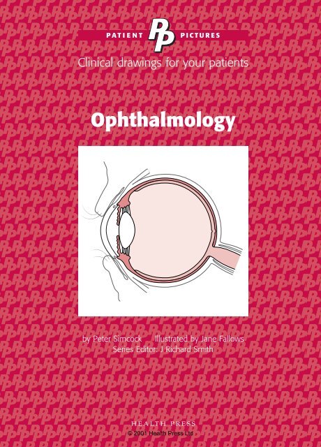
<strong>Ophthalmology</strong><br />
by Peter Simcock Illustrated by Jane Fallows<br />
Series Editor: J Richard Smith<br />
HEALTH PRESS<br />
PICTURES<br />
Clinical drawings for your patients<br />
© 2001 Health Press Ltd
PATIENT<br />
<strong>Ophthalmology</strong><br />
by Peter Simcock MRCP FRCS FRCOphth DO<br />
Consultant Ophthalmic Surgeon, West of England Eye Unit,<br />
Royal Devon and Exeter Hospital, Exeter, UK<br />
Series Editor: J Richard Smith MD MRCOG<br />
Consultant Gynaecologist,<br />
PICTURES<br />
Clinical drawings for your patients<br />
Chelsea and Westminster Hospital, London,<br />
and Honorary Senior Lecturer in Obstetrics and Gynaecology,<br />
Imperial College School of Medicine, London, UK<br />
Illustrated by Jane Fallows, MeDee Art, London, UK<br />
HEALTH PRESS<br />
Oxford<br />
© 2001 Health Press Ltd
Patient Pictures – <strong>Ophthalmology</strong><br />
First published 2001<br />
Text © 2001 Peter Simcock<br />
© 2001 in this edition Health Press Limited<br />
Health Press Limited, Elizabeth House, Queen Street,<br />
Abingdon, Oxford OX14 3JR, UK<br />
Tel: (01235) 523233<br />
Fax: (01235) 523238<br />
Patient Pictures is a trade mark of Health Press Limited.<br />
All rights reserved. No part of this publication may be<br />
reproduced, stored in a retrieval system, or transmitted in any<br />
form or by any means, electronic, mechanical, photocopying<br />
(except under the terms of a recognized photocopying<br />
licensing scheme), recording or otherwise, without express<br />
permission of the publisher.<br />
The right of Peter Simcock to be identified as the author of<br />
this work has been asserted in accordance with the<br />
Copyright, Designs & Patents Act 1988 Sections 77 and 78.<br />
The publisher and author have made every attempt to ensure<br />
the accuracy of this book, but cannot accept responsibility<br />
for any errors or omissions.<br />
A CIP catalogue record for this title is available from the<br />
British Library.<br />
ISBN 1-899541-95-0<br />
Jane Fallows thanks Dee McLean for her help with the<br />
illustrations.<br />
<strong>Print</strong>ed by Uniskill Ltd, Witney, UK.<br />
© 2001 Health Press Ltd
Reproduction authorization<br />
The purchaser of this Patient Pictures series title is hereby authorized to reproduce<br />
by photocopy only, any part of the pictorial and textual material contained in this<br />
work for non-profit, educational, or patient education use. Photocopying for these<br />
purposes only is welcomed and free from further permission requirements from<br />
the publisher and free from any fee.<br />
The reproduction of any material from this publication outside the guidelines<br />
above is strictly prohibited without the permission in writing of the publisher<br />
and is subject to minimum charges laid down by the Publishers Licensing Society<br />
Limited or its nominees.<br />
Publisher, Health Press Limited, Oxford<br />
© 2001 Health Press Ltd
Author’s preface<br />
Sight is perhaps the most important of the senses.<br />
Many patients who consult their doctor or<br />
optometrist with a visual disturbance have a genuine<br />
fear of blindness. Examination of the eye can reveal<br />
local problems, such as a cataract or glaucoma, but<br />
can also provide evidence of medical conditions<br />
including high blood pressure and diabetes.<br />
There are a variety of surgical and laser procedures now available to treat many<br />
eye conditions. The aim of this book is to help healthcare professionals explain<br />
these treatments simply and clearly to their patients, with the aid of informative<br />
diagrams and concise explanatory notes. Communication at a level that is easily<br />
understood by patients will help them to make informed judgements about their<br />
own healthcare and alleviate anxiety about proposed treatments.<br />
Peter Simcock MRCP FRCS FRCOphth DO<br />
Consultant Ophthalmic Surgeon, West of England Eye Unit,<br />
Royal Devon and Exeter Hospital, Exeter, UK<br />
© 2001 Health Press Ltd
The eye and eyelid<br />
• The eye works in a similar way to a camera. Light enters<br />
the eye and is focused onto the retina, the light-sensitive<br />
surface at the back of the eye.<br />
• The clear window at the front of the eye is the cornea,<br />
which provides coarse focusing power. A lens lies behind<br />
the iris, the coloured part of the eye, and does the fine<br />
focusing. Muscles attached to the lens can change its<br />
shape, so light can be focused when looking at close<br />
or distant objects.<br />
• The pupil is the black hole in the middle of the iris. Its<br />
size can change to control the amount of light reaching<br />
the retina, like the aperture in a camera. In dim light,<br />
the pupil gets wider to let more light into the eye, and<br />
in bright light it gets smaller to reduce the amount of<br />
light entering.<br />
• The retina changes the image of the outside world into<br />
electrical impulses, which travel to the brain along the<br />
optic nerve.<br />
• The eyelids protect and clean the front of the eye. The<br />
upper eyelid blinks and washes tears over the surface of<br />
the eye. Tears are formed by glands around the eye and<br />
in the eyelids. Tears drain out of the eyes and into the<br />
nose through channels called tear ducts.<br />
• Muscles attached to the surface of the eyes allow them<br />
to move exactly together. Both eyes are protected by the<br />
orbital bone, except at the front.<br />
© 2001 Health Press Ltd
Side view<br />
Eyelid<br />
Iris<br />
Pupil<br />
Watery fluid<br />
(aqueous)<br />
Cornea<br />
Muscle that<br />
changes shape<br />
of lens<br />
© Health Press Limited<br />
Eye<br />
Muscles that move eye<br />
Optic nerve<br />
Orbital bone<br />
surrounding eye<br />
Nerve pathway to brain<br />
Vision processing in brain<br />
© 2001 Health Press Ltd<br />
Retina<br />
Glands that produce tears<br />
Upper eyelid<br />
Pupil<br />
Iris<br />
Lower eyelid<br />
Tear duct into nose<br />
From above<br />
Clear jelly<br />
(vitreous)<br />
Lens<br />
Optic nerve<br />
to brain<br />
1