changing the course of stroke - New Jersey Medical School ...
changing the course of stroke - New Jersey Medical School ...
changing the course of stroke - New Jersey Medical School ...

You also want an ePaper? Increase the reach of your titles
YUMPU automatically turns print PDFs into web optimized ePapers that Google loves.
University <strong>of</strong> Medicine and Dentistry <strong>of</strong> <strong>New</strong> <strong>Jersey</strong> Volume 1 Number 1 Spring 2003<br />
<strong>New</strong><br />
<strong>Jersey</strong><br />
<strong>Medical</strong><br />
<strong>School</strong>pulse pulse<br />
INAUGURAL ISSUE<br />
<strong>School</strong><br />
CHANGING THE COURSE OF STROKE<br />
SAVING SIGHT • CME: MACULAR DEGENERATION • NEW COCHLEAR IMPLANTS
Russell T. J<strong>of</strong>fe, MD<br />
Message<br />
from<br />
<strong>the</strong> Dean<br />
IAM VERY PLEASED TO PRESENT <strong>the</strong> inaugural issue <strong>of</strong> NJMS<br />
Pulse. This new magazine embodies <strong>the</strong> pride that all <strong>of</strong> us feel<br />
for <strong>the</strong> academic accomplishments <strong>of</strong> our school. It also serves to<br />
highlight our successes in meeting <strong>the</strong> challenges <strong>of</strong> today’s healthcare<br />
environment.<br />
Despite <strong>the</strong> uncertainties and distractions around us, <strong>the</strong> faculty, students<br />
and staff <strong>of</strong> NJMS remain remarkably focused and dedicated to<br />
achieving our commitment to excellence in medical education, research<br />
and clinical care. Within <strong>the</strong>se pages you will read about our innovative<br />
biomedical research, exemplary medical care, and forward thinking revisions<br />
to our curriculum. You will be reminded <strong>of</strong> <strong>the</strong> dedication <strong>of</strong> our<br />
alumni to <strong>the</strong>ir patients and to <strong>the</strong>ir communities.<br />
Each section <strong>of</strong> this issue speaks ei<strong>the</strong>r directly or indirectly to <strong>the</strong><br />
passionate commitment this school has to its stakeholders, specifically<br />
our city <strong>of</strong> <strong>New</strong>ark neighbors and <strong>the</strong>ir families and <strong>the</strong> wider nor<strong>the</strong>rn<br />
<strong>New</strong> <strong>Jersey</strong> community.<br />
As you glance through <strong>the</strong>se pages, I am confident that you will<br />
appreciate, as I do, that NJMS is a vital resource to our community, to<br />
our state <strong>of</strong> <strong>New</strong> <strong>Jersey</strong>, and to our nation. Congratulations to all <strong>of</strong> you:<br />
our faculty, alumni, students, staff and community stakeholders for<br />
helping our school achieve excellence.<br />
ROBERT GLICK<br />
NJMS Pulse<br />
VOLUME 1, NUMBER 1, SPRING 2003<br />
UNIVERSITY OF MEDICINE AND DENTISTRY<br />
OF NEW JERSEY<br />
NEW JERSEY MEDICAL SCHOOL<br />
Dean, UMDNJ-<strong>New</strong> <strong>Jersey</strong> <strong>Medical</strong> <strong>School</strong><br />
Russell T. J<strong>of</strong>fe, MD<br />
Executive Editor and Vice Dean, NJMS<br />
William E. Reichman, MD<br />
Managing Editor and Director,<br />
Marketing and Media Relations<br />
UMDNJ-University Hospital<br />
Robin Preisler<br />
Senior Editor and Director,<br />
University Marketing Communications<br />
Barbara Hurley<br />
Editors, NJMS Pulse<br />
Eve Jacobs<br />
Mary Ann Littell<br />
AlumniFocus Editor<br />
Joseph V. DiTrolio, MD<br />
Coordinator, AlumniFocus<br />
Dianne Mink<br />
Contributing Writers<br />
Maryann Brinley<br />
Carole Walker<br />
Jill Spotz<br />
EDITORIAL BOARD<br />
Patrick Bower<br />
Vice President for Development<br />
Foundation <strong>of</strong> UMDNJ<br />
Suzanne H. Atkin, MD<br />
Associate Pr<strong>of</strong>essor <strong>of</strong> Medicine<br />
Department <strong>of</strong> Medicine<br />
President-Elect, University Hospital <strong>Medical</strong> Staff<br />
Linda Luciano, MBA<br />
Associate Dean and Chief Operating Officer<br />
<strong>New</strong> <strong>Jersey</strong> <strong>Medical</strong> <strong>School</strong><br />
Suresh Raina, MD<br />
Chief <strong>of</strong> Staff<br />
Associate Dean for Clinical Affairs<br />
University Hospital<br />
John Katz, MD<br />
Associate Pr<strong>of</strong>essor<br />
Department <strong>of</strong> Anes<strong>the</strong>siology<br />
President, Alumni Association <strong>of</strong> NJMS<br />
Harvey L. Ozer, MD<br />
Senior Associate Dean for Research<br />
Office <strong>of</strong> Research & Sponsored Programs<br />
Anthony Rosania<br />
NJMS’05<br />
Office <strong>of</strong> Student Affairs<br />
<strong>New</strong> <strong>Jersey</strong> <strong>Medical</strong> <strong>School</strong><br />
DESIGN<br />
Sherer Graphic Design<br />
KEEP IN TOUCH<br />
NJMS Pulse is published twice each year<br />
by UMDNJ-<strong>New</strong> <strong>Jersey</strong> <strong>Medical</strong> <strong>School</strong> (NJMS)<br />
and UMDNJ-University Hospital (UH). We<br />
welcome letters to <strong>the</strong> editor and suggestions for<br />
future articles. Send all correspondence to Pulse<br />
Editor, Office <strong>of</strong> University Marketing<br />
Communications, University Heights –P.O. Box<br />
1709, Room 1328, 65 Bergen Street, <strong>New</strong>ark, NJ,<br />
07101-1709 or via email to njmspulse@umdnj.edu<br />
NJMS Pulse is published twice a year, in spring and<br />
fall, by <strong>New</strong> <strong>Jersey</strong> <strong>Medical</strong> <strong>School</strong>, P.O. Box 1709,<br />
185 South Orange Avenue, <strong>New</strong>ark, NJ 07107-1709.
FEATURES<br />
12 Changing <strong>the</strong> Course <strong>of</strong> Stroke by Maryann B. Brinley<br />
The Brain Attack Team prevents death and disability<br />
from <strong>stroke</strong>.<br />
18 Through <strong>the</strong> Looking Glass by Eve Jacobs<br />
Eyeing <strong>the</strong> future <strong>of</strong> ophthalmology in <strong>the</strong> 21st century<br />
26 Hear! Hear! On <strong>the</strong> Cutting Edge <strong>of</strong> Cochlear<br />
Implant Technology by Maryann B. Brinley<br />
Innovative medical device brings patients like Dianne Mink<br />
back to <strong>the</strong> hearing world.<br />
DEPARTMENTS<br />
INSIDE INFORMATION<br />
2 <strong>New</strong> Trial Compares MS Therapies<br />
3 Faculty Star in TV Series<br />
3 TB Center Has 10th Anniversary<br />
4 Alum Gives $2 Million for Chair in Orthopaedics<br />
5 Treating Complex Head and Neck Malignancies<br />
6 <strong>New</strong> Company Launched<br />
6 Cardiac Surgery at UH Takes Leap Forward<br />
7 Prestigious Fellowship for Tom Denny<br />
8 Curriculum Changes for NJMS<br />
9 Streamlining Emergency Care<br />
9 <strong>New</strong> <strong>Jersey</strong>’s Virtual Defense Tower<br />
VOICES<br />
10 The Full-Body Scan Debate: Pros, Cons and<br />
O<strong>the</strong>r Cons by Stephen R. Baker, MD<br />
CONTINUING MEDICAL EDUCATION<br />
24 Age-Related Macular Degeneration:<br />
Update on Pathogenesis by Marco A. Zarbin, MD, PhD<br />
ALUMNIFOCUS<br />
32 <strong>New</strong>s <strong>of</strong> special interest to graduates<br />
END PAGE<br />
40 <strong>New</strong> Research Dean by Carole Walker<br />
Scientist, pr<strong>of</strong>essor, mentor—just a few words<br />
to describe Harvey Ozer<br />
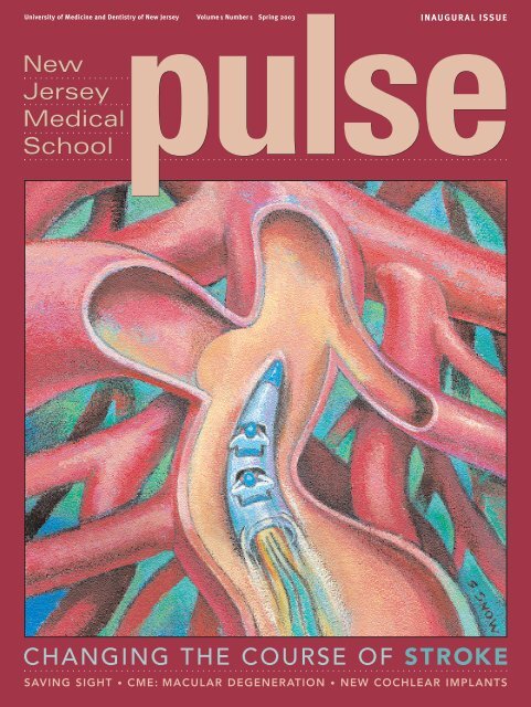
COVER ILLUSTRATION BY SCOTT SNOW<br />
pulse INAUGURAL ISSUE<br />
spring 2003<br />
For names and e-mail addresses <strong>of</strong> all NJMS experts who appear in this issue, see <strong>the</strong> inside back cover.<br />
k<br />
m<br />
N EW JERSEY MEDICAL SCHOOL 1
<strong>New</strong> Trial Compares<br />
MS Therapies<br />
2<br />
PULSE SPRING 2003<br />
insideinformation<br />
MRI study monitors lesions <strong>of</strong> patients taking<br />
two medications: Betaseron and Copaxone.<br />
P<br />
HYSICIANS AT NEW JERSEY MEDICAL SCHOOL (NJMS)<br />
have launched a $3,619,080 clinical trial comparing<br />
two FDA-approved treatments for multiple sclerosis<br />
(MS): interferon beta 1b (Betaseron) and glatiramer acetate<br />
(Copaxone). It is <strong>the</strong> first head-to-head, randomized study<br />
comparing any interferon with glatiramer acetate. The Neurological<br />
Institute <strong>of</strong> <strong>New</strong> <strong>Jersey</strong> at NJMS is one <strong>of</strong> two study<br />
sites in <strong>the</strong> state; <strong>the</strong> o<strong>the</strong>r is <strong>the</strong> Bernard W. Gimbel MS<br />
Center in Teaneck. Principal investigators are Leo J. Wolansky,<br />
MD, and Diego Cadavid, MD.<br />
The trial is unique because it uses state-<strong>of</strong>-<strong>the</strong>-art magnetic<br />
resonance imaging (MRI) technology to detect and monitor<br />
brain lesions in patients with MS. These lesions, also known<br />
as plaques, are scars in <strong>the</strong> central nervous system where<br />
inflammation has stripped axons <strong>of</strong> <strong>the</strong> insulating fatty protein<br />
known as myelin. Demyelinated axons do not function efficiently,<br />
and <strong>the</strong>se lesions are what cause <strong>the</strong> symptoms <strong>of</strong><br />
multiple sclerosis.<br />
MRI has proven to be a valuable tool for diagnosis, management<br />
and research <strong>of</strong> MS. “The 3-Tesla MRI on <strong>the</strong><br />
<strong>New</strong>ark campus is state-<strong>of</strong>-<strong>the</strong>-art in detecting brain lesions,”<br />
LONG TR, SHORT TE IMAGE<br />
OF STUDY PATIENT<br />
The high magnetic fieldstrength<br />
<strong>of</strong> <strong>the</strong> 3T system<br />
produces high signal to noise<br />
ratio images, even with thin<br />
3 mm thick sections. Use <strong>of</strong><br />
thin sections facilitates improved<br />
demonstration <strong>of</strong> small<br />
lesions, such as are commonly<br />
seen with MS (arrows).<br />
k<br />
m<br />
Leo J. Wolansky, MD (left), and<br />
Diego Cadavid, MD<br />
says Wolansky. “Within<br />
months <strong>of</strong> FDA approval, we<br />
installed <strong>the</strong> first 3-Tesla MRI scanner for clinical use in <strong>New</strong><br />
<strong>Jersey</strong>. Until now, <strong>the</strong>re was virtually no information about <strong>the</strong><br />
appearance <strong>of</strong> plaques using an MRI with a higher magnetic<br />
field strength than 2 Tesla.”<br />
To fur<strong>the</strong>r enhance <strong>the</strong> visibility <strong>of</strong> <strong>the</strong> lesions, <strong>the</strong> study<br />
will employ a triple dose <strong>of</strong> gadolinium, a contrast-enhancing<br />
agent used to detect active inflammation. This technique had<br />
been described several years ago at NJMS by Wolansky, working<br />
with UMDNJ President Stuart D. Cook, MD, who is also<br />
director <strong>of</strong> <strong>the</strong> MS Center at NJMS. Gadolinium, <strong>the</strong> 64th<br />
chemical in <strong>the</strong> periodic table <strong>of</strong> elements, is strongly paramagnetic,<br />
meaning that, like iron, it is attracted by magnetic<br />
fields, making it particularly useful in MRI.<br />
Participants will be randomized to receive one <strong>of</strong> <strong>the</strong> two<br />
drugs. “The study is clearly attractive to patients because<br />
everyone receives an effective, approved <strong>the</strong>rapy,” says<br />
Cadavid, who is assistant pr<strong>of</strong>essor <strong>of</strong> neurology and neurosciences.<br />
“There are no placebos.” They receive a baseline scan<br />
upon entry, and after that, once every four weeks. All MRIs<br />
will be carried out on <strong>the</strong> 3-Tesla unit, with Wolansky conducting<br />
a blinded evaluation <strong>of</strong> <strong>the</strong> scans, including visual as<br />
well as computer analysis.<br />
“From <strong>the</strong> scans, we can get feedback that might o<strong>the</strong>rwise<br />
take years to obtain,” says Wolansky, who is pr<strong>of</strong>essor <strong>of</strong> radiology<br />
and MRI section chief.<br />
The study is being funded by a grant from Berlex Laboratories,<br />
Inc., <strong>the</strong> manufacturer <strong>of</strong> Betaseron. A larger study is<br />
planned later to compare double-dose Betaseron with<br />
Copaxone. “MS is a field full <strong>of</strong> opinions about <strong>the</strong>se and<br />
o<strong>the</strong>r drugs, but <strong>the</strong>re is not much hard data to go by,” says<br />
Cadavid. “This study will give us <strong>the</strong> first insight into <strong>the</strong> relative<br />
efficacy <strong>of</strong> <strong>the</strong>se two drugs in a head-to-head trial.” ●<br />
TOP: PETER BYRON
Faculty Star in TV Series<br />
N THE EARLY 1990s, TB rates<br />
were on <strong>the</strong> rise. The state <strong>of</strong><br />
<strong>New</strong> <strong>Jersey</strong> had <strong>the</strong> seventh<br />
highest rate <strong>of</strong> TB infection in <strong>the</strong> country.<br />
And to make matters worse, a growing<br />
number <strong>of</strong> TB patients were coinfected<br />
with HIV or had a drug-resistant<br />
strain <strong>of</strong> TB, making treatment<br />
more difficult and time-consuming. Lee<br />
Reichman, MD, decided it was time to<br />
take action. And <strong>the</strong> <strong>New</strong> <strong>Jersey</strong><br />
<strong>Medical</strong> <strong>School</strong> National Tuberculosis<br />
Center was born.<br />
This January, <strong>the</strong> Center celebrated<br />
its 10th anniversary in brand-new, state<strong>of</strong>-<strong>the</strong>-art<br />
quarters at <strong>the</strong> International<br />
Center for Public Health in<br />
<strong>New</strong>ark (pictured at right). Its<br />
run has been impressive. It<br />
has improved medicationtaking<br />
compliance in<br />
<strong>New</strong>ark from 62 percent<br />
to 98 percent, lowered <strong>the</strong><br />
TB rates in <strong>the</strong> city by 62 percent, and<br />
achieved National Model Center designation<br />
from <strong>the</strong> federal government.<br />
“We now occupy <strong>the</strong> only clinical facility<br />
in <strong>the</strong> world designed from scratch<br />
for safe and effective treatment <strong>of</strong> TB<br />
and multi-drug resistant TB,” says<br />
Reichman, Executive Director <strong>of</strong> <strong>the</strong><br />
Center and a pr<strong>of</strong>essor <strong>of</strong> medicine and<br />
preventive medicine and community<br />
health at NJMS. The TB Center has<br />
also demonstrated <strong>the</strong> effectiveness <strong>of</strong> a<br />
widely used strategy called directly<br />
observed <strong>the</strong>rapy, or DOT, in<br />
which public health<br />
<strong>New</strong>s about NJMS events, faculty,<br />
grants, research and more<br />
THE FIRST THREE PROGRAMS <strong>of</strong> a cable television series<br />
focusing on <strong>the</strong> most critical health issues facing <strong>the</strong> state aired<br />
in February, featuring several <strong>of</strong> <strong>the</strong> medical school’s experts.<br />
Steve Adubato is <strong>the</strong> host. The first installment looked at <strong>the</strong> debate<br />
over hormone replacement <strong>the</strong>rapy and included two patients and<br />
three physicians, two from UMDNJ: Gerson Weiss, MD, chair <strong>of</strong><br />
OB/GYN at NJMS, and Gloria Bachman, MD, from Robert Wood<br />
Johnson <strong>Medical</strong> <strong>School</strong>. The second installment, examining <strong>the</strong> potential impact <strong>of</strong> weaponizing biological agents such as anthrax<br />
and smallpox, featured: Nancy Connell, PhD, Director <strong>of</strong> <strong>the</strong> Center for BioDefense, NJMS; Peter Wenger, MD, NJMS assistant pr<strong>of</strong>essor<br />
<strong>of</strong> preventive medicine; and David Perlin, PhD, Scientific Director <strong>of</strong> <strong>the</strong> Public Health Research Institute (PHRI). The third<br />
show examined <strong>the</strong> state’s and country’s level <strong>of</strong> preparedness for an attack using biological or chemical weapons. William<br />
Halperin, MD, DrPH, chair <strong>of</strong> preventive medicine at NJMS, and Leah Ziskin, PhD, SPH associate dean in south <strong>Jersey</strong>, participated.<br />
Transcripts <strong>of</strong> <strong>the</strong> programs can be accessed at <strong>the</strong> Healthy <strong>New</strong> <strong>Jersey</strong> Web site. UMDNJ is an underwriter <strong>of</strong> <strong>the</strong> series. ●<br />
TB Center Has 10th Anniversary<br />
I<br />
workers visit TB patients daily to<br />
observe <strong>the</strong>m taking medication.<br />
In 2001, 23.2 <strong>New</strong>ark residents<br />
per 100,000 had active tuberculosis,<br />
down from 71.8 per 100,000 in 1991.<br />
Experts believe DOT is <strong>the</strong> primary<br />
reason for <strong>the</strong> dramatic reduction.<br />
TB can be cured, but it requires taking<br />
several medications for many months.<br />
Unfortunately, once patients begin to<br />
feel better, <strong>the</strong>y stop taking <strong>the</strong> antibiotics,<br />
allowing <strong>the</strong> disease to mutate<br />
into drug-resistant strains which can<br />
spread.<br />
Reichman’s book, “Timebomb:<br />
The Global Epidemic <strong>of</strong> Multi-<br />
Drug-Resistant Tuberculosis”<br />
(McGraw-Hill), recently won<br />
first prize in <strong>the</strong> trade category<br />
<strong>of</strong> <strong>the</strong> American <strong>Medical</strong><br />
Writers Association 2002<br />
<strong>Medical</strong> Book Awards<br />
Competition. ●<br />
TOP: GETTY IMAGES; BOTTOM: DAN KATZ NEW JERSEY MEDICAL SCHOOL 3
4<br />
insideinformation<br />
Alum Gives<br />
$2 Million for Chair<br />
in Orthopaedics<br />
The co-inventor <strong>of</strong> <strong>the</strong> <strong>New</strong> <strong>Jersey</strong> Knee<br />
makes a gift to encourage human joint<br />
replacement research at NJMS.<br />
F<br />
REDERICK F. BUECHEL, MD, a 1972 NJMS graduate<br />
and co-inventor <strong>of</strong> <strong>the</strong> <strong>New</strong> <strong>Jersey</strong> LCS Total Knee<br />
Replacement System, has given <strong>the</strong> school <strong>the</strong> largest<br />
single gift ever received from an alum—a $2 million endowment<br />
to establish a chair in <strong>the</strong> Department <strong>of</strong> Orthopaedics.<br />
He completed <strong>the</strong> orthopaedic residency program at NJMS<br />
in 1977.<br />
The Frederick F. Buechel, MD, Chair for Joint Replacement<br />
will enhance a strong clinical program in total joint<br />
replacement at University Hospital with a basic science<br />
research program.<br />
“My wish for <strong>the</strong> chair holder is to push <strong>the</strong> frontiers <strong>of</strong><br />
human joint replacement research into practical, clinical applications,”<br />
says Buechel.<br />
To encourage scientific exchange and research collaborations,<br />
<strong>the</strong> chair holder will work in conjunction with <strong>the</strong> <strong>New</strong><br />
<strong>Jersey</strong> Institute <strong>of</strong> Technology (NJIT) to establish <strong>the</strong> Buechel-<br />
Pappas Biomechanical Engineering Liaison—a partnership<br />
between <strong>the</strong> NJMS orthopaedic department and <strong>the</strong> departments<br />
<strong>of</strong> Biomedical Engineering and Mechanical Engineering<br />
at NJIT.<br />
This liaison reinforces <strong>the</strong> importance Buechel places on <strong>the</strong><br />
value <strong>of</strong> collaboration. Engineer Michael Pappas, PhD, a pr<strong>of</strong>essor<br />
<strong>of</strong> mechanical engineering at NJIT, taught biomechanics<br />
to NJMS medical students and residents. Buechel, <strong>the</strong>n a surgical<br />
resident, was inspired by Pappas’ skill at solving complex<br />
structural problems. They joined forces when <strong>the</strong> resident<br />
asked his teacher for help in constructing a pros<strong>the</strong>tic ankle.<br />
Since <strong>the</strong>n, <strong>the</strong>y have received more than 100 patents for<br />
implants and instruments related to replacement ankles, hips,<br />
shoulders, knee and finger joints.<br />
PULSE SPRING 2003<br />
“My wish for <strong>the</strong> chair holder is to push<br />
<strong>the</strong> frontiers <strong>of</strong> human joint replacement<br />
research into practical, clinical applications.”<br />
Frederick F. Buechel, MD<br />
The endowment will also establish <strong>the</strong> Buechel-Pappas<br />
Award for Outstanding Biomedical Engineering Research,<br />
which will be given every o<strong>the</strong>r year to a surgeon-engineering<br />
team from <strong>the</strong> two schools. It will also allow for <strong>the</strong> development<br />
<strong>of</strong> a clinical research unit on <strong>the</strong> <strong>New</strong>ark campus to promote<br />
cutting-edge research.<br />
Buechel, who is a clinical pr<strong>of</strong>essor <strong>of</strong> orthopaedic surgery<br />
at NJMS, was inducted into <strong>the</strong> <strong>New</strong> <strong>Jersey</strong> Inventors Hall <strong>of</strong><br />
Fame with Pappas in 1998.<br />
Fred Behrens, MD, pr<strong>of</strong>essor and chair <strong>of</strong> <strong>the</strong> Department<br />
<strong>of</strong> Orthopaedics, comments: “This gift, which is a tribute to<br />
Dr. Buechel’s achievements and his remarkable career, will<br />
help to assure that our school remains at <strong>the</strong> forefront <strong>of</strong><br />
research and education in orthopaedics.” ●<br />
PADDY ECKERSLEY
Treating Complex<br />
Head and Neck Malignancies<br />
H<br />
ead and neck malignancies,<br />
including those found in <strong>the</strong><br />
voice box, throat, mouth, salivary<br />
glands and sinuses, account for<br />
about 5 percent <strong>of</strong> all cancers. However,<br />
at University Hospital (UH), head and<br />
neck malignancies total about 15 to 17<br />
percent <strong>of</strong> all cancers treated.<br />
What brings <strong>the</strong>se patients to <strong>New</strong>ark<br />
is a multidisciplinary program led by<br />
Soly Baredes, MD, chief <strong>of</strong> <strong>the</strong> division<br />
<strong>of</strong> otolaryngology-head and neck surgery<br />
and associate pr<strong>of</strong>essor <strong>of</strong> surgery at<br />
<strong>New</strong> <strong>Jersey</strong> <strong>Medical</strong> <strong>School</strong>. “Head and<br />
neck tumors can be difficult to manage<br />
in a community setting because <strong>of</strong> <strong>the</strong><br />
complexity <strong>of</strong> treatment,” says Baredes.<br />
For example, tumors at <strong>the</strong> base <strong>of</strong> <strong>the</strong><br />
skull require <strong>the</strong> skills <strong>of</strong> a neurosurgeon,<br />
ophthalmologist and plastic surgeon<br />
as well as an otolaryngologist.<br />
O<strong>the</strong>r head and neck malignancies may<br />
need treatment by oral and maxill<strong>of</strong>acial<br />
surgeons as well.<br />
Baredes says <strong>the</strong> collaboration among<br />
surgeons makes UH’s program unique<br />
in <strong>New</strong> <strong>Jersey</strong>. “For example, our skull<br />
base team includes a neurosurgeon,<br />
ophthalmologist and otolaryngologist,”<br />
he explains. “We collaborate on complex<br />
tumors that cross <strong>the</strong> barriers between<br />
<strong>the</strong> nasal cavities, sinuses, and <strong>the</strong><br />
brain.” UH is <strong>the</strong> only facility in <strong>the</strong><br />
state, and one <strong>of</strong> a few in <strong>the</strong> U.S., with<br />
intraoperative MRI technology, pioneered<br />
here by neurosurgeon Michael<br />
Schulder, MD. It allows surgeons to<br />
pinpoint <strong>the</strong> tumor location and remove<br />
it with minimal damage to surrounding<br />
tissue. Ano<strong>the</strong>r special feature <strong>of</strong> <strong>the</strong><br />
program is interventional radiology.<br />
Preoperative embolization <strong>of</strong> tumors<br />
decreases blood loss during surgery to<br />
remove lesions.<br />
A cornerstone <strong>of</strong> <strong>the</strong> program are <strong>the</strong><br />
weekly head and neck tumor confer-<br />
Left and above: The CT scan pictured<br />
is that <strong>of</strong> a parotid tumor.<br />
Below: Soly Baredes, MD, examining<br />
a patient<br />
ences, which are attended by representatives<br />
<strong>of</strong> <strong>the</strong> many specialties on <strong>the</strong><br />
interdisciplinary team. (In addition to<br />
those listed above, <strong>the</strong>se also include<br />
medical and radiation oncologists,<br />
pathologists, head and neck radiologists,<br />
and specialized nursing staff.)<br />
The Head and Neck Cancer Resource<br />
Center at UH, funded by a grant from<br />
<strong>the</strong> HealthCare Foundation <strong>of</strong> <strong>New</strong><br />
<strong>Jersey</strong>, provides patients and <strong>the</strong>ir families<br />
with a variety <strong>of</strong> rehabilitative services.<br />
“This type <strong>of</strong> surgery affects basic<br />
survival skills, such as <strong>the</strong> ability to eat,<br />
brea<strong>the</strong> and communicate,” says<br />
advanced practice nurse Ray Scarpa,<br />
MA, CNS, AOCN. “Some 40 to 60<br />
percent <strong>of</strong> patients may have tracheostomies,<br />
and many also have laryngectomies.”<br />
The Center staff, which<br />
includes four advance practice nurses,<br />
helps with <strong>the</strong> physical management <strong>of</strong><br />
supplemental devices, from feeding<br />
tubes to breathing aids. ●<br />
PETER BYRON NEW JERSEY MEDICAL SCHOOL 5
insideinformation<br />
<strong>New</strong> Company Launched<br />
UNE 25, 2002<br />
MARKED A MILE-<br />
STONE for UMDNJ’s<br />
technology transfer <strong>of</strong>fice<br />
when BioDelivery Sciences<br />
International, Inc. went public<br />
using stock ticker BDSI<br />
(NASDAQ). It is <strong>the</strong><br />
University’s first spin-<strong>of</strong>f company<br />
to go public. Raphael<br />
Mannino, PhD, and Susan<br />
Gould-Fogerite, PhD, both<br />
associate pr<strong>of</strong>essors <strong>of</strong> molecu-<br />
6<br />
J<br />
PULSE SPRING 2003<br />
lar biology at NJMS, are <strong>the</strong><br />
company’s founders.<br />
Mannino is executive vice<br />
president and chief scientific<br />
<strong>of</strong>ficer <strong>of</strong> BDSI, and serves on<br />
its board <strong>of</strong> directors. He is a<br />
world leader in cochleate<br />
technology and an expert in<br />
applying artificial lipid-based<br />
delivery systems to problems<br />
in biotechnology, including<br />
drug delivery, vaccine design<br />
and gene <strong>the</strong>rapy applications.<br />
Cardiac Surgery<br />
At UH Takes<br />
Leap Forward<br />
NJMS AND UNIVERSITY HOSPITAL (UH)<br />
Gould-Fogerite, co-developer<br />
<strong>of</strong> <strong>the</strong> cochleate technology,<br />
is vice president <strong>of</strong> innovation/discovery<br />
research at<br />
BDSI and serves on its board.<br />
The groundbreaking cochleate<br />
technology was developed<br />
by <strong>the</strong> partners while at<br />
Albany <strong>Medical</strong> College and<br />
NJMS and is licensed exclusively<br />
to BioDelivery Sciences<br />
International. Cochleate delivery<br />
vehicles (Bioral TM ) repre-<br />
have launched a state-<strong>of</strong>-<strong>the</strong>-art cardiac surgery<br />
program through a new partnership with<br />
Columbia Presbyterian <strong>Medical</strong> Center (CPMC). Columbia’s<br />
cardiac surgery program is currently ranked as one <strong>of</strong> <strong>the</strong> best<br />
in <strong>the</strong> U.S. Columbia surgeons performed <strong>the</strong> nation’s first robotically-assisted<br />
atrial septal defect repair without a chest incision, and<br />
CPMC is also <strong>the</strong> national training center for two <strong>of</strong> <strong>the</strong> three existing FDA trials <strong>of</strong> robotic cardiac<br />
surgery. Its work in <strong>the</strong> use <strong>of</strong> LVAD (left ventricular assist device) has helped thousands <strong>of</strong><br />
patients with end-term heart failure. These and o<strong>the</strong>r innovations will now be available at UH.<br />
Several Columbia faculty members have assumed leadership positions in <strong>the</strong> new program.<br />
They include Barry Esrig, MD, chief <strong>of</strong> <strong>the</strong> division <strong>of</strong> cardiothoracic surgery at UH and Douglas<br />
Jackson, MD, vice chair <strong>of</strong> critical care, anes<strong>the</strong>siology, and director <strong>of</strong> <strong>the</strong> new Cardiothoracic<br />
Surgery Intensive Care Unit (currently under construction). They join Michael Banker, MD,<br />
director <strong>of</strong> cardiac surgery at UH. Administration <strong>of</strong> <strong>the</strong> new division will be provided by Eric<br />
Rose, MD, chair <strong>of</strong> <strong>the</strong> department <strong>of</strong> surgery at Columbia.<br />
Says NJMS Dean Russell T. J<strong>of</strong>fe, MD: “This affiliation provides enormous opportunity for<br />
NJMS as well as University Hospital. The combination <strong>of</strong> <strong>the</strong> best <strong>of</strong> both schools allows us to<br />
move quickly in broadening our clinical, education and research capabilities in <strong>the</strong> entire range<br />
<strong>of</strong> <strong>the</strong> cardiac sciences.” ●<br />
sent a new technology platform<br />
for oral and systemic<br />
delivery <strong>of</strong> molecules with<br />
important <strong>the</strong>rapeutic biological<br />
activities.<br />
They are stable phospholipid-divalent<br />
cation precipitates<br />
composed <strong>of</strong> simple,<br />
naturally occurring materials.<br />
Their unique multi-layer<br />
structure is a large, continuous,<br />
solid lipid bi-layer sheet<br />
rolled up in a spiral, with no<br />
internal aqueous space.<br />
Therefore, <strong>the</strong> interior <strong>of</strong> <strong>the</strong><br />
cochleates is essentially free<br />
<strong>of</strong> water and resistant to penetration<br />
by oxygen. The structure<br />
remains intact, even<br />
though its outer layers may be<br />
exposed to harsh environmental<br />
conditions or enzymes.<br />
This includes protection from<br />
digestion in <strong>the</strong> stomach.<br />
The partners hold nine<br />
U.S. patents, two Australian,<br />
and one issued in <strong>the</strong> major<br />
European countries. ●<br />
Susan Gould-Fogerite, PhD, and<br />
Raphael Mannino, PhD<br />
TOP: GETTY IMAGES; BOTTOM: PETER BYRON
Prestigious<br />
Fellowship for<br />
Tom Denny<br />
The researcher began his career at <strong>New</strong><br />
<strong>Jersey</strong> <strong>Medical</strong> <strong>School</strong> in 1983. He’s now<br />
learning <strong>the</strong> political “ropes” in Washington<br />
and simultaneously managing a multi-million<br />
dollar federal appropriation to set up a<br />
vaccine reference lab at NJMS.<br />
HOMAS N. DENNY, MSc, assistant pr<strong>of</strong>essor <strong>of</strong> pathology,<br />
laboratory medicine and pediatrics at NJMS, was<br />
T one <strong>of</strong> seven health pr<strong>of</strong>essionals named Robert Wood<br />
Johnson Health Policy Fellows for 2002–2003 by <strong>the</strong> Institute<br />
<strong>of</strong> Medicine (IOM) <strong>of</strong> <strong>the</strong> National Academies.The fellows are<br />
all midcareer health pr<strong>of</strong>essionals working in academic and<br />
community-based settings and were chosen on a competitive<br />
basis from nominations.<br />
For three months starting in September, <strong>the</strong> researcher participated<br />
in an orientation designed to familiarize fellows with <strong>the</strong><br />
public-policy process and government health and biomedical<br />
research activities. Denny spent full days learning about current<br />
health issues, federal health and research agencies, principal congressional<br />
committees active in health affairs and major healthinterest<br />
groups.<br />
In December, <strong>the</strong> fellows moved on to part two <strong>of</strong> <strong>the</strong>ir orientation—organized<br />
by <strong>the</strong> American Political Science<br />
Association in conjunction with its Congressional Fellowship<br />
Program—to give <strong>the</strong>m a broadened perspective on <strong>the</strong> range <strong>of</strong><br />
public-policy issues and <strong>the</strong> political process. During this time,<br />
<strong>the</strong>y interviewed for <strong>the</strong>ir work assignments in <strong>the</strong> <strong>of</strong>fices <strong>of</strong> senators<br />
and representatives in Congress and <strong>the</strong> executive branch.<br />
Denny began working with <strong>the</strong> U.S. Senate Committee on<br />
Health, Education, Labor and Pensions on January 6. His first<br />
assignment was to work on a global HIV/AIDS bill, followed by<br />
biodefense, and vaccine compensation/liability legislation. As<br />
staff to <strong>the</strong> committee, he works closely with its members to get<br />
legislation passed.<br />
Denny, who is also director <strong>of</strong> <strong>the</strong> Center for Laboratory<br />
Investigation at NJMS, has studied host defense mechanisms in<br />
response to tumors and infectious diseases for 20 years. He has<br />
served on a number <strong>of</strong> committees for <strong>the</strong> NIH-NIAID Division<br />
<strong>of</strong> AIDS as part <strong>of</strong> <strong>the</strong> HIV clinical trials program. In<br />
1997, he received a NIH HIV Innovative Vaccine Grant award<br />
to study a new method <strong>of</strong> vaccine delivery. He has authored and<br />
co-authored more than 70 peer-reviewed papers.<br />
Denny has also been active in his home town, serving on <strong>the</strong><br />
Cranford Board <strong>of</strong> Education, <strong>the</strong> Cranford Township Committee<br />
and as mayor.<br />
In response to <strong>the</strong> terror attacks <strong>of</strong> 9/11, Denny established a<br />
study on UMDNJ’s <strong>New</strong>ark campus to assess immunologic<br />
memory/responses in individuals who had previously received<br />
smallpox vaccinations. He is currently analyzing interim data<br />
from <strong>the</strong> study. His next venture will be to set up a reference<br />
laboratory at NJMS to support NIH trials for smallpox vaccine<br />
and potential vaccines for anthrax and o<strong>the</strong>r agents. ●<br />
PETER BYRON NEW JERSEY MEDICAL SCHOOL 7
8<br />
insideinformation<br />
Curriculum Changes for NJMS<br />
T<br />
HINK QUICK: Can you recall<br />
three critical points presented<br />
during <strong>the</strong> last fact-driven lecture<br />
you attended? If not, take heart.<br />
Research indicates that you are more<br />
likely to retain material learned in an<br />
interactive environment than passive<br />
learning in lectures.<br />
Alex Stagnaro-Green, MD, <strong>the</strong> new<br />
associate dean <strong>of</strong> curriculum and faculty<br />
development at NJMS, joined <strong>the</strong><br />
administration in September, 2002, and<br />
has been working to develop and implement<br />
a new curriculum by August<br />
2004. His responsibilities also include<br />
reviewing <strong>the</strong> present curriculum to<br />
make sure that <strong>the</strong> school is in compliance<br />
with <strong>the</strong> Liaison Committee on<br />
<strong>Medical</strong> Education (LCME). The<br />
LCME is charged by Congress with <strong>the</strong><br />
accreditation for medical schools in <strong>the</strong><br />
U.S. and Canada, and has 125 standards<br />
all medical schools must meet.<br />
“Different students learn best with<br />
different technologies, and certain topics<br />
ought to be taught through one modality<br />
versus ano<strong>the</strong>r,” explains Stagnaro-<br />
Green, who believes in an eclectic<br />
approach. “Instead <strong>of</strong> relying heavily on<br />
any one technique, a medical school<br />
curriculum must incorporate numerous<br />
teaching tactics: lecture, small group,<br />
case-based, computer-assisted, and simulated<br />
patients.” A school wide curriculum<br />
retreat was held in January, and<br />
more than 100 faculty, students, and<br />
administrators participated.<br />
<strong>Medical</strong> school is a first step in a lifelong<br />
journey for <strong>the</strong> pr<strong>of</strong>essional. “What<br />
we teach will have a lasting impact,” he<br />
says. “We want to make sure that our<br />
graduates are not only well versed in <strong>the</strong><br />
medical knowledge <strong>of</strong> 2003, but also<br />
PULSE SPRING 2003<br />
“We have a<br />
wonderful opportunity and an<br />
awesome responsibility to recast <strong>the</strong> curriculum and<br />
learning environment which will shape <strong>the</strong><br />
physician <strong>of</strong> <strong>the</strong> future.” Alex Stagnaro-Green, MD<br />
have <strong>the</strong> skills, intellectual curiosity, and<br />
passion for learning to ensure <strong>the</strong>ir academic<br />
development throughout <strong>the</strong>ir<br />
careers.” To accomplish this, students<br />
must be active participants in <strong>the</strong> learning<br />
process and acquire skills <strong>the</strong>y can<br />
use forever.<br />
Stagnaro-Green’s 3 p.m. session on<br />
<strong>the</strong> endocrine system in “Introduction<br />
to Clinical” for 14 second-year students<br />
is just one model <strong>of</strong> <strong>the</strong> new curriculum<br />
already being put into place. In room B-<br />
617C <strong>of</strong> <strong>the</strong> <strong>Medical</strong> Science Building,<br />
<strong>the</strong>re are no wrong or right answers, just<br />
a developing story line for hypo<strong>the</strong>tical<br />
patients. Students sit around a wide<br />
table taking turns as <strong>the</strong>y read aloud,<br />
asking questions and considering case<br />
history details. In fact, one <strong>of</strong> <strong>the</strong> scenarios<br />
concerns an anxious med student<br />
who has lost her appetite and is exhibiting<br />
o<strong>the</strong>r disturbing symptoms. Is it<br />
stress? Is it an eating disorder? Could<br />
she be sleep-deprived? Is it her thyroid?<br />
As more details are <strong>of</strong>fered and <strong>the</strong> narrative<br />
reaches its diagnosis, <strong>the</strong> case<br />
becomes clearer and so do <strong>the</strong> intricacies<br />
<strong>of</strong> hormones and biochemistry. This<br />
woman has Graves disease. Following<br />
this, <strong>the</strong> entire class visits a patient in<br />
<strong>the</strong> hospital who presented with syncope<br />
and severe hypocalcemia. During <strong>the</strong><br />
<strong>course</strong> <strong>of</strong> this patient interaction, students<br />
became aware <strong>of</strong> <strong>the</strong> complex<br />
interplay between medical, interpersonal<br />
and societal factors which brought this<br />
man to University Hospital. These medical<br />
students also get a sense <strong>of</strong> <strong>the</strong> curricular<br />
shifts and interactive learning in<br />
store for NJMS. ●<br />
GETTY IMAGES
Streamlining<br />
Emergency Care<br />
I<br />
n terms <strong>of</strong> timeliness<br />
and customer<br />
service, emergency<br />
departments <strong>of</strong>ten earn bad<br />
marks. Providing stellar medical<br />
treatment quickly in an<br />
unpredictable environment<br />
poses enormous problems.<br />
But Ronald Bruce Low,<br />
MD, MS, believes we should<br />
borrow some tricks <strong>of</strong> <strong>the</strong><br />
trade from businesses where<br />
speed and uniformity <strong>of</strong><br />
results is crucial. “Computerization<br />
is <strong>the</strong> key,” says <strong>the</strong><br />
newly hired vice chair <strong>of</strong> surgery<br />
and chief <strong>of</strong> emergency<br />
medicine at University<br />
Hospital (UH). “Tracking<br />
systems used by companies<br />
like Federal Express and<br />
McDonald’s enable companies<br />
to meet <strong>the</strong> needs <strong>of</strong><br />
<strong>the</strong>ir customers. We need to<br />
automate an individual’s trip<br />
through <strong>the</strong> ER by improving<br />
staff access to patient<br />
information, ultimately<br />
allowing <strong>the</strong> patient to<br />
receive treatment faster.”<br />
Low has extensive Level I<br />
trauma center experience,<br />
coming from Kings County<br />
Hospital in Brooklyn and <strong>the</strong><br />
State University <strong>of</strong> <strong>New</strong> York<br />
at Brooklyn, where he served<br />
as vice chair <strong>of</strong> <strong>the</strong> department<br />
<strong>of</strong> emergency medicine.<br />
His plans for UH include<br />
starting an emergency medicine<br />
residency program.<br />
Since <strong>the</strong> majority <strong>of</strong> UH’s<br />
admissions and follow-up<br />
appointments result from<br />
emergency room visits,<br />
improving customer service is<br />
essential. “Patient representatives<br />
on staff will act as<br />
liaisons between <strong>the</strong> physician<br />
and family members,<br />
updating <strong>the</strong>m on <strong>the</strong> status<br />
<strong>of</strong> <strong>the</strong>ir loved ones, assisting<br />
with parking issues or even<br />
helping to find a pharmacy<br />
in <strong>the</strong> area,” says Lowe.<br />
As a Level I trauma center,<br />
UH’s emergency department<br />
is one <strong>of</strong> <strong>the</strong> busiest in <strong>the</strong><br />
state with more than 61,000<br />
visits annually. ●<br />
<strong>New</strong> <strong>Jersey</strong>’s<br />
Virtual Defense<br />
Tower<br />
NEW JERSEY<br />
HAS A POTENT<br />
NEW MEANS<br />
OF DEFENSE in any<br />
potential terror<br />
strike using biological,<br />
chemical or<br />
nuclear weapons.<br />
Medtower’s Biodefense<br />
Knowledge Platform, called<br />
INFORM, is likely to become <strong>New</strong><br />
<strong>Jersey</strong>’s preferred biodefense information site. The INFORM<br />
system is currently available in a scaled-down form at<br />
http://inform.medtower.com.<br />
Launched by UMDNJ’s Center for Biodefense at NJMS and<br />
Medtower, a company that builds Internet-based information<br />
systems aimed at targeted audiences, INFORM provides<br />
customized content prepared by experts in <strong>the</strong> field.<br />
Among <strong>the</strong> NJMS content specialists for <strong>the</strong> site are: biological<br />
experts David Alland, MD; Nancy Connell, PhD; William<br />
Halperin, MD, DrPH; and Peter Wenger, MD; chemical<br />
expert Steven Marcus, MD; response expert Brendan<br />
McCluskey, MPA; and radiation expert Lionel Zuckier, MD.<br />
The site posts both daily updates and almost-instantaneous<br />
“alerts” and “bulletins” as needed, as well as covering<br />
many topics in depth. Material is provided for three levels—<strong>the</strong><br />
basic (for <strong>the</strong> general public) is accessible to anyone.<br />
Individuals wanting access to <strong>the</strong> advanced level (for<br />
physicians and o<strong>the</strong>r healthcare providers) or that for emergency<br />
responders will need to register or send an e-mail to<br />
<strong>the</strong> site’s manager, and <strong>the</strong>ir credentials will be verified.<br />
Also available are educational modules and “expert moderator<br />
discussions.” Current presentations posted on <strong>the</strong><br />
Web site include: “Biological Weapons: <strong>the</strong> Agents and <strong>the</strong><br />
Threat,” “<strong>Medical</strong> Virology: An Introduction,” “Smallpox:<br />
An Overview,” and “Surveillance and Risk Communication:<br />
A Primer.” ●<br />
GETTY IMAGES NEW JERSEY MEDICAL SCHOOL 9
Stephen R. Baker, MD<br />
For example, <strong>the</strong> seemingly achievable<br />
cure for a common cold still eludes us.<br />
On <strong>the</strong> o<strong>the</strong>r hand, <strong>the</strong> pharmaceutical<br />
industry should be congratulated for<br />
introducing powerful drugs that cure a<br />
whole host <strong>of</strong> conditions. These collective<br />
achievements have been gained<br />
under governmental scrutiny, and with<br />
notable exceptions such regulatory<br />
efforts have been successful.<br />
In stark contrast, manufacturers <strong>of</strong><br />
advanced imaging equipment are not<br />
subject to <strong>the</strong> same intense federal<br />
inspection and oversight in <strong>the</strong> United<br />
States as <strong>the</strong> pharmaceutical industry. In<br />
fact, nowhere is <strong>the</strong> tension between<br />
public expectations, entrepreneurial<br />
expedience and incomplete scientific<br />
verification more acutely felt than in <strong>the</strong><br />
emerging arena <strong>of</strong> CT (computed<br />
tomography) screening. The amazing<br />
recent improvements in this technology<br />
have made it possible to obtain a com-<br />
10<br />
PULSE SPRING 2003<br />
njmsvoices<br />
The Full Body Scan Debate:<br />
Pros, Cons and O<strong>the</strong>r Cons<br />
THE INTERPLAY BETWEEN <strong>the</strong> public demand for medical<br />
innovation and <strong>the</strong> introduction <strong>of</strong> new technology<br />
is usually characterized by a dynamic disequilibrium<br />
in which wants and resources are <strong>of</strong>ten out <strong>of</strong> sync.<br />
prehensive visual assessment <strong>of</strong> <strong>the</strong> body<br />
through <strong>the</strong> generation <strong>of</strong> narrowly<br />
spaced images, painlessly rendered in<br />
only a few minutes. CT screening has<br />
captured <strong>the</strong> attention <strong>of</strong> many individuals<br />
eager to know as much about <strong>the</strong>ir<br />
innards as possible. Screening computed<br />
tomography is touted by its adherents to<br />
bypass a preliminary visit to a physician.<br />
It is so diagnostically incisive, advocates<br />
maintain, that <strong>the</strong> requirement for a<br />
physical examination or lab test is obviated<br />
because a screening CT can provide<br />
manifest evidence <strong>of</strong> a tumor, an abnormal<br />
calcification or an arterial bulge.<br />
No wonder <strong>the</strong> publicity about CT<br />
screening centers is convincing to <strong>the</strong><br />
worried well. Yet, is this innovation all<br />
to <strong>the</strong> good? It would seem so, according<br />
to <strong>the</strong> testimonials <strong>of</strong> celebrities who<br />
tell <strong>the</strong> world that <strong>the</strong>ir screening exam<br />
found a treatable condition before it was<br />
too late. Or is <strong>the</strong>re more to this issue?<br />
Actually, CT screening encompasses<br />
four different examinations, each with<br />
promises and limitations: CT colonography,<br />
o<strong>the</strong>rwise known as virtual<br />
colonoscopy, CT screening for lung<br />
nodules to detect small cancers, calcium<br />
scoring <strong>of</strong> coronary arteries and full<br />
body screening <strong>of</strong> <strong>the</strong> thorax and<br />
abdomen. These tests have been made<br />
possible by <strong>the</strong> deployment <strong>of</strong> very fast<br />
CT scanners, and none require <strong>the</strong><br />
administration <strong>of</strong> intravenous contrast<br />
material, which confers an increase in<br />
cost as well as <strong>the</strong> added risks <strong>of</strong> pain<br />
and allergic reaction. Let us assess each<br />
<strong>of</strong> <strong>the</strong>se new studies in turn.<br />
Colon cancer is <strong>the</strong> second leading<br />
cause <strong>of</strong> death from cancer in adults<br />
over age 40. In most cases, it occurs<br />
with <strong>the</strong> transformation <strong>of</strong> an initially<br />
benign polyp which becomes malignant<br />
as it grows beyond one centimeter in<br />
diameter. If such polyps are removed<br />
when still small, <strong>the</strong> likelihood <strong>of</strong> colon<br />
cancer is markedly reduced. In a traditional<br />
colonoscopic examination, first<br />
visual identification and <strong>the</strong>n polyp<br />
extirpation can be performed at <strong>the</strong><br />
same time. As <strong>the</strong> procedure is <strong>of</strong>ten<br />
painful, it is most <strong>of</strong>ten done under<br />
PETER BYRON
sedation. Screening CT, supporters say,<br />
can also discern polyps between 0.5 and<br />
1.0 centimeters in diameter. If none are<br />
seen, <strong>the</strong>n a traditional colonoscopy<br />
need not be performed.<br />
However, <strong>the</strong> jury is still out about<br />
whe<strong>the</strong>r CT colonography is as effective<br />
as conventional colonoscopy for <strong>the</strong><br />
detection <strong>of</strong> small polyps. The coexistence<br />
<strong>of</strong> feces is one problem, <strong>the</strong> common<br />
presence <strong>of</strong> numerous diverticula is<br />
ano<strong>the</strong>r. Until definitive pro<strong>of</strong> <strong>of</strong> CT<br />
colonography’s efficacy is established,<br />
one should be wary about relying on it<br />
as <strong>the</strong> only examination to perform as<br />
part <strong>of</strong> an every 5 to 10 year surveillance<br />
for early-stage large bowel cancer.<br />
Cancer <strong>of</strong> <strong>the</strong> lung is <strong>the</strong> most common<br />
lethal malignancy in <strong>the</strong> Western<br />
world. Screening CT can certainly find<br />
small nodules in <strong>the</strong> lung. And once<br />
detected, a thoracotomy can be performed<br />
to remove <strong>the</strong>se tiny tumefactions<br />
with <strong>the</strong> ultimate benefit <strong>of</strong> making<br />
<strong>the</strong> initially asymptomatic patient<br />
cancer-free.<br />
Yet, we do know that a thoracotomy<br />
is not necessarily an innocuous procedure.<br />
Many benign granulomas appear<br />
on CT identical to minuscule cancerous<br />
nodules. In individuals who were previously<br />
infected by tuberculosis or histoplasmosis,<br />
such nodules <strong>of</strong>ten remain as<br />
a legacy. Thus, a thoracotomy for such<br />
patients would be an unnecessary intrusion.<br />
What we do not know is <strong>the</strong><br />
growth rate <strong>of</strong> lung cancers. Do some<br />
stay small and o<strong>the</strong>rs grow even though<br />
all look <strong>the</strong> same under <strong>the</strong> microscope?<br />
Currently, a multi-center study is underway<br />
to answer questions about incipient<br />
bronchogenic carcinoma. Until <strong>the</strong><br />
results are in, one cannot be sure that<br />
CT screening creates more mischief<br />
than benefit.<br />
The rationale behind calcium scoring<br />
<strong>of</strong> <strong>the</strong> coronary arteries is that <strong>the</strong> presence<br />
<strong>of</strong> calcium salts in <strong>the</strong> walls <strong>of</strong><br />
<strong>the</strong>se vessels indicates that <strong>the</strong> patient,<br />
even one who has not experienced chest<br />
pain, is at heightened risk for a cardiac<br />
event. To be sure, it seems plausible that<br />
<strong>the</strong>re is such an association. Yet is it<br />
Why is <strong>the</strong> public so<br />
infatuated with CT screening?<br />
One reason is that a negative<br />
screening CT conveys a sense<br />
<strong>of</strong> immortality.<br />
An image from a full-body, non-contrast<br />
screening CT study. The kidneys are outlined<br />
by fat on ei<strong>the</strong>r side <strong>of</strong> <strong>the</strong> spine<br />
and <strong>the</strong> aorta is calcified.<br />
actual? Many studies have suggested that<br />
it is not <strong>the</strong> hardened vascular coat that<br />
presages an abrupt arterial occlusion but<br />
ra<strong>the</strong>r <strong>the</strong> presence <strong>of</strong> a s<strong>of</strong>t intimal<br />
plaque that is more apt to ulcerate and<br />
precipitate a luminal blockage. Even<br />
more recent refinements in CT technology<br />
have now allowed us, with an intravenous<br />
injection <strong>of</strong> contrast material, to<br />
not only observe <strong>the</strong> calcified vessel wall<br />
but also <strong>the</strong> lumen itself. In effect we<br />
can do a virtual coronary arteriogram<br />
without invading <strong>the</strong> arteries directly.<br />
NJMS experts speak out on topics<br />
<strong>of</strong> interest or controversy<br />
Whe<strong>the</strong>r this fur<strong>the</strong>r advance will be<br />
confirmed as effective is still unknown,<br />
but it has distinct abnormalities over<br />
simple calcium scoring. Moreover, it<br />
may also replace diagnostic cardiac<br />
ca<strong>the</strong>terization for <strong>the</strong> recognition <strong>of</strong><br />
coronary artery disease. The noncontrast<br />
CT scan limited only to vessel<br />
wall calcification has not yet been<br />
proven to be related to a propensity for<br />
coronary occlusion. Moreover, with<br />
newly realized achievements in CT<br />
instrumentation, calcium scoring is<br />
probably obsolete with respect to cardiac<br />
assessment, because <strong>the</strong> interior <strong>of</strong> coronary<br />
arteries can be viewed with a noninvasive<br />
CT test.<br />
We come now to total body screening.<br />
Unlike <strong>the</strong> o<strong>the</strong>r three types <strong>of</strong> CT<br />
evaluation in <strong>the</strong> asymptomatic individual,<br />
<strong>the</strong>re has been no attempt to undertake<br />
prospective blinded studies to<br />
gauge its efficacy. What does this test<br />
assess? In essence, it is a “fishing expedition”<br />
looking at all organs in <strong>the</strong> chest<br />
and abdomen for <strong>the</strong> signs <strong>of</strong> a tumefaction<br />
or an arterial bulge before symptoms<br />
ensue. The fact that it entails an<br />
unfocused search should not automatically<br />
render its dismissal. After all, big<br />
fish are sometimes caught in fishing<br />
expeditions. So, why not do it? Actually,<br />
it is highly unlikely that anything will<br />
be found <strong>of</strong> significance with respect to<br />
<strong>the</strong> saving <strong>of</strong> a life or even identifying a<br />
treatable lesion.<br />
Before proceeding with <strong>the</strong> assessment<br />
<strong>of</strong> its possible value for each organ<br />
under scrutiny we must acknowledge<br />
<strong>the</strong> relevance <strong>of</strong> <strong>the</strong> iron laws <strong>of</strong> testing.<br />
When <strong>the</strong> prevalence <strong>of</strong> a condition is<br />
low, most tests will be truly negative. Of<br />
<strong>the</strong> few that will be positive, most will<br />
be falsely positive. Even if <strong>the</strong> test is<br />
Continued on page 31<br />
N EW JERSEY MEDICAL SCHOOL<br />
11
pulse SPRING 2003<br />
STROKE<br />
BY MARYANN B. BRINLEY<br />
The campaign has been quietly ga<strong>the</strong>ring momentum.<br />
A small army <strong>of</strong> <strong>stroke</strong> specialists is now in<br />
<strong>New</strong>ark to put a stop to <strong>the</strong> number three cause <strong>of</strong><br />
death and <strong>the</strong> leading reason for disability in <strong>the</strong><br />
United States. “Stroke is life-threatening and 30<br />
percent <strong>of</strong> people who develop one, die. But quite apart from<br />
that, we accept disability from <strong>stroke</strong> too easily. It really wrecks<br />
somebody’s future and creates havoc in families. Even physicians<br />
don’t have <strong>the</strong> empathy or insight into this. We have <strong>the</strong><br />
treatment for this,” says Patrick Pullicino, MD, PhD, <strong>New</strong><br />
<strong>Jersey</strong> <strong>Medical</strong> <strong>School</strong> (NJMS) neurosciences chair. Generating<br />
millions <strong>of</strong> research grant dollars, NJMS and University<br />
Hospital (UH) have recruited neuro-interventionalists, neurologists,<br />
<strong>stroke</strong>-intensivists, laboratory and clinical researchers,<br />
and support staff to help win <strong>the</strong> race. Within UH, interventional<br />
neuro-radiologists and cardiologists are also joining forces<br />
to change <strong>the</strong> <strong>course</strong> <strong>of</strong> <strong>stroke</strong>s and improve chances <strong>of</strong> survival<br />
and recovery without disability.<br />
AT THE ACUTE STAGE<br />
S tep<br />
into <strong>the</strong> cerebrovascular angiography suite at UH where<br />
<strong>the</strong> speed <strong>stroke</strong> demands will send you flying to <strong>the</strong> center<br />
<strong>of</strong> a patient’s brain. According to <strong>the</strong> National Institute <strong>of</strong><br />
Health’s (NIH) guidelines for acute ischemic <strong>stroke</strong>, <strong>the</strong>re is a<br />
“golden hour” <strong>of</strong> rapid identification and treatment essential to<br />
saving lives. The NJMS-UH team has done this in half <strong>the</strong> time<br />
recommended by <strong>the</strong> NIH and with more accuracy.<br />
12<br />
CHANGING<br />
THE COURSE OF<br />
PULSE SPRING 2003<br />
A <strong>stroke</strong> occurs when blood flow to an area <strong>of</strong> <strong>the</strong> brain is<br />
interrupted, cutting <strong>of</strong>f oxygen and nutrients to <strong>the</strong> tissue<br />
(ischemic), or when a vessel breaks and spills blood into <strong>the</strong> surrounding<br />
cells (hemorrhagic). Both kinds <strong>of</strong> <strong>stroke</strong> create a cascade<br />
<strong>of</strong> biochemical disasters that call for <strong>the</strong> fastest response<br />
possible because <strong>the</strong> longer brain tissue remains under attack,<br />
<strong>the</strong> more likely <strong>the</strong> person is to die or suffer serious side effects.<br />
If <strong>the</strong>re is one message about <strong>stroke</strong> that has been hammered<br />
repeatedly to physicians and broadcast to <strong>the</strong> public, it is this<br />
window <strong>of</strong> treatment opportunity: from zero to three hours for<br />
intravenous (IV) tPA (tissue Plasminogen Activator). What isn’t<br />
being stressed is that IV tPA is only one possible answer with<br />
limited use. To receive IV tPA, patients must meet strict criteria<br />
and candidates are frequently excluded. There are new generations<br />
<strong>of</strong> thrombolytic drugs to open clogged vessels as well as<br />
neuroprotective agents under investigation. And in <strong>the</strong> hands <strong>of</strong><br />
skilled neuro-interventionalists, some <strong>of</strong> <strong>the</strong> miniature mechanical<br />
tools that can be placed in arteries—snares, guidewires,<br />
ca<strong>the</strong>ters, stents, lasers, and neuro-jets— can stage miracle<br />
recoveries in minimally invasive procedures. Because <strong>of</strong><br />
advanced—before, during and after—medical protocols, only<br />
a comprehensive <strong>stroke</strong> center <strong>of</strong>fers real options <strong>of</strong> survival<br />
without disabilities. Paralysis, pain, cognitive, language and<br />
emotional deficits aren’t every <strong>stroke</strong> victim’s fate.<br />
Jeffrey Farkas, MD, assistant pr<strong>of</strong>essor and chief <strong>of</strong> interventional<br />
neuro-radiology, worries that too <strong>of</strong>ten <strong>the</strong> standard ER<br />
approach to acute ischemic <strong>stroke</strong> is old medicine. Five years<br />
ago, <strong>the</strong> treatment for every <strong>stroke</strong> was IV tPA. However, “not<br />
ILLUSTRATIONS BY SCOTT SNOW
every patient with a<br />
<strong>stroke</strong> can be approached<br />
along one algorithmic pathway.<br />
A <strong>stroke</strong> is actually a host <strong>of</strong><br />
multiple different disorders. The reality<br />
is that you won’t find out what’s happening<br />
to your patient, pinpoint <strong>the</strong> exact problem, tailor<br />
<strong>the</strong> treatment and do <strong>the</strong> right thing without<br />
delay, within that important window <strong>of</strong> opportunity,<br />
unless you get to a comprehensive <strong>stroke</strong> center,” says Farkas.<br />
Meanwhile, <strong>the</strong> unique combination <strong>of</strong> clinical and research<br />
expertise on <strong>the</strong> Brain Attack, or BATeam, has moved beyond<br />
<strong>the</strong> mean, cutting <strong>the</strong>ir door-to-drug response on occasion to<br />
15 minutes and doubling <strong>the</strong> window <strong>of</strong> time for some patients.<br />
The word among BATeam members is that patients in <strong>the</strong> local<br />
<strong>New</strong>ark catchment area get better treatment for <strong>stroke</strong> than<br />
people living in Manhattan.<br />
Farkas points to his computer and quickly opens images <strong>of</strong><br />
brains to illustrate successes. Here’s an 88-year-old woman who<br />
came in three hours after <strong>stroke</strong> symptoms began and walked<br />
out soon after with all <strong>of</strong> her faculties intact. Here’s a 48-yearold<br />
police <strong>of</strong>ficer who arrived symptomatic and in <strong>the</strong> back <strong>of</strong> a<br />
squad car. He walked out <strong>of</strong> <strong>the</strong> hospital a week later. Here’s<br />
someone who was 12 hours out, Farkas says. Instead <strong>of</strong> using<br />
tPA, “I opened <strong>the</strong> artery by advancing a little wire and opening<br />
a balloon. See <strong>the</strong> tip <strong>of</strong> my ca<strong>the</strong>ter. We saved <strong>the</strong> patient<br />
from a drug that could cause him to bleed anywhere in his<br />
body.”<br />
Designed to<br />
unclot blood, intravenous<br />
tPA can also trigger<br />
bleeding elsewhere in <strong>the</strong> body.<br />
“Watch those arteries filling in,” Farkas<br />
says, pointing to his computer screen. “Now,<br />
you can see an area <strong>of</strong> brain damage but this patient<br />
did very well,” he adds. By restoring blood flow and oxygen<br />
quickly, collateral vessels can take over. “We can turn a bad<br />
<strong>stroke</strong> into a very recoverable one.” Every <strong>stroke</strong> patient is different,<br />
however. Depending upon where in <strong>the</strong> brain <strong>the</strong> <strong>stroke</strong><br />
has occurred, warning symptoms can range from paralysis to a<br />
full spectrum <strong>of</strong> sensory and motor difficulties. Thus, to see<br />
someone acutely ill with a severe <strong>stroke</strong> one day and <strong>the</strong>n better<br />
<strong>the</strong> very next is “amazing,” he admits. “I’ve had patients who<br />
NEW JERSEY MEDICAL SCHOOL 13
couldn’t move <strong>the</strong>ir arms or legs,<br />
who were having severe <strong>stroke</strong>s and<br />
were just very sick and symptomatic,<br />
walk away from <strong>the</strong> table and play tennis <strong>the</strong> next week.”<br />
Charles J. Prestigiacomo, MD, assistant pr<strong>of</strong>essor,<br />
Department <strong>of</strong> Neurological Surgery and Radiology, with specialties<br />
in cerebrovascular and endovascular surgery, explains,<br />
“The technology in <strong>the</strong> field <strong>of</strong> embolization has finally caught<br />
up with <strong>the</strong> ideas.” Following <strong>the</strong> lead <strong>of</strong> cardiologists who have<br />
been opening clogged vessels in <strong>the</strong> heart and o<strong>the</strong>r areas <strong>of</strong> <strong>the</strong><br />
body for years, “We manipulate ca<strong>the</strong>ters, going up inside<br />
blood vessels to take care <strong>of</strong> problems. We bring ca<strong>the</strong>ters to<br />
places in <strong>the</strong> vascular tree in <strong>the</strong> brain where we just couldn’t go<br />
before.” Being able to speed right to <strong>the</strong> area <strong>of</strong> blockage intraarterially<br />
can open that NIH window <strong>of</strong> survival wider than <strong>the</strong><br />
zero to three hour recommendation because blood flow will<br />
begin almost immediately, rescuing cortical tissue from potential<br />
neurological damage.<br />
In <strong>the</strong> angio suite designed for cerebrovascular events, windows<br />
along one wall and multiple computers above, behind and<br />
in front, allow observation <strong>of</strong> all <strong>the</strong> action. More than 10<br />
members <strong>of</strong> <strong>the</strong> BATeam—techs, interventional neuro-radiologists,<br />
anes<strong>the</strong>siologists, nurses, physician assistants—who are<br />
ready for <strong>stroke</strong>s 24 hours a day, seven days a week, wear protective<br />
lead aprons and vests as well as more traditional surgical<br />
garb. Banks <strong>of</strong> computers display nearly automatic threedimensional<br />
images <strong>of</strong> <strong>the</strong> vascular anatomy <strong>of</strong> <strong>the</strong> male patient<br />
on <strong>the</strong> o<strong>the</strong>r side <strong>of</strong> <strong>the</strong> windowed wall. What’s inside his head<br />
does remind you <strong>of</strong> a tree. This patient has a large, probably<br />
congenital, arterial-venous malformation (AVM) and while a<br />
<strong>stroke</strong> didn’t bring him to UH, it could have. Seizures and<br />
episodes <strong>of</strong> losing consciousness led him here even after ano<strong>the</strong>r<br />
hospital said nothing much could be done.<br />
The simple use <strong>of</strong> a computer mouse will turn <strong>the</strong> tree upside<br />
down, inside out, slowly twisting it 360 degrees. By applying a<br />
14<br />
PULSE SPRING 2003<br />
Jeffrey Farkas, Charles J. Prestigiacomo, and<br />
Adnan Qureshi (clockwise from top)<br />
substance similar to “crazy glue” in specific arteries, <strong>the</strong> vascular<br />
specialists will stop what Farkas calls “a big hose draining blood<br />
out <strong>of</strong> his brain.” The images are clear and allow for a look at<br />
<strong>the</strong> AVM, but also permit <strong>the</strong> physician to scan for o<strong>the</strong>r areas<br />
<strong>of</strong> blockage, clots, aneurysms, infarctions or blood that has<br />
spilled into brain tissue. “If you can’t see it, you can’t treat it,”<br />
explains Farkas. Ischemic <strong>stroke</strong>s, caused by blockages in <strong>the</strong><br />
vessels <strong>of</strong> <strong>the</strong> brain or possibly in <strong>the</strong> carotid arteries, can be<br />
interrupted and ended quickly to save brain cells. If <strong>the</strong> <strong>stroke</strong><br />
is hemorrhagic, <strong>the</strong> cause must be quickly identified and neurointerventionalists<br />
can do this. Picking <strong>the</strong> right patients for <strong>the</strong><br />
right procedures is key. “You’re on a subway system. As long as<br />
you know which way to turn and where to get <strong>of</strong>f, you can get<br />
anywhere in <strong>the</strong> brain today,” Farkas explains. From a puncture<br />
in <strong>the</strong> femoral artery in <strong>the</strong> groin, <strong>the</strong> approach will be up past<br />
<strong>the</strong> heart, through one <strong>of</strong> <strong>the</strong> carotid arteries in <strong>the</strong> neck and<br />
right up into <strong>the</strong> brain, in a minimally invasive technique.<br />
The speed in this race to open a clogged vessel causing an<br />
ischemic <strong>stroke</strong> and reperfuse <strong>the</strong> nearby tissue, to stop <strong>the</strong><br />
bleeding in a hemorrhagic <strong>stroke</strong>, to fix an aneurysm with a<br />
stent or coil, or to administer <strong>the</strong> newest generation <strong>of</strong> thrombolytic<br />
medication directly to <strong>the</strong> area affected instead <strong>of</strong> flooding<br />
<strong>the</strong> entire bloodstream intravenously, is faster than ever<br />
because <strong>of</strong> high tech tools. Farkas and his team work closely<br />
with industry representatives in designing and testing devices.<br />
Imaging modalities are also key. An initial CAT scan can quickly<br />
rule out hemorrhage or a brain tumor causing <strong>stroke</strong>-like<br />
symptoms and may even show areas that are in danger <strong>of</strong> dying<br />
but still salvageable. Catching a <strong>stroke</strong> about to happen, but<br />
before damage shows up on <strong>the</strong> scan, is a coup <strong>the</strong> BATeam<br />
treasures. “This is <strong>the</strong> most complex anatomy that exists and<br />
what we are doing with our equipment is building blood vessel<br />
maps right <strong>of</strong>f CAT scans. We started in 1996 at Massachusetts<br />
General Hospital. While some literature about CT-<br />
Angiography (CT-A) has appeared, not many people are really<br />
familiar with <strong>the</strong> procedure. Current medical literature is now<br />
mixed between CAT scans and MRIs, which can be good, but<br />
CT-A makes more intuitive sense,” says Farkas.<br />
CT-A has become so useful for <strong>stroke</strong> that it replaces traditional<br />
angiograms and MRIs. Even Prestigiacomo was surprised<br />
by <strong>the</strong> reliance on CT-A when he arrived last summer after 11<br />
years at Columbia University College <strong>of</strong> Physicians and Surgeons.<br />
Before any procedure, angiography used to be <strong>the</strong> gold<br />
standard for diagnosis, but Prestigiacomo says, “We can take<br />
patients straight to <strong>the</strong> OR with CT-A and it shows me all <strong>the</strong><br />
anatomy I am supposed to see.”<br />
BACKGROUND PHOTO © LESTER V. BERGMAN/CORBIS; PORTRAITS: PETER BYRON
BATeam members work staggered shifts to cover <strong>the</strong>ir unit<br />
around <strong>the</strong> clock. Minutes after a page by EMS, o<strong>the</strong>r mimicking<br />
conditions are ruled out, and <strong>the</strong> type <strong>of</strong> <strong>stroke</strong> is determined,<br />
“We are ready to go,” explains Michael Lee, angio and<br />
X-ray technologist. In <strong>the</strong> case <strong>of</strong> an acute ischemic <strong>stroke</strong>, “We<br />
use a local anes<strong>the</strong>tic and <strong>the</strong> patient, who is conscious, feels no<br />
pain from <strong>the</strong> procedure.” Fast-flowing jets <strong>of</strong> saline solution at<br />
<strong>the</strong> tip <strong>of</strong> <strong>the</strong> ca<strong>the</strong>ter and <strong>the</strong> pumping action <strong>of</strong> <strong>the</strong> body’s<br />
own bloodstream speed <strong>the</strong> trip.<br />
Inside <strong>the</strong> angio room, imaging equipment can move along<br />
ceiling tracks. Cabinets <strong>of</strong> devices, long guidewires, ca<strong>the</strong>ters<br />
and coils for mechanical clot disruption or retrieval and cerebral<br />
drug delivery are along one wall. At center stage beneath more<br />
computer monitors, <strong>the</strong> patient is draped in plastic. His body<br />
may be nearly invisible on <strong>the</strong> table but not his brain or <strong>the</strong> vascular<br />
system <strong>of</strong> highways which feed it. “Embolization has<br />
changed how we do AVMs,” Prestigiacomo explains. “I always<br />
embolize first. You can turn a 12 to 15 hour operation into a 6<br />
to 8 hour procedure and though it’s still challenging, it’s not <strong>the</strong><br />
blood bath that it used to be.” This man will wake up in good<br />
condition, go home in two days and return for fur<strong>the</strong>r surgery<br />
in a few weeks.<br />
Linda Foggie, angiotech supervisor, explains, “Ours is <strong>the</strong><br />
only hospital around with multiple views <strong>of</strong> <strong>the</strong> patient— AP<br />
(anterior/posterior) and lateral— at <strong>the</strong> same time,” as well as<br />
three dimensional. This AP monitor, a futuristic, big white arc,<br />
can swing panoramically around <strong>the</strong> patient to catch images<br />
from inside his head at every angle. “Old cerebral angiograms<br />
Linda Foggie, Mike Lee, and Andrea<br />
Hidalgo (clockwise from top)<br />
used to take three to four hours and <strong>the</strong>n diagnostic<br />
took two to three hours more,” she recalls.<br />
What has pushed UH past <strong>the</strong> standard <strong>stroke</strong><br />
medical response is this marriage <strong>of</strong> radiology and<br />
neurology. “The ability to accomplish results so fast<br />
takes technology and teamwork,” Farkas says. “For<br />
years, our team has been led by Andrea Hidalgo, MD, a<br />
<strong>stroke</strong> neurologist, who is still a front-line person. She<br />
meets <strong>the</strong> patient, quickly excludes all o<strong>the</strong>r conditions,<br />
consults with <strong>the</strong> family and stretches her responsibilities long<br />
into treatment.”<br />
As for Farkas, personal experience plays a part, too. As a 24year-old<br />
medical student, he recalls, “I watched my grandmo<strong>the</strong>r<br />
collapse from a <strong>stroke</strong>. She died even though <strong>the</strong> EMS arrived<br />
within minutes.” Back <strong>the</strong>n, <strong>the</strong>re was nothing anyone could do<br />
for her. “It was a quick and probably painless way to die, but I<br />
always felt that she had more to give me. So now, I spend my<br />
time navigating <strong>the</strong> blood vessels <strong>of</strong> <strong>the</strong> brain and spinal cord.<br />
Stroke is <strong>the</strong> most rewarding thing that I could do.”<br />
DOWN THE RESEARCH ROAD<br />
O n<br />
<strong>the</strong> back <strong>of</strong> a 10 page packet <strong>of</strong> <strong>New</strong> <strong>Jersey</strong> Health<br />
Department statistics showing <strong>the</strong> sad reality <strong>of</strong> <strong>stroke</strong> in<br />
<strong>the</strong> state and a belt <strong>of</strong> even higher numbers for <strong>the</strong> <strong>New</strong>ark area,<br />
Adnan Qureshi, MD, quickly sketches a brain under attack. A<br />
neurologist and researcher trained in both <strong>stroke</strong> and endovascular<br />
work, Qureshi has just arrived from <strong>the</strong> University <strong>of</strong><br />
Buffalo <strong>Medical</strong> <strong>School</strong> to head <strong>the</strong> fast-growing <strong>stroke</strong> unit.<br />
BATeam leaders, like Patrick Pullicino, MD, PhD, and<br />
Abutaher M. Yahia, MD, University Hospital’s new director <strong>of</strong><br />
neuro-critical care for <strong>stroke</strong> (who is also from Buffalo), call<br />
Qureshi a <strong>stroke</strong> pioneer.<br />
With pen in hand, he points to <strong>the</strong> core <strong>of</strong> primary cell death<br />
being deprived <strong>of</strong> oxygen (anoxia) because <strong>of</strong> a thrombus in a<br />
vessel. Here’s <strong>the</strong> penumbra, or surrounding tissue, in danger <strong>of</strong><br />
dying from a secondary wave <strong>of</strong> toxic chemicals being released<br />
by <strong>the</strong> brain’s own cells.<br />
“This cascade <strong>of</strong> events within <strong>the</strong> brain is so damaging,” he<br />
explains, “and we are trying to understand <strong>the</strong>se processes<br />
which start immediately in a <strong>stroke</strong> and can continue for many<br />
hours.” Brain cell injury will result from this disruption in<br />
blood flow at <strong>the</strong> core. There may be no way to save those first<br />
cells at <strong>the</strong> center from dying within three to five minutes and<br />
becoming non-functioning, necrotic, fluid-filled cavities<br />
(infarcts). However, just outside that dead zone, this penumbra<br />
N EW JERSEY MEDICAL SCHOOL 15
may be salvageable for hours. Yet, without increased blood flow<br />
or some form <strong>of</strong> protection, in a cruel trick <strong>of</strong> natural brain<br />
chemistry, <strong>the</strong> circle <strong>of</strong> penumbra cells will come under attack<br />
by over-excited amino acids, such as glutamate, in <strong>the</strong> brain<br />
itself. The same supportive neurochemicals, which once carried<br />
communication signals and defended against infections and<br />
trauma, will turn and kill <strong>the</strong> normal, neighboring tissue. Cell<br />
membranes burst, fill with water and start to trigger brain<br />
swelling, <strong>the</strong> number one cause <strong>of</strong> death in <strong>the</strong> first week after<br />
a <strong>stroke</strong>.<br />
Research and clinical teams have been asking questions about<br />
<strong>the</strong> best ways to protect this penumbra. Along with mechanical<br />
intervention to reperfuse brain tissue, are <strong>the</strong>re o<strong>the</strong>r ways to<br />
keep it alive? Antithrombotics prevent formation <strong>of</strong> blood clots.<br />
Antiplatelets decrease clotting capability in blood and so do<br />
anticoagulants. To dissolve clots, recombinant tissue plasminogen<br />
activator (rtPA), a genetically engineered form <strong>of</strong> tPA,<br />
which is made naturally by <strong>the</strong> body, was first used in 1996.<br />
The bad news: “If you look at <strong>the</strong> numbers, 58 percent <strong>of</strong><br />
patients, despite receiving tPA in <strong>the</strong> recommended window <strong>of</strong><br />
opportunity, still die or end up with severe disabilities. So <strong>the</strong>re<br />
is a lot <strong>of</strong> room to improve treatment modalities,” Qureshi says.<br />
What combinations <strong>of</strong> medication and delivery technique<br />
will improve <strong>the</strong> odds <strong>of</strong> preventing death and disability from<br />
<strong>stroke</strong>? (Someone in <strong>the</strong> U.S. dies every 3.3 minutes from<br />
<strong>stroke</strong> and, for those who survive, <strong>the</strong> range <strong>of</strong> frustrating disabilities<br />
can be so difficult that suicide becomes a concern.)<br />
Most thrombolytic medications employed now for IV or<br />
intra-arterial rescue are second generation drugs (citocoline,<br />
dizocilpine, nimodipine, tirilazad and o<strong>the</strong>rs) and<br />
improvements on <strong>the</strong> original rtPA. “We now have<br />
approval for clinical, experimental use <strong>of</strong> third generation<br />
thrombolytics,” Qureshi says. A thrombus,<br />
or embolism, is made <strong>of</strong> two components: blood<br />
cells called platelets and fibrin strands which hold<br />
it toge<strong>the</strong>r. “Thrombolytics only act on <strong>the</strong> fibrin<br />
strands, but what about <strong>the</strong> platelets?” Qureshi<br />
asks. “This is something we’ve been thinking about<br />
all along. If you are going to push clots with only<br />
one kind <strong>of</strong> medication, <strong>the</strong> platelets are totally unaffected,<br />
which may explain why most <strong>of</strong> <strong>the</strong> blood vessels<br />
are not opening. The third generation <strong>of</strong> thrombolytics<br />
break <strong>the</strong> fibrin and also have a strong antiplatelet<br />
capability.”<br />
Ano<strong>the</strong>r area under investigation is <strong>the</strong> role <strong>of</strong> neuroprotectants<br />
(calcium channel blockers and o<strong>the</strong>r inhibitors) which<br />
16<br />
PULSE SPRING 2003<br />
might extend <strong>the</strong> window <strong>of</strong> viability for that penumbra. So far,<br />
<strong>the</strong> trials have failed but, according to Qureshi, <strong>the</strong> neuroprotectants<br />
were unable to get to <strong>the</strong> area <strong>of</strong> <strong>the</strong> brain suffering<br />
damage because passage was blocked, so no one really knows<br />
whe<strong>the</strong>r or not <strong>the</strong>y might work.<br />
And on <strong>the</strong> top <strong>of</strong> Pullicino and Yahia’s research agenda lists:<br />
what is <strong>the</strong> connection between <strong>stroke</strong> and <strong>the</strong> heart? “I’m very<br />
interested in developing this new area <strong>of</strong> neurology and cardiology,”<br />
Pullicino says. “People doing <strong>stroke</strong> feel that cardiac care<br />
belongs to <strong>the</strong> cardiologist and cardiologists feel that <strong>stroke</strong><br />
areas belong to <strong>the</strong> neurologists.” Not thinking across departmental<br />
lines is clearly a mistake because <strong>stroke</strong> is a major complication<br />
<strong>of</strong> heart failure. Investigating <strong>the</strong>se borderlines<br />
between <strong>the</strong> heart and <strong>the</strong> brain, Pullicino’s team has just kicked<br />
<strong>of</strong>f a multi-center $30 million trial from National Institutes <strong>of</strong><br />
Health (NIH) to study 2,860 patients with cardiac heart failure.<br />
“We’re going to have 70 sites in <strong>the</strong> United States and Canada,”<br />
he says. Cardiac heart failure (CHF) causes 250,000 deaths a<br />
year and 75,000 <strong>stroke</strong>s. Among 50- to 59-year-olds, CHF<br />
jumps <strong>the</strong> risk <strong>of</strong> <strong>stroke</strong> up four times higher than average.<br />
THE COST OF STROKE<br />
> Total cost <strong>of</strong> <strong>stroke</strong> to <strong>the</strong> United States: estimated at<br />
about $43 billion per year<br />
> Direct costs for medical care and <strong>the</strong>rapy: estimated at<br />
about $28 billion per year<br />
> Indirect costs from lost productivity and o<strong>the</strong>r factors:<br />
estimated at about $15 million per year<br />
> Average cost <strong>of</strong> care for a patient up to 90 days after<br />
<strong>stroke</strong>: $15,000*<br />
> For 10% <strong>of</strong> patients, cost <strong>of</strong> care for <strong>the</strong> first 90 days<br />
after a <strong>stroke</strong>: $35,000*<br />
> Percentage <strong>of</strong> direct cost <strong>of</strong> care for <strong>the</strong> first 90 days*:<br />
initial hospitalization = 43%<br />
rehabilitation = 16%<br />
physician costs = 14%<br />
hospital readmission = 14%<br />
medications and o<strong>the</strong>r expenses = 13%<br />
Courtesy <strong>of</strong> <strong>the</strong> NINDS<br />
* From “The Stroke/Brain Attack Reporter’s Handbook,”<br />
National Stroke Association.
IN RECOVERY<br />
“A<br />
s soon as <strong>the</strong> patient hits <strong>the</strong> ER, your critical care<br />
work has set in,” Yahia explains, emphasizing <strong>the</strong><br />
importance <strong>of</strong> a comprehensive neuro-intensive care unit (ICU)<br />
at all stages and especially in recovery. “You can have a wonderful<br />
intervention team quickly fix every blood vessel—intra-arterially,<br />
intravenously or mechanically—but that will account for<br />
only 30 percent <strong>of</strong> <strong>the</strong> patient’s chances for survival.” Outcome<br />
is still going to be determined by what happens afterward.<br />
Blood pressure, oxygen, infection control, body temperature,<br />
nutrition, and even management <strong>of</strong> fluids are critical.<br />
Meanwhile, protocols for care may differ from standard ICU<br />
strategies. “You need a neuro-intensivist in charge,” says Yahia,<br />
a trained neurologist who is also an endovascular care specialist.<br />
By becoming <strong>the</strong> patient’s advocate, a single neuro-intensivist<br />
can manage critically ill neurological patients better.<br />
In <strong>the</strong> case <strong>of</strong> a <strong>stroke</strong>, that calls for special knowledge. For<br />
example, <strong>stroke</strong> patients routinely suffer from cardiac suppression<br />
or dysfunction, and while <strong>the</strong>se heart problems may be<br />
transient, experience has shown that <strong>the</strong> need for oxygenation is<br />
greater than normal in such complex situations. In addition,<br />
“Someone with a <strong>stroke</strong> cannot communicate easily. Swallowing<br />
or breathing coordination can be difficult. Mental status is low.”<br />
A patient with a small hemorrhagic <strong>stroke</strong> and impaired swallowing<br />
can develop pneumonia, <strong>the</strong>n sepsis and end up in <strong>the</strong><br />
hospital for weeks.<br />
Swelling after a <strong>stroke</strong> is a common phenomenon and <strong>the</strong><br />
first 36 to 72 hours are critical. When <strong>the</strong> brain swells, it causes<br />
pressure on normal tissue and can lead to fur<strong>the</strong>r cell damage.<br />
The main goal for that initial period is to keep normal parts<br />
<strong>of</strong> <strong>the</strong> brain from being damaged, and to prevent <strong>the</strong> situation<br />
from becoming life-threatening. To get victims safely past <strong>the</strong><br />
swelling, medication can lower metabolism and oxygen requirement.<br />
Hypo<strong>the</strong>rmia, or reducing body temperature, also helps.<br />
Research on hibernating animals, who exist for months in states<br />
<strong>of</strong> hypo<strong>the</strong>rmia, show that slowing <strong>the</strong> metabolism <strong>of</strong> <strong>the</strong> brain<br />
and blood flow can give some injured tissue time to recover.<br />
However, don’t picture a body packed in ice. Yahia and Qureshi<br />
have used a ca<strong>the</strong>ter with cooling power in <strong>the</strong> large distal vein.<br />
Inserting this special ca<strong>the</strong>ter into one <strong>of</strong> <strong>the</strong> veins in <strong>the</strong> groin<br />
and sliding it up to <strong>the</strong> abdomen, <strong>the</strong>y have managed to lower<br />
internal temperature and maintain it that way for 24 to 48<br />
hours. Results are encouraging. In critical situations when medical<br />
management has been exhausted, temporarily removing<br />
part <strong>of</strong> <strong>the</strong> skull to allow tissue to protrude is a life-saving<br />
option.<br />
Abutaher M. Yahia, Brian Greenwald, and<br />
Patrick Pullicino (clockwise from top right)<br />
Part <strong>of</strong> every recovery plan must<br />
include a comprehensive <strong>stroke</strong><br />
evaluation workup. “Patients are<br />
going home only to return with ano<strong>the</strong>r <strong>stroke</strong>,” Yahia laments,<br />
explaining <strong>the</strong> high occurrence <strong>of</strong> recurrent attacks in <strong>the</strong> first<br />
weeks. Approximately 25 percent <strong>of</strong> people who have <strong>stroke</strong>s<br />
will have ano<strong>the</strong>r within five years, and that risk is greatest right<br />
after <strong>the</strong> first. Releasing a patient from <strong>the</strong> ICU, without first<br />
determining where <strong>the</strong> clot originated is an error made too<br />
<strong>of</strong>ten in hospitals not designed for comprehensive <strong>stroke</strong> management.<br />
“Of all <strong>stroke</strong>s, <strong>the</strong> carotid artery occlusive diseases<br />
contribute 10 percent and <strong>the</strong> intracranial occlusive diseases<br />
contribute ano<strong>the</strong>r 10 percent. But in <strong>the</strong> remaining 80 percent<br />
<strong>of</strong> acute ischemic <strong>stroke</strong>s, <strong>the</strong>re are cardiac sources <strong>of</strong><br />
embolism.” Relying only on a carotid Doppler and an MRI<br />
scan <strong>of</strong> <strong>the</strong> brain, and finding nothing, means “<strong>the</strong> investigation<br />
has not been thorough enough,” Yahia says.<br />
“Look at this,” he says. The image on his computer <strong>of</strong> <strong>the</strong><br />
beating heart <strong>of</strong> a patient who suffered a <strong>stroke</strong> earlier, clearly<br />
predicts danger. Anyone can see <strong>the</strong> waxy, gelatinous buildup <strong>of</strong><br />
plaque right <strong>the</strong>re on <strong>the</strong> screen. Like a swatch <strong>of</strong> fabric caught<br />
and waving in a stream <strong>of</strong> water, <strong>the</strong> fatty deposit actually flutters<br />
precariously with each beat <strong>of</strong> <strong>the</strong> man’s heart. “A piece<br />
probably broke <strong>of</strong>f to give him <strong>the</strong> <strong>stroke</strong>, but notice how much<br />
more is still <strong>the</strong>re on his aorta. Eighty percent <strong>of</strong> <strong>stroke</strong> patients<br />
have o<strong>the</strong>r sources <strong>of</strong> emboli, and <strong>the</strong> simplest diagnostic thing<br />
that I can do is probe and check.”<br />
Under conscious sedation and local anes<strong>the</strong>sia, using a transesophageal<br />
echocardiogram, cardiac sources <strong>of</strong> clots can be<br />
identified. Yahia takes only 10 to 15 minutes with a tool that<br />
has been on <strong>the</strong> market for 10 years. In a few hours, a plan to<br />
modify <strong>stroke</strong> risk factors (hypertension, heart disease, diabetes,<br />
blood cholesterol) as well as lifestyle indicators (smoking, alcohol<br />
consumption, exercise, diet) can be based on more than<br />
Continued on page 23<br />
N EW JERSEY MEDICAL SCHOOL<br />
17
Through <strong>the</strong><br />
looking<br />
glass By Eve Jacobs<br />
lLenses, lasers, implants and restorative drops<br />
are sometimes not a quick fix for aging, or o<strong>the</strong>rwise failing,<br />
orbs. While saving sight is <strong>of</strong>ten complex, restoring lost vision<br />
can be <strong>the</strong> stuff <strong>of</strong> research and miracles. As diabetes rates soar<br />
and millions <strong>of</strong> baby boomers make <strong>the</strong> challenging trek<br />
through <strong>the</strong>ir 50s and beyond, <strong>the</strong> incidence <strong>of</strong> failing eyesight<br />
is surging and better answers are desperately needed.<br />
The successes <strong>of</strong> blind heroes and artists (among <strong>the</strong> better<br />
known—Homer, John Milton and Stevie Wonder) are notable,<br />
but for most, thoughts <strong>of</strong> a sightless life are daunting. A better<br />
grasp <strong>of</strong> <strong>the</strong> cellular and genetic underpinnings <strong>of</strong> visual loss is<br />
starting to translate into new treatments; and researchers are<br />
also developing novel methods for eye repair. Pioneering <strong>the</strong><br />
state-<strong>of</strong>-<strong>the</strong>-art and helping to set <strong>the</strong> direction <strong>of</strong> <strong>the</strong> science<br />
are driving forces <strong>of</strong> academic programs, among <strong>the</strong>m <strong>the</strong> state’s<br />
sole residency training program for ophthalmologists at <strong>the</strong><br />
NJMS Institute <strong>of</strong> Ophthalmology and Visual Science.<br />
“As Sick As An Eye Can Get”<br />
The eye is a target for traumatic injury, which can set <strong>of</strong>f<br />
a chain <strong>of</strong> inflammatory responses that produce additional<br />
massive damage. Among <strong>the</strong> aspirations <strong>of</strong> ocular<br />
immunologist David Chu, MD, is halting <strong>the</strong> cascade <strong>of</strong> once<br />
irreparable impairment inflicted by chemical and <strong>the</strong>rmal burns<br />
to <strong>the</strong> eyes. His newest tools include amniotic membrane and<br />
stem cells transplanted to <strong>the</strong> site <strong>of</strong> <strong>the</strong> wounds. What he calls<br />
“no magic bullet,” never<strong>the</strong>less can stop encroaching blindness<br />
and even restore sight.<br />
Take <strong>the</strong> case <strong>of</strong> a factory worker whose eyes were accidentally<br />
splashed with ammonia. Repeatedly washing out <strong>the</strong><br />
MARK OWEN/ILLUSTRATION WORKS, INC./GETTY IMAGES<br />
chemical was <strong>the</strong> logical first, second, even third step taken by<br />
his local hospital. But Chu says that <strong>the</strong> chemical stripped <strong>of</strong>f<br />
corneal surface cells needed to maintain clarity <strong>of</strong> vision. A<br />
wash-out may not suffice as treatment in severe cases.<br />
Fur<strong>the</strong>rmore, Chu explains that <strong>the</strong> inflammation caused by<br />
chemical or <strong>the</strong>rmal burns is hard to control with drops and<br />
pills because <strong>of</strong> <strong>the</strong> delicate nature <strong>of</strong> <strong>the</strong> ocular surface, leaving<br />
scars and seriously impaired vision, which can develop over<br />
weeks and months following <strong>the</strong> initial injury.<br />
Amniotic membrane, which helps to protect fetuses in <strong>the</strong><br />
womb, improves tissue-healing without scarring. Amniotic<br />
membrane is derived from placentas after children have been<br />
born. It has been used in burn care for decades, but was not<br />
introduced into Western ophthalmology until fairly recently.<br />
The use <strong>of</strong> amniotic membrane as a base for limbal stem cell<br />
transplants is a novel idea for repairing scarred corneas. Limbal<br />
stem cells—which give rise to new corneal cells— are depleted<br />
following certain kinds <strong>of</strong> eye trauma and disease.<br />
“A combined amniotic membrane and limbal cell transplant<br />
is a delicate surgery with complex follow-up,” Chu explains.<br />
“It’s only <strong>the</strong> beginning <strong>of</strong> a long battle. This is as sick as an eye<br />
can get.”<br />
When <strong>the</strong> cells are taken from <strong>the</strong> patient’s healthy eye, <strong>the</strong>re<br />
is no problem with rejection and <strong>the</strong> chances for success are<br />
high. But when <strong>the</strong> o<strong>the</strong>r eye is too badly damaged, cells come<br />
from a cadaver or a living relative, and tissue rejection is a significant<br />
problem. “It’s treated much like a solid organ transplant,”<br />
says Chu. “The key to keeping <strong>the</strong> cells alive is to closely<br />
monitor <strong>the</strong> immuno-suppression.”<br />
No magic bullet, but when it works, it may appear to be<br />
NEW JERSEY MEDICAL SCHOOL<br />
19
nothing short <strong>of</strong> a miracle. One <strong>of</strong> Chu’s patients—an engineer<br />
whose entire body was burned in a chemical accident—had visited<br />
many ophthalmologists over a period <strong>of</strong> years, but was told<br />
nothing could be done to restore his sight. His left eye was completely<br />
scarred, and <strong>the</strong> only thing he could see was if <strong>the</strong> light<br />
was on or <strong>of</strong>f. With a limbal cell transplant (cells were donated<br />
by his daughter, who is in her 20s) combined with <strong>the</strong> amniotic<br />
membrane transplant, <strong>the</strong> patient’s vision in <strong>the</strong> left eye was<br />
restored to 20/50, and he is now undergoing plastic surgery.<br />
Chu, also a member <strong>of</strong> <strong>the</strong> department’s Section <strong>of</strong> Corneal<br />
Wound Healing and Ocular Immunology, has been trained in<br />
<strong>the</strong> use <strong>of</strong> chemo<strong>the</strong>rapy for suppression <strong>of</strong> <strong>the</strong> immune system<br />
and frequently treats manifestations <strong>of</strong> autoimmune diseases—<br />
such as lupus, Crohn’s disease, pemphagoid, and rheumatoid<br />
arthritis—on <strong>the</strong> cornea and o<strong>the</strong>r visible portions <strong>of</strong> <strong>the</strong> eye.<br />
Administering chemo<strong>the</strong>rapy, such as cytoxan and methotrexate,<br />
for <strong>the</strong>se conditions is not mainstream ophthalmology, he<br />
says, but can save sight.<br />
Inventors at Work<br />
Glaucoma can generally be remedied with drugs that<br />
ei<strong>the</strong>r decrease <strong>the</strong> amount <strong>of</strong> fluid in <strong>the</strong> eye or<br />
increase <strong>the</strong> amount <strong>of</strong> fluid flowing out <strong>of</strong> <strong>the</strong> eye. In<br />
both cases, <strong>the</strong> aim is to reduce fluid pressure build-up, which<br />
can damage <strong>the</strong> optic nerve. When drug <strong>the</strong>rapy doesn’t do <strong>the</strong><br />
Eye diagram (right).<br />
20<br />
PULSE SPRING 2003<br />
Proliferative retinopathy, an<br />
advanced form <strong>of</strong> diabetic retinopathy,<br />
occurs when abnormal new blood<br />
vessels and scar tissue form on <strong>the</strong><br />
surface <strong>of</strong> <strong>the</strong> retina (left).<br />
trick, lasers are <strong>of</strong>ten used to improve drainage and lower<br />
pressure.<br />
What happens when <strong>the</strong> usual methods don’t work?<br />
According to glaucoma specialist Robert D. Fechtner, MD, 50<br />
percent <strong>of</strong> glaucoma surgeries fail within five years. That has<br />
been his incentive to explore a better way.<br />
Just this past fall, he pulled <strong>of</strong>f a first in this part <strong>of</strong> <strong>the</strong> world.<br />
A young deaf woman was losing her sight to glaucoma and was<br />
not responsive to <strong>the</strong> standard <strong>the</strong>rapies. Using a 2 ml metal<br />
tube fashioned by an Israeli company that manufactures cardiac<br />
stents, he implanted <strong>the</strong> “drain” into her eye employing a needle.<br />
The tube passes from <strong>the</strong> front <strong>of</strong> <strong>the</strong> eye (<strong>the</strong> anterior<br />
chamber) into <strong>the</strong> space between <strong>the</strong> eyeball and <strong>the</strong> skin-like<br />
covering <strong>of</strong> <strong>the</strong> eye (<strong>the</strong> subconjunctival space). The excess fluid<br />
in <strong>the</strong> eye flows out through <strong>the</strong> miniature tube, lessening pressure.<br />
The young woman’s vision has been restored and <strong>the</strong> outlook<br />
is very promising.<br />
Fechtner enjoys wrestling with <strong>the</strong> unusual problems. He is<br />
part <strong>of</strong> an inventors’ alliance, which includes optical technology<br />
specialists Richard Greene, MD, PhD, and Gordon A.<br />
Thomas, PhD, both at NJIT. Their immediate aim is to devise<br />
a means for diabetics to test blood sugar without piercing <strong>the</strong>ir<br />
skin. The team’s concept involves a small device—to be integrated<br />
into an eyeglass lens—that uses a type <strong>of</strong> laser light that<br />
will change color when it hits glucose molecules in <strong>the</strong> aqueous<br />
portion <strong>of</strong> <strong>the</strong> eye. The device will function as an artificial pancreas,<br />
taking constant readings <strong>of</strong> <strong>the</strong> patient’s glucose levels in<br />
<strong>the</strong> aqueous humour, and triggering insulin release from a<br />
pump as needed. The work is underwritten by a grant from <strong>the</strong><br />
Pfeiffer Research Foundation.<br />
The team also has a $296,362 grant from <strong>the</strong> National<br />
<strong>Medical</strong> Technology Test Bed to develop two devices to improve<br />
glaucoma monitoring. The “self-tonometer” uses a technology<br />
called piezo-spectroscopy to allow glaucoma patients to measure<br />
<strong>the</strong>ir own intra-ocular pressure (IOP) by touching <strong>the</strong> device to<br />
<strong>the</strong>ir eyelids. Currently glaucoma patients have <strong>the</strong>ir pressure<br />
measured three to four times yearly by an ophthalmologist.<br />
More frequent readings will mean less damage to <strong>the</strong> optic<br />
nerve should <strong>the</strong>re be a rise in intra-ocular pressure. Patients<br />
will be instructed to call <strong>the</strong>ir physician if <strong>the</strong>ir IOP rises.<br />
The second device looks and functions like a miniaturized<br />
movie camera. For use by physicians, it records “moving pictures”<br />
<strong>of</strong> <strong>the</strong> inside <strong>of</strong> <strong>the</strong> eye, zooming in on areas <strong>of</strong> <strong>the</strong> retina<br />
damaged by obstructed blood flow. The “retinal mapper”<br />
also records changes in <strong>the</strong> color <strong>of</strong> <strong>the</strong> retina, an early indication<br />
<strong>of</strong> glaucoma and o<strong>the</strong>r eye diseases. In addition, it will cor-<br />
NATIONAL EYE INSTITUTE, NIH
elate patterns <strong>of</strong> colors inside <strong>the</strong> eye with changes in heartbeat,<br />
allowing <strong>the</strong> inventors to hone in on a relationship between<br />
heart rhythms and eye pressure, perhaps yielding new insights.<br />
A <strong>New</strong> Strategy for “Eye Stroke”<br />
Some evidence suggests that “eye <strong>stroke</strong>” calls for much <strong>the</strong><br />
same plan <strong>of</strong> action as a “brain attack,” and time is as<br />
much <strong>of</strong> <strong>the</strong> essence. More sophisticated diagnostic<br />
equipment and a coordinated team approach can frequently<br />
halt <strong>the</strong> progressive cascade <strong>of</strong> harmful events, radically <strong>changing</strong><br />
<strong>the</strong> outcome <strong>of</strong> a <strong>stroke</strong> from bleak to bright.<br />
Like a brain attack, eye <strong>stroke</strong> (central retinal artery occlusion)<br />
results from a blocked blood vessel, but its effects are typically<br />
localized at <strong>the</strong> retina and can result in permanent blindness.<br />
“It cuts <strong>of</strong>f <strong>the</strong> blood and oxygen supply to <strong>the</strong> retina and<br />
causes blinding visual loss in <strong>the</strong> affected eye,” says neuro-ophthalmologist<br />
and orbital surgeon Roger Turbin, MD.<br />
The newest generation <strong>of</strong> radiological equipment, that<br />
quickly produces clear views <strong>of</strong> <strong>the</strong> affected area, and a highly<br />
skilled <strong>stroke</strong> team that is available around <strong>the</strong> clock mean that<br />
complex procedures can be performed shortly after <strong>the</strong> patient<br />
reaches <strong>the</strong> hospital. In <strong>the</strong> case <strong>of</strong> a clot in <strong>the</strong> ophthalmic<br />
artery, a very thin ca<strong>the</strong>ter is threaded through <strong>the</strong> femoral<br />
DAN KATZ<br />
Learning About Diabetes From<br />
The Eyes <strong>of</strong> The <strong>New</strong> <strong>Jersey</strong> 725<br />
Diabetes is disproportionately prevalent and severe<br />
among African Americans, and can trigger o<strong>the</strong>r<br />
complications that are killers. <strong>Medical</strong> retina specialist<br />
Monique Roy, MD, has been following a group <strong>of</strong> 725<br />
African Americans with Type 1 diabetes since 1993. The participants—now<br />
numbering 600—ranged in age from 10 through<br />
70, with <strong>the</strong> majority being in <strong>the</strong>ir 30s, when <strong>the</strong> study began.<br />
Roy has uncovered some information vital to her study population.<br />
Her data shows that African Americans with Type 1 diabetes,<br />
particularly women, have more diabetic retinopathy<br />
(especially vision-threatening retinopathy) than whites with<br />
Type 1 diabetes; and that African American women with Type 1<br />
diabetes are 12 times more likely to die early than African<br />
American women in <strong>the</strong> general population. African American<br />
men with <strong>the</strong> disease are at 6.1 times greater risk <strong>of</strong> dying<br />
early than <strong>the</strong>ir peers without <strong>the</strong> disease, and those with a<br />
small amount <strong>of</strong> kidney disease when <strong>the</strong>y were first seen were<br />
more likely to die <strong>of</strong> renal complications than women. “It looks<br />
like many African American men with diabetes start with renal<br />
disease,” she explains. “It’s also what kills <strong>the</strong>m—so protecting<br />
<strong>the</strong>ir kidneys early on is very important.”<br />
Roy says that researchers have looked at <strong>the</strong> environmental<br />
predictors <strong>of</strong> disease progression, and now need to figure out<br />
<strong>the</strong> causes. “How can we use genes to predict complications?”<br />
she asks. The retina specialist<br />
says that frequently<br />
those with Type 1 diabetes<br />
have few complications in<br />
<strong>the</strong> first 10 years <strong>of</strong> <strong>the</strong>ir disease.<br />
Then some will turn a<br />
corner for <strong>the</strong> worst, and<br />
o<strong>the</strong>rs will continue to do<br />
fine. “We need to determine who has turned this corner after<br />
10 years and try to find out why.”<br />
Roy says her research is now focusing on genetic factors,<br />
and <strong>the</strong> interaction between genes and <strong>the</strong> environment.<br />
Investigators are finding that <strong>the</strong>re are many subtypes within<br />
<strong>the</strong> large group <strong>of</strong> those afflicted with diabetes. There is a<br />
small percent <strong>of</strong> African Americans who have an acute onset <strong>of</strong><br />
<strong>the</strong> disease during adolescence or <strong>the</strong>ir 20s, become insulindependent,<br />
and <strong>the</strong>n go into remission 10 to 15 years later, no<br />
longer needing insulin.<br />
The researcher is participating in a world diabetes genetic<br />
consortium. The focus is to ga<strong>the</strong>r as many families as possible<br />
who are affected by diabetes to aid in <strong>the</strong> hunt for <strong>the</strong> diabetes<br />
gene(s). Roy explains that <strong>the</strong> researchers would like to<br />
collect blood not only from <strong>the</strong> person with Type 1 diabetes,<br />
but from his parents, and affected and unaffected siblings. ●<br />
artery in <strong>the</strong> leg, to <strong>the</strong> carotid artery, and finally into <strong>the</strong> very<br />
narrow ophthalmic artery, so that a clot-dissolving enzyme, tissue<br />
plasminogen activator (tPA), can be applied through <strong>the</strong><br />
ca<strong>the</strong>ter to dissolve <strong>the</strong> clot. The procedure restores ocular circulation<br />
and can be highly successful at restoring sight if it is<br />
done within four hours <strong>of</strong> <strong>the</strong> eye <strong>stroke</strong>.<br />
Turbin says that <strong>the</strong> team treated two patients recently who<br />
arrived after <strong>the</strong> prescribed interval—one within eight hours,<br />
<strong>the</strong> o<strong>the</strong>r within 24 hours—and that both improved, although<br />
vision loss could not be entirely reversed.<br />
<strong>New</strong> surgical techniques also restore sight in idiopathic intracranial<br />
hypertension (pseudotumor cerebri). If left untreated,<br />
<strong>the</strong> condition can lead to visual loss or blindness. Medication<br />
generally works to lower cerebrospinal fluid pressure, says Larry<br />
Frohman, MD, director <strong>of</strong> <strong>the</strong> division <strong>of</strong> neuro-ophthalmology,<br />
who treats many patients with <strong>the</strong> disorder. But continuing<br />
loss <strong>of</strong> vision may necessitate orbital surgery. According to<br />
Turbin, a new variation <strong>of</strong> <strong>the</strong> surgical technique called optic<br />
nerve sheath fenestration creates a “window” in <strong>the</strong> sheath-like<br />
covering <strong>of</strong> <strong>the</strong> optic nerve with better surgical exposure, which<br />
permits less tissue trauma and a chance for improved visual outcomes.<br />
The optic nerve sheath serves as an escape hatch for <strong>the</strong><br />
cerebrospinal fluid in <strong>the</strong> region <strong>of</strong> <strong>the</strong> nerve governing vision,<br />
NEW JERSEY MEDICAL SCHOOL<br />
21
and by creating an escape conduit, preserves and/or restores lost<br />
sight.<br />
Turbin says that <strong>the</strong>re is also a constant influx <strong>of</strong> very severe<br />
trauma that is extremely challenging for <strong>the</strong> surgical team at<br />
University Hospital. The eye trauma surgeons are part <strong>of</strong> that<br />
team. “You need aggressive techniques to put a person back<br />
toge<strong>the</strong>r after high speed car crashes, and penetrating gun and<br />
knife injuries, many <strong>of</strong> which cause serious damage to <strong>the</strong> eyes,”<br />
he comments. “These kinds <strong>of</strong> challenges improve our abilities<br />
as thinkers and as surgeons.”<br />
LASIK and Beyond<br />
The sight restoring Excimer laser wowed doctors and<br />
consumers alike when it came on <strong>the</strong> scene with much<br />
fanfare in <strong>the</strong> mid-’90s. “The initial application <strong>of</strong> <strong>the</strong><br />
laser to eye care was a giant leap,” says Peter Hersh, MD, pr<strong>of</strong>essor<br />
<strong>of</strong> ophthalmology at NJMS and <strong>the</strong> lead author <strong>of</strong> <strong>the</strong><br />
clinical study which led to FDA approval <strong>of</strong> <strong>the</strong> Excimer laser<br />
for correction <strong>of</strong> nearsightedness in 1995.<br />
LASIK uses <strong>the</strong> Excimer laser to sculpt <strong>the</strong> cornea’s surface<br />
“much like sandpaper works on wood,” explains Hersh, who is<br />
director <strong>of</strong> refractive and corneal surgery. LASIK is done under<br />
a “corneal flap.” In PRK, <strong>the</strong> cornea is reshaped following <strong>the</strong><br />
removal <strong>of</strong> surface epi<strong>the</strong>lial cells. PRK is used if a patient has a<br />
thin cornea or problems with <strong>the</strong> surface <strong>of</strong> <strong>the</strong> cornea. In <strong>the</strong><br />
new LASEK procedure, <strong>the</strong> epi<strong>the</strong>lial cells are preserved while<br />
<strong>the</strong> laser treatment is done without a LASIK flap.<br />
Over <strong>the</strong> years, laser technology has become “more dependable<br />
and safe,” according to Hersh. Wave front technology—<br />
adapted from astronomy—provides more accurate measurements<br />
to use in programming <strong>the</strong> laser with <strong>the</strong> hope <strong>of</strong><br />
improving results still more and minimizing side effects.<br />
Hersh’s fascination with new devices is still leading him into<br />
uncharted territory. His was one <strong>of</strong> five sites nationwide selected<br />
to perform trials on conductive keratoplasty (CK), which<br />
uses radi<strong>of</strong>requency energy, ra<strong>the</strong>r than lasers, to reshape <strong>the</strong><br />
cornea for <strong>the</strong> treatment <strong>of</strong> farsightedness. The procedure does<br />
not require ei<strong>the</strong>r a surgical flap or removal <strong>of</strong> surface epi<strong>the</strong>lial<br />
cells, and can be done in 5 to 10 minutes per eye in <strong>the</strong> physician’s<br />
<strong>of</strong>fice. Hersh presented <strong>the</strong> CK clinical study results to <strong>the</strong><br />
FDA last year, and <strong>the</strong> procedure was recently approved by <strong>the</strong><br />
FDA. This procedure is designed for patients over 40 years old<br />
who have farsightedness and need reading glasses.<br />
“We now have a variety <strong>of</strong> procedures and technologies to<br />
use, and we can suggest <strong>the</strong> treatment option best for that person,”<br />
he says. “We are also better at picking and choosing who<br />
22<br />
PULSE SPRING 2003<br />
Diabetic macular<br />
edema (above).<br />
“Technological<br />
development<br />
is so fast that <strong>the</strong><br />
ability to be at<br />
<strong>the</strong> forefront <strong>of</strong><br />
new technologies<br />
is very important.”<br />
will do well, so <strong>the</strong><br />
— Peter Hersh<br />
range <strong>of</strong> people who can<br />
be successfully treated is<br />
greatly increasing.”<br />
Hersh feels that <strong>the</strong> speed at which laboratory and clinical<br />
research findings move into <strong>the</strong> realm <strong>of</strong> patient care is one <strong>of</strong><br />
<strong>the</strong> greatest advantages <strong>of</strong> <strong>the</strong> academic setting. Leading <strong>the</strong> trials<br />
that establish <strong>the</strong> safety and efficacy <strong>of</strong> new technologies and<br />
devices means more experience and so more skill in handling<br />
<strong>the</strong> complex cases safely. He also does about 120 corneal transplants<br />
each year and is research director <strong>of</strong> <strong>the</strong> <strong>New</strong> <strong>Jersey</strong> Eye<br />
Bank, which he says is one <strong>of</strong> <strong>the</strong> best in <strong>the</strong> country.<br />
“Technological development is so fast that <strong>the</strong> ability to be at<br />
<strong>the</strong> forefront <strong>of</strong> new technologies is very important,” he concludes.<br />
“We are helping to lead <strong>the</strong> development <strong>of</strong> technologies<br />
to restore sight.”<br />
The NJMS Institute<br />
<strong>of</strong> Ophthalmology and<br />
Visual Science<br />
Dealing with <strong>the</strong> unusual, <strong>the</strong> exceptionally complex,<br />
<strong>the</strong> seemingly unresolvable, <strong>the</strong> horrific, and disorders<br />
and injuries requiring a multifaceted team are what an<br />
academic medical program is charged with. Teaching <strong>the</strong> next<br />
generation <strong>of</strong> eye specialists <strong>the</strong> state-<strong>of</strong>-<strong>the</strong>-art and inspiring in<br />
<strong>the</strong>m a devotion to <strong>the</strong> science demand patience, skill, knowledge,<br />
lots <strong>of</strong> hours, many hands, and more than a few flashes <strong>of</strong><br />
brilliance. Among <strong>the</strong> cast:<br />
Marco Zarbin, MD, PhD, Chair • A visionary (no pun<br />
intended) who has built up <strong>the</strong> academic department and led<br />
<strong>the</strong> group to <strong>the</strong> top <strong>of</strong> <strong>the</strong> charts in record time. A retinal surgeon<br />
specializing in age related macular degeneration (AMD),<br />
his research focuses on sight-restoring <strong>the</strong>rapies for those in <strong>the</strong><br />
later stages <strong>of</strong> <strong>the</strong> disease. During <strong>the</strong> last eight years, his laboratory<br />
has made major progress in transplantation <strong>of</strong> retinal pigment<br />
epi<strong>the</strong>lium (RPE) cells. His research goal for <strong>the</strong> department<br />
is “to cure blindness.” He’s <strong>the</strong> author <strong>of</strong> <strong>the</strong> CME module<br />
on age-related macular degeneration following this story.<br />
Larry Frohman, MD, Vice Chair • A highly respected<br />
neuro-ophthalmologist, his clinical focus ranges from <strong>the</strong> effects<br />
<strong>of</strong> neurological, infectious, and immunological systemic dis-<br />
NATIONAL EYE INSTITUTE, NIH
eases on vision to unexplained visual loss. He has established<br />
such a reputation for treating sarcoid, vasculitis, and autoimmune<br />
disease affecting <strong>the</strong> optic nerve and retina, that o<strong>the</strong>r<br />
medical schools refer patients to him. His interests and expertise<br />
also extend to development <strong>of</strong> Web-based systems for physicians<br />
to share clinical resources on neuro-ophthalmological and<br />
rare conditions. He is currently President-elect <strong>of</strong> <strong>the</strong> North<br />
American Neuro-Ophthalmology Society.<br />
Paul Langer, MD • An ophthalmic plastic and reconstructive<br />
surgeon who is a valued member <strong>of</strong> <strong>the</strong> University Hospital<br />
trauma and skull base surgery teams, he heads up <strong>the</strong> only residency<br />
training program in <strong>the</strong> state for ophthalmologists. The<br />
residency recently earned its five-year accreditation with no citations,<br />
a feat achieved by only 10 percent <strong>of</strong> such programs in <strong>the</strong><br />
U.S. This formerly “unrecognized gem” is attracting <strong>the</strong> best<br />
and brightest, who value: <strong>the</strong>ir exposure to a high volume <strong>of</strong><br />
complex medical and surgical cases; <strong>the</strong> commitment <strong>of</strong> <strong>the</strong> faculty<br />
to residency teaching; <strong>the</strong> high standards <strong>of</strong> practice in all<br />
subspecialties; and <strong>the</strong> wide spectrum <strong>of</strong> clinical and laboratory<br />
research. Graduates say it is <strong>the</strong> top program in <strong>the</strong> tri-state area<br />
and among <strong>the</strong> most elite in <strong>the</strong> country.<br />
Neelakshi Bhagat, MD • A medical and surgical retinal specialist,<br />
and a graduate <strong>of</strong> <strong>the</strong> residency program, she is a shining<br />
example <strong>of</strong> <strong>the</strong> caliber <strong>of</strong> <strong>the</strong> training here. When she completed<br />
her residency in 1998, <strong>the</strong>re were no women in <strong>the</strong> surgical<br />
retinal field in <strong>New</strong> <strong>Jersey</strong>. Twelve-to 14-hour days are <strong>the</strong><br />
norm. Currently, she spends 25 percent <strong>of</strong> her day teaching, 55<br />
to 60 percent on patient care, and 15 to 20 percent on clinical<br />
research, with some overlap between areas. She is hoping to<br />
spend even more <strong>of</strong> her time teaching. As part <strong>of</strong> <strong>the</strong> hospital’s<br />
trauma surgery team, she now works side-by-side with her role<br />
model, Marco Zarbin.<br />
Paul Lama, MD • Associate Director <strong>of</strong> <strong>the</strong> Glaucoma<br />
Service, Dr. Lama is one <strong>of</strong> <strong>the</strong> few glaucoma specialists in <strong>the</strong><br />
U.S. who is also an internist certified by <strong>the</strong> America Board <strong>of</strong><br />
Internal Medicine. His unique medical background has enabled<br />
him to become an authority on <strong>the</strong> systemic complications <strong>of</strong><br />
medicines used to treat glaucoma.<br />
Ronald Rescigno, MD • Director <strong>of</strong> <strong>the</strong> Uveitis Service, Dr.<br />
Rescigno is also a principal investigator in <strong>the</strong> Studies <strong>of</strong> <strong>the</strong><br />
Ocular Complications <strong>of</strong> AIDS, a multicenter study funded by<br />
<strong>the</strong> National Eye Institute. Uveitis refers to a number <strong>of</strong> infectious<br />
and noninfectious diseases that affect <strong>the</strong> portion <strong>of</strong> <strong>the</strong><br />
eye termed <strong>the</strong> uveal tract. Uveitis patients also include those<br />
with diseases such as sarcoidosis, which is particularly prevalent<br />
in nor<strong>the</strong>rn <strong>New</strong> <strong>Jersey</strong>. ●<br />
STROKE<br />
Continued from page 17<br />
general recommendations and carry <strong>the</strong> weight <strong>of</strong> concrete evidence:<br />
a picture <strong>of</strong> a heart ready to deliver more clots to <strong>the</strong><br />
brain.<br />
This <strong>stroke</strong> recovery plan also includes early rehabilitation.<br />
“Within <strong>the</strong> first day a <strong>stroke</strong> patient is admitted,” explains<br />
Brian D. Greenwald, MD, Director <strong>of</strong> Trauma Rehabilitation,<br />
“<strong>the</strong> team is consulted to hasten recovery <strong>of</strong> swallowing, speech,<br />
motor and cognitive impairments.” Though <strong>the</strong> brain does have<br />
great ability to relearn and change (plasticity), rehabilitation<br />
<strong>the</strong>rapy is such hard work that specialists like Greenwald are<br />
finding important roles for mood altering medications, such as<br />
stimulants or anti-depressants. “Psychopharmacology can be<br />
used to maximize a patient’s participation.”<br />
In <strong>the</strong> best <strong>of</strong> all possible worlds, <strong>the</strong> strange, debilitating<br />
results that can mar life after some <strong>stroke</strong>s wouldn’t happen at<br />
all. Recovery would not need to incorporate consideration <strong>of</strong><br />
paralysis (hemiplegia, hemiparesis, dysphagia), cognitive<br />
deficits (apraxia, agnosia), lost language abilities (dysarthria,<br />
aphasia), emotional trauma or pain. Neuro-radiologist Jeffrey<br />
Farkas, MD, couldn’t agree more. Think about <strong>the</strong> actor Kirk<br />
Douglas, he says. “Douglas wrote a book about pulling himself<br />
up by <strong>the</strong> bootstraps after a <strong>stroke</strong>, and having to relearn everything.<br />
The agony <strong>of</strong> his recovery was so bad that he wanted to<br />
kill himself. Had he come here,” Farkas poses hypo<strong>the</strong>tically,<br />
“and <strong>the</strong>re is no 100 percent certainty ever in life, <strong>of</strong> <strong>course</strong>, but,<br />
had Kirk Douglas been admitted to University Hospital in that<br />
window <strong>of</strong> opportunity, he wouldn’t have had very much to<br />
write about.” ●<br />
Contributing researcher and writer: Jill Spotz<br />
FOR PHYSICIANS ONLY!<br />
1-866-27-STROKE (7-8765) is a hotline for physicians’ use<br />
only, answered by <strong>the</strong> University Hospital’s Emergency<br />
<strong>Medical</strong> System’s (EMS) dispatch center and designed to<br />
expedite patient transfers from o<strong>the</strong>r hospitals or directly<br />
from <strong>the</strong> field in acute <strong>stroke</strong> situations.<br />
NEW JERSEY MEDICAL SCHOOL 23
24<br />
PULSE SPRING 2003<br />
continuingmedicaleducation<br />
Marco A. Zarbin, MD, PhD • Chair, Department <strong>of</strong> Ophthalmology<br />
Age-Related Macular Degeneration:<br />
Update on Pathogenesis<br />
ACHALLENGE TO ALL PHYSICIANS is keeping current in an environment <strong>of</strong><br />
constant change. There is more information available, but less time to read<br />
it. So, to ease <strong>the</strong> burden, each issue <strong>of</strong> NJMS Pulse will <strong>of</strong>fer a CME<br />
module (worth one credit hour) by a notable NJMS specialist/researcher.<br />
If you would like credit for this <strong>course</strong>, or just want to finish reading <strong>the</strong> article,<br />
go to <strong>the</strong> online address on <strong>the</strong> bottom <strong>of</strong> page 25. The purpose <strong>of</strong> this program is<br />
to review <strong>the</strong> cell biology <strong>of</strong> age-related macular degeneration, and address questions<br />
about AMD pathogenesis. The <strong>course</strong> is designed for all physicians.<br />
Overall Learning Objectives<br />
Upon completion <strong>of</strong> this <strong>course</strong>, participants should be able to:<br />
> explain <strong>the</strong> difference between changes to <strong>the</strong> eye caused by aging and those<br />
caused by age-related macular degeneration (AMD);<br />
> understand <strong>the</strong> five general concepts relevant to <strong>the</strong> cell biology <strong>of</strong> AMD;<br />
> recognize <strong>the</strong> opportunities for early <strong>the</strong>rapeutic interventions and <strong>the</strong><br />
potential for later, sight-restoring <strong>the</strong>rapies.<br />
For more information on <strong>the</strong> Center for Continuing & Outreach Education<br />
(CCOE), visit <strong>the</strong>ir Web site at ccoe.umdnj.edu.<br />
Accreditation The UMDNJ-Center for Continuing and Outreach Education (UMDNJ-CCOE) is accredited by<br />
<strong>the</strong> Accreditation Council for Continuing <strong>Medical</strong> Education (ACCME) to sponsor continuing medical education<br />
(CME) for physicians.<br />
UMDNJ-CCOE designates this educational activity for a maximum <strong>of</strong> one category 1 credit toward <strong>the</strong><br />
AMA Physician’s Recognition Award. Each physician should claim only those credits that he/she actually<br />
spent in <strong>the</strong> activity.<br />
Disclosure In accordance with <strong>the</strong> disclosure policies <strong>of</strong> UMDNJ and to conform with ACCME and FDA<br />
guidelines, all program faculty are required to disclose to <strong>the</strong> activity participants;<br />
1) <strong>the</strong> existence <strong>of</strong> any financial interest or o<strong>the</strong>r relationship with <strong>the</strong> manufacturer <strong>of</strong> any commercial<br />
product or provider <strong>of</strong> commercial services that relates to <strong>the</strong> content <strong>of</strong> <strong>the</strong>ir presentation material; or<br />
commercial contributors <strong>of</strong> this activity that could be perceived as a real or apparent conflict <strong>of</strong> interest.<br />
2) <strong>the</strong> identification <strong>of</strong> a commercial product that is unlabeled to use or an investigational use <strong>of</strong> a<br />
product or device that is not yet approved.<br />
Marco Zarbin, MD, PhD, reported no significant relationships. ●<br />
SUMMARY<br />
Age-related macular degeneration<br />
(AMD) is <strong>the</strong> leading cause <strong>of</strong> blindness<br />
among persons over age 55 years in <strong>the</strong><br />
United States and Europe. Five general<br />
concepts relevant to <strong>the</strong> cell biology <strong>of</strong><br />
AMD are identified. First, AMD<br />
involves aging changes plus additional<br />
pathology (i.e., AMD is not just an<br />
aging change). Second, in aging and<br />
AMD, oxidative stress causes retinal pigment<br />
epi<strong>the</strong>lium (RPE) and, possibly,<br />
choriocapillaris injury. Third, in AMD,<br />
RPE and choriocapillaris injury result in<br />
a chronic inflammatory response within<br />
Bruch’s membrane and <strong>the</strong> choroid.<br />
Fourth, in AMD, RPE and possibly,<br />
choriocapillaris injury and inflammation<br />
lead to formation <strong>of</strong> an abnormal extracellular<br />
matrix. This abnormal extracellular<br />
matrix causes altered diffusion <strong>of</strong><br />
nutrients to <strong>the</strong> retina and RPE, which<br />
may precipitate fur<strong>the</strong>r RPE and retinal<br />
damage. Fifth, <strong>the</strong> abnormal extracellular<br />
matrix results in altered RPE biology<br />
leading ultimately to atrophy <strong>of</strong> <strong>the</strong><br />
retina, RPE, and choriocapillaris and/or<br />
CNV growth. In this pathogenetic<br />
sequence <strong>of</strong> events, both environment<br />
and genetics can alter any given patient’s<br />
PETER BYRON
susceptibility to <strong>the</strong> disease. At this<br />
time, it seems likely that multiple genes<br />
play a role in determining susceptibility<br />
to and resistance from AMD.<br />
Manipulation <strong>of</strong> environmental variables<br />
(e.g., antioxidant levels) provides<br />
an opportunity for early <strong>the</strong>rapeutic<br />
intervention. Gene and/or cellular <strong>the</strong>rapy<br />
provide an opportunity for later,<br />
sight-restoring treatment. Implicit in<br />
this characterization <strong>of</strong> AMD pathogenesis<br />
is <strong>the</strong> concept <strong>the</strong>re is linear progression<br />
from one stage <strong>of</strong> <strong>the</strong> disease to<br />
<strong>the</strong> next. This assumption may be incorrect<br />
and different biochemical pathways<br />
leading to geographic atrophy and/or<br />
CNVs may operate simultaneously.<br />
Additional experimentation with in<br />
vitro and in vivo models will prove or<br />
refute <strong>the</strong> concept <strong>of</strong> linear progression<br />
and will establish <strong>the</strong> identities <strong>of</strong> <strong>the</strong><br />
various pathways that lead to CNVs and<br />
geographic atrophy.<br />
INTRODUCTION<br />
Age-related macular degeneration<br />
(AMD) is a condition(s) characterized<br />
by accumulation <strong>of</strong> membranous debris<br />
on both sides <strong>of</strong> <strong>the</strong> retinal pigment<br />
epi<strong>the</strong>lium (RPE) basement membrane.<br />
Clinical manifestations <strong>of</strong> drusen<br />
(Figure 1), atrophy <strong>of</strong> <strong>the</strong> RPE/choriocapillaris,<br />
RPE detachment, and<br />
choroidal new vessel (CNV) formation<br />
(see figure 2 on Web) occur after age 55.<br />
In <strong>the</strong> U.S., AMD is <strong>the</strong> most important<br />
cause <strong>of</strong> new cases <strong>of</strong> blindness in<br />
patients over 55. Usually, visual loss is<br />
confined to loss <strong>of</strong> central vision with<br />
preservation <strong>of</strong> peripheral vision.<br />
Decreased central vision results in loss<br />
<strong>of</strong> <strong>the</strong> ability to read, to drive, to recognize<br />
faces, and, in many cases, to live<br />
independently. Approximately 30 percent<br />
<strong>of</strong> patients over <strong>the</strong> age <strong>of</strong> 74 have<br />
some clinical sign <strong>of</strong> AMD, and six mil-<br />
A. B.<br />
Figure 1 • Confluent s<strong>of</strong>t drusen. A. Fundus<br />
photograph demonstrating confluent, large s<strong>of</strong>t<br />
drusen with overlying foci <strong>of</strong> RPE hyperpigmentation.<br />
These drusen resemble an exudative RPE<br />
detachment. RPE hyperpigmentation is a sign <strong>of</strong><br />
photoreceptor degeneration. B. Late phase fluorescein<br />
angiogram <strong>of</strong> A demonstrating uneven<br />
hyperfluorescence and drusen staining. Uneven<br />
hyperfluorescence may signify variations in <strong>the</strong><br />
degree <strong>of</strong> Bruch's membrane permeability in<br />
<strong>the</strong> macula.<br />
lion Americans have <strong>the</strong> disease. Over<br />
80% <strong>of</strong> cases <strong>of</strong> severe visual loss result<br />
from <strong>the</strong> growth <strong>of</strong> abnormal blood vessels,<br />
CNVs, under <strong>the</strong> macula. Approximately<br />
600,000 Americans have AMDinduced<br />
CNVs.<br />
WHAT IS THE MACULA?<br />
The macula has no histological counterpart,<br />
but by convention, most ophthalmologists<br />
use <strong>the</strong> term to refer to <strong>the</strong><br />
central portion <strong>of</strong> <strong>the</strong> area centralis. The<br />
area centralis refers to a specialized area<br />
<strong>of</strong> <strong>the</strong> retina that is ~5500 µm in diameter,<br />
comparable to <strong>the</strong> size <strong>of</strong> <strong>the</strong> head<br />
<strong>of</strong> a nail, and contains two or more layers<br />
<strong>of</strong> ganglion cells. The fovea is ~1500<br />
µm in diameter and is a central depression<br />
in <strong>the</strong> macula. The fovea contains<br />
<strong>the</strong> foveal avascular zone, which is ~500<br />
µm in diameter, and <strong>the</strong> central foveola,<br />
which is ~350 µm in diameter. The<br />
term macula lutea (“yellow spot” in<br />
Latin) derives its name from <strong>the</strong> presence<br />
<strong>of</strong> a high concentration <strong>of</strong> yellow<br />
pigments, primarily carotenoids, in <strong>the</strong><br />
foveolar area. These pigments (mainly<br />
lutein and zeaxanthin) have antioxidant<br />
activity, filter out photic damage-causing<br />
blue light, and may be derived from<br />
<strong>the</strong> plasma.<br />
The foveola contains specialized photoreceptors.<br />
Photons are captured by <strong>the</strong><br />
photoreceptor outer segment, a columnar<br />
structure consisting <strong>of</strong> stacks <strong>of</strong><br />
membranous discs containing <strong>the</strong> photopigments.<br />
The foveola provides high<br />
acuity (20/10–20/20) vision necessary<br />
for reading, sewing, performing surgery,<br />
etc. The extrafoveolar part <strong>of</strong> <strong>the</strong> macula<br />
provides less sharp vision, ranging from<br />
20/40–20/200. For photoreceptors to<br />
function properly, <strong>the</strong>y must be in intimate<br />
contact with <strong>the</strong> RPE. The photoreceptors<br />
and RPE exchange nutrients<br />
and o<strong>the</strong>r materials. The choroid is a<br />
vascular layer <strong>of</strong> <strong>the</strong> eye wall interposed<br />
between <strong>the</strong> sclera and RPE, and its<br />
capillaries, termed <strong>the</strong> choriocapillaris,<br />
provide <strong>the</strong> blood supply to <strong>the</strong> RPE<br />
and photoreceptors. The RPE is separated<br />
from <strong>the</strong> choriocapillaris by a thin<br />
layer <strong>of</strong> collagenous connective tissue<br />
called Bruch’s membrane (see figure 3 on<br />
Web). Bruch’s membrane is composed<br />
<strong>of</strong> an inner and outer collagenous zone<br />
separated by an elastic lamina. The<br />
basement membranes <strong>of</strong> <strong>the</strong> RPE and<br />
choriocapillaris line <strong>the</strong> inner and<br />
outer aspect <strong>of</strong> Bruch’s membrane,<br />
respectively.<br />
When <strong>the</strong> macula malfunctions, people<br />
experience blurring or darkness in<br />
<strong>the</strong> center <strong>of</strong> <strong>the</strong>ir visual field. AMD<br />
affects both distance and near vision,<br />
but it results in total blindness only<br />
rarely. The condition may hardly be<br />
noticeable in its early stages. Sometimes<br />
only one eye loses vision while <strong>the</strong> o<strong>the</strong>r<br />
eye retains good vision for many years.<br />
Some common ways vision loss is<br />
detected are: words on a page appear<br />
blurred; a dark or empty area appears in<br />
<strong>the</strong> center <strong>of</strong> vision; straight lines appear<br />
distorted. > > ><br />
To finish reading <strong>the</strong> article and/or take<br />
<strong>the</strong> test, log on to http://ccoe.umdnj.edu/<br />
ccoe/online_learning.html<br />
Supported in part by Research to Prevent Blindness, Inc.,<br />
<strong>the</strong> Eye Institute <strong>of</strong> <strong>New</strong> <strong>Jersey</strong>, and <strong>the</strong> <strong>New</strong> <strong>Jersey</strong> Lions<br />
Eye Research Foundation.<br />
N EW JERSEY MEDICAL SCHOOL 25
26<br />
PULSE SPRING 2003<br />
EAR PHOTO: ROBERT DISCALFANI/STONE/GETTY IMAGES; PHOTO ILLUSTRATION: SHEILA JOHNSTON SHERER
ON THE CUTTING EDGE OF<br />
COCHLEAR IMPLANT TECHNOLOGY<br />
BY MARYANN B. BRINLEY<br />
Jed A. Kwartler, MD, NJMS ’83, likens cochlear implant surgery<br />
to “installing <strong>the</strong> light switch that allows someone to turn<br />
on a light.” Kwartler is a clinical associate pr<strong>of</strong>essor in otolaryngology<br />
at <strong>New</strong> <strong>Jersey</strong> <strong>Medical</strong> <strong>School</strong> (NJMS) and a University<br />
Hospital (UH) staff surgeon. “It’s a small thing to be able to do.<br />
But, for a deaf patient, it means all <strong>the</strong> world. To take someone<br />
from <strong>the</strong> isolation <strong>of</strong> non-hearing to hearing…well, that is very<br />
satisfying.”<br />
Close your eyes, he suggests. Keep <strong>the</strong>m closed. “We can<br />
continue talking and you are still connected to <strong>the</strong> world.”<br />
Now, put your fingers in your ears. Don’t take <strong>the</strong>m out. Try<br />
to have a conversation.<br />
“Being deaf can cut you <strong>of</strong>f from <strong>the</strong> world,” Kwartler says.<br />
“Even when Helen Keller was once asked if she had a choice <strong>of</strong><br />
being blind or deaf, she chose blindness.”<br />
Sometimes, his patients are as close to home as <strong>the</strong> Alumni<br />
Office on B Level in <strong>the</strong> <strong>Medical</strong> Science Building where coordinator<br />
Dianne Mink had anguished over her diminishing ability<br />
to hear for years. No one knew exactly why this was happening<br />
(an auto-immune factor? <strong>the</strong> residue <strong>of</strong> childhood ear<br />
infections? a genetic connection? hereditary link? hormones?<br />
stress?)—and <strong>the</strong>y still don’t. Gradually, she went from one<br />
hearing aid to two and <strong>the</strong>n suddenly, not even <strong>the</strong> newest,<br />
most sophisticated device helped. That was last year.<br />
“Nothing!” she says, “No improvement. I’d be panicked.<br />
Your whole way <strong>of</strong> life changes. People think you are normal<br />
because you look normal but you are out <strong>of</strong> touch. That isolation<br />
is <strong>the</strong> worst. To be honest, I wanted this cochlear implant<br />
so badly that I was just fine, not nervous at all, on that morn-<br />
PHOTO COURTESY OF COCHLEAR LIMITED<br />
ing <strong>of</strong> surgery in June,” she says. Her three grown daughters<br />
“were bananas!” she laughs, “and wondering why I would do<br />
this to myself. Yet, <strong>the</strong> situation had become so stressful and<br />
even in terms <strong>of</strong> connecting to <strong>the</strong>m, I just feel this is a miracle<br />
I needed.”<br />
Like a complicated melody in a symphony <strong>of</strong> sounds, a<br />
cochlear implant is a complex medical solution with a rich,<br />
rocky history and success doesn’t begin or end with <strong>the</strong> surgery.<br />
The brain itself may have to rewire <strong>the</strong> lost or missing neural<br />
circuitry which interprets sound waves, not to mention all <strong>the</strong><br />
high-tech auditory computer mapping required for noise to be<br />
understood. “Surgeons get too much credit,” Kwartler says. For<br />
a patient like Mink, a master student in her own aural recovery,<br />
teamwork and timing are certainly factors which promised <strong>the</strong><br />
best <strong>of</strong> what this cutting edge technology can bring.<br />
Consider this: Mink even owes her current ability to hear<br />
that ice maker in <strong>the</strong> freezer section <strong>of</strong> her refrigerator, <strong>the</strong><br />
directional turn signals clicking in her car, and her daughters’<br />
voices on <strong>the</strong> telephone, to a host <strong>of</strong> medical scientists dating all<br />
<strong>the</strong> way back to Allessandro Volta, <strong>the</strong> Italian physicist in <strong>the</strong><br />
1880s who invented <strong>the</strong> electro-magnetic battery, among o<strong>the</strong>r<br />
things. “Volta,” Kwartler explains, “took his electrodes, stuck<br />
<strong>the</strong>m in his own ears and created a low level electrical charge.<br />
He described <strong>the</strong> sound as something like gurgling porridge.<br />
Obviously, <strong>the</strong> technology languished a little,” he jokes, before<br />
emerging in <strong>the</strong> 1950s in Europe and Australia.<br />
According to <strong>the</strong> Food and Drug Administration (FDA),<br />
approximately 70,000 people worldwide now have cochlear<br />
implants and this technology is evolving rapidly. Like <strong>the</strong> dot<br />
NEW JERSEY MEDICAL SCHOOL 27
matrix printers which once produced all <strong>of</strong> our computer printouts,<br />
<strong>the</strong>se auditory nerve pros<strong>the</strong>ses are metamorphosing into<br />
clearer and clearer possible answers for 28 million deaf<br />
Americans. Advances in our understanding <strong>of</strong> <strong>the</strong> central auditory<br />
system in <strong>the</strong> brain, how inner-ear hair cells grow, live and<br />
die, as well as breakthroughs in neuroscience, molecular biology,<br />
biochemistry and even genetics have revolutionized “what is<br />
really a team process,” says Kwartler.<br />
As a medical student at NJMS, Kwartler always knew he<br />
wanted to be a surgeon and found that microsurgical techniques<br />
and miniaturization satisfied <strong>the</strong> technical demands he loved.<br />
After specializing in ear, nose and throat (ENT) medicine, he<br />
went west for a fellowship at <strong>the</strong> House Ear Clinic and Institute<br />
in Los Angeles under <strong>the</strong> legendary Bill House, MD. “I was his<br />
last clinical fellow before his retirement and very fortunate to be<br />
under his tutelage. Cochlear implants were first performed <strong>the</strong>re<br />
in <strong>the</strong> early 1960s and Dr. House, a godfa<strong>the</strong>r <strong>of</strong> this technology,<br />
invented and tried more things than you can imagine.”<br />
Kwartler returned to <strong>New</strong> <strong>Jersey</strong> in 1990. “One <strong>of</strong> <strong>the</strong> reasons<br />
we waited to start this intense program at NJMS is that <strong>the</strong><br />
technology had improved dramatically.” While hearing aids<br />
amplify sound, cochlear implants, even <strong>the</strong> early, simplistic versions,<br />
aimed to compensate for damaged or non-functioning<br />
parts <strong>of</strong> <strong>the</strong> inner ear. Every implant system has an internal<br />
receiver, or surgically implanted element; an externally worn<br />
processor with its battery; and a headpiece with a microphone<br />
and transmitter. The processor, worn ei<strong>the</strong>r behind <strong>the</strong> ear or on<br />
a belt at <strong>the</strong> waist, is a mini-computer that takes sound information,<br />
codes it into electrical signals, transmits <strong>the</strong> signals to<br />
<strong>the</strong> implanted receiver behind <strong>the</strong> ear and <strong>the</strong>n sends <strong>the</strong>m fur<strong>the</strong>r<br />
down to an electrode coiled within <strong>the</strong> cochlea itself.<br />
Most deaf people have lost some or all <strong>of</strong> <strong>the</strong> four parallel<br />
rows <strong>of</strong> cochlear hair cells (so-called because <strong>of</strong> <strong>the</strong> hair-like cilia<br />
on <strong>the</strong>se auditory nerve fibers). Without those 30,000 auditory<br />
nerve fibers to get excited enough to change <strong>the</strong> acoustic energy<br />
<strong>of</strong> sound into electrical energy, <strong>the</strong> brain can’t hear.<br />
“Sound waves are just like ripples on water,” explains<br />
Kwartler. Normally, <strong>the</strong>se pressure waves vibrate down <strong>the</strong> ear<br />
canal, to <strong>the</strong> ear drum with its tympanic membrane, and to <strong>the</strong><br />
three small bones <strong>of</strong> <strong>the</strong> middle ear (malleus, incus and stapes)<br />
which pick up <strong>the</strong> vibrations and pass <strong>the</strong>m on into <strong>the</strong> snail<br />
shaped cochlea <strong>of</strong> <strong>the</strong> inner ear. “The electrode in an implant,<br />
which has been snaked down inside <strong>the</strong> cochlea, is electrically<br />
stimulating <strong>the</strong> surviving nerve fibers,” he explains. Beyond <strong>the</strong><br />
cochlea, research is also investigating <strong>the</strong> role <strong>of</strong> a large bundle<br />
<strong>of</strong> nerve tissue, called <strong>the</strong> efferent system, which takes <strong>the</strong> infor-<br />
28<br />
PULSE SPRING 2003<br />
mation into <strong>the</strong> brain to make real sense <strong>of</strong> <strong>the</strong> sound.<br />
Uncoiled, <strong>the</strong> cochlea would break down into discrete sections<br />
designed to discern various vibrational energies by frequency.<br />
Sound, especially speech, naturally covers a wide<br />
frequency, but <strong>the</strong> early implants had an electrode that delivered<br />
only a single, stimulating current and didn’t target discrete<br />
areas. Thus, <strong>the</strong> implant user would be alerted to some sound<br />
but unable to discriminate speech. “We could show that <strong>the</strong><br />
patient even identified tones at a normal level but couldn’t<br />
understand a single word or sentence,” he explains.<br />
“Technically, <strong>the</strong> operation would be a success but functionally,<br />
<strong>the</strong> computers weren’t powerful enough to code all <strong>the</strong> information<br />
delivered by speech.” What scientists have learned is<br />
that high pitches are picked up near <strong>the</strong> base <strong>of</strong> <strong>the</strong> cochlea<br />
while lower sounds are detected at <strong>the</strong> apex. Meanwhile, different<br />
formulas for trying to determine which was <strong>the</strong> most important<br />
information to deliver to each patient were debated as well<br />
as placement <strong>of</strong> <strong>the</strong> electrode array. The analogy, Kwartler<br />
points out, was like playing Wheel <strong>of</strong> Fortune. (The TV game<br />
show challenges players to guess a word or expression without<br />
seeing every one <strong>of</strong> its letters.) “What ‘letters’ do you need to<br />
know to figure out speech? We couldn’t put all <strong>the</strong> ‘letters’ into<br />
<strong>the</strong> processor but with <strong>the</strong> growth <strong>of</strong> faster and faster, and smaller<br />
and smaller computers, we are able to include more information.<br />
Our understanding <strong>of</strong> what patients need to hear, as well<br />
as what <strong>the</strong>y can live without, has grown as well as <strong>the</strong> speed <strong>of</strong><br />
<strong>the</strong>se processors.”<br />
Produced by two licensed manufacturers in <strong>the</strong> U.S.<br />
(Cochlear Ltd. and Advanced Bionics Corp.), cochlear implant<br />
WHAT CAUSES<br />
HEARING LOSS?<br />
The reason for Dianne Mink’s deafness was never<br />
determined. However, experts do know that <strong>the</strong>re are<br />
two kinds <strong>of</strong> hearing loss: conductive (caused by<br />
middle ear infections, traumatic head injury or birth<br />
defects such as otosclerosis) and sensorineural<br />
(due to heredity, genes, aging, exposure to loud<br />
noise or ototoxic medications.)
systems are similar in design, with processors designed to dispatch<br />
more than a single shot <strong>of</strong> stimulation inside. Meanwhile,<br />
on <strong>the</strong> outside, a small ear level microphone can filter, analyze<br />
and digitize sound, emphasizing pitch, loudness and timing<br />
cues. Audiologists also work with <strong>the</strong> implant patient for<br />
months after surgery to map, set and repeatedly tune <strong>the</strong>se very<br />
personal listening programs. They are like musical instruments,<br />
Kwartler suggests. And, as this new world <strong>of</strong> sound travels<br />
inside <strong>the</strong> brain, <strong>the</strong> neural network is stimulated, reviving, or<br />
perhaps, in <strong>the</strong> case <strong>of</strong> a baby born deaf, even creating hearing<br />
memory. Like a muscle, <strong>the</strong> brain grows only in response to<br />
stimuli. While <strong>the</strong> architecture <strong>of</strong> stored memory is still an<br />
unsolved mystery, <strong>the</strong> truth is: in absolute silence, your sensory<br />
brain forgets how to hear.<br />
“Remember those first calculators?” Kwartler asks. They were<br />
huge by comparison to today’s multi-functional minis.<br />
Miniaturization in circuitry and science have brought us many<br />
smaller, faster devices. “Ideally, <strong>the</strong> cochlear implant battery<br />
should last longer (not just 12 or 6 hours). Ultimately, we’ll<br />
<strong>of</strong>fer a totally implantable system, like a heart pacemaker.”<br />
In <strong>the</strong> future, physicians may also place devices behind both<br />
ears, instead <strong>of</strong> choosing <strong>the</strong> ear which has been deaf for <strong>the</strong><br />
shortest period <strong>of</strong> time. Clinical trials for bilateral, or double,<br />
cochlear implants are taking place and Kwartler’s Ear Specialty<br />
Group is taking part in <strong>the</strong> pediatric bilateral implant program,<br />
sponsored by Cochlear Ltd. For <strong>the</strong> moment at NJMS, “We<br />
tend to go with <strong>the</strong> ear which may have some residual hearing<br />
and have more surviving nerves to stimulate. That can be hard<br />
for patients who fear losing what little bit <strong>of</strong> hearing <strong>the</strong>y have<br />
left because <strong>of</strong> <strong>the</strong> surgery itself.”<br />
At 9:30 am on Thursday, June 20th, Mink harbored no fears<br />
about losing <strong>the</strong> bare minimal residual hearing in her left ear,<br />
<strong>the</strong> side chosen by Kwartler to implant. She had lost her right<br />
ear’s function seven years before. After hearing in <strong>the</strong> left ear<br />
first began to decline and aids didn’t work, she failed to qualify<br />
for surgery. That was in <strong>the</strong> fall <strong>of</strong> 2001 and by March, she was<br />
back in Kwartler’s suburban Springfield <strong>of</strong>fice to plead, “You<br />
have to test me again.” This time, she qualified.<br />
To become a candidate for a cochlear implant, adults must<br />
have severe hearing loss in both ears, limited benefit from hearing<br />
aids, no medical contraindications, and a strong desire to be<br />
part <strong>of</strong> <strong>the</strong> hearing world. For children ages 12 months to 17<br />
years, physicians look for lack <strong>of</strong> progress in auditory skills, supportive<br />
families with appropriate expectations, and ask that<br />
children be enrolled in educational programs emphasizing auditory<br />
skills. “For children who are born deaf, this is a long learn-<br />
MITTERMEIER/TAXI/GETTY IMAGES<br />
INSIDE<br />
THE EAR<br />
ing process,” Kwartler says. Controversy within <strong>the</strong> deaf community<br />
about whe<strong>the</strong>r cochlear implants are appropriate for<br />
babies born deaf, or even if <strong>the</strong> inability to hear is really a handicap,<br />
also exists. Kwartler says, “Some parents expect children to<br />
start talking right away.” That won’t happen, <strong>of</strong> <strong>course</strong>. “You<br />
have to think <strong>of</strong> that surgical date as a hearing birthday.”<br />
Though <strong>the</strong> brain is capable <strong>of</strong> an enormous potential for<br />
understanding sound patterns at birth, babbling always has to<br />
come before comprehension or real language skills. <strong>New</strong> tools<br />
are being built into some implant processors, like <strong>the</strong> Nucleus<br />
24, which takes some <strong>of</strong> <strong>the</strong> guesswork out <strong>of</strong> mapping a preverbal<br />
human being. With Neural Response Telemetry (NRT),<br />
signals are sent to and from <strong>the</strong> hearing fibers in <strong>the</strong> cochlea and<br />
measure <strong>the</strong> response. The child isn’t asked to provide verbal<br />
feedback.<br />
As an adult with a rich hearing history, Mink would be a different<br />
story, <strong>of</strong> <strong>course</strong>.<br />
Surgery is done on an outpatient basis and can last from two<br />
to three hours. “It’s no riskier than a tonsillectomy, among <strong>the</strong><br />
most common procedures done in <strong>the</strong> U.S.,” Kwartler explains.<br />
The existing dangers all relate to <strong>the</strong> structures a surgeon could<br />
run into on <strong>the</strong> way to <strong>the</strong> cochlea. Though reports last year at<br />
a conference in Europe raised <strong>the</strong> specter <strong>of</strong> bacterial meningitis<br />
following cochlear implant surgery, those risks were dismissed<br />
after an investigation and FDA probe. “What’s changed<br />
is that patients are now being advised to get vaccinated before<br />
surgery for pneumoccocal bacteria which causes meningitis,” he<br />
adds. “The odds now <strong>of</strong> a patient getting this are about zero.”<br />
N EW JERSEY MEDICAL SCHOOL 29
The surgery itself, and <strong>the</strong> approach into <strong>the</strong> skull, are used<br />
for a variety <strong>of</strong> o<strong>the</strong>r problems. Kwartler, one <strong>of</strong> only a few<br />
physicians in <strong>New</strong> <strong>Jersey</strong> who perform this procedure, explains<br />
that a small area <strong>of</strong> <strong>the</strong> head behind <strong>the</strong> ear is shaved and a<br />
curved incision about six to eight centimeters, or three and a<br />
half inches, in length is made to expose <strong>the</strong> mastoid. This bone<br />
behind <strong>the</strong> ear is hollow, like a corrogated cardboard box, and<br />
after <strong>the</strong> top to <strong>the</strong> box is taken <strong>of</strong>f, Kwartler and his team go<br />
deeper. Along <strong>the</strong> way, <strong>the</strong>y identify structures like <strong>the</strong> sigmoid<br />
sinus, <strong>the</strong> vein that drains blood out <strong>of</strong> <strong>the</strong> head, <strong>the</strong> lining <strong>of</strong><br />
<strong>the</strong> brain which is just to <strong>the</strong> north <strong>of</strong> <strong>the</strong> mastoid cavity, and<br />
<strong>the</strong> floor <strong>of</strong> <strong>the</strong> temporal lobe where balance canals sit. During<br />
surgery, “you can see <strong>the</strong>m, as well as <strong>the</strong> nerve which is responsible<br />
for movement <strong>of</strong> <strong>the</strong> face and cheek sensations. Once we<br />
have identified <strong>the</strong>se structures, we know we are in <strong>the</strong> right<br />
spot. We can see <strong>the</strong> end <strong>of</strong> <strong>the</strong> cochlea, an area called <strong>the</strong> round<br />
window, through which <strong>the</strong> sealed membrane <strong>of</strong> <strong>the</strong> cochlea can<br />
be entered.”<br />
After carving a little seat for <strong>the</strong> tiny receiver into <strong>the</strong> mastoid<br />
bone and making a tiny—about a millimeter in size—<br />
opening into <strong>the</strong> basal, or first, turn <strong>of</strong> <strong>the</strong> spiral cochlea,<br />
Kwartler slides <strong>the</strong> electrode into this snail-shape structure.<br />
Then, “you’re done,” he says. “Just coil it down and close up.”<br />
Though dizzy and in some pain, Mink was in <strong>the</strong> recovery<br />
area by 11 am and back home by 5 pm with a little help from<br />
her children. She stayed close to bed all weekend with her “head<br />
bandaged and wrapped in a turban. I don’t like to be on<br />
painkillers and I remember being in tears at one point. I had no<br />
sense <strong>of</strong> balance.” Her instructions were to drink fluids, take<br />
Tylenol and not wash her hair. As Kwartler explains, “There is<br />
nothing we would be doing in <strong>the</strong> hospital except mo<strong>the</strong>ring<br />
her and a patient can get better mo<strong>the</strong>ring at home.”<br />
By Sunday, Mink felt a little better. Then, energized and anx-<br />
30<br />
PULSE SPRING 2003<br />
Jed A. Kwartler, MD, and audiologist<br />
Christine H<strong>of</strong>fman (center) fine-tune<br />
Dianne Mink’s cochlear implant.<br />
ious to reclaim an ordinary life, “On Tuesday<br />
morning, I got into my car and drove to <strong>the</strong><br />
supermarket to buy blueberries,” she says.<br />
“The store is only a half mile from my house<br />
but I felt as if I had accomplished something.”<br />
A week later, she went back for a<br />
check-up but cochlear implants are never<br />
turned on until healing occurs, which can<br />
take up to five weeks.<br />
Mink’s appointment was set for July 18<br />
and <strong>the</strong> audiologist Christine H<strong>of</strong>fman, her<br />
“angel,” would do <strong>the</strong> honors. Kwartler<br />
believes that <strong>the</strong> hardest part <strong>of</strong> making a<br />
cochlear implant work comes alongside <strong>the</strong> audiologist and lies<br />
with <strong>the</strong> patient, who must be persistent and fight <strong>the</strong> urge to<br />
turn away from all <strong>the</strong> sound. Some users describe <strong>the</strong> first<br />
experiences to a clamor <strong>of</strong> noise, like a pinball game, or <strong>the</strong><br />
middle <strong>of</strong> a raucous gambling casino. What is sometimes called<br />
“cross chatter,” as electrodes jump between fields, can also<br />
occur. For sure, implants still can’t absolutely restore normal<br />
hearing.<br />
Yet, “it was amazing,” Mink recalls. Having been warned not<br />
to expect perfection, “Christine’s voice sounded a little like<br />
Minnie Mouse but my brain quickly began to readjust and<br />
compensate.” She could hear even better after months <strong>of</strong> dedicated<br />
speech processor mapping, practice, and hours <strong>of</strong> testing.<br />
Voices are clear though all three <strong>of</strong> her daughters still sound <strong>the</strong><br />
same on <strong>the</strong> telephone. Music is a bit <strong>of</strong> a mystery. The beat in<br />
a Gladys Knight concert is clear but <strong>the</strong> melody is missing.<br />
“I have a tiny toggle,” she explains from her desk in <strong>the</strong><br />
Alumni Office, gesturing to <strong>the</strong> side <strong>of</strong> her implanted ear, “that<br />
can be boosted up or down.” The site <strong>of</strong> <strong>the</strong> implant incision<br />
was sore for awhile and until <strong>the</strong> fall, she couldn’t sleep with her<br />
head to her left side. Designed to last forever, <strong>the</strong> external parts<br />
are taken <strong>of</strong>f when she sleeps. She is also careful in <strong>the</strong> rain.<br />
“You can’t get it wet,” she explains. “So I wear a hooded rain<br />
jacket and always keep that umbrella handy.” O<strong>the</strong>rwise, her<br />
life is more normal than it has been in years. She even went to<br />
<strong>the</strong> movies, a simple excursion that had become frustrating.<br />
The buzz <strong>of</strong> students in <strong>the</strong> hallway, knocks on <strong>the</strong> door, and<br />
telephones ringing are more than welcome intrusions now that<br />
she is back in <strong>the</strong> hearing world.<br />
When two surgical roads appeared in his career path back in<br />
<strong>the</strong> 1980s, Kwartler’s instinct to go small has carried no small<br />
payback. “I am really able to positively impact people’s lives,” he<br />
says. And Dianne Mink would be <strong>the</strong> first to agree. ●<br />
DAN KATZ
njmsvoices<br />
highly sensitive and specific, when <strong>the</strong><br />
prevalence is low <strong>the</strong> false positive issue<br />
cannot be minimized.<br />
Fur<strong>the</strong>rmore, full body CT screening<br />
does not take advantage <strong>of</strong> <strong>the</strong> added<br />
diagnostic capability <strong>of</strong> intravenous contrast<br />
material. Thus, to find a tumor one<br />
can only search for a bulge or a hole or<br />
a calcification, because without contrast,<br />
we cannot assess <strong>the</strong> sometimes crucial<br />
factor <strong>of</strong> heightened or diminished<br />
vascularity.<br />
With that in mind let us first consider<br />
<strong>the</strong> chest. Full body CT screening is<br />
incapable <strong>of</strong> diagnosing cancer <strong>of</strong> <strong>the</strong><br />
esophagus at an early, potentially curable<br />
stage. If not <strong>the</strong> thorax, perhaps <strong>the</strong><br />
real pay<strong>of</strong>f is in <strong>the</strong> abdomen. Colon<br />
cancer detection requires a colonoscopy,<br />
ei<strong>the</strong>r conventional or possibly virtual.<br />
The presence <strong>of</strong> stomach cancer is<br />
assessed endoscopically to inspect <strong>the</strong><br />
mucosa. It is not readily diagnosed by<br />
non-contrast CT. Gallbladder cancer is<br />
rare except in individuals at risk for it.<br />
Cancer <strong>of</strong> <strong>the</strong> pancreas has never been<br />
recognized as an incidental finding for<br />
which removal <strong>of</strong> <strong>the</strong> tumor has resulted<br />
in a cure. Characteristically, <strong>the</strong> onset <strong>of</strong><br />
symptoms precedes CT recognition and<br />
by <strong>the</strong>n it is usually too late to extend<br />
life by surgery or chemo<strong>the</strong>rapy. Cancer<br />
<strong>of</strong> <strong>the</strong> adrenal gland is so rare—only<br />
500 cases per year nationwide—that a<br />
CT screening would not be likely to<br />
find it. Ninety percent <strong>of</strong> lymphomas<br />
appear with non-Hodgkins histology. In<br />
this group <strong>of</strong> neoplasms <strong>the</strong> cell type<br />
ra<strong>the</strong>r than <strong>the</strong> extent <strong>of</strong> <strong>the</strong> tumor is<br />
<strong>the</strong> determinant <strong>of</strong> ultimate survival.<br />
Cancer <strong>of</strong> <strong>the</strong> bladder typically comes to<br />
attention by <strong>the</strong> presence <strong>of</strong> infection or<br />
blood in <strong>the</strong> urine, not by <strong>the</strong> incidental<br />
discovery <strong>of</strong> a mass. Malignancies <strong>of</strong> <strong>the</strong><br />
ovary, if detected very early as stage one<br />
lesions, are amenable to cure. However,<br />
continued from page 11<br />
CT scanning does not reveal <strong>the</strong>se small<br />
tumors. Cancers <strong>of</strong> <strong>the</strong> cervix and uterus<br />
are diagnosed initially by o<strong>the</strong>r means.<br />
Primary liver cancer is found predominantly<br />
in certain at risk populations.<br />
Moreover, even <strong>the</strong> best imaging tests<br />
employing a contrast material protocol<br />
can only detect two-thirds to threefourths<br />
<strong>of</strong> cases. Non-contrast CT does<br />
not play a role here.<br />
This leaves only renal cancer for<br />
which <strong>the</strong> detection <strong>of</strong> <strong>the</strong> malignancy<br />
in an asymptomatic individual can be<br />
crucial, mandating <strong>the</strong>rapy to effect a<br />
cure. Here, too, small lesions have a better<br />
prognosis than large ones. Yet, small<br />
cancers are more readily seen when contrast<br />
material is administered. Fur<strong>the</strong>rmore,<br />
between ages 40 and 60, <strong>the</strong><br />
chance <strong>of</strong> developing a renal cancer is<br />
only one in 500 in men and one in 700<br />
in women. CT screening, <strong>of</strong> <strong>course</strong>, is<br />
not done across an interval but at a particular<br />
point in time. In any one year in<br />
that 20-year period, <strong>the</strong> chance <strong>of</strong> finding<br />
a renal cancer by non-contrast CT<br />
in an asymptomatic individual is only<br />
one in 20,000. Hence, it is much more<br />
likely that if a CT screening study suggests<br />
<strong>the</strong> presence <strong>of</strong> a suspicious lesion<br />
it will probably be a false positive. Such<br />
a distribution <strong>of</strong> findings was revealed<br />
by a study conducted by a leading promoter<br />
<strong>of</strong> CT screening who evaluated<br />
nearly 2,000 patients. Twenty renal<br />
masses were noted. The author stated<br />
that stone disease and simple cysts were<br />
excluded from <strong>the</strong> renal mass group. Of<br />
<strong>the</strong> 20 lesions, two were cancers and one<br />
was an angiomyolipoma. What were <strong>the</strong><br />
o<strong>the</strong>r 17? Undoubtedly, <strong>the</strong>y were not<br />
tumors at all, but simulations <strong>of</strong> masses:<br />
in o<strong>the</strong>r words, false positives.<br />
By <strong>the</strong> way, CT screening can also<br />
identify and measure an aneurysm <strong>of</strong><br />
<strong>the</strong> aorta. This is an important finding,<br />
because when small, aneurysms can be<br />
resected and <strong>the</strong> condition cured.<br />
However, ultrasonography is a valid<br />
competitive test which is inexpensive,<br />
readily available and deposits no radiation.<br />
Hence, it should be <strong>the</strong> examination<br />
<strong>of</strong> choice.<br />
Why is <strong>the</strong> public so infatuated with<br />
CT screening? For many individuals <strong>the</strong><br />
test is in accord with <strong>the</strong>ir cherished<br />
sensibilities about <strong>the</strong>ir bodies, even if<br />
both <strong>the</strong> American College <strong>of</strong> Radiology<br />
and <strong>the</strong> FDA have stated categorically,<br />
that full body CT screening has no<br />
scientific value. Americans with means<br />
are willing to pay for it because <strong>the</strong>y<br />
put great faith in new technology, especially<br />
if it is relatively pain-free. Moreover,<br />
since a screening exam does not<br />
require a physician’s referral, it is congenial<br />
to <strong>the</strong> fashionable notion <strong>of</strong><br />
patient empowerment. Moreover, a<br />
negative screening CT conveys a sense<br />
<strong>of</strong> immortality.<br />
There are important psychological<br />
factors which have helped screening CT<br />
become a burgeoning industry.<br />
Consumers have a right to know what<br />
<strong>the</strong>y are getting for <strong>the</strong>ir money because<br />
no third party will support screening.<br />
They must pay up front. Some patients<br />
will get <strong>the</strong> assurance that comes with a<br />
negative scan. However, that does not<br />
mean that <strong>the</strong>y are disease-free. CT<br />
imaging will not detect hypertension,<br />
hypercholesterolemia and diabetes. And<br />
if a positive finding is observed, can a<br />
consumer be sure without subsequent<br />
surgery that it is really real? Unless all<br />
<strong>the</strong>se issues are explained to prospective<br />
patients, <strong>the</strong>y should beware that while<br />
<strong>the</strong>ir wants may be served, <strong>the</strong>y are really<br />
being conned. ●<br />
Stephen R. Baker, MD, is Chair <strong>of</strong> <strong>the</strong><br />
Department <strong>of</strong> Radiology at NJMS.<br />
NEW JERSEY MEDICAL SCHOOL 31
<strong>School</strong>alumnifocus<br />
<strong>New</strong><br />
<strong>Jersey</strong><br />
<strong>Medical</strong><br />
Joseph V. DiTrolio, MD ’79<br />
A Message to Alumni<br />
AS PAST PRESIDENT <strong>of</strong> <strong>the</strong> Alumni Association and founder and<br />
editor-in-chief <strong>of</strong> AlumniFocus magazine, I am proud to<br />
introduce <strong>the</strong> first issue <strong>of</strong> NJMS Pulse. We have expanded<br />
your alumni magazine to keep you up to date on a broad range <strong>of</strong> clinical<br />
and research developments, pr<strong>of</strong>essional issues and alumni news.<br />
We are keeping <strong>the</strong><br />
AlumniFocus name for <strong>the</strong><br />
pages with information <strong>of</strong><br />
particular interest to NJMS<br />
alumni, and I am very<br />
pleased to serve as editor <strong>of</strong><br />
this special section.<br />
I hope you enjoy <strong>the</strong> new<br />
look <strong>of</strong> <strong>the</strong> magazine and<br />
agree with me that <strong>the</strong> editorial<br />
staff has succeeded in<br />
launching a fine publication.<br />
This is a joint venture<br />
between <strong>New</strong> <strong>Jersey</strong> <strong>Medical</strong><br />
<strong>School</strong> and <strong>the</strong> Alumni Association.<br />
Your support is critical<br />
in helping <strong>the</strong> Alumni<br />
Association fulfill its mission,<br />
and we encourage your<br />
involvement and participation.<br />
Let us hear from you.<br />
In past issues <strong>of</strong> Alumni-<br />
Focus, you have heard from<br />
me on many topics <strong>of</strong> impor-<br />
32<br />
PULSE SPRING 2003<br />
tance to physicians. Today I<br />
have no doubt that <strong>the</strong> cost<br />
<strong>of</strong> malpractice insurance tops<br />
<strong>the</strong> list and impacts all <strong>of</strong> us.<br />
How did this issue reach<br />
<strong>the</strong> crisis stage that prompted<br />
a doctor walk-out? I believe<br />
<strong>the</strong> process started more than<br />
15 years ago, when cost constraints<br />
were first implemented.<br />
At first, it didn’t seem<br />
like much, but slowly <strong>the</strong><br />
screws were turned. Fees<br />
were cut back and doctors<br />
corralled into managed care<br />
programs. The supposed carrot<br />
was that reduced fees<br />
could be <strong>of</strong>fset by increased<br />
volume.<br />
Healthcare costs continued<br />
to escalate, and doctors<br />
contributed to some <strong>of</strong> <strong>the</strong>se<br />
increases. The business <strong>of</strong><br />
healthcare required a new<br />
support system, and <strong>the</strong><br />
expanding expenses to go<br />
with it. As a group, physicians<br />
have never been known<br />
to be good business managers.<br />
As collectable fees have<br />
decreased over <strong>the</strong> years, it<br />
was just a matter <strong>of</strong> time<br />
until it would be impossible<br />
to practice medicine and<br />
make a reasonable living.<br />
Remember, <strong>the</strong> average<br />
medical student graduates at<br />
28 with about $100,000 <strong>of</strong><br />
educational debt. Ahead is<br />
three to seven years <strong>of</strong> specialty<br />
training before new<br />
physicians make a competitive<br />
salary and start <strong>the</strong> kind<br />
<strong>of</strong> life many <strong>of</strong> <strong>the</strong>ir contemporaries<br />
did 12 years earlier.<br />
Now physicians face <strong>the</strong><br />
dramatic increase in <strong>the</strong> cost<br />
<strong>of</strong> malpractice insurance. Are<br />
increased jury awards to<br />
blame? Partly, as I have seen<br />
cases with no wrongdoing<br />
settled for fear <strong>of</strong> greater losses.<br />
The system that sets fee<br />
premiums is terribly flawed.<br />
Any physician named in a<br />
lawsuit—won, lost or<br />
dropped—is considered a<br />
risk and subjected to<br />
increased premiums. One<br />
could win every case and still<br />
be priced out <strong>of</strong> existence.<br />
This is wrong and unhealthy<br />
for our national system.<br />
Could lawsuits be controlled<br />
by requiring prior<br />
review? I believe those who<br />
advocate <strong>the</strong> “Certificate <strong>of</strong><br />
Merit” approach are creating<br />
a Catch-22 process. Besides<br />
<strong>the</strong> fact that <strong>the</strong> MD who<br />
signs such a certificate does<br />
not have to be board certified<br />
or even licensed, if <strong>the</strong> lawsuit<br />
is later found to have no<br />
merit and is dismissed, malpractice<br />
premiums will still<br />
go up.<br />
What about patient<br />
responsibility? When a physician<br />
diagnoses a serious illness,<br />
<strong>the</strong> patient’s first reac-<br />
PADDY ECKERSLEY
tion is <strong>of</strong>ten, “Why didn’t<br />
you find it sooner?” If a surgical<br />
procedure does not have<br />
<strong>the</strong> same successful outcome<br />
reported on <strong>the</strong> six o’clock<br />
news, <strong>the</strong> patient wants to<br />
know, “Who screwed up?”<br />
Sure <strong>the</strong>re are some bad doctors.<br />
We all do our best to<br />
remove <strong>the</strong>m from practicing<br />
to protect <strong>the</strong> public, and<br />
I believe we can do better.<br />
However, life is a risk, and<br />
bad things can happen.<br />
Patient expectations have<br />
become totally unrealistic,<br />
sometimes encouraged by<br />
lawyers.<br />
Now we have healthcare<br />
costs increasing, physician<br />
incomes decreasing and a<br />
malpractice insurance crisis<br />
that is <strong>the</strong> straw breaking <strong>the</strong><br />
camel’s back. If we wish to<br />
continue to attract <strong>the</strong> best<br />
and brightest to medicine,<br />
we must correct <strong>the</strong> balance.<br />
O<strong>the</strong>rwise, <strong>the</strong> system will<br />
spiral out <strong>of</strong> control. Doctors<br />
will pass increased costs on<br />
to <strong>the</strong>ir patients, who in turn<br />
will look to insurance companies.<br />
Rest assured, changes<br />
would be mandated immediately<br />
if <strong>the</strong> general public<br />
had to bear this cost. ●<br />
A Popular Choice<br />
A30 percent increase in<br />
applications for <strong>the</strong><br />
coming year puts an<br />
emphatic halt to a six-year<br />
decline at NJMS and far outpaces<br />
an anticipated increase nationwide.<br />
According to <strong>the</strong> Association<br />
<strong>of</strong> American <strong>Medical</strong> <strong>School</strong>s<br />
(AAMC), preliminary data for<br />
2003 projects a 4 to 6 percent<br />
increase next year, reversing a<br />
downward trend since 1996.<br />
Applications at NJMS total 2934 compared to 2248 for last<br />
year. The largest number <strong>of</strong> applicants was 4062 for <strong>the</strong> entering<br />
class in 1995. George Heinrich, MD, associate dean for admissions,<br />
suggests that <strong>the</strong> number <strong>of</strong> medical school applications<br />
reflects job opportunities in o<strong>the</strong>r areas. “With <strong>the</strong> fizzle <strong>of</strong> dot<br />
coms and financial services, applicants seem to be considering<br />
medical school more seriously again,” he notes.<br />
Despite <strong>the</strong> drop in submitted applications, <strong>the</strong> AAMC reports<br />
that <strong>the</strong> number <strong>of</strong> accepted applicants to U.S. medical schools<br />
has remained stable at around 17,000 since 1980. ●<br />
<strong>New</strong>s <strong>of</strong> special interest to<br />
NJMS graduates<br />
NJMS calendar <strong>of</strong> events<br />
SUMMARY OF MARCH EVENTS<br />
March was a busy month for alumni and students alike.<br />
Career Night on March 4 was a great success, with 35 physicians<br />
telling more than 100 students about <strong>the</strong>ir pr<strong>of</strong>essional<br />
experiences. The excitement <strong>of</strong> Match Day on March 20 is<br />
still fresh in everyone’s mind, as <strong>the</strong> fourth-year students celebrated<br />
<strong>the</strong>ir successful residency matches.<br />
In our next issue we will share photos <strong>of</strong> <strong>the</strong> Alumni<br />
Association Reunion, which was held on March 29 at <strong>the</strong><br />
Dolce-Hamilton Park Hotel.<br />
April 3 Exit interview for 4th year students<br />
April 7, 8, Phonathons<br />
10, 14 & 15<br />
April 24 Residency Fair—MSB Grand Foyer, 5 p.m.<br />
April 26 Parents Day—MSB Grand Foyer, 8 a.m.<br />
May 2 NJMS Awards and Scholarship Reception<br />
May 16 Alumni Lectures<br />
Continental Breakfast, 9 a.m.<br />
15th Annual Stuart S. Stevenson Memorial<br />
Lecture—MSB-B610, 9:30 a.m.<br />
8th Annual Benjamin F. Rush Surgical Society<br />
Lecture—MSB-B552, 11 a.m.<br />
35th Annual Harold J. Jeghers Memorial<br />
Lecture—MSB-B610, Noon<br />
Rush Society Luncheon—MSB Grand Foyer,<br />
1 p.m.<br />
May 19 Convocation - NJPAC - 2 p.m.<br />
May 21 Commencement - Sovereign Bank Arena,<br />
Trenton, NJ, 10 a.m.<br />
June 4 Alumni Association Annual Board <strong>of</strong> Trustees<br />
Dinner Meeting, MSB B515, 6 p.m.<br />
All alumni are encouraged to attend.<br />
For information or to register for any <strong>of</strong> <strong>the</strong>se events, please call <strong>the</strong><br />
Alumni Office at 973-972-6864; from outside NJ, call 800-477-7040.<br />
JOHN EMERSON N EW JERSEY MEDICAL SCHOOL 33
alumnifocus EVENTS<br />
34<br />
2<br />
3<br />
4<br />
5<br />
PULSE SPRING 2003<br />
1<br />
6<br />
7<br />
A Memorable Evening<br />
for 98 Scholarship<br />
Recipients<br />
The Annual Alumni Scholarship Awards Dinner<br />
October 17, 2002<br />
THE SUPPORT AND GENEROSITY <strong>of</strong> <strong>the</strong> alumni and<br />
friends <strong>of</strong> <strong>New</strong> <strong>Jersey</strong> <strong>Medical</strong> <strong>School</strong> has made<br />
it possible for <strong>the</strong> Alumni Association to award 98<br />
student scholarships totaling more than $130,000.<br />
These awards provide financial assistance to students<br />
as <strong>the</strong>y pursue <strong>the</strong>ir educational endeavors,<br />
including summer research positions, externships<br />
and international health elective <strong>course</strong>s.<br />
The following comments are from John W. Katz, MD’75, President<br />
<strong>of</strong> <strong>the</strong> Alumni Association, NJMS.<br />
THIS YEAR’S ALUMNI<br />
AWARDS DINNER was <strong>the</strong><br />
best ever, <strong>the</strong> result <strong>of</strong> careful<br />
planning and hard work. The<br />
lion’s share <strong>of</strong> thanks and credit<br />
goes to <strong>the</strong> many large and<br />
small benefactors whose generosity<br />
swelled <strong>the</strong> funds, and<br />
especially to those endowing<br />
<strong>the</strong> 44 Named Scholarships.<br />
The students applied for <strong>the</strong><br />
awards with a wonderful combination<br />
<strong>of</strong> scholarship, community<br />
and medical school<br />
service. Need was also a factor<br />
in <strong>the</strong> selection process. The<br />
Scholarship Committee reviewed<br />
<strong>the</strong> applications, referring<br />
its recommendations to<br />
<strong>the</strong> Board <strong>of</strong> Trustees <strong>of</strong> <strong>the</strong><br />
Alumni Association for <strong>the</strong>ir<br />
approval.<br />
This year, many donors<br />
attended <strong>the</strong> dinner, and had<br />
<strong>the</strong> pleasure <strong>of</strong> awarding <strong>the</strong>ir<br />
scholarships in person.<br />
Following <strong>the</strong> event, all donors<br />
received framed photos <strong>of</strong><br />
<strong>the</strong>ir awardees. It was most<br />
gratifying for me to announce<br />
<strong>the</strong> names <strong>of</strong> those who<br />
received scholarships and<br />
PHOTOS: A.J. SUNDSTROM
shake <strong>the</strong>ir hands, and I<br />
believe a good time was had<br />
by all.<br />
Scholarships are <strong>the</strong> tangible<br />
results <strong>of</strong> giving real help<br />
to real students. In <strong>the</strong> months<br />
to come, we will be contacting<br />
you to give. Pledge help, <strong>the</strong><br />
need is great. Please consider<br />
endowing a Named Scholarship,<br />
or even better, endowing<br />
a scholarship to last for perpetuity.<br />
For fur<strong>the</strong>r information,<br />
contact <strong>the</strong> Alumni Office at<br />
973-972-6864. ●<br />
1. Alumni class scholarship<br />
recipients<br />
2. Donor Dr. Richard Pelosi’61 congratulates<br />
Chirag Badami’03 on<br />
being awarded <strong>the</strong> Dr. Charles<br />
Berry Memorial Scholarship.<br />
3. Dr. Louis Amorosa’69 poses<br />
with <strong>the</strong> recipients <strong>of</strong> <strong>the</strong> Anna<br />
Amorosa Scholarships, Frank<br />
Caputo’03 and George Dakwar’03.<br />
4. Sidney Glas<strong>of</strong>er’03 and Sarah<br />
Lubitz’03, recipients <strong>of</strong> <strong>the</strong> Dr.<br />
Kurt A. Heinrich Memorial<br />
Scholarships, received <strong>the</strong>ir<br />
awards from Dr. George F.<br />
Heinrich’72.<br />
5. Richard Agag’04 is congratulated<br />
by donor Dr. Rene Chalom’89.<br />
Richard was awarded <strong>the</strong> Mayer<br />
Chalom Memorial Scholarship.<br />
6. Dr. John W. Katz’75, Alumni<br />
Association President, presents<br />
<strong>the</strong> Angelo and Theresa Lina<br />
Memorial Scholarship to Omar<br />
Akhtar’03. Donor: Dr. John Lina’73.<br />
7. Alumni Named Scholarship<br />
recipients<br />
Words <strong>of</strong> Thanks<br />
The Scholarship Committee would like to share<br />
just a few <strong>of</strong> <strong>the</strong> many heartfelt thank you<br />
notes sent by NJMS scholarship recipients to<br />
<strong>the</strong> donors <strong>of</strong> <strong>the</strong>ir awards.<br />
Dear Class <strong>of</strong> 1963:<br />
The scholarship is significant to me for<br />
several reasons: It <strong>of</strong>fers <strong>the</strong> peace <strong>of</strong> mind<br />
that added financial support brings, and it<br />
implies recognition <strong>of</strong> my dedication to<br />
medicine. Most important, however, it<br />
reinforces my belief that people are essentially<br />
willing to give <strong>of</strong> <strong>the</strong>mselves for <strong>the</strong><br />
betterment <strong>of</strong> our community. Thank you.<br />
Lee Chadrick Chua ’04<br />
Dear Members <strong>of</strong> <strong>the</strong> Class <strong>of</strong> 1971:<br />
This scholarship will certainly help ease<br />
<strong>the</strong> financial burden <strong>of</strong> medical school. My<br />
wife and I especially appreciate this scholarship<br />
as she is also a fourth-year student<br />
at UMDNJ-<strong>New</strong> <strong>Jersey</strong> Dental <strong>School</strong>. As<br />
a student, it means a lot to be recognized,<br />
encouraged and supported by alumni <strong>of</strong><br />
NJMS.<br />
Jason Hade ’04<br />
Dear Benefactors <strong>of</strong> <strong>the</strong> Class <strong>of</strong> 1983:<br />
In a world and time flooded with<br />
reminders <strong>of</strong> how precious life is and how<br />
incomparably commendable it is to give,<br />
you are an inspiration for your generosity,<br />
kindness and <strong>the</strong> difference you truly<br />
make. As physicians, not only do you heal<br />
<strong>the</strong> pain <strong>of</strong> a broken and afflicted<br />
humankind, but you give even more by<br />
your example.<br />
Lori Vales ’04<br />
Dear Dr. Amorosa ’69:<br />
You serve an even more<br />
special role than simply as<br />
a donor; as a faculty member <strong>of</strong> UMDNJ,<br />
you provide something that cannot have a<br />
price attached to it—you provide knowledge<br />
and training. These gifts are priceless!<br />
You truly are a great role model to all<br />
<strong>of</strong> us.<br />
George Dakwar ’03<br />
Dear Dr. Alger ’64:<br />
I am humbled to receive <strong>the</strong> Howard and<br />
Edna Alger Memorial Scholarship. It is<br />
especially rewarding as you have been a<br />
part <strong>of</strong> my medical education since my<br />
first days as a student in <strong>the</strong> seven-year<br />
medical program with <strong>the</strong> College <strong>of</strong><br />
<strong>New</strong> <strong>Jersey</strong>.<br />
Ricky Madhok ’03<br />
Dear Dr. Swerdlin ’72:<br />
I cannot thank you enough for this scholarship,<br />
particularly at this time, when <strong>the</strong>re<br />
is <strong>the</strong> increased financial burden <strong>of</strong> residency<br />
application, interviews and my rotation<br />
abroad. By using <strong>the</strong> scholarship to<br />
fur<strong>the</strong>r my goals <strong>of</strong> helping children in <strong>the</strong><br />
U.S. and overseas, I hope I will honor <strong>the</strong><br />
individuals for whom <strong>the</strong> gift was named.<br />
Rashmi Shetgiri ’03<br />
NEW JERSEY MEDICAL SCHOOL 35
alumnifocus PROFILE<br />
JeanMarie Houghton, MD, PhD<br />
Career<br />
Times Two<br />
EARNING TWO ADVANCED SCIENCE DEGREES may<br />
be <strong>the</strong> academic equivalent <strong>of</strong> climbing <strong>the</strong> Himalayas.<br />
Even with <strong>the</strong> ultimate tools <strong>of</strong> <strong>the</strong> trade in hand,<br />
achieving <strong>the</strong> peak is reserved for <strong>the</strong> most determined.<br />
Yet, at <strong>New</strong> <strong>Jersey</strong> <strong>Medical</strong> <strong>School</strong>, <strong>the</strong><br />
numbers <strong>of</strong> students currently in dual<br />
degree doctoral programs is soaring.<br />
This is <strong>the</strong> question that begs to be<br />
answered: Are two advanced degrees<br />
really better than one?<br />
Why would medical students—<br />
overworked, short <strong>of</strong> sleep and cash,<br />
and challenged with absorbing an everincreasing<br />
load <strong>of</strong> complex information—find<br />
value in pursuing a PhD?<br />
Why dedicate more than a decade (seven<br />
years <strong>of</strong> academic studies plus residency<br />
training) to graduate education?<br />
To find answers to <strong>the</strong>se questions,<br />
you may want to ask JeanMarie<br />
Houghton, MD, PhD. She was <strong>the</strong> first<br />
graduate <strong>of</strong> <strong>the</strong> physician-scientist program<br />
jointly run by NJMS and<br />
36<br />
PULSE SPRING 2003<br />
UMDNJ’s Graduate <strong>School</strong> <strong>of</strong> Biological<br />
Sciences. <strong>New</strong> <strong>Jersey</strong> born and<br />
bred, she grew up in Edison, graduated<br />
from Douglass College <strong>of</strong> Rutgers<br />
University in 1985, and entered medical<br />
school that year. “Initially, my goal was<br />
to become a good, old-fashioned practicing<br />
doctor,” she remembers.<br />
After earning her MD degree in<br />
1989, she completed a residency in<br />
internal medicine at University Hospital<br />
in 1992, serving as chief resident for<br />
one year. This was followed by a gastroenterology<br />
fellowship which she finished<br />
in 1994. With five years <strong>of</strong> handson<br />
clinical experience, she was ready—<br />
and looking forward—to hanging out<br />
her shingle.<br />
But Buzz Johanson, MD, <strong>the</strong>n <strong>the</strong><br />
chair <strong>of</strong> <strong>the</strong> Department <strong>of</strong> Medicine at<br />
NJMS, noticed Houghton’s tenaciousness<br />
and spark. He was convinced that<br />
not only would she make a great physician,<br />
but sure that she had <strong>the</strong> smarts<br />
and stamina to do original research and<br />
make contributions to academic medicine<br />
as well. He encouraged her to apply<br />
for a K11 Physician-Scientist Training<br />
Award from <strong>the</strong> National Cancer<br />
Institute and she was successful, garnering<br />
a $440,000, five-year grant.<br />
Winning grant funds is a necessary step<br />
to proceeding with this program.<br />
“Training physician-researchers is a<br />
national trend,” comments Jeff Wilusz,<br />
PhD, director <strong>of</strong> <strong>the</strong> MD/PhD program<br />
at NJMS and GSBS. “It’s certainly not<br />
something we invented here. The NIH<br />
and medical schools across <strong>the</strong> country<br />
believe this is important.” Forty percent<br />
<strong>of</strong> U.S. medical schools <strong>of</strong>fer NIHfunded<br />
dual doctoral degree programs.<br />
Historically, physicians diagnose disease<br />
and administer medical care to<br />
patients. Biomedical researchers work in<br />
PHOTOS BY DAN KATZ
laboratories to study how cells—and<br />
<strong>the</strong>ir component parts—function and<br />
malfunction. The physician-researcher<br />
brings <strong>the</strong> two toge<strong>the</strong>r—taking observations<br />
from patient care to <strong>the</strong> laboratory<br />
for investigation and more quickly<br />
“translating” <strong>the</strong> discoveries <strong>of</strong> <strong>the</strong> lab<br />
into actual <strong>the</strong>rapies to combat disease.<br />
For Wilusz, <strong>the</strong> reasoning behind <strong>the</strong><br />
MD/PhD trend is crystal clear. “Dual<br />
training means that future<br />
research will be guided by <strong>the</strong> real<br />
clinical issues,” he explains.<br />
The desire to do original translational<br />
research is <strong>of</strong>ten <strong>the</strong> driving<br />
force for those committed to<br />
<strong>the</strong> long haul; and <strong>the</strong>re are two<br />
distinct roads to get <strong>the</strong>re. The<br />
MD/PhD track takes students<br />
through two years <strong>of</strong> medical<br />
school, three to four years <strong>of</strong> PhD<br />
training, <strong>the</strong>n two more years <strong>of</strong><br />
medical school, followed by residency<br />
training. Students earn <strong>the</strong> MD<br />
degree after completion <strong>of</strong> medical<br />
school and <strong>the</strong> PhD ei<strong>the</strong>r after completion<br />
<strong>of</strong> <strong>the</strong> PhD training or when <strong>the</strong>y<br />
receive <strong>the</strong> MD degree. The physicianscientist<br />
track takes a fully trained physician<br />
with an MD degree, and trains him<br />
or her to be an independent researcher,<br />
culminating in <strong>the</strong> acquisition <strong>of</strong> <strong>the</strong><br />
PhD.<br />
Houghton spent two years in <strong>the</strong> lab<br />
<strong>of</strong> mentor Harvey Ozer, MD (<strong>the</strong>n <strong>the</strong><br />
NJMS chair <strong>of</strong> microbiology and molecular<br />
genetics, and currently <strong>the</strong> NJMS<br />
Senior Associate Dean for Research),<br />
participating in his investigation <strong>of</strong><br />
“gene expression in immortal human<br />
cells.” Meanwhile, she began her PhD<br />
studies at UMDNJ’s Graduate <strong>School</strong> <strong>of</strong><br />
Biomedical Sciences (GSBS). Back to<br />
school was her motto: At age 31,<br />
Houghton was sitting in <strong>the</strong> classroom<br />
with students 10 years her junior, many<br />
<strong>of</strong> whom had just graduated from<br />
college.<br />
“She was a ball <strong>of</strong> fire—very dynamic,”<br />
recalls Wilusz. “We had an MD/<br />
PhD program, but no program for<br />
physicians who get <strong>the</strong>ir research calling<br />
later. We modeled <strong>the</strong> program to fit<br />
Jean.”<br />
But even for someone so motivated,<br />
<strong>the</strong> terrain was rough. “I hated it my<br />
“I’m very stubborn,” says<br />
JeanMarie Houghton. “If I start<br />
something, I don’t let go.”<br />
That seems to be <strong>the</strong> stuff <strong>of</strong><br />
success in research.<br />
first year,” she admits. “I went from<br />
being in charge to being a first year student<br />
sitting in a classroom again, and<br />
from being relatively independent to<br />
working on my mentor’s project.” She<br />
thought <strong>of</strong> dropping out <strong>of</strong> <strong>the</strong> program,<br />
but <strong>the</strong>n “it all started to click.”<br />
After two years <strong>of</strong> work in Ozer’s lab,<br />
she created her own project focusing on<br />
<strong>the</strong> host response in Helicobacter infection.<br />
“It gave me autonomy and forced<br />
me to take charge <strong>of</strong> what I was doing,”<br />
she said.<br />
How did she finally know she was on<br />
<strong>the</strong> right track for herself? “I’m very<br />
stubborn—exceptionally tenacious,” she<br />
explains. “If I start something, I don’t let<br />
go. And if I don’t get an answer, I’ll try<br />
15 more ways.” That seems to be <strong>the</strong><br />
stuff <strong>of</strong> success in research.<br />
Houghton says <strong>the</strong> educational<br />
process was long and sometimes painful.<br />
But she also uses words like “fantastic”<br />
and “phenomenal” to describe her<br />
classes and <strong>the</strong> faculty, and credits<br />
Wilusz with being a great advisor.<br />
He in turn calls her a “pioneer,”<br />
saying that it takes guts<br />
and stamina to blaze <strong>the</strong> trail.<br />
The dual degree programs are<br />
flourishing now, with 15 students<br />
enrolled and five or more beginning<br />
in <strong>the</strong> fall. “We’ve made <strong>the</strong><br />
leap into being a major national<br />
program,” he says. “The applicants<br />
are all high quality and<br />
some have amazing credentials.”<br />
Houghton very happily earned her<br />
PhD in May 2001 and was recruited to<br />
<strong>the</strong> University <strong>of</strong> Massachusetts, where<br />
she has new grants, conducts research in<br />
her own lab with three people working<br />
for her, teaches classes, cares for patients<br />
and is involved with <strong>the</strong> MD/PhD program<br />
<strong>the</strong>re. “It was all that training that<br />
got me here,” she states.<br />
Would she do it this way again?<br />
“Yes,” she concludes. “Getting <strong>the</strong> clinical<br />
training, <strong>the</strong>n <strong>the</strong> entire PhD training<br />
is <strong>the</strong> best. You get <strong>the</strong> experience,<br />
knowledge and skills <strong>of</strong> both.”<br />
Along <strong>the</strong> way, Houghton even managed<br />
to find <strong>the</strong> time to marry fellow<br />
NJMS alum Kyung Kim, MD, whom<br />
she met during her second year <strong>of</strong> medical<br />
school, and to have three children,<br />
Nicole, now 7, and twins, Ashley and<br />
Amanda, age 5. ●<br />
NEW JERSEY MEDICAL SCHOOL 37
alumnifocus CLASS NOTES<br />
THE 1960S<br />
Daniel D. Cowell, MD’60<br />
resigned as Chair, Department <strong>of</strong><br />
Psychiatry and Behavioral<br />
Medicine (after eight years) at <strong>the</strong><br />
Edwards <strong>School</strong> <strong>of</strong> Medicine,<br />
Marshall University, WV. He<br />
remains as Associate Dean for<br />
Graduate <strong>Medical</strong> Education, clinician<br />
(Pr<strong>of</strong>essor <strong>of</strong> Psychiatry) and<br />
educator. His wife, Diana, is a hospice<br />
social worker and son Dana is<br />
at West Virginia University.<br />
Leo M. Pisculli, MD’60 writes<br />
that his youngest daughter, Jenny,<br />
is entering her 4th year at Ben<br />
Gurion/Columbia University<br />
<strong>School</strong> <strong>of</strong> International Medicine<br />
38<br />
In Memoriam<br />
PULSE SPRING 2003<br />
in Beer Sheva, Israel.<br />
George R. Haddad, MD’61 says<br />
his son Jerry and wife Rosemarie<br />
welcomed twin boys on July 18,<br />
2002.<br />
Gerald S. Levey, MD’61 is in his<br />
ninth year as Dean <strong>of</strong> <strong>the</strong> David<br />
Geffen <strong>School</strong> <strong>of</strong> Medicine at<br />
UCLA and Provost for <strong>Medical</strong><br />
Sciences. His wife, Barbara, is<br />
Assistant Vice Chancellor for<br />
Biomedical Affairs at UCLA and<br />
current President <strong>of</strong> <strong>the</strong> <strong>Medical</strong><br />
Society for Clinical Pharmacology<br />
and Therapeutics.<br />
Bill Cantor, MD’62 is still practicing<br />
otolaryngology after 32 years<br />
in Woodcliff Lake, NJ. He has<br />
Michael J. Auletta, MD’84 passed away on November 7, 2002.<br />
Dr. Auletta received an advanced degree from <strong>the</strong> University <strong>of</strong><br />
Michigan in 1987 and completed a fellowship in 1988 at <strong>the</strong><br />
University <strong>of</strong> California. He was a dermatologist in Freehold, NJ<br />
and an assistant clinical pr<strong>of</strong>essor at <strong>the</strong> Robert Wood Johnson<br />
<strong>Medical</strong> <strong>School</strong> Division <strong>of</strong> Dermatology.<br />
Franklin Charles Behrle, MD passed away on October 2, 2002, in<br />
Grantham, NH. Dr. Behrle served as <strong>the</strong> executive director for<br />
Statewide Perinatal Services and Research Center <strong>of</strong> NJ. He was an<br />
associate pr<strong>of</strong>essor <strong>of</strong> pediatrics at <strong>the</strong> University <strong>of</strong> Kansas <strong>School</strong><br />
<strong>of</strong> Medicine and pr<strong>of</strong>essor and chairman <strong>of</strong> pediatrics at NJMS.<br />
Humbert L. Riva, MD passed away at <strong>the</strong> age <strong>of</strong> 88 on January<br />
21, 2003. Dr. Riva practiced at St. Michael’s <strong>Medical</strong> Center in<br />
<strong>New</strong>ark for over three decades and was <strong>the</strong> oldest working obstetrician/gynecologist<br />
in NJ when he retired in 1998. He was <strong>the</strong> chairman<br />
<strong>of</strong> obstetrics and gynecology at UMDNJ, held <strong>the</strong> same position<br />
at <strong>the</strong> Seton Hall College <strong>of</strong> Medicine in <strong>Jersey</strong> City, NJ, and<br />
was awarded <strong>the</strong> title <strong>of</strong> pr<strong>of</strong>essor emeritus by both schools.<br />
Robert P. Rossbaum, MD’62 died at his home in South<br />
Glastonbury, CT on February 16, 2003, at <strong>the</strong> age <strong>of</strong> 73. Dr.<br />
Rossbaum, an anes<strong>the</strong>siologist, was a partner in <strong>the</strong> Hartford Anes<strong>the</strong>siology<br />
Association, Inc., retiring after over 30 years <strong>of</strong> service.<br />
He is survived by his wife Elizabeth, children Robert Ross and<br />
Kathleen Vella, and four grandchildren.<br />
William E. Sorrel, MD, PhD, president <strong>of</strong> <strong>the</strong> Pan American<br />
<strong>Medical</strong> Association, passed away on November 16, 2002. He is<br />
remembered as an inspired leader, brilliant psychiatrist, distinguished<br />
educator and humanitarian.<br />
taken over <strong>the</strong> Fifth Pathway<br />
<strong>Medical</strong> Student Program at<br />
Pascack Valley Hospital, and enjoys<br />
teaching. He is also involved in<br />
<strong>the</strong>ater, music and grandfa<strong>the</strong>ring.<br />
Wife Paula is a watercolor artist.<br />
Dr. Cantor is in contact with<br />
Frank Blackburn, MD, Karl<br />
(Bruno) Brandenberg, MD, and<br />
Tony Cuccuzzella, MD, all <strong>of</strong> <strong>the</strong><br />
Seton Hall Class <strong>of</strong> 1962.<br />
William J. Carey, MD’62 is<br />
enjoying solo, non-HMO primary<br />
care and interaction with house<br />
staff at Berkshire <strong>Medical</strong> Center<br />
in Pittsfield, MA.<br />
Joan Bender Cracco, MD’63 is<br />
Pr<strong>of</strong>essor <strong>of</strong> Neurology, Pediatrics,<br />
Physiology and Pharmacology and<br />
Director <strong>of</strong> <strong>the</strong> Division <strong>of</strong><br />
Pediatric Neurology at <strong>the</strong> State<br />
University, <strong>New</strong> York Downstate<br />
<strong>Medical</strong> Center and <strong>the</strong> Kings<br />
County Hospital Center, Brooklyn.<br />
Joseph Cudia, MD’63 retired<br />
from ob-gyn practice and is active<br />
with family, hobbies and travel.<br />
Donald P. Schwartz, MD’63 is<br />
retired from <strong>the</strong> practice <strong>of</strong> asthma<br />
and allergy, except for three hours<br />
a week. He resides in Sanibel<br />
Island, FL.<br />
Anthony J. Passannante, MD’66<br />
is Director <strong>of</strong> Nuclear Cardiology<br />
at <strong>the</strong> <strong>New</strong> Brunswick Cardiology<br />
Group. His son,. A. J. Passannante,<br />
Jr., MD, is <strong>the</strong> managing partner<br />
<strong>of</strong> <strong>the</strong> group, and daughter, Marian<br />
Passannante, PhD, is an Assistant<br />
Pr<strong>of</strong>essor at NJMS.<br />
Andre Vanderzanden, MD’68 is<br />
still practicing pediatrics. His<br />
daughter Jacqueline, in medical<br />
school in NY, will also pursue her<br />
fa<strong>the</strong>r’s specialty.<br />
Dennis P. Quinlan, Sr., MD’69<br />
was named Vice-Chair <strong>of</strong> <strong>the</strong><br />
Department <strong>of</strong> Medicine at NJMS.<br />
THE 1970S<br />
Arthur J. Torre, MD’70 received<br />
<strong>the</strong> Sir William Osler Award for<br />
excellence at <strong>the</strong> American Lung<br />
Association’s “Breath <strong>of</strong> Spring”<br />
Ball on March 23 at <strong>the</strong> East<br />
Brunswick Hilton. He is <strong>the</strong> first<br />
allergist to receive <strong>the</strong> award. Dr.<br />
Torre is also co-chair <strong>of</strong> <strong>the</strong><br />
Pediatric Asthma Coalition <strong>of</strong> NJ.<br />
Peter J. Kurzweil, MD’71, CMD,<br />
was named Certified <strong>Medical</strong><br />
Director in Long Term Care by<br />
<strong>the</strong> American <strong>Medical</strong> Directors<br />
Certification Program.<br />
Richard H. Steip, MD’73 writes<br />
that <strong>the</strong>ir oldest son, Brian, 28, is a<br />
licensed physical <strong>the</strong>rapist in CA,<br />
and <strong>the</strong>y were expecting <strong>the</strong>ir first<br />
grandchild in December 2002.<br />
Robert M. Carvalho, MD’75 is<br />
<strong>the</strong> <strong>Medical</strong> Director <strong>of</strong> <strong>the</strong> Vista<br />
del Mar Hospital in Ventura and<br />
Harmony Place in Malibu, CA.<br />
Larry D. Gruenwald, MD’75<br />
and Ann Marie Comandatore,<br />
MD’90 are partners in a new<br />
pediatric practice at 90 Millburn<br />
Avenue, Millburn, NJ.<br />
David L. Isralowitz, MD’75 is an<br />
Assistant Clinical Pr<strong>of</strong>essor <strong>of</strong><br />
internal medicine at NJMS.<br />
Anthony R. Scillia, MD’75 was<br />
named <strong>Medical</strong> Director <strong>of</strong> <strong>the</strong><br />
new in-patient MICA Program<br />
at St. Clare’s Hospital. He is<br />
looking forward to helping with<br />
<strong>the</strong> Psychiatric Residency Training<br />
Program with his film lecture<br />
series, “Psychiatry and <strong>the</strong><br />
Cinema.”<br />
Audrey Kriegman, MD’76 is<br />
Director, U.S. Clinical<br />
Development and <strong>Medical</strong> Affairs<br />
for Osteoporosis at Novartis<br />
Pharmaceuticals Company.
Paul J. LoVerme, MD’78 is Vice-<br />
President <strong>of</strong> <strong>the</strong> NJ Chapter <strong>of</strong> <strong>the</strong><br />
American College <strong>of</strong> Surgeons.<br />
Richard Stark, MD’79 is now<br />
Associate Chief <strong>of</strong> Staff,<br />
Ambulatory Care, VA <strong>New</strong> <strong>Jersey</strong><br />
Health Care System.<br />
THE 1980S<br />
Linda J. Griffith, MD’81 is<br />
<strong>Medical</strong> Director, Mercy Hospice,<br />
Springfield, OH.<br />
Frank L. Kane, MD’82 is <strong>the</strong> first<br />
physician from NJ to be elected to<br />
<strong>the</strong> Board <strong>of</strong> <strong>the</strong> American Board<br />
<strong>of</strong> Family Practice.<br />
Robert E. Morrow, MD’83 is<br />
Chairman <strong>of</strong> <strong>the</strong> Department <strong>of</strong><br />
Psychiatry at <strong>the</strong> Pocono <strong>Medical</strong><br />
Center and was appointed to <strong>the</strong><br />
Board <strong>of</strong> Directors <strong>of</strong> <strong>the</strong><br />
Physicians Health Programs for <strong>the</strong><br />
Pennsylvania <strong>Medical</strong> Society.<br />
Allan R. Tunkel, MD’84 wrote an<br />
editorial, “Corticosteroids for<br />
Everyone with Meningitis?” which<br />
was published in <strong>the</strong> November<br />
14, 2002 issue <strong>of</strong> <strong>the</strong> <strong>New</strong> England<br />
Journal <strong>of</strong> Medicine.<br />
Douglas R. Mailly, MD’85 wrote<br />
that he and wife <strong>of</strong> 4 years,<br />
Mande, were expecting twins.<br />
Louis Potters, MD’85 was inducted<br />
as a Fellow in <strong>the</strong> American<br />
College <strong>of</strong> Radiology. A clinical<br />
associate member <strong>of</strong> <strong>the</strong> Department<br />
<strong>of</strong> Radiation Oncology at<br />
Memorial Sloan-Kettering Cancer<br />
Center in NY, he also is medical<br />
director for radiation oncology at<br />
Mercy <strong>Medical</strong> Center, Rockville<br />
Centre, NY.<br />
James D. Murray, MD’86 is in<br />
practice as a vascular surgeon at<br />
Kaiser Permanente in Baldwin<br />
Park, CA and was recently promot-<br />
ed to <strong>the</strong> rank <strong>of</strong> Captain in <strong>the</strong><br />
U.S. Naval Reserves.<br />
Fred Caruso, MD’87 completed<br />
15 years <strong>of</strong> Army duty and is<br />
now founder and President <strong>of</strong><br />
All American Imaging, P.A., in<br />
Fayetteville, NC.<br />
Lauren D. LaPorta, MD’88, a<br />
Clinical Assistant Pr<strong>of</strong>essor in <strong>the</strong><br />
Department <strong>of</strong> Psychiatry at<br />
NJMS and Chief Psychiatrist for<br />
Consultation Liaison Services at<br />
Bergen Regional <strong>Medical</strong> Center,<br />
was elected a fellow in <strong>the</strong><br />
American Psychiatric Association.<br />
Paul Kovatis, MD’89, who practices<br />
orthopaedics, was featured in<br />
Best Doctors 2002 in “Better<br />
Living Magazine.” He is Secretary/<br />
Treasurer <strong>of</strong> <strong>the</strong> Bergen County<br />
<strong>Medical</strong> Society and FDA clinical<br />
investigator, non-unions in fracture<br />
surgery.<br />
James R. Smith, MD’89 is board<br />
certified in internal medicine, specializing<br />
in sleep disorders. He and<br />
his wife share a photo (above) <strong>of</strong><br />
<strong>the</strong>ir three daughters.<br />
THE 1990S<br />
Sylvie D. Khorenian, MD’91<br />
opened an <strong>of</strong>fice for dermatology<br />
and cosmetic laser surgery at 630<br />
Palisades Avenue, Englewood, NJ.<br />
Paul J. Molinaro, MD’91 is currently<br />
a first-year student at<br />
Western State College <strong>of</strong> Law in<br />
Fullerton, CA.<br />
Stay in Touch!<br />
Let us know what you have been doing recently. Mail this form to: Alumni<br />
Association <strong>of</strong> <strong>New</strong> <strong>Jersey</strong> <strong>Medical</strong> <strong>School</strong>, 185 S. Orange Avenue, MSB-B504,<br />
<strong>New</strong>ark, NJ 07103-2714. Photos are welcome. You can also send your news<br />
via e-mail to: njmsalum@umdnj.edu or fax us at (973) 972-2251.<br />
Name Graduation Year<br />
Home Phone Office Fax<br />
E-Mail Address<br />
Mailing Address<br />
What I Have Been Doing (enclose photos)<br />
I would like to be a representative for my class.<br />
Please join <strong>the</strong> Alumni Association <strong>of</strong> <strong>New</strong> <strong>Jersey</strong> <strong>Medical</strong> <strong>School</strong>.<br />
General Dues $50.00<br />
Resident in Training $15.00<br />
Lifetime Membership $1,000.00<br />
Visit http://njms.umdnj.edu/, click on Alumni Association and <strong>the</strong>n On-Line<br />
Dues Payment to pay your dues on line.<br />
The Lifetime Membership is now being <strong>of</strong>fered to our alumni as a means to<br />
perpetuate <strong>the</strong> goals <strong>of</strong> <strong>the</strong> Alumni Association and enable its members to<br />
sustain <strong>the</strong>ir support in a more meaningful way. All categories <strong>of</strong> membership<br />
will afford you <strong>the</strong> opportunity to keep connected with us. You will continue<br />
to receive all membership benefits, including NJMS Pulse magazine,<br />
information about upcoming events and reunions, and library privileges.<br />
Rosanne F. Giannuzzi, MD’93<br />
became a partner with <strong>the</strong><br />
Montclair Anes<strong>the</strong>sia Associates,<br />
PA, at Mountainside Hospital,<br />
Montclair, NJ in October 2002.<br />
James G. Roros, MD’93 and<br />
wife Beth welcomed <strong>the</strong>ir son<br />
Constantinos on September 26,<br />
2001. Dr. Roros accepted a<br />
position at Piedmont Surgical<br />
Associates, Eden, NC.<br />
Rosaline Ahkami, MD’94, a new<br />
mom to baby boy Ryan Kelly<br />
Whitworth, practices dermatology<br />
part-time in Livingston and<br />
Warren, NJ.<br />
Colleen Krol, MD’94 is active in<br />
support <strong>of</strong> The BEAR Group in<br />
Hewitt, NJ, and welcomes all<br />
inquiries. (See <strong>the</strong>ir Web site at<br />
http://www.savenjbears.com/).<br />
Stephen Sun, MD’96 works at<br />
Organon Pharmaceuticals and is<br />
involved in a new startup company<br />
called Physician’s Discount Book.<br />
E-mail him for a copy.<br />
Timothy Kanter, MD’97 completed<br />
his residency in internal<br />
medicine at Northwestern<br />
University. After working with a<br />
community health center in<br />
Manhattan, he took an academic<br />
position at Bronx Lebanon<br />
Hospital, where he serves as an<br />
HIV specialist.<br />
Brian Valerian, MD’97 is pursuing<br />
a fellowship in colon and rectal<br />
surgery at Cleveland Clinic Florida<br />
in Weston, FL. He and his wife<br />
Sharmon are enjoying <strong>the</strong>ir first<br />
baby, Michaela Rose, born<br />
September 20, 2002.<br />
Sonya Strassberg, MD’98 married<br />
pediatrician John Welter in April<br />
2001, and started a neonatology<br />
fellowship at Westchester <strong>Medical</strong><br />
Center, Valhalla, NY.<br />
THE 2000S<br />
Gautam Malhotra, MD’01 and<br />
his bride honeymooned in Greece.<br />
NEW JERSEY MEDICAL SCHOOL 39
last word<br />
<strong>New</strong> Research Dean<br />
for NJMS<br />
WHO IS HARVEY LEON OZER, <strong>the</strong> new senior associate<br />
dean for research at NJMS? His colleagues, enthusiastic<br />
over his appointment, say that “he is someone who<br />
clearly puts <strong>the</strong> institution and department needs in front <strong>of</strong> his<br />
own personal agenda” and <strong>the</strong>y are “really looking forward to his<br />
leadership in this critical position.” The accolades go on and on.<br />
He’s described as an intellectual, a role<br />
model and mentor, honest, fair and supportive.<br />
All this and a respected research<br />
scientist, too. He attributes <strong>the</strong>se aspects<br />
<strong>of</strong> his character to motivation provided<br />
by his parents who stressed that <strong>the</strong>ir<br />
children do something with <strong>the</strong>ir lives.<br />
Ozer graduated cum laude from<br />
Harvard College in 1960, and earned<br />
his MD from Stanford <strong>Medical</strong> <strong>School</strong><br />
in 1965. While <strong>the</strong>re he became interested<br />
in genetics research, completing a<br />
fellowship in genetics and a year as a visiting<br />
research fellow at <strong>the</strong> Institute for<br />
Tumor Biology at <strong>the</strong> Karolinska<br />
Institute in Stockholm, Sweden.<br />
He interned at Boston Children’s<br />
Hospital <strong>Medical</strong> Center and served in<br />
<strong>the</strong> U.S. Public Health Service as a<br />
research associate and staff fellow at <strong>the</strong><br />
NIH, and as a senior scientist at <strong>the</strong><br />
Worcester Foundation <strong>of</strong> Experimental<br />
Biology. His career also includes stints at<br />
<strong>the</strong> University <strong>of</strong> Massachusetts <strong>School</strong><br />
<strong>of</strong> Medicine, Hunter College, <strong>the</strong><br />
Graduate Center <strong>of</strong> <strong>the</strong> City University<br />
<strong>of</strong> <strong>New</strong> York, <strong>the</strong> Weizmann Institute in<br />
Israel and Johns Hopkins <strong>School</strong> <strong>of</strong><br />
Medicine.<br />
40<br />
PULSE SPRING 2003<br />
In 1988, NJMS recruited Ozer as a<br />
pr<strong>of</strong>essor and chair <strong>of</strong> <strong>the</strong> Department<br />
<strong>of</strong> Microbiology and Molecular Genetics.<br />
“I made <strong>the</strong> decision to come here<br />
because I knew that <strong>the</strong> department was<br />
good and anticipated that I could possibly<br />
make it even better,” he comments.<br />
He holds a joint appointment at<br />
UMDNJ’s Graduate <strong>School</strong> <strong>of</strong> Biomedical<br />
Sciences and also <strong>New</strong> <strong>Jersey</strong> Dental<br />
<strong>School</strong>, and considers <strong>the</strong> recent relocation<br />
<strong>of</strong> <strong>the</strong> Department <strong>of</strong> Microbiology<br />
and Molecular Genetics into <strong>the</strong> newly<br />
constructed state-<strong>of</strong>-<strong>the</strong>-art facilities<br />
at <strong>the</strong> International Center for Public<br />
Health in Science Park as one <strong>of</strong> his<br />
major accomplishments.<br />
In his new position, in addition to<br />
focusing on research growth, he looks<br />
forward to being a spokesperson for <strong>the</strong><br />
NJMS research community. One <strong>of</strong> his<br />
first recruits, Jeffrey Wilusz, PhD, now<br />
pr<strong>of</strong>essor <strong>of</strong> microbiology and molecular<br />
genetics, and assistant dean <strong>of</strong> <strong>the</strong><br />
MD/PhD Program at NJMS, says that<br />
Ozer was one <strong>of</strong> <strong>the</strong> top reasons he<br />
accepted a position here. Wilusz says<br />
that <strong>the</strong> new dean’s “knowledge <strong>of</strong><br />
NJMS inside and out, combined with<br />
his experience as a scientist, a grant<br />
reviewer, and an administrator, will help<br />
to spearhead <strong>the</strong> growth <strong>of</strong> research at<br />
this school.”<br />
Nancy Connell, PhD, agrees. Currently<br />
vice-chair <strong>of</strong> research in <strong>the</strong><br />
Department <strong>of</strong> Medicine, she was also<br />
one <strong>of</strong> Ozer’s early faculty appointments.<br />
She says that as a department<br />
chair he was generous to a fault with<br />
his time and energy and support for<br />
faculty, staff and students. “He recognized<br />
<strong>the</strong> contributions <strong>of</strong> all members<br />
in <strong>the</strong> department. He thinks carefully<br />
about everything he does and says, and<br />
I’ve never known him to react in haste.”<br />
Jant Fant, MS, assistant dean for<br />
research, says: “He is a facilitator who<br />
brings investigators toge<strong>the</strong>r to work<br />
toward <strong>the</strong> good <strong>of</strong> <strong>the</strong> school.”<br />
Ozer’s long scientific career means he<br />
knows <strong>the</strong> challenges <strong>of</strong> <strong>the</strong> job. To this<br />
day, he finds his field <strong>of</strong> research exciting.<br />
For more than 30 years he and his<br />
coworkers collaborated in a research<br />
program combining <strong>the</strong> approaches <strong>of</strong><br />
genetics, viruses, cancer and more recently,<br />
aging, supported by grants from<br />
<strong>the</strong> NIH’s National Cancer Institute<br />
and <strong>the</strong> National Institute on Aging.<br />
He finds academic medicine rewarding,<br />
and also a way to give back something.<br />
“I am proud <strong>of</strong> my personal successes,<br />
but I also take pride in my colleagues’<br />
accomplishments.” ●<br />
—Carole Walker<br />
JOHN EMERSON
�<br />
Our faculty welcomes your comments, suggestions and observations. We have provided email<br />
Keep in Touch<br />
addresses for faculty members featured in this issue and have included patient referral contact<br />
information where appropriate. We look forward to hearing from you.<br />
David Alland, MD allandda@umdnj.edu<br />
Stephen Baker, MD bakersr@umdnj.edu<br />
Michael Banker, MD bankermc@umdnj.edu<br />
Soly Baredes, MD baredeso@umdnj.edu<br />
Neelakshi Bhagat, MD bhagatne@umdnj.edu<br />
Diego Cadavid, MD cadavidi@umdnj.edu<br />
David Chu, MD chuda@umdnj.edu<br />
Nancy Connell, PhD connell@umdnj.edu<br />
Thomas Denny, Msc dennytn@umdnj.edu<br />
Barry Esrig, MD esrigba@umdnj.edu<br />
Jeffrey Farkas, MD farkasje@umdnj.edu<br />
Robert D. Fechtner, MD fechtnrd@umdnj.edu<br />
Susan Gould-Fogerite, PhD fogerisu@umdnj.edu<br />
Larry Frohman, MD frohman@umdnj.edu<br />
Brian Greenwald, MD greenwbd@umdnj.edu<br />
Andrea Hidalgo, MD hidalgan@umdnj.edu<br />
William Halperin, MD, DrPH halperwe@umdnj.edu<br />
Douglas Jackson, MD jacksod1@umdnj.edu<br />
Jed Kwartler, MD kwartlja@umdnj.edu<br />
Paul Lama, MD lamapj@umdnj.edu<br />
�Patient Referrals<br />
Paul Langer, MD planger@umdnj.edu<br />
Ronald Low, MD, MS lowro@umdnj.edu<br />
Raphael Mannino, PhD manninrj@umdnj.edu<br />
Steven Marcus, MD marcussm@umdnj.edu<br />
Harvey Ozer, MD ozerhl@umdnj.edu<br />
Charles Prestigiacomo, MD presticj@umdnj.edu<br />
Patrick Pullicino, MD, PhD pullic@umdnj.edu<br />
Adnan Qureshi, MD qureshai@umdnj.edu<br />
Lee Reichman, MD, MPH reichmlb@umdnj.edu<br />
Ronald Rescigno, MD rescigrj@umdnj.edu<br />
Monique Roy, MD roymo@umdnj.edu<br />
Michael Schulder, MD schulder@umdnj.edu<br />
Alex Stagnaro-Green, MD stagnaas@umdnj.edu<br />
Roger Turbin, MD turbinre@umdnj.edu<br />
Gerson Weiss, MD weissge@umdnj.edu<br />
Peter Wenger, MD wengerpn@umdnj.edu<br />
Leo Wolansky, MD wolanslj@umdnj.edu<br />
Abutaher Yahia, MD yahiaam@umdnj.edu<br />
Marco Zarbin, MD, PhD zarbin@umdnj.edu<br />
Lionel Zuckier, MD zuckier@umdnj.edu<br />
Center for Otolaryngology/Head and Neck Surgery: 973-226-3444<br />
Cardiac Surgery/<strong>New</strong> <strong>Jersey</strong> Cardiothoracic Institute: 973-972-5742<br />
Acute Stroke Program at University Hospital (emergencies only): 1-866-27-STROKE (277-8765)<br />
Cerebrovascular Disease Management Practice at The Neurological Institute <strong>of</strong> <strong>New</strong> <strong>Jersey</strong> (NINJ): 973-972-2550<br />
Institute <strong>of</strong> Ophthalmology and Visual Science: 973-972-2065<br />
Cochlear Implant Center <strong>of</strong> <strong>New</strong> <strong>Jersey</strong>: 973-972-0189
University <strong>of</strong> Medicine and Dentistry <strong>of</strong> <strong>New</strong> <strong>Jersey</strong><br />
<strong>New</strong> <strong>Jersey</strong> <strong>Medical</strong> <strong>School</strong><br />
185 South Orange Avenue<br />
P.O. Box 1709<br />
<strong>New</strong>ark, <strong>New</strong> <strong>Jersey</strong> 07101-1709<br />
http://njms.umdnj.edu<br />
DAN KATZ<br />
Non-pr<strong>of</strong>it Organization<br />
U.S. Postage<br />
PAID<br />
Permit No. 524<br />
Burlington, VT 05401