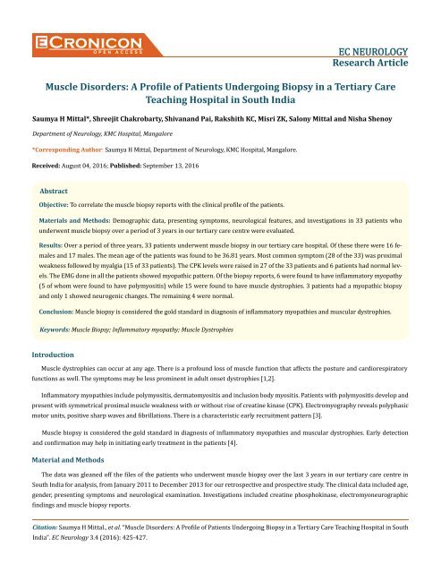
Muscle Disorders- A Profile of Patients Undergoing Biopsy in a Tertiary Care Teaching Hospital in South India
Create successful ePaper yourself
Turn your PDF publications into a flip-book with our unique Google optimized e-Paper software.
Cronicon<br />
OPEN ACCESS<br />
EC NEUROLOGY<br />
Research Article<br />
<strong>Muscle</strong> <strong>Disorders</strong>: A <strong>Pr<strong>of</strong>ile</strong> <strong>of</strong> <strong>Patients</strong> <strong>Undergo<strong>in</strong>g</strong> <strong>Biopsy</strong> <strong>in</strong> a <strong>Tertiary</strong> <strong>Care</strong><br />
Teach<strong>in</strong>g <strong>Hospital</strong> <strong>in</strong> <strong>South</strong> <strong>India</strong><br />
Saumya H Mittal*, Shreejit Chakrobarty, Shivanand Pai, Rakshith KC, Misri ZK, Salony Mittal and Nisha Shenoy<br />
Department <strong>of</strong> Neurology, KMC <strong>Hospital</strong>, Mangalore<br />
*Correspond<strong>in</strong>g Author: Saumya H Mittal, Department <strong>of</strong> Neurology, KMC <strong>Hospital</strong>, Mangalore.<br />
Received: August 04, 2016; Published: September 13, 2016<br />
Abstract<br />
Objective: To correlate the muscle biopsy reports with the cl<strong>in</strong>ical pr<strong>of</strong>ile <strong>of</strong> the patients.<br />
Materials and Methods: Demographic data, present<strong>in</strong>g symptoms, neurological features, and <strong>in</strong>vestigations <strong>in</strong> 33 patients who<br />
underwent muscle biopsy over a period <strong>of</strong> 3 years <strong>in</strong> our tertiary care centre were evaluated.<br />
Results: Over a period <strong>of</strong> three years, 33 patients underwent muscle biopsy <strong>in</strong> our tertiary care hospital. Of these there were 16 females<br />
and 17 males. The mean age <strong>of</strong> the patients was found to be 36.81 years. Most common symptom (28 <strong>of</strong> the 33) was proximal<br />
weakness followed by myalgia (15 <strong>of</strong> 33 patients). The CPK levels were raised <strong>in</strong> 27 <strong>of</strong> the 33 patients and 6 patients had normal levels.<br />
The EMG done <strong>in</strong> all the patients showed myopathic pattern. Of the biopsy reports, 6 were found to have <strong>in</strong>flammatory myopathy<br />
(5 <strong>of</strong> whom were found to have polymyositis) while 15 were found to have muscle dystrophies. 3 patients had a myopathic biopsy<br />
and only 1 showed neurogenic changes. The rema<strong>in</strong><strong>in</strong>g 4 were normal.<br />
Conclusion: <strong>Muscle</strong> biopsy is considered the gold standard <strong>in</strong> diagnosis <strong>of</strong> <strong>in</strong>flammatory myopathies and muscular dystrophies.<br />
Keywords: <strong>Muscle</strong> <strong>Biopsy</strong>; Inflammatory myopathy; <strong>Muscle</strong> Dystrophies<br />
Introduction<br />
<strong>Muscle</strong> dystrophies can occur at any age. There is a pr<strong>of</strong>ound loss <strong>of</strong> muscle function that affects the posture and cardiorespiratory<br />
functions as well. The symptoms may be less prom<strong>in</strong>ent <strong>in</strong> adult onset dystrophies [1,2].<br />
Inflammatory myopathies <strong>in</strong>clude polymyositis, dermatomyositis and <strong>in</strong>clusion body myositis. <strong>Patients</strong> with polymyositis develop and<br />
present with symmetrical proximal muscle weakness with or without rise <strong>of</strong> creat<strong>in</strong>e k<strong>in</strong>ase (CPK). Electromyography reveals polyphasic<br />
motor units, positive sharp waves and fibrillations. There is a characteristic early recruitment pattern [3].<br />
<strong>Muscle</strong> biopsy is considered the gold standard <strong>in</strong> diagnosis <strong>of</strong> <strong>in</strong>flammatory myopathies and muscular dystrophies. Early detection<br />
and confirmation may help <strong>in</strong> <strong>in</strong>itiat<strong>in</strong>g early treatment <strong>in</strong> the patients [4].<br />
Material and Methods<br />
The data was gleaned <strong>of</strong>f the files <strong>of</strong> the patients who underwent muscle biopsy over the last 3 years <strong>in</strong> our tertiary care centre <strong>in</strong><br />
<strong>South</strong> <strong>India</strong> for analysis, from January 2011 to December 2013 for our retrospective and prospective study. The cl<strong>in</strong>ical data <strong>in</strong>cluded age,<br />
gender, present<strong>in</strong>g symptoms and neurological exam<strong>in</strong>ation. Investigations <strong>in</strong>cluded creat<strong>in</strong>e phosphok<strong>in</strong>ase, electromyoneurographic<br />
f<strong>in</strong>d<strong>in</strong>gs and muscle biopsy reports.<br />
Citation: Saumya H Mittal., et al. “<strong>Muscle</strong> <strong>Disorders</strong>: A <strong>Pr<strong>of</strong>ile</strong> <strong>of</strong> <strong>Patients</strong> <strong>Undergo<strong>in</strong>g</strong> <strong>Biopsy</strong> <strong>in</strong> a <strong>Tertiary</strong> <strong>Care</strong> Teach<strong>in</strong>g <strong>Hospital</strong> <strong>in</strong> <strong>South</strong><br />
<strong>India</strong>”. EC Neurology 3.4 (2016): 425-427.
<strong>Muscle</strong> <strong>Disorders</strong>: A <strong>Pr<strong>of</strong>ile</strong> <strong>of</strong> <strong>Patients</strong> <strong>Undergo<strong>in</strong>g</strong> <strong>Biopsy</strong> <strong>in</strong> a <strong>Tertiary</strong> <strong>Care</strong> Teach<strong>in</strong>g <strong>Hospital</strong> <strong>in</strong> <strong>South</strong> <strong>India</strong><br />
426<br />
The muscle biopsy was done from the moderately affected muscles after a written consent and fixed <strong>in</strong> formal<strong>in</strong>. Masson’s trichrome<br />
sta<strong>in</strong><strong>in</strong>g was done for all cases.<br />
None <strong>of</strong> the patients had molecular genetic studies. Electron microscopy was not available at out centre and therefore not performed.<br />
The retrospective study was performed to correlate between the cl<strong>in</strong>ical features, CPK levels, electrophysiological details and the biopsy<br />
data. The data was analyzed us<strong>in</strong>g the standard means <strong>of</strong> statistics.<br />
Results<br />
Over a period <strong>of</strong> three years, 33 patients underwent muscle biopsy <strong>in</strong> our tertiary care hospital. 7 biopsies were performed <strong>in</strong> the 1st<br />
year <strong>of</strong> the study. 13 studies were performed <strong>in</strong> the subsequent 2 years each. Of these there were 16 females and 17 males. The mean age<br />
<strong>of</strong> the patients was found to be 36.81 years (range from 7 years to 66 years <strong>of</strong> age). The disease had a progressive course <strong>in</strong> all the patients.<br />
The mean delay between the onset <strong>of</strong> symptoms and presentation to the hospital was 4.9 years.<br />
Almost all patients (28 <strong>of</strong> the 33) presented with compla<strong>in</strong>ts <strong>of</strong> proximal weakness. The other 5 presented with compla<strong>in</strong>ts <strong>of</strong> myalgia.<br />
A total <strong>of</strong> 15 patients had myalgia. Arthralgia was a compla<strong>in</strong>t <strong>in</strong> 4 <strong>of</strong> the patients while weight loss was a compla<strong>in</strong>t <strong>in</strong> 1 patient. None <strong>of</strong><br />
them had fever, respiratory <strong>in</strong>volvement.<br />
The patients underwent biopsy at different sites. In our study, the most common site <strong>of</strong> biopsy was the vastus lateralis. 18 <strong>of</strong> the 33<br />
patients underwent biopsy from the vastus lateralis, only 1 <strong>of</strong> them be<strong>in</strong>g from the right sided and the rema<strong>in</strong><strong>in</strong>g from the left side. 8<br />
patients underwent biopsy from the left biceps brachii, 5 from the deltoid (4 from the left side and 1 from the right side). The rema<strong>in</strong><strong>in</strong>g<br />
2 were performed from the right gastrocnemius.<br />
The CPK levels were done <strong>in</strong> all the patients. 6 <strong>of</strong> them had normal levels <strong>of</strong> CPK. The rema<strong>in</strong><strong>in</strong>g 27 were high. Of these 27, 16 patients<br />
had CPK levels <strong>of</strong> more than 1000 U/L, and the rema<strong>in</strong><strong>in</strong>g 11 had CPK levels less than 1000 U/L.<br />
The EMG done <strong>in</strong> all the patients showed myopathic pattern.<br />
Of the biopsy reports, 6 were found to have <strong>in</strong>flammatory myopathy (5 <strong>of</strong> whom were found to have polymyositis) while 15 were found<br />
to have muscle dystrophies. 3 patients had a myopathic biopsy and only 1 showed neurogenic changes. The rema<strong>in</strong><strong>in</strong>g 4 were normal<br />
(these patients were found to have hypothyroidism <strong>in</strong> 3 cases and hyperparathyroidism <strong>in</strong> 1 case).<br />
Based on the cl<strong>in</strong>ical features and exam<strong>in</strong>ation f<strong>in</strong>d<strong>in</strong>gs, the patients <strong>of</strong> muscular dystrophies could be identified as 5 patients with<br />
Becker muscular dystrophy. Another 5 were found to have limb girdle dystrophy. Amongst the rema<strong>in</strong><strong>in</strong>g 5, 2 patients had facioscapulohumeral<br />
dystrophy and 3 patients had myotonic dystrophy.<br />
22 patients who underwent biopsy showed a preserved fascicular structure. Other 11 showed altered fascicular structure. Masson’s<br />
trichrome sta<strong>in</strong> showed normal endomysial collagen pattern <strong>in</strong> 15 patients. It was found to be mildly <strong>in</strong>crease <strong>in</strong> 9 patients, moderately<br />
<strong>in</strong>creased <strong>in</strong> 4 and markedly <strong>in</strong>creased <strong>in</strong> 5 patients. My<strong>of</strong>iber size was preserved <strong>in</strong> 7 patients. It was found to have mild variation <strong>in</strong> 12,<br />
moderate variation <strong>in</strong> 3 and marked variation <strong>in</strong> 11 patients. 12 patients showed lymphocytic <strong>in</strong>filtration and a similar number <strong>of</strong> patients<br />
were found to have myophagocytosis.<br />
Discussion<br />
Over time, the diagnosis and diagnostic tests available have seen changes. Start<strong>in</strong>g from rout<strong>in</strong>e histology, 1960s came up with enzyme<br />
histochemistry and 1990s brought up immunohistochemistry. Use <strong>of</strong> MHCs antigen immunosta<strong>in</strong><strong>in</strong>g has been used now recently [4].<br />
Citation: Saumya H Mittal., et al. “<strong>Muscle</strong> <strong>Disorders</strong>: A <strong>Pr<strong>of</strong>ile</strong> <strong>of</strong> <strong>Patients</strong> <strong>Undergo<strong>in</strong>g</strong> <strong>Biopsy</strong> <strong>in</strong> a <strong>Tertiary</strong> <strong>Care</strong> Teach<strong>in</strong>g <strong>Hospital</strong> <strong>in</strong> <strong>South</strong><br />
<strong>India</strong>”. EC Neurology 3.4 (2016): 425-427.
<strong>Muscle</strong> <strong>Disorders</strong>: A <strong>Pr<strong>of</strong>ile</strong> <strong>of</strong> <strong>Patients</strong> <strong>Undergo<strong>in</strong>g</strong> <strong>Biopsy</strong> <strong>in</strong> a <strong>Tertiary</strong> <strong>Care</strong> Teach<strong>in</strong>g <strong>Hospital</strong> <strong>in</strong> <strong>South</strong> <strong>India</strong><br />
Inflammatory myopathies were considered common on <strong>India</strong>. They are potentially reversible and therefore need to be diagnosed.<br />
However, they may not be that frequent <strong>in</strong> <strong>India</strong>. Muscular dystrophies are more common. Duchenne muscular dystrophy <strong>in</strong> childhood<br />
and limb girdle muscle dystrophy <strong>in</strong> adults are known to be the commonest muscular dystrophies [5]. In our study too, most <strong>of</strong> the patients<br />
(15 <strong>of</strong> 33) were found to have muscular dystrophy. This was higher than that found <strong>in</strong> a larger study by Das where 27.4% <strong>of</strong> all<br />
myopathies were muscular dystrophies. Whereas <strong>in</strong> comparison only 6 <strong>of</strong> our 33 patients had <strong>in</strong>flammatory myopathies [6].<br />
427<br />
Polymyositis biopsy shows <strong>in</strong>flammatory cells (that <strong>in</strong>clude CD8+ T cells and macrophages) which can destroy the normal appear<strong>in</strong>g<br />
my<strong>of</strong>ibers. In contrast, dermatomyositis shows atrophic, degenerat<strong>in</strong>g and regenerat<strong>in</strong>g my<strong>of</strong>ibers <strong>in</strong> a perifascicular distribution. This<br />
results due to the destruction <strong>of</strong> the capillaries <strong>of</strong> the area caus<strong>in</strong>g hypoxia and subsequent my<strong>of</strong>ibril damage. Neovascularisation may be<br />
seen [3]. In our study 5 patients showed the changes suggestive <strong>of</strong> polymyositis and 1 patient had dermatomyositis.<br />
Muscular dystrophies are characterized by ‘dystrophic’ changes that specifically <strong>in</strong>clude variation <strong>in</strong> size <strong>of</strong> the fibre, <strong>in</strong>creased <strong>in</strong>ternal<br />
nuclei and presence <strong>of</strong> regeneration and degeneration <strong>of</strong> fibres. Inflammatory cellular <strong>in</strong>filtrate may be seen along with fibre splitt<strong>in</strong>g.<br />
These features occur due to recurrent muscle fibre necrosis and regeneration [1,2]. 15 <strong>of</strong> our patients showed such changes.<br />
Conclusion<br />
It is clear that wide varieties <strong>of</strong> myopathies exist. And few neurology consultants that <strong>India</strong> can boast <strong>of</strong> will usually have their hands<br />
full. Few centres have laboratories equipped to evaluate myopathies [5]. Most <strong>of</strong> the time, the patients <strong>of</strong> <strong>India</strong> are unable to afford extensive<br />
<strong>in</strong>vestigations which can run up the costs. <strong>Muscle</strong> biopsy is considered the gold standard <strong>in</strong> diagnosis <strong>of</strong> <strong>in</strong>flammatory myopathies<br />
and muscular dystrophies [3].<br />
Proximal weakness rema<strong>in</strong>s the commonest symptom with which a patient would present to the hospital. Muscular dystrophy was<br />
found to be more common than <strong>in</strong>flammatory myopathies. Amongst <strong>in</strong>flammatory myopathies, polymyositis was found to be the commonest.<br />
Bibliography<br />
1. Leigh B. Waddell., et al. “Diagnosis <strong>of</strong> the Muscular Dystrophies, Muscular Dystrophy”. Dr. Madhuri Hegde (Ed.), InTech (2012).<br />
2. Elizabeth M McNally and Peter Pytel. “<strong>Muscle</strong> Diseases: The Muscular Dystrophies. Annu. Rev Pathol”. Mechanisms <strong>of</strong> Disease 2<br />
(2007): 87-109.<br />
3. Mammen AL. “Dermatomyositis and polymyositis: Cl<strong>in</strong>ical presentation, autoantibodies, and pathogenesis”. Annals <strong>of</strong> the New York<br />
Academy <strong>of</strong> Sciences 1184 (2010): 134-153.<br />
4. Nagappa M., et al. “Major histocompatibility complex and <strong>in</strong>flammatory cell subtype expression <strong>in</strong> <strong>in</strong>flammatory myopathies and<br />
muscular dystrophies”. Neurology <strong>India</strong> 61.6 (2013): 614-621.<br />
5. Khadilkar S. “Bird’s eye view <strong>of</strong> myopathies <strong>in</strong> <strong>India</strong>”. Neurology <strong>India</strong> 56.3 (2008): 225-228.<br />
6. Das S. “Diagnosis <strong>of</strong> muscular dystrophies: The chang<strong>in</strong>g concepts”. Neurology <strong>India</strong> 46.3 (1998): 165-176.<br />
Volume 3 Issue 4 September 2016<br />
© All rights reserved by Saumya H Mittal., et al.<br />
Citation: Saumya H Mittal., et al. “<strong>Muscle</strong> <strong>Disorders</strong>: A <strong>Pr<strong>of</strong>ile</strong> <strong>of</strong> <strong>Patients</strong> <strong>Undergo<strong>in</strong>g</strong> <strong>Biopsy</strong> <strong>in</strong> a <strong>Tertiary</strong> <strong>Care</strong> Teach<strong>in</strong>g <strong>Hospital</strong> <strong>in</strong> <strong>South</strong><br />
<strong>India</strong>”. EC Neurology 3.4 (2016): 425-427.


