Salony paper- Anemia in SLE based on Iron Studies and Soluble Transferrin Receptor Levels
You also want an ePaper? Increase the reach of your titles
YUMPU automatically turns print PDFs into web optimized ePapers that Google loves.
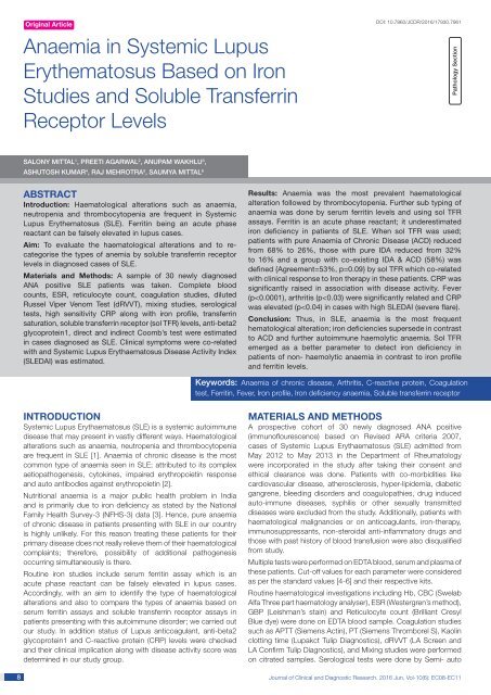
Orig<str<strong>on</strong>g>in</str<strong>on</strong>g>al Article<br />
Anaemia <str<strong>on</strong>g>in</str<strong>on</strong>g> Systemic Lupus<br />
Erythematosus Based <strong>on</strong> Ir<strong>on</strong><br />
<strong>Studies</strong> <strong>and</strong> <strong>Soluble</strong> Transferr<str<strong>on</strong>g>in</str<strong>on</strong>g><br />
<strong>Receptor</strong> <strong>Levels</strong><br />
DOI: 10.7860/JCDR/2016/17930.7961<br />
Pathology Secti<strong>on</strong><br />
<str<strong>on</strong>g>Sal<strong>on</strong>y</str<strong>on</strong>g> Mittal 1 , Preeti Agarwal 2 , Anupam Wakhlu 3 ,<br />
Ashutosh Kumar 4 , Raj Mehrotra 5 , Saumya Mittal 6<br />
ABSTRACT<br />
Introducti<strong>on</strong>: Haematological alterati<strong>on</strong>s such as anaemia,<br />
neutropenia <strong>and</strong> thrombocytopenia are frequent <str<strong>on</strong>g>in</str<strong>on</strong>g> Systemic<br />
Lupus Erythematosus (<str<strong>on</strong>g>SLE</str<strong>on</strong>g>). Ferrit<str<strong>on</strong>g>in</str<strong>on</strong>g> be<str<strong>on</strong>g>in</str<strong>on</strong>g>g an acute phase<br />
reactant can be falsely elevated <str<strong>on</strong>g>in</str<strong>on</strong>g> lupus cases.<br />
Aim: To evaluate the haematological alterati<strong>on</strong>s <strong>and</strong> to recategorise<br />
the types of anemia by soluble transferr<str<strong>on</strong>g>in</str<strong>on</strong>g> receptor<br />
levels <str<strong>on</strong>g>in</str<strong>on</strong>g> diagnosed cases of <str<strong>on</strong>g>SLE</str<strong>on</strong>g>.<br />
Materials <strong>and</strong> Methods: A sample of 30 newly diagnosed<br />
ANA positive <str<strong>on</strong>g>SLE</str<strong>on</strong>g> patients was taken. Complete blood<br />
counts, ESR, reticulocyte count, coagulati<strong>on</strong> studies, diluted<br />
Russel Viper Venom Test (dRVVT), mix<str<strong>on</strong>g>in</str<strong>on</strong>g>g studies, serological<br />
tests, high sensitivity CRP al<strong>on</strong>g with ir<strong>on</strong> profile, transferr<str<strong>on</strong>g>in</str<strong>on</strong>g><br />
saturati<strong>on</strong>, soluble transferr<str<strong>on</strong>g>in</str<strong>on</strong>g> receptor (sol TFR) levels, anti-beta2<br />
glycoprote<str<strong>on</strong>g>in</str<strong>on</strong>g>1, direct <strong>and</strong> <str<strong>on</strong>g>in</str<strong>on</strong>g>direct Coomb’s test were estimated<br />
<str<strong>on</strong>g>in</str<strong>on</strong>g> cases diagnosed as <str<strong>on</strong>g>SLE</str<strong>on</strong>g>. Cl<str<strong>on</strong>g>in</str<strong>on</strong>g>ical symptoms were co-related<br />
with <strong>and</strong> Systemic Lupus Erythaematosus Disease Activity Index<br />
(<str<strong>on</strong>g>SLE</str<strong>on</strong>g>DAI) was estimated.<br />
Results: Anaemia was the most prevalent haematological<br />
alterati<strong>on</strong> followed by thrombocytopenia. Further sub typ<str<strong>on</strong>g>in</str<strong>on</strong>g>g of<br />
anaemia was d<strong>on</strong>e by serum ferrit<str<strong>on</strong>g>in</str<strong>on</strong>g> levels <strong>and</strong> us<str<strong>on</strong>g>in</str<strong>on</strong>g>g sol TFR<br />
assays. Ferrit<str<strong>on</strong>g>in</str<strong>on</strong>g> is an acute phase reactant; it underestimated<br />
ir<strong>on</strong> deficiency <str<strong>on</strong>g>in</str<strong>on</strong>g> patients of <str<strong>on</strong>g>SLE</str<strong>on</strong>g>. When sol TFR was used;<br />
patients with pure Anaemia of Chr<strong>on</strong>ic Disease (ACD) reduced<br />
from 68% to 26%, those with pure IDA reduced from 32%<br />
to 16% <strong>and</strong> a group with co-exist<str<strong>on</strong>g>in</str<strong>on</strong>g>g IDA & ACD (58%) was<br />
def<str<strong>on</strong>g>in</str<strong>on</strong>g>ed {Agreement=53%, p=0.09} by sol TFR which co-related<br />
with cl<str<strong>on</strong>g>in</str<strong>on</strong>g>ical resp<strong>on</strong>se to Ir<strong>on</strong> therapy <str<strong>on</strong>g>in</str<strong>on</strong>g> these patients. CRP was<br />
significantly raised <str<strong>on</strong>g>in</str<strong>on</strong>g> associati<strong>on</strong> with disease activity. Fever<br />
(p
www.jcdr.net<br />
analyser (Hitachi 911/912) which <str<strong>on</strong>g>in</str<strong>on</strong>g>cluded LDH (ELITech) <strong>and</strong><br />
renal functi<strong>on</strong> tests (PZ Cormay S.A.). High sensitivity CRP was<br />
estimated by Calbiotech hsCRP ELISA. Ir<strong>on</strong> studies: Serum ferrit<str<strong>on</strong>g>in</str<strong>on</strong>g><br />
(Accub<str<strong>on</strong>g>in</str<strong>on</strong>g>d ELISA kit), TIBC, Serum ir<strong>on</strong>, % transferr<str<strong>on</strong>g>in</str<strong>on</strong>g> saturati<strong>on</strong>,<br />
soluble transferr<str<strong>on</strong>g>in</str<strong>on</strong>g> receptor levels (TSZ ELISA kit) were also<br />
performed.<br />
Additi<strong>on</strong>al parameters estimated were: anti beta 2 glycoprote<str<strong>on</strong>g>in</str<strong>on</strong>g>-1<br />
levels (Dr Fenn<str<strong>on</strong>g>in</str<strong>on</strong>g>g ELISA kit), direct & Indirect Coomb’s (Erycl<strong>on</strong>e<br />
Anti Human globul<str<strong>on</strong>g>in</str<strong>on</strong>g> Tulip Diagnostics). All the test results were<br />
<str<strong>on</strong>g>in</str<strong>on</strong>g>terpreted <str<strong>on</strong>g>based</str<strong>on</strong>g> <strong>on</strong> the respective kit st<strong>and</strong>ards.<br />
Cl<str<strong>on</strong>g>in</str<strong>on</strong>g>ical symptoms were noted, disease activity score (<str<strong>on</strong>g>SLE</str<strong>on</strong>g>DAI)<br />
was estimated. The results were presented <str<strong>on</strong>g>in</str<strong>on</strong>g> mean (±SD) <strong>and</strong><br />
percentages. The Chi-square test was used to compare the<br />
categorical variables. The Kruskal-Wallis test was used to compare<br />
more than two groups <strong>and</strong> Tukey’s multiple comparis<strong>on</strong> tests was<br />
for pair wise comparis<strong>on</strong>s. The Spearman correlati<strong>on</strong> coefficient<br />
was used to f<str<strong>on</strong>g>in</str<strong>on</strong>g>d the correlati<strong>on</strong> between two c<strong>on</strong>t<str<strong>on</strong>g>in</str<strong>on</strong>g>uous variables.<br />
The p-value less than 0.05 were c<strong>on</strong>sidered as significant. All the<br />
analyses were carried out by us<str<strong>on</strong>g>in</str<strong>on</strong>g>g SPSS 16.0 versi<strong>on</strong>.<br />
RESULTS<br />
Prevalence of <str<strong>on</strong>g>SLE</str<strong>on</strong>g> was predom<str<strong>on</strong>g>in</str<strong>on</strong>g>ant <str<strong>on</strong>g>in</str<strong>on</strong>g> females (F:M-29:1). The<br />
mean age of disease <strong>on</strong>set was 29 years. A total of 21 out of<br />
30 (70%) patients were found to have anaemia, 7 out of 30<br />
(23%) patients had thrombocytopenia, <strong>and</strong> 5 out of 30 (16.7%)<br />
patients had both anaemia <strong>and</strong> thrombocytopenia. No patient had<br />
leucopoenia. However, 30% (9 out 30) patients were found to have<br />
leucocytosis. All anaemic patients were further assessed as per flow<br />
diagram <str<strong>on</strong>g>in</str<strong>on</strong>g> [Table/Fig-1]. [Table/Fig-1] also summarises our results<br />
of Ir<strong>on</strong> Deficiency Anaemia (IDA), Anaemia of Chr<strong>on</strong>ic Disease<br />
(ACD) <strong>on</strong> rout<str<strong>on</strong>g>in</str<strong>on</strong>g>e ir<strong>on</strong> studies <strong>and</strong> special tests which <str<strong>on</strong>g>in</str<strong>on</strong>g>cluded<br />
soluble transferr<str<strong>on</strong>g>in</str<strong>on</strong>g> receptor assays (Sol tFR) <strong>and</strong> <strong>Soluble</strong> transferr<str<strong>on</strong>g>in</str<strong>on</strong>g><br />
receptor <str<strong>on</strong>g>in</str<strong>on</strong>g>dex (Sol tFR <str<strong>on</strong>g>in</str<strong>on</strong>g>dex= Sol tFR/ log ferrit<str<strong>on</strong>g>in</str<strong>on</strong>g>) which were d<strong>on</strong>e<br />
<strong>on</strong> n<strong>on</strong>-haemolytic cases. Features of haemolysis (LDH>480IU/l,<br />
Indirect bilirub<str<strong>on</strong>g>in</str<strong>on</strong>g>>2mg/dl, corrected reticulocyte count>2.5%<br />
<strong>and</strong> presence of spherocytes <strong>on</strong> peripheral smear study) were<br />
seen <str<strong>on</strong>g>in</str<strong>on</strong>g> 2 out of 21 anaemia cases; out of which 1 case showed<br />
positive Coomb’s test. As per the diagnostic criteria for anaemia<br />
(as calculated from soluble transferr<str<strong>on</strong>g>in</str<strong>on</strong>g> receptor >5mcg/ml) 3 out<br />
of 19 (16%)patients were found to have IDA (soluble transferr<str<strong>on</strong>g>in</str<strong>on</strong>g><br />
receptor=3<br />
but 1.5mcg/ml) 15(79%) out of 19 patients were found to have<br />
co-exist<str<strong>on</strong>g>in</str<strong>on</strong>g>g IDA <strong>and</strong> ACD or <strong>on</strong>ly IDA, while 4 (21%) out of 19<br />
patients were found to have pure anaemia of chr<strong>on</strong>ic disease<br />
(soluble transferr<str<strong>on</strong>g>in</str<strong>on</strong>g> receptor <str<strong>on</strong>g>in</str<strong>on</strong>g>dex1.4mg/dl) while <str<strong>on</strong>g>in</str<strong>on</strong>g> seven<br />
cases <strong>on</strong>ly beta2 glycoprote<str<strong>on</strong>g>in</str<strong>on</strong>g>-1 levels was positive. Out of the<br />
3 patients who showed positivity for both LA <strong>and</strong> anti-beta2<br />
glycoprote<str<strong>on</strong>g>in</str<strong>on</strong>g>-1, two patients had history of thrombosis. The patients<br />
with high beta2 glycoprote<str<strong>on</strong>g>in</str<strong>on</strong>g>-1 levels had low haemoglob<str<strong>on</strong>g>in</str<strong>on</strong>g> as well<br />
[Table/Fig-2]. The cl<str<strong>on</strong>g>in</str<strong>on</strong>g>ical features of patients were studied <strong>and</strong><br />
disease activity was determ<str<strong>on</strong>g>in</str<strong>on</strong>g>ed <str<strong>on</strong>g>based</str<strong>on</strong>g> <strong>on</strong> <str<strong>on</strong>g>SLE</str<strong>on</strong>g>DAI score. Out of<br />
all cl<str<strong>on</strong>g>in</str<strong>on</strong>g>ical signs <strong>and</strong> symptoms of <str<strong>on</strong>g>SLE</str<strong>on</strong>g>, we found history of fever<br />
(p
<str<strong>on</strong>g>Sal<strong>on</strong>y</str<strong>on</strong>g> Mittal et al., Haematological Alterati<strong>on</strong>s <str<strong>on</strong>g>in</str<strong>on</strong>g> <str<strong>on</strong>g>SLE</str<strong>on</strong>g><br />
In patients with no flare, fever (91.7%) was the most comm<strong>on</strong><br />
present<str<strong>on</strong>g>in</str<strong>on</strong>g>g symptom followed by history of new rash (41.7%).<br />
Patients with mild to moderate flare had polyarthritis, alopecia<br />
<strong>and</strong> mucosal ulcers which were distributed equally (50% each).<br />
In patients with severe flare, serositis (21.4%), nephritis (35.7%),<br />
neurological manifestati<strong>on</strong>s (35.7%) were the most comm<strong>on</strong><br />
present<str<strong>on</strong>g>in</str<strong>on</strong>g>g features [Table/Fig-3].<br />
DISCUSSION<br />
As far as demographic profile of the study group was c<strong>on</strong>cerned,<br />
female predom<str<strong>on</strong>g>in</str<strong>on</strong>g>ance was observed. The <str<strong>on</strong>g>in</str<strong>on</strong>g>creased frequency of<br />
<str<strong>on</strong>g>SLE</str<strong>on</strong>g> am<strong>on</strong>g women has been attributed <str<strong>on</strong>g>in</str<strong>on</strong>g> part to estrogens [7,8].<br />
The mean age of <strong>on</strong>set of disease of patients <str<strong>on</strong>g>in</str<strong>on</strong>g> our study was 29<br />
years.<br />
On thorough review of literature; different authors have studied<br />
haematological alterati<strong>on</strong>s <str<strong>on</strong>g>in</str<strong>on</strong>g> <str<strong>on</strong>g>SLE</str<strong>on</strong>g> patients <strong>and</strong> we found our<br />
f<str<strong>on</strong>g>in</str<strong>on</strong>g>d<str<strong>on</strong>g>in</str<strong>on</strong>g>gs comparable to their results to a great extent [Table/Fig-4]<br />
[2,9-14]. The major divergence we did was that we analysed our<br />
results for sol TFR as a tool to categorize anaemia <str<strong>on</strong>g>in</str<strong>on</strong>g> <str<strong>on</strong>g>SLE</str<strong>on</strong>g> patients<br />
that had not been <str<strong>on</strong>g>in</str<strong>on</strong>g>vestigated by any of these authors. To the<br />
best of our knowledge/<str<strong>on</strong>g>in</str<strong>on</strong>g>formati<strong>on</strong> no Indian <strong>and</strong> western studies<br />
<strong>on</strong> sol TFR levels has been d<strong>on</strong>e <str<strong>on</strong>g>in</str<strong>on</strong>g> <str<strong>on</strong>g>SLE</str<strong>on</strong>g> patients. However, various<br />
<str<strong>on</strong>g>in</str<strong>on</strong>g>vestigators like Ja<str<strong>on</strong>g>in</str<strong>on</strong>g> et al., have studied <strong>on</strong> sol TFR assays <strong>and</strong><br />
sol TFR <str<strong>on</strong>g>in</str<strong>on</strong>g>dex <str<strong>on</strong>g>in</str<strong>on</strong>g> anaemic patients <strong>and</strong> compared the results of<br />
ferrit<str<strong>on</strong>g>in</str<strong>on</strong>g> from sol TFR assays <strong>and</strong> sol TFR <str<strong>on</strong>g>in</str<strong>on</strong>g>dex [15]. When sol TFR<br />
was used to determ<str<strong>on</strong>g>in</str<strong>on</strong>g>e the type of anaemia results were more<br />
<str<strong>on</strong>g>in</str<strong>on</strong>g>formative when compared to c<strong>on</strong>venti<strong>on</strong>al tests used (Serum<br />
ferrit<str<strong>on</strong>g>in</str<strong>on</strong>g> levels, total ir<strong>on</strong> b<str<strong>on</strong>g>in</str<strong>on</strong>g>d<str<strong>on</strong>g>in</str<strong>on</strong>g>g capacity, % transferr<str<strong>on</strong>g>in</str<strong>on</strong>g> saturati<strong>on</strong>)<br />
<strong>and</strong> c<strong>on</strong>secutively the newly identified subgroup of patients with IDA<br />
<strong>and</strong> ACD overlap benefited with additi<strong>on</strong> of Ir<strong>on</strong> supplements.<br />
Ferrit<str<strong>on</strong>g>in</str<strong>on</strong>g> be<str<strong>on</strong>g>in</str<strong>on</strong>g>g an acute phase reactant, diagnosis of ir<strong>on</strong> deficiency<br />
<str<strong>on</strong>g>in</str<strong>on</strong>g> hospitalized or ill patients can be missed as; such patients have<br />
normal or <str<strong>on</strong>g>in</str<strong>on</strong>g>creased ferrit<str<strong>on</strong>g>in</str<strong>on</strong>g> values even when ir<strong>on</strong> deficient. The<br />
low sensitivity of ferrit<str<strong>on</strong>g>in</str<strong>on</strong>g> for ir<strong>on</strong> deficiency <str<strong>on</strong>g>in</str<strong>on</strong>g> these patients may<br />
require an <str<strong>on</strong>g>in</str<strong>on</strong>g>vasive procedure such as b<strong>on</strong>e marrow biopsy <strong>and</strong>/<br />
or a trial of ir<strong>on</strong> therapy to differentiate ir<strong>on</strong> deficiency from other<br />
causes of anaemia [16]. To overcome this drawback of ferrit<str<strong>on</strong>g>in</str<strong>on</strong>g> as<br />
a marker of decreased marrow Ir<strong>on</strong> stores, many serum markers<br />
have been <str<strong>on</strong>g>in</str<strong>on</strong>g>vestigated till date. Kohgo et al., proposed that the<br />
soluble transferr<str<strong>on</strong>g>in</str<strong>on</strong>g> receptor, a truncated form of the membraneassociated<br />
transferr<str<strong>on</strong>g>in</str<strong>on</strong>g> receptor as a sensitive <str<strong>on</strong>g>in</str<strong>on</strong>g>dicator of ir<strong>on</strong><br />
deficiency <strong>and</strong> is not an acute-phase reactant [17]. S<str<strong>on</strong>g>in</str<strong>on</strong>g>ce, our<br />
study group c<strong>on</strong>sisted of <str<strong>on</strong>g>SLE</str<strong>on</strong>g> patients, where ferrit<str<strong>on</strong>g>in</str<strong>on</strong>g> could be<br />
raised as an acute phase reactant, we additi<strong>on</strong>ally performed sol<br />
TFR assays <strong>on</strong> their samples to reduce the false positive cases of<br />
ACD.<br />
www.jcdr.net<br />
As summarized <str<strong>on</strong>g>in</str<strong>on</strong>g> [Table/Fig-1] by sol TFR assays the number of<br />
patients with pure ACD reduced from 68% to 26% <strong>and</strong> the number<br />
of patients with pure IDA reduced from 32% to 16%; 58% patients<br />
with co-exist<str<strong>on</strong>g>in</str<strong>on</strong>g>g IDA & ACD were identified {Agreement=53%,<br />
p=0.09}. By sol TFR <str<strong>on</strong>g>in</str<strong>on</strong>g>dex the number of patients with pure ACD<br />
reduced from 68% to 21% <strong>and</strong> the rest of the patients were<br />
categorised as pure IDA or with co-existent IDA & ACD which<br />
could not be determ<str<strong>on</strong>g>in</str<strong>on</strong>g>ed separately {Agreement=24%, p=0.11}.<br />
Based <strong>on</strong> our data we c<strong>on</strong>clude that the sol tFR levels al<strong>on</strong>g with<br />
the sol tFR <str<strong>on</strong>g>in</str<strong>on</strong>g>dex can be very useful <str<strong>on</strong>g>in</str<strong>on</strong>g> differentiat<str<strong>on</strong>g>in</str<strong>on</strong>g>g pure IDA, ACD<br />
<strong>and</strong> identify<str<strong>on</strong>g>in</str<strong>on</strong>g>g those patients who had co-exist<str<strong>on</strong>g>in</str<strong>on</strong>g>g ACD with IDA,<br />
thus provid<str<strong>on</strong>g>in</str<strong>on</strong>g>g a n<strong>on</strong>-<str<strong>on</strong>g>in</str<strong>on</strong>g>vasive alternative to b<strong>on</strong>e marrow ir<strong>on</strong>.<br />
Usual f<str<strong>on</strong>g>in</str<strong>on</strong>g>d<str<strong>on</strong>g>in</str<strong>on</strong>g>gs like thrombosis <str<strong>on</strong>g>in</str<strong>on</strong>g> cases with positive LA (2/3) <strong>and</strong><br />
significant correlati<strong>on</strong> between fever <strong>and</strong> polyarthritis with disease<br />
severity <strong>and</strong> flare was noted <str<strong>on</strong>g>in</str<strong>on</strong>g> our study group. A significant<br />
negative correlati<strong>on</strong> between Beta2glycoprote<str<strong>on</strong>g>in</str<strong>on</strong>g>1 <strong>and</strong> haemoglob<str<strong>on</strong>g>in</str<strong>on</strong>g><br />
levels has been reported by Suszek et al., (p = 0.0003; r = -0.67)<br />
[18]. We also found similar correlati<strong>on</strong> though <str<strong>on</strong>g>in</str<strong>on</strong>g>significant (p=0.12),<br />
which can be due to small sample size (n=30).<br />
C reactive prote<str<strong>on</strong>g>in</str<strong>on</strong>g> was found to be raised <str<strong>on</strong>g>in</str<strong>on</strong>g> 18 out of 30 (60%)<br />
patients while 12 out of 30 (40%) patients had normal levels. CRP,<br />
Ferrit<str<strong>on</strong>g>in</str<strong>on</strong>g> <strong>and</strong> ESR levels were compared with the disease activity<br />
<str<strong>on</strong>g>based</str<strong>on</strong>g> <strong>on</strong> <str<strong>on</strong>g>SLE</str<strong>on</strong>g>DAI score am<strong>on</strong>g which; <strong>on</strong>ly CRP levels were found<br />
to be <str<strong>on</strong>g>in</str<strong>on</strong>g> statistically significant associati<strong>on</strong> with the disease activity<br />
score <str<strong>on</strong>g>SLE</str<strong>on</strong>g>DAI (p
www.jcdr.net<br />
References<br />
[1] Mans<strong>on</strong> JJ, Rahman A. Systemic lupus erythaematosus. Orphanet J Rare Dis.<br />
2006;1:6.<br />
[2] Giannouli S, Voulgarelis M, Ziakas PD, et al. Anaemia <str<strong>on</strong>g>in</str<strong>on</strong>g> systemic lupus<br />
erythaematosus: from pathophysiology to cl<str<strong>on</strong>g>in</str<strong>on</strong>g>ical assessment. Ann Rheum Dis.<br />
2006; 65:144–48.<br />
[3] Nati<strong>on</strong>al Family Health Survey-3; Guidel<str<strong>on</strong>g>in</str<strong>on</strong>g>es for c<strong>on</strong>trol of Ir<strong>on</strong> Deficiency Anaemia<br />
<str<strong>on</strong>g>in</str<strong>on</strong>g> India; Nati<strong>on</strong>al Nutriti<strong>on</strong> M<strong>on</strong>itor<str<strong>on</strong>g>in</str<strong>on</strong>g>g Bureau Survey (NNMBS) 2006.<br />
[4] L<strong>on</strong>go, Fauci, Kasper, Hauser, James<strong>on</strong>, Loscalzo. Harris<strong>on</strong>’s pr<str<strong>on</strong>g>in</str<strong>on</strong>g>ciples of<br />
Internal Medic<str<strong>on</strong>g>in</str<strong>on</strong>g>e. 18 th editi<strong>on</strong>. United States of America: The McGraw-Hill;<br />
2012.<br />
th<br />
[5] Lewis SM, Ba<str<strong>on</strong>g>in</str<strong>on</strong>g> BJ, Bates I. Dacie <strong>and</strong> Lewis Practical Haematology.10 editi<strong>on</strong>.<br />
Philadelphia: Churchill Liv<str<strong>on</strong>g>in</str<strong>on</strong>g>gst<strong>on</strong>e; 2006.<br />
[6] Richard A. McPhers<strong>on</strong>, Matthew R. P<str<strong>on</strong>g>in</str<strong>on</strong>g>cus. Henry’s Cl<str<strong>on</strong>g>in</str<strong>on</strong>g>ical Diagnosis <strong>and</strong><br />
Management by Laboratory Methods. 21 st editi<strong>on</strong>. Philadelphia: Saunders;<br />
2007.<br />
[7] Lahita RG. The role of sex horm<strong>on</strong>es <str<strong>on</strong>g>in</str<strong>on</strong>g> systemic lupus erythaematosus. Curr<br />
Op<str<strong>on</strong>g>in</str<strong>on</strong>g> Rheumatol. 1999;11:352.<br />
[8] Costenbader KH, Feskanich D, Stampfer MJ, et al. Reproductive <strong>and</strong> menopausal<br />
factors <strong>and</strong> risk of systemic lupus erythaematosus <str<strong>on</strong>g>in</str<strong>on</strong>g> women. Arthritis Rheum.<br />
2007;56:1251.<br />
[9] Malaviya AN, S<str<strong>on</strong>g>in</str<strong>on</strong>g>gh RR, S<str<strong>on</strong>g>in</str<strong>on</strong>g>gh YN, et al. Prevalence of systemic lupus<br />
erythaematosus <str<strong>on</strong>g>in</str<strong>on</strong>g> India. Lupus. 1993;2(2):115-18.<br />
[10] Benseler SM, Silverman ED. Systemic lupus erythaematosus. Rheum Dis Cl<str<strong>on</strong>g>in</str<strong>on</strong>g> N<br />
Am. 2007;33:471–98.<br />
[11] Voulgarelis M, Kokori SI, Ioannidis JP, Tzioufas AG, Kyriaki D, Moutsopoulos HM.<br />
Anaemia <str<strong>on</strong>g>in</str<strong>on</strong>g> systemic lupus erythaematosus: aetiological profile <strong>and</strong> the role of<br />
erythropoiet<str<strong>on</strong>g>in</str<strong>on</strong>g>. Ann Rheum Dis. 2000;59: 217–22.<br />
[12]<br />
[13]<br />
[14]<br />
[15]<br />
[16]<br />
[17]<br />
[18]<br />
[19]<br />
<str<strong>on</strong>g>Sal<strong>on</strong>y</str<strong>on</strong>g> Mittal et al., Haematological Alterati<strong>on</strong>s <str<strong>on</strong>g>in</str<strong>on</strong>g> <str<strong>on</strong>g>SLE</str<strong>on</strong>g><br />
Beyan E, Beyan C, Turan M. Haematological presentati<strong>on</strong> <str<strong>on</strong>g>in</str<strong>on</strong>g> systemic lupus<br />
erythaematosus <strong>and</strong> its relati<strong>on</strong>ship with disease activity. Haematology.<br />
2007;12(3):257-61.<br />
Shaikh MA, Mem<strong>on</strong> I, Ghori RA. Frequency of anaemia <str<strong>on</strong>g>in</str<strong>on</strong>g> patients with<br />
systemic lupus erythaematosus at tertiary care hospitals. J Pak Med Assoc.<br />
2010;60(10):822-25.<br />
Sasidharan PK, B<str<strong>on</strong>g>in</str<strong>on</strong>g>dya M, Sajeeth Kumar KG. Haematological manifestati<strong>on</strong>s<br />
of sle at <str<strong>on</strong>g>in</str<strong>on</strong>g>itial presentati<strong>on</strong>: is it underestimated? ISRN Haematol.<br />
2012;2012:961872.<br />
Ja<str<strong>on</strong>g>in</str<strong>on</strong>g> S, Narayan S, Ch<strong>and</strong>ra J, et al. Evaluati<strong>on</strong> of serum transferr<str<strong>on</strong>g>in</str<strong>on</strong>g> receptor<br />
<strong>and</strong> sTFR ferrit<str<strong>on</strong>g>in</str<strong>on</strong>g> <str<strong>on</strong>g>in</str<strong>on</strong>g>dices <str<strong>on</strong>g>in</str<strong>on</strong>g> diagnos<str<strong>on</strong>g>in</str<strong>on</strong>g>g <strong>and</strong> differentiat<str<strong>on</strong>g>in</str<strong>on</strong>g>g ir<strong>on</strong> deficiency anaemia<br />
from anaemia of chr<strong>on</strong>ic disease. Indian J Pediatr. 2010;77(2):179-83<br />
Mast AE, Bl<str<strong>on</strong>g>in</str<strong>on</strong>g>der MA, Gr<strong>on</strong>owski AM, et al. Scott. Cl<str<strong>on</strong>g>in</str<strong>on</strong>g>ical utility of the soluble<br />
transferr<str<strong>on</strong>g>in</str<strong>on</strong>g> receptor <strong>and</strong> comparis<strong>on</strong> with serum ferrit<str<strong>on</strong>g>in</str<strong>on</strong>g> <str<strong>on</strong>g>in</str<strong>on</strong>g> several populati<strong>on</strong>s.<br />
Cl<str<strong>on</strong>g>in</str<strong>on</strong>g>ical Chemistry. 1998;44:145–51.<br />
Kohgo Y, Niitsu Y, K<strong>on</strong>do H, et al. Serum transferr<str<strong>on</strong>g>in</str<strong>on</strong>g> receptor as a new <str<strong>on</strong>g>in</str<strong>on</strong>g>dex of<br />
erythropoiesis. Blood. 1987;70:1955–58.<br />
Suszek D, Majdan M, Chudzik D, et al. Relati<strong>on</strong>s between severity of anaemia <strong>and</strong><br />
certa<str<strong>on</strong>g>in</str<strong>on</strong>g> antiphospholipid antibodies presence <str<strong>on</strong>g>in</str<strong>on</strong>g> systemic lupus erythaematosus<br />
patients. Pol Arch Med Wewn. 2006;115(5):426-31.<br />
Amezcua-Guerra LM, Spr<str<strong>on</strong>g>in</str<strong>on</strong>g>gall R, Arrieta-Alvarado AA, et al. C-reactive prote<str<strong>on</strong>g>in</str<strong>on</strong>g><br />
<strong>and</strong> complement comp<strong>on</strong>ents but not other acute-phase reactants discrim<str<strong>on</strong>g>in</str<strong>on</strong>g>ate<br />
between cl<str<strong>on</strong>g>in</str<strong>on</strong>g>ical subsets <strong>and</strong> organ damage <str<strong>on</strong>g>in</str<strong>on</strong>g> systemic lupus erythaematosus.<br />
Cl<str<strong>on</strong>g>in</str<strong>on</strong>g> Lab. 2011;57(7-8):607-13.<br />
PARTICULARS OF CONTRIBUTORS:<br />
1. Assistant Professor, Department of Pathology, Kasturba Medical College, Mangalore, Karnataka, India.<br />
2. Assistant Professor, DNB, Department of Pathology, K<str<strong>on</strong>g>in</str<strong>on</strong>g>g George Medical University, Lucknow, UP, India.<br />
3. Assistant Professor, Department of Rheumatology, K<str<strong>on</strong>g>in</str<strong>on</strong>g>g George Medical University, Lucknow, UP, India.<br />
4. Professor, Department of Pathology, K<str<strong>on</strong>g>in</str<strong>on</strong>g>g George Medical University, Lucknow, UP, India.<br />
5. Professor, Department of Pathology, Kasturba Medical College, Mangalore, Karnataka, India.<br />
6. Registrar, Department of Medic<str<strong>on</strong>g>in</str<strong>on</strong>g>e, Indraprastha Apollo Hospital, New Delhi, India.<br />
NAME, ADDRESS, E-MAIL ID OF THE CORRESPONDING AUTHOR:<br />
Dr. <str<strong>on</strong>g>Sal<strong>on</strong>y</str<strong>on</strong>g> Mittal,<br />
Assistant Professor, Department of Pathology, KMC, Light House Hill Road,<br />
Mangalore—575001, Karnataka, India.<br />
E-mail: drsal<strong>on</strong>ymittal23@gmail.com<br />
F<str<strong>on</strong>g>in</str<strong>on</strong>g>ancial OR OTHER COMPETING INTERESTS: N<strong>on</strong>e.<br />
Date of Submissi<strong>on</strong>: Nov 22, 2015<br />
Date of Peer Review: Jan 11, 2016<br />
Date of Acceptance: Jan 27, 2016<br />
Date of Publish<str<strong>on</strong>g>in</str<strong>on</strong>g>g: Jun 01, 2016<br />
Journal of Cl<str<strong>on</strong>g>in</str<strong>on</strong>g>ical <strong>and</strong> Diagnostic Research. 2016 Jun, Vol-10(6): EC08-EC11 11


