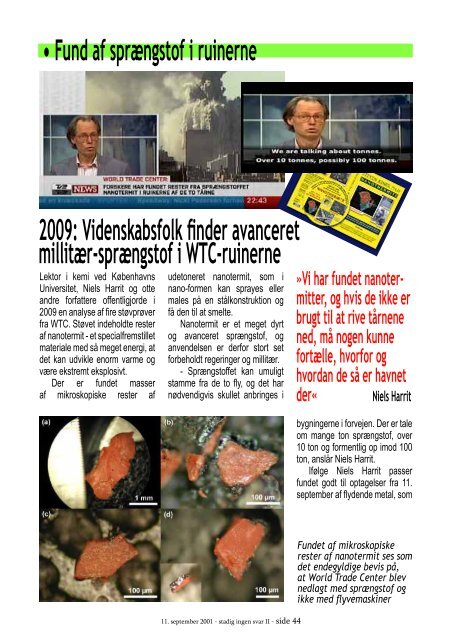
• Fund af sprængstof i ruinerne 2009: Videnskabsfolk finder avanceret millitær-sprængstof i WTC-ruinerne Lektor i kemi ved Københavns Universitet, Niels Harrit og otte andre forfattere offentligjorde i 2009 en analyse af fire støvprøver fra WTC. Støvet indeholdte rester af nanotermit - et specialfremstillet materiale med så meget energi, at det kan udvikle enorm varme og være ekstremt eksplosivt. Der er fundet masser af mikroskopiske rester af udetoneret nanotermit, som i nano-formen kan sprayes eller males på en stålkonstruktion og få den til at smelte. Nanotermit er et meget dyrt og avanceret sprængstof, og anvendelsen er derfor stort set forbeholdt regeringer og millitær. - Sprængstoffet kan umuligt stamme fra de to fly, og det har nødvendigvis skullet anbringes i 11. september 2001 - stadig ingen svar II - side 44 »Vi har fundet nanotermitter, og hvis de ikke er brugt til at rive tårnene ned, må nogen kunne fortælle, hvorfor og hvordan de så er havnet der« Niels Harrit bygningerne i forvejen. Der er tale om mange ton sprængstof, over 10 ton og formentlig op imod 100 ton, anslår Niels Harrit. Ifølge Niels Harrit passer fundet godt til optagelser fra 11. september af flydende metal, som Fundet af mikroskopiske rester af nanotermit ses som det endegyldige bevis på, at World Trade Center blev nedlagt med sprængstof og ikke med flyvemaskiner
Nanotermit er en pyroteknisk s t o f s a m m e n s æ t n i n g af metal-oxid (rust, red) og ultra-fintkornet metalpulver. Når de to indholdsstoffer bringes til kemisk reaktion udledes en meget stor energimasse hvilket fører til ekspansion samt kraftig varmeudvikling. Termit har evnen til at smelte eller skære i beton, stål og andre metaller. Nanotermit har større effekt end traditionel termit og kan bruges som sprængstof. Ordet nano indikerer at stofferne forekommer i meget små og sub-mikro skopiske partikelstørrelser Kilde: Wikipedia løber ud af sydtårnet umiddelbart inden, det styrter sammen. Og det forklarer, hvorfor der mange uger senere stadig var søer af smeltet jern i ruinerne af tårnene. - Resultaterne fra vores analyser er helt i overensstemmelse med, at der var tale om en kontrolleret nedrivning af tårnene, siger Niels Harrit. Støvprøver med de karakteristiske røde elementer af nanotermit er indsamlet fra gader og en lejlighed på Manhattan fra ti minutter efter det andet tårn faldt og frem til en uge senere. The Open Chemical Physics Journal, 2009, 2, 7-31 7 Open Access Active Thermitic Material Discovered in Dust from the 9/11 World Trade Center Catastrophe Niels H. Harrit *,1 , Jeffrey Farrer 2 , Steven E. Jones *,3 , Kevin R. Ryan 4 , Frank M. Legge 5 , Daniel Farnsworth 2 , Gregg Roberts 6 , James R. Gourley 7 and Bradley R. Larsen 3 Active Thermitic Material Found in WTC Dust The Open Chemical Physics Journal, 2009, Volume 2 11 1 Department of Chemistry, University of Copenhagen, Denmark 2 Department of Physics and Astronomy, Brigham Young University, Provo, UT 84602, USA 3 S&J Scientific Co., Provo, UT, 84606, USA 4 9/11 Working Group of Bloomington, Bloomington, IN 47401, USA 5 Logical Systems Consulting, Perth, Western Australia 6 Architects & Engineers for 9/11 Truth, Berkeley, CA 94704, USA 7 International Center for 9/11 Studies, Dallas, TX 75231, USA Abstract: We have discovered distinctive red/gray chips in all the samples we have studied of the dust produced by the destruction of the World Trade Center. Examination of four of these samples, collected from separate sites, is reported in this paper. These red/gray chips show marked similarities in all four samples. One sample was collected by a Manhattan resident about ten minutes after the collapse of the second WTC Tower, two the next day, and a fourth about a week later. The properties of these chips were analyzed using optical microscopy, scanning electron microscopy (SEM), X-ray energy dispersive spectroscopy (XEDS), and differential scanning calorimetry (DSC). The red material contains grains approximately 100 nm across which are largely iron oxide, while aluminum is contained in tiny plate-like structures. Separation of components using methyl ethyl ketone demonstrated that elemental aluminum is present. The iron oxide and aluminum are intimately mixed in the red material. When ignited in a DSC device the chips exhibit large but narrow exotherms occurring at approximately 430 C, far below the normal ignition temperature for conventional thermite. Numerous iron-rich spheres are clearly observed in the residue following the ignition of these peculiar red/gray chips. The red portion of these chips is found to be an unreacted thermitic material and highly energetic. Keywords: Scanning electron microscopy, X-ray energy dispersive spectroscopy, Differential scanning calorimetry, DSC analysis, World Trade Center, WTC dust, 9/11, Iron-rich microspheres, Thermite, Super-thermite, Energetic nanocomposites, Nano-thermite. INTRODUCTION publicized but are no less important to the outstanding obligation that remains to the victims of that tragedy, to determine The destruction of three skyscrapers (WTC 1, 2 and 7) on September 11, 2001 was an immensely tragic catastrophe that not only impacted thousands of people and families directly, due to injury and loss of life, but also provided the motivation for numerous expensive and radical changes in domestic and foreign policy. For these and other reasons, knowing what really happened that fateful day is of grave importance. A great deal of effort has been put forth by various government-sponsored and -funded investigations, which led, in large part, to the reports released by FEMA [1] and NIST [2]. Other studies of the destruction have been less well *Address correspondence to these authors (NH) Department of Chemistry, University of Copenhagen, Copenhagen, DK-2100, Denmark; Tel: (+45)35321846; Fax: (+45)35320460; E-mail: harrit@nano.ku.dk, (SEJ) at S&J Scientific Co., Provo, UT, 84606, USA; Tel: 801-735-5885; E-mail: Hardevidence@gmail.com 10 The Open Chemical Physics Journal, 2009, Volume 2 Harrit et al. Some samples were also tested in a differential scanning crographs of red/gray chips from each of the four WTC dust calorimeter (Netzsch DSC 404C) to measure heat flow into samples. Note the scale marker in each image as they were or out of the red/gray chips. The DSC tests were conducted acquired at different magnifications. At approximately with a linear heating rate of 10 ˚C per minute up to a tem- 2.5 mm in length, the chip in Fig. (2a) was one of the larger perature of 700 ˚C. During heating, the samples were con- chips collected. The mass of this chip was approximately 0.7 tained in alumina pans and air was allowed to flow at 55 mg. All of the chips used in the study had a gray layer and a milliliters per minute during the heating. The plots were gen- red layer and were attracted by a magnet. The inset image in erated by acquiring data points at a rate of 20 points per ˚C Fig. (2d) shows the chip in cross section, which reveals the or 200 points per minute. The equipment was calibrated to gray layer. The gray layer is also partially visible in Fig. display the data in watts per gram. The plots were set to dis- (2b). Similarities between the samples are already evident play positive heat flow out of the sample such that exother- from these photographs. mic behavior of the sample would yield a peak and endo- Fig. (3) shows three images for comparison of views of thermic behavior a trough. the same set of chips using different methods. Fig. (3a) is a The dust samples were also examined by visible-light VLM photomicrograph of a group of particles, which shows microscopy (VLM) through a Nikon Epiphot 200 stereomi the red material and in some cases the adhering gray matecroscope, an Olympus BX60 stereomicroscope and a Nikon rial. Fig. (3b, c) are, respectively, a secondary electron (SE) Labophot microscope and camera. image and a backscattered electron (BSE) image of the same group of particles, using a scanning electron microscope RESULTS (SEM) without a conductive coating over the sample. It can 1. Characterization of the Red/Gray Chips be seen in the SE image that the red layer of the particles has very bright regions caused by a slight accumulation of Red/gray chips were found in all of the dust samples col charge under the electron beam, owing to the relatively poor lected. An analysis of the chips was performed to assess the conductivity of the red layer (see Discussion section). The similarity of the chips and to determine the chemistry and BSE image shows the red layer darker than the gray layer, materials that make up the chips. Fig. (2) displays photomi- Fig. (3). A series of images of the same group of particles extracted by magnet from sample 2. The color photomicrograph in (a), obtained by VLM, locates and identifies the red/gray particles. An SE image (b) acquired by SEM gives a better indication of size and shape of the particles, and a BSE image (c) shows, by grayscale intensity, the difference in average atomic number between the red layer, gray layer and other dust particles. indicating that the red layer is composed of material that has Newly fractured cross sections of red/gray chips from the a relatively lower average atomic number than the gray four different dust samples are shown by BSE imaging in layer. Fig. (5). These four cross sections are representative of all the red/gray chips studied from the dust samples. The BSE A higher-magnification BSE image of the corner of one of the chips, shown in Fig. (4), allows for closer examination images illustrate the finding that all of the red layers studied of the difference in grayscale intensity of the two layers and contained small bright particles or grains characterized by a high average atomic number. The size and presence of the confirms the higher average atomic number of the gray layer. particles was found to be consistent throughout the layers, The red material also shows specks and other heterogene- but the concentration of the particles was found to vary loities, in marked contrast to the smooth gray layer. cally, as can be seen from the images. the whole truth of the events of that day [3-10]. A number of these studies have appropriately focused attention on the remaining physical material, and on available photographs and video footage, as sources of evidence still in public hands, relating to the method of destruction of the three skyscrapers. The collapses of the three tallest WTC buildings were remarkable for their completeness, their near free-fall speed [11] their striking radial symmetry [1, 12] and the surprisingly large volume of fine toxic dust [13] that was generated. In order to better understand these features of the destruction, the authors initiated an examination of this dust. In June 2007, Dr. Steven Jones observed distinctive bi-layered chips, with both a red and a gray layer, in a sample of the WTC dust. Initially, it was suspected these might be dried paint chips, but after closer inspection and testing, it was shown that this was not the case. Further testing was then performed on the red/gray chips in an attempt to ascertain their compo- Red Layer Gray Layer Acc.V Spot Magn Det WD Exp 20.0 kV 6.0 2400x BSE 7.5 1 1874-4125/09 2009 Bentham Open Fig. (4). Higher magnification BSE image of one of the chips in previous image. The red layer appears darker and is on top of the gray layer. Artiklen om fundet af nanotermit i ruinerne efter World Trade Center blev første gang offentliggjort i The Open Chemical Physics Journal, Vol 2, 2009, Bentham Open [DOI: 10.2174/1874412500902010007]. Offentliggørelsen fik Bentham’s daværende chefredaktør, Marie-Paule Pileni, til at opsige sin stilling i protest mod, at hun ikke havde læst artiklen først. Men artiklen eller dens konklusioner er aldrig blevet fagligt bestredet. 11. september 2001 - stadig ingen svar II - side 45 20 mm C C O C Fe O Active Thermitic Material Found in WTC Dust The Open Chemical Physics Journal, 2009, Volume 2 13 C Fe O O Fe Fe 0 1 2 3 4 5 6 7 8 9 keV Active Thermitic Material Found in WTC Dust The Open Chemical Physics Journal, 2009, Volume 2 15 Fig. (9). SE image of the cross section shown in Fig. (8a). from a cluster of the smaller bright faceted grains. Again it was observed that the thin sheet-like particles are rich in Al and Si whereas the bright faceted grains are rich in Fe. Both Active Thermitic Material Found in WTC Dust The Open Chemical Physics Journal, 2009, Volume 2 17 determined. The initial objective was to compare the behavtor of roughly 5 times its original thickness. The photomiior of the red layer with paint when soaked in a strong orcrograph shown in Fig. (13) also shows the chip after the ganic solvent known to soften and dissolve paint. Red/gray MEK soak. The red layer can be seen extending out from the chips were soaked in methyl ethyl ketone (MEK) for 55 gray layer. hours with frequent agitation and subsequently dried in air over several days. The chips showed significant swelling of the red layer, but with no apparent dissolution. In marked contrast, paint chips softened and partly dissolved when similarly soaked in MEK. It was discovered in this process that a significant migration and segregation of aluminum had occurred in the red-chip material. This allowed us to assess whether some of the aluminum was in elemental form. The chip that was used for this experiment was extracted from dust sample 2 and is shown in the images below. Fig. (12a) shows an SE image of the chip prior to the MEK treatment. It is positioned with the interface between the red and gray layers nearly parallel to the plane of the image. Fig. (12b) shows a BSE image of the chip after the MEK soak. Note that the chip fractured during the MEK treatment and handling. In this image the red layer and gray layer are side by side so that the interface between the layers is edge-on (perpendicular to the plane of the image) with the gray layer Fig. (13). Photomicrograph of the MEK treated chip. on the right. The red layer of the chip was found, by visual Prior to soaking the chip in MEK an XEDS spectrum was inspection, to have swelled out from the gray layer by a fac- acquired from an area of the red-layer surface. The resulting spectrum, shown in Fig. (14), produced the expected peaks (a) for Fe, Si, Al, O, and C. Other peaks included calcium, sulfur, zinc, chromium and potassium. The occurrence of these elements could be attributed to surface contamination due to the fact that the analysis was performed on the as-collected surface of the red layer. The large Ca and S peaks may be due to contamination with gypsum from the pulverized wallboard material in the buildings. O Fig. (8). BSE images of cross sections of the red layer from each of the dust samples 1-4 shown in (a)-(d) respectively. Fig. (6). XEDS spectra obtained from the gray layers from each of the four WTC dust samples, with (a) corresponding to sample 1, and so on (b-d). Fig. (2). Photomicrographs of red/gray chips from samples 14 of the WTC dust involved in this study, shown in (a)-(d) respectively. The inset in (d) shows the chip edge on, which reveals the gray layer. The red/gray chips are mounted on an aluminum pedestal, using a carbon conductive tab, for viewing in the scanning electron microscope (SEM). Det kan koste dyrt at tale for højt om uønskede emner, hvilket to af rapportens medforfattere har oplevet. Laboratorieleder Kevin Ryan (ansat ved firmaet som havde certificeret stålet til World Trade Center) skrev et brev til National Institute for Standards and Technology (NIST) og påpegede det umulige i den officielle forklaring - og mistede sit job. Dr. Steven E. Jones, fysiker ved Brigham Young University, stillede i en artikel spørgsmål ved, hvordan flybrændstof kunne udløse nok energi til at pulverisere tre højhuse. Han blev afskediget Fe Fe Fe Fe Fe Fe Fe Fe Acc.V Spot Magn Det WD Exp 10.0 kV 3.0 50000x SE 8.0 1 (d) (c) (b) spectra display significant carbon and oxygen, which may be partially due to the beam spreading and receiving an overlapping X-ray signal from the matrix material as well as particles below the surface. The beam energy (20 keV) is such that the volume of material from which the X-ray signal is generated is larger than the particles. Hence, some Al and Si are seen in Fig. (11b) which may not be inherent in the faceted grains, and some Fe is seen in Fig. (11a), which may not be inherent in the plate-like particles. The consistently rhombic-shaped, faceted appearance of C Ca Fe 500 mm Si Fe S Al (b) Zn Ca Cr Fe Zn 1 2 3 4 5 6 7 8 9 keV Fig. (14). XEDS spectrum of red side before soaking in MEK. Notice the presence of Zn and Cr, which are sometimes seen in the red layers. The large Ca and S peaks may be due to surface contamination with wallboard material. XEDS maps were acquired from the swollen red material at a beam energy of 10 kV, in order to determine the locations of various elements following the MEK treatment. The Acc.V Spot Magn Det WD Exp data shown in Fig. (15) illustrate regions where iron, alumi- 20.0 kV 3.0 400x BSE 9.8 1 100 mm num and silicon are concentrated. Furthermore, the data indicate that wherever silicon or iron is concentrated, oxygen Fig. (12). SE images of the red/gray chip that was soaked in methyl is also concentrated. On the other hand, there also exist re- ethyl ketone for 55 hours, (a) prior to and (b) after MEK soaking. gions where the aluminum is concentrated but where the 1 mm (a) the iron-rich grains strongly suggests that they are crystalline. From these data, it is determined that the red/gray chips from different WTC dust samples are extremely similar in their chemical and structural makeup. It is also shown that within the red layer there is an intimate mixing of the Fe-rich grains and Al/Si platelike particles and that these particles are embedded in a carbon-rich matrix. 2. Test Using Methyl Ethyl Ketone Solvent By employing some means to separate the different components of the material, the chemical compositions of the different particles in the red layer were more accurately »Resultaterne fra vores analyser er helt i overensstemmelse med, at der var tale om en kontrolleret nedrivning af tårnene« Niels Harrit Sandheder med konsekvenser og hans artikel er i dag fjernet fra universitets databaser. Danske Niels Harrit er dog fortsat ved Kemisk Institut under Københavns Universitet og holder derudover foredrag om bygning 7.