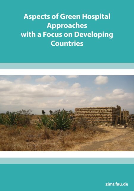
Aspects of Green Hospital Approaches with a Focus on Developing ...
Aspects of Green Hospital Approaches with a Focus on Developing ...
Aspects of Green Hospital Approaches with a Focus on Developing ...

Sie wollen auch ein ePaper? Erhöhen Sie die Reichweite Ihrer Titel.
YUMPU macht aus Druck-PDFs automatisch weboptimierte ePaper, die Google liebt.
<str<strong>on</strong>g>Aspects</str<strong>on</strong>g> <str<strong>on</strong>g>of</str<strong>on</strong>g> <str<strong>on</strong>g>Green</str<strong>on</strong>g> <str<strong>on</strong>g>Hospital</str<strong>on</strong>g><br />
<str<strong>on</strong>g>Approaches</str<strong>on</strong>g><br />
<str<strong>on</strong>g>with</str<strong>on</strong>g> a <str<strong>on</strong>g>Focus</str<strong>on</strong>g> <strong>on</strong> <strong>Developing</strong><br />
Countries<br />
zimt.fau.de
Imprint<br />
Publisher:<br />
Central Institute <str<strong>on</strong>g>of</str<strong>on</strong>g> Healthcare Engineering (ZiMT) at FAU Erlangen-Nuernberg<br />
Managing Director: Dr.-Ing. Kurt Höller, MBA<br />
Henkestraße 91<br />
D-91052 Erlangen<br />
Tel.: +49 9131 85-26868<br />
Fax: +49 9131 85-26862<br />
hoeller@zimt.uni-erlangen.de<br />
www.zimt.fau.de<br />
2
Preface<br />
In 2012, a students’ seminar at the University <str<strong>on</strong>g>of</str<strong>on</strong>g> Erlangen-<br />
Nuernberg started <str<strong>on</strong>g>with</str<strong>on</strong>g> a focus <strong>on</strong> “green hospital” developments.<br />
Main topic was a stable and sustainable energy supply in operating<br />
rooms or hospitals since most diagnostic and anesthetic devices are<br />
useless <str<strong>on</strong>g>with</str<strong>on</strong>g>out power. In the first step, an evaluati<strong>on</strong> <str<strong>on</strong>g>of</str<strong>on</strong>g> the basic needs<br />
<str<strong>on</strong>g>of</str<strong>on</strong>g> a hospital should be d<strong>on</strong>e. This could happen in accordance to existing<br />
Siemens <str<strong>on</strong>g>Green</str<strong>on</strong>g> <str<strong>on</strong>g>Hospital</str<strong>on</strong>g> projects. The knowledge <strong>on</strong> real ec<strong>on</strong>omic<br />
and envir<strong>on</strong>mental efficiency in each process allows the eliminati<strong>on</strong> <str<strong>on</strong>g>of</str<strong>on</strong>g><br />
specific deficits. In a sec<strong>on</strong>d step, the descripti<strong>on</strong> and basic design <str<strong>on</strong>g>of</str<strong>on</strong>g> devices<br />
and methods will be implemented that fulfill the needs <str<strong>on</strong>g>of</str<strong>on</strong>g> a special<br />
envir<strong>on</strong>ment.<br />
In order to provide a real scenario, we cooperated <str<strong>on</strong>g>with</str<strong>on</strong>g> a group <str<strong>on</strong>g>of</str<strong>on</strong>g> physicians who support an<br />
installati<strong>on</strong> and the maintenance <str<strong>on</strong>g>of</str<strong>on</strong>g> a rural hospital in Camero<strong>on</strong>. This made a precise plan <str<strong>on</strong>g>of</str<strong>on</strong>g><br />
a minimally equipped operating room and powering c<strong>on</strong>cept for developing countries possible.<br />
So far, the hospital uses an electricity supply <str<strong>on</strong>g>of</str<strong>on</strong>g> the closest major city Tibati and also an own<br />
emergency generator. However, due to frequent power outages, <str<strong>on</strong>g>with</str<strong>on</strong>g> additi<strong>on</strong>al failure <str<strong>on</strong>g>of</str<strong>on</strong>g> the<br />
emergency generator, and independent power supply would be better and preferable. Another<br />
idea is to divide the building in different secti<strong>on</strong>s <str<strong>on</strong>g>of</str<strong>on</strong>g> energy supply. Operating room, laboratory,<br />
etc. should be powered by an internal power supply, while patient‘s room could be supplied by<br />
external power. The reducti<strong>on</strong> <str<strong>on</strong>g>of</str<strong>on</strong>g> energy c<strong>on</strong>sumpti<strong>on</strong> <str<strong>on</strong>g>of</str<strong>on</strong>g> air-c<strong>on</strong>diti<strong>on</strong>ed operati<strong>on</strong> rooms using<br />
heat and cold insulati<strong>on</strong> is also a challenge since the building materials are rarely available.<br />
Our aim was simple and robust technology that is innovative and clever. Since diesel and electricity<br />
in Camero<strong>on</strong> are very expensive, we focused <strong>on</strong> the greatest possible use <str<strong>on</strong>g>of</str<strong>on</strong>g> sustainable<br />
energy. An obvious c<strong>on</strong>cept is the installati<strong>on</strong> <str<strong>on</strong>g>of</str<strong>on</strong>g> solar panels since Camero<strong>on</strong> lies near the equator.<br />
However, the set up an energy store for the night is still an open questi<strong>on</strong>. For this purpose,<br />
a complete package should be addressed including a diesel generator, solar panels and batteries.<br />
We invited outstanding experts from research institutes, companies and n<strong>on</strong>-governmental organizati<strong>on</strong>s<br />
who talked about their field <str<strong>on</strong>g>of</str<strong>on</strong>g> work which is related to the project. They presented<br />
c<strong>on</strong>cepts that inspired our students in finding soluti<strong>on</strong>s for the implementati<strong>on</strong> in Camero<strong>on</strong>. The<br />
students learned to create cross-references between the various topics in order to develop their<br />
own ideas. Each participant had to give a talk and to submit an article <strong>on</strong> a particular topic.<br />
Due to the lack <str<strong>on</strong>g>of</str<strong>on</strong>g> any financial resources in order to realize those ideas it is important to make the<br />
project „<str<strong>on</strong>g>Green</str<strong>on</strong>g> <str<strong>on</strong>g>Hospital</str<strong>on</strong>g>“ public and to acquire adequate funding. After completi<strong>on</strong> <str<strong>on</strong>g>of</str<strong>on</strong>g> the project<br />
in Camero<strong>on</strong> the results should be extended to similar projects also in other regi<strong>on</strong>s <str<strong>on</strong>g>of</str<strong>on</strong>g> Africa and<br />
Latin America.<br />
Dr.-Ing. Kurt Höller, MBA<br />
Managing Director<br />
Central Institut <str<strong>on</strong>g>of</str<strong>on</strong>g> Healthcare Engineering<br />
3
C<strong>on</strong>tent<br />
Self-sufficient energy c<strong>on</strong>cept using solar heat in<br />
combinati<strong>on</strong> <str<strong>on</strong>g>with</str<strong>on</strong>g> a stirling engine for developing countries...............................................5<br />
Dose Reducti<strong>on</strong> in Radiology as <str<strong>on</strong>g>Green</str<strong>on</strong>g> <str<strong>on</strong>g>Hospital</str<strong>on</strong>g> Requirement<br />
in <strong>Developing</strong> Countries.........................................................................................................13<br />
Air-Management in a surgery.................................................................................................33<br />
Functi<strong>on</strong>al c<strong>on</strong>cepti<strong>on</strong> <str<strong>on</strong>g>of</str<strong>on</strong>g> a minimally equipped operati<strong>on</strong> room.....................................43<br />
Einsatz einer Low-cost Funduskamera in Entwicklungsländern.......................................53<br />
Sterilisati<strong>on</strong> in Entwicklungsländern.....................................................................................61<br />
Energieverbrauch in der Radiologie......................................................................................73<br />
Medizinethische Betrachtung v<strong>on</strong> Entwicklungshilfe.........................................................83<br />
Kamerun – Eine allgemeine Vorstellung eines Landes.......................................................97<br />
4
Students‘ Seminar <str<strong>on</strong>g>Green</str<strong>on</strong>g> <str<strong>on</strong>g>Hospital</str<strong>on</strong>g> in <strong>Developing</strong> Countries Erlangen 2012<br />
Self-sufficient energy c<strong>on</strong>cept using solar<br />
heat in combinati<strong>on</strong> <str<strong>on</strong>g>with</str<strong>on</strong>g> a stirling engine<br />
for developing countries<br />
Thomas Bindl<br />
Central Institute <str<strong>on</strong>g>of</str<strong>on</strong>g> Healthcare Engineering<br />
Friedrich-Alexander University <str<strong>on</strong>g>of</str<strong>on</strong>g> Erlangen-Nuremberg<br />
Erlangen, Germany<br />
thomas.tb.bindl@studium.uni-erlangen.de<br />
Abstract – In this article an integrated energy c<strong>on</strong>cept will be introduced which<br />
c<strong>on</strong>tains recommended practice for technical and human issues for developing<br />
countries. Furthermore a low temperature stirling system supplied <str<strong>on</strong>g>with</str<strong>on</strong>g> heat from<br />
solar thermal and different recovery systems is discussed. Finally an outlook for<br />
further stirling motor developments is given.<br />
Keywords: NOTES, <str<strong>on</strong>g>Green</str<strong>on</strong>g> <str<strong>on</strong>g>Hospital</str<strong>on</strong>g>, self-sufficient, energy c<strong>on</strong>cept, stirling engine, solar<br />
thermal.<br />
Introducti<strong>on</strong><br />
Transferring high-technology to developing coutries causes different kinds <str<strong>on</strong>g>of</str<strong>on</strong>g> problems.<br />
In industrial countries the most products are developed for moderate climate. Installing<br />
technical systems in countries <str<strong>on</strong>g>with</str<strong>on</strong>g> extreme high or low temperature could influence the<br />
functi<strong>on</strong>ality. On the technical side it is also a problem to repair damaged machines. The<br />
replacement parts and technical know-how <str<strong>on</strong>g>of</str<strong>on</strong>g> the locals are missing.<br />
In developing countries we have to c<strong>on</strong>sider special requirements to an energy c<strong>on</strong>cept.<br />
Therefore we divide it into three parts, installati<strong>on</strong>, operati<strong>on</strong> and error. In the installati<strong>on</strong><br />
phase it has to be checked how the system could be transported. For heavy and huge<br />
parts the roads must be sufficient paved and adequate transporting vehicles are needed.<br />
If its possible local materials should be used. The system must be running at extreme<br />
envir<strong>on</strong>mental circumstances. It is also important that n<strong>on</strong>-technical experts can run the<br />
system intuitively. In case <str<strong>on</strong>g>of</str<strong>on</strong>g> error locals must be able to repair it so that less dependency<br />
to others is given.<br />
Therefore an integrated and sustainable energy c<strong>on</strong>cept for developing countries is needed,<br />
which unifies a technical and human c<strong>on</strong>cept. Furthermore we analyzed a lowtemperature<br />
stirling engine in combinati<strong>on</strong> <str<strong>on</strong>g>with</str<strong>on</strong>g> solar thermal as a possible soluti<strong>on</strong>.<br />
5
Integrated energy system<br />
Material and Methods<br />
In developing countries an uninterrupted power supply isn’t guaranteed. Both in town<br />
and in rural areas a self-sufficient energy supply is reas<strong>on</strong>able. Another requirement is<br />
low running costs and easy technologies. The most underestimated part is to develop an<br />
appropriate human c<strong>on</strong>cept. In most cases experts from industrial coutries bring hightechnology<br />
stuff <str<strong>on</strong>g>with</str<strong>on</strong>g>out an adequate training <str<strong>on</strong>g>of</str<strong>on</strong>g> locals, who are running these systems.<br />
Therefore we present the recommended practice for developing an energy strategy in<br />
developing countries [Figure 1].<br />
Figure 1: Our sustainable energy strategy is divided in the human and technical c<strong>on</strong>cept. Especially<br />
the human part is mostly underestimated.<br />
Energy self-sufficient system<br />
Der Minassians [3] and He and Sanders [4] has shown in their work that its possible to<br />
built up a low-temperature stirling engine <str<strong>on</strong>g>with</str<strong>on</strong>g> electrical power output about 2,5 kW.<br />
The Sunventi<strong>on</strong> company developed a running stirling engine which delivers 1,5 kW.<br />
The principle <str<strong>on</strong>g>of</str<strong>on</strong>g> their c<strong>on</strong>cept is to heat up water to 150° and store it in a hot reservoir.<br />
For the cold reservoir ground water is used. A stirling engine uses this temperature<br />
difference to generate mechanical energy to run an electrical generator. This engine<br />
doesn’t burn up fossil fuels and produces no emissi<strong>on</strong>. The working gas inside is typically<br />
air or helium. Water is also not wasted because it is a sealed, closed cycle system.<br />
6
Robust systems<br />
With respect to in the introducti<strong>on</strong> part menti<strong>on</strong>ed requirements we checked the robustness<br />
<str<strong>on</strong>g>of</str<strong>on</strong>g> our system. First the stirling engine c<strong>on</strong>sists <strong>on</strong>ly <str<strong>on</strong>g>of</str<strong>on</strong>g> mechanical parts. There are<br />
less electr<strong>on</strong>ic parts which are difficult to install or to repair. The size <str<strong>on</strong>g>of</str<strong>on</strong>g> stirling engine,<br />
solar thermal collectors, hot and cold reservoir isn’t as big as wind power modules for<br />
example and they are easy to transport. For the cold reservoir a n<strong>on</strong>insulated water tank<br />
could be used. The hot reservoir tank must be isolated. To heat up the water parabolic<br />
mirrors are used. The simple installati<strong>on</strong> and the robustness are the main advantages <str<strong>on</strong>g>of</str<strong>on</strong>g><br />
this soluti<strong>on</strong>.<br />
Involve locals<br />
In developing countries not <strong>on</strong>ly the educati<strong>on</strong>al background is important. In the early<br />
planning phase the local situati<strong>on</strong> must be checked. How stable is the political situati<strong>on</strong><br />
in this country Is it safe for our workers Are there religious c<strong>on</strong>venti<strong>on</strong>s or traditi<strong>on</strong>s<br />
which should be respected How is the mentality <str<strong>on</strong>g>of</str<strong>on</strong>g> the locals These issues are <str<strong>on</strong>g>of</str<strong>on</strong>g>ten<br />
underestimated and therefore we recommend to spend enough time to clear these topics.<br />
It is reas<strong>on</strong>able to instruct locals in installati<strong>on</strong> and running the system. Therefore technical<br />
adept people should be recruited.<br />
Pr<str<strong>on</strong>g>of</str<strong>on</strong>g>essi<strong>on</strong>al support<br />
It make sense to inspect the system in regular time intervals. Thus a stable c<strong>on</strong>necti<strong>on</strong> to<br />
technical experts <str<strong>on</strong>g>of</str<strong>on</strong>g> the development assistance organizati<strong>on</strong> must be guaranteed. Local<br />
pr<str<strong>on</strong>g>of</str<strong>on</strong>g>essi<strong>on</strong>als could charged <str<strong>on</strong>g>with</str<strong>on</strong>g> this task too.<br />
Stirling cycle process<br />
The ideal stirling process c<strong>on</strong>sists <str<strong>on</strong>g>of</str<strong>on</strong>g> two isochore and two isothermal processes which<br />
are shown in the pressure volume diagram in [Figure 2]<br />
Figure 2: Stirling process shown in pressure-volume and temperature-entropy diagram. The<br />
numbers 1, 2, 3 and 4 mark the processes[3].<br />
7
In the compressi<strong>on</strong> process the gas is compressed by the pist<strong>on</strong> while the displacer is at<br />
the top <str<strong>on</strong>g>of</str<strong>on</strong>g> the cylinder [6]. The temperature Tc is c<strong>on</strong>stant during this process because<br />
Figure 3: Compressi<strong>on</strong> process [6]<br />
the gas is cooled. The green area under the P-V curve marks the required work W1-2.<br />
Process 2-3 is isochore <str<strong>on</strong>g>with</str<strong>on</strong>g> an inner heat transport from the regenerator. During this<br />
process no work is d<strong>on</strong>e. The following process 3-4 is an isothermal expansi<strong>on</strong>. TH is<br />
c<strong>on</strong>stant again and W3-4 is d<strong>on</strong>e by the system [6].<br />
Figure 5 shows the two isochore processes. In process 2-3 heat is added and in 4-1 heat<br />
is removed from the gas over the regenerator.<br />
Figure 4: Expansi<strong>on</strong> process [6]<br />
8
Reas<strong>on</strong>s for the real stirling process [Figure 7][8]<br />
--<br />
High velocity <str<strong>on</strong>g>of</str<strong>on</strong>g> gas can‘t reached at high motor speed<br />
--<br />
Efficiency <str<strong>on</strong>g>of</str<strong>on</strong>g> the regenerator couldn‘t reach 100%<br />
--<br />
Death space effects<br />
--<br />
Dissipati<strong>on</strong> <str<strong>on</strong>g>of</str<strong>on</strong>g> working gas and pressure losses<br />
--<br />
Dissipati<strong>on</strong> <str<strong>on</strong>g>of</str<strong>on</strong>g> real gas<br />
--<br />
Dissipati<strong>on</strong> because <str<strong>on</strong>g>of</str<strong>on</strong>g> mechanical fricti<strong>on</strong><br />
--<br />
Thermal loss <str<strong>on</strong>g>of</str<strong>on</strong>g> the material<br />
--<br />
Pendulum losses<br />
--<br />
Adiabatic losses<br />
Advantages <str<strong>on</strong>g>of</str<strong>on</strong>g> a stirling engine:<br />
Ability to use all heat producer which generates a temperature difference. It doesn’t<br />
matter if sun, chemical reacti<strong>on</strong>s, or gases are used. There are no burning processes for<br />
a low-temperature stirling motor which uses heat which is generated by the sun. Another<br />
benefit is, that <str<strong>on</strong>g>with</str<strong>on</strong>g>out pressure peaks there are no exceeding vibrati<strong>on</strong>s. Because <str<strong>on</strong>g>of</str<strong>on</strong>g><br />
the advantageous ideal process the efficiency is relative high compared to other thermal<br />
engines. The easy maintenance is a huge c<strong>on</strong>venience c<strong>on</strong>cerning the operati<strong>on</strong> in developing<br />
countries. Due to low-temperatures ceramic could be used for building the engine.<br />
Also the closed cycle system avoids wasting water or oil. But the most outstanding<br />
advantage is that low-temperature differences could be c<strong>on</strong>vert into mechanical energy.<br />
Disadvantages <str<strong>on</strong>g>of</str<strong>on</strong>g> a stirling engine:<br />
Figure 5: Isochore processes 2-3 and 4-1<br />
On the <strong>on</strong>e hand the engine is easy to build and no expensive parts are used, but <strong>on</strong> the<br />
9
Figure 6: Energy c<strong>on</strong>cept for operati<strong>on</strong> room c<strong>on</strong>sists <str<strong>on</strong>g>of</str<strong>on</strong>g> three parts. The regular power supply<br />
system includes solar thermal collectors generating 150-200° hot water for the hot reservoir.<br />
Groundwater is used for the cold reservoir. Stirling motor supplies 1,5 kW output power. A biomass<br />
burner could heat the hot reservoir. In case <str<strong>on</strong>g>of</str<strong>on</strong>g> emergency a diesel generator and batteries is<br />
used as backup system.<br />
other hand there hasn’t been a serial producti<strong>on</strong> for now. The most engines are <strong>on</strong> development<br />
stage. Thus it is relative expensive to install such a system. The systems are<br />
relative big and the electrical output is relative low.<br />
Three stages energy c<strong>on</strong>cept<br />
The main system <str<strong>on</strong>g>of</str<strong>on</strong>g> our energy c<strong>on</strong>cept c<strong>on</strong>sists <str<strong>on</strong>g>of</str<strong>on</strong>g> the stirling motor. It is combined<br />
<str<strong>on</strong>g>with</str<strong>on</strong>g> a biomass burner which generates heat for the hot reservoir in case <str<strong>on</strong>g>of</str<strong>on</strong>g> a l<strong>on</strong>g period<br />
<str<strong>on</strong>g>of</str<strong>on</strong>g> no sun. Batteries could safe the power supply till the biomass burner is working. In<br />
case <str<strong>on</strong>g>of</str<strong>on</strong>g> emergency or maintenance a diesel generator is used [Figure 6].<br />
10<br />
Results<br />
Stirling system is more robust than photovoltaic systems. Their are less electr<strong>on</strong>ic<br />
parts which are weak at high temperatures. Therefore robust mechanical parts are used.<br />
Another weak part is the power c<strong>on</strong>verter which needs to be cooled.<br />
Power supply runs round the clock. In isolated tanks heated water could be stored and<br />
used at night for electrical power generati<strong>on</strong>.<br />
Different safety steps ensure energy supply. The first two steps are run <strong>on</strong>ly <str<strong>on</strong>g>with</str<strong>on</strong>g> renewable<br />
energy sources.The third step is a diesel generator, which runs in case <str<strong>on</strong>g>of</str<strong>on</strong>g> failure<br />
in the core system c<strong>on</strong>sisting stirling motor, cold and hot reservoir. [Figure 2]<br />
Stirling systems can be c<strong>on</strong>structed and repaired in the third world. High costs and<br />
the difficulty to realize it because <str<strong>on</strong>g>of</str<strong>on</strong>g> no serial producti<strong>on</strong> are the problems.
Figure 7: Energy flow diagram for an motor design <str<strong>on</strong>g>with</str<strong>on</strong>g> 2,5 kW electrical power output. [4]<br />
Outlook<br />
At the moment the most low-temperature stirling motors are in the development state.<br />
A serial producti<strong>on</strong> could lower the producti<strong>on</strong> costs. So far an usage in industrial countries<br />
is unec<strong>on</strong>omical. Whereas in developing countries <str<strong>on</strong>g>with</str<strong>on</strong>g> high solar radiati<strong>on</strong> these<br />
systems are reas<strong>on</strong>able. Finally if an energy system is planned at these areas the local<br />
situati<strong>on</strong> has to be checked and integrated c<strong>on</strong>cept unifying technically and human aspects.<br />
References<br />
[1] http://www.bsrsolar.com/index_d.html<br />
[2] http://www.inspiritenergy.com/index.php<br />
[3] http://www.powerfromthesun.net/Book/chapter12/chapter12.html<br />
[4] Der Minassians, A., Stirling engine for low-temperature solar-thermal-electric power generati<strong>on</strong><br />
University <str<strong>on</strong>g>of</str<strong>on</strong>g> California PhD thesis, Berkeley (2007)<br />
[5] He, M., Sanders, S., Design <str<strong>on</strong>g>of</str<strong>on</strong>g> a 2.5 kW Low Temperature Stirling for Distributed Solar Generati<strong>on</strong><br />
Engine, University <str<strong>on</strong>g>of</str<strong>on</strong>g> California – Berkeley, Berkeley, CA, 94720, USA (2011)<br />
[6] K<strong>on</strong>tragool B., W<strong>on</strong>gwises S., A four power-pist<strong>on</strong> low-temperature differential Stirling engine<br />
using simulated solar energy as a heat source (2008)<br />
[7] http://www.ohio.edu/mechanical/thermo/Intro/Chapt.1_6/Chapter3b.html<br />
[8] Werdich M., Kübler K., Stirling -Maschinen – Grundlagen Technik Anwendungen, ökobuch Verlag,<br />
Staufen bei Freiburg (2007) ISBN 978-3-936896-29-9<br />
11
Students‘ Seminar <str<strong>on</strong>g>Green</str<strong>on</strong>g> <str<strong>on</strong>g>Hospital</str<strong>on</strong>g> in <strong>Developing</strong> Countries Erlangen 2012<br />
Dose Reducti<strong>on</strong> in Radiology as <str<strong>on</strong>g>Green</str<strong>on</strong>g><br />
<str<strong>on</strong>g>Hospital</str<strong>on</strong>g> Requirement in <strong>Developing</strong><br />
Countries<br />
Comparis<strong>on</strong> <str<strong>on</strong>g>of</str<strong>on</strong>g> old and new devices regarding<br />
radiati<strong>on</strong> dose and possibilities for refurbishing<br />
Alexandra Grimm<br />
Central Institute <str<strong>on</strong>g>of</str<strong>on</strong>g> Healthcare Engineering<br />
Friedrich-Alexander University <str<strong>on</strong>g>of</str<strong>on</strong>g> Erlangen-Nuremberg<br />
Erlangen, Germany<br />
grimm@zimt.uni-erlangen.de<br />
Abstract – D<strong>on</strong>ated medical equipment can be essential for hospitals in developing<br />
nati<strong>on</strong>s – but not in every case. Outmoded apparatus which are energy-intensive<br />
or dose inefficient (X-ray units or computed tomography (CT)) are <str<strong>on</strong>g>of</str<strong>on</strong>g>f no help<br />
and even involves the danger <str<strong>on</strong>g>of</str<strong>on</strong>g> a vicious circle. We analyzed the development <str<strong>on</strong>g>of</str<strong>on</strong>g><br />
radiology systems over the last decades. Thereby, we identified and evaluated the<br />
possibilities to refurbish old systems <str<strong>on</strong>g>with</str<strong>on</strong>g> the aim to keep radiati<strong>on</strong> dose as low as<br />
reas<strong>on</strong>able achievable and potential for applicati<strong>on</strong> in developing countries like<br />
Camero<strong>on</strong> (Africa) in our case. Despite the particular c<strong>on</strong>diti<strong>on</strong> in developing nati<strong>on</strong>s<br />
we found promising techniques to reduce radiati<strong>on</strong> exposure, while maintaining<br />
optimum image quality and diagnostic c<strong>on</strong>fidence.<br />
Keywords: <str<strong>on</strong>g>Green</str<strong>on</strong>g> <str<strong>on</strong>g>Hospital</str<strong>on</strong>g>, <strong>Developing</strong> Country, <strong>Developing</strong> Nati<strong>on</strong>, Radiati<strong>on</strong> Dose,<br />
Computed Tomography (CT), X-Ray Technology, Digital Imaging, Automatic Exposer<br />
C<strong>on</strong>trol (AEC), Iterative Rec<strong>on</strong>structi<strong>on</strong>.<br />
Introducti<strong>on</strong><br />
Placement in Overall C<strong>on</strong>text “<str<strong>on</strong>g>Green</str<strong>on</strong>g> <str<strong>on</strong>g>Hospital</str<strong>on</strong>g>”<br />
The final goal <str<strong>on</strong>g>of</str<strong>on</strong>g> “<str<strong>on</strong>g>Green</str<strong>on</strong>g> <str<strong>on</strong>g>Hospital</str<strong>on</strong>g>s” is sustainability by fulfilling the three criteria Efficiency,<br />
Quality and <str<strong>on</strong>g>Green</str<strong>on</strong>g> illustrated in Figure 1. Efficiency means an aware use <str<strong>on</strong>g>of</str<strong>on</strong>g><br />
the scarce resources (5.60% <str<strong>on</strong>g>of</str<strong>on</strong>g> GDP (2009) for health expenditures in Camero<strong>on</strong> 2012<br />
[1]).The most efficient way to safe resources in radiology is to diagnose quick and<br />
c<strong>on</strong>fident. Wr<strong>on</strong>g diagnoses followed by inappropriate therapies threaten resources and<br />
health as described by the 1-10-100 rule <str<strong>on</strong>g>of</str<strong>on</strong>g> quality management. Also in Europe and<br />
USA <str<strong>on</strong>g>with</str<strong>on</strong>g> 14% a c<strong>on</strong>siderable proporti<strong>on</strong> <str<strong>on</strong>g>of</str<strong>on</strong>g> deaths are caused by the wr<strong>on</strong>g diagnose<br />
[2]. Generally the criteria Quality stands for keeping radiati<strong>on</strong> dose as low as reas<strong>on</strong>ab-<br />
13
ly achievable (ALARA), c<strong>on</strong>sistent <str<strong>on</strong>g>with</str<strong>on</strong>g> the diagnostic task by ensuring diagnostic accuracy,<br />
also for inexperienced radiologists <str<strong>on</strong>g>with</str<strong>on</strong>g> independent automatically imaging <str<strong>on</strong>g>of</str<strong>on</strong>g><br />
high quality. The lack <str<strong>on</strong>g>of</str<strong>on</strong>g> medical educati<strong>on</strong> in developing countries can be bridged by<br />
providing radiology systems which are easy c<strong>on</strong>trollable and select appropriate scanning<br />
parameter automatically. In Additi<strong>on</strong>, developing solid and reliable basic technologies<br />
is required <str<strong>on</strong>g>with</str<strong>on</strong>g>out overestimating the worth <str<strong>on</strong>g>of</str<strong>on</strong>g> new technologies [2]. Finally, a<br />
<str<strong>on</strong>g>Green</str<strong>on</strong>g> <str<strong>on</strong>g>Hospital</str<strong>on</strong>g> has to be <str<strong>on</strong>g>Green</str<strong>on</strong>g>, i.e. envir<strong>on</strong>mentally c<strong>on</strong>scious. It has to be empathized<br />
that scrapped radiological systems from developed nati<strong>on</strong>s represent <str<strong>on</strong>g>of</str<strong>on</strong>g>ten e-waste<br />
instead <str<strong>on</strong>g>of</str<strong>on</strong>g> being “green” and are also unusable in developing nati<strong>on</strong>s. Outmoded apparatus<br />
which work energy-intensive and apply additi<strong>on</strong>al radiati<strong>on</strong> dose are a widely unrealized<br />
outset <str<strong>on</strong>g>of</str<strong>on</strong>g> a vicious circle <str<strong>on</strong>g>with</str<strong>on</strong>g> <str<strong>on</strong>g>of</str<strong>on</strong>g>ten disregarded ethical aspects. C<strong>on</strong>cerns have<br />
been raised regarding the potential risk from radiati<strong>on</strong> dose due to the uninformed use<br />
in medicine. Thus, although in rural hospitals a critical need <str<strong>on</strong>g>of</str<strong>on</strong>g> medical equipment <str<strong>on</strong>g>of</str<strong>on</strong>g> all<br />
sorts exists, radiology d<strong>on</strong>ated items have a peculiarity [3, 4].<br />
Relevance <str<strong>on</strong>g>of</str<strong>on</strong>g> Dose Reducti<strong>on</strong> in Radiology<br />
Dose-inducing radiology technologies are X-ray radiography, computed tomography<br />
(CT), positr<strong>on</strong> emissi<strong>on</strong> tomography (PET) and single phot<strong>on</strong> emissi<strong>on</strong> computed tomography<br />
(SPECT). Due to the relevance for developing countries, especially for Camero<strong>on</strong><br />
<strong>on</strong>ly the X-ray radiography and CT are analyzed in the following. If developing<br />
countries follow the trend <str<strong>on</strong>g>of</str<strong>on</strong>g> developed nati<strong>on</strong>s regarding medical technologies,<br />
the importance <str<strong>on</strong>g>of</str<strong>on</strong>g> CT will increase in Camero<strong>on</strong> so<strong>on</strong> [2]. With its short scanning time<br />
and isotropic resoluti<strong>on</strong>, X-ray techniques and CT allow physicians to diagnose injuries<br />
and diseases quick, safe and accurate. However, radiati<strong>on</strong> dose is <strong>on</strong>e <str<strong>on</strong>g>of</str<strong>on</strong>g> the most<br />
significant factors determining image quality and thereby diagnostic c<strong>on</strong>fidence and<br />
interventi<strong>on</strong>al accuracy. Hence, it is important to choose the right balance between radiati<strong>on</strong><br />
dose exposure and required image quality. As simple as it may sound, finding<br />
this appropriate target image quality demands a thorough understanding <str<strong>on</strong>g>of</str<strong>on</strong>g> the image<br />
14<br />
Figure 1: Criteria <str<strong>on</strong>g>of</str<strong>on</strong>g> sustainability soluti<strong>on</strong>s in radiology
quality requirements for each diagnostic task and how radiati<strong>on</strong> dose, as determined by<br />
scanning parameters such as tube current, exposure time and tube potential, is related<br />
to image quality. In this matter unexpired physicians can be assisted by the support <str<strong>on</strong>g>of</str<strong>on</strong>g><br />
radiology systems.<br />
However, why is the reducti<strong>on</strong> <str<strong>on</strong>g>of</str<strong>on</strong>g> radiati<strong>on</strong> dose in developing countries <str<strong>on</strong>g>of</str<strong>on</strong>g> special interest<br />
Reducti<strong>on</strong> <str<strong>on</strong>g>of</str<strong>on</strong>g> radiati<strong>on</strong> dose is particularly relevant in younger populati<strong>on</strong> groups<br />
like the average populati<strong>on</strong> in Camero<strong>on</strong>. The median age <str<strong>on</strong>g>of</str<strong>on</strong>g> the populati<strong>on</strong> in Camero<strong>on</strong><br />
is <str<strong>on</strong>g>with</str<strong>on</strong>g> 19.6 years (in 2012) widely below the average in developed nati<strong>on</strong>s, like in<br />
Germany <str<strong>on</strong>g>with</str<strong>on</strong>g> 45.3 years [5]. For younger patients the risk <str<strong>on</strong>g>of</str<strong>on</strong>g> cancer due to radiati<strong>on</strong><br />
exposure is up to three times higher because these patients have a l<strong>on</strong>ger life expectancy<br />
and their organs are more sensitive to radiati<strong>on</strong> damage [6 - 8]. Due to the higher<br />
birthrate in developing countries (Camero<strong>on</strong>: 32.49‰ in 2012) compared to our standards<br />
(Germany: 8.33‰ in 2012) the amount <str<strong>on</strong>g>of</str<strong>on</strong>g> pregnant patients is significant higher<br />
[9]. Imaging pregnant patient is a unique challenge to radiologists due to the c<strong>on</strong>cerns<br />
<str<strong>on</strong>g>of</str<strong>on</strong>g> radiati<strong>on</strong> risks to the embryo/fetus. Potential effects <str<strong>on</strong>g>of</str<strong>on</strong>g> radiati<strong>on</strong> <strong>on</strong> the embryo/fetus<br />
include prenatal death, intrauterine growth restricti<strong>on</strong>, severe mental retardati<strong>on</strong>, organ<br />
malformati<strong>on</strong> and childhood cancer [7, 10].<br />
Promising techniques to reduce associated radiati<strong>on</strong> exposure, while maintaining optimum<br />
image quality are needed to diagnose c<strong>on</strong>fidently. Since radiati<strong>on</strong> dose is determined<br />
by many factors, there are various ways to reduce it. We analyzed the available<br />
techniques for X-ray units and for CT and evaluated the accompanied possibilities for<br />
engineering refurbished attachments <str<strong>on</strong>g>with</str<strong>on</strong>g> reduced dose – potential for applicati<strong>on</strong> in<br />
developing countries like Camero<strong>on</strong> (Africa).<br />
Materials and Methods<br />
In the following the methods analyzed to reduce radiati<strong>on</strong> dose in radiology are listed<br />
for X-ray technology and for computed tomography (CT).<br />
A. X-Ray Technology<br />
a) X-Ray Tube and Generators<br />
b) Collimati<strong>on</strong> <str<strong>on</strong>g>of</str<strong>on</strong>g> X-Ray Beam<br />
c) X-Ray Beam Filtrati<strong>on</strong><br />
d) Detector Efficiency<br />
e) Automatic Exposure C<strong>on</strong>trol<br />
f) Shielding <str<strong>on</strong>g>of</str<strong>on</strong>g> Radiosensitive Organs<br />
B. Computed Tomography (CT)<br />
B.1 Hardware<br />
a) X-Ray Beam Filtratiom<br />
b) Collimati<strong>on</strong> <str<strong>on</strong>g>of</str<strong>on</strong>g> X-Ray Beam<br />
c) Detector Efficiency<br />
d) Shielding <str<strong>on</strong>g>of</str<strong>on</strong>g> Radiosensitive Organs<br />
15
B.2 System C<strong>on</strong>trol<br />
a) Tube Current Modulati<strong>on</strong><br />
b) Automatic Exposure C<strong>on</strong>trol<br />
c) Phot<strong>on</strong> Energy Optimizati<strong>on</strong><br />
d) Helical Beam Pitch<br />
B.3 S<str<strong>on</strong>g>of</str<strong>on</strong>g>tware<br />
c) Noise Reducti<strong>on</strong> Filters<br />
d) Iterative Image Rec<strong>on</strong>structi<strong>on</strong> Techniques<br />
A. X-Ray Technology<br />
Results and Discussi<strong>on</strong><br />
Regarding radiati<strong>on</strong> dose X-ray radiography has a big advantage. Unlike images acquired<br />
<str<strong>on</strong>g>with</str<strong>on</strong>g> CT, radiographs look overexposed in the sense <str<strong>on</strong>g>of</str<strong>on</strong>g> being too dark, if patient<br />
radiati<strong>on</strong> dose was too high. CT data are normalized and represent a fixed amount <str<strong>on</strong>g>of</str<strong>on</strong>g><br />
attenuati<strong>on</strong> relative to that <str<strong>on</strong>g>of</str<strong>on</strong>g> water which ensures that the image always appears properly<br />
exposed. In additi<strong>on</strong>, CT image quality improves noticeably <str<strong>on</strong>g>with</str<strong>on</strong>g> regard to the<br />
amount <str<strong>on</strong>g>of</str<strong>on</strong>g> noise <str<strong>on</strong>g>with</str<strong>on</strong>g> increasing dose. C<strong>on</strong>trary, in case <str<strong>on</strong>g>of</str<strong>on</strong>g> X-ray radiography the user<br />
is compelled to adapt the exposure time and dose according to diagnostic task and patient<br />
anatomy to prevent over- or under-exposure. Hence, here the risk <str<strong>on</strong>g>of</str<strong>on</strong>g> unnecessary<br />
patient dose is much less than in CT and the issue <str<strong>on</strong>g>of</str<strong>on</strong>g> reducing radiati<strong>on</strong> dose is also less<br />
relevant. Additi<strong>on</strong>ally, radiography is generally characterized by a significantly lower<br />
radiati<strong>on</strong> exposure than CT. As a result the amount <str<strong>on</strong>g>of</str<strong>on</strong>g> possible dose reducti<strong>on</strong> is also far<br />
below the reducible amount in CT. Nevertheless, also in radiography some useful refurbishing<br />
potential, discussed in the following, exists.<br />
a) Digital Imaging<br />
Although analog X-ray systems are still in usage even in developed nati<strong>on</strong>, further use<br />
has to be avoided, especially in countries like Camero<strong>on</strong>. Caused by the different climatically<br />
c<strong>on</strong>diti<strong>on</strong>s old analogous working apparatus are difficult to use in Camero<strong>on</strong><br />
[2, 11]. An additi<strong>on</strong>al reas<strong>on</strong> why digital X-ray systems are worth the investment is the<br />
16<br />
Figure 2: Knee radiography before (A) and after (B) image processing [12]
enabled interpers<strong>on</strong>al communicati<strong>on</strong> <str<strong>on</strong>g>of</str<strong>on</strong>g> the physicians. This ensures c<strong>on</strong>fident diagnoses,<br />
a steadily self-improvement and additi<strong>on</strong>al purpose psychical ex<strong>on</strong>erati<strong>on</strong> for the<br />
physician if he can diagnose serious cases in cooperati<strong>on</strong>. C<strong>on</strong>sequently, radiologists<br />
who work hundreds <str<strong>on</strong>g>of</str<strong>on</strong>g> kilometers apart can discuss diagnoses, which is comm<strong>on</strong> also in<br />
Figure 3: Analog (film-screen) chest radiograph, used protocol: 120 kV, 0.50 mAs, 5.06 mR (A)<br />
and digital chest radiograph, used protocol: 85 kV, 0.045 mAs, 1.12 mR (B) [13]<br />
developed nati<strong>on</strong>s [2]. Additi<strong>on</strong>ally, there are the general benefits <str<strong>on</strong>g>of</str<strong>on</strong>g> digital X-ray systems.<br />
Hence, image quality can be improved by post-processing and editing (Figure 2).<br />
Here, the underexposed image in Figure 2A was improved to ensure a c<strong>on</strong>fident diagnosis<br />
(Figure 2B) <str<strong>on</strong>g>with</str<strong>on</strong>g>out additi<strong>on</strong>al examinati<strong>on</strong>.<br />
Moreover, <str<strong>on</strong>g>with</str<strong>on</strong>g> digital imaging systems, examinati<strong>on</strong>s may be performed <str<strong>on</strong>g>with</str<strong>on</strong>g> reduced<br />
dose because the needed exposure is no l<strong>on</strong>ger determined by film density [13]. Hence,<br />
the image c<strong>on</strong>trast is enhanced <str<strong>on</strong>g>with</str<strong>on</strong>g>out additi<strong>on</strong>al exposure and dose can be significant<br />
Figure 4: Digital (A) and analogous (B) skull radiography, both acquired <str<strong>on</strong>g>with</str<strong>on</strong>g> 3.2 mAs [14]<br />
reduced as dem<strong>on</strong>strated in the chest radiograph in Figure 3. Generally, using digital radiography<br />
systems dose reducti<strong>on</strong>s up to 90% can be achieved compared to analogous<br />
systems.<br />
Figure 4 outlines the enormous radiati<strong>on</strong> dose reducti<strong>on</strong> by improved exposure utilizati<strong>on</strong>.<br />
While the digital radiograph is already well exposed (Figure 4A), the same current<br />
is insufficient to expose the film <str<strong>on</strong>g>of</str<strong>on</strong>g> the analogous radiograph and the image looks underexposed<br />
(Figure 4B).<br />
Furthermore, savings <str<strong>on</strong>g>of</str<strong>on</strong>g> chemicals for film processing is especially an advantage where<br />
it is a challenge to dispose chemicals appropriately. Finally, the clinical workflow can be<br />
17
improved in various ways. Faster imaging <str<strong>on</strong>g>with</str<strong>on</strong>g>out time requirement for film processing<br />
provides efficiency by a 30% increase in patient throughput [15]. This is big advantage<br />
in developing countries because for example in Camero<strong>on</strong> the physician density is <str<strong>on</strong>g>with</str<strong>on</strong>g><br />
0.19 per 1000 inhabitants <strong>on</strong>ly 5% <str<strong>on</strong>g>of</str<strong>on</strong>g> the density in Germany (3.531%) [16]. Additi<strong>on</strong>ally,<br />
the digital storage <str<strong>on</strong>g>of</str<strong>on</strong>g> patient data, examinati<strong>on</strong>s and diagnoses leads to a further<br />
improvement <str<strong>on</strong>g>of</str<strong>on</strong>g> the workflow. This is especially a benefit because <str<strong>on</strong>g>of</str<strong>on</strong>g> the insufficient<br />
organizati<strong>on</strong> in Camero<strong>on</strong> [4]. Due to the digital storage, material costs can be reduced<br />
additi<strong>on</strong>ally. It is expected that the reducti<strong>on</strong> in film costs results in an over 40%<br />
reducti<strong>on</strong> <str<strong>on</strong>g>of</str<strong>on</strong>g> the total examinati<strong>on</strong> costs [14]. Thus, digital imaging is a starting point<br />
for refurbishing dose reduced radiology systems by ec<strong>on</strong>omic retooling. Thereby, great<br />
benefits are assumed for patients and radiologists in developing nati<strong>on</strong>s.<br />
b) X-Ray Tubes and Generators<br />
X-ray tubes and generator are comp<strong>on</strong>ents <str<strong>on</strong>g>of</str<strong>on</strong>g> X-ray systems which are already wellengineered.<br />
Hence, there is no big potential for further improvements. Additi<strong>on</strong>ally, it<br />
has to be taken into account that these hardware comp<strong>on</strong>ents are well-matched which<br />
makes retooling challenging and unec<strong>on</strong>omical [17].<br />
c) Collimati<strong>on</strong> <str<strong>on</strong>g>of</str<strong>on</strong>g> X-Ray Beam<br />
Pre-patient collimators are positi<strong>on</strong>ed between X-ray source and patient to define the<br />
X-ray beam coverage and shape to avoid unnecessary radiati<strong>on</strong> dose. Retooling and refurbishing<br />
<str<strong>on</strong>g>of</str<strong>on</strong>g> collimators is again challenging as discussed in the previous secti<strong>on</strong> [17].<br />
d) X-Ray Beam Filtrati<strong>on</strong><br />
X-ray beam filter attenuates and “hardens” the absorpti<strong>on</strong> spectrum resulting in an efficient<br />
penetrati<strong>on</strong> <str<strong>on</strong>g>of</str<strong>on</strong>g> the X-ray beams through the patient. “S<str<strong>on</strong>g>of</str<strong>on</strong>g>t X-rays”, i.e. radiati<strong>on</strong><br />
<str<strong>on</strong>g>with</str<strong>on</strong>g> low energy and a high chance <str<strong>on</strong>g>of</str<strong>on</strong>g> being absorbed <str<strong>on</strong>g>with</str<strong>on</strong>g>in the patient are decreased.<br />
X-ray filters <str<strong>on</strong>g>of</str<strong>on</strong>g> aluminum or cooper selectively remove these s<str<strong>on</strong>g>of</str<strong>on</strong>g>t X-rays which never<br />
reach the detectors and therefore do not c<strong>on</strong>tribute to the image and thus decrease radiati<strong>on</strong><br />
dose. Especially for thinner patients, like comm<strong>on</strong> in developing nati<strong>on</strong>s, prefiltrati<strong>on</strong><br />
can be <str<strong>on</strong>g>of</str<strong>on</strong>g> notable advantage [17].<br />
e) Detector Efficiency<br />
The detector is <strong>on</strong>e <str<strong>on</strong>g>of</str<strong>on</strong>g> the most important comp<strong>on</strong>ents <str<strong>on</strong>g>of</str<strong>on</strong>g> the dose performance <str<strong>on</strong>g>of</str<strong>on</strong>g> a<br />
X-ray system. Two dose-relevant characteristics <str<strong>on</strong>g>of</str<strong>on</strong>g> a detector are quantum detecti<strong>on</strong><br />
efficiency and geometrical efficiency, which together describe the effectiveness <str<strong>on</strong>g>of</str<strong>on</strong>g> the<br />
detector <strong>on</strong> c<strong>on</strong>verting incident X-ray energy into signals referred to as dose efficiency.<br />
The geometric efficiency describes the relati<strong>on</strong> between active detector area and total<br />
detector area while the detector efficiency describes the amount <str<strong>on</strong>g>of</str<strong>on</strong>g> the X-rays reaching<br />
the detector and are indeed absorbed and translated into signal for the image.<br />
Table 1 gives an overview <str<strong>on</strong>g>of</str<strong>on</strong>g> the detector development for digital X-ray units and CT<br />
systems. Over the last decades gas detectors, scintillati<strong>on</strong> detectors and direct c<strong>on</strong>ver-<br />
18
Example materials<br />
Gas detector<br />
xen<strong>on</strong><br />
Scintillati<strong>on</strong><br />
detector<br />
cesium iodine,<br />
gadolinium oxysulfide<br />
geometric efficiency high 80-90% high<br />
detector efficiency 40 - 60% > 90% 97 - 99 %<br />
Resoluti<strong>on</strong> low > 500 µm<br />
Table 1: Overview <str<strong>on</strong>g>of</str<strong>on</strong>g> the detector development in radiology<br />
Direct<br />
c<strong>on</strong>verting detector<br />
cadmium telluride,<br />
amorphous selenium<br />
very high<br />
< 100 µm<br />
ting detectors were in c<strong>on</strong>cern <str<strong>on</strong>g>of</str<strong>on</strong>g> research and usage. Gas detectors are still in patches<br />
in clinical use, especially in CT. Here, the noble gas xen<strong>on</strong> is getting i<strong>on</strong>ized by X-ray.<br />
Then, the current between anode and cathode is measured which is proporti<strong>on</strong>al to the<br />
attenuati<strong>on</strong> <str<strong>on</strong>g>of</str<strong>on</strong>g> the X-ray beam <str<strong>on</strong>g>with</str<strong>on</strong>g>in the patient’s body. Their advantage is a rapid resp<strong>on</strong>se,<br />
which results in a fast scanning speed. However, their drawback <str<strong>on</strong>g>of</str<strong>on</strong>g> detector<br />
efficiency between 40 and 60% caused their replacement by solid state detectors, also<br />
referred to as scintillati<strong>on</strong> detectors, <str<strong>on</strong>g>with</str<strong>on</strong>g> detector efficiency greater than 90%. Here,<br />
the X-ray is c<strong>on</strong>verted into light and is detected by photodiodes. The occurring current<br />
is again proporti<strong>on</strong>al to attenuati<strong>on</strong>. Their disadvantage compared to the gas detectors<br />
is a slow resp<strong>on</strong>se and a high afterglow which limits the detecti<strong>on</strong> efficiency. Additi<strong>on</strong>ally,<br />
if used in CT systems, a scatter grid is necessary to ensure the square angle <str<strong>on</strong>g>of</str<strong>on</strong>g> the<br />
X-ray. This grid limits the resoluti<strong>on</strong> (> 500 µm) and reduces the geometric efficiency.<br />
Furthermore, a trade-<str<strong>on</strong>g>of</str<strong>on</strong>g>f between quantum efficiency and resoluti<strong>on</strong> has to be made regulated<br />
by the thickness <str<strong>on</strong>g>of</str<strong>on</strong>g> the scintillati<strong>on</strong> layer. Currently, these detectors are widely<br />
used. However, to improve the resoluti<strong>on</strong> and dose efficiency direct c<strong>on</strong>verting detectors<br />
are already under development. These detectors c<strong>on</strong>vert X-rays not into light but<br />
directly into electrical charge. By setting a minimum energy threshold electr<strong>on</strong>ic noise,<br />
dark current and scattered rays can be excluded. Therefore, there is no need for a scatter<br />
grid, which is resp<strong>on</strong>sible for dead spaces between single pixels and limits the resoluti<strong>on</strong><br />
and geometric efficiency. [18 - 23].<br />
Because <str<strong>on</strong>g>of</str<strong>on</strong>g> the advantages <str<strong>on</strong>g>of</str<strong>on</strong>g> direct c<strong>on</strong>verting detectors and the practical possibility <str<strong>on</strong>g>of</str<strong>on</strong>g><br />
exchanging a detector, they have high potential as refurbishing candidate for dose reduced<br />
X-ray units <str<strong>on</strong>g>with</str<strong>on</strong>g> improved image quality. This extends to CT in the same manner.<br />
Figure 5: Adapti<strong>on</strong> <str<strong>on</strong>g>of</str<strong>on</strong>g> exposure according to patient size [26]<br />
19
f) Automatic Exposure C<strong>on</strong>trol<br />
With the use <str<strong>on</strong>g>of</str<strong>on</strong>g> automatic exposure c<strong>on</strong>trol (AEC) the brightness <str<strong>on</strong>g>of</str<strong>on</strong>g> an image is c<strong>on</strong>trolled<br />
automatically. Thus, the scanner is adjusting the exposure automatically and<br />
stops before overexposure by itself. Illustrated in Figure 5, for smaller patients (Figure<br />
5A), less exposure, less current and therefore less dose is sufficient to obtain the desired<br />
exposure. In c<strong>on</strong>trast, for larger patients (Figure 5B) attenuati<strong>on</strong> is higher and radiati<strong>on</strong><br />
dose must be increased. Hence, applicati<strong>on</strong> <str<strong>on</strong>g>of</str<strong>on</strong>g> a single protocol is inherently inefficient.<br />
Especially the failure <str<strong>on</strong>g>of</str<strong>on</strong>g> transfer scanning protocols from developed nati<strong>on</strong>, where the<br />
average patient is much more corpulent than in developing nati<strong>on</strong>s has to be avoided.<br />
Therefore, exposure times must be adjusted according to body size, weight and body<br />
circumference. Additi<strong>on</strong>ally, exposure also has to be adapted according to medical examinati<strong>on</strong>s<br />
[24, 25]. This individualizati<strong>on</strong> <str<strong>on</strong>g>of</str<strong>on</strong>g> scanning parameters is important to achieve<br />
high quality images and prevent under- or over-exposure and unnecessary patient dose.<br />
Hence, <str<strong>on</strong>g>with</str<strong>on</strong>g> the use <str<strong>on</strong>g>of</str<strong>on</strong>g> AEC it is also possible for unexpired physicians to get images<br />
<str<strong>on</strong>g>of</str<strong>on</strong>g> high quality <str<strong>on</strong>g>with</str<strong>on</strong>g> low dose. Caused by the opportunity <str<strong>on</strong>g>of</str<strong>on</strong>g> ec<strong>on</strong>omically retooling this<br />
technique, AEC has potential to be used in developing nati<strong>on</strong>s like Camero<strong>on</strong>.#<br />
g) Shielding <str<strong>on</strong>g>of</str<strong>on</strong>g> Radiosensitive Organs<br />
Selective protecti<strong>on</strong> <str<strong>on</strong>g>of</str<strong>on</strong>g> radiosensitive tissue and organs, such as the breast, eye lenses<br />
and g<strong>on</strong>ads, is particularly relevant for younger populati<strong>on</strong> groups like the average populati<strong>on</strong><br />
in Camero<strong>on</strong>. Shields made <str<strong>on</strong>g>of</str<strong>on</strong>g> thin sheets <str<strong>on</strong>g>of</str<strong>on</strong>g> flexible latex impregnated <str<strong>on</strong>g>with</str<strong>on</strong>g><br />
bismuth and shaped to cover the eye lens, thyroid, or breasts can be used, respectively,<br />
during period<strong>on</strong>tal, skull, cervical spine, or chest radiography exams to reduce the effective<br />
organ dose [27, 28].<br />
B. Computed Tomography (CT)<br />
The following paragraph shows and discusses the results <str<strong>on</strong>g>of</str<strong>on</strong>g> the CT system analysis. It<br />
is divided into three parts; first hardware, followed by system c<strong>on</strong>trol and c<strong>on</strong>cluded by<br />
s<str<strong>on</strong>g>of</str<strong>on</strong>g>tware soluti<strong>on</strong>s.<br />
B.1 Hardware – Optimizati<strong>on</strong> <str<strong>on</strong>g>of</str<strong>on</strong>g> the CT System<br />
a) X-Ray Beam Filtrati<strong>on</strong><br />
In CT beam hardening filters are implemented like already described for X-ray units and<br />
targets again at ”hardening“ the beam spectra to reduce dose which do not c<strong>on</strong>tribute to<br />
the image data. Itoh et al. [29] compared radiati<strong>on</strong> exposure <str<strong>on</strong>g>with</str<strong>on</strong>g> an aluminum filter and<br />
that <str<strong>on</strong>g>with</str<strong>on</strong>g> a c<strong>on</strong>venti<strong>on</strong>al filter. The study showed a 17% reducti<strong>on</strong> in patient dose and a<br />
9% decrease in image noise <str<strong>on</strong>g>with</str<strong>on</strong>g> the aluminum filter. For beam shaping in CT additi<strong>on</strong>al<br />
bow-tie filters are used. Since the cross-secti<strong>on</strong> <str<strong>on</strong>g>of</str<strong>on</strong>g> patients is typically oval in shape,<br />
the attenuati<strong>on</strong> <str<strong>on</strong>g>of</str<strong>on</strong>g> a fan beam in the peripheral regi<strong>on</strong> is less than that passing through<br />
the central regi<strong>on</strong>. As illustrated in Figure 6: Bow-tie filter or beam-shaping filter mi-<br />
20
nimize radiati<strong>on</strong> exposure in the peripheral regi<strong>on</strong>s <str<strong>on</strong>g>of</str<strong>on</strong>g> the patient’s anatomy [32], bowtie<br />
or beam-shaping filters minimize radiati<strong>on</strong> exposure in the peripheral regi<strong>on</strong>s <str<strong>on</strong>g>of</str<strong>on</strong>g> the<br />
patient’s anatomy, thus providing better noise c<strong>on</strong>sistency <str<strong>on</strong>g>with</str<strong>on</strong>g>in the image while saving<br />
in average 50% radiati<strong>on</strong> dose [30, 31]. Hence, bow-tie filters are an effective technology<br />
for reducing patient dose and are already <str<strong>on</strong>g>of</str<strong>on</strong>g>ten used in comm<strong>on</strong> CT scanners.<br />
Figure 6: Bow-tie filter or beam-shaping filter minimize radiati<strong>on</strong> exposure in the peripheral<br />
regi<strong>on</strong>s <str<strong>on</strong>g>of</str<strong>on</strong>g> the patient’s anatomy [32]<br />
b) Collimati<strong>on</strong> <str<strong>on</strong>g>of</str<strong>on</strong>g> X-Ray Beam<br />
Additi<strong>on</strong>al to the facts already discussed for collimators in X-ray units pre-patient<br />
collimators in CT must be carefully selected to address specific clinical requirements<br />
because there is a trade-<str<strong>on</strong>g>of</str<strong>on</strong>g>f between radiati<strong>on</strong> dose and spatial resoluti<strong>on</strong>. Generally,<br />
thicker beam collimati<strong>on</strong> in multi-detector row CT results in a more dose-efficient examinati<strong>on</strong>.<br />
However, it limits the width <str<strong>on</strong>g>of</str<strong>on</strong>g> the thinnest secti<strong>on</strong>s that can be rec<strong>on</strong>structed.<br />
On the other hand, although thin collimati<strong>on</strong> increases the proporti<strong>on</strong> <str<strong>on</strong>g>of</str<strong>on</strong>g> over-beaming<br />
X-rays, it allows rec<strong>on</strong>structi<strong>on</strong> <str<strong>on</strong>g>of</str<strong>on</strong>g> thinner slices [33]. Hence, it has to be carefully implemented<br />
<strong>on</strong> scanners for developing nati<strong>on</strong> regarding this trade-<str<strong>on</strong>g>of</str<strong>on</strong>g>f.<br />
c) Detector Efficiency<br />
The capabilities to improve detector efficiency for CT systems are equal to the capabilities<br />
for X-ray units as already described in III.A.e). Additi<strong>on</strong>ally, in case <str<strong>on</strong>g>of</str<strong>on</strong>g> CT it has<br />
to be menti<strong>on</strong>ed that <str<strong>on</strong>g>of</str<strong>on</strong>g> course multi-slice detectors CTs are more dose-efficient because<br />
over-beaming can be reduced. Although, caused by incompatibility single-detector<br />
scanners cannot be refurbished into multi-detector scanners [34].<br />
d) Shielding <str<strong>on</strong>g>of</str<strong>on</strong>g> Radiosensitive Organs<br />
Shields for the protecti<strong>on</strong> <str<strong>on</strong>g>of</str<strong>on</strong>g> radiosensitive tissue and organs like eye lens, thyroid, or<br />
breasts can be used, during brain, cervical spine, or chest CT exams as well as during<br />
X-ray examinati<strong>on</strong>s as already discussed in III.A.g). However, in CT their use is not generally<br />
recommended because dose reducti<strong>on</strong> by these filters is accompanied by increased<br />
noise and artifacts for the simple reas<strong>on</strong> that the dose shield attenuates the anterior<br />
21
X-ray beam and hence decreases anterior organ dose, it also attenuates X-rays coming<br />
from the posterior directi<strong>on</strong> that have already c<strong>on</strong>tributed to organ dose and c<strong>on</strong>tain important<br />
image informati<strong>on</strong> [35 - 37]. Additi<strong>on</strong>ally, the dose reducti<strong>on</strong> provided by these<br />
shields can be readily achieved by decreasing X-ray tube current <str<strong>on</strong>g>with</str<strong>on</strong>g>out introducing<br />
noise or increasing beam-hardening artifacts, as explained in the following paragraph.<br />
Hence, it has to be well c<strong>on</strong>sidered to use such shields in developing countries.<br />
B.2 System C<strong>on</strong>trol – Optimizati<strong>on</strong> <str<strong>on</strong>g>of</str<strong>on</strong>g> Scanning Parameters and Scanning Protocol<br />
Lowering tube current or voltage or increasing the helical beam pitch is the most direct<br />
way <str<strong>on</strong>g>of</str<strong>on</strong>g> achieving radiati<strong>on</strong> dose reducti<strong>on</strong>. This paragraph will analyze these possibilities<br />
to improve the examinati<strong>on</strong> c<strong>on</strong>diti<strong>on</strong>s in developing nati<strong>on</strong>.<br />
a) Tube Current Modulati<strong>on</strong><br />
By adjusting the tube current for each patient individually, radiati<strong>on</strong> dose can be reduced<br />
because patient size varies greatly, especially between developed and developing<br />
countries. For smaller patients, less tube current and therefore less dose is sufficient to<br />
obtain the desired image quality (Figure 7A). For larger patients, to ensure adequate<br />
image quality, the radiati<strong>on</strong> dose must be increased (Figure 7B). Hence, applicati<strong>on</strong> <str<strong>on</strong>g>of</str<strong>on</strong>g> a<br />
single protocol is inherently inefficient. Especially the failure <str<strong>on</strong>g>of</str<strong>on</strong>g> transfer scanning protocols<br />
from developed nati<strong>on</strong> where the average patient is much more corpulent than in<br />
developing nati<strong>on</strong>s has to be avoided. Otherwise the mainly small patients in Camero<strong>on</strong><br />
and other developing nati<strong>on</strong>s will have redundant exposure to radiati<strong>on</strong>. Therefore,<br />
scanning parameters must be adjusted according to body size, weight and cross-secti<strong>on</strong>al<br />
body dimensi<strong>on</strong>s (Figure 7A, B) [29, 38].<br />
Figure 7: Tube current modulati<strong>on</strong>: l<strong>on</strong>gitudinal (z-axis) and angularly (x- and y-axis) [26]<br />
22
Additi<strong>on</strong>ally, to reduce dose further, the tube current has to be modulated <str<strong>on</strong>g>with</str<strong>on</strong>g>in a patient.<br />
Thereby, dose modulati<strong>on</strong> occur l<strong>on</strong>gitudinal (z-axis) (Figure 7C) and angularly<br />
(x- and y-axis) (Figure 7D). L<strong>on</strong>gitudinal tube current modulati<strong>on</strong> involves variati<strong>on</strong><br />
<str<strong>on</strong>g>of</str<strong>on</strong>g> the radiati<strong>on</strong> dose am<strong>on</strong>g different anatomic regi<strong>on</strong>s (e.g. shoulders vs. abdomen<br />
vs. pelvis) by varying the tube current al<strong>on</strong>g the z-axis <str<strong>on</strong>g>of</str<strong>on</strong>g> the patient. Hence, the userselected<br />
quantum noise level is maintained at each slice in the image data and dose can<br />
be reduced [39]. Angularly modulati<strong>on</strong> adjusts tube current in <strong>on</strong>e tube rotati<strong>on</strong> to account<br />
for attenuati<strong>on</strong> inc<strong>on</strong>sistencies at different angles <str<strong>on</strong>g>of</str<strong>on</strong>g> gantry rotati<strong>on</strong>, e.g. from the<br />
anterior-posterior directi<strong>on</strong> to the lateral directi<strong>on</strong>. In the human anatomy that is highly<br />
asymmetric. For example in the shoulders the X-rays are much less attenuated in the<br />
anterior-posterior directi<strong>on</strong> than in the lateral directi<strong>on</strong> [40].<br />
b) Automatic Exposure C<strong>on</strong>trol<br />
Automatic exposure c<strong>on</strong>trol (AEC) in CT aims to automatically modulate the tube current<br />
to accommodate differences in attenuati<strong>on</strong> due to patient anatomy, shape and size,<br />
as explained previously. The user <strong>on</strong>ly determines the image quality requirements (as<br />
regards c<strong>on</strong>trast to-noise ratio) and the CT system modulates the tube current in realtime<br />
automatically by using a feedback mechanism. Hence, in resp<strong>on</strong>se to variati<strong>on</strong>s in<br />
X-ray intensity at the detector according to the measured attenuati<strong>on</strong> from the 180° previous<br />
projecti<strong>on</strong> the tube current is adjusted [23, 41]. Figure 8 illustrates this modulati<strong>on</strong>:<br />
A higher attenuati<strong>on</strong> level at a projecti<strong>on</strong> view increases the tube current (red line)<br />
and a lower attenuati<strong>on</strong> decreases it.<br />
It has been shown that radiati<strong>on</strong> dose can be reduced efficiently up to 40% by maintaining<br />
image quality <str<strong>on</strong>g>with</str<strong>on</strong>g> AEC [40, 43]. Figure 9 compares the variati<strong>on</strong> <str<strong>on</strong>g>of</str<strong>on</strong>g> noise and<br />
Figure 8: Illustrati<strong>on</strong> <str<strong>on</strong>g>of</str<strong>on</strong>g> AEC in a helical scan [42]<br />
23
dose, respectively in standard CT <str<strong>on</strong>g>with</str<strong>on</strong>g>out AEC and <str<strong>on</strong>g>with</str<strong>on</strong>g> the use <str<strong>on</strong>g>of</str<strong>on</strong>g> AEC. While noise<br />
varies str<strong>on</strong>gly (red dots) <str<strong>on</strong>g>with</str<strong>on</strong>g> the anatomic regi<strong>on</strong> for a c<strong>on</strong>stant tube current scan (Figure<br />
9A) it can be kept relatively c<strong>on</strong>stant in an automatic and pre-determined fashi<strong>on</strong><br />
when modulating the current both <str<strong>on</strong>g>with</str<strong>on</strong>g> projecti<strong>on</strong> angle and in the z-directi<strong>on</strong> (Figure<br />
9B). The respective dose distributi<strong>on</strong>s (Figure 9C, D) show that dose is implicitly reduced<br />
automatically for regi<strong>on</strong>s <str<strong>on</strong>g>of</str<strong>on</strong>g> lower attenuati<strong>on</strong>.<br />
Automatic tube current modulati<strong>on</strong> promises to be an important development in the optimizati<strong>on</strong><br />
<str<strong>on</strong>g>of</str<strong>on</strong>g> scanning parameters that will help to eliminate the guesswork involved in<br />
the parameter selecti<strong>on</strong> which is a challenge especially for inexperienced radiologists.<br />
Thus, <str<strong>on</strong>g>with</str<strong>on</strong>g> AEC we have a reliable tool to generate images <str<strong>on</strong>g>of</str<strong>on</strong>g> high quality which definitely<br />
has to be used in developing countries for the reducti<strong>on</strong> <str<strong>on</strong>g>of</str<strong>on</strong>g> radiati<strong>on</strong> dose. Thereby,<br />
the general tendency to ensure low noise levels by cauti<strong>on</strong>ary choosing a higher dose<br />
is obviated.<br />
c) Phot<strong>on</strong> Energy Optimizati<strong>on</strong><br />
The idea <str<strong>on</strong>g>of</str<strong>on</strong>g> lowering the tube potential is that dose is approximately proporti<strong>on</strong>al to<br />
the square <str<strong>on</strong>g>of</str<strong>on</strong>g> the tube voltage [33, 34]. Moreover, in CT exams involving the use <str<strong>on</strong>g>of</str<strong>on</strong>g><br />
iodinated c<strong>on</strong>trast media, the superior enhancement <str<strong>on</strong>g>of</str<strong>on</strong>g> iodine at lower tube potentials<br />
improves the c<strong>on</strong>spicuity <str<strong>on</strong>g>of</str<strong>on</strong>g> hypervascular or hypovascular pathologies because the attenuati<strong>on</strong><br />
coefficient <str<strong>on</strong>g>of</str<strong>on</strong>g> iodine increases as phot<strong>on</strong> energy decreases toward the k-edge<br />
energy <str<strong>on</strong>g>of</str<strong>on</strong>g> 33 keV. However, a lower voltage may not increase the c<strong>on</strong>trast <str<strong>on</strong>g>of</str<strong>on</strong>g> tissues,<br />
lesi<strong>on</strong>s and other pathological structures <str<strong>on</strong>g>with</str<strong>on</strong>g>out iodine uptake. Additi<strong>on</strong>ally, images<br />
24<br />
Figure 9: Comparis<strong>on</strong> <str<strong>on</strong>g>of</str<strong>on</strong>g> noise and dose in standard CT and <str<strong>on</strong>g>with</str<strong>on</strong>g> the use <str<strong>on</strong>g>of</str<strong>on</strong>g> AEC [44]
obtained using a lower tube voltage tends to be much noisier, mainly due to the higher<br />
absorpti<strong>on</strong> <str<strong>on</strong>g>of</str<strong>on</strong>g> low-energy phot<strong>on</strong>s by the patient, thus noise change is approximately inversely<br />
proporti<strong>on</strong>al to the tube voltage change. It was shown that for thin adult patients<br />
(< 55 kg) applying a low-tube-voltage protocol (100 kV, 200mA) results in an image<br />
quality comparable to that <str<strong>on</strong>g>of</str<strong>on</strong>g> standard voltage (120 kV, 200 mAs). C<strong>on</strong>trary, when the<br />
patient size is above a particular weight (> 80 kg), the benefit <str<strong>on</strong>g>of</str<strong>on</strong>g> the c<strong>on</strong>trast enhancement<br />
<str<strong>on</strong>g>of</str<strong>on</strong>g> iodine is negated by the increased noise level [6, 38]. Thus, the use <str<strong>on</strong>g>of</str<strong>on</strong>g> lower<br />
voltages has to be carefully evaluated by expired radiologist for every particular type <str<strong>on</strong>g>of</str<strong>on</strong>g><br />
examinati<strong>on</strong> and patient before being applied to small patients in developing countries.<br />
d) Helical Beam Pitch<br />
In helical CT scanners the pitch factor is defined as table increment (table feed) per<br />
gantry rotati<strong>on</strong> divided by the X-ray beam width. In principle, raising the pitch can c<strong>on</strong>tribute<br />
to radiati<strong>on</strong> dose reducti<strong>on</strong> by reducing exposure time. However, scanning at a<br />
higher pitch tends to cause helical artifacts, degradati<strong>on</strong> <str<strong>on</strong>g>of</str<strong>on</strong>g> the secti<strong>on</strong>-sensitivity pr<str<strong>on</strong>g>of</str<strong>on</strong>g>ile<br />
(secti<strong>on</strong> broadening) and creates larger effective slice thickness which results in a decrease<br />
in spatial resoluti<strong>on</strong>. Hence, caused by several factors an increased pitch may limit<br />
lesi<strong>on</strong> detectability and reduce image quality. Therefore, instead <str<strong>on</strong>g>of</str<strong>on</strong>g> increasing the pitch<br />
other methods should be used to reduce radiati<strong>on</strong> dose [45].<br />
B.3 S<str<strong>on</strong>g>of</str<strong>on</strong>g>tware – Improvement <str<strong>on</strong>g>of</str<strong>on</strong>g> Image Rec<strong>on</strong>structi<strong>on</strong> and Data Processing<br />
Compared to hardware comp<strong>on</strong>ents, image rec<strong>on</strong>structi<strong>on</strong> and data processing have the<br />
highest potential for further improvements regarding radiati<strong>on</strong> dose. Optimally designed<br />
data processing and image rec<strong>on</strong>structi<strong>on</strong> methods can generate images <str<strong>on</strong>g>with</str<strong>on</strong>g> lower<br />
noise levels <str<strong>on</strong>g>with</str<strong>on</strong>g>out sacrificing other image properties, thus improving the overall<br />
image quality, which can further be translated into radiati<strong>on</strong> dose reducti<strong>on</strong>. Additi<strong>on</strong>ally,<br />
they are more easily to refurbish compared to hardware comp<strong>on</strong>ents which is in<br />
general the most noticeable benefit <str<strong>on</strong>g>of</str<strong>on</strong>g> s<str<strong>on</strong>g>of</str<strong>on</strong>g>tware-updates.<br />
a) Noise Reducti<strong>on</strong> Filters<br />
As discussed earlier, radiati<strong>on</strong> dose reducti<strong>on</strong> is limited by increased image noise that<br />
can obscure lesi<strong>on</strong>s otherwise visible <strong>on</strong> images obtained <str<strong>on</strong>g>with</str<strong>on</strong>g> standard higher dose parameters.<br />
Spatial domain filters solve this problem by manipulating data in the rec<strong>on</strong>structed<br />
images and achieve an improved noise level. However, the price <str<strong>on</strong>g>of</str<strong>on</strong>g> this noise<br />
reducti<strong>on</strong> is a decreased sharpness and lesi<strong>on</strong>-to-background c<strong>on</strong>trast which causes decreased<br />
lesi<strong>on</strong> c<strong>on</strong>spicuity as illusFigure 10: Example scan <str<strong>on</strong>g>with</str<strong>on</strong>g>out (A) and <str<strong>on</strong>g>with</str<strong>on</strong>g> (B)<br />
usage <str<strong>on</strong>g>of</str<strong>on</strong>g> a s<str<strong>on</strong>g>of</str<strong>on</strong>g>tware noise reducti<strong>on</strong> filter [48] Figure 10. Hence, before introducing this<br />
technique in developing countries further improvement is needed to maintain image<br />
c<strong>on</strong>trast while decreasing image noise and optimize image quality necessary for diagnostic<br />
accuracy [46, 47].<br />
Instead <str<strong>on</strong>g>of</str<strong>on</strong>g> manipulating the data in the rec<strong>on</strong>structed images raw-data based filters modulate<br />
the raw-data domain (projecti<strong>on</strong>-data) before rec<strong>on</strong>structi<strong>on</strong> by smoothing the<br />
projecti<strong>on</strong> data <str<strong>on</strong>g>with</str<strong>on</strong>g> an optimized likelihood functi<strong>on</strong> or using a projecti<strong>on</strong>-adaptive<br />
25
Figure 10: Example scan <str<strong>on</strong>g>with</str<strong>on</strong>g>out (A) and <str<strong>on</strong>g>with</str<strong>on</strong>g> (B) usage <str<strong>on</strong>g>of</str<strong>on</strong>g> a s<str<strong>on</strong>g>of</str<strong>on</strong>g>tware noise reducti<strong>on</strong> filter [48]<br />
n<strong>on</strong>linear rec<strong>on</strong>structi<strong>on</strong> filter based <strong>on</strong> a statistical model. Thereby, these filters enhance<br />
the signal-dependent noise in the rec<strong>on</strong>structi<strong>on</strong> data up to 30-60% and minimize the<br />
loss <str<strong>on</strong>g>of</str<strong>on</strong>g> image quality and resoluti<strong>on</strong> to less than 5% [49]. However, it is more promising<br />
to work directly in the raw data domain than <strong>on</strong> the rec<strong>on</strong>structed image such as spatialdata<br />
based filters. Thus, if a s<str<strong>on</strong>g>of</str<strong>on</strong>g>tware noise filter is used in developing nati<strong>on</strong>s it should<br />
be a raw-data based filter.<br />
b) Iterative Image Rec<strong>on</strong>structi<strong>on</strong> Techniques<br />
The reas<strong>on</strong> why the currently clinical widely used filtered back projecti<strong>on</strong> (FBP) algorithm<br />
is dose inefficient is that it assumes a perfect signal. FBP presents an overly<br />
simplistic view <str<strong>on</strong>g>of</str<strong>on</strong>g> reality, where ideal system optics feature a point source, point voxels<br />
and point detector elements, all linked together by an infinitely small pencil beam. This<br />
beam is assumed to expose the detector array in a square angle. It also assumes perfect<br />
projecti<strong>on</strong> samples, ignoring the noise inherent to X-ray attenuati<strong>on</strong> and detecti<strong>on</strong> electr<strong>on</strong>ics<br />
[50].<br />
On c<strong>on</strong>trary, the peculiarity <str<strong>on</strong>g>of</str<strong>on</strong>g> iterative rec<strong>on</strong>structi<strong>on</strong> (IR) algorithms is that they are<br />
able to correct image data by incorporating an assortment <str<strong>on</strong>g>of</str<strong>on</strong>g> physical models <str<strong>on</strong>g>of</str<strong>on</strong>g> the CT<br />
system into the rec<strong>on</strong>structi<strong>on</strong> process that can accurately characterize the data acquisiti<strong>on</strong><br />
process including noise, beam hardening, scatter, etc. Even so, a current limitati<strong>on</strong><br />
<str<strong>on</strong>g>of</str<strong>on</strong>g> IR is the l<strong>on</strong>g computing time. Therefore, modified and computati<strong>on</strong>ally faster<br />
Figure 11: Filtered back projecti<strong>on</strong> (A) and iterative rec<strong>on</strong>structi<strong>on</strong> (ASIR) (B), both scans <str<strong>on</strong>g>with</str<strong>on</strong>g><br />
same dose <str<strong>on</strong>g>of</str<strong>on</strong>g> CDTI vol<br />
= 9 mGy [54]<br />
26
IR techniques, such as the adaptive statistical iterative rec<strong>on</strong>structi<strong>on</strong> (ASIR) were developed<br />
and clinically implemented by several companies. This first generati<strong>on</strong> <str<strong>on</strong>g>of</str<strong>on</strong>g> IR<br />
involves <strong>on</strong>ly <strong>on</strong>e corrective model which is addressed to image noise and keeps complexity<br />
down to a minimum by using the same idealized system optic representati<strong>on</strong> as<br />
FBP. The noise reducti<strong>on</strong> is achieved by explicit modeling the statistical distributi<strong>on</strong> <str<strong>on</strong>g>of</str<strong>on</strong>g><br />
the acquired data from the physicals <str<strong>on</strong>g>of</str<strong>on</strong>g> the interacti<strong>on</strong> <str<strong>on</strong>g>of</str<strong>on</strong>g> radiati<strong>on</strong> <str<strong>on</strong>g>with</str<strong>on</strong>g> matter driven<br />
Figure 12: Transverse (A) and cor<strong>on</strong>al (B) chest CT images, rec<strong>on</strong>structed <str<strong>on</strong>g>with</str<strong>on</strong>g> FBP (9.1 mGy,<br />
88 mA, 120 kV) and transverse (C) and cor<strong>on</strong>al (D) images rec<strong>on</strong>structed <str<strong>on</strong>g>with</str<strong>on</strong>g> ASIR (2.9 mGy,<br />
55 mA, 120 kV) [55]<br />
by photoelectric adsorpti<strong>on</strong>, Compt<strong>on</strong> Effect and coherent scatter, as well as detector<br />
integrati<strong>on</strong> and system electr<strong>on</strong>ics. Hence, ASIR evaluates noise formati<strong>on</strong> and iteratively<br />
reduce it by comparing the acquired image to a statistically modeled projecti<strong>on</strong>.<br />
Thus, the corrupt signal is accurately identified and minimized during the IR process<br />
<str<strong>on</strong>g>with</str<strong>on</strong>g>out affecting other properties such as resoluti<strong>on</strong> [51-53]. Figure 11 compares the effect<br />
using ASIR (Figure 11B) instead <str<strong>on</strong>g>of</str<strong>on</strong>g> FBP (Figure 11A). Both images result from the<br />
same projecti<strong>on</strong> data, despite the noise levels differentiate significantly, clearly pointing<br />
out the preeminence <str<strong>on</strong>g>of</str<strong>on</strong>g> iterative rec<strong>on</strong>structi<strong>on</strong> [54].<br />
Otherwise,Figure 12: Transverse (A) and cor<strong>on</strong>al (B) chest CT images, rec<strong>on</strong>structed<br />
<str<strong>on</strong>g>with</str<strong>on</strong>g> FBP (9.1 mGy, 88 mA, 120 kV) and transverse (C) and cor<strong>on</strong>al (D) images rec<strong>on</strong>structed<br />
<str<strong>on</strong>g>with</str<strong>on</strong>g> ASIR (2.9 mGy, 55 mA, 120 kV) [55] as shown in Figure 12, <str<strong>on</strong>g>with</str<strong>on</strong>g> the<br />
use <str<strong>on</strong>g>of</str<strong>on</strong>g> ASIR technique image quality and diagnostic c<strong>on</strong>fidence can be maintained by<br />
significantly reducing the number <str<strong>on</strong>g>of</str<strong>on</strong>g> required projecti<strong>on</strong> views and radiati<strong>on</strong> dose. This<br />
benefit has l<strong>on</strong>g been utilized by nuclear medicine imaging, where the phot<strong>on</strong> numbers<br />
are much smaller than in CT and FBP is already replaced. The example in Figure 12A,C<br />
uses ASIR instead <str<strong>on</strong>g>of</str<strong>on</strong>g> FBP images (Figure 12B, D) which resulted in 68% reducti<strong>on</strong> <str<strong>on</strong>g>of</str<strong>on</strong>g><br />
radiati<strong>on</strong> dose. The ill-defined ground-glass opacity (arrow) <str<strong>on</strong>g>of</str<strong>on</strong>g> rhabdomyosarcoma is<br />
still shown exactly, thus the diagnostic accuracy is maintained [55].<br />
One <str<strong>on</strong>g>of</str<strong>on</strong>g> the most noticeable benefit <str<strong>on</strong>g>of</str<strong>on</strong>g> adapted iterative image rec<strong>on</strong>structi<strong>on</strong> technique<br />
is that it keep computing time down to a minimum and is therefore easy to implement<br />
27
<strong>on</strong> older scanners. Thus, <str<strong>on</strong>g>with</str<strong>on</strong>g> ASIR we have a high potential refurbishing candidate for<br />
dose improved CT scanners also in developing nati<strong>on</strong>s.<br />
However, the first model-based-IR (MBIR) technique is also already clinically introduced<br />
from GE Healthcare. Here, the algorithms use a model <str<strong>on</strong>g>of</str<strong>on</strong>g> the entire CT scanner physics<br />
and optics which includes the empirically determined detector and tube resp<strong>on</strong>se<br />
pr<str<strong>on</strong>g>of</str<strong>on</strong>g>iles, detector geometry, the focal spot <str<strong>on</strong>g>of</str<strong>on</strong>g> the tube, slice thickness, beam hardening,<br />
Figure 13: Comparis<strong>on</strong> <str<strong>on</strong>g>of</str<strong>on</strong>g> FBP (A) and IR (B) <str<strong>on</strong>g>with</str<strong>on</strong>g> Veo from GE. Protocol: 0.61 mSv, 120 kV,<br />
10 mA, CTDIvol: 0.74 mGy [57]<br />
scatter, attenuati<strong>on</strong> through metal, etc. Such higher data utilizati<strong>on</strong> improves the overall<br />
statistics and helps to reduce noise in the final image. Additi<strong>on</strong>ally, MBIR algorithms<br />
improve the low-c<strong>on</strong>trast detecti<strong>on</strong>ability and spatial resoluti<strong>on</strong> significantly and thereby<br />
help clinicians to achieve a c<strong>on</strong>fident diagnosis at substantial lower dose [50, 56].<br />
As compared andFigure 13: Comparis<strong>on</strong> <str<strong>on</strong>g>of</str<strong>on</strong>g> FBP (A) and IR (B) <str<strong>on</strong>g>with</str<strong>on</strong>g> Veo from GE.<br />
Protocol: 0.61 mSv, 120 kV, 10 mA, CTDIvol: 0.74 mGy [57] illustrated in Figure 13:<br />
Comparis<strong>on</strong> <str<strong>on</strong>g>of</str<strong>on</strong>g> FBP (A) and IR (B) <str<strong>on</strong>g>with</str<strong>on</strong>g> Veo from GE. Protocol: 0.61 mSv, 120 kV, 10<br />
mA, CTDIvol: 0.74 mGy [57]Figure 13, MBIR (Figure 13B) enables brilliant image<br />
quality <str<strong>on</strong>g>with</str<strong>on</strong>g> radiati<strong>on</strong> dose below 1 mSv (exactly 0.61 mSv) for the upper body. While<br />
28<br />
Figure 14: Interacti<strong>on</strong>al strategy for reaching the goal <str<strong>on</strong>g>of</str<strong>on</strong>g> a<br />
“<str<strong>on</strong>g>Green</str<strong>on</strong>g> Radiology”
the image rec<strong>on</strong>structed by filtered bFigure 13: Comparis<strong>on</strong> <str<strong>on</strong>g>of</str<strong>on</strong>g> FBP (A) and IR (B) <str<strong>on</strong>g>with</str<strong>on</strong>g><br />
Veo from GE. Protocol: 0.61 mSv, 120 kV, 10 mA, CTDIvol: 0.74 mGy [57]ack projecti<strong>on</strong><br />
in Figure 13A is very noisy. To realize the enormous dose reducti<strong>on</strong>, the value has<br />
to be compared to the scans in Figure 11 and Figure 12 where the needed radiati<strong>on</strong> dose<br />
was above twelve times higher. Radiati<strong>on</strong> dose <str<strong>on</strong>g>of</str<strong>on</strong>g> 0.61 mSv is less than the effective<br />
dose <str<strong>on</strong>g>of</str<strong>on</strong>g> a radiography examinati<strong>on</strong> <str<strong>on</strong>g>of</str<strong>on</strong>g> the upper body. These dose reducti<strong>on</strong> capabilities<br />
open up exciting new possibilities in CT by changing the previous rules <str<strong>on</strong>g>of</str<strong>on</strong>g> CT which<br />
leads to a paradigm shift compared to present rec<strong>on</strong>structi<strong>on</strong> techniques. With computati<strong>on</strong>al<br />
power growing quickly the final steps will be made and the clinical implementati<strong>on</strong><br />
<str<strong>on</strong>g>of</str<strong>on</strong>g> MBIR will be widely available.<br />
C<strong>on</strong>clusi<strong>on</strong><br />
In c<strong>on</strong>clusi<strong>on</strong>, a multitude <str<strong>on</strong>g>of</str<strong>on</strong>g> techniques and strategies are available to optimize dose<br />
in radiology while maintaining satisfactory image quality. Thus, there is a large potential<br />
for substantially reducti<strong>on</strong> <str<strong>on</strong>g>of</str<strong>on</strong>g> radiati<strong>on</strong> dose. However, to reach the final goal <str<strong>on</strong>g>of</str<strong>on</strong>g> a<br />
“<str<strong>on</strong>g>Green</str<strong>on</strong>g> Radiology”, the crucial issue <str<strong>on</strong>g>of</str<strong>on</strong>g> radiati<strong>on</strong> dose optimizati<strong>on</strong> must be addressed<br />
by both, radiologists and manufacturers. Thus, as illustrated in Figure 14, an interacti<strong>on</strong>al<br />
strategy which not <strong>on</strong>ly cares about the radiology system is necessary. The first<br />
step is to ensure the c<strong>on</strong>structi<strong>on</strong>al radiati<strong>on</strong> protecti<strong>on</strong> in the radiology department.<br />
This includes a sufficient distance to the radiati<strong>on</strong> source, separati<strong>on</strong> <str<strong>on</strong>g>of</str<strong>on</strong>g> examinati<strong>on</strong><br />
and c<strong>on</strong>trol room, entry barriers, warning signs, protective walls, ceilings, doors and<br />
windows made <str<strong>on</strong>g>of</str<strong>on</strong>g> lead or c<strong>on</strong>crete. Moreover, we have to provide protective clothes<br />
and capes made <str<strong>on</strong>g>of</str<strong>on</strong>g> lead. The sec<strong>on</strong>d step is an adequate instructi<strong>on</strong>, training and educati<strong>on</strong><br />
before using X-ray modalities. Medical pers<strong>on</strong>nel involved in radiologic imaging<br />
have to be regularly instructed about the risks regarding radiati<strong>on</strong> dose and protecti<strong>on</strong><br />
methods. Additi<strong>on</strong>ally, the stuff should be familiar <str<strong>on</strong>g>with</str<strong>on</strong>g> the variety <str<strong>on</strong>g>of</str<strong>on</strong>g> methods and<br />
techniques for radiati<strong>on</strong> dose reducti<strong>on</strong> to ensure that radiati<strong>on</strong> exposure is kept as low<br />
as reas<strong>on</strong>ably achievable. In Germany for example the “Röntgenverordnung” (RöV),<br />
“Strahlenschutzverordnung” (StrlSchV) and “Bundesamt für Strahlenschutz” c<strong>on</strong>trols<br />
and ensures m<strong>on</strong>itoring. Also in developing countries we need comparable adapted<br />
methods to ensure a carefully and aware use <str<strong>on</strong>g>of</str<strong>on</strong>g> radiology systems. Moreover, a maintenance<br />
service and a reparati<strong>on</strong> and spare parts service ensure usability <str<strong>on</strong>g>of</str<strong>on</strong>g> the systems<br />
over the whole time [2]. With this interacti<strong>on</strong>al strategy the challenge <str<strong>on</strong>g>of</str<strong>on</strong>g> “green” soluti<strong>on</strong>s<br />
in radiology will be manageable.<br />
Acknowledgment<br />
I owe my gratitude to Dr. Kurt Höller and Thomas Kästner (Central Institute <str<strong>on</strong>g>of</str<strong>on</strong>g> Healthcare<br />
Engineering) for the organizati<strong>on</strong> <str<strong>on</strong>g>of</str<strong>on</strong>g> the seminar “<str<strong>on</strong>g>Green</str<strong>on</strong>g> <str<strong>on</strong>g>Hospital</str<strong>on</strong>g>” and to Dr. Martin<br />
Hupfer, Dr. Fabian Eisa and Dr. Marcel Beister (Institute <str<strong>on</strong>g>of</str<strong>on</strong>g> Medical Physics) for the<br />
helpful discussi<strong>on</strong>s.<br />
29
References<br />
[1] Central Intelligence Agency, Office <str<strong>on</strong>g>of</str<strong>on</strong>g> Public Affairs. The World Factbook: Health expenditures.<br />
https://www.cia.gov/library/publicati<strong>on</strong>s/the-world-factbook/rankorder/2225rank.htmlcountryName=Camero<strong>on</strong>&c<br />
ountryCode=cm®i<strong>on</strong>Code=afr&rank=125 accessed 12 Aug 2012.<br />
[2] M. Krämer, R. Singer. Unser Verständnis v<strong>on</strong> Nachhaltigkeit. ZiMT <str<strong>on</strong>g>Green</str<strong>on</strong>g> <str<strong>on</strong>g>Hospital</str<strong>on</strong>g> Seminar, Erlangen 11 June 2012.<br />
[3] S. Harper. Global Import Regulati<strong>on</strong>s for Pre-Owned (Used and Refurbished) Medical Devices. Internati<strong>on</strong>al Trade<br />
Administrati<strong>on</strong> and Trade Development 2003, 5: 29–30.<br />
[4] M. Niechzial, K. Gesing. Möglichkeiten der Privatsektorbeteiligung im Krankenhausbetrieb in Entwicklungsländern<br />
KfW Bankengruppe, Frankfurt am Main 2007: 13-14.<br />
[5] Central Intelligence Agency, Office <str<strong>on</strong>g>of</str<strong>on</strong>g> Public Affairs. The World Factbook: Median Age.<br />
https://www.cia.gov/library/publicati<strong>on</strong>s/the-world-factbook/fields/2177.html accessed 12 Aug 2012.<br />
[6] P. Vock. CT dose reducti<strong>on</strong> in children. Eur Radiol 2005, 15: 2330–2340.<br />
[7] L. Berlin. Radiati<strong>on</strong> Exposure and the Pregnant Patient. American Journal <str<strong>on</strong>g>of</str<strong>on</strong>g> Roentgenology 1996, 167: 1377–1379.<br />
[8] K. A. Gogos, E. N. Yakoumakis, I. A. Tsalafoutas, T. K. Makri. Radiati<strong>on</strong> dose c<strong>on</strong>siderati<strong>on</strong>s in comm<strong>on</strong> paediatric<br />
X-ray examinati<strong>on</strong>s. Pediatric Radiology 2003, 33: 236–240.<br />
[9] Central Intelligence Agency, Office <str<strong>on</strong>g>of</str<strong>on</strong>g> Public Affairs. The World Factbook: Birth Rate.<br />
https://www.cia.gov/library/publicati<strong>on</strong>s/the-world-factbook/fields/2054.html accessed 12 Aug 2012.<br />
[10] S. J. Patel, D. L. Reede, D. S. Katz, R. Subramaniam, J. K. Amorosa. Imaging the Pregnant Patient for N<strong>on</strong>obstetric<br />
C<strong>on</strong>diti<strong>on</strong>s: Algorithms and Radiati<strong>on</strong> Dose C<strong>on</strong>siderati<strong>on</strong>s. Radiographics 2007, 27: 1705–1722.<br />
[11] L. R. Goodman, W. D. Foley, C. R. Wils<strong>on</strong>, A. A. Rimm, T. L. Laws<strong>on</strong>. Digital and c<strong>on</strong>venti<strong>on</strong>al chest images: observer<br />
performance <str<strong>on</strong>g>with</str<strong>on</strong>g> Film Digital Radiography System. Radiology 1986, 158: 27–33.<br />
[12] M. Kothari, A. Guermazi, G. Ingersleben, Y. Miaux, M. Sieffert, J. Block, R. Stevens, C. Peterfy. Fixed-flexi<strong>on</strong> radiography<br />
<str<strong>on</strong>g>of</str<strong>on</strong>g> the knee provides reproducible joint space width measurements in osteoarthritis. Eur Radiol 2004, 14:<br />
[13] M. S. Kogutt, J. P. J<strong>on</strong>es, D. D. Perkins. Low-Dose Digital Computed Radiography in Pediatric Chest Imaging. American<br />
Journal <str<strong>on</strong>g>of</str<strong>on</strong>g> Roentgenology 1988, 151: 775–779.<br />
[14] C. R. B. Merritt, C. Matthews, R. H. Tutt<strong>on</strong>, K. D. Miller, K. A. Bell, M. S. Kogutt, E. i. Bluth, S. Balter, J. A. Kalmar.<br />
Clinical applicati<strong>on</strong> <str<strong>on</strong>g>of</str<strong>on</strong>g> digital radiography: Computed radiographic imaging. Radiographics 1985, 5: 397–414.<br />
[15] K. P. Andriole. Productivity and Cost Assessment <str<strong>on</strong>g>of</str<strong>on</strong>g> Computed Radiography, Digital Radiography, and Screen-Film<br />
for Outpatient Chest Examinati<strong>on</strong>s. Journal <str<strong>on</strong>g>of</str<strong>on</strong>g> Digital Imaging 2002, 15: 161–169.<br />
[16] Central Intelligence Agency, Office <str<strong>on</strong>g>of</str<strong>on</strong>g> Public Affairs. The World Factbook: Physicians Density.<br />
https://www.cia.gov/library/publicati<strong>on</strong>s/the-world-factbook/fields/2226.html accessed 12 Aug 2012.<br />
[17] M. Siedband. Purchasing dilemma: replace or refurbish. Radiology Management 1981, 4: 2–3.<br />
[18] W. Kalender. Computed tomography: Fundamentals, system technology, image quality, applicati<strong>on</strong>s. 3rd ed. Publicis<br />
Corporate Pub., Erlangen 2011: 47-55.<br />
[19] M. J. Yaffe, J. A. Rowlands. X-ray detectors for digital radiography. Phys. Med. Biol. 1997, 42: 1–39.<br />
[20] K. Bacher, P. Smeets, K. B<strong>on</strong>narens, A. de Hauwere, K. Verstraete, H. Thierens. Dose Reducti<strong>on</strong> in Patients Undergoing<br />
Chest Imaging: Digital Amorphous Silic<strong>on</strong> Flat-Panel Detector Radiography Versus C<strong>on</strong>venti<strong>on</strong>al Film-Screen<br />
Radiography and Phosphor-Based Computed Radiography. American Journal <str<strong>on</strong>g>of</str<strong>on</strong>g> Roentgenology 2003, 181: 923–929.<br />
[21] H. G. Chotas, J. T. Dobbins, C. E. Ravin. Principles <str<strong>on</strong>g>of</str<strong>on</strong>g> Digital Radiography <str<strong>on</strong>g>with</str<strong>on</strong>g> Large-Area, Electr<strong>on</strong>ically Readable<br />
Detectors: A Review <str<strong>on</strong>g>of</str<strong>on</strong>g> the Basics. Radiology 1999, 210: 595–599.<br />
[22] M. Garmer, S. P. Hennigs, H. J. Jäger, Schrick F., T. van de Loo, A. Jacobs, A. Hanusch, A. Christmann, K. Mathias.<br />
Digital Radiography Versus C<strong>on</strong>venti<strong>on</strong>al Radiography in Chest Imaging: Diagnostic Performance <str<strong>on</strong>g>of</str<strong>on</strong>g> a Large-Area<br />
Silic<strong>on</strong> Flat-Panel Detector in a Clinical CT-C<strong>on</strong>trolled Study. American Journal <str<strong>on</strong>g>of</str<strong>on</strong>g> Roentgenology 2000, 174: 75–80.<br />
[23] T. H. Mulkens, P. Bellinck, M. Baeyaert, D. Ghysen, X. van Dijck, E. Mussen, C. Venstermans, J.-L. Termote. Use <str<strong>on</strong>g>of</str<strong>on</strong>g><br />
an Automatic Exposure C<strong>on</strong>trol Mechanism for Dose Optimizati<strong>on</strong> in Multi-Detector Row CT Examinati<strong>on</strong>s: Clinical<br />
Evaluati<strong>on</strong>. Radiology 2005, 237: 213–223.<br />
[24] P. Doyle, D. Gentle, C. J. Martin. Optimising automatic exposure c<strong>on</strong>trol in computed radiography and the impact <strong>on</strong><br />
patient dose. Radiati<strong>on</strong> Protecti<strong>on</strong> Dosimetry 2005, 114: 236–239.<br />
[25] P. Doyle, C. J. Martin. Calibrating automatic exposure c<strong>on</strong>trol devices for digital radiography. Phys. Med. Biol. 2006,<br />
51: 5475–5485.<br />
[26] A. Britten. CT Systems and Hardware. Medical Devices Agency 2002, 83: 1–8.<br />
[27] A. Doolan, P. C. Brennan, L. A. Rainford, J. Healy. G<strong>on</strong>ad protecti<strong>on</strong> for the anteroposterior projecti<strong>on</strong> <str<strong>on</strong>g>of</str<strong>on</strong>g> the pelvis<br />
in diagnostic radiography in Dublin hospitals. Radiography 2004, 10: 15–21.<br />
[28] N. W. Marshall, K. Faulkner, P. Clarke. An investigati<strong>on</strong> into the effect <str<strong>on</strong>g>of</str<strong>on</strong>g> protective devices <strong>on</strong> the dose to radiosensitive<br />
organs in the head and neck. British Journal <str<strong>on</strong>g>of</str<strong>on</strong>g> Radiology 1992, 65: 799–802.<br />
[29] S. Itoh, S. Koyama, M. Ikeda, M. Ozaki, A. Sawaki, S. Iwano, T. Ishigaki. Further Reducti<strong>on</strong> <str<strong>on</strong>g>of</str<strong>on</strong>g> Radiati<strong>on</strong> Dose in<br />
Helical CT for Lung Cancer Screening Using Small Tube Current and a Newly Designed Filter. Journal <str<strong>on</strong>g>of</str<strong>on</strong>g> Thoracic<br />
Imaging 2001, 16: 81–88.<br />
[30] T. L. Toth. Dose reducti<strong>on</strong> opportunities for CT scanners. Pediatric Radiology 2002, 32: 261–267.<br />
[31] N. Mail, D. J. Moseley, J. H. Siewerdsen, D. A. Jaffray. The influence <str<strong>on</strong>g>of</str<strong>on</strong>g> bowtie filtrati<strong>on</strong> <strong>on</strong> c<strong>on</strong>e-beam CT image<br />
quality. Medical Physics 2009, 36: 22–32.<br />
30
[32] M. M. Maher, M. K. Kalra, T. L. Toth, C. Wittram, S. Saini, J.-A. Shepard. Applicati<strong>on</strong> <str<strong>on</strong>g>of</str<strong>on</strong>g> rati<strong>on</strong>al practice and technical<br />
advances for optimizing radiati<strong>on</strong> dose for chest CT. Journal <str<strong>on</strong>g>of</str<strong>on</strong>g> Thoracic Imaging 2004, 19: 16–23.<br />
[33] M. F. McNitt-Gray. AAPM/RSNA Physics Tutorial for Residents: Topics in CT: Radiati<strong>on</strong> Dose in CT. Radiographics<br />
2002, 22: 1541–1553.<br />
[34] L. M. Hamberg, J. T. Rhea, G. J. Hunter, J. H. Thrall. Multi-Detector Row CT: Radiati<strong>on</strong> Dose Characteristics. Radiology<br />
2003, 226: 762–772.<br />
[35] E. Hein, P. Rogalla, R. Klingebiel, B. Hamm. Low-dose CT <str<strong>on</strong>g>of</str<strong>on</strong>g> the paranasal sinuses <str<strong>on</strong>g>with</str<strong>on</strong>g> eye lens protecti<strong>on</strong>: effect<br />
<strong>on</strong> image quality and radiati<strong>on</strong> dose. Eur Radiol 2002, 12: 1693–1696.<br />
[36] K. D. Hopper, S. H. King, M. E. Lobell, T. R. TenHave, J. S. Weaver. The breast: in-plane x-ray protecti<strong>on</strong> during<br />
diagnostic thoracic CT - shielding <str<strong>on</strong>g>with</str<strong>on</strong>g> bismuth radioprotective garments. Radiology 1997, 205: 853–858.<br />
[37] D. J. McLaughlin, R. B. Mo<strong>on</strong>ey. Dose reducti<strong>on</strong> to radiosensitive tissues in CT. Do commercially available shields<br />
meet the users’ needs Clinical Radiology 2004, 59: 446–450.<br />
[38] A. M. Di<strong>on</strong>, F. Berger, O. Hélie, D. Ott, A. Spiegel, Y. S. Cordoliani. Dose reducti<strong>on</strong> at abdominal CT imaging: reduced<br />
tensi<strong>on</strong> (kV) or reduced intensity (mAs) Journal de Radiologie 2004, 85: 375–380.<br />
[39] M. K. Kalra, S. Rizzo, M. M. Maher, E. F. Halpern, T. L. Toth, J.-A. O. Shepard, S. L. Aquino. Chest CT Performed<br />
<str<strong>on</strong>g>with</str<strong>on</strong>g> Z-Axis Modulati<strong>on</strong>: Scanning Protocol and Radiati<strong>on</strong> Dose. Radiology 2005, 237: 303–308.<br />
[40] W. A. Kalender, H. Wolf, C. Suess. Dose reducti<strong>on</strong> in CT by anatomically adapted tube current modulati<strong>on</strong>. II. Phantom<br />
measurements. Medical Physics 1999, 2248–2253.<br />
[41] M. K. Kalra, Naz Nausheen, S. M. Rizzo, M. A. Blake. Computed Tomography Radiati<strong>on</strong> Dose Optimizati<strong>on</strong>: Scanning<br />
Protocols and Clinical Applicati<strong>on</strong>s <str<strong>on</strong>g>of</str<strong>on</strong>g> Automatic Exposure C<strong>on</strong>trol. Curr Probl Diagn Radiol. 2005, 34: 171–<br />
181.<br />
[42] L. Yu, X. Liu, S. Leng, J. M. K<str<strong>on</strong>g>of</str<strong>on</strong>g>ler, J. C. Ramirez-Giraldo, M. Qu, J. Christner, J. G. Fletcher, C. H. McCollough.<br />
Radiati<strong>on</strong> dose reducti<strong>on</strong> in computed tomography: techniques and future perspective. Imaging in Medicine 2009, 1:<br />
65–84.<br />
[43] W. A. Kalender, H. Wolf, C. Suess, M. Gies, D. Hentschel, W. Bautz. Dose reducti<strong>on</strong> in CT by anatomically adapted<br />
tube current modulati<strong>on</strong>: experimental results and first patient studies. Radiology 1997, 205: 471–476.<br />
[44] W. Kalender. Computed tomography: Fundamentals, system technology, image quality, applicati<strong>on</strong>s. 3rd ed. Publicis<br />
Corporate Pub., Erlangen 2011: 132-136.<br />
[45] M. Mahesh, J. C. Scatarige, J. Cooper, E. K. Fishman. Dose and Pitch Relati<strong>on</strong>ship for a Particular Multislice CT<br />
Scanner. American Journal <str<strong>on</strong>g>of</str<strong>on</strong>g> Roentgenology 2001, 177: 1273–1275.<br />
[46] M. K. Kalra, M. M. Maher, D. V. Sahani, M. A. Blake, P. F. Hahn, G. B. Avinash, T. L. Toth, E. Halpern, S. Saini.<br />
Low-Dose CT <str<strong>on</strong>g>of</str<strong>on</strong>g> the Abdomen: Evaluati<strong>on</strong> <str<strong>on</strong>g>of</str<strong>on</strong>g> Image Improvement <str<strong>on</strong>g>with</str<strong>on</strong>g> Use <str<strong>on</strong>g>of</str<strong>on</strong>g> Noise Reducti<strong>on</strong> Filters Pilot Study.<br />
Radiology 2003, 228: 251–256.<br />
[47] M. K. Kalra, C. Wittram, M. M. Maher, A. Sharma, G. B. Avinash, K. Karau, T. L. Toth, E. Halpern, S. Saini, J.-A.<br />
Shepard. Can Noise Reducti<strong>on</strong> Filters Improve Low-Radiati<strong>on</strong>-Dose Chest CT Images Pilot Study. Radiology 2003,<br />
228: 257–264.<br />
[48] Clar<strong>on</strong> Technology Inc. CT Denoiser.<br />
www.clar<strong>on</strong>tech.com/applicati<strong>on</strong>s_denoiser.php accessed 12 Aug 2012.<br />
[49] M. Kachelrieß, O. Watze, W. A. Kalender. Generalized multi-dimensi<strong>on</strong>al adaptive filtering for c<strong>on</strong>venti<strong>on</strong>al and spiral<br />
single-slice, multi-slice, and c<strong>on</strong>e-beam CT. Medical Physics 2001, 28: 475–490.<br />
[50] M. Beister, D. Kolditz, W. A. Kalender. Iterative rec<strong>on</strong>structi<strong>on</strong> methods in X-ray CT. European Journal <str<strong>on</strong>g>of</str<strong>on</strong>g> Medical<br />
Physics 2012, 28: 94–108.<br />
[51] A. R. Kambadak<strong>on</strong>e, N. A. Chaudhary, G. S. Desai, D. D. Nguyen, N. M. Kulkarni, D. V. Sahani. Low-Dose MDCT<br />
and CT Enterography <str<strong>on</strong>g>of</str<strong>on</strong>g> Patients With Crohn Disease: Feasibility <str<strong>on</strong>g>of</str<strong>on</strong>g> Adaptive Statistical Iterative Rec<strong>on</strong>structi<strong>on</strong>.<br />
American Journal <str<strong>on</strong>g>of</str<strong>on</strong>g> Roentgenology 2011, 196: W743–W752.<br />
[52] J. Leipsic, T. M. LaBounty, B. Heilbr<strong>on</strong>, J. K. Min, G. B. J. Mancini, F. Y. Lin, C. Taylor, A. Dunning, J. P. Earls. Estimated<br />
Radiati<strong>on</strong> Dose Reducti<strong>on</strong> Using Adaptive Statistical Iterative Rec<strong>on</strong>structi<strong>on</strong> in Cor<strong>on</strong>ary CT Angiography:<br />
The ERASIR Study. American Journal <str<strong>on</strong>g>of</str<strong>on</strong>g> Roentgenology 2010, 195: 655–660.<br />
[53] J. Leipsic, G. Nguyen, J. Brown, D. Sin, J. R. Mayo. A Prospective Evaluati<strong>on</strong> <str<strong>on</strong>g>of</str<strong>on</strong>g> Dose Reducti<strong>on</strong> and Image Quality<br />
in Chest CT Using Adaptive Statistical Iterative Rec<strong>on</strong>structi<strong>on</strong>. American Journal <str<strong>on</strong>g>of</str<strong>on</strong>g> Roentgenology 2010, 195:<br />
1095–1099.<br />
[54] S. Singh, M. K. Kalra, M. D. Gilman, J. Hsieh, H. H. Pien, Digumarthy SR, J. A. Shepard. Adaptive statistical iterative<br />
rec<strong>on</strong>structi<strong>on</strong> technique for radiati<strong>on</strong> dose reducti<strong>on</strong> in chest CT: a pilot study. Radiology 2011, 259: 565–573.<br />
[55] S. Singh, M. K. Kalra, A. S. Shenoy-Bhangle, A. Saini, D. A. Gervais, S. J. Westra, J. H. Thrall. Radiati<strong>on</strong> Dose Reducti<strong>on</strong><br />
<str<strong>on</strong>g>with</str<strong>on</strong>g> Hybrid Iterative Rec<strong>on</strong>structi<strong>on</strong> for Pediatric CT. Radiology 2012, 263: 537–546.<br />
[56] D. Fleischmann, F. E. Boas. Computed tomography - old ideas and new technology. Eur Radiol 2011, 21: 510–517.<br />
[57] B. Nafziger. Ultra-low-Dose CT Technology <str<strong>on</strong>g>with</str<strong>on</strong>g> Greatly Improved Image Clarity. Medical Electr<strong>on</strong>ic Device Soluti<strong>on</strong>s<br />
2012, 3: 8–13.<br />
31
Students‘ Seminar <str<strong>on</strong>g>Green</str<strong>on</strong>g> <str<strong>on</strong>g>Hospital</str<strong>on</strong>g> in <strong>Developing</strong> Countries Erlangen 2012<br />
Air-Management in a surgery<br />
Bastian Schöneberger<br />
Central Institute <str<strong>on</strong>g>of</str<strong>on</strong>g> Healthcare Engineering<br />
Friedrich-Alexander University <str<strong>on</strong>g>of</str<strong>on</strong>g> Erlangen-Nuremberg<br />
Erlangen, Germany<br />
b.schoeneberger@alles-relativ.de<br />
Abstract – In this article there will be given an overview <str<strong>on</strong>g>of</str<strong>on</strong>g> a c<strong>on</strong>cept to manage the<br />
stream and temperature <str<strong>on</strong>g>of</str<strong>on</strong>g> the air in surgeries. This c<strong>on</strong>cept divides temperature<br />
and circulati<strong>on</strong> <str<strong>on</strong>g>of</str<strong>on</strong>g> the air in two parts. The temperature will c<strong>on</strong>trolled via earth<br />
pipes and stack effect. This idea will be used in older Persian buildings, but for<br />
modern surgeries they must adapted. The circulati<strong>on</strong> will be managed from an industrial<br />
ventilati<strong>on</strong> system, also in this part the temperature <str<strong>on</strong>g>of</str<strong>on</strong>g> the air will be used<br />
to c<strong>on</strong>trol the air-temperature. Both parts together use the DIN 1964-4 as guideline<br />
for the air management in developing countries.<br />
Keywords: Cooling <str<strong>on</strong>g>of</str<strong>on</strong>g> air, Circulati<strong>on</strong> <str<strong>on</strong>g>of</str<strong>on</strong>g> air, stack effect, bernoulli effect, air filtering, DIN<br />
1964-4, earth cooling<br />
Introducti<strong>on</strong><br />
For patients it´s important, that they get healthy out <str<strong>on</strong>g>of</str<strong>on</strong>g> the hospital. Inside the hospital,<br />
the surgery is <strong>on</strong>e <str<strong>on</strong>g>of</str<strong>on</strong>g> the main reas<strong>on</strong>s to get sick. For this, there are two determining<br />
factors. One are the direct c<strong>on</strong>tacts <str<strong>on</strong>g>of</str<strong>on</strong>g> surgery pers<strong>on</strong>al <str<strong>on</strong>g>with</str<strong>on</strong>g> the injury, the other are microbes<br />
and bacteria in the air. For the first point, everybody can sanitize himself or the<br />
surgical instruments. Not so easy is to get clean and fresh air in the room.<br />
In hospitals in the industrial countries is this problem not so important, because everything<br />
can build for a good air c<strong>on</strong>diti<strong>on</strong>ing and stable energy supply is normal. In<br />
developing countries is this not self-evident. In the opposite <str<strong>on</strong>g>of</str<strong>on</strong>g> the industrial countries,<br />
developing countries are usually in very different climate z<strong>on</strong>es (Fig. 1). For example<br />
in Siberia it´s cold, tropic climate in H<strong>on</strong>duras and hot and dry in Africa. So no general<br />
c<strong>on</strong>cept can be build for the whole world. But is it possible to get a guideline for airmanagement<br />
in surgeries worldwide<br />
The main reas<strong>on</strong> to analyze this was to create an air c<strong>on</strong>diti<strong>on</strong>ing c<strong>on</strong>cept for a surgery<br />
in a bush hospital in northern Cameroun. These regi<strong>on</strong> is very dry and <str<strong>on</strong>g>with</str<strong>on</strong>g> an average<br />
<str<strong>on</strong>g>of</str<strong>on</strong>g> 32,2°C hot. Up to now, open the door and the window was the <strong>on</strong>ly chance to get<br />
fresh air in the surgery. But <str<strong>on</strong>g>with</str<strong>on</strong>g> this air microbes and bacteria get in touch <str<strong>on</strong>g>with</str<strong>on</strong>g> the<br />
33
patient.<br />
Figure 1: Map <str<strong>on</strong>g>of</str<strong>on</strong>g> the developing countries worldwide 2011 [6]<br />
Material and Methods<br />
The basic for finding an air c<strong>on</strong>cept was the German industry norm. In the DIN 1946-4<br />
“ventilati<strong>on</strong> and air c<strong>on</strong>diti<strong>on</strong>ing – Ventilati<strong>on</strong>s in buildings and rooms <str<strong>on</strong>g>of</str<strong>on</strong>g> health care”<br />
[2] are the written the German rules for air in surgery. The surgery rooms are divided<br />
in two different classes, Ia and Ib (Tab. 1). The main difference is the air stream, for Ia<br />
is a laminar and for Ib a turbulent air streaming required. The air stream is important to<br />
eliminate the bacteria and microbes. The laminar air stream pushes these particles out <str<strong>on</strong>g>of</str<strong>on</strong>g><br />
the way in <strong>on</strong>ly <strong>on</strong>e directi<strong>on</strong>. The turbulent air stream attenuates the c<strong>on</strong>centrati<strong>on</strong> <str<strong>on</strong>g>of</str<strong>on</strong>g><br />
the airborne germ. But there is no evidence that the laminar stream is better than turbulent.<br />
Only for big operati<strong>on</strong>s like endoprosthesis makes it sense. [8] Also the temperature,<br />
the air rate and the filtrati<strong>on</strong> are important for fresh and clean air in a surgery room.<br />
The German Institute for Standardizati<strong>on</strong> is claiming 19 to 26°C, 1200 m 3 /h outside air,<br />
34<br />
Room class Ib Ia<br />
Effect <strong>on</strong> air germ<br />
Eliminati<strong>on</strong> by diluti<strong>on</strong><br />
Table 1: Room requierements <str<strong>on</strong>g>of</str<strong>on</strong>g> DIN 1964-4 [2]<br />
Eliminati<strong>on</strong> by displacement<br />
Air stream Turbulence Low turbulence<br />
Filter F5/F7, F9, H13 F5/F7, F9, H13<br />
Temperature 19°C – 26°C 19°C – 26°C<br />
Air rate ≥ 2400 m 3 /h ≥ 8000 m 3 /h<br />
Fresh air ≥ 1200 m 3 /h ≥ 1200 m 3 /h
more than 2400 m 3 /h and 3-point filtering (F5/F7, F9 and H13 at the end).<br />
These points can be divided in two main secti<strong>on</strong>s, because the air rate, filtering and the<br />
kind <str<strong>on</strong>g>of</str<strong>on</strong>g> the air are worldwide possible. But the outside temperature is very different in<br />
the different developing countries. So the idea is to separate the temperature from the<br />
other points.<br />
Activity OP Heat output [W]<br />
dormant Patient 80<br />
Relaxed sitting Anesthetist 100<br />
Relaxed standing Anesthetist, springer 125<br />
Low physical work,<br />
sitting<br />
Low physical work, standing<br />
Moderate physical work<br />
Anesthetist, surge<strong>on</strong> 125<br />
Anesthetist, springer, assistent,<br />
instrument nurse<br />
Surge<strong>on</strong>, assistent, instrument<br />
nurse<br />
145<br />
200<br />
Heavy physical work surge<strong>on</strong> 300<br />
Table 2: Room requirements <str<strong>on</strong>g>of</str<strong>on</strong>g> DIN 1964-4 [10]<br />
Cooling <str<strong>on</strong>g>of</str<strong>on</strong>g> the room air<br />
The temperature is important, because if it´s to cold, the surgery pers<strong>on</strong>al freeze. Also<br />
the patient cardiovascular-system reacts and enlarges the vessels to c<strong>on</strong>trol the body<br />
temperature. But in a surgery this is not optimal because more blood is flood out at the<br />
wounded part <str<strong>on</strong>g>of</str<strong>on</strong>g> the body. To hot temperature is not <strong>on</strong>ly for the pers<strong>on</strong>al undesired,<br />
alike the reacti<strong>on</strong> <str<strong>on</strong>g>of</str<strong>on</strong>g> the patient body is to slow down, to cool down. In booth situati<strong>on</strong>s<br />
it will be stress for the patient and the surgery pers<strong>on</strong>al. Not <strong>on</strong>ly the machines in the<br />
surgery is heating up the room, also the pers<strong>on</strong>al and patient, a summery is given in Table<br />
2.<br />
To find a c<strong>on</strong>cept to c<strong>on</strong>trol the temperature in rooms, especially in case <str<strong>on</strong>g>of</str<strong>on</strong>g> a surgery<br />
it´s interesting how the nature managed these problem. Cooling c<strong>on</strong>cepts are observed<br />
at the buildings <str<strong>on</strong>g>of</str<strong>on</strong>g> termites, prairie dog and suricate. Bears, foxes and bunnies buildings<br />
are known for stable und warm temperature. If booth c<strong>on</strong>cepts can be unified it will be<br />
a c<strong>on</strong>cept for human buildings. [4]<br />
The termite-idea is, that hot air is bubble up and retaining cold air. Assisting to these,<br />
different air pressure can activate an airstream. Termite buildings can be up to 8 meters.<br />
The air pressure is in this height smaller then at the ground and the temperature is higher.<br />
So the air stream goes from the ground to the top. This effect is called “stack effect”.<br />
The density <str<strong>on</strong>g>of</str<strong>on</strong>g> air is [5][13][14]:<br />
35
With and for the density difference follows [5][13][14]<br />
Approximately for the range <str<strong>on</strong>g>of</str<strong>on</strong>g> nearly 20°C:<br />
The pressure in different heights can be calculated <str<strong>on</strong>g>with</str<strong>on</strong>g> [5][13][14]:<br />
With these two formulas the pressure efficiency Φ p<br />
is [5][13][14]<br />
If the pressure difference is nearly linear to the temperature difference the pressure efficiency<br />
is [5][13][14]<br />
This formula describes how str<strong>on</strong>g the laid <str<strong>on</strong>g>of</str<strong>on</strong>g>f energy is <strong>on</strong> the middle room temperature.<br />
With the law <str<strong>on</strong>g>of</str<strong>on</strong>g> Bernoulli can the velocity <str<strong>on</strong>g>of</str<strong>on</strong>g> the air calculated [5][13][14]<br />
But this is not the single effect the prairie dogs and the suricate use also the temperature<br />
<str<strong>on</strong>g>of</str<strong>on</strong>g> the earth to cool down the buildings. On the ground the earth has the temperature <str<strong>on</strong>g>of</str<strong>on</strong>g><br />
the air. But up to 10 meters the temperature decrease to 10°C. In the depth <str<strong>on</strong>g>of</str<strong>on</strong>g> 15 meters<br />
it will be round 8°C. The building <str<strong>on</strong>g>of</str<strong>on</strong>g> the prairie dogs goes down to 5 meters and so<br />
they have cooler and stable temperature. Because the daily or seas<strong>on</strong>al fluctuati<strong>on</strong> <str<strong>on</strong>g>of</str<strong>on</strong>g><br />
the temperature is in the deeper earth not very distinct. This effect is the same, which<br />
bears or foxes use in winter. There dens lie under the earth and the soil is an isolator for<br />
the cold temperature.<br />
A technical implementati<strong>on</strong> <str<strong>on</strong>g>of</str<strong>on</strong>g> a combinati<strong>on</strong> <str<strong>on</strong>g>of</str<strong>on</strong>g> both effects can be realized (Fig. 2).<br />
One problem is that the air stream is not usable for surgeries. Because in such rooms<br />
it´s important that the air comes through the ceiling and leave the room in height <str<strong>on</strong>g>of</str<strong>on</strong>g> the<br />
floor. But this is not the <strong>on</strong>ly problem the inlet <str<strong>on</strong>g>of</str<strong>on</strong>g> the air must be safe <str<strong>on</strong>g>of</str<strong>on</strong>g> heavy gases,<br />
water and damage. To solve these limitati<strong>on</strong>s the inlet must be 1 meter above the ground<br />
in a separate area. From this inlet the pipe system must go down in the earth. The depth<br />
36
depends <strong>on</strong> the regi<strong>on</strong> where this c<strong>on</strong>cept will be realized, the ground and the desired<br />
temperature.<br />
For a good temperature exchange it´s make sense to take metal pipes, but it´s important<br />
to look for the ground character. In dry regi<strong>on</strong>s the ground is no problem, but in<br />
Figure 2: C<strong>on</strong>cept <str<strong>on</strong>g>of</str<strong>on</strong>g> the cooling system: a) inlet <str<strong>on</strong>g>of</str<strong>on</strong>g> the air, b) cool down by transfer through the<br />
earth, c) heat exchang via metal wall, d) exhaust <str<strong>on</strong>g>of</str<strong>on</strong>g> the hot air<br />
wet regi<strong>on</strong>s the metal will be rusting. Metal is also used in the surgery after leaving the<br />
ground. To air streaming must be canalized in metal walls. The walls can change the<br />
temperature and the air is enclosed, so they can´t disturb the air stream in the surgery.<br />
The last step for the warm air is the chimney where they leave the system. To support<br />
the whole system the chimney should be painted black. So the air will be hotter and they<br />
draw more cold air from the inlet. Chimney and inlet must be covered by a ro<str<strong>on</strong>g>of</str<strong>on</strong>g> that no<br />
rain inside. [1][3]<br />
Circulati<strong>on</strong> <str<strong>on</strong>g>of</str<strong>on</strong>g> the room air<br />
In the DIN 1964 is requested an air stream <str<strong>on</strong>g>of</str<strong>on</strong>g> 2400 m 3 /h (Class Ib) or more than 8000<br />
m 3 /h (Class Ia). Versus no air stream, it´s a target to get more than 2000 m 3 /h air in a<br />
surgery in developing countries. The requested 1200 m 3 /h fresh air from outside is not<br />
a huge problem. The greater handicap is the temperature <str<strong>on</strong>g>of</str<strong>on</strong>g> the air. So the idea <str<strong>on</strong>g>of</str<strong>on</strong>g> the<br />
c<strong>on</strong>cept is to use the earth cooling. [5] In c<strong>on</strong>trast to the pure room cooling it´s important<br />
to canalize the air in the right way, from top to the bottom (Fig. 3). In this part it´s<br />
not possible <strong>on</strong>ly use power <str<strong>on</strong>g>of</str<strong>on</strong>g> nature [8][10]. In developing countries it´s important to<br />
use robust machines, because the envir<strong>on</strong>ment is not the best and the technician are not<br />
the best-trained pers<strong>on</strong>s. For this reas<strong>on</strong> it make sense that it will be used an industrial<br />
ventilati<strong>on</strong> system.<br />
The whole system is a nearly closed system. Only <strong>on</strong>e entry for the outside air is in the<br />
system. From this point the air is canalized to the air ventilati<strong>on</strong> system. Inside this, there<br />
is the filtering system. For this system the DIN requested three filters, two (F5/F7,<br />
F9) <str<strong>on</strong>g>of</str<strong>on</strong>g> them are for particles from 1 to 10 μm. F5 filters reduce 40-60%, F7 80-90% and<br />
F9 >95% <str<strong>on</strong>g>of</str<strong>on</strong>g> 4 μm particles. The third filter must be a H13 filter, this <strong>on</strong>e reduce 99,95%<br />
<str<strong>on</strong>g>of</str<strong>on</strong>g> particles smaller than 1 μm. The disadvantage <str<strong>on</strong>g>of</str<strong>on</strong>g> three-stage filtering is, that every<br />
37
Figure 3: Air stream in a surgery [9]<br />
filter reduces the power <str<strong>on</strong>g>of</str<strong>on</strong>g> the air stream. So the ventilati<strong>on</strong> system must have a greater<br />
airflow than the airstream in the surgery. The next stage after the air ventilati<strong>on</strong> system<br />
is the surgery. Through the ceiling the air enters the room. It makes sense that <strong>on</strong>ly<br />
over the workplace air comes in. So the bacteria and microbes pushed outside this area.<br />
For an Ia room request the German Institute for Standardizati<strong>on</strong> [2] 9 m 2 area over the<br />
operati<strong>on</strong>-bed. Also it must be a TAV-ceiling, it´s a low turbulence displacement current<br />
ceiling. In the surgery room the air is heating up, so it must be sucking up in the height<br />
<str<strong>on</strong>g>of</str<strong>on</strong>g> the floor. This warm air will be tunneled in the earth to cool down before they canalized<br />
back to the ventilati<strong>on</strong> system. Short before the ventilati<strong>on</strong> system is reached, the<br />
used air will be mixed <str<strong>on</strong>g>with</str<strong>on</strong>g> fresh air. (Fig.4) The fresh air must be 1200 m 3 /h.<br />
Figure 4: C<strong>on</strong>cept <str<strong>on</strong>g>of</str<strong>on</strong>g> the air circulati<strong>on</strong>: a) air ventilati<strong>on</strong> system, b) air stream in the surgery,<br />
c) warm air, d) sucti<strong>on</strong> in height <str<strong>on</strong>g>of</str<strong>on</strong>g> the ground, e) cool down in earth pipes, f) air routed back to<br />
the ventilati<strong>on</strong> system, g) mixing <str<strong>on</strong>g>with</str<strong>on</strong>g> fresh air<br />
That the whole system works effective, the air pressure in the surgery must be higher<br />
than the pressure outside the room. When the door will be opened, <strong>on</strong>ly the filtered air<br />
goes out and no air <str<strong>on</strong>g>with</str<strong>on</strong>g> bacteria comes in the room. For a higher security <str<strong>on</strong>g>of</str<strong>on</strong>g> the medical<br />
pers<strong>on</strong>al it will be better to have a sliding door.<br />
38
Cooling <str<strong>on</strong>g>of</str<strong>on</strong>g> the room air<br />
Results and Discussi<strong>on</strong><br />
With the earth cooling system it´s possible to c<strong>on</strong>trol the temperature <str<strong>on</strong>g>of</str<strong>on</strong>g> rooms and<br />
buildings worldwide. The nature uses this system in many different c<strong>on</strong>cepts in every<br />
regi<strong>on</strong> worldwide. So the human can use this system to c<strong>on</strong>trol temperature <str<strong>on</strong>g>with</str<strong>on</strong>g>out any<br />
external energy. Without metal walls in rooms, this system is used in Persia in different<br />
c<strong>on</strong>cepts. In Ab Anbar´s <strong>on</strong>ly use the difference <str<strong>on</strong>g>of</str<strong>on</strong>g> pressure to ventilate the hall inside.<br />
A Bâdgir (Fig. 5)[11][12] is a windcatcher, this persian c<strong>on</strong>cept d<strong>on</strong>´t use <strong>on</strong>ly the pressure<br />
difference also cooling the air by earth. In additi<strong>on</strong> to the “normal” ground temperature<br />
this buildings use evaporati<strong>on</strong> <str<strong>on</strong>g>of</str<strong>on</strong>g> the groundwater. But not <strong>on</strong>ly old buildings use<br />
this technic, the “Eastgate Center” in Harare (Zimbabwe) get fresh, cold air <str<strong>on</strong>g>with</str<strong>on</strong>g>out the<br />
metal walls and the groundwater. A lot <str<strong>on</strong>g>of</str<strong>on</strong>g> parking houses use this method to get fresh<br />
air inside the platforms. So this c<strong>on</strong>cept can be used for fresh and cold air in hospitals<br />
worldwide. Also the hospitals in industrial countries can use the c<strong>on</strong>cept to reduce the<br />
cost <str<strong>on</strong>g>of</str<strong>on</strong>g> energy for climate c<strong>on</strong>trol units.<br />
Figure 5: Air stream <str<strong>on</strong>g>of</str<strong>on</strong>g> a windcatcher [7]<br />
For surgeries it´s necessary to canalizes the cold air, because the cold air and the clean<br />
<strong>on</strong>e shouldn´t mix. To manage this problem it´s possible to takes a wall mad out <str<strong>on</strong>g>of</str<strong>on</strong>g> metal<br />
in the surgery room. In opposite <str<strong>on</strong>g>of</str<strong>on</strong>g> the DIN 1964, the temperature can´t c<strong>on</strong>trol exactly,<br />
<strong>on</strong>ly the velocity and the speed <str<strong>on</strong>g>of</str<strong>on</strong>g> the air. These two parameters can c<strong>on</strong>trol the<br />
temperature.<br />
To build such a system, metal, pipes, st<strong>on</strong>es and workers are required. St<strong>on</strong>es are to<br />
build the chimney this part can also be used to be a symbol for the hospital and as a marker<br />
to find the hospital in big regi<strong>on</strong>s. Workers are important to dig the holes and ditch<br />
39
for the pipes. This would be the most expensive part to build this c<strong>on</strong>cept. On the other<br />
hand, this system is service reduced after building it, d<strong>on</strong>´t need any energy and can adapted<br />
to all buildings and room in the hospital. In some regi<strong>on</strong>s the metal pipes can be<br />
reduced and tunnels can be build out <str<strong>on</strong>g>of</str<strong>on</strong>g> st<strong>on</strong>es. Then the ground water can flood into and<br />
cool the air down, like in bâdgirs. This change can reduce the building costs, but it needs<br />
more service, because the mud in the canals must eliminate sometimes. This is a idea for<br />
regi<strong>on</strong>s, where the ground water is not so deep in the earth and big tunnels can be build.<br />
In cold regi<strong>on</strong>s the freezing ground can damage the pipes in the earth, because in summer<br />
the ground is s<str<strong>on</strong>g>of</str<strong>on</strong>g>t and in winter it´s solid. So the earth is moving and the pipes are<br />
stable in the ground. The system must be adapted to the envir<strong>on</strong>ment.<br />
Circulati<strong>on</strong> <str<strong>on</strong>g>of</str<strong>on</strong>g> the room air<br />
In c<strong>on</strong>trast to the air cooling is required for the circulati<strong>on</strong> external energy sources. So<br />
the price is higher than for the cooling. It will be used an industrial ventilati<strong>on</strong> system<br />
<str<strong>on</strong>g>with</str<strong>on</strong>g> a filtering system. If it´s a mobile system, it can be upgrade simple. More important<br />
is the tunnel system <str<strong>on</strong>g>of</str<strong>on</strong>g> the pipes, because the whole system needs round curves to reduce<br />
dead air. In this areas bacteria and microbes can settle down and pollute the filtered<br />
air. So, the whole system needs a lot <str<strong>on</strong>g>of</str<strong>on</strong>g> space round the surgery, this is to c<strong>on</strong>sider in the<br />
planning <str<strong>on</strong>g>of</str<strong>on</strong>g> the building. The earth cooling system can be build at the same time, where<br />
the room cooling system will be build, but here it´s necessary to use metal pipes in the<br />
ground, because this air will be used in several times.<br />
If a 3-stage filtering is necessary for the first time is a questi<strong>on</strong> <str<strong>on</strong>g>of</str<strong>on</strong>g> the price. A standard<br />
industrial ventilati<strong>on</strong> system has a 2-stage-filter this is much better than no <strong>on</strong>e. For the<br />
third filter the machine needs more power and that’s a problem, when the power supply<br />
is old or not stable. For the hospital in Cameroun it´s an advance to get a 2-stage filter<br />
system, because today the use no filter and <strong>on</strong>ly fresh air c<strong>on</strong>trolled by door and window.<br />
Window and doors are not useful for surgeries, because the loss <str<strong>on</strong>g>of</str<strong>on</strong>g> temperature and<br />
pressure is high. Window glass is a very good heating bridge, so temperature grows up<br />
or falls down. This difference must be braced through the cooling system. Doors destroy<br />
the pressure in the room and it makes sense to have a higher air pressure in the surgery<br />
than in the rooms around. The airborne germ from outside can´t come in and the exhaust<br />
cools down the other rooms <str<strong>on</strong>g>with</str<strong>on</strong>g> the surgery air. A sliding door can´t bang, in this pressure<br />
system, so it´s useful to use such <strong>on</strong>e.<br />
C<strong>on</strong>clusi<strong>on</strong><br />
The German Institute <str<strong>on</strong>g>of</str<strong>on</strong>g> Standardizati<strong>on</strong> DIN 1964-4 is a good guideline to c<strong>on</strong>cept<br />
a surgery air-management system. It can be adapted to the envir<strong>on</strong>ment <str<strong>on</strong>g>of</str<strong>on</strong>g> the regi<strong>on</strong><br />
where the hospital exists.<br />
Due the separati<strong>on</strong> <str<strong>on</strong>g>of</str<strong>on</strong>g> air cooling and circulati<strong>on</strong> by increasing energy efficiency. Managing<br />
the room temperature and the air stream can be realized <str<strong>on</strong>g>with</str<strong>on</strong>g> ground cooling respectively<br />
heating. Depending <strong>on</strong> the depth <str<strong>on</strong>g>of</str<strong>on</strong>g> the pipes in the earth, the temperature is<br />
stable the whole year. So the system works <str<strong>on</strong>g>with</str<strong>on</strong>g> nearly stable parameters.<br />
40
The sucking effect <str<strong>on</strong>g>of</str<strong>on</strong>g> the chimneys depends <strong>on</strong> the air pressure and the temperature <str<strong>on</strong>g>of</str<strong>on</strong>g><br />
the in- and out-going air. It can affect by build a black chimney and a white inlet. This<br />
c<strong>on</strong>cept can adaptable to all buildings. The important part for surgeries is the metal wall,<br />
that no mixing <str<strong>on</strong>g>of</str<strong>on</strong>g> the cold and the filtered air is possible. In other buildings this is not<br />
necessary.<br />
The <strong>on</strong>ly part where external energy will be used is the air ventilati<strong>on</strong> system <str<strong>on</strong>g>of</str<strong>on</strong>g> the circulati<strong>on</strong>.<br />
For this is a compact industrial machine useful, because these are robust, have<br />
a 2-stage filtering system, needs little space, can be adapted for the regi<strong>on</strong>al requirement<br />
and are service reduced.<br />
Outlook<br />
Systemair, a firm for ventilati<strong>on</strong> systems, has a new series <str<strong>on</strong>g>of</str<strong>on</strong>g> compact industrial ventilati<strong>on</strong><br />
systems (Table 2). These machines have as standard a 2-stage filter system, expandable<br />
to a 3-stage filter or sand filter. Without a heating system they need <strong>on</strong>ly a 230V<br />
voltage <str<strong>on</strong>g>with</str<strong>on</strong>g> 50Hz and the max airflow goes up to 7000 m 3 /h. The heaviest weight is<br />
683 kg, this is important for the shipping to Cameroun, but 260 kg is the mass <str<strong>on</strong>g>of</str<strong>on</strong>g> a possible<br />
system. The price <str<strong>on</strong>g>of</str<strong>on</strong>g> <strong>on</strong>e <str<strong>on</strong>g>of</str<strong>on</strong>g> the possible machines is 16.000 €, but Systemair will<br />
Voltage/Frequency 230 V / 50 Hz<br />
Phase 1<br />
Input power motors 2 x 780 W<br />
Filter supply<br />
F7<br />
Filter extract<br />
F5<br />
Weight<br />
260 kg<br />
Min airflow 600 m 3 /h<br />
Max airflow 2200 m 3 /h<br />
Table 2: Technical data <str<strong>on</strong>g>of</str<strong>on</strong>g> Systemair Topvex SR04, as an example<br />
support us and the price is not fixed.<br />
For the hospital in Cameroun is an air management system <str<strong>on</strong>g>with</str<strong>on</strong>g> DIN 1964-4 as guideline<br />
possible.<br />
To see how good this c<strong>on</strong>cept works, it must be implement in the architecture c<strong>on</strong>cept<br />
<str<strong>on</strong>g>of</str<strong>on</strong>g> the TU Munich.<br />
The c<strong>on</strong>cept is a possible way to realize a cheap and durable c<strong>on</strong>cept <str<strong>on</strong>g>of</str<strong>on</strong>g> an air-management<br />
system for hospitals in Cameroun and worldwide, especially in developing countries.<br />
41
References<br />
[1] Zimmermann, M.: “Handbuch der passiven Kühlung”, EMPA ZEN, Ch-8600 Dübendorf (1999)<br />
[2] DIN 1946-4: Ventilati<strong>on</strong> and air c<strong>on</strong>diti<strong>on</strong>ing – Ventilati<strong>on</strong> in buildings and rooms <str<strong>on</strong>g>of</str<strong>on</strong>g> health care, Ausgabe<br />
2008-12<br />
[3] Nothvogel, J.: Nutzung regenerativer Energien in Gebäuden: Passive und hybride Gebäudekühlung,<br />
Diplomarbeit, Zentrum für umweltbewusstes Bauen (ZUB), Universität Kassel (2002)<br />
[4] Nachtigal, W.: Bau-Bi<strong>on</strong>ik, Springer Verlag Berlin, Heidelberg, New York (2003)<br />
[5] Fiedler, E.: Natürliche Lüftung großer Gebäude – Die Auslegung unter Berücksichtigung v<strong>on</strong> Wettereinflüssen,<br />
tab – Das Fachmagazin der TGA-Branche, 7-8 2010, 42ff.<br />
[6] http://en.wikipedia.org/wiki/Human_Development_Index<br />
[7] http://en.wikipedia.org/wiki/Windcatcher<br />
[8] Scherrer, M. et al: Empfehlung des Netzwerkes Zukunft Hygiene (NZH) zu Planung, Betrieb und Abnahme<br />
v<strong>on</strong> raumlufttechnischen Anlagen im OP, Hygiene und Medizin 2009-34, 188-191<br />
[9] http://www.bhks.de/almanach/2010/016-019.pdf<br />
[10] Scherrer, M.: Raumlufttechnik im OP – sinnvolle Neuerungen, Hygiene Forum B<strong>on</strong>n 2010<br />
[11] A´zami A.: Badgi in traditi<strong>on</strong>al Iranian architecture, Internati<strong>on</strong>al C<strong>on</strong>ference “Passive and Low Energy<br />
Cooling for the Built Envir<strong>on</strong>ment, Santorini, Greece, May 2005<br />
[12] Abdel-m<strong>on</strong>iem El-Shorbagy: Design <str<strong>on</strong>g>with</str<strong>on</strong>g> Nature: Wincatcher as a Paradigm <str<strong>on</strong>g>of</str<strong>on</strong>g> Natural Ventilati<strong>on</strong><br />
Device in Buildings, Internati<strong>on</strong>al Journal <str<strong>on</strong>g>of</str<strong>on</strong>g> Civil & Envir<strong>on</strong>mental Engineering IJCEE-IJENS, Vol<br />
10, No 3, 2010, 26-31<br />
[13] Halliday D., Resnick R., Walker J.: Halliday Physik – Bachelor Editi<strong>on</strong>, Wiley VCH Verlag, Weinheim,<br />
2007<br />
[14] Kuchling H.: Taschenbuch der Physik 18. Auflage, Carl Hanser Verlag, München<br />
42
Students‘ Seminar <str<strong>on</strong>g>Green</str<strong>on</strong>g> <str<strong>on</strong>g>Hospital</str<strong>on</strong>g> in <strong>Developing</strong> Countries Erlangen 2012<br />
Functi<strong>on</strong>al c<strong>on</strong>cepti<strong>on</strong> <str<strong>on</strong>g>of</str<strong>on</strong>g> a minimally<br />
equipped operati<strong>on</strong> room<br />
Johannes Häußermann<br />
Central Institute <str<strong>on</strong>g>of</str<strong>on</strong>g> Healthcare Engineering<br />
Friedrich-Alexander University <str<strong>on</strong>g>of</str<strong>on</strong>g> Erlangen-Nuremberg<br />
Erlangen, Germany<br />
Abstract – This article gives an overview over the possibilities <str<strong>on</strong>g>of</str<strong>on</strong>g> equipping an operating<br />
room, to fulfill the minimum requirements for fundamental surgeries. Especially<br />
in envir<strong>on</strong>ments <str<strong>on</strong>g>with</str<strong>on</strong>g> problematic c<strong>on</strong>diti<strong>on</strong>s as fragmentary electrical support<br />
or insufficient medical care over wide regi<strong>on</strong>s.<br />
Keywords: surgery, developing country, independence from electrical supply, anesthesia,<br />
m<strong>on</strong>itoring, medical imaging.<br />
Introducti<strong>on</strong><br />
For the surgical treatment <str<strong>on</strong>g>of</str<strong>on</strong>g> patients a specialized envir<strong>on</strong>ment is a certain necessity.<br />
This envir<strong>on</strong>ment should accomplish some c<strong>on</strong>diti<strong>on</strong>s. For operati<strong>on</strong>s c<strong>on</strong>cerning inner<br />
organs, and therefore the opening <str<strong>on</strong>g>of</str<strong>on</strong>g> the human body, e.g. heart surgery or intestinal<br />
operati<strong>on</strong>s, an extremely clean operating field has to be assured. In order to reduce the<br />
stress <strong>on</strong> the patients body and the opportunity for hazardous agents to infiltrate the organism,<br />
in the past years several minimal invasive procedures where developed. Whereas<br />
the endoscope is the gold standard for minimal invasive surgery in industrial nati<strong>on</strong>s,<br />
the method cannot be applied in developing countries, due to the technical effort and the<br />
omitted training <str<strong>on</strong>g>of</str<strong>on</strong>g> the surge<strong>on</strong>s.<br />
It is not possible to compare the c<strong>on</strong>diti<strong>on</strong>s we know to these applying in operating<br />
rooms in developing countries like Camero<strong>on</strong>. Despite that, an optimal treatment <str<strong>on</strong>g>of</str<strong>on</strong>g><br />
the patients has to be enabled. In order to manage this, the operating room has to be accommodated<br />
to the envir<strong>on</strong>mental requirements and the training status <str<strong>on</strong>g>of</str<strong>on</strong>g> the operating<br />
pers<strong>on</strong>nel. Envir<strong>on</strong>mental difficulties may be the lack <str<strong>on</strong>g>of</str<strong>on</strong>g> energy supply or the general<br />
supply <str<strong>on</strong>g>with</str<strong>on</strong>g> drugs for a post-operative care.<br />
Requirements<br />
For performing a surgery, some measures are required. Simple as it sounds, the patient<br />
has to be stored properly during a surgery. Modern operating tables provide the opportunity<br />
for modular storage <str<strong>on</strong>g>of</str<strong>on</strong>g> the patient’s body. Furthermore it is important, that the<br />
43
surface <str<strong>on</strong>g>of</str<strong>on</strong>g> the operating table is s<str<strong>on</strong>g>of</str<strong>on</strong>g>t. Due to the fact that the patient is anesthetized she<br />
or he is not moving during the operati<strong>on</strong> at all. In this case decubitus ulcers can occur<br />
by suppressi<strong>on</strong> <str<strong>on</strong>g>of</str<strong>on</strong>g> blood flow in the affected area just by the applicati<strong>on</strong> <str<strong>on</strong>g>of</str<strong>on</strong>g> the weight<br />
<str<strong>on</strong>g>of</str<strong>on</strong>g> the body part over it. Because <str<strong>on</strong>g>of</str<strong>on</strong>g> this, the perfusi<strong>on</strong> <str<strong>on</strong>g>of</str<strong>on</strong>g> the tissue decreases and ischemia<br />
occurs. With a s<str<strong>on</strong>g>of</str<strong>on</strong>g>tened surface <str<strong>on</strong>g>of</str<strong>on</strong>g> the operating table, this effect is weakened by a<br />
better pressure distributi<strong>on</strong>.<br />
Despite the s<str<strong>on</strong>g>of</str<strong>on</strong>g>tness, the surface has to be sterilizable, because <str<strong>on</strong>g>of</str<strong>on</strong>g> the unavoidable c<strong>on</strong>tact<br />
<str<strong>on</strong>g>with</str<strong>on</strong>g> body fluids. In fact that means, that it should have closed pores and has to <str<strong>on</strong>g>with</str<strong>on</strong>g>stand<br />
alcohol c<strong>on</strong>taining sterilizing agents. Besides it should be durable.<br />
Furthermore, the operati<strong>on</strong> table has to provide an unfettered access to the patient and<br />
the operating field. For this purpose it should be designed like it can be seen in Figure 1.<br />
Here the arms <str<strong>on</strong>g>of</str<strong>on</strong>g> the patient are spread and easily approachable for intravenous access,<br />
which is important for a proper anesthesia. All other parts <str<strong>on</strong>g>of</str<strong>on</strong>g> the patient’s body are also<br />
accessible for agitati<strong>on</strong>s like ventilati<strong>on</strong>, c<strong>on</strong>trol <str<strong>on</strong>g>of</str<strong>on</strong>g> the vital signs or, above all, the<br />
operati<strong>on</strong> itself.<br />
This table and all other devices should be placed in a room <str<strong>on</strong>g>with</str<strong>on</strong>g> restricted access and<br />
a size <str<strong>on</strong>g>of</str<strong>on</strong>g> at least 25 square meters. And since medical devices can be turned into much<br />
m<strong>on</strong>ey <strong>on</strong> the black market, perhaps measurements <str<strong>on</strong>g>of</str<strong>on</strong>g> safeguarding should be c<strong>on</strong>sidered.<br />
This also includes items like pressurized gas cylinders or anything that could be<br />
44<br />
Figure 1: Design <str<strong>on</strong>g>of</str<strong>on</strong>g> operating room and positi<strong>on</strong>ing <str<strong>on</strong>g>of</str<strong>on</strong>g> the staff [1]
used <strong>on</strong> a different purpose. So it should be kept inside the locked operating room until<br />
a better soluti<strong>on</strong> maintains the same amount <str<strong>on</strong>g>of</str<strong>on</strong>g> protecti<strong>on</strong> against larceny, which is also<br />
a problem in industrial countries.<br />
Anesthesia and M<strong>on</strong>itoring<br />
Depending <strong>on</strong> the operati<strong>on</strong> and to reduce pain and the emoti<strong>on</strong>al and physical stress,<br />
the patient should be kept <strong>on</strong> anesthesia during the whole operati<strong>on</strong>. For small procedures,<br />
for example suturing, a local anesthesia is sufficient. For bigger operati<strong>on</strong>s, e.g. the<br />
treatment <str<strong>on</strong>g>of</str<strong>on</strong>g> inner injuries or an appendectomy, a general anesthesia is indicated. The<br />
former requires just the injecti<strong>on</strong> <str<strong>on</strong>g>of</str<strong>on</strong>g> a local anesthetic into the tissue surrounding the affected<br />
area. The patient remains resp<strong>on</strong>sive and needs no further m<strong>on</strong>itoring <str<strong>on</strong>g>of</str<strong>on</strong>g> the vital<br />
signs. The anesthetic causes a loss <str<strong>on</strong>g>of</str<strong>on</strong>g> nocicepti<strong>on</strong> by blocking the pain receptors in the<br />
c<strong>on</strong>cerned tissue.<br />
The performance <str<strong>on</strong>g>of</str<strong>on</strong>g> a general anesthesia requires more equipment. Here the patient is<br />
made and kept unc<strong>on</strong>scious by affecting the GABA and NMDA systems in the brain.<br />
By this a relaxati<strong>on</strong> <str<strong>on</strong>g>of</str<strong>on</strong>g> skeletal muscles, loss <str<strong>on</strong>g>of</str<strong>on</strong>g> motor reflexes, memory, c<strong>on</strong>sciousness<br />
and resp<strong>on</strong>se to pain is achieved. In order to maintain this c<strong>on</strong>diti<strong>on</strong>, permanent administrati<strong>on</strong><br />
<str<strong>on</strong>g>of</str<strong>on</strong>g> the anesthetic agent, e.g. Prop<str<strong>on</strong>g>of</str<strong>on</strong>g>ol, is necessary. This can be provided by<br />
usage <str<strong>on</strong>g>of</str<strong>on</strong>g> an infusi<strong>on</strong> pump. On this, the amount <str<strong>on</strong>g>of</str<strong>on</strong>g> administered agent per time is adjustable.<br />
To ensure the autarky <str<strong>on</strong>g>of</str<strong>on</strong>g> the used infusi<strong>on</strong> pump from the energy supply, it should<br />
be equipped <str<strong>on</strong>g>with</str<strong>on</strong>g> a battery. These well known devices, used every hospital all over the<br />
world, provide an administrati<strong>on</strong> <str<strong>on</strong>g>of</str<strong>on</strong>g> anesthetic agents up to eight hours, depending <strong>on</strong><br />
the delivery rate [2] and the device used.<br />
By using an infusi<strong>on</strong> pump, initiati<strong>on</strong> and maintenance <str<strong>on</strong>g>of</str<strong>on</strong>g> an anesthesia is nearly automatically<br />
feasible. On the other hand, the manual performance is almost impossible, due<br />
to the necessary precisi<strong>on</strong> <str<strong>on</strong>g>of</str<strong>on</strong>g> the administrati<strong>on</strong> <str<strong>on</strong>g>of</str<strong>on</strong>g> drugs. The manual injecti<strong>on</strong> <str<strong>on</strong>g>of</str<strong>on</strong>g> Prop<str<strong>on</strong>g>of</str<strong>on</strong>g>ol<br />
<strong>on</strong> demand would not maintain a c<strong>on</strong>stant level <str<strong>on</strong>g>of</str<strong>on</strong>g> the drug in the organism, and<br />
<str<strong>on</strong>g>with</str<strong>on</strong>g>out this, not a c<strong>on</strong>stant state <str<strong>on</strong>g>of</str<strong>on</strong>g> anesthesia. Furthermore it would bind the attenti<strong>on</strong><br />
<str<strong>on</strong>g>of</str<strong>on</strong>g> a staff member, which could not do anything else. Furthermore, the infusi<strong>on</strong> pump<br />
can be used to administrate other agents, for example antibiotics, to prevent inflammatory<br />
effects due to the surgical procedure, saline or packed red blood cells, both for the<br />
compensati<strong>on</strong> <str<strong>on</strong>g>of</str<strong>on</strong>g> volume losses.<br />
Because <str<strong>on</strong>g>of</str<strong>on</strong>g> muscle relaxati<strong>on</strong> menti<strong>on</strong>ed earlier, another problem occurs. Inspirati<strong>on</strong> is<br />
an active task, which demands the stimulati<strong>on</strong> and acti<strong>on</strong> <str<strong>on</strong>g>of</str<strong>on</strong>g> the respiratory musculature,<br />
even when it is subc<strong>on</strong>scious. In order to perfuse the patient’s body <str<strong>on</strong>g>with</str<strong>on</strong>g> oxygenated<br />
blood, the gas exchange in the patient’s lungs is required to work properly. To ensure<br />
that the he or she is adequately supplied <str<strong>on</strong>g>with</str<strong>on</strong>g> oxygen, medical air or pure oxygen has to<br />
be pressed in the patient’s lungs.<br />
Bringing this pressure up requires energy, which is either, afforded by a machine or, in<br />
the case <str<strong>on</strong>g>of</str<strong>on</strong>g> a missing electrical power, by a staff member <str<strong>on</strong>g>of</str<strong>on</strong>g> the surgical team (bag valve<br />
mask). The latter is the less comfortable and man power binding alternative. So the pers<strong>on</strong><br />
ventilating the patient is not capable to do any other tasks c<strong>on</strong>cerning the care <str<strong>on</strong>g>of</str<strong>on</strong>g> the<br />
patient. Furthermore it is exhausting, especially when the operati<strong>on</strong> takes l<strong>on</strong>g hours. In<br />
45
c<strong>on</strong>clusi<strong>on</strong> it is recommended that the ventilati<strong>on</strong> <str<strong>on</strong>g>of</str<strong>on</strong>g> the patient is automatically taken<br />
care <str<strong>on</strong>g>of</str<strong>on</strong>g>. This is possible by the use <str<strong>on</strong>g>of</str<strong>on</strong>g> emergency ventilators. These are c<strong>on</strong>ceived for<br />
the transport <str<strong>on</strong>g>of</str<strong>on</strong>g> patients from the operating room to the intensive care unit or from the<br />
site <str<strong>on</strong>g>of</str<strong>on</strong>g> an accident to the hospital. Thus they are designed rugged and compact. Fitted<br />
to their spectrum <str<strong>on</strong>g>of</str<strong>on</strong>g> usage, these can be c<strong>on</strong>nected to a piped medical gas system, or<br />
more applicable in the regarding envir<strong>on</strong>ment, to gas cylinders. These supply <str<strong>on</strong>g>with</str<strong>on</strong>g> ether<br />
medical air or pure oxygen. With such systems sustainment <str<strong>on</strong>g>with</str<strong>on</strong>g> oxygen over up to five<br />
hours [3], depending <strong>on</strong> the device, the volume <str<strong>on</strong>g>of</str<strong>on</strong>g> the gas cylinder, the battery and the<br />
patient’s needs, can be assured.<br />
Once the patient gets ventilated, it is inevitable to m<strong>on</strong>itor her or his vital signs. Parameters<br />
for this m<strong>on</strong>itoring are blood oxygenati<strong>on</strong>, blood pressure, electrical heart activity<br />
and heart rate.<br />
Pulse oximetry is a sancti<strong>on</strong> to measure the blood oxygenati<strong>on</strong>. The red blood cells,<br />
which c<strong>on</strong>tain hemoglobin, are resp<strong>on</strong>sible for the transport <str<strong>on</strong>g>of</str<strong>on</strong>g> oxygen throughout the<br />
body. Hemoglobin, the red dye in blood <str<strong>on</strong>g>of</str<strong>on</strong>g> the most mammals, is capable <str<strong>on</strong>g>of</str<strong>on</strong>g> binding<br />
up to four oxygen atoms to <strong>on</strong>e unit. By accumulati<strong>on</strong> <str<strong>on</strong>g>of</str<strong>on</strong>g> oxygen its spectral light absorpti<strong>on</strong><br />
changes. The two different physiological types <str<strong>on</strong>g>of</str<strong>on</strong>g> the protein show clearly<br />
discriminable absorpti<strong>on</strong> spectra. By measuring the absorpti<strong>on</strong> <str<strong>on</strong>g>of</str<strong>on</strong>g> light <str<strong>on</strong>g>of</str<strong>on</strong>g> two different<br />
wavelengths (660·10 -9 m and 940·10 -9 m), the ratio <str<strong>on</strong>g>of</str<strong>on</strong>g> oxygenated blood (HbO 2<br />
) to deoxygenated<br />
blood (Hb) can be determined (Figure 2). This is important to c<strong>on</strong>trol and<br />
ensure the proper supply <str<strong>on</strong>g>of</str<strong>on</strong>g> tissue <str<strong>on</strong>g>with</str<strong>on</strong>g> oxygen.<br />
Figure 2: Absorpti<strong>on</strong> spectra <str<strong>on</strong>g>of</str<strong>on</strong>g> hemoglobin [4]<br />
Another parameter to m<strong>on</strong>itor is the electrical activity <str<strong>on</strong>g>of</str<strong>on</strong>g> the heart. The heart is the driving<br />
force for the circulati<strong>on</strong> <str<strong>on</strong>g>of</str<strong>on</strong>g> the blood. It is a hollow muscle organ, which pumps the<br />
blood by c<strong>on</strong>tracti<strong>on</strong>. Its proper functi<strong>on</strong> can be determined by the use <str<strong>on</strong>g>of</str<strong>on</strong>g> a device called<br />
electrocardiogram (ECG). For its c<strong>on</strong>tracti<strong>on</strong> the cardiac muscle gets signals from the<br />
aut<strong>on</strong>omic nervous system (even in anesthesia). These signals can be measured by the<br />
46
ECG. The ECG is a kind <str<strong>on</strong>g>of</str<strong>on</strong>g> oscilloscope, c<strong>on</strong>nected to the patient <str<strong>on</strong>g>with</str<strong>on</strong>g> at least four<br />
leads. By this it is possible to measure and display the hearts stimulus c<strong>on</strong>ducti<strong>on</strong>, as<br />
seen in Figure 3 ECG m<strong>on</strong>itor <str<strong>on</strong>g>with</str<strong>on</strong>g> heart rate and pulse oxymetry [5]. This data allows<br />
c<strong>on</strong>clusi<strong>on</strong>s about the heart activity (heart rate and heart rhythm <str<strong>on</strong>g>with</str<strong>on</strong>g> four leads, electrical<br />
axis <str<strong>on</strong>g>with</str<strong>on</strong>g> at least six leads). Furthermore it is a precise and real time measurement <str<strong>on</strong>g>of</str<strong>on</strong>g><br />
the activities <str<strong>on</strong>g>of</str<strong>on</strong>g> the heart, which most <str<strong>on</strong>g>of</str<strong>on</strong>g> all is inexpensive in n<strong>on</strong> invasive.<br />
During an operati<strong>on</strong> it is very important to m<strong>on</strong>itor the ECG due to recognize any<br />
change in the patient’s status. So, the heart reacts to several stages <str<strong>on</strong>g>of</str<strong>on</strong>g> shock, e.g. like<br />
hypovolemic shock caused by an inner injury, before these can be noticed by the surge<strong>on</strong>.<br />
According to [6], displaying an ECG during an anesthesia is unavoidable to c<strong>on</strong>trol<br />
the patient’s circulatory functi<strong>on</strong>. Here also is stated, that the blood pressure and heart<br />
Figure 3: ECG m<strong>on</strong>itor <str<strong>on</strong>g>with</str<strong>on</strong>g> heart rate and pulse oxymetry [5]<br />
rate should be determined every 5 minutes. Changes in the blood pressure also indicate<br />
a leakage <str<strong>on</strong>g>of</str<strong>on</strong>g> the circulatory system or other systemic situati<strong>on</strong>s, as reacti<strong>on</strong>s to medicati<strong>on</strong><br />
(e.g. vaso-dilating drugs).<br />
Here also apply the special requirements <str<strong>on</strong>g>of</str<strong>on</strong>g> a minimally equipped operating room. Especially<br />
the independence from electrical supply is really important in our case. Again,<br />
modern emergency devices are the equipment <str<strong>on</strong>g>of</str<strong>on</strong>g> choice. The devices are specially designed<br />
for emergency services all over the world. They are reliable, robust and independent<br />
<str<strong>on</strong>g>of</str<strong>on</strong>g> an electrical supply. Additi<strong>on</strong>ally they combine some functi<strong>on</strong>s in <strong>on</strong>e unit. For<br />
example pr<str<strong>on</strong>g>of</str<strong>on</strong>g>essi<strong>on</strong>al emergency defibrillators <str<strong>on</strong>g>with</str<strong>on</strong>g> the ability to measure ECG, blood<br />
pressure, oxygen saturati<strong>on</strong> and heart rate are available. In additi<strong>on</strong> <strong>on</strong>e has the possibility<br />
(<str<strong>on</strong>g>with</str<strong>on</strong>g> some devices) <str<strong>on</strong>g>of</str<strong>on</strong>g> an extracorporeal pacemaker (Figure 4). Usually engineered<br />
for just short periods <str<strong>on</strong>g>of</str<strong>on</strong>g> time, e.g. the transport <str<strong>on</strong>g>of</str<strong>on</strong>g> a patient from the operating room to<br />
the post anesthesia care unit (PACU) or a flight in an rescue helicopter from the accident<br />
site to the emergency room, its batteries last up to 3.5 hours [7], depending <strong>on</strong> the usage<br />
(less if using the defibrillator).<br />
47
Figure 4: Emergency defibrillator [8]<br />
Devices to perform the Surgery<br />
Furthermore, during an open surgery the discharge <str<strong>on</strong>g>of</str<strong>on</strong>g> body fluids, because <str<strong>on</strong>g>of</str<strong>on</strong>g> the cut<br />
tissue, is unavoidable. To cherish the surge<strong>on</strong>s’ ability to work, she or he needs a clean<br />
operating field. A device called sucti<strong>on</strong> can provide this. There are some operated manually.<br />
With these, a maximal vacuum <str<strong>on</strong>g>of</str<strong>on</strong>g> -800·10 2 Pa/-0.8 bar (200·10 2 Pa/0.2 bar absolute<br />
pressure) at a free airflow <str<strong>on</strong>g>of</str<strong>on</strong>g> 70 l/min is producible. In this setup it is possible to aspirate<br />
up to 1000 ml <str<strong>on</strong>g>of</str<strong>on</strong>g> liquids [9]. These features make such a sucti<strong>on</strong> pump sufficient for a<br />
modulus <str<strong>on</strong>g>of</str<strong>on</strong>g> applicati<strong>on</strong> as discussed before. The surgical assistant operates the pump by<br />
foot while she or he is performing the aspirati<strong>on</strong> <strong>on</strong> the operating field.<br />
When body fluids, e.g. blood, are lost, their loss has to be compensated from a certain<br />
amount. This fact yields a problem. The, normally for blood transfusi<strong>on</strong>s used, packed<br />
red blood cells (PRBC) have to be cooled, in order not to be destroyed. They should not<br />
be kept at room temperature for more than four hours [10]. At refrigerated temperatures<br />
they can be kept for 42 days. This declares a need for refrigerators that can cool not<br />
<strong>on</strong>ly packed red blood cells, but drugs as well. The inherent problem <str<strong>on</strong>g>of</str<strong>on</strong>g> a refrigerator is<br />
that it needs energy to produce a closed room <str<strong>on</strong>g>with</str<strong>on</strong>g> less temperature than the surrounding<br />
room. Generally, electrical energy is taken in most household refrigerators. If this<br />
energy source is not available, the refrigerator can keep the inside temperature, depending<br />
<strong>on</strong> the efficiency <str<strong>on</strong>g>of</str<strong>on</strong>g> the sealing, in a satisfying range for some hours. A little bit<br />
more independence is given <str<strong>on</strong>g>with</str<strong>on</strong>g> a compensati<strong>on</strong> refrigerator. It can be powered by gas,<br />
sunlight or even the waste heat <str<strong>on</strong>g>of</str<strong>on</strong>g> other devices, for example the autoclave or a water<br />
heater. But even when it uses thermal energy instead <str<strong>on</strong>g>of</str<strong>on</strong>g> electrical energy, it yet needs<br />
c<strong>on</strong>stant energy supply.<br />
48
Each locati<strong>on</strong> where c<strong>on</strong>tact <str<strong>on</strong>g>with</str<strong>on</strong>g> potentially infectious material, as in an operating<br />
room, occurs, has to be rated Biosafety level 2 (BSL 2). A locati<strong>on</strong> where material <str<strong>on</strong>g>with</str<strong>on</strong>g><br />
known infectiousness is handled, has to be leveled BLS 3 [11]. The former means that<br />
<strong>on</strong>ly authorized pers<strong>on</strong>nel is granted access to this locati<strong>on</strong> (<str<strong>on</strong>g>with</str<strong>on</strong>g> appropriate protective<br />
measures) and that any equipment used in the operating room which comes in c<strong>on</strong>tact<br />
<str<strong>on</strong>g>with</str<strong>on</strong>g> potential infectious material, has to be sterilizable. A comm<strong>on</strong> method for sterilizing<br />
surgical equipment is autoclaving. In this procedure, the surgical instruments are<br />
sterilized by steam at a typical temperature <str<strong>on</strong>g>of</str<strong>on</strong>g> 121 °C for 15 minutes. This inactivates all<br />
bacteria, fungi, spores and viruses, but does not destroy pri<strong>on</strong>s. This process has a high<br />
significance in the medical daily routine in industrial nati<strong>on</strong>s. Thus, it should be applied<br />
in the discussed envir<strong>on</strong>ment as well. Though, because <str<strong>on</strong>g>of</str<strong>on</strong>g> the difficult power supply situati<strong>on</strong><br />
it is a complicated topic. Because <str<strong>on</strong>g>of</str<strong>on</strong>g> the voluptuously character <str<strong>on</strong>g>of</str<strong>on</strong>g> this matter,<br />
the author refers to a fellow seminar paper c<strong>on</strong>cerning this topic.<br />
The disinfecti<strong>on</strong> <str<strong>on</strong>g>of</str<strong>on</strong>g> the clothes worn by the surge<strong>on</strong>s should be c<strong>on</strong>sidered as well. In<br />
industry nati<strong>on</strong>s the surgical clothes are burned after every use. This is impossible for<br />
cost reas<strong>on</strong>s in a developing country. But there are cott<strong>on</strong> fabrics available to transfer<br />
bacteria col<strong>on</strong>ies from <strong>on</strong>e Petri dish to another, which can be autoclaved. Tailoring<br />
clothes <str<strong>on</strong>g>of</str<strong>on</strong>g> these fabrics could be a soluti<strong>on</strong> for this problem, although these fabrics are<br />
very expensive.<br />
But there are also item, which cannot be put in the autoclave because <str<strong>on</strong>g>of</str<strong>on</strong>g> their size. These<br />
have at least to be sanitized by a disinfecti<strong>on</strong> agent. That presumes, that the surfaces <str<strong>on</strong>g>of</str<strong>on</strong>g><br />
these items have to <str<strong>on</strong>g>with</str<strong>on</strong>g>stand these chemical agents. This also includes items, which do<br />
not have direct c<strong>on</strong>tact <str<strong>on</strong>g>with</str<strong>on</strong>g> the patient, as like as repository shelves or crash carts, used<br />
to place surgical instruments <strong>on</strong> during the surgery.<br />
With the combinati<strong>on</strong> <str<strong>on</strong>g>of</str<strong>on</strong>g> these devices, especially infusi<strong>on</strong> pump, emergency ventilator,<br />
emergency defibrillator and manual sucti<strong>on</strong> pump, a surgery <str<strong>on</strong>g>with</str<strong>on</strong>g> general anesthesia is<br />
possible. The range <str<strong>on</strong>g>of</str<strong>on</strong>g> functi<strong>on</strong>s is not as big as in a stati<strong>on</strong>ary setup, but the operating<br />
staff is independent <str<strong>on</strong>g>of</str<strong>on</strong>g> electrical power for at least 2 hours and the setup is suitable for<br />
surgeries as appendectomies or repositi<strong>on</strong> <str<strong>on</strong>g>of</str<strong>on</strong>g> broken b<strong>on</strong>es.<br />
For operating, the surge<strong>on</strong> needs enough light to see the operating field. This light source<br />
has to be flexibly positi<strong>on</strong>able to fit the changing requirements <str<strong>on</strong>g>of</str<strong>on</strong>g> the procedure. Without<br />
a surgical lighting it is difficult for the surge<strong>on</strong> to perform the operati<strong>on</strong>, especially<br />
at night or even cloudy weather. Because <str<strong>on</strong>g>of</str<strong>on</strong>g> the bad efficiency <str<strong>on</strong>g>of</str<strong>on</strong>g> comm<strong>on</strong> light bulbs<br />
the energy c<strong>on</strong>sumpti<strong>on</strong> <str<strong>on</strong>g>of</str<strong>on</strong>g> surgical lighting is very high. So it is difficult to run such a<br />
light source <str<strong>on</strong>g>with</str<strong>on</strong>g>out c<strong>on</strong>stant electrical supply. The LED technology could provide a remedy<br />
by being less energy c<strong>on</strong>sumptive. Since it is still really expensive it is not a good<br />
surrogate. However, the technology in portable LED light sources improves steadily. By<br />
now headlamps are available, which could compensate the lack <str<strong>on</strong>g>of</str<strong>on</strong>g> lighting for the surge<strong>on</strong>.<br />
Another advantage would be, that the doctor would radiate light in the directi<strong>on</strong><br />
she or he looks and could not block the light <str<strong>on</strong>g>with</str<strong>on</strong>g> her or his body. These headlamps cannot<br />
afford the 100.000 lux postulated in [12], but are still sufficient for the illuminati<strong>on</strong><br />
<str<strong>on</strong>g>of</str<strong>on</strong>g> the operati<strong>on</strong> field. Additi<strong>on</strong>ally the LED developers can adapt the light temperature<br />
<str<strong>on</strong>g>of</str<strong>on</strong>g> c<strong>on</strong>venti<strong>on</strong>al lighting and, by using RGB-arrays, even produce any color the surge<strong>on</strong><br />
needs. Furthermore this technology does not produce that much heat as the well-known<br />
49
light bulb, which is absorbed by the illuminated tissue.<br />
Indeed, there are c<strong>on</strong>cepts <str<strong>on</strong>g>of</str<strong>on</strong>g> emergency power supplies, which are able to bridge a<br />
blackout, but they are not applicable for situati<strong>on</strong>s in which the electrical supply is not<br />
available most <str<strong>on</strong>g>of</str<strong>on</strong>g> the time. In this c<strong>on</strong>text, for examinati<strong>on</strong> and surgery a compromise<br />
has to be made. Again, the author refers to a fellow paper c<strong>on</strong>cerning the autarky <str<strong>on</strong>g>of</str<strong>on</strong>g><br />
energy supply under complicated c<strong>on</strong>diti<strong>on</strong>s.<br />
Medical Imaging<br />
Except for interventi<strong>on</strong>al equipment there is a need for diagnostic devices as well. As<br />
well in emergency diagnostics as in standard examinati<strong>on</strong>s, s<strong>on</strong>ography has a huge value.<br />
The s<strong>on</strong>ography device uses ultrasound waves. It emits and receives the waves and<br />
can determine properties <str<strong>on</strong>g>of</str<strong>on</strong>g> the tissue, the wave was reflected by. By this, it can calculate<br />
a cross-secti<strong>on</strong> picture <str<strong>on</strong>g>of</str<strong>on</strong>g> the structures, which the ultras<strong>on</strong>ic probe c<strong>on</strong>tacts.<br />
The standard procedures for polytraumatic patients are called p-FAST (präklinische<br />
fokussuierte abdominelle S<strong>on</strong>ographie bei Trauma, English: prehospital focused abdominal<br />
s<strong>on</strong>ography for trauma) and FEEL (<str<strong>on</strong>g>Focus</str<strong>on</strong>g>ed Echocardiography for Life Support)<br />
[13]. These two procedures provide a comprehensive overview <str<strong>on</strong>g>of</str<strong>on</strong>g> the intra-abdominal<br />
injuries <str<strong>on</strong>g>of</str<strong>on</strong>g> a trauma patient. Therefore they foresee scanning at selected focus areas in<br />
order to s<strong>on</strong>ographically exam the patient’s abdomen and thorax for inner injuries and<br />
hemorrhages, as for example a cardiac tamp<strong>on</strong>ade.<br />
For this reas<strong>on</strong>, portable lightweight s<strong>on</strong>ography devices were developed, to provide rapid<br />
examinati<strong>on</strong>s <strong>on</strong> patients at the scene <str<strong>on</strong>g>of</str<strong>on</strong>g> an accident. These are battery powered (up<br />
to two hours scan time), small and even provide the ability <str<strong>on</strong>g>of</str<strong>on</strong>g> Doppler s<strong>on</strong>ography, to,<br />
for example, measure the blood flow [14]. Furthermore, a physician can c<strong>on</strong>nect several<br />
transducers to the devices. Themselves, as well as the batteries and the transducers can<br />
be disinfected. Besides, as <strong>on</strong>e <str<strong>on</strong>g>of</str<strong>on</strong>g> the few, the producer <str<strong>on</strong>g>of</str<strong>on</strong>g> the device menti<strong>on</strong>ed in [14]<br />
sp<strong>on</strong>sors n<strong>on</strong>pr<str<strong>on</strong>g>of</str<strong>on</strong>g>it healthcare organizati<strong>on</strong>s <str<strong>on</strong>g>with</str<strong>on</strong>g> its products.<br />
Since the high mortality <str<strong>on</strong>g>of</str<strong>on</strong>g> severe and or polytraumatic injuries and the insufficient<br />
area-wide medical care, s<strong>on</strong>ography plays a minor role in emergency medicine in the<br />
discussed envir<strong>on</strong>ment. But for prenatal and perinatal examinati<strong>on</strong>s <str<strong>on</strong>g>of</str<strong>on</strong>g> the fetus, s<strong>on</strong>ography<br />
is the gold standard in gynecology. Such a device, as discussed before, would<br />
improve the situati<strong>on</strong> in developing countries exceedingly. Through this, the mortality<br />
<str<strong>on</strong>g>of</str<strong>on</strong>g> avoidable complicati<strong>on</strong>s in pregnancy and delivery <str<strong>on</strong>g>of</str<strong>on</strong>g> children could be lowered.<br />
Another standard device for medical imaging is the x-ray unit. As the most used diagnostic<br />
method throughout the world, x-ray is short-wave radiati<strong>on</strong>, capable to penetrate<br />
human tissue, b<strong>on</strong>e and even metal. A receiver, ether chemical or electr<strong>on</strong>ic, makes<br />
the tissue informati<strong>on</strong> visual. The optical denser medium attenuates radiati<strong>on</strong> str<strong>on</strong>ger<br />
(bright in the x-ray image) as an optical less dense structure (dark <strong>on</strong> the x-ray image).<br />
It is received as a differently weakened signal <strong>on</strong> the detector. By this, <strong>on</strong>e can visualize<br />
structures inside the human body. As, for example b<strong>on</strong>es, to check for fractures, or the<br />
lungs, to check for tuberculosis.<br />
Borrowed from the veterinary secti<strong>on</strong>, battery powered x-ray units exist. Portable and<br />
lightweight, they <str<strong>on</strong>g>of</str<strong>on</strong>g>fer the opportunity to perform x-ray examinati<strong>on</strong>s in every locati<strong>on</strong>.<br />
50
With these devices it is possible to generate x-ray irradiati<strong>on</strong> <str<strong>on</strong>g>with</str<strong>on</strong>g> energies up to 100<br />
keV [15]. With a fully charged battery up to 500 exposures are possible. As a compromise<br />
it would be very suitable, c<strong>on</strong>cerning a situati<strong>on</strong> <str<strong>on</strong>g>with</str<strong>on</strong>g> short-term energy supply,<br />
which just had to be satisfactory for charging the batteries <str<strong>on</strong>g>of</str<strong>on</strong>g> the devices.<br />
But batteries are always a delicate topic in hot and humid envir<strong>on</strong>ments. It can possibly<br />
happen, that they do not have the total capacity or heat up in a manner, that incorporated<br />
self-protecti<strong>on</strong> mechanisms shut down the device in order not to destroy it by overheating.<br />
Engineers should take this in c<strong>on</strong>siderati<strong>on</strong> for the next generati<strong>on</strong> <str<strong>on</strong>g>of</str<strong>on</strong>g> devices.<br />
C<strong>on</strong>clusi<strong>on</strong> and Outlook<br />
The written before is just a brief collecti<strong>on</strong> <str<strong>on</strong>g>of</str<strong>on</strong>g> some devices and methods to improve or<br />
respectively apply a widespread medical care in developing countries. There are many<br />
devices not menti<strong>on</strong>ed used to perform a surgery <str<strong>on</strong>g>with</str<strong>on</strong>g> the, to us, well known standards,<br />
but the mostly needed <strong>on</strong>es to perform it at all. C<strong>on</strong>cerning the topic <str<strong>on</strong>g>of</str<strong>on</strong>g> a functi<strong>on</strong>al c<strong>on</strong>cepti<strong>on</strong><br />
<str<strong>on</strong>g>of</str<strong>on</strong>g> a minimal equipped operating room, observing the difficulty <str<strong>on</strong>g>of</str<strong>on</strong>g> a not stable<br />
and not always available electrical infra structure, it is important to make the surge<strong>on</strong><br />
independent from electrical power. At least, for a part <str<strong>on</strong>g>of</str<strong>on</strong>g> the surgery, so she or he can<br />
do the surgery at all.<br />
One has to c<strong>on</strong>sider, that organ transplantati<strong>on</strong> is not possible in this setup or envir<strong>on</strong>ment.<br />
But a fundamental care can be provided for allegedly mortal indicati<strong>on</strong>s as appendicitis.<br />
Also, the reader has to keep in mind, that the best-equipped operating room<br />
has no value <str<strong>on</strong>g>with</str<strong>on</strong>g>out a surge<strong>on</strong> performing operati<strong>on</strong>s. So it is the industrial nati<strong>on</strong>s<br />
obligati<strong>on</strong>, to sensitize their highly qualified and trained surge<strong>on</strong>s and medical staff for<br />
the needs in the not that well developed rest <str<strong>on</strong>g>of</str<strong>on</strong>g> the world. A sustainable effect can be<br />
achieved, if our doctors and paramedics give some part <str<strong>on</strong>g>of</str<strong>on</strong>g> their knowledge to form an<br />
aut<strong>on</strong>omous and gapless medical care system <strong>on</strong> site.<br />
This solely can be reached by a collaborati<strong>on</strong> <str<strong>on</strong>g>of</str<strong>on</strong>g> medical pers<strong>on</strong>nel and medical engineering.<br />
It is important to adopt c<strong>on</strong>cepts to the demands <str<strong>on</strong>g>of</str<strong>on</strong>g> this special envir<strong>on</strong>ment.<br />
The export <str<strong>on</strong>g>of</str<strong>on</strong>g> old medical technology is a step in the right directi<strong>on</strong>, but has to be<br />
tailored to the needs <str<strong>on</strong>g>of</str<strong>on</strong>g> the recipient. With no electricity, a mobile tethered x-ray unit<br />
is not worth anything. In this case, it will not be used. Furthermore, it is the engineers<br />
and help organizati<strong>on</strong>s obligati<strong>on</strong> to set up c<strong>on</strong>cepts that are easily deliverable, easy and<br />
inexpensive to use and affordable to purchase. So a homogenous distributi<strong>on</strong> can be<br />
achieved and the circumstances can be improved.<br />
One could imagine a standard setup <str<strong>on</strong>g>of</str<strong>on</strong>g> portable battery fed devices to build up a minimally<br />
equipped operating room (tailored to the local requirements), shipped as a package,<br />
funded by d<strong>on</strong>ati<strong>on</strong>s and explained by pr<str<strong>on</strong>g>of</str<strong>on</strong>g>essi<strong>on</strong>als, which travel <str<strong>on</strong>g>with</str<strong>on</strong>g> the equipment,<br />
to perform the most suitable transfer <str<strong>on</strong>g>of</str<strong>on</strong>g> knowledge. This could be the foundati<strong>on</strong><br />
<str<strong>on</strong>g>of</str<strong>on</strong>g> a health care system in less developed countries.<br />
51
References<br />
[1] Philip R. Schauer, William Gourash, Giselle G. Hamad and S. Ikramuddin. “Operating Room Setup<br />
and Patient Positi<strong>on</strong>ing for Laparoscopic Gastric Bypasses and Laparoscopic Gastric Banding”. In:<br />
Richard L. Whelan, James W. Fleshman and Dennis L. Fowler, eds., The Sages Manual, ISBN 978-0-<br />
387-29050-8, pp. 76-84, Springer New York, USA, 2006.<br />
[2] B. Braun Melsungen AG. “Perfusor ® Space and Accessory, S<str<strong>on</strong>g>of</str<strong>on</strong>g>tware 688D”. Instructi<strong>on</strong>s for Use, p. 24,<br />
Melsungen, Germany, January 2006.<br />
[3] Dräger Medical AG & Co. KGaA. “Oxylog ® 3000, Emergency and transport ventilator, S<str<strong>on</strong>g>of</str<strong>on</strong>g>tware 1.n”.<br />
Instructi<strong>on</strong>s for use. 2nd Editi<strong>on</strong>, pp. 23 & 91, Lübeck, Germany, April 2002.<br />
[4] http://www.ariz<strong>on</strong>askywatch.com/articles/articles/morgell<strong>on</strong>s%20and%20chemtrails%20carnicom.<br />
htm, retrieved August 1, 2012<br />
[5] Schiller AG. “Defigard 5000 ® , Der vielseitige Lebensretter”. P. 17, Baar, Switzland, July 2010.<br />
[6] American Society <str<strong>on</strong>g>of</str<strong>on</strong>g> Anesthesiologists. “Standards for basic anesthetic m<strong>on</strong>itoring”. P. 3, July 2011.<br />
[7] Schiller AG. “Defigard 5000 ® , Der vielseitige Lebensretter”. P. 2, Baar, Switzland, July 2010.<br />
[8] Schiller AG. “Defigard 5000 ® , Der vielseitige Lebensretter”. P. 14, Baar, Switzland, July 2010.<br />
[9] Ambu Internati<strong>on</strong>al A/S. “Ambu ® TwinPump & Ambu ® Twinpump 1000”. Directi<strong>on</strong>s fur use, p. 3, Ballerup,<br />
Denmark, March 2002.<br />
[10] American Associati<strong>on</strong>s <str<strong>on</strong>g>of</str<strong>on</strong>g> Blood Banks (AABB). “Circular <str<strong>on</strong>g>of</str<strong>on</strong>g> Informati<strong>on</strong> for the use <str<strong>on</strong>g>of</str<strong>on</strong>g> human blood<br />
and blood comp<strong>on</strong>ents”. Pp. 8 & 9, Bethesda, USA, December 2009.<br />
[11] J<strong>on</strong>athan Y. Richm<strong>on</strong>d, Robert W. McKinney, eds., Biosafety in Microbiological and Biomedical Laboratories,<br />
4th ed. ed., ISBN 0-7881-8513-6, Diane Publishing Co., USA, January 1999.<br />
[12] DIN 5035 Teil 3. “Belechtung mit künstlichem Licht – Beleuchtung im Gesundheitswesen”. Beuth<br />
Berlin, Germany, July 2006.<br />
[13] R. Breitkreutz, H. Ilper, F. H. Seeger and F. Walcher. “Ultraschall für Notfälle: Anwendungen im Rettungsdienst“.<br />
In: Notfallmedizin up2date, 3, Thieme Stuttgart, Germany, 2008.<br />
[14] S<strong>on</strong>oSite, Inc. “NanoMaxx Ultrasound System”. User Guide, pp. 3 & 34 ff, Bothell, USA, 2010.<br />
[15] United Radiology Systems. “Battery Powered Portable Generator, PXM-BT Family”. Deerfield, USA,<br />
2006.<br />
52
Students‘ Seminar <str<strong>on</strong>g>Green</str<strong>on</strong>g> <str<strong>on</strong>g>Hospital</str<strong>on</strong>g> in <strong>Developing</strong> Countries Erlangen 2012<br />
Einsatz einer Low-cost Funduskamera in<br />
Entwicklungsländern<br />
Funkti<strong>on</strong>sweise und Anforderung an die Telemedizin<br />
Jana Schmitt<br />
Central Institute <str<strong>on</strong>g>of</str<strong>on</strong>g> Healthcare Engineering<br />
Friedrich-Alexander University <str<strong>on</strong>g>of</str<strong>on</strong>g> Erlangen-Nuremberg<br />
Erlangen, Germany<br />
Abstract – Im Rahmen des Projekts „Low-Cost Funduskamera für die dritte Welt“<br />
werden technische Möglichkeiten zur Fundusskopie mit telemedizinischer Anbindung<br />
in Entwicklungsländern erforscht und dem<strong>on</strong>striert. Die dabei erreichten<br />
Fortschritte, wie Verbesserung der Bildqualität, Aufbau aus Komp<strong>on</strong>enten des<br />
Massenmarkts und Verbesserung der telemedizinischen Wege, werden im Folgenden<br />
genauer beleuchtet.<br />
Keywords: FUNDUSKAMERA, low-cost, Entwicklungsland<br />
Einleitung<br />
Das Auge ist für viele Menschen das wichtigste Sinnesorgan. Wir empfangen ca. 80%<br />
aller Informati<strong>on</strong>en aus der Umwelt über das Auge. Eine Beeinträchtigung des Sehvermögens<br />
oder der Verlust dessen, würde unseren Alltag sehr stark beeinflussen. Das Auge<br />
als einziges transparentes Organ macht es einem Arzt möglich, einen direkten Einblick<br />
in den Körper zu erhalten. [1]<br />
Das Auge selbst (vergleiche Abbildung 1) lässt sich mit einer Kamera vergleichen.<br />
Beim Sehen fällt das Licht durch die Linse auf die Netzhaut. Dort wird das Bild v<strong>on</strong><br />
den ca. 125 Milli<strong>on</strong>en Photorezeptoren aufgenommen, und über den Sehnerv ans Ge-<br />
Abbildung 1: Bild der Netzhaut [2]<br />
53
Abbildung 2: Aufbau des menschlichen Auges [2]<br />
hirn zur Verarbeitung weitergeleitet. [1]<br />
Die Funduskamera macht es möglich in das Auge hinein, und damit den Augenhintergrund<br />
(Fundus) bzw. die Netzhaut, wie in Abbildung 2, zu fotografieren. Dabei können<br />
der Sehnerv, die Netzhaut und die Aderhaut gut dargestellt und fotografisch festgehalten<br />
werden.<br />
Mit Hilfe dieser Fundusaufnahmen besteht die Möglichkeit viele Krankheiten zu Diagnostizieren<br />
und früh zu erkennen. Auch eine Verlaufsdokumentati<strong>on</strong> ist anhand der<br />
Bilder sehr gut möglich. Ein Beispiel ist der grüne Star (Glaukom), die häufigste Erblindungsursache<br />
in Deutschland. Diese Krankheit bemerkt der Patient in einem frühen<br />
Stadium nicht, und bei späterer Diagnose bleiben irreversible Schäden. Auch andere<br />
Krankheiten des Augenhintergrundes können mit dieser Methode diagnostiziert werden.<br />
Unter Anderem, die altersabhängige Makuladegenerati<strong>on</strong>, Arterien- und Venenverschlüsse<br />
am Augenhintergrund, Schäden der Netzhaut durch Diabetes mellitus und<br />
vieles mehr. [1, 3]<br />
Abbildung 3: Prinzip der Funduskamera [4]<br />
54
Funkti<strong>on</strong>sweise der Funduskamera<br />
Die Funduskamera funkti<strong>on</strong>iert ganz allgemein nach dem Prinzip der direkten Augenspiegelung,<br />
wie in Abbildung 3 dargestellt. Bei der Untersuchung mit dem Augenspiegel<br />
wird das Auge über einen teildurchlässigen Spiegel beleuchtet. Wenn sich die Beleuchtungsrichtung<br />
und die Beobachtungsrichtung überlagern, kann der Arzt durch das<br />
Auge hindurch auf die Netzhaut sehen.<br />
Um qualitativ hochwertige Aufnahmen zu erhalten, muss das Patientenauge möglichst<br />
stillhalten und die Pupille möglichst groß sein. Um die Pupille auf zu weiten, werden<br />
häufig Medikamente eingesetzt. Trotz der weiten Pupille sieht der Arzt immer nur einen<br />
kleinen Teil der Netzhaut, je nach Winkel in dem in das Auge hinein sieht. Das größte<br />
Abbildung 4: Schema der indirekten Augenspiegelung [4]<br />
Problem ist allerdings nicht, dass der Arzt nur einen kleinen Ausschnitt sieht, s<strong>on</strong>dern,<br />
dass das einfallende Licht sich auf der Cornea reflektiert und damit Spiegelungen erzeugt.<br />
Diese Reflexe können so stark sein, dass sie das reflektierte Bild der Netzhaut<br />
überstrahlen.<br />
Um den Bereich zu vergrößern, der betrachtet werden kann, wird das Prinzip der indirekten<br />
Augenspiegelung angewendet. Dabei wird, wie in Abbildung 4 zu erkennen<br />
ist, eine Linse vor das Patientenauge gehalten. Dadurch kann ein größerer Bereich der<br />
Netzhaut beleuchtet und auch gesehen werden. Allerdings sieht der behandelnde Arzt<br />
die Netzhaut bei dieser Methode auf dem Kopf, da der Einsatz der Linse zu einem Zwischenbild<br />
führt. [4]<br />
Um eine Funduskamera zu erhalten, kann nun statt dem Arztauge eine Kamera eingesetzt<br />
werden um das Bild der Netzhaut fotografisch festzuhalten.<br />
55
Verbesserungen und Fortschritte des Projekts „Low-Cost<br />
Funduskamera für die dritte Welt“<br />
Abbildung 5: Aufnahme der Netzhaut mit Pupillenteilung [6]<br />
Zur Aufnahme des Fundus soll eine möglichst auf dem Massenmarkt verfügbare kostengünstige<br />
digitale Bilderfassungseinheit verwendet werden. Die digitalen Fundusbilder<br />
sollen durch neue Methoden und Technologien der Bildnachbearbeitung in ihrem<br />
medizinischen Wert gesteigert und durch telemedizinische Verfahren einem Expertenkreis<br />
zugänglich gemacht werden. Durch die so erreichte Fernbefundung ist es möglich,<br />
in Regi<strong>on</strong>en mit schlechter medizinischer Versorgung eine ophthalmologische Untersuchung<br />
einer großen Zahl v<strong>on</strong> Menschen zu erreichen, obwohl kein Ophtalmologe vor<br />
Ort ist. [5]<br />
Für die in diesem Projekt entwickelte Funduskamera wird das Prinzip der indirekten<br />
Augenspiegelung angewendet. Als Linsensystem, das vor das Patientenauge gehalten<br />
wird, wird ein Okular aus dem Bereich der Amateurastr<strong>on</strong>omie verwendet. Eine Anfertigung<br />
optischer Gläser ist sehr teuer, daher ist geschickt auf die günstigeren Okulare<br />
des Massenmarktes zurückzugreifen. Mit dem Ausgewählten Augenstück ist es möglich<br />
68° der Netzhaut zu sehen. Die meisten bisher verwendetet Funduskameras können bis<br />
maximal 50° aufnehmen. Mit dieser Komp<strong>on</strong>ente wird also das Sichtfeld vergrößert<br />
und der Low-Cost Aspekt berücksichtigt. Für die Aufnahmen mit diesem Augenstück<br />
ist keine Medikamentengabe mehr nötig, sodass der Augenhintergrund n<strong>on</strong>-myadrisch<br />
dargestellt werden kann. [7]<br />
Das Problem, der Cornea-Reflexe wird dadurch gelöst, dass das Prinzip der Pupillenteilung<br />
angewendet wird. Dabei wird die Pupille in der Mitte optisch geteilt, und nur eine<br />
der beiden Hälften wird beleuchtet, sodass nur auf dieser Hälfte Reflexi<strong>on</strong>en entstehen<br />
können. Da das Licht allerdings im gesamten Auge ankommt, ist es möglich, die andere<br />
Hälfte mit der Kamera aufzunehmen. Nach der ersten Aufnahme werden Beleuchtungsseite<br />
und Aufnahmeseite getauscht, sodass das gesamte Auge ohne Spiegelungen<br />
abgebildet werden kann. In Abbildung 5 wird zum Beispiel die obere Hälfte der Pupille<br />
beleuchtet und lediglich die Untere für das Ergebnis ausgewertet. Darauf folgt eine<br />
Aufnahme, bei der der untere Teil des Auges beleuchtet werden würde und der Obere<br />
56
aufgenommen wird. Die beiden Aufnahmen werden digital passend zusammengesetzt,<br />
sodass der gesamte Augenhintergrund dargestellt werden kann. [6, 7]<br />
Für die Befundung sind Farbbilder sehr wichtig, da nur auf ihnen entscheidende Details<br />
Abbildung 6: Generieren eines Farbbildes [6]<br />
sichtbar sind. Um die Aufnahmequalität zu verbessern wird hier eine Graustufenkamera<br />
verwendet, sodass die Bilder eine dreifache Auflösung erreichen. Die höhere Auflösung<br />
ist wichtig, für die genaue und detailierte Befundung. Um nicht auf die Farbbilder verzichten<br />
zu müssen, werden mit der Graustufenkamera drei Bilder mit unterschiedlich<br />
farbiger Beleuchtung gemacht, und diese Bilder zum Schluss, wie in Abbildung 6, übereinander<br />
gelegt. Dabei wird der Augenhintergrund einmal mit rotem einmal mit grünem<br />
und einmal mit blauem Licht beleuchtet. [6, 7]<br />
Beleuchtet wird der Augenhintergrund mit LEDs, da diese sehr klein und trotzdem sehr<br />
leuchtstark sind. Die Aufnahme mit einer Graustufenkamera hat auch noch den Vorteil,<br />
dass weniger Licht benötigt wird, und damit das Patientenauge nicht unnötig belastet<br />
wird. [7]<br />
Das Versenden der Daten aus den Entwicklungsländern via Internet ist in den meisten<br />
Fällen nicht möglich, da in den Krankenhäusern vor Ort kein Internetzugang vorhanden<br />
ist. Auch per Satellit die Daten zu versenden ist zu aufwendig. Deshalb soll für diese<br />
Funduskamera das in den Entwicklungsländern gut ausgebaute Handynetz zur Übermittlung<br />
der Daten genutzt werden. [7]<br />
Einsatz in Entwicklungsländern und Anforderungen an die<br />
Telemedizin<br />
Für den Einsatz eines Medizingerätes und speziell einer Funduskamera in Entwicklungsländern<br />
müssen bestimmte Rahmenbedingungen erfüllt sein.<br />
Die erste Schwierigkeit besteht darin, Pers<strong>on</strong>al zu finden das ein technisches Gerät richtig<br />
bedienen kann. Dieses Problem wird gelöst da bei der Realisierung der Funduskamera<br />
in bes<strong>on</strong>derem Maße auf einen hohen Automatisierungsgrad, Robustheit und leichte<br />
57
Bedienbarkeit Wert gelegt wird. Die Funduskamera kann somit auch v<strong>on</strong> einem medizinischen<br />
Laien angewandt werden, welcher auch die Anamnese durchführen und zusammen<br />
mit den Fundusbildern übermitteln kann. Durch die im Ausland oder einer größeren<br />
Stadt erfolgende Diagnose können die geringen vor Ort vorhandenen medizinischen<br />
Kapazitäten auf die Therapie der bedürftigen Menschen k<strong>on</strong>zentriert werden. [5]<br />
Eine weitere Herausforderung ist die Stromversorgung. Die neu entwickelte Funduskamera<br />
kann mit 12 Volt betrieben werden. [7] Eine Idee ist es, zum Besipiel die Kamera<br />
mit Akkus zu versorgen, diese können aufgeladen werden, sollte das Krankenhaus mit<br />
wenigstens einigen Stunden Strom in der Woche versorgt werden. Dabei könnte die Kamera<br />
nahezu durchgehend einsatzbereit sein, ohne direkt v<strong>on</strong> einer Energieversorgung<br />
abhängig zu sein.<br />
Die Anforderungen an die Telemedizin sind sehr hoch. Und sind auch stark abhängig<br />
v<strong>on</strong> dem tatsächlichen Einsatz der Kamera. Für den Einsatz gibt es zum einen die Möglichkeit<br />
eine Funduskamera in einem bereits bestehenden Krankenhaus bzw. einer bestehenden<br />
Praxis anzuwenden, oder mit der Kamera durch das Land zu reisen.<br />
Sollte die Kamera in einem Krankenhaus oder einer Praxis stati<strong>on</strong>iert sein, können v<strong>on</strong><br />
den kritischen Patienten, oder wenn möglich, v<strong>on</strong> allen Menschen ambulant als Vorsorge<br />
Fundusbilder gemacht werden. Diese würden dann zur Befundung über das bestehende<br />
Handynetz, beispielsweise nach Erlangen, verschickt und befundet werden.<br />
Danach würde der Befund wieder zurückgeschickt. Dieser Befundungsvorgang müsste<br />
allerdings sehr schnell gehen, da die Menschen, wenn ihnen nicht bekannt ist, dass sie<br />
krank sind, die Krankenstati<strong>on</strong> sehr schnell wieder verlassen und dann für die Mediziner<br />
vor Ort nicht mehr erreichbar sind.<br />
Die Alternative dazu ist es, mit einem Geländewagen oder Ähnlichem durch die Siedlungen<br />
zu fahren und die Leute zu untersuchen. Hierbei würden ebenfalls die Bilder<br />
über das Handynetz aus dem Entwicklungsland heraus gesendet, extern befundet und<br />
dann der Befund wieder zurückgeschickt. Hierbei könnte man sich vorstellen, entweder<br />
mehrere Tage an einem Ort zu bleiben, bis die Befunde angekommen sind, oder eine<br />
Route ein zweites Mal nach erhalten der Befunde abzufahren. Beim verkünden der Befunde<br />
könnten Patienten gegebenenfalls gleich mit in ein Krankenhaus oder zu einem<br />
Spezialisten genommen werden.<br />
Unabhängig welche Variante realisiert werden kann, es besteht in beiden Fällen die Notwendigkeit,<br />
dass möglichst schnell und präzise befundet werden kann, sodass in den<br />
Entwicklungsländern schnell gehandelt werden kann.<br />
58<br />
Zusammenfassung und Ausblick<br />
Es wurde erreicht, ein System zu entwickeln, bei dem das Auge ohne Medikamentengabe<br />
untersucht werden kann. Um das Gerät möglichst kostengünstig herstellen zu können,<br />
wurden Standardkomp<strong>on</strong>enten vom Massenmarkt eingesetzt. Des Weiteren wurde<br />
durch den Einsatz einer m<strong>on</strong>ochromen Kamera und der unterschiedlich farbigen Beleuchtungen<br />
ein Bild mit einer höheren Auflösung, und dabei eine Redukti<strong>on</strong> der benötigten<br />
Lichtmenge erreicht. Durch den Einsatz der Pupillenteilung werden außerdem
größere Reflexi<strong>on</strong>en vermieden. [6]<br />
Das Projekt „Low‐Cost Funduskamera für die dritte Welt“ hat bereits große Fortschritte<br />
für die Entwicklung Funduskamera gebracht. Bis der Prototyp in Serie gehen kann<br />
müssen noch einige Schritte getan werden. Es muss noch ein Gehäuse und eine bedienerfreundliche<br />
Oberfläche entwickelt werden. Außerdem muss noch geklärt werden, ob<br />
die Bilder in den Entwicklungsländern vor Ort zusammengerechnet werden, oder ob der<br />
Rechenaufwand ebenfalls mit ausgelagert werden soll. Wenn diese und andere Feinheiten<br />
geklärt sind, besteht die Möglichkeit für eine Flächendeckende Fundusskopie auch<br />
in Entwicklungsländern.<br />
59
Literatur und Abbildungen<br />
[1] http://augenarzt-scheler.de/index.phpid=132(zuletzt aufgerufen am 30.07.12)<br />
[2] Poster zur Langen Nacht der Wissenschaften: Digitaler Augenspiegel: Blick ins Innere des Auges, FAU<br />
Erl.-Nbg., 2011<br />
[3] Vorlesung Biologisches und Technisches Sehen, SS 2011, Pr<str<strong>on</strong>g>of</str<strong>on</strong>g>. Dr. G. Michels<strong>on</strong><br />
[4] J. Blaß: Erstellung einer Abbildungsoptik für einen digitalen Augenspiegel, FAU Erl.-Nbg., 2010<br />
[5] https://medical-valley-emn.de/node/2947 (zuletzt aufgerufen am 30.07.12)<br />
[6] Poster: B. Höher: N<strong>on</strong>-mydriatic, 68° wide fundus imaging <str<strong>on</strong>g>with</str<strong>on</strong>g> low cost comp<strong>on</strong>ents, FAU Erl.-Nbg.,<br />
2012<br />
[7] Persönliches Gespräch mit Dipl.-Ing. B. Höher am 26.07.12<br />
60
Students‘ Seminar <str<strong>on</strong>g>Green</str<strong>on</strong>g> <str<strong>on</strong>g>Hospital</str<strong>on</strong>g> in <strong>Developing</strong> Countries Erlangen 2012<br />
Sterilisati<strong>on</strong> in Entwicklungsländern<br />
Sabine Weingärtner<br />
Central Institute <str<strong>on</strong>g>of</str<strong>on</strong>g> Healthcare Engineering<br />
Friedrich-Alexander University <str<strong>on</strong>g>of</str<strong>on</strong>g> Erlangen-Nuremberg<br />
Erlangen, Germany<br />
s.weingaertner@alles-relativ.de<br />
Abstract – Sterilisati<strong>on</strong> in Krankenhäusern ist weltweit ein wichtiges Thema. Jedoch<br />
muss in Entwicklungsländern auch auf die Rahmenbedingungen und die Bedienbarkeit<br />
geachtet werden.<br />
Ressourcensch<strong>on</strong>end und effektiv ist das K<strong>on</strong>zept der Dampfsterilisati<strong>on</strong>. Die Temperaturregelung<br />
wird durch ein Überdruckventil sicher gestellt . Der dort entweichende<br />
Dampf treibt eine kleine Turbine zur Stromerzeugung an und kann zusätzlich<br />
zur Erhitzung eines Wasserspeichers genutzt werden. Der erzeugte Strom soll<br />
eine K<strong>on</strong>trolllampe und einen Schließmechanismus am Autoklav betreiben. Nachdem<br />
der Dampf abgekühlt ist, wird er gefiltert und für die Sterilisati<strong>on</strong> wieder<br />
verwendet. Der Dampf zur Erwärmung des Autoklaven wird durch ein Trägeröl<br />
erzeugt, das auf dem Hausdach mittels Solarthermie in einem Parabolrinnenkraftwerk<br />
erhitzt wird. Dieses soll zudem ein Dampfkraftwerk zur Stromerzeugung<br />
betreiben.<br />
Keywords: Sterilisati<strong>on</strong>, Dampf, Regenerative Energie, Solarthermie, Parabolrinnen.<br />
Einleitung<br />
Zur Vermeidung v<strong>on</strong> Infekti<strong>on</strong>en ist Hygiene ein wichtiges Thema in Krankenhäusern.<br />
Untrennbar verbunden mit dem Thema Hygiene sind die Desinfekti<strong>on</strong> und die Sterilisati<strong>on</strong>.<br />
Erst die Anwendung dieser Prozesse auf zum Beispiel Materialien, Geräte oder<br />
Gefäße ermöglicht es, diese in die Versorgung der Patienten mit einzubringen. Typisches<br />
Sterilgut sind u. a. Fadenziehsets, OP- Besteck und OP- Kleidung.<br />
Desinfekti<strong>on</strong> und Sterilisati<strong>on</strong><br />
Definiti<strong>on</strong>en<br />
Bei der Desinfekti<strong>on</strong> besteht das Ziel, die Zahl v<strong>on</strong> Infekti<strong>on</strong>serregern soweit zu reduzieren,<br />
dass eine Übertragung bzw. eine Infekti<strong>on</strong> ausgeschlossen werden kann. Eine<br />
61
100%ige Keimreduzierung findet bei der Desinfekti<strong>on</strong> nicht statt. [1]<br />
Die Sterilisati<strong>on</strong> hat zum Ziel, ein zu sterilisierendes Gut v<strong>on</strong> allen lebenden Mikroorganismen<br />
einschließlich ihrer Ruhestadien (Sporen) zu befreien, d. h. Keimfreiheit<br />
herzustellen. [1]<br />
In der Realität ist mit 100% Sicherheit völlige Sterilität nicht zu erreichen. Sterilisati<strong>on</strong>,<br />
wie auch Desinfekti<strong>on</strong>, werden daher mathematisch definiert. Materialien oder Gegenstände<br />
gelten als steril, wenn in einer Milli<strong>on</strong> gleichbehandelter Einheiten des selben<br />
Sterilgutes maximal ein vermehrungsfähiger Mikroorganismus enthalten ist; d. h. die<br />
Wahrscheinlichkeit, dass maximal ein vermehrungsfähiger Mikroorganismus in einer<br />
Milli<strong>on</strong> gleichbehandelter Sterilgute enthalten ist, liegt bei 10-6 [2]. Für die Desinfekti<strong>on</strong><br />
liegt die Wahrscheinlichkeit des maximalen Enthaltens eines vermehrungsfähigen<br />
Mikroorganismus bei 10-5. [2]<br />
Keimarme, sterile Aufbereitung<br />
„Gemäß § 3 Absatz 14 Medizinproduktegesetz ist die Aufbereitung v<strong>on</strong> bestimmungsgemäß<br />
keimarm oder steril zur Anwendung kommenden Medizinprodukten die nach<br />
deren Inbetriebnahme zum Zwecke der erneuten Anwendung durchgeführte Reinigung,<br />
Desinfekti<strong>on</strong> und Sterilisati<strong>on</strong> einschließlich der damit zusammenhängenden Arbeitsschritte<br />
sowie die Prüfung und Wiederherstellung der technisch-funkti<strong>on</strong>ellen Sicherheit.“<br />
Zitat: [3]<br />
„Eine ordnungsgemäße Aufbereitung wird laut Medizinprodukte-Betreiberverordnung<br />
(§ 4 Absatz 2) vermutet, wenn die [im folgenden Absatz beschriebene] gemeinsame<br />
Empfehlung der Kommissi<strong>on</strong> für Krankenhaushygiene und Infekti<strong>on</strong>spräventi<strong>on</strong> am<br />
Robert Koch-Institut und des Bundesinstitutes für Arzneimittel und Medizinprodukte<br />
zu den „Anforderungen an die Hygiene bei der Aufbereitung v<strong>on</strong> Medizinprodukten“<br />
beachtet wird.“ Zitat: [3]<br />
Grundlagen der Sterilisati<strong>on</strong><br />
62<br />
Materialien und Methoden<br />
Laut der „Anforderungen an die Hygiene bei der Aufbereitung v<strong>on</strong> Medizinprodukten“<br />
des Robert-Koch-Institutes, sind die Medizinprodukte, die eine Aufbereitung benötigen,<br />
folgende<br />
• „die mit dem menschlichen Körper in Berührung kommen oder in diesen eingebracht<br />
werden,<br />
• die für die Durchleitung, die Veränderung der biologischen oder chemischen Zusammensetzung<br />
oder die Aufbewahrung v<strong>on</strong> Blut, Blutbestandteilen, anderer Körperflüssigkeiten<br />
oder Körpergeweben zur späteren Anwendung am Mensch oder<br />
• für die Durchleitung v<strong>on</strong> Flüssigkeiten, Gasen oder anderer Zubereitungen zum<br />
Zweck der Infusi<strong>on</strong>, Perfusi<strong>on</strong> oder s<strong>on</strong>stigen Verabreichung oder Einleitung in den<br />
menschlichen Körper angewendet zu werden“<br />
Zitat: [4]
Die Aufzählungen des Robert- Koch Institutes beinhalten eine Vielzahl an Objekten, die<br />
eine Aufbereitung benötigen. Es werden aber weder die Art der Aufbereitung noch das<br />
Verfahren genauer spezifiziert. Auch wird nicht differenziert, ob es sich um eine Desinfekti<strong>on</strong><br />
oder um eine Sterilisati<strong>on</strong> handelt.<br />
Ich werde mich in meinen weiteren Ausführungen auf die Wiederaufbereitung, d. h. die<br />
Sterilisati<strong>on</strong>, v<strong>on</strong> Gebrauchsgegenständen beschränken.<br />
• „Die Wirkung einer Desinfekti<strong>on</strong> und Sterilisati<strong>on</strong> ist abhängig v<strong>on</strong>:“<br />
• „der angewendeten Temperatur,“<br />
• „dem angewendeten Verfahren,“<br />
• „dem chemischen Mittel,“<br />
• „der Einwirkzeit“<br />
• „dem Durchdringungsvermögen der Desinfekti<strong>on</strong>smittel“<br />
• „der Durchdringungsmöglichkeit des Desinfekti<strong>on</strong>s- oder sterilisati<strong>on</strong>sgutes“<br />
• „der Anzahl der Mikroorganismen“<br />
• „Resistenz und Zustand der Keime (vegetativ oder Sporen)“<br />
• „Umgebungsbedingungen (Schmutz, Feuchte, Reinigung etc.)“<br />
Die ersten sieben Punkte sind z.T. v<strong>on</strong>einander abhängig. Zitat: [5 S.61, S.68]<br />
„Die Aufbereitung umfasst in der Regel folgende Einzelschritte:<br />
• das sachgerechte Vorbereiten (Vorbehandeln, Sammeln, Vorreinigen und gegebenenfalls<br />
Zerlegen der angewendeten Medizinprodukte und deren zügigen, sicher<br />
umschlossenen und Beschädigungen vermeidenden Transport zum Ort der Aufbereitung,<br />
• die Reinigung/Desinfekti<strong>on</strong>, Spülung und Trocknung,<br />
• die Prüfung auf Sauberkeit und Unversehrtheit (z. B. Korrosi<strong>on</strong>, Materialbeschaffenheit),<br />
gegebenenfalls Wiederholung v<strong>on</strong> Schritt b) und die Identifikati<strong>on</strong>, z. B.<br />
Abbildung 1: [6]<br />
63
zum Zwecke der Entscheidung über eine erneute Aufbereitung bei deren zahlenmäßiger<br />
Begrenzung,<br />
• die Pflege und Instandsetzung,<br />
• die Funkti<strong>on</strong>sprüfung und, je nach Erfordernis,<br />
• die Kennzeichnung sowie<br />
• das Verpacken und die Sterilisati<strong>on</strong> (Kat. IB).“ Zitat: [4]<br />
Anhand der drei Grundlagen (Anforderungen an die Hygiene bei der Aufbereitung v<strong>on</strong><br />
Medizinprodukten; Abhängigkeit der Sterilisati<strong>on</strong>swirkung; Aufbereitung des Sterilisati<strong>on</strong>sgutes),<br />
kann man ein geeignetes Sterilisati<strong>on</strong>sverfahren bestimmen.<br />
Dabei ist es hilfreich , dass man das Sterilgut in drei Gruppen einteilen kann:<br />
• Unkritisch<br />
• Semikritisch<br />
• Kritisch.<br />
Ansätze zur Bestimmung des Sterilisati<strong>on</strong>sverfahrens zeigen die beiden folgenden Graphiken<br />
des DGSV. (Abbildungen 1 und 2)<br />
64<br />
Abbildung 2: [6]
Verfahren der Sterilisati<strong>on</strong><br />
Im Weiteren möchte ich nun genauer auf die einzelnen Verfahren der Sterilisati<strong>on</strong> eingehen.<br />
Ich beziehe mich dabei hauptsächlich auf Steuer & Schubert, 2010 [5]. Man unterscheidet<br />
im Wesentlichen folgende Verfahren:<br />
Physikalische Sterilisati<strong>on</strong>:<br />
Die Physikalische Sterilisati<strong>on</strong> unterteilt sich in Thermische Sterilisati<strong>on</strong>, Gassterilisati<strong>on</strong>,<br />
Plasmasterilisati<strong>on</strong> und I<strong>on</strong>isierende Strahlung. Die Thermische Sterilisati<strong>on</strong> lässt<br />
sich noch einmal in Dampfsterilisati<strong>on</strong> und Heißluftsterilisati<strong>on</strong> gliedern.<br />
Thermische Sterilisati<strong>on</strong>:<br />
Bei thermischen Verfahren zeigen die Mikroorganismen einen logarithmischen Zusammenhang<br />
zwischen Einwirkdauer und Temperatur, weswegen eine Abtötung der Keime<br />
zu 100% nicht möglich ist. Die Dezimalredukti<strong>on</strong>szeit (D-Wert) ist dabei die Zeit in<br />
Minuten, die ein Mikroorganismus bei einer bestimmten Temperatur gehalten werden<br />
muss, um eine Redukti<strong>on</strong> der Ausgangskeimzahl v<strong>on</strong> 90% zu erlangen. [7]<br />
mit N 0<br />
= Anfangskeimwert, N t<br />
=Endkeimgehalt, t = Hitzebehandlungszeit in Minuten,<br />
T = Temperatur in °C.<br />
Die Abhängigkeit des D-Wertes v<strong>on</strong> der Temperatur wir durch den z-Wert gegeben. Der<br />
z-Wert gibt an, um welchen Betrag die Temperatur erhöht werden muss, um den D-Wert<br />
auf ein Zehntel zu reduzieren<br />
Sowohl der D-Wert als auch der z-Wert sind charakteristisch für verschiedene Mikroorganismen.<br />
[2]<br />
Dampfsterilisati<strong>on</strong>:<br />
Bei der Dampfsterilisati<strong>on</strong> wird das Medizinprodukt strömendem Dampf ausgesetzt.<br />
Hierbei ist es wichtig, vorher alle Schmutzschichten zu entfernen. Weiterhin ist zu beachten,<br />
dass das Medizinprodukt Wasser und Hitze verträgt.<br />
Der Wirkungsbereich dieser Methode umfasst ABC und wird bevorzugt auch bei kritischen<br />
Medizinprodukten angewendet. Die Einwirkzeit und die Temperatur hängen<br />
vom zu sterilisierenden Gegenstand ab. Hierbei werden mehrere Phasen unterschieden,<br />
die Aufheiz-, Halte- und Kühlphase. Es tragen alle Phasen zur Sterilisati<strong>on</strong> bei, jedoch<br />
ist die Dauer der einzelnen Phasen abhängig vom Sterilgut unterschiedlich und hängt<br />
z. B. v<strong>on</strong> der Durchdringung ab. Ein Wert hierfür ist der sogenannte F- Wert, er wird in<br />
Minuten angegeben.<br />
Der Wirkungsbereich dieser Methode umfasst ABC und wird bevorzugt auch bei kriti-<br />
65
schen Medizinprodukten angewendet. Die Einwirkzeit und die Temperatur hängen vom<br />
zu sterilisierenden Gegenstand ab. Hierbei werden mehrere Phasen unterschieden, die<br />
Aufheiz-, Halte- und Kühlphase. Es tragen alle Phasen zur Sterilisati<strong>on</strong> bei, jedoch ist<br />
die Dauer der einzelnen Phasen abhängig vom Sterilgut unterschiedlich und hängt z. B.<br />
v<strong>on</strong> der Durchdringung ab. Ein Wert hierfür ist der sogenannte F- Wert, er wird in Minuten<br />
angegeben.<br />
Zur Herstellung der Reindampfumgebung gibt es zwei Verfahren,<br />
• das Vakuumverfahren, bei dem der Dampf in ein Vakuum einströmt, und<br />
• das Strömungs- oder Gravitati<strong>on</strong>sverfahren, bei dem nach dem Dampfkochtopfprinzip,<br />
die Luft durch Sattdampf verdrängt wird. [8]<br />
Bei der Lagerung der Teile muss gewährleistet sein, dass diese gänzlich vom Dampf<br />
umströmt und durchdrungen werden können.<br />
Die Bauweise als Autoklaven ist Voraussetzung um die nötigen Temperaturen zu erreichen.<br />
Hierbei ist der Druck p abhängig vom Volumen V des Autoklaven, der Menge an<br />
Wasser m, die sich darin befindet und der gewünschten Sterilisati<strong>on</strong>stemperatur T. R s<br />
ist<br />
dabei die spezifische Gask<strong>on</strong>stante für Wasser.<br />
Nach DIN 58953 7/8 können so sterilisierte Medizinprodukte bei geeigneter Lagerung<br />
ohne Umverpackung bis zu einem Tag gelagert werden. (Tabelle 1)<br />
Tabelle 1: Thermoresistenzstufen [5]<br />
66<br />
Heißluftsterilisati<strong>on</strong>:<br />
Auch diese Methode funkti<strong>on</strong>iert über Wärme, jedoch sind durch die fehlende Feuchtigkeit<br />
deutlich höhere Temperaturen notwendig, „da die Widerstandsfähigkeit v<strong>on</strong>
Zellproteinen gegen eine Hitzekoagulati<strong>on</strong> durch die gleichzeitig einsetzende Dehydrati<strong>on</strong><br />
heraufgesetzt wird.“ [7]<br />
Es werden Temperaturen v<strong>on</strong> deutlich über 200°C bei der Heißluftsterilisati<strong>on</strong> benötigt.<br />
Bekannt ist dieses Verfahren im Hausgebrauch durch das „Abflammen“ v<strong>on</strong> z. B. Messern<br />
oder Nadeln.<br />
Gassterilisati<strong>on</strong>:<br />
Die Gassterilisati<strong>on</strong> erfolgt entweder bei 60-75°C maximal 90 Minuten lang und erfordert<br />
Formaldehyd, einen Unterdruck v<strong>on</strong> 0,2 bar und eine relative Luftfeuchtigkeit v<strong>on</strong><br />
60-80%. Oder die Verfahrensdauer beträgt bei 60°C 20min-6 Stunden lang, s<str<strong>on</strong>g>of</str<strong>on</strong>g>ern eine<br />
Ethylenoxid- K<strong>on</strong>zentrati<strong>on</strong> v<strong>on</strong> 100-200mg/l bei einer relativen Luftfeuchtigkeit v<strong>on</strong><br />
55-85% zugeführt wird.<br />
Durch die Drücke v<strong>on</strong> 53 mbar-7 bar sind Unter-, Gleich- und Überdruckverfahren<br />
möglich.<br />
Plasmasterilisati<strong>on</strong>:<br />
Die Plasmasterilisati<strong>on</strong> erfolgt bei 45°C, 75-90 Minuten lang, und erfordert die Verwendung<br />
v<strong>on</strong> 58% Wasserst<str<strong>on</strong>g>of</str<strong>on</strong>g>fperoxid.<br />
I<strong>on</strong>isierende Strahlung;<br />
Die I<strong>on</strong>isierende Strahlung funkti<strong>on</strong>iert mit Beta- und Gamma-Strahlung. Die Anwendung<br />
erfolgt je nach Strahlendosis, danach richten sich auch die Parameter der Anwendung.<br />
Chemische Sterilisati<strong>on</strong>:<br />
Die Chemische Sterilisati<strong>on</strong> kann Anwendung finden, wenn durch Feuchtigkeit und<br />
Hitze eine thermische Sterilisati<strong>on</strong> nicht möglich ist. Hierbei werden chemische St<str<strong>on</strong>g>of</str<strong>on</strong>g>fe<br />
benötigt, die das Absterben der Mikroorganismen bewirkt. Die Chemikalien werden<br />
normalerweise dampfförmig zum Einsatz gebracht.<br />
Das Sterilgut muss vorher gereinigt werden. Für den Sterilisati<strong>on</strong>sprozess selbst muss<br />
es trocken sein. Die Physikalische, insbes<strong>on</strong>dere die Dampfsterilisati<strong>on</strong>, ist der Chemischen<br />
Sterilisati<strong>on</strong> vorzuziehen.<br />
Sterilisati<strong>on</strong> in Entwicklungsländern<br />
„Ein Entwicklungsland ist nach allgemeinem Verständnis ein Land, das hinsichtlich<br />
seiner wirtschaftlichen, sozialen und politischen Entwicklung einen relativ niedrigen<br />
Stand aufweist. Dabei handelt es sich um einen Sammelbegriff für Länder, die nach<br />
allgemeinem Sprachgebrauch als „arm“ gelten. Welches Land als Entwicklungsland<br />
einzustufen ist oder nicht, hängt vom Maßstab ab, an dem man die Entwicklung eines<br />
Landes misst.” [9]<br />
„Die sogenannten Entwicklungsländer, also die Länder mit dem niedrigsten Pro-Kopf-<br />
Einkommen, geben im Jahr durchschnittlich 5 bis 50 Dollar pro Einwohner an Gesund-<br />
67
heitsausgaben aus. Innerhalb dieses Budgets geht wiederum nur ein Bruchteil in die<br />
operative Medizin bzw. gilt der Bekämpfung operativ zu behandelnder Erkrankungen.<br />
Die Chirurgie gilt als teuer, weil sie viele technische Voraussetzungen und speziell ausgebildetes<br />
Pers<strong>on</strong>al braucht. Ihre erfolgreiche Durchführung setzt ein hohes Maß an Infrastruktur<br />
voraus (Strom, Sterilisati<strong>on</strong>, bildgebende Diagnostik, Ambulanzservice u.a.),<br />
die schwer dauerhaft aufrecht zu erhalten ist. Erfolge gelten als schlecht messbar und<br />
wenig nachhaltig und chirurgische Erkrankungen gehören nicht in die Reihe der „killer<br />
diseases“. Diese werden v<strong>on</strong> kardiovaskulären und Infekti<strong>on</strong>skrankheiten angeführt,<br />
die folglich auch einen Großteil der staatlichen und humanitären Hilfe auf sich ziehen.<br />
Dies hat zur Folge, dass etwa 2 Milliarden Menschen (!) weltweit keinen Zugang zu einer<br />
chirurgischen Grundversorgung haben.“ [10]<br />
Ein Großteil der Entwicklungsländer liegt in Regi<strong>on</strong>en der Erde mit einem hohen Anteil<br />
an S<strong>on</strong>neneinstrahlung. (Abbildung 3)<br />
Abbildung 3: Ärmsten Staaten der Welt, ermittelt anhand des Einkommens pro Einwohner,<br />
Einkommen unter 745US$ (Weltbank, 2001) [9]<br />
Für diese Länder würde es sich anbieten Sterilisati<strong>on</strong>sverfahren einzusetzen, die auf die<br />
Ressource S<strong>on</strong>neneinstrahlung zurückgreifen können. Ein derartiges Verfahren wäre<br />
die Dampfsterilisati<strong>on</strong>, wobei der notwendige Dampf für den Autoklaven mittels Solarthermie<br />
gew<strong>on</strong>nen werden könnte.<br />
68<br />
Stromerzeugung mittels Solarthermie<br />
Solarthermie bezeichnet die Umwandlung v<strong>on</strong> S<strong>on</strong>nenenergie in thermische Energie.<br />
Diese kann zur Erzeugung v<strong>on</strong> Strom oder Wärme genutzt werden.<br />
Parabolrinnenkraftwerke<br />
Parabolrinnen sind eine Form v<strong>on</strong> Kollektoren zur K<strong>on</strong>zentrati<strong>on</strong> der S<strong>on</strong>nenstrahlung.<br />
Es handelt sich hierbei um Hohlspiegel mit einem parabelförmigen Querschnitt. Eine
Abbildung 4: Funkti<strong>on</strong>sweise einer Parabolrinne [14]<br />
Bes<strong>on</strong>derheit der Spiegel ist, dass durch die Bauform auch Randstrahlung genutzt werden<br />
kann.<br />
Durch den Brennpunkt des Spiegels verläuft eine mit Trägeröl oder Wasser gefüllte Leitung.<br />
Die S<strong>on</strong>nenstrahlen werden fokussiert und auf diese Leitung gelenkt und um etwa<br />
ein Vierzigfaches k<strong>on</strong>zentriert, so dass das Leitungsmedium Temperaturen v<strong>on</strong> 200 -<br />
500°C erreichen kann. Ausgerichtet werden die Rinnen normal in Nord-Süd- Richtung.<br />
Für eine möglichst hohe Effizienz wird ihre Neigung der S<strong>on</strong>ne nach verändert. Die<br />
heiße Flüssigkeit wird zur Nutzung an einen Wärmetauscher weitergeleitet. [11] (Abbildung<br />
4)<br />
Im Wärmetauscher wird in einem Sekundärkreis Dampf erzeugt. Mit dem Dampf kann<br />
mit einer Turbine Strom gew<strong>on</strong>nen werden. Der maximale Wirkungsgrad einer solchen<br />
Anlage liegt bei 66%, bei gleichzeitiger Nutzung der Abwärme. Im allgemeinen<br />
Betrieb ist allerdings ein niedrigerer Wirkungsgrad zu erwarten, da der Nutzungsgrad<br />
stark v<strong>on</strong> den örtlichen Gegebenheiten am Aufstellungsort und v<strong>on</strong> der Wärmeeinstrahlung<br />
abhängt. [12] (Abbildung 5)<br />
Abbildung 5: Direktnormalstrahlung [13]<br />
69
Sterilisati<strong>on</strong> als Gesamtsystem<br />
Ein Trägeröl kann z. B. auf dem Hausdach mittels Solarthermie in einer Parabolrinne<br />
erwärmt werden. Dieses Trägeröl wird durch das Haus zu einem Wärmetauscher geführt.<br />
Am Wärmetauscher gibt das Öl seine Wärme an das kühlere Wasser ab, erkaltet<br />
so selbst wieder, bevor es zurück auf das Hausdach geführt wird.<br />
Durch diesen ersten Wärmetauscher fließt kaltes Wasser, welches verdampft. Der gew<strong>on</strong>nene<br />
Wasserdampf kann zur Sterilisati<strong>on</strong> in einen Autoklaven geleitet werden. Dieser<br />
wird zuvor über eine Seitenklappe beladen. Im Autoklaven wird die Temperatur zum<br />
Sterilisieren über ein Druckventil geregelt. Der hier entweichende Dampf kann eine<br />
kleine Turbine antreiben. um den Strom für den Schließmechanismus des Autoklaven<br />
und eine Leuchtanzeige zu liefern.<br />
Wenn der Autoklav nicht benötigt wird, kann der Wasserdampf auch zum Betreiben eines<br />
kleinen Dampfkraftwerkes genutzt werden. Hierzu wird der Dampf auf eine Turbine<br />
zur Stromerzeugung umgelenkt.<br />
Der Dampf v<strong>on</strong> beiden Turbinen wird im Kreislauf durch einen zweiten Wärmetauscher<br />
geführt. Dort gibt der Dampf seine Restwärme ab und k<strong>on</strong>densiert. Das K<strong>on</strong>densat<br />
kann gefiltert werden, bevor es dem ersten Wärmetauscher wieder zugeführt wird.<br />
(Abbildung 6)<br />
Das warme Wasser aus dem zweiten zusätzlichen Wärmetauscher kann ebenfalls im<br />
Krankenhaus genutzt werden.<br />
Abbildung 6: Komplettes K<strong>on</strong>zept<br />
70<br />
Resultate und Diskussi<strong>on</strong><br />
Die Schwierigkeiten der Sterilisati<strong>on</strong> in Entwicklungsländern liegen darin, dass an vielen<br />
Orten keine Infrastruktur vorhanden ist. Die Anlieferung v<strong>on</strong> Chemikalien ist aufwändig<br />
oder überhaupt nicht möglich, zudem kann die Bevölkerung die Kosten dafür<br />
nicht tragen. Auch eine permanente Stromversorgung ist meist nicht sichergestellt. In
Notfällen, wie der Betreibung v<strong>on</strong> diagnostischen Geräten oder OP Ausstattung, kann<br />
unter Umständen auf ein Notstromaggregat zurückgegriffen werden. Aber auch hierbei<br />
ist die Anlieferung v<strong>on</strong> Diesel ein Problem. Zudem kann ein solches Gerät nur eine sehr<br />
begrenzte Menge Energie bereitstellen. Sterilisati<strong>on</strong> muss in diesen Ländern also als<br />
ein gesamtheitliches K<strong>on</strong>zept gesehen werden. Es muss sichergestellt werden, dass das<br />
Gerät betrieben werden kann, sowohl v<strong>on</strong> den Ressourcen, als auch vom technischen<br />
Sachverständnis des Bedieners her. Auch mögliche Reparaturen und Wartungen müssen<br />
bedacht werden, da diese in der Regel nicht adhoc v<strong>on</strong> ausgebildeten Technikern<br />
vor Ort vorgenommen werden können. Eine sichere und robuste K<strong>on</strong>strukti<strong>on</strong> ist somit<br />
gefordert.<br />
Dampfsterilisati<strong>on</strong> ist in Entwicklungsländern wohl das am einfachsten umsetzbare und<br />
sinnvollste Sterilisati<strong>on</strong>sverfahren. Es ermöglicht eine Sterilisati<strong>on</strong> v<strong>on</strong> unkritischen,<br />
semikritischen und kritischen Medizinprodukten und kann bei den meisten Materialien<br />
angewendet werden. Es braucht außerdem außer Wasser keine weiteren Substanzen.<br />
Zudem kann die Dampfsterilisati<strong>on</strong> mit einer regenerativen stromerzeugenden Komp<strong>on</strong>enten<br />
verknüpft werden, um einen deutlich erhöhten Gesamtnutzen zu erzielen. Die<br />
Verbindung mit einem Parabolrinnenkraftwerk, mit dem über ein Fließmedium in einem<br />
Sekundärkreis Dampf erzeugt wird, ermöglicht tagsüber einen stromfreien Sterilisati<strong>on</strong>svorgang.<br />
Nach DIN 58953 7/8 kann man bereits tagsüber Medizinprodukte sterilisieren<br />
die nachts gebraucht werden, da bei geeigneter Umgebung die Sterilisati<strong>on</strong> über<br />
24 Stunden bestand hat. Es ist somit möglich, das Sterilgut ohne weitere Umverpackung<br />
zu lagern. Um eine saubere Umgebung zu gewährleisten, kann man z.B. ein Tuch mit<br />
sterilisieren und beim Herausnehmen die sterilisierten Gegenstände darin einwickeln.<br />
Somit könnte man die Medizinprodukte dann wieder im OP bereit legen. Ans<strong>on</strong>sten ist<br />
bei nächtlichen Notfällen eine Sterilisati<strong>on</strong> durch Strom oder Feuer ebenfalls möglich.<br />
Bei der Dampfsterilisati<strong>on</strong> mittels Parabolrinnenkraftwerk sind alle Flüssigkeitssysteme<br />
in sich geschlossen. Eine Vermischung der Medien Öl und Wasser oder v<strong>on</strong> keimbelasteten<br />
Wasser mit Frischwasser wird dadurch ausgeschlossen. Hierdurch wird das<br />
System so einfach wie möglich gehalten, was wiederum die Reparaturfreundlichkeit<br />
erhöht und die Reparaturkomplexität senkt..<br />
Zudem kann das Wasser des Sekundärkreislaufes durch die Filtrati<strong>on</strong> immer wieder zur<br />
Sterilisati<strong>on</strong> und Stromerzeugung genutzt werden, so dass dieses System auch in wasserarmen<br />
Regi<strong>on</strong>en Anwendung finden kann.<br />
Ein weiterer Vorteil dieses Gesamtsystems ist, dass der mit der Turbine erzeugte Strom<br />
dem Krankenhaus zur weiteren Nutzung zur Verfügung gestellt werden kann. Das<br />
Krankenhaus ist dadurch weniger v<strong>on</strong> einer externen Versorgung mit Strom oder Diesel<br />
zur eigenen Stromerzeugung abhängig und damit auch weniger in seiner eigentlichen<br />
Arbeit, Kranke zu behandeln, eingeschränkt.<br />
Letztlich steht auch noch das warme Wasser aus dem zweiten Wärmetauscher dem<br />
Krankenhaus zur Verfügung. Mit ausreichend warmen Wasser kann die allgemeine Hygiene<br />
im Krankenhaus verbessert werden.<br />
71
72<br />
Zusammenfassung<br />
Sterilisati<strong>on</strong> ist unbestritten ein sehr wichtiges und entscheidendes Thema bei der Krankenhaushygiene.<br />
Jedoch können für Sterilisati<strong>on</strong> in allen Teilen der Welt nicht überall<br />
die gleichen Systeme genutzt werden. In Industrieländern ist es möglich, auf Grund der<br />
vorhandenen Ressourcen, für jedes Medizinprodukt ein perfekt geeignetes Verfahren zu<br />
haben. Dies ist in Entwicklungsländern nur sehr begrenzt der Fall. Jedoch sollte auch<br />
in diesen Ländern ein hoher Hygienestandart möglich sein. Systeme für Entwicklungsländer<br />
müssen ressourcensch<strong>on</strong>end, einfach zu bedienen, wartungsarm und einfach zu<br />
reparieren sein. Zudem ist die Auswahl der dort genutzten Medizinprodukte auch geringer.<br />
Die Idee war ein K<strong>on</strong>zept für ein funkti<strong>on</strong>ierendes Sterilisati<strong>on</strong>sverfahren zu finden,<br />
dass auch in Entwicklungsländern möglichst die Sterilisati<strong>on</strong> aller Gebrauchsgegenstände<br />
abdeckt, v<strong>on</strong> Kleidung oder Decken bis zum Operati<strong>on</strong>sbesteck. Das K<strong>on</strong>zept<br />
der Dampfsterilisati<strong>on</strong> mittels Parabolrinnenkraftwerk wurde bewusst einfach gehalten,<br />
ohne dass dabei Funkti<strong>on</strong> eingebüßt wurde. Stattdessen wurde neben der Sterilisati<strong>on</strong><br />
auf einen hohen Zusatznutzen Wert gelegt, der sich in einer unabhängigen und zusätzlichen<br />
Stromversorgung und der Nutzung v<strong>on</strong> Warmwasser zeigt.<br />
Literatur<br />
[1] Abipur. Abgerufen am 05.August.2012. http://www.abipur.de/referate/stat/651968578.html<br />
[2] Wikipedia. (16. Mai 2012). Wikipedia: Sterilisati<strong>on</strong>. Abgerufen am 2. August 2012 v<strong>on</strong> Wikipedia:<br />
de.wikipedia.org/wiki/Sterilisati<strong>on</strong><br />
[3] Robert Koch Institut. (1. 5 2012). Robert Koch Institut. Abgerufen am 2. August 2012 v<strong>on</strong> Robert Koch<br />
Institut: http://www.rki.de/DE/C<strong>on</strong>tent/Infekt/Krankenhaushygiene/Aufb_MedProd/Aufb_MedProd_<br />
node.html<br />
[4] Robert- Koch- Institut. (2001). Anforderungen an die Hygiene der der Aufbereitung v<strong>on</strong> Medizinprodukten.<br />
Bundesgesundheitsblatt- Gesundheitsforschung- Gesundheitsschutz (44), S. 1115–1126.<br />
[5] Steuer, W., & Schubert, F. (2010). Leitfaden der Desinfekti<strong>on</strong>, Sterilisati<strong>on</strong> und Entwesung (8 Ausg.).<br />
(F. Schubert, Hrsg.) Deutschland, Deutschland: Behr‘s Verlag.<br />
[6] DGSV Arbeitskreis Qualität. (Oktober 2003). DGSV. (D. D. Sterilgutversorgung, Hrsg.) Abgerufen am<br />
02. August 2012 v<strong>on</strong> http://www.rki.de/DE/C<strong>on</strong>tent/Infekt/Krankenhaushygiene/Kommissi<strong>on</strong>/Downloads/DGSV_pdf.pdf__blob=publicati<strong>on</strong>File<br />
[7] Einschütz, K. (03. November 2004). FU Berlin. Abgerufen am 02. August 2012 v<strong>on</strong> http://www.diss.<br />
fu-berlin.de/diss/servlets/MCRFileNodeServlet/FUDISS_derivate_000000001501/<br />
[8] Wikipedia. (19. Mai 2012). Wikipedia: Autoklav. Abgerufen am 02. August 2012 v<strong>on</strong> http://de.wikipedia.<br />
org/wiki/Autoklav<br />
[9] Wikipedia. (24.Juli.2012). Wikipedia:Entwicklungsland. Abgerufen am 06.August.2012 v<strong>on</strong> http://<br />
de.wikipedia.org/wiki/Entwicklungsland<br />
[10] Ziele und Perspektiven, Autor Mothes H, Die Deutsche Gesellschaft für Tropenchirurgie, Berufsverband<br />
deutscher Chirurgen e.V.,BDC|Online - 01.04.2012<br />
[11] Wikipedia. (7. Juli 2012). Wikipedia:Solarthermie. Abgerufen am 02. August 2012 v<strong>on</strong> http://<br />
de.wikipedia.org/wiki/Solarthermie<br />
[12] Wikipedia. (28. Juli 2012). Wikipedia: Wärmekraftwerk. Abgerufen am 02. August 2012 v<strong>on</strong> http://<br />
de.wikipedia.org/wiki/W%C3%A4rmekraftwerk<br />
[13] Bayer, & Knies. (2009). Direktnormalstrahlung auf der Erdoberfläche als Jahresmittelwert.<br />
[14] Herzberg, H. (1914). Kol<strong>on</strong>ie und Heimat (35), S. 5.<br />
[15] named. (kein Datum). Normen. Abgerufen am 02. August 2012 v<strong>on</strong> http://www.named.din.de/sixcms_<br />
upload/media/2616/Anhang%20B%20Normen%20110201.pdf<br />
[16] Robert Koch Institut. (kein Datum). Krankenhaushygiene und Infekti<strong>on</strong>spräventi<strong>on</strong>.
Students‘ Seminar <str<strong>on</strong>g>Green</str<strong>on</strong>g> <str<strong>on</strong>g>Hospital</str<strong>on</strong>g> in <strong>Developing</strong> Countries Erlangen 2012<br />
Energieverbrauch in der Radiologie<br />
Richard Koopmann<br />
Central Institute <str<strong>on</strong>g>of</str<strong>on</strong>g> Healthcare Engineering<br />
Friedrich-Alexander University <str<strong>on</strong>g>of</str<strong>on</strong>g> Erlangen-Nuremberg<br />
Erlangen, Germany<br />
richard-koopmann@t-<strong>on</strong>line.de<br />
Abstract – In diesem Artikel werde ich das Potential v<strong>on</strong> Energieeinsparung in<br />
der Radiologie näher erläutern. Hierbei werde ich die einzelnen diagnostischen<br />
Verfahren, wie k<strong>on</strong>venti<strong>on</strong>elles und digitales Röntgen, Computertomographie, Ultraschall<br />
und die Magnetres<strong>on</strong>anztomographie kurz beschreiben und die Möglichkeiten<br />
der Energieeinsparung näher beleuchten. Im Bes<strong>on</strong>deren werde ich auf die<br />
das Programm Refurbished-System, zur Energieeinsparung v<strong>on</strong> älteren Geräten<br />
eingehen.<br />
Keywords: Energieeinsparung, Röntgen, Ultraschall, CT, MRT, Refurbis hed-Systems<br />
Einleitung<br />
Was würden wir nur ohne „Strom“ machen Unser gesamtes Leben ist vollständig abhängig<br />
v<strong>on</strong> einer sicheren Energieversorgung. Doch leider ist Energie nicht im Übermaß<br />
verfügbar. Fossile Brennst<str<strong>on</strong>g>of</str<strong>on</strong>g>fe verknappen sich, was zu einer Preissteigerung führt.<br />
Abbildung 1: Prognose zur Strompreisentwicklung<br />
73
Aus Abb.1 kann man ablesen, dass in den nächsten Jahren mit einem deutlichen Anstieg<br />
der Stromkosten zu rechnen ist. Noch können wir den immer weiter steigenden „Energiehunger“<br />
nicht mit regenerativen Energien stillen. Auch aus dem Gesichtspunkt der<br />
Klimaerwärmung ist es wichtig, dass wir so viel wie möglich an Energie einsparen, um<br />
unsere Umwelt nicht unnötig mit CO2-Ausstoß zu belasten. Moderne Krankenhäuser<br />
haben einen sehr hohen Energieverbrauch. Zwar macht der Energieverbrauch relativ<br />
zu den Gesamtkosten nur wenig aus[1], aber diese Kosten kann man verhältnismäßig<br />
einfach reduzieren. Fixkosten, wie die Kosten für Pers<strong>on</strong>al, die den größten Anteil ausmachen,<br />
kann man nur schwer und meist auf Kosten v<strong>on</strong> Qualitätseinbußen reduzieren.<br />
Der Energieverbrauch eines Krankenhauses wird im Wesentlichen durch den Entwurf<br />
des Gebäudes geprägt. Doch auch die Diagnosegeräte in der Radiologie benötigen enorme<br />
Mengen an Energie. Wenn man diese Kosten senken könnte, würde man viel Geld<br />
sparen. Für niedriger entwickelte Länder, ähnlich Kamerun, wo die Energiepreise bis<br />
zu zehnmal höher sind als in Deutschland ist eine Energieeinsparung sehr dementsprechend<br />
noch wertvoller. Jeder gesparte Dollar ist enorm wichtig, da in afrikanischen<br />
Ländern deutlich weniger Geld für die Gesundheit ausgegeben werden kann.<br />
Energieverbrauch in der Radiologie<br />
Die Radiologie in einem Krankenhaus erfüllt wichtige diagnostische Aufgaben. Neben<br />
dem OP gehört die Radiologie zu den Abteilungen, die sich durch den Fortschritt<br />
in der Medizintechnik am meisten Verändert haben. Zudem k<strong>on</strong>venti<strong>on</strong>ellen Röntgen<br />
kamen im Laufe der Jahre, der Ultraschall, die Computertomographie und Magnetres<strong>on</strong>anztomographie.<br />
Es ist schwierig exakt zu bestimmen wie viel Energie die einzelnen<br />
Geräte in der Radiologie benötigen, da es stark v<strong>on</strong> den Betriebsstunden abhängig<br />
ist. Bei Radiologisches- Abteilung mit allen aufgezählten Untersuchungsmethoden liegt<br />
74<br />
Abbildung 2: Energiekosten in der Radiologie
der durchschnittliche Energieverbrauch der Geräte bei knapp 91.000kWh/a [1]. Relativ<br />
zur gesamten Radiologisches-Abteilung benötigen die Diagnosegeräte knapp die<br />
Hälfte(siehe Abb.2) der gesamten Energiekosten [1]. Außerdem benötigt die Radiologie<br />
neben der Allgemeinstrom- auch eine Ersatzstromversorgung, um eine unterbrechungsfreie<br />
Stromversorgung sicher zu stellen. Denn die CT- und MRT-Geräte können nicht<br />
vollständig abgeschaltet werden.<br />
Röntgen<br />
Die Entdeckung der Röntgenstrahlung des Würzburger Physikpr<str<strong>on</strong>g>of</str<strong>on</strong>g>essors Wilhelm C<strong>on</strong>rad<br />
Röntgen im Jahre 1895 gilt als eine der bedeutendsten Erfindungen in der Medizin.<br />
Ein Röntgengerät erzeugt Röntgenstrahlungen mit der die zu untersuchende Körperregi<strong>on</strong><br />
durchdrungen wird. Durch die unterschiedlichen Absorpti<strong>on</strong>en und Streuungseffekte<br />
werden die Röntgenstrahlen unterschiedlich stark geschwächt. Ein Röntgengerät<br />
besteht aus Grundkomp<strong>on</strong>ente Hochspannungserzeuger, Röntgenröhre und Detektor[2].<br />
Der Hochspannungserzeuger wurde früher durch Funkenindukti<strong>on</strong> oder Stromunterbrecher<br />
realisiert, heute verwendet man Gleichspannungsgeneratoren um die Röntgenröhre<br />
mit Energie zu versorgen. Bei den ersten Röntgengeräten wurde die Röntgenstrahlung<br />
durch Gasentladung erzeugt(I<strong>on</strong>enröhre), diese wurde v<strong>on</strong> der Drehanoden-Glühkathodenröhre<br />
abgelöst. Die Anode erhitzt sich dabei auf bis zu 2700 C° und muss daher<br />
stark herunter gekühlt werden. Durch die Spannung zwischen Anode und Kathode lässt<br />
sich die Wellenlänge der erzeugten Röntgenstrahlung modellieren. Typischerweise liegt<br />
die Spannung dann zwischen 30 und 400 kV[2]. Je höher die Röhrenspannung gewählt<br />
wird, umso kleiner wird die Wellenlänge. Kurzwellige Strahlung durchdringt dabei den<br />
Körper leichter als langwellige und wird als harte Strahlen bezeichnet. Es gibt zwei unterschiedliche<br />
K<strong>on</strong>zepte die Röntgenstrahlung aufzunehmen, die k<strong>on</strong>venti<strong>on</strong>elle und<br />
die digitale Röntgentechnik. Bei der k<strong>on</strong>venti<strong>on</strong>ellen Röntgentechnik wird ein Röntgenfilm<br />
belichtet.<br />
Dieser Film besitzt ca. 220000 Silberbromidkörnchen pro cm² [3], die durch auftreffende<br />
Röntgenstrahlung belichtet werden. Die Auswahl des Formats und der Empfindlichkeitsklasse<br />
der Röntgenfolie hängt dav<strong>on</strong> ab welche Körperteile man durchleuchten<br />
Abbildung 3: Digitales Röntgen<br />
75
möchte. Durch die hohe Zahl an Silberbromidkörnchen, die als Bildpunkte fungieren,<br />
war die Auflösung des k<strong>on</strong>venti<strong>on</strong>ellen Röntgen recht lange dem digitalen Röntgen<br />
überlegen. Durch den technischen Fortschritt und Preisverfall in der Mikroelektr<strong>on</strong>ik<br />
würde die digitale Röntgentechnik immer attraktiver.<br />
Heutzutage werden sch<strong>on</strong> mehr als die Hälfte aller Röntgenuntersuchungen digital<br />
durchgeführt. Bei der digitalen Röntgentechnik werden die Hauptfunkti<strong>on</strong>en, Bilddetekti<strong>on</strong>,<br />
Bilddarstellung und Archivierung entkoppelt. Digitale Speicherfolien, Röntgenverstärker<br />
und Flächendetektoren nehmen die Röntgenstrahlen auf und erzeugen ein<br />
analoges Signal. Ein Analog-Digital-C<strong>on</strong>verter wandelt dies entstandene Signal in ein<br />
digitales Signal um [3].<br />
Dieses Signal kann nun auf digitalen Speichermedien, wie einer Festplatte gespeichert<br />
werden. Ein großer Vorteil an dem digitalen Röntgen ist, dass diese Aufnahmen sich<br />
digital nachbearbeitet lassen, sodass selbst bei schlecht beleuchteten Röntgenbildern<br />
meist ein neues Röntgenbild nicht notwendig ist. Dies hat nicht nur den Vorteil, dass der<br />
Patient nicht nochmal einer Röntgenstrahlung ausgesetzt ist, s<strong>on</strong>dern spart auch Energie<br />
und Zeit. Durch die höhere Empfindlichkeit beim digitalen Röntgen lassen sich im Gegensatz<br />
zu k<strong>on</strong>venti<strong>on</strong>ellen Röntgen die Strahlendosis um bis zu 90% verringern, wobei<br />
Helligkeit und K<strong>on</strong>trast unverändert bleiben.<br />
Energieverbrauch k<strong>on</strong>venti<strong>on</strong>elles vs. Digitales Röntgen<br />
Abbildung 4: Röntgengerät<br />
Die Erzeugung der Röntgenstrahlung ist bei beiden Röntgentechniken dieselbe, jedoch<br />
benötigt man für das digitale Röntgen weniger Röntgendosis, wodurch weniger Energie<br />
verbraucht wird. Das kov. Röntgen nutzt die Film-Folien-Technik, bei der die Folie<br />
die Funkti<strong>on</strong>en der Bilddetekti<strong>on</strong>, Bilddarstellung und Archivierung übernimmt. Doch<br />
um etwas auf der Folie zu erkennen muss der Röntgenfilm, wie bei einem normalen<br />
76
Fot<str<strong>on</strong>g>of</str<strong>on</strong>g>ilm, erst durch ein Filmentwicklungssystem entwickelt und danach v<strong>on</strong> hinten beleuchtet<br />
werden. Sowohl die das Beleuchten als auch das Entwickeln kosten zusätzlich<br />
Strom der bei digitalen Röntgen nicht notwendig ist. Auch aus ökologischer und langfristig<br />
auch ök<strong>on</strong>omischer Sicht ist das digitale Röntgen zu favorisieren, da ein Röntgenfilm<br />
nur einmal belichtet werden kann und die Farbe für die Entwicklung überflüssig<br />
werden. Beim digitalen Röntgen hingegen ist der Detektor s<str<strong>on</strong>g>of</str<strong>on</strong>g>ort wieder für die nächste<br />
Untersuchung bereit. Desweiteren lässt sich durch das digitale Röntgen Telemedizin<br />
realisieren.<br />
Telemedizin<br />
Unter Telemedizin versteht man die zeitliche und räumliche Überbrückung v<strong>on</strong> Akteuren<br />
im Gesundheitssystem. Sie kann dadurch verschieden Krankenhäuser miteinander<br />
verbinden [5]. Dieser Technik wird wahrscheinlich in den nächsten Jahren stark an<br />
Bedeutung gewinnen [5]. Grade bei Krankenhäusern wo die Einzugsgebiete mehreren<br />
hundert Kilometern haben, wie bei unserem Buschkrankenhaus in Kamerun, könnte<br />
man bei komplexen Diagnosen sich zusätzlichen Rat eines anderen Arztes einholen.<br />
Der große Vorteil an der Telemedizin ist, dass alle notwenigen Komp<strong>on</strong>enten nicht erst<br />
erforsch werden müssen, s<strong>on</strong>dern sch<strong>on</strong> bereit liegen. Um für die Telemedizin gerüstet<br />
zu sein benötigt man digitales Röntgen. Diese Tendenz zum digitalen Röntgen bestätigen<br />
auch die Verkaufszahlen v<strong>on</strong> Siemens in Entwicklungsländer.<br />
Computer-Tomographie<br />
Durch die Erfindung der Computertomographie im Jahr 1972 leitete G<str<strong>on</strong>g>of</str<strong>on</strong>g>rey N. Housfield<br />
eine neue Epoche der bildgebenden Diagnostik ein [6]. Sie liefert überlagerungsfreie,<br />
errechnete Schichtbilder des Körpers. In der ersten Generati<strong>on</strong> (Translati<strong>on</strong>-Rotati<strong>on</strong>s-Scanner),<br />
die meist reine „Schädelscanner“ waren, bewegten sich Detektor und<br />
Röhre im festen Abstand zueinander. Hierbei wurde Zeile für Zeile abgetastet, daher erforderte<br />
es 180 Projekti<strong>on</strong>en, die 300s beanspruchten [7]. Anfangs wurden v<strong>on</strong> Translati<strong>on</strong>-Rotati<strong>on</strong>s-Scanner<br />
ein Nadelstrahl und bei späteren CT`s ein Flächenstrahl v<strong>on</strong> der<br />
Röntgenröhre ausgesendet. Der nächste große Meilenstein war die erste Spiral- Computertomographie<br />
mit k<strong>on</strong>tinuierlichem Tischplattenvorschub in den neunziger Jahren.<br />
Auf diesem Prinzip beruhen noch heute alle auf dem Markt angebotenen Computertomographien.<br />
Durch die Spiral-CT`s, wobei Röhren-Detektor-System eine Helix abfährt,<br />
könnte die Untersuchungszeit nochmal reduziert werden. Eine weitere Halbierung<br />
der Durchleuchtungszeit wurde durch die Dual-Source- Technologie erreicht, bei<br />
der statt nur einer Röntgenröhre jetzt zwei Röntgenröhren rotierten. CT- Untersuchungen<br />
waren und sind sehr energieintensiv, denn moderne CT-Scanner haben eine Röhrenleistung<br />
v<strong>on</strong> 20-60kW bei Spannungen v<strong>on</strong> 80-140kV [6]. Hinzu kommt noch die<br />
Energie die für Rotati<strong>on</strong> des Röhren-Detektor-System benötigt wird(moderne Geräte<br />
benötigen hierfür nur noch 0,4 Sekunden [6]). Bei Untersuchungen entsteht obendrein<br />
eine große Abwärme, die unter Energieaufwand herunter gekühlt werden muss, um die<br />
Funkti<strong>on</strong>alität der Computertomographien beizubehalten. Statt den Energie einzusparen,<br />
sind aktuell die größten Forschungsleitlinien die Reduzierung der Strahlendosis<br />
77
und Untersuchungszeiten. Es wurde beispielsweise erreicht, dass bei der Herzbildgebung<br />
nur Dosis-Werte v<strong>on</strong> einem mSv benötigt werden [6]. Bei modernen Computertomographien<br />
gelang es die Aufnahmezeit für einen Ganzkörperscan auf 4 bis 5 Sekunden<br />
reduziert [6]. Diese Fortschritte kommen nicht nur dem Patienten zugute, s<strong>on</strong>dern auch<br />
den Abläufen in den Krankenhäuser. Durch die Reduzierung der Aufnahmezeit, die bei<br />
älteren Geräten teilweise mehr als eine Stunde betrug, lässt sich heute viel Energie und<br />
Zeit einsparen.<br />
Magnetres<strong>on</strong>anztomographie<br />
Die Magnetres<strong>on</strong>anztomographie ist das bildgebende Verfahren, welches die meiste<br />
Energie der vorgestellten bildgebenden Verfahren benötigt. Beim MRT werden genau<br />
wie bei Computertomographie Schichtbilder des Körpers erstellt. Hierfür wird<br />
jedoch nicht die Röntgenstrahlung genutzt, s<strong>on</strong>dern mithilfe v<strong>on</strong> angeregten Wasserst<str<strong>on</strong>g>of</str<strong>on</strong>g>fatomen<br />
die Schichtbilder errechnet. Durch ein MRT lassen sich bes<strong>on</strong>ders gut<br />
Weichteile darstellen. Dieses diagnostische Verfahren erfordert für die Kühlung große<br />
Energiemengen, die im Betrieb bei 40-100kW und im Standby bei etwa 10kW liegen<br />
[4]. Darüber hinaus benötigt man eine permanente Stromversorgung um die Supraleitung<br />
aufrecht zu erhalten. Diese Anforderungen machen das MRT für Entwicklungsländer<br />
mit einem instabilen Stromnetz relativ uninteressant.<br />
Ultraschall<br />
Der medizinische Einsatz v<strong>on</strong> Ultraschall wird als S<strong>on</strong>ographie bezeichnet. Sie basiert<br />
auf dem Prinzip, dass der Ultraschall unterschiedlichen Materialien verschieden stark<br />
absorbiert, reflektiert oder hindurch tritt. Als Ultraschall bezeichnet man dabei Frequenzen<br />
oberhalb der menschlichen Hörgrenze. Für diagnostische Zwecke verwendet man<br />
Frequenzen zwischen 1 und 40 MHz [4]. Die Einsatzbereiche der S<strong>on</strong>ographie sind genauso<br />
vielfältig, wie die Größe der Ultraschallgeräte, v<strong>on</strong> kleinen mobilen Geräten bis<br />
zu Geräte die Rollen benötigen um sie zu verschieben. Man kann nicht generalisieren<br />
78<br />
Abbildung 5: Computertomographie
wie viel Strom ein Ultraschallgerät benötigt, da es die verschiedensten Anwendungsfelder<br />
für die S<strong>on</strong>ographie gibt. Doch insgesamt bleibt festzustellen, dass ein Ultraschallgerät<br />
verhältnismäßig wenig Strom benötigt.<br />
Refurbished-Systems<br />
Im Bereich der Wiederaufbereitung v<strong>on</strong> medizintechnischen Geräten gibt es sch<strong>on</strong> jetzt<br />
einen großen Wettbewerb. Denn viele medizinische habe nicht mehr die Mittel um Neugeräte<br />
zu erwerben und greifen immer häufiger, um nicht auf Diagnose- Geräte verzichten<br />
zu müssen, auf generalüberholte Geräte zurück. Diese Refurbished-Systems sind<br />
deutlich billiger als die Neugeräte. Im folgenden wird der Prozess des Refurbishment<br />
anhand des Programms v<strong>on</strong> Siemens näher erläutert werden. Auch die anderen großen<br />
Medizintechnikunternehmen haben ähnliche Programme, die sich nur geringfügig v<strong>on</strong>einander<br />
unterscheiden. Nicht nur die großen „Player“ in der Medizintechnikbranche<br />
bieten die Wideraufbereitung v<strong>on</strong> alten Geräten an, s<strong>on</strong>dern auch eine Vielzahl v<strong>on</strong> kleineren<br />
mittelständischen Unternehmen. Siemens versucht mit seinem Programm „Proven<br />
Excellence“, durch einen hohe Qualitätsstandart und ein günstiges Preis-Leistungsverhältnis,<br />
sich am Markt zu etablieren. Bei dem Programm werden durchschnittlich<br />
90% der alten Materialien in dem überholten Gerät wiederverwendet. „Proven Excellence“<br />
ist fünf Prozessschritte untergliedert, wobei der komplette Kreislauf geschlossen<br />
ist[ 8].<br />
Der erste Schritt ist die „Selecti<strong>on</strong>“[8], wobei das Alter, der Zustand, die Wartungsgeschichte<br />
geprüft werden. Nachdem alle notwendigen Daten ermittelt wurden, wird das<br />
Gerät inspiziert, dem<strong>on</strong>tiert und in den Geschäftsbereich Siemens Refurbished Systems<br />
geliefert(„De-Installati<strong>on</strong>“). Im Werk in Forchheim wird nun der wichtigste Prozessschritt,<br />
das „Refurbishment“, durchgeführt. Hierbei wird das Gerät im ersten Schritt gereinigt,<br />
desinfiziert und neu lackiert. Nachdem man nun das System optisch nicht mehr<br />
v<strong>on</strong> einem Neugerät unterscheiden kann, werden abgenutzte und funkti<strong>on</strong>suntüchtige<br />
Komp<strong>on</strong>enten durch Orginalersatzteile ausgetauscht. Schlussendlich wird die S<str<strong>on</strong>g>of</str<strong>on</strong>g>tware<br />
auf den neusten Stand gebracht und alle Funkti<strong>on</strong>en getestet[9]. In Schritt vier wird das<br />
Gerät v<strong>on</strong> Technikern am Bestimmungsort m<strong>on</strong>tiert und wiederum auf Funkti<strong>on</strong>alität<br />
überprüft. Falls auch hier alles reibungsfrei funkti<strong>on</strong>iert, erhält der Kunde ein Qualitätszertifikat[8].<br />
Die Garantie und der Kundendienst werden in dem letzten Schritt „Warranty“<br />
beschrieben.<br />
Refurbished-Systems sind nicht nur deshalb sehr interessant, weil sie große Mengen an<br />
C02 sparen, (Siemens hat mit seinen 1745 wiederaufbereiten Systemen in den Jahren<br />
2007-2008 knapp 20000 T<strong>on</strong>nen C02 gespart [10]), s<strong>on</strong>dern auch der Energieverbrauch<br />
der Geräte lässt sich durch das Wiederaufbereiten senken. Dabei sind die Geräte um bis<br />
zu 30% billiger als die Neugeräte, also auch aus wirtschaftlichen Gesichtspunkten sehr<br />
attraktiv. Den letzten Punkt halte ich für bes<strong>on</strong>ders wichtig, da die medizinischen Instituti<strong>on</strong>en<br />
im Gegensatz zu Privatanwendern deutlich weniger auf den Umweltschutz<br />
achten. Falls auch der wirtschaftliche Anreiz besteht, schütz jeder gern die Umwelt.<br />
79
Fazit<br />
Eingangs wurde bereits beschrieben, dass die Fixkosten in einem Krankenhaus den<br />
deutlich größten Anteil ausmachen.Der Sachenkostenanteil (z.B. Wasser, Energie, Verwaltungsbedarf,<br />
Lebensmittel, etc.) beträgt 33%. V<strong>on</strong> diesen 33% liegt der Anteil der<br />
Kosten für die Energie bei knapp 6%, das heißt, dass insgesamt nur ungefähr 2% der<br />
Gesamtkosten für Energie ausgegeben werden [1]. Man könnte glauben, dass diese 2%<br />
zu vernachlässigen seien, doch bei den Energiekosten kommt es zu starken anstiegen.<br />
Abbildung 6: Proven Excellence v<strong>on</strong> Siemens<br />
Beispielweise stieg der Aufwand für Energiekosten in einem Krankenhaus in Baden-<br />
Württemberg binnen eines Jahres um 11,3% [11]. Dieser Zuwachs ist nicht einmalig,<br />
s<strong>on</strong>dern durch die Veränderungen im Energiesektor ist mit weiteren Zuwächsen zu<br />
rechnen. Es wird vermutet, dass Energieanteil an den Gesamtkosten eines Krankenhauses<br />
auf 4-5% in den nächsten Jahren ansteigen wird. Alle deutschen Krankenhäuser benötigen<br />
zusammen rund 4,5 Milli<strong>on</strong>en MWh an elektrischer Energie, damit könnte man<br />
1,3 Milli<strong>on</strong>en Haushalte versorgen [11]. Für ein Krankhaus bedeutet es, dass es durchschnittlich<br />
so viel wie ca. 600 Haushalte verbraucht [11]. Wenn man sich nun diese Dimensi<strong>on</strong>en<br />
bewusst macht, kann man verstehen warum Energieeinsparungen absolut<br />
notwendig sind. Jede Möglichkeit der Energieeinsparung sollte genutzt werden um die<br />
Energieeffizienz zu steigern. Dabei sollte man auch nicht die bildgebenden Verfahren<br />
auslassen. Hierbei muss jedoch stark abwägt werden, denn es ist zwar lobenswert die<br />
Kosten für eine Untersuchung durch Energieeinsparungen zu senken, doch dies darf<br />
niemals auf Kosten der Diagnosesicherheit gehen.<br />
Denn jede Fehldiagnose kostet unverhältnismäßig viel mehr Geld als sie Energie einsparen<br />
könnte. Wie bereits sch<strong>on</strong> beschrieben haben die Medizintechnikunternehmen<br />
nicht primär das Anliegen allein den Stromverbrauch zu senken. Doch beispielweise<br />
ging dies beim Fortschritt v<strong>on</strong> Computertomographien mit der Reduzierung v<strong>on</strong> der<br />
Strahlenbelastung und der Aufnahmezeit einher. Ein Umrüsten v<strong>on</strong> älteren bildgebenden<br />
Verfahren allein um Energie einzusparen ist nicht ök<strong>on</strong>omisch und wird daher auch<br />
80
nicht v<strong>on</strong> den Herstellern angeboten. Jedoch bietet das vorgestellte Programm der Refurbished-Systems<br />
ist ein interessanter Ansatz, wobei zusätzlich die Energieeffizienz<br />
gesteigert wird. Es bleibt festzustellen, dass gerade bei den sehr energieintensiven bildgebenden<br />
Verfahren das Potential der Energieeinsparmöglichkeiten noch nicht komplett<br />
ausgereizt wurde. Für das Buschkrankenhaus in Kamerun, wäre es aber auch illusorisch<br />
Abbildung 7: Mobilett XP<br />
über Computertomographien oder Magnetress<strong>on</strong>anztomographen zu sprechen. Diese<br />
Verfahren benötigen zusätzlich eine unterbrechungsfreie Stromversorgen, die bisher<br />
noch nicht existiert. Vielmehr wäre es wichtig solide, stabile, wartungsarme, leichtbedienbare<br />
Röntgenanlage zu installieren, die den hohe Temperaturen und dem Staubbelastungen<br />
standhält. Mobile Röntgengeräte, wie der ,,MOBILETT XP“(siehe Abb. 7)<br />
v<strong>on</strong> Siemens erfüllen diese Anforderungen. Darüber hinaus wird das, MOBILETT XP“<br />
auch mit leistungsstarken Akku angeboten, sodass selbst in den Phasen, in denen kein<br />
Strom vorhanden ist, nicht auf die Röntgensysteme verzichtet werden muss. Außerdem<br />
ließe sich hiermit ein Umstieg auf digitales Röntgen leicht realisieren.<br />
81
Quellen<br />
[1] Stefan Holeck „Energieoptimierung in Krankenhäusern Qualität und Quantität des Energiebedarfs<br />
v<strong>on</strong> Krankenhäusern unter bes<strong>on</strong>derer Berücksichtigung des Einflusses des architekt<strong>on</strong>ischen<br />
und bauk<strong>on</strong>struktiven Entwurfes. Weimar, Juni 2007<br />
[2] Markus Kühner, Stefan Meißner, Alexander Vogl „Klassisches Röntgen“ 2001<br />
[3] Rüdiger Kramme, „Medizintechnik” ISBN 978-3-642-16186-5, Springer, 2011<br />
[4] Erich Wintermantel, Suk-Woo Ha, „Medizintechnik: Life Science Engeering, Interdisziplinarität,<br />
Biokompatibilität, Technologien, Implantate, Diagnostik, Werkst<str<strong>on</strong>g>of</str<strong>on</strong>g>fe, Zertifizirung,<br />
Business“ ISBN 978-3540939351, Springer 2009<br />
[5] Bettina Reiter, Jürgen Turek, Werner Weidenfed „Telemedizin – Zukunftsgut im Gesundheitswesen“<br />
Forschungsgruppe Zukunftsfragen, Ausgabe 1 Januar 2011<br />
[6] „Computertomographie Geschichte und Technologie“, Siemens Medical<br />
[7] Marc Kachelrieß „CT-Technik“ Institut für Medizinische Physik (IMP) Friedrich-Alexander<br />
Universität Erlangen- Nürnberg<br />
[8] „Proven Excellence“ – herausragende Qualität, Zuverlässigkeit und Flexibilität“, www.siemens.de/medical-<br />
magazine, 2006<br />
[9] „Quality up – costs down“, www.siemens.com/proven- excellence<br />
[10] Abigail Weld<strong>on</strong>, Going <str<strong>on</strong>g>Green</str<strong>on</strong>g> from all Angles“, www.siemens.com/healthcaremagazine,Article<br />
from the customer magazine Medical Soluti<strong>on</strong>s, December 2009<br />
[11] Pr<str<strong>on</strong>g>of</str<strong>on</strong>g>. Dr. Rudolf Schmid „Energiekosten – ein vernachlässigtes Zukunftsthema“ das Krankenhaus,<br />
2008<br />
[12] „Excellence in CT-SOMATOM Definiti<strong>on</strong>“ www.siemens.com/medical<br />
82
Students‘ Seminar <str<strong>on</strong>g>Green</str<strong>on</strong>g> <str<strong>on</strong>g>Hospital</str<strong>on</strong>g> in <strong>Developing</strong> Countries Erlangen 2012<br />
Medizinethische Betrachtung v<strong>on</strong><br />
Entwicklungshilfe<br />
Untersuchung medizinethischer Fragestellungen im<br />
Zusammenhang mit technischer Ausbauhilfe in der<br />
Gesundheitsversorgung v<strong>on</strong> Entwicklungsländern<br />
Nelida Somesfalean<br />
Central Institute <str<strong>on</strong>g>of</str<strong>on</strong>g> Healthcare Engineering<br />
Friedrich-Alexander University <str<strong>on</strong>g>of</str<strong>on</strong>g> Erlangen-Nuremberg<br />
Erlangen, Germany<br />
somesfalean.nelida@gmail.de<br />
Abstract – Dieser Artikel behandelt den Einsatz v<strong>on</strong> möglichen medizinethischen<br />
Betrachtungspunkten in der heutigen Entwicklungshilfe. Ein bes<strong>on</strong>deres Augenmerk<br />
soll dabei auf den Gebrauch v<strong>on</strong> Technik, k<strong>on</strong>kreter Medizintechnik, in Entwicklungsländer<br />
gelegt werden. Im Rahmen des Seminares „<str<strong>on</strong>g>Green</str<strong>on</strong>g> <str<strong>on</strong>g>Hospital</str<strong>on</strong>g>” an<br />
der Friedrich-Alexander Universität in Erlangen soll diese Ausarbeitung, wie viele<br />
andere Themen, die z.B. die Energieversorgung betreffen oder den Standort analysieren,<br />
dabei helfen in Kamerun einen Operati<strong>on</strong>ssaal zu bauen. Dieser soll der<br />
Umgebung entsprechen, die nötige Versorgung gewährleisten und den Anforderungen,<br />
wie beispielsweise die Reduzierung v<strong>on</strong> Energieverlusten, der Einsatz regenerativer<br />
Energieträger, Einsatz v<strong>on</strong> umweltfreundlichen Materialien und die<br />
Anpassung an medizinische, räumliche und organisatorische Erwartung der Patienten<br />
und der Arzte, gerecht werden.<br />
Die in diesem Beitrag erörterte Thematik, oder vielmehr Problematik, soll einige<br />
Ansatzmöglichkeiten aufzeigen, wie man nach ethischen Prinzipien Entwicklungshilfe,<br />
in der Medizintechnik eingesetzt wird, leisten kann.<br />
Keywords: Medizinethik, Entwicklungshilfe, ethisch-moralische Prinzipien, technische<br />
Ausbauhilfe<br />
“In Wahrheit heißt etwas wollen, ein Experiment machen, um zu erfahren, was wir können.[1]“<br />
–– Friedrich Nietzsche<br />
Einleitung: Definiti<strong>on</strong>en<br />
Es bedarf zuerst der Erklärung einiger grundlegender Begriffe, bevor man an die Erröterung<br />
der eigentlichen Fragestellung herangeht. So werden im Folgenden die Termini<br />
“Ethik”, “Moral”, “Medizinische Ethik” und “Ethisches Dilemma” näher erläutert.<br />
83
Ethik stammt vom Altgriechischem “Ethos” ab und bedeutet Charakter. Der Ausdruck<br />
Moral kommt aus dem Lateinischen “Moralis” und heißt Sitte oder Benehmen. Der Unterschied<br />
zwischen Moral und Ethik besteht darin, dass Moral sich auf definierte Themen<br />
bezieht, z.B. Sexualität, und Gefahren beschreibt, die abgewendet werden sollen.<br />
Ethik hingegen beschreibt das geistige oder objektive Verhalten und Ideale, die angestrebt<br />
werden sollen. Ethik beurteilt die Moral. Sie ist eine einheitliche Norm für alle,<br />
moralische Werte hingegen entwickeln sich bei jedem Menschen individuell. Eine ganz<br />
klare Unterscheidung ist jedoch kaum möglich, da beide Begriffe sich auf den Charakter<br />
einer Pers<strong>on</strong> und auf menschliche Verhaltensweisen beziehen. Die Ethik ist ein großer<br />
Teil der Philosophie und befasst sich mit Moral und dem menschlichen Handeln.<br />
Sie baut allein auf dem Prinzip der Vernunft auf. Die Aufgabe ist es, Kriterien für gutes<br />
und schlechtes Handeln und die Bewertung seiner Motive und Folgen aufzustellen. Ziel<br />
der Ethik ist die Erarbeitung v<strong>on</strong> allgemeingültigen Normen und Werten. Sie soll dem<br />
Menschen Hilfen für seine sittlichen Entscheidungen liefern [2].<br />
Medizinische Ethik beschäftigt sich dementsprechend mit den sittlichen Normsetzungen,<br />
die für das Gesundheitswesen gelten sollen. Als grundlegende Werte gelten das<br />
Wohlergehen des Menschen, das Verbot zu schaden und das Recht auf Selbstbestimmung,<br />
besser gesagt das Prinzip der Aut<strong>on</strong>omie, das im Verlauf des Essays in einem<br />
anderen Zusammenhang auftreten wird [3].<br />
84<br />
Definiti<strong>on</strong>: Ethisches Dilemma<br />
Ethisches Dilemma, ein außerordentlich wichtiger Begriff für die Ausübung v<strong>on</strong> Entwicklungshilfe,<br />
liegt vor, wenn ethische Prinzipien in K<strong>on</strong>flikt geraten, d.h. man befindet<br />
sich in einer ethischen Entscheidungssituati<strong>on</strong>, in der man gegen ein Prinzip<br />
verstößt um ein Anderes zu erfüllen [4]. Als Beispiel soll hier der Transport v<strong>on</strong> medizintechnischen<br />
Geräten genannt werden. Das eine ethische Prinzip ist etwas gutes in<br />
einem Entwicklungsland tun zu wollen, Menschen zu helfen und die medizinische Versorgungssituati<strong>on</strong><br />
in Gegenden dieser Welt, in der diese suboptimal ist, zu verbessern.<br />
Das andere soll der Gedanke an die Umwelt und die Ressourcen sein, die bei einem<br />
Transportflug darunter leiden. In diesem Fall kann man sich unübersehbar keinesfalls<br />
für ein Handeln entscheiden ohne gegen eines der Prinzipien zu verstoßen. Auch wenn<br />
der Umweltgedanke hier unpassend wirkt, muss bei der reinen Überlegung nach ethischen<br />
Gesichtspunkten auch dieser berücksichtigt werden. Medizinethik, ob mit Betrachtung<br />
auf medizintechnische Geräte oder die rein medizinische Form, die die Pflichten<br />
des Behandelnden Arztes weitestgehend aufzuzeigen versucht oder die Beachtung<br />
der Wertvorstellungen und Wünsche des Patienten fordert, ist Teil der Ethik. Und Ethik<br />
umfasst jegliche Komp<strong>on</strong>enten, Handlungsweisen und Gedankenk<strong>on</strong>strukte, die den<br />
Menschen betreffen. Auch die Umwelt. Denn eine Schädigung der Umwelt, ist eine<br />
Schädigung des menschlichen Lebensraumes und impliziert auch die Gesundheit des<br />
Menschen. Wenn also auf Dauer der Umwelt, durch verschieden Faktoren, geschadet<br />
wird, wird dies unweigerlich zu einer Reduzierung der Lebensqualität, einem Anstieg
der Krankheiten und folglich zu einer Veränderung des Gesundheitswesens und der Gesundheitsversorgung<br />
führen. Im Ganzen ist dies, wenn auch nicht auf den ersten Blick<br />
zu erkennen, eine medizinethische Überlegung eines ethischen Dilemmas.<br />
Medizinethische Dilemmata, um k<strong>on</strong>kreter ein rein die Medizin betreffendes ethisches<br />
Sujet zu erläutern, treten systematisch in Entscheidungssituati<strong>on</strong>en auf, in denen zum<br />
einen das Wohlergehen des Patienten durch einen Eingriff oder eine Behandlung gefördert<br />
und verbessert werden soll, zum anderen, jedoch, die Lebensqualität eines Patienten<br />
durch eben diese eine Therapie oder Operati<strong>on</strong> erheblich eingeschränkt werden<br />
kann und sein Recht auf Selbstbestimmung in den Vordergrund tritt. In der Intensivmedizin,<br />
der Krebstherapie, der invasiven Pränataldiagnostik sowie bei Transplantati<strong>on</strong>svorgängen<br />
sind diese Entscheidungssituati<strong>on</strong>en allgegenwärtig. Als extremes Beispiel<br />
bedient man sich im Folgenden der therapeutischen Behandlung v<strong>on</strong> Krebs und kann<br />
aus dem entstehenden Dilemma alle anderen medizinisch ethischen Dilemmata folgern.<br />
Wird Krebs im späten oder sehr späten Stadium erkannt, ist die Sterblichkeit des Patienten<br />
abzusehen. Durch Therapien und invasive Maßnahmen, kann man den Punkt der<br />
Sterblichkeit mit den heutigen medizinischen Mitteln und nach heutigem Wissenstand,<br />
je nach Schwere des Stadiums und Verfassung des Patienten, hinauszögern. Die Therapien,<br />
meist Strahlentherapien mit Röntgenstrahlen, radioaktiven St<str<strong>on</strong>g>of</str<strong>on</strong>g>fen, I<strong>on</strong>en- oder<br />
Prot<strong>on</strong>enstrahlen, Strahlung mit Neutr<strong>on</strong>en oder Elektr<strong>on</strong>en, sowie die Strahlung mit<br />
Hyperthermie, können zu erheblichen Einschränkung der Lebensqualität führen. Auch<br />
wenn es Palliativmedizinische Behandlungen gibt, in der die Lebensqualität gefördert<br />
werden soll, verändert sich das Leben des Patienten durch beträchtliche Dezimierung<br />
des Vermögens alltägliche Tätigkeiten zu verrichten oder durch phänotypische Veränderungen,<br />
immens. Eine solche Entscheidungssituati<strong>on</strong> entsteht nun in diesem Beispiel,<br />
indem man abwägen muss, ob Arzt oder Patient, früher und möglicherweise schmerzvoller<br />
zu sterben, aber das Leben so uneingeschränkt wie möglich noch genießen zu<br />
können oder länger zu leben und die enormen Einschränkungen akzeptieren. Hierbei<br />
gibt es kein Richtig oder Falsch- es ist ein Dilemma, in dem egal für welchen Handlungsstrang<br />
man sich entscheidet, der Mensch letztendlich leidet oder, je nach Sichtweise,<br />
erlöst wird. Nicht anders zeigt sich das ethische Dilemma bei den anderen Beispielen.<br />
In der Intensivmedizin z.B. die ständige Frage nach dem Selbstbestimmungsrecht<br />
des Patienten, das möglicherweise nicht mehr geschützt und respektiert wird oder werden<br />
kann, wenn dieser lediglich künstlich durch Geräte am Leben gehalten wird, bishin<br />
zu der Sterbebegleitung oder der gerechten Verteilung begrenzter Ressourcen, wie z.B.<br />
wenn ein Mangel an Bluttransfusi<strong>on</strong>en vorliegt. Es entstehen immerwährende K<strong>on</strong>flikte<br />
zwischen möglichen Handlungs- und Entscheidungssituati<strong>on</strong>en.<br />
Entwicklungshilfe betreffend treten ähnliche Situati<strong>on</strong>en auf, doch anstatt k<strong>on</strong>krete Beispiele<br />
zu nennen, welche K<strong>on</strong>flikte entstehen können, ist es vor jedem Aufbau eines<br />
Projekts erforderlich, sich im Voraus mögliche ethische Dilemmata zu überlegen und<br />
diese zu analysieren. Durch die Arbeit in anderen Ländern entsteht eine Arbeit im Rahmen<br />
v<strong>on</strong> anderen Kulturen und Traditi<strong>on</strong>en und somit ist ein Gedankenk<strong>on</strong>strukt nach<br />
unseren westlich orientierten moralischen und ethischen Werten weder sinnvoll noch<br />
gewollt. Das ethische Dilemma liegt hier vielmehr in der Diskrepanz zwischen dem,<br />
was das Ziel eines Projekts in einem Entwicklungsland ist und dem, was die Betr<str<strong>on</strong>g>of</str<strong>on</strong>g>-<br />
85
fenen wirklich wollen. Wie vereinbart man also die Entwicklungsziele und Interessen<br />
einer Organisati<strong>on</strong>, eines Projekts oder des Auftragsgebers mit den möglicherweise abweichenden<br />
Vorstellungen der Zielgruppe Im Falle des angestrebten Projektes „<str<strong>on</strong>g>Green</str<strong>on</strong>g><br />
<str<strong>on</strong>g>Hospital</str<strong>on</strong>g>“ in Kamerun, würde dies also bedeuten, dass der Aufbau eines modernen Operati<strong>on</strong>ssaals<br />
als Entwicklungsziel den Menschen vor Ort helfen soll. Jdie Frage ist aber:<br />
ist es mit den Wünschen und Vorstellungen der Ansässigen vereinbar Wenn nicht, wie<br />
soll man Kompromisse schaffen Und ist ein Kompromiss auch eine Lösung, die langfristig<br />
der Zielgruppe Vorteile verschafft oder eher Probleme verursacht Außerdem<br />
muss man sich bei der Frage nach Kompromissen, die Frage nach der Kompromissbereitschaft<br />
der Einheimischen stellen. Es gibt Regi<strong>on</strong>en die Entwicklungshilfe nicht<br />
wünschen oder gar nicht dulden, die dieser sogar feindlich gegenüberstehen. Ein klassisches<br />
Dilemma, dem auch die „Ärzte ohne Grenzen“ regelmäßig ausgesetzt sind[5].<br />
In Krisengebieten ist dies ein häufig gesehenes Phänomen, wie z.B. in Bürgerkriegsregi<strong>on</strong>en,<br />
in der die Politik keine Hilfe wünscht, Hilfe jedoch dringend benötigt wird. So<br />
steht man vor der Entscheidung Entwicklungshilfe zu leisten und die Situati<strong>on</strong> durch<br />
die eigene Anwesenheit möglicherweise zu verschärfen und sich im schlimmsten Fall<br />
in Lebensgefahr zu begeben oder die Hilfe zu unterlassen und damit die medizinische<br />
Versorgung zu verweigern, die jedoch akut wäre. Im Falle v<strong>on</strong> technischer Ausbauhilfe<br />
ist dies ein sehr überzogenes Beispiel, doch nicht gänzlich auszuschließen, denn eine<br />
Zusammenarbeit v<strong>on</strong> Hilfsorganisati<strong>on</strong>en, die sich auf den technischen Ausbau spezialisieren,<br />
mit den „Ärzten ohne Grenzen“, wäre sinnvoll und denkbar. Was jedoch die<br />
Feindlichkeit gegenüber Projekten wie dem „<str<strong>on</strong>g>Green</str<strong>on</strong>g> <str<strong>on</strong>g>Hospital</str<strong>on</strong>g>“-K<strong>on</strong>zept angeht, steht<br />
man lediglich vor der Überlegung ob die Einheimischen einen solchen Bau wünschen<br />
oder ob es ihnen missfallen könnte und sie dieses sabotieren könnten.<br />
Ebenso hat man bei ähnlichen Projekten durch beispielsweise begrenzte Finanzen einen<br />
erheblichen Implementierungsdruck. Dieser steht jedoch in einem immensen K<strong>on</strong>flikt<br />
zu der Nachhaltigekit, die man gewähren möchte. Einer Regi<strong>on</strong> in einem Entwicklungsland<br />
eine medizintechnische Ausstattung zur Verfügung stellen reicht nicht aus. Nachhaltig<br />
arbeiten heißt, für die Wartung und der fachgerechten Bedienung solcher Geräte<br />
zu sorgen, auch nach der Fertigstellung des Projektes und wenn das Team nicht mehr<br />
vor Ort ist. Eine Ausbildung v<strong>on</strong> Einheimischen ist erforderlich um dies zu gewährleisten,<br />
genauso wie die Weiterbildung v<strong>on</strong> Ärzten vor Ort, damit diese die Instrumente und<br />
technischen Geräte auch betätigen können. Das erfordert Zeit und Zeit ist auch in der<br />
Entwicklungshilfe mit Kosten verbunden, die man ab einem bestimmten Punkt nicht<br />
mehr aufbringen kann. Somit muss man sich der Problematik bewusst werden, dass der<br />
Grundsatz v<strong>on</strong> Entwicklungshilfe, Nachhaltigkeit, nicht erfüllt werden könnte.<br />
Ethische Dilemmata treten also sowohl in der Medizin, als auch in der Medizintechnik<br />
und der Entwicklungshilfe auf. Wie man allgemein methodisch medizinethische Überlegungen<br />
herleiten kann, die die technische Ausbauhilfe in der Gesundheitsversorgung<br />
v<strong>on</strong> Entwicklungsländern implizieren, soll im Folgenden aufgezeigt werden.<br />
86
Methode nach Beauchamp und Childress<br />
Aufgrund fehlender Methoden v<strong>on</strong> medizinethischer Betrachtungsweisen, die sich k<strong>on</strong>kret<br />
auf Entwicklungshilfe beziehen und -noch k<strong>on</strong>kreter- auf der in diesem Zusammenhang,<br />
technischer Ausbauhilfe, bedient man sich dem K<strong>on</strong>zept der Bioethiker Beauchamp<br />
und Childress, die 1977 in ihrem Buch “Principles <str<strong>on</strong>g>of</str<strong>on</strong>g> Biomedical Ethics” die<br />
vier ethisch-moralischen Prinzipien aufgesetzt haben, die auch heute noch als Orientierungshilfe<br />
für ethisches Handeln dienen.[6]<br />
Die vier ethisch-moralischen Prinzipien nach Beauchamp und Childress:<br />
• Aut<strong>on</strong>omy<br />
• N<strong>on</strong>maleficence<br />
• Beneficence<br />
• Justice<br />
Diese vier Begriffe sind auch unter dem Namen “Georgetown-Mantra” bekannt, benannt<br />
nach der Georgetown University in Washingt<strong>on</strong> D.C., USA, an der die zwei Bioethiker<br />
ihre Thesen aufgestellt haben[7]. Mit diesen Begriffen versuchen Tom L. Beauchamp<br />
und James Childress systematisch eine Analyse jener Prinzipien darzustellen,<br />
die helfen sollen das breite Spektrum abzudecken, das bei Entscheidungen in der Biomedizin<br />
auftritt. Diese Prinzipien sollen als universalistische Werte gelten[8].<br />
“Respect for aut<strong>on</strong>omy is not a mere ideal in health care; it is a pr<str<strong>on</strong>g>of</str<strong>on</strong>g>essi<strong>on</strong>al obligati<strong>on</strong>.<br />
Aut<strong>on</strong>omous choice is a right, not a duty <str<strong>on</strong>g>of</str<strong>on</strong>g> patients”[9]. Beauchamp und Childress<br />
appelieren mit dem Prinzip der Selbstbestimmung an die Dringlichkeit das Recht auf<br />
Aut<strong>on</strong>omie zu respektieren und zu achten. Der Arzt kann nicht beliebig wählen ob und<br />
wenn er den Patienten selbst bestimmen lässt, s<strong>on</strong>dern es ist seine Pflicht. Das Prinzip<br />
spricht also jedem das Recht auf Entscheidungsfreiheit zu und beinhaltet die Pflicht vor<br />
jeder Behandlung oder jedem Eingriff, das Einverständnis des Patienten zu erfragen<br />
und seine Wünsche zu respektieren.<br />
Nach Beauchamp und Childress ist das Prinzip des Nicht-Schadens, n<strong>on</strong>maleficence,<br />
die Forderung nach der Unterlassung v<strong>on</strong> Eingriffen, die Schaden verursachen können.<br />
Dieses Prinzip steht im ständigen K<strong>on</strong>flikt mit dem Prinzip der Fürsorge. In der Medizin<br />
kann man jedoch durch den Einsatz v<strong>on</strong> beispielsweise invasiven Therapien nicht<br />
nicht schaden. Es ist ein Widerspruch in sich, doch trotzdem ein Prinzip, an dem man<br />
sich orienieren soll. Greift man in einem kleinen Exkurs auf das ethische Dilemma der<br />
Krebstherapie zurück, lässt sich dieses Beispiel auch hier anwenden, denn nach den<br />
Gedanken der Bioethiker, wäre die Chemotherapie ein gutes Beispiel für den K<strong>on</strong>flikt<br />
zwischen dem Prinzip gutes zu tun und in dem Fall zu heilen und Nicht-Schaden. Schaden<br />
wird hier jedoch mit dem schwerwiegenden Eingriff in den menschlichen Körper<br />
sehr wohl angerichtet[ 10].<br />
Beneficence, das Prinzip der Fürsorge und Hilfeleistung, behandelt Tätigkeit wie „[...]<br />
mercy, kindness, and charity”[ 11], zu deutsch Barmherzigkeit, Freundlichkeit und<br />
Wohltätigkeit. Medizinethisch betrachtet folglich die Pflicht auf das Wohl des Patienten<br />
zu achten und dieses zu fördern. Wie bereits erläutert, kollidiert dieses Prinzip mit<br />
dem Prinzip der Schadensvermeidung. An dieser Stelle muss man zwischen Nutzen<br />
87
und Risiko abwägen und dabei stets auf die Wünsche des Patienten eingehen und diese<br />
miteinbeziehen. Durch die Abwägung der beiden Prinzipien und dem Einbeziehen des<br />
Prinzips der Aut<strong>on</strong>omie, lassen sich die drei bisher vorgestellten Prinzipien vereinen.<br />
So gilt es nun noch das letzte verbleibende Prinzip zu erklären, das Prinzip der Gerechtigkeit,<br />
Justice. Dieses Prinzip befasst sich mit der Frage nach der gerechten Verteilung<br />
v<strong>on</strong> knappen Ressourcen in der Medizin, dem Respekt und der Achtung der Menschenrechte<br />
sowie dem Respekt vor moralisch akzeptablen Gesetzen. Die Philosophen<br />
und Ethiker Beauchamp und Childress fordern mit dem Prinzip der Gerechtigkeit, die<br />
Gleichbehandlung der Patienten und einen vernünftigen Umgang mit medizinischen<br />
Ressourcen, so dass jeder Patient die Möglichkeit hat entsprechend versorgt zu werden<br />
und nicht benachteiligt ist[12].<br />
Diese vier Prinzipien sind gleichwertig und man kann sie nacheinander abhandeln. Um<br />
eine Gewichtung zwischen den Prinzipien herzustellen, müssen Prioritäten k<strong>on</strong>kretisiert<br />
werden und anschließend im Einzelfall abgewogen werden.<br />
Ein solcher Einzelfall zeigt sich auch in der Ausarbeitung dieses Themas und so treten<br />
im weiteren Verlauf des Essays die Begriffe nicht in der aufgezeigten Reihenfolge auf.<br />
Methode im Bezug auf Entwicklungshilfe<br />
Beneficence – Gutes tun<br />
“Gutes tun” ist das essentielste, vordergründigste und wichtigste Anliegen v<strong>on</strong> Entwicklungshilfe<br />
bzw. Entwicklungshilfeleistenden.<br />
Beneficence ist das Prinzip der Fürsorge und verpflichtet den Handelnden, das Wohl<br />
des Hilfebedürftigen, Hilfesuchenden oder -medizinethisch betrachtet- den Patienten<br />
zu fördern.<br />
So ist man auch in der Entwicklungshilfe, in der Medizintechnik zum Einsatz kommt,<br />
darauf bedacht, gutes zu tun im Sinne v<strong>on</strong> Krankheiten erkennen, behandeln und heilen,<br />
Schmerzen lindern und, im besten Falle, Leiden und Beschwerden gar vorzubeugen. Im<br />
besten Fall kann mit dem Einsatz v<strong>on</strong> Medizintechnik in Entwicklungsländern eine erhebliche<br />
Verbesserung der Gesundheitssituati<strong>on</strong> bewirkt werden. Durch den Gebrauch<br />
v<strong>on</strong> modernen technischen Geräten, können Krankheiten wesentlich schneller diagnostiziert<br />
und therapiert werden. Dies würde sowohl dem Patienten, als auch dem behandelnden<br />
Arzt zugutekommen. Nicht nur Diagnose- und Therapiegeräte, würden dem<br />
Arzt dazu verhelfen, schneller und effizienter zu behandeln und mit dem Zeitersparnis<br />
mehr Patienten in kürzerer Zeit zu untersuchen, auch Operati<strong>on</strong>en könnten durch den<br />
Gebrauch v<strong>on</strong> Medizintechnik erleichtert werden. Gerade in kritischen Gebieten, in denen<br />
sterile Räume eine Seltenheit sind, sind Operati<strong>on</strong>en im größeren Umfang sehr kritisch<br />
zu betrachten und für den Patienten nicht selten eine Gefahr. Der mögliche Einsatz<br />
v<strong>on</strong> minimalinvasiven Methoden im OP würden riskante und umfangreiche Eingriffe<br />
reduzieren. Die Gefahr einer K<strong>on</strong>taminati<strong>on</strong> aufgrund eines nicht ausreichend sterilen<br />
Raumes wäre ebenfalls geringer, sowie sich ebenfalls die Operati<strong>on</strong>szeit in den meisten<br />
Fällen verkürzen ließe. Dies ist vor allem in Entwicklungsländer wichtig, da Unternährung<br />
ein akutes Problem ist und im Falle einer Operati<strong>on</strong> die Verabreichung v<strong>on</strong> Nar-<br />
88
kosemitteln bei einem mehrstündigen Eingriff tödlich sein kann für einen nicht ausreichend<br />
ernährten Menschen, dessen Immunsystem geschwächt ist. Auch der Einsatz v<strong>on</strong><br />
Telemedizin würde Patienten in Entwicklungsländer zugutekommen. Gerade in ländlichen<br />
Regi<strong>on</strong>en. So könnten sich Ärzte, die in diesen Ländern durch den bestehenden<br />
Ärztemangel räumlich sehr weit v<strong>on</strong>einander getrennt sind, bei Unsicherheiten, die die<br />
Diagnose oder Therapie betreffen, absprechen und sich somit eine zweite Meinung<br />
einholen. Dies ist bis dato kaum praktizierbar in Ländern der Dritten Welt, doch sehr<br />
wohl machbar, da die Infrastruktur der Mobilfunknetze in Afrika sehr gut ausgebaut<br />
ist. Das Prinzip der Fürsorge soll also im Falle v<strong>on</strong> Entwicklungshilfe mit Einsatz v<strong>on</strong><br />
Medizintechnik darauf abzielen, dem Patienten vor Ort mit allen möglichen Mitteln zu<br />
helfen und dabei stets die Verhältnisse, die am betreffenden Ort herrschen, im Auge zu<br />
behalten, so dass man vernünftig handeln kann.<br />
Beneficence, Gutes tun, steht jedoch, auch die Entwicklungshilfe betrachtend, im häufigen<br />
K<strong>on</strong>flikt mit N<strong>on</strong>maleficence.<br />
N<strong>on</strong>maleficence – Schaden vermeiden<br />
Nicht selten kommt es vor, dass man gutes tut, sprich dem Prinzip der Fürsorge nachkommen<br />
will, doch dass man den Punkt des “Nicht-Schadens”, das Prinzip der Schadensvermeidung,<br />
übersieht. Man darf sich im Gedanken der -überspitzt gesagt- Aufopferung<br />
oder des Altruismus nicht verlieren und den Begriff N<strong>on</strong>maleficence außer<br />
Acht lassen. Die Gefahr besteht darin, diese zwei Prinzipien als Eines zu sehen und zu<br />
mutmaßen, dass wenn man Gutes tut auch Schaden vermeidet. Dem ist deutlich zu widersprechen.<br />
Setzt man wieder an dem K<strong>on</strong>zept der Entwicklungshilfe mit technischer Ausbauhilfe<br />
an, gibt es ethisch betrachtet durchaus Ansatzpunkte, die Hilfeleistung zu überdenken.<br />
So kann man sich ein gedankliches K<strong>on</strong>strukt aufbauen und die Frage aufkommen lassen<br />
“Inwiefern könnte ich mit meinem Handeln, das eigentlich Gutes bezweckt, schaden”<br />
Das Projekt „<str<strong>on</strong>g>Green</str<strong>on</strong>g> <str<strong>on</strong>g>Hospital</str<strong>on</strong>g>“ zielt darauf ab, ein Krankenhaus in Kamerun zu bauen,<br />
das u.a. über einen Operati<strong>on</strong>ssaal, der mit relativ modernen medizintechnischen Geräten<br />
ausgestattet ist, verfügt und damit eine effiziente und schnelle Behandlung und<br />
Therapie ermöglichen kann. Den Patienten sollte dies zugutekommen. Betrachtet man<br />
jedoch andere Aspekte näher, stellt sich die Frage, unter anderem, nach der Wetterbeständigkeit<br />
der Geräte. Können diese der extremen Hitze standhalten, oder war der<br />
Transport im Endeffekt ums<strong>on</strong>st, wenn man die Kosten und die Umweltverschmutzung<br />
betrachtet Denn auch ök<strong>on</strong>omische und ökologische Fragestellungen sind Teil<br />
ethischer Denkweisen. Die Ök<strong>on</strong>omie und Ökologie ist hier jedoch nicht als primäres<br />
Problem zu betrachten, s<strong>on</strong>dern, der Schaden am Patienten selbst, der entstehen kann<br />
wenn ein Gerät durch physikalische Einflüsse, hier thermische, fehlerhaft arbeitet und<br />
dadurch falsche Diagnosen aufgestellt werden. Dies kann lebensgefährlich sein, wenn<br />
beispielsweise die Isolati<strong>on</strong> nicht mehr gewährleistet werden kann und es zu Stromstößen<br />
kommt, sprich Stromfluss durch den menschlichen Körper. Doch auch die gesellschaftliche<br />
Ebene ist zu beachten. Schade ich Einheimischen, die nicht zwangsläu-<br />
89
fig Patienten sind Wird z.B. einheimischen Ärzten/Heilern geschadet, die durch die<br />
K<strong>on</strong>kurrenz eines westlichen Projekts keine Einnahmen mehr erzielen können Die<br />
Betrachtung gesellschaftlicher Komp<strong>on</strong>enten muss daher unbedingt bei der Verwirklichung<br />
v<strong>on</strong> Entwicklungshilfe einbezogen und berücksichtigt werden. Und zwar nicht<br />
nur gesellschaftlichen Schaden, den man mit Entwicklungshilfe vor Ort erzeugen kann,<br />
s<strong>on</strong>dern gesellschaftliche Fehler unsererseits, der Industrienati<strong>on</strong>en. Spenden v<strong>on</strong> medizintechnischer<br />
Ausrüstung erfolgt nicht selten aus den falschen Gründen. Wenn neue,<br />
verbesserte Geräte den Markt stürmen und Kliniken sich, wenn die Finanzen vorhanden<br />
sind, diese anschaffen, bleibt die Frage <str<strong>on</strong>g>of</str<strong>on</strong>g>fen, was mit den alten Geräten passiert. Der<br />
Gedanke diese zu spenden liegt nahe. Die Motivati<strong>on</strong> liegt nicht immer darin Entwicklungsländern<br />
zu helfen, s<strong>on</strong>dern es sind <str<strong>on</strong>g>of</str<strong>on</strong>g>tmals finanzielle Beweggründe. Die Einsparung<br />
v<strong>on</strong> Lager- oder Entsorgungskosten sind zwei der Wichtigsten. Und trotzdem soll<br />
der reine Vorgang des Spendens hier im Vordergrund stehen, mit der man etwas Gutes<br />
bewirken möchte, nämlich hier nicht mehr benötigte jedoch voll funkti<strong>on</strong>sfähige Medizingeräte<br />
in ein Land zu transportieren, in dem die medizinische Versorgung desaströs<br />
ist. Robert Malkin, Pr<str<strong>on</strong>g>of</str<strong>on</strong>g>essor für Medizintechnik an der Duke University, Durham, North<br />
Carolina und Gründer der Spendenorganisati<strong>on</strong> „Engineering World Health“ schilderte<br />
in einem Beitrag vom 1. August 2012 der medizinischen Fachzeitschrift „The<br />
Lancet“ die Zustände, die aus solchen Spenden entstehen können[13]. Er kritisiert den<br />
Transport v<strong>on</strong> Medizingeräten in Entwicklungsländer, die dort vor Ort unbrauchbar<br />
sind oder fehlerhaft funkti<strong>on</strong>ieren. So waren bis dato beispielsweise gespendete Geräte,<br />
die Sauerst<str<strong>on</strong>g>of</str<strong>on</strong>g>f k<strong>on</strong>zentrieren sollen, in Gambia unbrauchbar, da die Medizingeräte nicht<br />
für die dortige Stromspannung ausgelegt waren. Der Schaden ist hier geringfügiger als<br />
in einem weiteren Beispiel in dem OP-Leuchten nach Nicaragua gebracht wurden, jedoch<br />
keine Ersatzleuchten. In Folge dessen, fingen diese, während einer Herzoperati<strong>on</strong><br />
an einem Mädchen, feuer. „[...] technology should be combined <str<strong>on</strong>g>with</str<strong>on</strong>g> other innovati<strong>on</strong>s,<br />
such as effective delivery mechanisms and novel approaches to financing if it is to be<br />
scaled-up and have a substantial effect <strong>on</strong> global health. [14]” Technologien sollten<br />
nur dann gespendet werden, wenn man sich dessen bewusst ist, welche Systeme man<br />
miteinander verknüpfen muss, um eine fehlerfreie Funkti<strong>on</strong> zu gewährleisten. Denn der<br />
erste und wichtigste Gedanke, Gutes zu tun und Menschen medizinisch mit modernen<br />
Mitteln zu helfen, ist alles andere als falsch, jedoch muss man ebenfalls im Vornherein<br />
beachten, welche Hilfe wirklich hilfreich ist und welche womöglich sogar Schaden anrichten<br />
könnte.<br />
Aut<strong>on</strong>omy – Respekt vor der Aut<strong>on</strong>omie<br />
Aut<strong>on</strong>omie ist die Fähigkeit des Menschen, seine persönlichen Ziele frei zu bestimmen<br />
und im Wissen um die K<strong>on</strong>sequenzen zu handeln. Das beinhaltet dann auch die Entscheidungsfreiheit<br />
des Patienten und die Pflicht, seine Wünsche und Wertvorstellungen<br />
zu achten und nicht ohne seine Einverständniserklärung zu handeln.<br />
In der Entwicklungshilfe läuft man Gefahr, dieses Prinzip zu missachten. Nicht mit Absicht,<br />
doch ist das Recht der Selbstbestimmung eines Patienten in einem Dritteweltland<br />
durch Sprachbarrieren gefährdet, genauso wie durch Überforderung durch zu großen<br />
90
Ansturm des <str<strong>on</strong>g>Hospital</str<strong>on</strong>g>s. Häufig besteht Entwicklungshilfe aus „Akkordarbeit“. In der<br />
Anspannung und in dem Stress möglichst vielen Patienten helfen zu wollen, ist die<br />
Kommunikati<strong>on</strong> zwischen Patient und Arzt häufig sehr schwierig. Man verständigt sich<br />
nicht selten eher durch Mimik und Gestik als durch Sprache. Dabei entscheiden Ärzte<br />
oder Helfende sehr schnell und gehen nicht ausreichend auf die Wünsche oder Symptome<br />
ein. In schweren Fällen kann dies zu Fehldiagnosen und falschen Therapien führen.<br />
Eben dieser Stress, der die Kommunikati<strong>on</strong> nicht im erwünschten Umfang zulässt, ist<br />
auch für die schnelle „Abfertigung“ v<strong>on</strong> Patienten verantwortlich. Viele Patienten bedeuten<br />
viele Diagnosen und entsprechend auch sehr viel Zeitaufwand, doch im Rahmen<br />
v<strong>on</strong> Entwicklungshilfe ist diese Zeit <str<strong>on</strong>g>of</str<strong>on</strong>g>tmals nicht vorhanden. Einheimische nehmen<br />
meist eine weite Reise auf sich, um einen Arzt zu besuchen, vor allem wenn in der Regi<strong>on</strong><br />
bekannt wird, dass ausländische Ärzte vor Ort sind oder, wie im Falle des „<str<strong>on</strong>g>Green</str<strong>on</strong>g><br />
<str<strong>on</strong>g>Hospital</str<strong>on</strong>g>s“, wahrscheinlich auch dann, wenn ein westliches Krankenhaus gebaut wird.<br />
Diese Patienten haben bei einer weiten Anreise nur den einen Tag um behandelt zu werden.<br />
Das ist den Ärzten bekannt und bei einer erhöhten Anzahl v<strong>on</strong> Patienten entsteht<br />
nicht selten Chaos und der Zwang diese schnellstmöglich zu behandeln. Dabei übergeht<br />
man nicht selten die Aut<strong>on</strong>omie des Patienten.<br />
Hierbei stellt sich nicht die Frage nach dem primären Anliegen – Helfen – , s<strong>on</strong>dern es<br />
soll aufgezeigt werden, dass ethische Prinzipien häufig nicht eingehalten werden können.<br />
So auch, wenn man die Aut<strong>on</strong>omie eines Einzelnen respektieren will, dies jedoch aus<br />
bestimmten Gründen nicht gelingt. Ein Patient, der ein medizintechnisches Gerät nicht<br />
kennt, zum ersten Mal gesehen hat und nicht versteht, was ein solches Gerät mit ihm<br />
macht, hat auch im Moment der Zustimmung zur Untersuchung oder Behandlung, die<br />
Behandlung zu akzeptieren, nicht aut<strong>on</strong>om gehandelt. Dabei ist es äußerst wichtig dem<br />
Recht der Selbstbestimmung mehr Aufmerksamkeit zukommen zu lassen, vor allem vor<br />
dem Hintergrund, dass man im Grunde nicht punktuell, soll heißen nicht nur an einem<br />
Standort und in einem Fall, die Entwicklung eines Landes fördern will, s<strong>on</strong>dern – wie<br />
bereits erwähnt- im Grunde dem ganzen Land dazu verhelfen will, eigenständig zu sein<br />
und aut<strong>on</strong>om handeln zu können. Dies soll so weit führen, dass es ohne Entwicklungshilfe<br />
wirtschaften kann.<br />
Justice – Gleichheit und Gerechtigkeit<br />
Gerechtigkeit ist die Anerkennung und Achtung der Rechte und der Interessen einer<br />
Pers<strong>on</strong>. Gerechtigkeit hat einen immensen Bedeutungsgehalt. Sie bezeichnet die Rechte<br />
und Pflichten gegenüber den Mitmenschen und ist somit das Grundprinzip des Zusammenlebens<br />
in Instituti<strong>on</strong>en wie Ländern, Regi<strong>on</strong>en oder Gemeinden.<br />
Dazu gehört auch eine gerechte Verteilung v<strong>on</strong> Gesundheitsleistungen. Gleichberechtigt<br />
heißt hier: die gerechte Verteilung v<strong>on</strong> Ressourcen, und jeden Patienten seiner Bedürfnisse<br />
gemäß zu behandeln.<br />
Daraus ergeben sich grundlegende Fragestellungen im Bezug auf Entwicklungshilfe.<br />
Kann man jeden gerecht behandeln Bekommt jeder der eine Behandlung benötigt auch<br />
eine Gibt es genug Räume in dem Krankenhaus Ist für genügend Ärzte gesorgt Die<br />
91
technische Ausbauhilfe v<strong>on</strong> Entwicklungsländern ist dav<strong>on</strong> nicht minder betr<str<strong>on</strong>g>of</str<strong>on</strong>g>fen. Ist<br />
für genügend Instrumente gesorgt Kann man alle gravierenden Fälle in einem sterilen<br />
Raum versorgen Reicht die technische Ausrüstung aus, um Jeden, der diese für die Diagnose<br />
benötigt, zu behandeln Hat man für Ersatzgeräte gesorgt, damit beim Ausfall<br />
eines Medizingerätes die Versorgung annähernd nahtlos fortgesetzt werden kann und<br />
der nächste Patient die gleiche Behandlung bekommen kann, wie die Patienten davor<br />
Entwicklungspolitik erfüllt selten das Prinzip der Gleichberechtigung. Nicht etwa, weil<br />
der Wille nicht vorhanden ist, s<strong>on</strong>dern weil die Realisierung äußerst schwierig ist. Die<br />
Entwicklungshilfe zielt <str<strong>on</strong>g>of</str<strong>on</strong>g>tmals darauf ab, als Erstes die schlimmsten Nöte zu minimieren<br />
und dies meist auch noch unter Zeitdruck. Nicht nur die fehlende Zeit ist ein<br />
Problem, s<strong>on</strong>dern auch der Mangel an medizinischen Ressourcen in Entwicklungsländern.<br />
Material kann selten in dem Mengen gespendet werden, in denen diese wirklich<br />
gebraucht werden. Das macht den Mangel aus. Aber auch der unnötige Transport<br />
v<strong>on</strong> unbrauchbaren Geräten, die Platz für wichtigere, vielleicht simplere, Mittel rauben,<br />
ist nicht gerecht. In Entwicklungsländern fehlt es auch an Technologie für Jedermann.<br />
Krankenhäuser können mit modernen Medizingeräten ausgestattet sein, akute Krankheiten<br />
- wie die HIV-Erkrankung – kann, jedoch nicht behandelt werden. Auch in reichen<br />
Industrieländern ist dies ein Problem, doch die teuren Medikamente, die die Symptome<br />
lindern, können aus Kostengründen nicht in Dritteweltländer gespendet werden.<br />
Gründe für die Ungleichverteilung v<strong>on</strong> Technologie können folgende sein<br />
[Abbildung 1]:<br />
Zum einen, dass die benötigte Technologie nicht existiert, so wie die fehlenden Möglichkeits<br />
AIDS zu heilen. In diesem Fall, gilt es die Forschung soweit voranzutreiben,<br />
Abbildung 1: Barriers to greater use <str<strong>on</strong>g>of</str<strong>on</strong>g> technology for global health<br />
dass Mittel und Wege gefunden werden, diese Barriere zu überwinden. Da dieses Problem<br />
auch in den Industrienati<strong>on</strong>en herrscht, wird es, selbst nach dessen Lösung, durch<br />
erhebliche Kosten verursacht, immer noch ein langer Weg sein, bis die benötigten Mittel<br />
ihren Einsatz auch in der Entwicklungspolitik finden. Die zweite Barriere stellt die<br />
Problematik dar, dass vorhandene Medizingeräte nicht eingesetzt werden können. Dies<br />
92
kann in Entwicklungsländern, wie am Beispiel der Sauerst<str<strong>on</strong>g>of</str<strong>on</strong>g>fgeräte gezeigt, an abweichenden<br />
Stromspannungen liegen. Häufig fehlen auch Ersatzteile, die für die Inbetriebnahme<br />
v<strong>on</strong> bereits vorhandenen Medizingeräte nötig sind. In der Tabelle angeführt sind<br />
unter diesem Punkt, die Kosten ein Problem. Gerade in Entwicklungsländern führt die<br />
Kostenfrage zu erheblichen Problemen, wenn es um die Umsetzung v<strong>on</strong> Medizintechnik<br />
in Krankenhäusern geht. Entwicklungshilfe heißt jedoch auch in andere Kulturen<br />
und damit in andere Traditi<strong>on</strong>en eindringen. Dabei gilt es diese zu achten und zu respektieren.<br />
Vielerorts in der Dritten Welt sind hochmoderne Technologien nicht gewollt oder<br />
akzeptiert. Wenn der Einsatz v<strong>on</strong> Medizintechnik mit den herrschenden Traditi<strong>on</strong>en<br />
kollidiert, entsteht ein Dilemma. Nach ethischen Gesichtspunkten sollte man den Gebrauch<br />
v<strong>on</strong> Medizingeräten, wenn diese nicht erwünscht sind, unterlassen. Nichtsdestotrotz<br />
entsteht eine Ungleichverteilung v<strong>on</strong> Gütern. In der Regi<strong>on</strong>, in der Medizintechnik<br />
nicht eingesetzt werden kann oder darf, dies jedoch prinzipiell möglich wäre, können<br />
die Menschen auch nicht gleichermaßen behandelt werden, wie die in Regi<strong>on</strong>en, in denen<br />
der Gebrauch v<strong>on</strong> Technologie im Gesundheitswesen akzeptiert wird.<br />
Bisher wurde versucht an medizinethische Fragestellungen heranzugehen, die Probleme<br />
eines technischen Ausbaus in Entwicklungsländern aufzeigen sollen.<br />
Doch stellt sich die Frage ob Entwicklungshilfe, auf lange Sicht betrachtet, die Probleme<br />
in den betreffenden Länder lösen kann oder ob man vielmehr neue Probleme schafft.<br />
Die Gesellschaft für Außenwirtschaftsbeförderung der Bundesrepublik Deutschland,<br />
Germany Trade and Invest, unterstütz deutsche Unternehmen, die ausländische Märkte<br />
erschließen wollen und informiert diese über die außenwirtschaftliche Situati<strong>on</strong>. In einer<br />
Bröschure der GTAI über den Markt v<strong>on</strong> Medizinprodukten in Südafrika, wird deutlich,<br />
dass auch in Entwicklungsregi<strong>on</strong>en die Wirtschaft angekurbelt werden kann[15].<br />
So stellt sich, ethisch betrachtet, die Frage, ob man im Rahmen v<strong>on</strong> Entwicklungshilfe<br />
Medizintechnik aus Deutschland spenden, oder wirtschaftlich gesehen, exportieren<br />
sollte, oder ob man die Entwicklungshilfe nicht darauf auslegt, einem Land dazu zu<br />
verhelfen eigenständig zu wirtschaften und einen eigenen Markt aufzubauen, um die<br />
Wirtschaft und dadurch die Politik und das Gesundheitswesen langfristig stabilisieren<br />
zu können.<br />
Marktentwicklung<br />
Germany Trade and Invest unterrichtet in der Publikati<strong>on</strong> (Juli 2011) über den Absatzmarkt<br />
an Medizintechnik in Südafrika. Daraus ergeben sich erstaunliche Zahlen zum<br />
Gesamtumsatz mit Medizingeräten. Dieser steige „[..] jährlich um rund 7% und wird<br />
für 2011 auf rund 800 Mio. US$ geschätzt.[16]“ Dies weist ein enormes Potential auf.<br />
Krankenhäuser sollen in Südafrika bis 2013 modernisiert werden, und dafür will die<br />
Regierung im Rahmen des „<str<strong>on</strong>g>Hospital</str<strong>on</strong>g> Revitalisati<strong>on</strong>“- Programms Milliarden investieren[17].<br />
Die Anschaffung v<strong>on</strong> Medizingeräten in Entwicklungsländern orientiert sich jedoch<br />
sehr stark an den Kosten, so dass günstigere Geräte bevorzugt werden. Da eine Lin-<br />
93
Abbdildung 2: Führende Lieferländer v<strong>on</strong> Medizintechnik in Südafrika<br />
zensierung ebenfalls fehlt, leidet die Qualität der eingeführten Geräte im hohen Maße.<br />
Große und einflussreiche Unternehmen fordern die Einführung der Linzensierung, um<br />
wenigstens eine Mindestqualität gewährleisten zu können. Geräte, die bereits in der EU<br />
oder den USA geprüft wurden, stellen eher selten das Problem dar, s<strong>on</strong>dern die Geräte,<br />
die aufgrund geringerer Kosten, aus Japan importiert werden [Abbildung 2].<br />
Zwar zeigt sich aus der obigen Tabelle, dass aus Japan bei weitem nicht so viel importiert<br />
wird, wie aus den großen Industrienati<strong>on</strong>en, doch fehlen bei der Lieferung aus Japan<br />
<str<strong>on</strong>g>of</str<strong>on</strong>g>t die nötigen Prüfungen der Medizingeräte und so kann Ware mit Mängel sehr<br />
schnell in einen Operati<strong>on</strong>ssaal landen, woraus sich immense Gefahrensituati<strong>on</strong>en ergeben<br />
können.<br />
Ethisch zeigt sich genau hier ein erhebliches Problem. Wieso wird Entwicklungshilfe<br />
nicht soweit ausgebaut, dass, beispielsweise Südafrika, einen eigenen Markt entwickeln,<br />
aufbauen und selbständig wirtschaften kann Mit einer zuerst ausländischen<br />
Überwachungsbehörde für die Lizenensierung vor Ort, könnte man bereits erwähnte<br />
Schwierigkeiten umgehen, Qualität fördern, Arbeitsplätze schaffen und, auf lange Sicht<br />
betrachtet, die Wirtschaft stabilisieren. Auch ökologische und ök<strong>on</strong>omische Faktoren<br />
wären dav<strong>on</strong> betr<str<strong>on</strong>g>of</str<strong>on</strong>g>fen. Der Verbrauch v<strong>on</strong> Ressourcen, wie Erdöl, der durch den Transport<br />
entsteht könnte, reduziert und Transport- und Lieferkosten eingespart werden. Zu<br />
Anfang muss es auch kein Hightech-Markt sein, den man aufzubauen und zu fördern<br />
versucht, s<strong>on</strong>dern einfache Hilfsmittel. „The Lancet“ publizierte Anfang August einen<br />
Bericht „Technologies for global health“, in dem die Autoren appellieren keine<br />
High-End-Geräte zu spenden, da diese nur sehr selten benötigt werden. Wichtiger seien<br />
schlichte Hilfsmittel, die günstig aber im betreffendem Land sehr hilfreich sind[18]. So<br />
wie den „Joipur Foot“, einer Prothese für Unterschenkel und Fuß aus Gummi. Eine solche<br />
Prothese ist sehr einfach in nur einer Stunde herzustellen und ermöglicht dem Patienten<br />
zusätzlich das Laufen auf unebenen Wegen und kann ohne Schuhe getragen werden.<br />
Mit hochwertigen Prothesen aus Industrieländern ist dies meist nicht möglich[19].<br />
Hier sollte es jedoch nicht um Spenden gehen, s<strong>on</strong>dern darum soweit Entwicklungshilfe<br />
zu leisten, dass zu Anfang ein Markt entstehen kann, in dem einfache Medizinprodukte<br />
vor Ort hergestellt und vertrieben werden können. Im Idealfall sollte dieser Markt expandieren<br />
und eine florierende Wirtschaft ermöglichen.<br />
94
Fazit<br />
Medizinethische Betrachtungsweisen in Entwicklungsländern erweisen sich als hoch<br />
komplex und lassen sich nur sehr selten miteinander vereinbaren oder führen zu einem<br />
zufriedenstellenden Ziel. Auch mit Aussicht auf technische Ausbauhilfe v<strong>on</strong> Entwicklungsländern<br />
wird der Sachverhalt nicht einfacher. Teilweise erschwert sich sogar die<br />
Problematik. Ärzte, wie die „Ärzte ohne Grenzen“, sind vor Ort und können mit meist<br />
sch<strong>on</strong> einfachen medizinischen Hilfsmitteln helfen, die Gesundheit fördern und Krankheiten<br />
heilen oder Beschwerden lindern. Der Einbezug v<strong>on</strong> Medizingeräten erfordert<br />
weitaus mehr ethische Betrachtungsweisen, als die Frage nach der ök<strong>on</strong>omischen und<br />
ökologischen Sinnhaftigkeit des Transportes und Einsatzes medizintechnischer Geräte,<br />
der Aussicht nach dem optimalen Einsatz der Geräte um Hilfe zu leisten, doch Schaden<br />
möglichst abzuwenden, Ressourcen gerecht aufzuteilen und den Menschen vor Ort, die<br />
mit einer solchen Technik nicht vertraut sind, diese näher zu bringen und ihnen die Vorgänge<br />
zu erklären. Man soll keine Angst und kein Misstrauen schaffen, durch ein westliches<br />
Mammutprojekt, das man einfach aufbaut und es den Einheimischen überlässt.<br />
Auch in technischer Hinsicht muss man Aufklärung leisten und die Menschen damit<br />
vertraut machen. Viel wichtiger ist die Nachhaltigkeit. Die „Ärzte ohne Grenzen“, beispielsweise,<br />
arbeiten nicht nachhaltig. Sie helfen punktuell und in Gegenden, in denen<br />
es v<strong>on</strong> Nöten ist. Der technische Ausbau in Entwicklungsländern soll nachhaltig sein.<br />
Damit muss man sich dessen bewusst sein, dass man sich trotz eventuellem Implementierungsdruck<br />
und finanziellen Grenzen, darum bemühen muss, Menschen vor Ort auszubilden,<br />
die die Geräte weiterhin bedienen und warten können. Man wird sich trotz<br />
alle dem ständig in ethischen Grauz<strong>on</strong>en befinden, die durch Entscheidungssituati<strong>on</strong>en<br />
und ethische Dilemmata, nicht klar abzuwägen sind. Medizinethische Betrachtungen<br />
v<strong>on</strong> Entwicklungshilfen sind sehr subjektiv und ein allgemein gültiges K<strong>on</strong>strukt ist dadurch<br />
nicht aufzustellen, durch die vorgestellten Prinzipien und die Beispiele, wie man<br />
diese anzuwenden hat, jedoch vielleicht leichter nachzuvollziehen und für jeden Einzelnen<br />
individuell nach seinen Vorstellungen für ein Projekt anzuwenden. Die Prinzipien<br />
sollen lediglich dabei helfen, einen roten Faden durch den Dschungel ethischer Denkweisen<br />
zu finden, im Hinblick auf eine gelungene und erfolgreiche Entwicklungspolitik<br />
mit technischer Ausbauhilfe. Und diese Entwicklungspolitik sollte auf lange Sicht<br />
darauf bedacht sein, dass das Entwicklungsland ein entwickeltes Land werden kann.<br />
„Die auf rein technisch-materiellen Prinzipien aufgebaute Entwicklungshilfe des<br />
Westens […] hat erst die Dritte Welt zur Dritten Welt im heutigen Sinn gemacht.[20]“<br />
Papst Benedikt XVI<br />
95
Referenzen<br />
[1] Nietzsches Writings as a Student. Essays and autobiographical works written from 1858-1868. Auszug aus<br />
dem Internet. URL: http://www.thenietzschechannel.com/notebooks/german/nachc/nachc3b.html, Stand:<br />
06.08.2012.<br />
[2] Der Grosse Brockhaus in einem Band, 3., aktualisierte Ausgabe, Verlag F.A. Brockhaus GmbH, ISBN<br />
978-3-7653-3143-5, Mannheim 2008.<br />
[3] Kaatsch, Hans-Jürgen, Rosenau, Hartmut, „Medizinethik“, LIT Verlag, ISBN 978-3-8258-1034-4, Berlin<br />
2008, S. 7.<br />
[4] Ethisches Dilemma- Was ist das eigentlich , Auszug aus dem Internet. URL: http://ethify.org/c<strong>on</strong>tent/<br />
ethisches-dilemma-was-ist-das-eigentlich, Stand: 06.08.2012.<br />
[5] Gefahren im Einsatz für Ärzte ohne Grenzen, Auszug aus dem Internet. URL: http://www.wissenswertes.<br />
at/index.phpid=msf-gefahren, Stand: 06.08.2012.<br />
[6] Beauchamp, Tom L., Childress, James, „Principles <str<strong>on</strong>g>of</str<strong>on</strong>g> Biomedical Ethics“, Fifth Editi<strong>on</strong>, Oxford Univerity<br />
Press, Inc., ISBN 0-19-514331-0, New York 2001.<br />
[7] May, Arndt T., „Aut<strong>on</strong>omie und Selbstbestimmung bei medizinischen Entscheidungen für Nichteinwilligungsfähige“,<br />
2., überarbeitet, aktualisierte Auflage, LIT Verlag Münster-Hamburg-Berlin-L<strong>on</strong>d<strong>on</strong>, ISBN<br />
3-8258-4915-5, Bochum 2000, S. 36.<br />
[8] May, Arndt T., „Aut<strong>on</strong>omie und Selbstbestimmung bei medizinischen Entscheidungen für Nichteinwilligungsfähige“,<br />
2., überarbeitet, aktualisierte Auflage, LIT Verlag Münster-Hamburg-Berlin-L<strong>on</strong>d<strong>on</strong>, ISBN<br />
3-8258-4915-5, Bochum 2000, S. 36.<br />
[9] Beauchamp, Tom L., Childress, James, „Principles <str<strong>on</strong>g>of</str<strong>on</strong>g> Biomedical Ethics“, Fifth Editi<strong>on</strong>, Oxford Univerity<br />
Press, Inc., ISBN 0-19-514331-0, New York 2001, S. 63.<br />
[10] Beauchamp, Tom L., Childress, James, „Principles <str<strong>on</strong>g>of</str<strong>on</strong>g> Biomedical Ethics“, Fifth Editi<strong>on</strong>, Oxford Univerity<br />
Press, Inc., ISBN 0-19-514331-0, New York 2001, S. 130 ff. .<br />
[11] Beauchamp, Tom L., Childress, James, „Principles <str<strong>on</strong>g>of</str<strong>on</strong>g> Biomedical Ethics“, Fifth Editi<strong>on</strong>, Oxford Univerity<br />
Press, Inc., ISBN 0-19-514331-0, New York 2001, S. 166.<br />
[12] Beauchamp, Tom L., Childress, James, „Principles <str<strong>on</strong>g>of</str<strong>on</strong>g> Biomedical Ethics“, Fifth Editi<strong>on</strong>, Oxford Univerity<br />
Press, Inc., ISBN 0-19-514331-0, New York 2001, S. 230 ff. .<br />
[13] The Lancet, „Technologies <str<strong>on</strong>g>of</str<strong>on</strong>g> Global Health“, Internetquelle, URL: http://www.thelancet.com/journals/<br />
lancet/article/PIIS0140-6736(12)61127-1/fulltext, Stand: 13.08.2012.<br />
[14] The Lancet, „Technologies <str<strong>on</strong>g>of</str<strong>on</strong>g> Global Health“, Internetquelle, URL: http://www.thelancet.com/journals/<br />
lancet/article/PIIS0140-6736(12)61127-1/fulltext, Stand: 13.08.2012.<br />
[15] Germany Trade and Invest, „Medizintechnik. Südafrika.“, Im Internet veröffentlichte Bröschüre, URL:<br />
http://www.gtai.de/wwwroot/archiv-<strong>on</strong>line-news/www.gtai.de/DE/C<strong>on</strong>tent/Online-news/2011/14/medien/s4-suedafrika-branche-medizin-2011,templateId%3Draw,property%3Dpublicati<strong>on</strong>File.pdf/s4-suedafrika-branche-medizin-20115f88.pdfshow=true,<br />
Stand: 05.08.2012.<br />
[16] Germany Trade and Invest, „Medizintechnik. Südafrika.“, Im Internet veröffentlichte Bröschüre, URL:<br />
http://www.gtai.de/wwwroot/archiv-<strong>on</strong>line-news/www.gtai.de/DE/C<strong>on</strong>tent/Online-news/2011/14/medien/s4-suedafrika-branche-medizin-2011,templateId%3Draw,property%3Dpublicati<strong>on</strong>File.pdf/s4-suedafrika-branche-medizin-20115f88.pdfshow=true,<br />
Stand: 05.08.2012, S. 1.<br />
[17] Germany Trade and Invest, „Medizintechnik. Südafrika.“, Im Internet veröffentlichte Bröschüre, URL:<br />
http://www.gtai.de/wwwroot/archiv-<strong>on</strong>line-news/www.gtai.de/DE/C<strong>on</strong>tent/Online-news/2011/14/medien/s4-suedafrika-branche-medizin-2011,templateId%3Draw,property%3Dpublicati<strong>on</strong>File.pdf/s4-suedafrika-branche-medizin-20115f88.pdfshow=true,<br />
Stand: 05.08.2012, S. 1.<br />
[18] The Lancet, „Technologies <str<strong>on</strong>g>of</str<strong>on</strong>g> Global Health“, Internetquelle, URL: http://www.thelancet.com/journals/<br />
lancet/article/PIIS0140-6736(12)61127-1/fulltext, Stand: 13.08.2012.<br />
[19] The Lancet, „Technologies <str<strong>on</strong>g>of</str<strong>on</strong>g> Global Health“, Internetquelle, URL: http://www.thelancet.com/journals/<br />
lancet/article/PIIS0140-6736(12)61127-1/fulltext, Stand: 13.08.2012.<br />
[20] Zitateheft 2009, Joseph Ratzinger, Zitat aus dem Internet, URL: http://www.normanrentrop.de/zitate/probleme.html,<br />
Stand: 06.08.2012.<br />
[Abbildung 1] The Lancet, „Technologies <str<strong>on</strong>g>of</str<strong>on</strong>g> Global Health“, Internetquelle, URL: http://www.thelancet.com/journals/lancet/article/PIIS0140-6736(12)61127-1/fulltext,<br />
Stand: 13.08.2012.<br />
[Abbildung 2] Germany Trade and Invest, „Medizintechnik. Südafrika.“, Im Internet veröffentlichte Bröschüre,<br />
URL: http://www.gtai.de/wwwroot/archiv-<strong>on</strong>line-news/www.gtai.de/DE/C<strong>on</strong>tent/Online-news/2011/14/<br />
medien/s4-suedafrika-branche-medizin-2011,templateId%3Draw,property%3Dpublicati<strong>on</strong>File.pdf/s4-<br />
suedafrika-branche-medizin-20115f88.pdfshow=true, Stand: 05.08.2012.<br />
96
Students‘ Seminar <str<strong>on</strong>g>Green</str<strong>on</strong>g> <str<strong>on</strong>g>Hospital</str<strong>on</strong>g> in <strong>Developing</strong> Countries Erlangen 2012<br />
Kamerun<br />
Eine allgemeine Vorstellung eines Landes<br />
Silas Ledoux Kenmoé<br />
Central Institute <str<strong>on</strong>g>of</str<strong>on</strong>g> Healthcare Engineering<br />
Friedrich-Alexander University <str<strong>on</strong>g>of</str<strong>on</strong>g> Erlangen-Nuremberg<br />
Erlangen, Germany<br />
ksilasledoux@yahoo.fr<br />
Abstract – In diesem Handout wollen wir Ihnen das Land Kamerun im Allgemeinen<br />
vorstellen. Wir werden zuerst über die Geschichte v<strong>on</strong> Kamerun, die soziale<br />
und ök<strong>on</strong>omische Lage erzählen, sowie über die soziale und gesundheitliche Lage<br />
berichten. Am Ende werden wir auf die Frage eingehen, ob man sich eine Zukunft<br />
in Kamerun vorstellen kann und was die Faktoren sind, die dafür eine Rolle spielen.<br />
Allgemeines<br />
Kamerun ist ein zentralafrikanisches Land auf dem afrikanischen K<strong>on</strong>tinent und grenzt<br />
im Norden an den Tschad, im Westen an Nigeria, im Osten an die Zentralafrikanische<br />
Republik und im Süden an die Republik K<strong>on</strong>go, Gabun, Äquatorial Guinea und durch<br />
die Bucht v<strong>on</strong> B<strong>on</strong>ny an den Atlantischen Ozean, wie auf dem neben stehenden Bild<br />
zu sehen ist.<br />
Das Land an sich liegt genau oberhalb des Äquators. Mit einer Fläche v<strong>on</strong> 475.442 km²<br />
hat Kamerun eine Anzahl v<strong>on</strong> 20.030.463 Einwohnern und somit ist Kamerun ca. 1,3-<br />
mal größer als die Bundesrepublik Deutschland. Die Amtssprachen der Präsidialrepublik<br />
Kamerun sind Französisch und Englisch, des Weiteren gibt es in diesem Land ca.<br />
280 lokale Landessprachen.<br />
Die politische Hauptstadt des Landes ist Yaoundé; die wirtschaftlich größte Stadt ist<br />
Douala. Kameruns Küste ist 240 km lang. Kamerun hat eine bes<strong>on</strong>dere Positi<strong>on</strong> in Zentralafrika<br />
und wurde weltweit als „Afrika in klein“ betrachtet.<br />
97
Geographie<br />
Kamerun besteht geographisch aus drei verschiedenen Landschaften:<br />
• Im Norden befindet sich eine schmale Tiefebene, die zum Becken des Tschadsees<br />
gehört.<br />
• Über den Süden und das Zentrum erstreckt sich eine Hochebene mit maximal 600m<br />
erreichenden Erhebungen.<br />
• Im Westen des Landes befindet sich eine Kette v<strong>on</strong> Bergen mit dem Kamerunberg<br />
als Höhepunkt mit 4.070 m.<br />
Kamerun ist landschaftlich und geographisch in verschiedene Regi<strong>on</strong>en unterteilt. Das<br />
Landesinnere besteht vorwiegend aus flachen Plateaus, die sich nördlich zum Adamaua-<br />
Hochland erheben. Im Westen ragen ein vulkanisches Gebirge und die höchste Erhebung<br />
Westafrikas, der Kamerunberg, empor. Der Süden wird in Küstennähe mit hohen<br />
Plateaus in den Ausläufen v<strong>on</strong> tropischen Regenwäldern überdeckt. Durch die verschiedenen<br />
klimatischen Bedingungen wachsen auch hier verschiedene Baumarten - Ölpalmen,<br />
Bambus und Ebenholz- die eine wichtige Einnahmequelle darstellen. Außerdem<br />
gibt es die Niederungen des Tschadsees im äußersten Norden.<br />
Die Tierwelt in dieser Regi<strong>on</strong> des Regenwaldes besteht überwiegend aus Affen, Schim-<br />
98
pansen, Gorillas, Löwen, Elefanten, Antilopen sowie vielen Vogel- und Schlangenarten.<br />
Anhand dieser kleinen Aufstellung ist es verständlich, dass Kamerun durch die verschiedenen<br />
Klimaz<strong>on</strong>en und geographischen Regi<strong>on</strong>en ein „kleines Afrika“ widerspiegelt<br />
und deshalb ein ganz bes<strong>on</strong>deres Land ist.<br />
Im Norden ist Kamerun mit einer Durchschnittstemperatur v<strong>on</strong> 30 Grad in der Trockenzeit<br />
ähnlich der Sahelz<strong>on</strong>e. Hier dauert die Regenzeit v<strong>on</strong> Mai bis September. In<br />
Südkamerun dauert die Trockenzeit v<strong>on</strong> November bis Februar und Nordkamerun ist<br />
dann eine regenärmere Regi<strong>on</strong> bei einer Durchschnittstemperatur v<strong>on</strong> 20 Grad in der<br />
Regenzeit. Die unten stehenden Bilder stehen symbolisch für die geographischen Bes<strong>on</strong>derheiten<br />
Kameruns.<br />
Geschichte<br />
Der Osten blieb den Massa, einer Volksgruppe, vorbehalten, die sich dort im 15. Jahrhundert<br />
niederließen und der Süden wurde v<strong>on</strong> Bantus, Moundang und Toupouris in<br />
Anspruch genommen und urbar gemacht. Die ersten Europäer, die das Land entdeckten,<br />
waren die Portugiesen. Im Jahr 1472 kam der Portugiese Fernando do Poo v<strong>on</strong> Douala<br />
nach Kamerun, hat dann in Douala den Fluss Wouri gesehen und nannte ihn aufgrund<br />
seines Krabben- und Garnelenreichtums „Rio dos Camaroes“ (Krabbenfluss). Dav<strong>on</strong><br />
ausgehend nannten die auf die portugiesische Herrschaft folgenden Spanier den Namen<br />
„Camaroes“ in „Camer<strong>on</strong>es“ geändert. Dieser Name wurde dann schließlich v<strong>on</strong><br />
Deutschen in „Kamerun“, v<strong>on</strong> den Franzosen in „Cameroun“ und schließlich v<strong>on</strong> den<br />
Engländern in „Camero<strong>on</strong>“ übersetzt. Aber der Name „Cameroun“ bezeichnete anfangs<br />
nur die Küstenstadt Douala, viel später erst wurde er für das ganze Land verwendet.<br />
Ab 1472 fingen europäische Mächte an, das Land Kamerun zu kol<strong>on</strong>ialisieren. Die Portugiesen<br />
waren die ersten, die 1520 den Handel mit Sklaven, Palmöl und Efenbeim aus<br />
Kamerun beg<strong>on</strong>nen haben. Erst als ein Jahrhundert später in Amerika und Europa die<br />
Sklaverei abgeschafft wurde, verließen die Portugiesen Kamerun. Obwohl das Hauptinteresse<br />
der Portugiesen auf der Sklaverei lag, bleibt dennoch positiv zu erwähnen, dass<br />
sie mit ihrem technischen Know-How die ersten Zuckerrohrplantagen anlegten.<br />
Nachdem Kamerun v<strong>on</strong> den Portugiesen verlassen wurde, wurde das Land durch England<br />
und Spanien kol<strong>on</strong>ialisiert. Aufgrund der geringen natürlichen Bodenschätze Kameruns<br />
orientierten sich die beiden Kol<strong>on</strong>ialmächte schnell in Richtung der mehr Ausbeute<br />
versprechenden Nachbarländer.<br />
Erst die Deutschen, die durch den Sahelforscher Heinrich Barth im Jahre 1815 das<br />
Land Kamerun entdeckten, schlossen im Juli 1884 durch Gustav Nachtigall Verträge<br />
mit den Douala- Fürsten ab und wurden so unter deutschen Schutz gestellt. Als Zeichen<br />
der Verbundenheit wurde somit eine deutsche Flagge in Douala gehisst (siehe Bild auf<br />
der nächsten Seite). Durch den deutschen Einfluss auf das Land wurde die Infrastruktur<br />
wesentlich verbessert. Straßen wurden gebaut, Schulen errichtet, Kirchen und Krankenhäuser<br />
geschaffen, um somit der Bevölkerung zu helfen. Auch wurde ein Schienensystem<br />
erbaut, das noch heute in Betrieb ist. Durch deutsche „Entwicklungshilfe“ wurde<br />
99
dem Land Kamerun das technische Wissen für den effizienten Anbau v<strong>on</strong> Kaffee, Kakao<br />
und Baumwolle, die Gewinnung des Kautschuk und der verschiedenen Gemüsearten<br />
vermittelt. So k<strong>on</strong>nte der Ertrag der Ernten wesentlich gesteigert werden, um Hunger,<br />
Not und Armut vorzubeugen.<br />
Die ersten deutschen Missi<strong>on</strong>are in Kamerun waren Alfred Sacker und Joseph Merrick,<br />
sie sind im Jahre 1841 angekommen. Die erste Niederlassung des deutschen Handels<br />
war das Handelshaus Woermann, welches im Jahre 1868 gegründet wurde.<br />
Hissung der deutschen Flagge in Douala 1884<br />
Deutschland ist vom 14. Juli 1884 bis 1918 in Kamerun geblieben. Während dieser Kol<strong>on</strong>isati<strong>on</strong>szeit<br />
wurden insgesamt 41 deutsche Gouverneure in Kamerun tätig und sechs<br />
Kommandeure der Schutztruppen. Ihr Regierungssitz war Buea und die Hauptstädte<br />
waren nacheinander Douala, Viktoria und Tiko. Nach dem Abkommen mit den Douala-<br />
Fürsten ließen sich die Deutschen zum Schutz ihrer Geschäftsleute in Douala nieder.<br />
Bald fingen Sie damit an, das Hinterland zu erobern und stießen dabei auf erhebliche<br />
Widerstände der Ureinwohner des Landes.<br />
Deshalb ist es an dieser Stelle nicht unwichtig, zwei nati<strong>on</strong>ale Helden des Widerstandes<br />
zu erwähnen: der erste war der König Rudolph Douala Manga Bell, der über 10 Jahre<br />
gegen die deutsche Regierung in Kamerun protestiert hat. Er wurde zum Helden, als er<br />
letztlich am 7.August 1913 in Douala verhaftet und erhängt wurde. Daneben ist Martin<br />
Paul Samba zu nennen, dessen Widerstand nach einigen Jahren wegen mangelnder Waffen<br />
und Ausrüstung zu Ende ging. Auch er wurde schließlich verhaftet und in Ebolowa<br />
erschossen. Damit war der Widerstand gegen die deutsche Herrschaft in Kamerun gebrochen.<br />
Bis zum Ersten Weltkrieg dauerte die deutsche Präsenz in Kamerun an.<br />
Bis zum ersten Weltkrieg hat Deutschland Kamerun kol<strong>on</strong>ialisiert. Durch die deutsche<br />
Niederlage verlor Deutschland seinen Machtanspruch in allen Kol<strong>on</strong>ien, woraufhin<br />
Frankreich und England ihre Stellung übernahm. Die Versailler Verträge v<strong>on</strong> 1919 teilten<br />
Kamerun auf Frankreich und England auf, wobei 1/5 des Landes unter britischer<br />
100
Herrschaft stand und die restlichen 4/5 v<strong>on</strong> Frankreich k<strong>on</strong>trolliert wurden.<br />
Während der französischen und englischen Herrschaft in Kamerun hat sich kaum etwas<br />
geändert. Nach dem zweiten Weltkrieg blieben die beiden Mächte weiterhin unter dem<br />
Mandat der Vereinten Nati<strong>on</strong>en in Kamerun involviert. Ziel dieser Fremdverwaltung<br />
sollte es sein, Kamerun auf dem Weg zur Selbstverwaltung nach demokratischen Prinzipien<br />
anzuleiten. Die erste politische Partei in Kamerun wurde im Jahre 1948 gegründet<br />
und hieß UPC (Uni<strong>on</strong> des Populati<strong>on</strong>s du Cameroun). Grund dieser Parteigründung<br />
war es, gegen Frankreich und England zu kämpfen, um die Unabhängigkeit zu erreichen.<br />
Die Partei wurde daher 1955 verboten. 1957 dann hat sich die erste kamerunische<br />
Regierung gebildet und als Ministerpräsident wurde Andre-Marie Mbida eingesetzt.<br />
Am 1. Januar 1960 erhielt das französische Kamerun durch eine Volksabstimmung die<br />
Unabhängigkeit. Der erste Präsident des unabhängig gewordenen Ost-Kamerun war<br />
Ahmadou Ahidjo. Präsident Ahidjo gelang es am 20. Mai 1972 die beide Kameruns zur<br />
„Republique du Camerun“ zu vereinigen. Am diesem Tag wurde in Jaunde einen Denkmal<br />
errichtet und dieses Datum wird jährlich als Nati<strong>on</strong>alfeiertag gefeiert.<br />
Politik<br />
Denkmal der Vereinigung in Jaunde<br />
Kameruns erster Präsident war Ahmadou Ahidjo, der v<strong>on</strong> 1960 bis 1982 in Kamerun<br />
regierte. Ahidjo hat es während seiner Machtausübung geschafft, die beiden Teile Kameruns<br />
wiederzuvereinigen. Daneben hat er die Wirtschaft des Landes vorangetrieben,<br />
Straßen bauen lassen und es war ihm ein Herzensanliegen, die Bildung der kamerunischen<br />
Jugend voranzutreiben. Bis heute ist sein Nachfolger, Paul Biya im Amt. In den<br />
über 30 Jahren seiner Herrschaft hat er Parteienpluralismus in Kamerun eingeführt.<br />
Derzeit gibt es in Kamerun über 15 unterschiedliche Parteien. Das Land Kamerun ist<br />
zurzeit Mitglied des Comm<strong>on</strong>wealth und Francoph<strong>on</strong>ie, denn sowohl Französisch als<br />
auch Englisch sind Amtssprachen. Seit 1960 ist Kamerun auch Mitglied der Vereinten<br />
Nati<strong>on</strong>en.<br />
101
Soziale Lage<br />
Was die soziale Lage angeht, gibt es in Kamerun zahlreiche ethnische Gruppen: ca. 286<br />
verschiedene Volks-und Sprachgruppen. Kaum ein Dorf hat nicht seine eigene Sprache,<br />
die sich deutlich v<strong>on</strong> den anderen Dialekten unterscheidet. Die bekanntesten v<strong>on</strong> diesen<br />
sind die Douala, Bassa, Ngemba, Bafang, Kotoko, Dschang, Mandoumba, Ew<strong>on</strong>do,<br />
Haoussa, Foulbe und Batie. Daneben gibt es auch zwei Umgangssprachen, welche vergleichbar<br />
mit Pidgin-Englisch aus dem Englischen und Camfranglais aus dem Französischen<br />
sind. Diese beiden kamerunischen Dialekte werden nur in Kamerun gesprochen<br />
und nur v<strong>on</strong> Einheimischen verstanden. Die übrigen lokalen Dialekte werden auch in<br />
anderen afrikanischen Ländern gesprochen.<br />
Die verschiedenen Ethnien v<strong>on</strong> Kamerun sind im ganzen Land verteilt. Im Süden leben<br />
die Bantu (Duala, Bassa, Mbo…), im Mittelteil und Norden leben Semi-Bantu (Bamiléké,<br />
Bamun…) und der südliche Regenwald ist mit Pygmäen bevölkert. Hierbei ist es<br />
wichtig zu erwähnen, dass Pygmäen eigentlich die Ureinwohner des Landes sind. Sie<br />
waren der erste Volksstamm, der in diesem Gebiet ansässig wurde. Im Norden leben die<br />
Moundang, Toupouri und Foulbe.<br />
Die Geburtenrate in Kamerun ist sehr hoch, bei 39,3 Geburten pro Jahr pro 1000 Menschen.<br />
(zum Vergleich: der Weltdurchschnitt liegt 25,0). 43 Prozent der Bevölkerung ist<br />
jünger als 14 Jahre alt. 54 Prozent der Kameruner sind zwischen 15 und 64 Jahre alt und<br />
nur 3 Prozent leben bis über das Alter v<strong>on</strong> 64 Jahre hinaus. Die Todesrate liegt bei 11,9<br />
je 1000 Menschen (zum Vergleich: der Weltdurchschnitt liegt bei 9,3). Die Lebenserwartung<br />
ist 56,7 Jahre für Männer und 58 Jahre für Frauen. Der Großteil der Bevölkerung<br />
sind Christen, fast 50 Prozent, über 22 Prozent sind Muslime und die restlichen<br />
Einwohner sind Anhänger afrikanischer Religi<strong>on</strong> und Atheisten.<br />
Wirtschaft<br />
Zu den natürlichen Ressourcen des Landes gehören unter anderem Erdöl (zwischen<br />
34 – 41 Mio. Barrel jährlich), Bauxit, Eisenerz, Kaffee, Kakao, Baumwolle, Bananen,<br />
Kautschuk, Holz, Gold und Diamanten. Kamerun ist weltweit fünfter Exporteur v<strong>on</strong><br />
Kakao und Kaffee. Wegen diesem unfassbaren Reichtum liegt Kamerun laut einer bayerischen<br />
Zeitung im Zentrum der Zentralafrikanischen Wirtschaftsgemeinschaft CE-<br />
MAC (Communité Ec<strong>on</strong>omique et M<strong>on</strong>etaire de l‘Afrique Centrale), weil Kamerun<br />
nicht nur der wichtigste Produkti<strong>on</strong>sstandort, s<strong>on</strong>dern auch die stärkste Volkswirtschaft<br />
in Zentralafrika ist. Die kamerunische Währung ist Franc CFA. Ein Euro entspricht<br />
derzeit 655,957 Franc CFA.<br />
Das Bruttoinlandsprodukt (BIP) betrug im Jahre 2004 rund 12,7 Mrd. Euro und das<br />
durchschnittliche Jahreseinkommen pro Einwohner betrugt 760 Euro (498560 Franc<br />
CFA). Dieses BIP setzt sich zusammen aus 28% Landwirtschaft, 30% Industrie und<br />
42% Dienstleistung.<br />
102
Die meisten Industrieunternehmen in Kamerun liegen im Süd-Westen, genauer gesagt<br />
in Limbe und Edea. Die wichtigsten v<strong>on</strong> ihnen sind SONARA (Ölraffinerie) und<br />
ALUCAM (Aluminiumschmelze). Die Arbeitslosigkeit in Kamerun ist im Vergleich zu<br />
anderen zentralafrikanischen Ländern niedrig und lag 1992 bei bei 25% der Bevölkerung<br />
und ist heute sogar gestiegen. Da sehr wenige Rohst<str<strong>on</strong>g>of</str<strong>on</strong>g>fe v<strong>on</strong> Kamerun im Land<br />
selbst bearbeitet werden, muss Kamerun auch viele fertige Produkte vom Ausland importieren.<br />
Die wichtigsten dav<strong>on</strong> sind Mineralien und andere Rohst<str<strong>on</strong>g>of</str<strong>on</strong>g>fe, Halbfertigwaren,<br />
industrielle Verbrauchsgüter, Nahrungsmittel, Getränke, Tabak und Transportausrüstungen.<br />
Der Alltag ist v<strong>on</strong> selbstständigen Tätigkeiten in Kleinunternehmen geprägt, da die Regierung<br />
nicht in der Lage ist, die ganze Bevölkerung einzustellen. Die meisten Kameruner<br />
sind daher selbständig. 75% der Wertflüsse werden in diesem Sektor registriert und<br />
60% aller Haushalte werden damit ernährt. Während der Schulferien sind die meisten<br />
Schüler entweder in diesem Bereich tätig oder helfen ihren Eltern bei den landwirtschaftlichen<br />
Aktivitäten.<br />
Ist eine Zukunft in Kamerun vorstellbar<br />
Um sich eine glückliche und h<str<strong>on</strong>g>of</str<strong>on</strong>g>fnungsvolle Zukunft in Kamerun vorzustellen, müssen<br />
noch viele Herausforderungen überwunden werden. Zuerst Krankheiten, was die<br />
wohl schwierigste der zu überwindenden Hürden in Kamerun ist. Trotz der hohen Geburtenrate,<br />
erreichen die wenigsten Kameruner das Alter v<strong>on</strong> 50 Jahren. Die Mehrheit<br />
stirbt noch jung wegen schlechter Krankenversorgung, schlechter Hygiene und Mangel<br />
an ausgewogener Ernährung. Die medizinische Versorgung ist wie eine pyramidale<br />
Struktur in Kamerun verteilt. In großen Städten oder Hauptstädten sind die besten<br />
Krankenhäuser zu finden, welche gut ausgerüstet sind und sehr gut qualifizierte Ärzte<br />
beschäftigen. Die Krankenhäuser entsprechen europäischen Standards und man kann<br />
sich ohne Bedenken behandeln lassen. Dann kommen die Provinz-regi<strong>on</strong>alen Krankenhäuser,<br />
die ebenso gut entwickelt sind und mit begabten Ärzten belegt sind. Als letztes<br />
sind schließlich zahlreiche Landkreis- und Bezirks-Krankenhäuser zu nennen, die zwar<br />
staatliche Krankenhäuser sind, aber sehr schlecht gebaut und nicht gut ausgerüstet sind.<br />
103
Diese Krankenhäuser haben sehr <str<strong>on</strong>g>of</str<strong>on</strong>g>t nur einen allgemeinen Arzt, der alle Krankheitsfälle<br />
allein behandeln muss und manchmal auch in Bereichen, in denen ihm die fachliche<br />
Qualifikati<strong>on</strong> fehlt.<br />
Die schlechte Ernährung liegt daran, dass fast alle Leute in Kamerun Feldarbeiten erledigen<br />
und die meisten ernähren sich nur v<strong>on</strong> dem, was sie vom eigenen Feld geerntet<br />
haben und somit können sie nicht verschiedenen Lebensmittel essen und müssen das<br />
ganzen Jahre nur eine spezielles Speise essen.<br />
Allgemein gibt es in Kamerun sieben Referenzkrankenhäuser in Jaunde und Duala. V<strong>on</strong><br />
den 10 Provinzen, die es in Kamerun gibt, haben 8 Provinzen ein Krankenhaus und 168<br />
Gesundheitsbezirke, die ein Landkreis- oder Bezirks-Krankenhaus haben. Alle diese<br />
Krankenhäuser haben zumindest einen Dienst v<strong>on</strong> Mutter und Kind Schutz. In Jaunde,<br />
gibt es eine medizinische Fakultät mit 60 Betrieben Ausbildung Medizinischen Pers<strong>on</strong>als.<br />
Aber es gibt nicht nur staatliche Krankenhäuser in Kamerun. Neben den staatlichen<br />
gibt es auch private Kliniken und traditi<strong>on</strong>elle Medizin, die manchmal eine bessere<br />
Behandlung als die in den staatlichen Krankenhäusern leisten. Sie sind meistens für<br />
private Krankenhäuser v<strong>on</strong> reichen Ausländern und traditi<strong>on</strong>elle Krankenhäuser v<strong>on</strong><br />
traditi<strong>on</strong>ellen Anhängern gegründet und haben eine sehr gute Behandlungsqualität. Die<br />
gefürchtetsten Krankheiten in Kamerun sind Malaria und AIDS/HIV. 35% bis 40% der<br />
Bevölkerung sterben an Malaria und 6 % an AIDS/HIV.<br />
Abbildung 3: Fußballmannschaft v<strong>on</strong> Kamerun<br />
Einer der wichtigen Punkte ist auch, dass es in Kamerun kaum gute Krankenhäuser in<br />
den Dörfern gibt. Wenn jemand eine starke Krankheit hat, muss er in die Großstädte<br />
fahren. Die Landbevölkerung ist sehr arm und kann sich die lange Reise und die bessere<br />
Behandlung in einem modernen Krankenhaus nicht leisten. V<strong>on</strong> Ngouabela zu Jaunde<br />
oder Duala zum Beispiel, fährt man ein bis zwei Tage. Die Leute bevorzugen dann<br />
meistens traditi<strong>on</strong>elle Medizin oder provinziale Krankenhäuser, obwohl sie nicht gut<br />
behandelt werden. Es gibt dort kaum Medikamente und kaum gut qualifizierte Mediziner.<br />
An zweite Stelle stehen Korrupti<strong>on</strong> und Kriminalität, die auch eben die Entwicklung<br />
und die Zukunft v<strong>on</strong> Kamerun verdüstern. Fast alle Kameruner sind korrupt und leben<br />
104
damit. Um sich eine bessere Zukunft vorzustellen, müssen Korrupti<strong>on</strong> und Kriminalität<br />
abgeschafft werden. Ein wichtiger Punkte wäre auch die öffentliche Veruntreuung, wie<br />
die Bayerische Zeitung „Lt. Bayern Landesbank“ veröffentlichte: „Laut dem aktuellen<br />
Weltbankreport reiht sich Kamerun in die Gruppe der ersten 20, v<strong>on</strong> 145 betrachteten<br />
Ländern, mit den schlechtesten Investiti<strong>on</strong>sbedingungen ein. Bewertungskriterien sind<br />
dabei unter anderem Korrupti<strong>on</strong>, Behinderungen durch die Justiz und die Finanzverwaltung<br />
sowie hohe Faktorkosten.“ (Länderanalyse Kamerun April 2005) Zudem muss die<br />
hohe Verschuldung gesenkt werden. Bereits in Jahre 1983 ist die Auslandverschuldung<br />
v<strong>on</strong> 4,5 auf 10,3 Mio. Euro gestiegen.<br />
Aber trotz aller dieser Herausforderungen ist es wichtig zu erwähnen, dass eine Zukunft<br />
immer noch vorstellbar in Kamerun ist. Obwohl es einige Hindernisse gibt, kann Kamerun<br />
doch auf folgenden Dingen aufbauen und H<str<strong>on</strong>g>of</str<strong>on</strong>g>fnung fassen:<br />
• Junge und dynamische Bevölkerung:<br />
• Hohe Geburtsrate: je 1000 Menschen beträgt 39,3 (Weltdurchschnitt 25,0)<br />
• Ca. 52% der Bevölkerung ist jünger als 20 Jahre alt<br />
• Hoher Alphabetisierungsgrad 79% (73,4% Frauen, 84,7% Männer)<br />
• Viele junge Kameruner studieren im Ausland. Die kamerunische studentische<br />
• Gemeinschaft ist mit 6000 Studenten (2008), die sechstgrößte in Deutschland.<br />
• Es gibt sch<strong>on</strong> circa 2500 Ingenieure aus Kamerun, die in Deutschland eingestellt<br />
sind<br />
• Wirtschaft:<br />
• Lt. Bayern Landesbank: „Innerhalb der zentralafrikanischen Wirtschaftsgemeinschaft<br />
CEMAC (…) ist Kamerun nicht nur der wichtigste Produkti<strong>on</strong>sstandort,<br />
s<strong>on</strong>dern auch die stärkste Volkswirtschaft. Mit einem realen Wirtschaftswachstum<br />
v<strong>on</strong> geschätzten 4,3% (2004), einer niedrigen Inflati<strong>on</strong>srate<br />
sowie einer stetig sinkenden Auslandsverschuldung … “<br />
• Tourismus: Ca. ½ Mio. Touristen besuchen das Land jährlich.<br />
• Sport: Der Sport ist zu einem Exportprodukt für Kamerun im letzten Jahrzehnt<br />
geworden. Reichlich wird das Land durch die „Unbezähmbaren Löwen“ bekannt.<br />
Quellen<br />
http://raft.hcuge.ch/cm/pages/sysSanteCam.htm<br />
http://de.wikipedia.org/wiki/Cameroun<br />
http://en.wikipedia.org/wiki/Health_in_Camero<strong>on</strong><br />
http://www.camero<strong>on</strong>-today.com/camero<strong>on</strong>-health.html<br />
http://fr.wikipedia.org/wiki/Sant%C3%A9_et_niveau_de_vie_au_Cameroun<br />
http://apad.revues.org/181<br />
http://de.wikipedia.org/wiki/Kamerun<br />
105
106
107