Microstructural evolution in creep exposed IN617 - University of ...
Microstructural evolution in creep exposed IN617 - University of ...
Microstructural evolution in creep exposed IN617 - University of ...

Create successful ePaper yourself
Turn your PDF publications into a flip-book with our unique Google optimized e-Paper software.
Vickers hardness tests were conducted us<strong>in</strong>g a 20kgf load on longitud<strong>in</strong>al cross-sections <strong>of</strong> the<br />
<strong>creep</strong> fractured specimens.<br />
Metallographic specimens were prepared from the head and gauge length <strong>of</strong> each <strong>creep</strong> specimen.<br />
Specimens were mounted <strong>in</strong> phenolic res<strong>in</strong>, polished, and etched <strong>in</strong> glyceregia which is a mixture <strong>of</strong><br />
hydrochloric acid, glycerol and nitric acid <strong>in</strong> a 3:2:1 ratio.<br />
Electron backscattered diffraction orientation images were obta<strong>in</strong>ed us<strong>in</strong>g an HKL Channel 5<br />
EBSD system attached to an FEI Sirion 200 field emission gun scann<strong>in</strong>g electron microscope. The<br />
accelerat<strong>in</strong>g voltage used was 20kV. A step size <strong>of</strong> 4.3µm was used. Full automatic <strong>in</strong>dex<strong>in</strong>g <strong>of</strong> the<br />
microstructure was obta<strong>in</strong>ed us<strong>in</strong>g proprietary s<strong>of</strong>tware: Flamenco was used for image acquisition and<br />
<strong>in</strong>dex<strong>in</strong>g and Tango was used for orientation maps. Index<strong>in</strong>g was performed us<strong>in</strong>g an fcc nickel structure<br />
with a lattice parameter a= 0.352 nm. Typical <strong>in</strong>dex<strong>in</strong>g rates were 97-98%.<br />
3mm th<strong>in</strong> foils <strong>of</strong> specimens mechanically polished to 40-60µm thickness were prepared from the<br />
gauge length <strong>of</strong> the <strong>creep</strong> tested specimens for transmission electron microscopy. F<strong>in</strong>al electro-polish<strong>in</strong>g<br />
to perforation was performed with tw<strong>in</strong>-jet electro polish<strong>in</strong>g (Model: 120; Fischione Instrument, USA)<br />
us<strong>in</strong>g an electro-polish<strong>in</strong>g mixture <strong>of</strong> perchloric acid (HClO4) and methanol (MeOH) <strong>in</strong> 1:4 ratios at 35 V<br />
and -40 ° C. The specimens were then exam<strong>in</strong>ed us<strong>in</strong>g a JEOL (Japan) JEM 2100 Model LaB6 TEM with<br />
an operat<strong>in</strong>g voltage <strong>of</strong> 200 kV.<br />
Thermodynamic calculations were performed to predict the phase stability <strong>in</strong> the alloy at different<br />
temperatures us<strong>in</strong>g Thermo-Calc s<strong>of</strong>tware [25].<br />
3. Results and discussion<br />
3.1 As-received microstructure <strong>of</strong> <strong>IN617</strong><br />
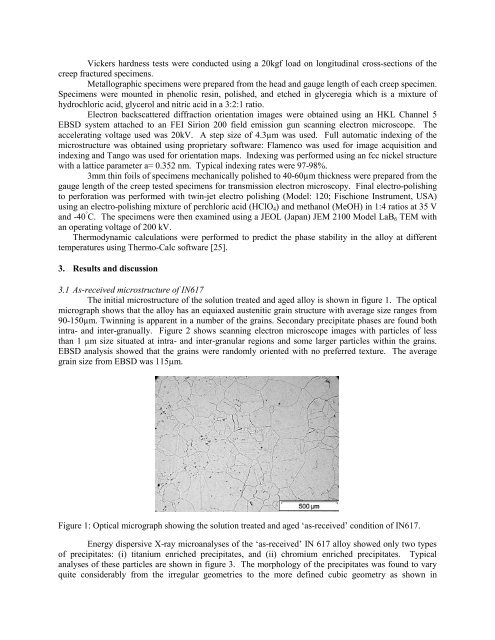
The <strong>in</strong>itial microstructure <strong>of</strong> the solution treated and aged alloy is shown <strong>in</strong> figure 1. The optical<br />
micrograph shows that the alloy has an equiaxed austenitic gra<strong>in</strong> structure with average size ranges from<br />
90-150µm. Tw<strong>in</strong>n<strong>in</strong>g is apparent <strong>in</strong> a number <strong>of</strong> the gra<strong>in</strong>s. Secondary precipitate phases are found both<br />
<strong>in</strong>tra- and <strong>in</strong>ter-granually. Figure 2 shows scann<strong>in</strong>g electron microscope images with particles <strong>of</strong> less<br />
than 1 µm size situated at <strong>in</strong>tra- and <strong>in</strong>ter-granular regions and some larger particles with<strong>in</strong> the gra<strong>in</strong>s.<br />
EBSD analysis showed that the gra<strong>in</strong>s were randomly oriented with no preferred texture. The average<br />
gra<strong>in</strong> size from EBSD was 115µm.<br />
Figure 1: Optical micrograph show<strong>in</strong>g the solution treated and aged ‘as-received’ condition <strong>of</strong> <strong>IN617</strong>.<br />
Energy dispersive X-ray microanalyses <strong>of</strong> the ‘as-received’ IN 617 alloy showed only two types<br />
<strong>of</strong> precipitates: (i) titanium enriched precipitates, and (ii) chromium enriched precipitates. Typical<br />
analyses <strong>of</strong> these particles are shown <strong>in</strong> figure 3. The morphology <strong>of</strong> the precipitates was found to vary<br />
quite considerably from the irregular geometries to the more def<strong>in</strong>ed cubic geometry as shown <strong>in</strong>