Electromagnetism Electromagnetism
Electromagnetism Electromagnetism
Electromagnetism Electromagnetism

Create successful ePaper yourself
Turn your PDF publications into a flip-book with our unique Google optimized e-Paper software.
Think about what<br />
it would be like<br />
to peer inside<br />
the human body to<br />
locate a tumor, find<br />
tiny blockages in blood<br />
vessels, or even identify<br />
damage to the brain.<br />
Medical technology<br />
known as magnetic<br />
resonance imaging<br />
(MRI) gives doctors a<br />
quick and painless way<br />
to see and diagnose<br />
these problems and<br />
more.<br />
Magnetic<br />
Images<br />
Like X rays, MRI creates pictures of a person’s<br />
internal organs and skeleton. But MRI produces<br />
clearer pictures than X rays do, and MRI does<br />
not expose the body to the potentially harmful<br />
radiation of X rays. Instead, MRI uses powerful<br />
electromagnets and radio waves to create<br />
images.<br />
The patient is placed in a large machine. An<br />
electric current in the electromagnet creates a<br />
powerful magnetic field around the patient.<br />
Because the human body is composed mostly of<br />
fat and water, there are many hydrogen atoms in<br />
the body. The magnetic field causes the nuclei of<br />
the hydrogen atoms to align in the direction of<br />
the magnetic field. Then another, weaker magnetic<br />
signal is sent out to the cells. The energy in<br />
this signal causes some hydrogen nuclei to change<br />
their position. As the signal's energy is absorbed<br />
and then released by the hydrogen nuclei, the<br />
MRI machine collects the signals and its computer<br />
converts the information into an image.<br />
Copyright © by Holt, Rinehart and Winston. All rights reserved.<br />
Magnets in Medicine<br />
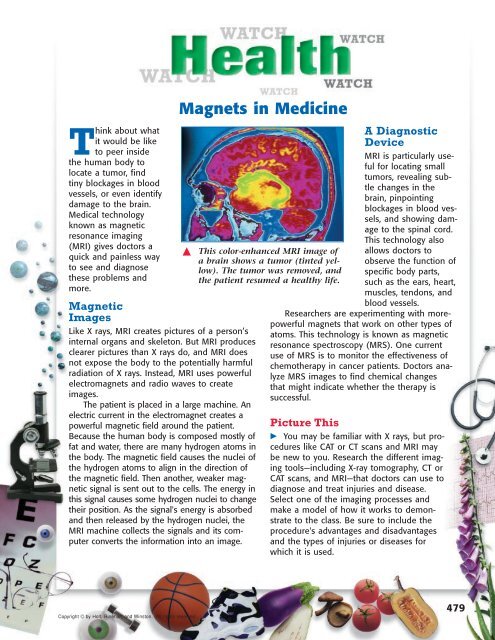
This color-enhanced MRI image of<br />
a brain shows a tumor (tinted yellow).<br />
The tumor was removed, and<br />
the patient resumed a healthy life.<br />
A Diagnostic<br />
Device<br />
MRI is particularly useful<br />
for locating small<br />
tumors, revealing subtle<br />
changes in the<br />
brain, pinpointing<br />
blockages in blood vessels,<br />
and showing damage<br />
to the spinal cord.<br />
This technology also<br />
allows doctors to<br />
observe the function of<br />
specific body parts,<br />
such as the ears, heart,<br />
muscles, tendons, and<br />
blood vessels.<br />
Researchers are experimenting with morepowerful<br />
magnets that work on other types of<br />
atoms. This technology is known as magnetic<br />
resonance spectroscopy (MRS). One current<br />
use of MRS is to monitor the effectiveness of<br />
chemotherapy in cancer patients. Doctors analyze<br />
MRS images to find chemical changes<br />
that might indicate whether the therapy is<br />
successful.<br />
Picture This<br />
You may be familiar with X rays, but procedures<br />
like CAT or CT scans and MRI may<br />
be new to you. Research the different imaging<br />
tools—including X-ray tomography, CT or<br />
CAT scans, and MRI—that doctors can use to<br />
diagnose and treat injuries and disease.<br />
Select one of the imaging processes and<br />
make a model of how it works to demonstrate<br />
to the class. Be sure to include the<br />
procedure's advantages and disadvantages<br />
and the types of injuries or diseases for<br />
which it is used.<br />
479