Follicular Hyperplasia, Follicular Lysis, and Progressive - American ...
Follicular Hyperplasia, Follicular Lysis, and Progressive - American ...
Follicular Hyperplasia, Follicular Lysis, and Progressive - American ...

Create successful ePaper yourself
Turn your PDF publications into a flip-book with our unique Google optimized e-Paper software.
Chang et al / FOLLICULAR HYPERPLASIA, FOLLICULAR LYSIS, AND PROGRESSIVE TRANSFORMATION OF GERMINAL CENTERS<br />
A B C<br />
D E F<br />
G H I<br />
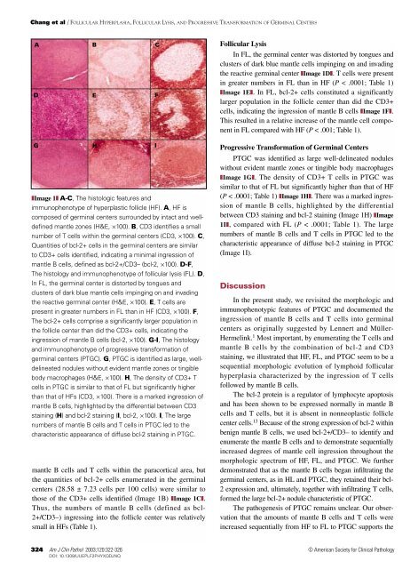
❚Image 1❚ A-C, The histologic features <strong>and</strong><br />
immunophenotype of hyperplastic follicle (HF). A, HF is<br />
composed of germinal centers surrounded by intact <strong>and</strong> welldefined<br />
mantle zones (H&E, ×100). B, CD3 identifies a small<br />
number of T cells within the germinal centers (CD3, ×100). C,<br />
Quantities of bcl-2+ cells in the germinal centers are similar<br />
to CD3+ cells identified, indicating a minimal ingression of<br />
mantle B cells, defined as bcl-2+/CD3– (bcl-2, ×100). D-F,<br />
The histology <strong>and</strong> immunophenotype of follicular lysis (FL). D,<br />
In FL, the germinal center is distorted by tongues <strong>and</strong><br />
clusters of dark blue mantle cells impinging on <strong>and</strong> invading<br />
the reactive germinal center (H&E, ×100). E, T cells are<br />
present in greater numbers in FL than in HF (CD3, ×100). F,<br />
The bcl-2+ cells comprise a significantly larger population in<br />
the follicle center than did the CD3+ cells, indicating the<br />
ingression of mantle B cells (bcl-2, ×100). G-I, The histology<br />
<strong>and</strong> immunophenotype of progressive transformation of<br />
germinal centers (PTGC). G, PTGC is identified as large, welldelineated<br />
nodules without evident mantle zones or tingible<br />
body macrophages (H&E, ×100). H, The density of CD3+ T<br />
cells in PTGC is similar to that of FL but significantly higher<br />
than that of HFs (CD3, ×100). There is a marked ingression of<br />
mantle B cells, highlighted by the differential between CD3<br />
staining (H) <strong>and</strong> bcl-2 staining (I, bcl-2, ×100). I, The large<br />
numbers of mantle B cells <strong>and</strong> T cells in PTGC led to the<br />
characteristic appearance of diffuse bcl-2 staining in PTGC.<br />
mantle B cells <strong>and</strong> T cells within the paracortical area, but<br />
the quantities of bcl-2+ cells enumerated in the germinal<br />
centers (28.58 ± 7.23 cells per 100 cells) were similar to<br />
those of the CD3+ cells identified (Image 1B) ❚Image 1C❚.<br />
Thus, the numbers of mantle B cells (defined as bcl-<br />
2+/CD3–) ingressing into the follicle center was relatively<br />
small in HFs (Table 1).<br />
324 Am J Clin Pathol 2003;120:322-326<br />
324 DOI: 10.1309/UUEPLF2PHYXQDJNQ<br />
<strong>Follicular</strong> <strong>Lysis</strong><br />
In FL, the germinal center was distorted by tongues <strong>and</strong><br />
clusters of dark blue mantle cells impinging on <strong>and</strong> invading<br />
the reactive germinal center ❚Image 1D❚. T cells were present<br />
in greater numbers in FL than in HF (P < .0001; Table 1)<br />
❚Image 1E❚. In FL, bcl-2+ cells constituted a significantly<br />
larger population in the follicle center than did the CD3+<br />
cells, indicating the ingression of mantle B cells ❚Image 1F❚.<br />
This resulted in a relative increase of the mantle cell component<br />
in FL compared with HF (P < .001; Table 1).<br />
<strong>Progressive</strong> Transformation of Germinal Centers<br />
PTGC was identified as large well-delineated nodules<br />
without evident mantle zones or tingible body macrophages<br />
❚Image 1G❚. The density of CD3+ T cells in PTGC was<br />
similar to that of FL but significantly higher than that of HF<br />
(P < .0001; Table 1) ❚Image 1H❚. There was a marked ingression<br />
of mantle B cells, highlighted by the differential<br />
between CD3 staining <strong>and</strong> bcl-2 staining (Image 1H) ❚Image<br />
1I❚, compared with FL (P < .0001; Table 1). The large<br />
numbers of mantle B cells <strong>and</strong> T cells in PTGC led to the<br />
characteristic appearance of diffuse bcl-2 staining in PTGC<br />
(Image 1I).<br />
Discussion<br />
In the present study, we revisited the morphologic <strong>and</strong><br />
immunophenotypic features of PTGC <strong>and</strong> documented the<br />
ingression of mantle B cells <strong>and</strong> T cells into germinal<br />
centers as originally suggested by Lennert <strong>and</strong> Müller-<br />
Hermelink. 1 Most important, by enumerating the T cells <strong>and</strong><br />
mantle B cells by the combination of bcl-2 <strong>and</strong> CD3<br />
staining, we illustrated that HF, FL, <strong>and</strong> PTGC seem to be a<br />
sequential morphologic evolution of lymphoid follicular<br />
hyperplasia characterized by the ingression of T cells<br />
followed by mantle B cells.<br />
The bcl-2 protein is a regulator of lymphocyte apoptosis<br />
<strong>and</strong> has been shown to be expressed normally in mantle B<br />
cells <strong>and</strong> T cells, but it is absent in nonneoplastic follicle<br />
center cells. 13 Because of the strong expression of bcl-2 within<br />
benign mantle B cells, we used bcl-2+/CD3– to identify <strong>and</strong><br />
enumerate the mantle B cells <strong>and</strong> to demonstrate sequentially<br />
increased degrees of mantle cell ingression throughout the<br />
morphologic spectrum of HF, FL, <strong>and</strong> PTGC. We further<br />
demonstrated that as the mantle B cells began infiltrating the<br />
germinal centers, as in HL <strong>and</strong> PTGC, they retained their bcl-<br />
2 expression <strong>and</strong>, ultimately, together with infiltrating T cells,<br />
formed the large bcl-2+ nodule characteristic of PTGC.<br />
The pathogenesis of PTGC remains unclear. Our observation<br />
that the amounts of mantle B cells <strong>and</strong> T cells were<br />
increased sequentially from HF to FL to PTGC supports the<br />
© <strong>American</strong> Society for Clinical Pathology