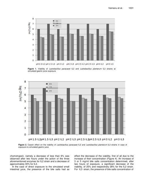
1030 Afr. J. Microbiol. Res. physiological <strong>and</strong> physico-chemical processes. <strong>The</strong> physiological parameters comprise pH, concentrations <strong>of</strong> gastric <strong>and</strong> small intestinal enzymes, concentrations <strong>of</strong> bile salts <strong>and</strong> <strong>the</strong> kinetics <strong>of</strong> passage through stomach <strong>and</strong> intestine (Dunne et al., 2001). Unfortunately, most studies on probiotic actions ignore <strong>the</strong>se facts <strong>and</strong>, as a result, data on <strong>the</strong> tolerance <strong>of</strong> probiotic strains are quite rare ( y elewicz et al., 2010). <strong>The</strong> purpose <strong>of</strong> this research was represented by <strong>the</strong> establishment <strong>of</strong> <strong>the</strong> <strong>viability</strong> <strong>of</strong> <strong>Lactobacillus</strong> <strong>paracasei</strong> <strong>IL2</strong> <strong>and</strong> <strong>Lactobacillus</strong> plantarum IL3 strains during <strong>the</strong>ir transit through <strong>the</strong> stomach <strong>and</strong> small intestine. <strong>The</strong> conditions at <strong>the</strong> gastric level were simulated by <strong>the</strong> use <strong>of</strong> pepsin, at various pH values between 1.5 <strong>and</strong> 3. <strong>The</strong> simulated pancreatic juice contained pancreatin <strong>and</strong> bile salts, in various concentrations, between 1.5 <strong>and</strong> 5. Moreover, <strong>the</strong> influences <strong>of</strong> casein <strong>and</strong> mucin, as protectors <strong>of</strong> <strong>the</strong> probiotic cells, on <strong>the</strong> <strong>viability</strong> were tested. Finally, <strong>the</strong> combined effect <strong>of</strong> <strong>the</strong> action <strong>of</strong> <strong>the</strong> simulated gastric <strong>and</strong> small intestine juice was determined <strong>and</strong> <strong>the</strong> ma<strong>the</strong>matical parameters <strong>of</strong> <strong>the</strong> cell <strong>viability</strong> <strong>and</strong> mortality were calculated. MATERIALS AND METHODS Biological materials <strong>The</strong> bacterial strains L. <strong>paracasei</strong> <strong>IL2</strong> <strong>and</strong> L. plantarum IL3 were maintained in glycerol 20% (Collection <strong>of</strong> <strong>the</strong> Faculty <strong>of</strong> Biotechnology, Bucharest), at -82°C. <strong>The</strong> strain was revitalized by two successive cultures in MRS broth, at 37°C. <strong>The</strong> experiments were performed in <strong>the</strong> Industrial Biotechnology Laboratory <strong>of</strong> <strong>the</strong> Department <strong>of</strong> Biotechnology, in <strong>the</strong> second half <strong>of</strong> 2010. <strong>The</strong> gastric <strong>and</strong> small intestine juice were prepared according to <strong>the</strong> method described by Kos et al. (2000). In case <strong>of</strong> simulated gastric juice (pepsin 3 g/l), various pH values, <strong>of</strong> 1.5, 2, 2.5 <strong>and</strong> 3.0 were used. <strong>The</strong> simulation <strong>of</strong> <strong>the</strong> small intestine juice (pancreatin 1 g/l) was made at various bile salts concentrations (1.5, 2, 3 <strong>and</strong> 5 mg/ml). <strong>The</strong> mucin <strong>and</strong> casein influences on <strong>the</strong> strain <strong>viability</strong> in <strong>the</strong> gastric <strong>and</strong> small intestine juice were determined. A concentration <strong>of</strong> 1 g/liter in NaOH 0.5% was used <strong>and</strong> <strong>the</strong> determination was performed according to <strong>the</strong> method described by Kos et al. (2000). <strong>The</strong> cumulated effect <strong>of</strong> <strong>the</strong> simulated gastric <strong>and</strong> small intestine juice was determined at pH 2 <strong>and</strong> bile salts concentration <strong>of</strong> 3 mg/ml in <strong>the</strong> pancreatic juice. All tests were performed in Durham tubes, provided with silicone membrane meant for sampling (Kos et al., 2000; Sarahroodi et al., 2010; Puangpronpitag et al., 2009; Movsesyan et al., 2010; Vamanu <strong>and</strong> Vamanu, 2010). Fur<strong>the</strong>rmore, <strong>the</strong> effects <strong>of</strong> trypsin, chymotrypsin, <strong>and</strong> pronase on <strong>viability</strong> were determined separately for each enzyme. Thus, in a Durham tube, 1 ml <strong>of</strong> enzyme solution at a concentration <strong>of</strong> 1 mg/ml, 0.3 ml NaOH 0.5% <strong>and</strong> 0.2 ml cell suspension were added. Within two hours, <strong>the</strong> <strong>viability</strong> was determined in <strong>the</strong> presence <strong>of</strong> mucin <strong>and</strong> casein (Kos et al., 2000; Sarahroodi et al., 2010; Philip et al., 2009). <strong>The</strong> <strong>viability</strong> <strong>and</strong> mortality were determined at various pH values according to <strong>the</strong> method described by Kos et al. (2000), in <strong>the</strong> presence <strong>of</strong> pepsin <strong>and</strong> respectively <strong>of</strong> pancreatin, toge<strong>the</strong>r with various concentrations <strong>of</strong> bile salts. <strong>The</strong> same ma<strong>the</strong>matical indices were calculated as well in <strong>the</strong> presence <strong>of</strong> mucin <strong>and</strong> casein, according to <strong>the</strong> protection <strong>of</strong>fered to <strong>the</strong> cell <strong>viability</strong>. <strong>The</strong> critical points were represented by <strong>the</strong> crossing between <strong>the</strong> <strong>viability</strong> <strong>and</strong> mortality curves (Kos et al., 2000; Sarahroodi et al., 2010; Yateem et al., 2008; Vamanu <strong>and</strong> Vamanu, 2010). <strong>The</strong> <strong>viability</strong> was determined by insemination in double layer, in MRS broth, hourly. <strong>The</strong> plates were incubated for 48 h at 37°C <strong>and</strong> <strong>the</strong> results were read using <strong>the</strong> ColonyQuant equipment <strong>and</strong> <strong>the</strong>y were registered as <strong>the</strong> log (CFU/ml) (Kos et al., 2000; Sarahroodi et al., 2010; Otles <strong>and</strong> Ozlem, 2003; Vamanu <strong>and</strong> Vamanu, 2010). RESULTS AND DISCUSSION <strong>The</strong> tested strains must have a good <strong>viability</strong> in order to be used as probiotic, since one <strong>of</strong> <strong>the</strong> greatest problems <strong>of</strong> <strong>the</strong>se strains is <strong>the</strong>ir resistance in <strong>the</strong> conditions <strong>of</strong> <strong>the</strong> gastric <strong>and</strong> intestinal transit. <strong>The</strong> effect <strong>of</strong> <strong>the</strong> gastrointestinal transit, which begins in <strong>the</strong> stomach, was exercised by pepsin, at pH between 1.5 <strong>and</strong> 3. <strong>The</strong> stationary time at this level did not exceed 2 h. Thus, Figure 1 presents <strong>the</strong> <strong>viability</strong> <strong>of</strong> <strong>IL2</strong> <strong>and</strong> IL3 strains at gastric level. <strong>The</strong> <strong>viability</strong> <strong>of</strong> <strong>the</strong> strains, especially that <strong>of</strong> <strong>IL2</strong> strain, was directly influenced by <strong>the</strong> pH value. At pH <strong>of</strong> 1.5, <strong>the</strong> <strong>IL2</strong> strain showed only 72% <strong>of</strong> <strong>the</strong> <strong>viability</strong> registered at 0 h <strong>of</strong> exposure, in comparison with 70% for IL3. <strong>The</strong> value <strong>of</strong> <strong>the</strong> <strong>viability</strong> was in such situation higher for IL3 in comparison with <strong>IL2</strong>. At pH higher than 2, <strong>the</strong> strains maintained <strong>the</strong>ir <strong>viability</strong> constant after one h <strong>of</strong> exposure to <strong>the</strong> simulated gastric juice. After two hours, as pH increased from 1.5 to 2, <strong>the</strong> <strong>viability</strong> also increased <strong>and</strong> remained constant after pH reached 2.5, being 77% <strong>of</strong> <strong>the</strong> initial one for <strong>IL2</strong> <strong>and</strong> 72% <strong>of</strong> <strong>the</strong> initial one for IL3, respectively. <strong>The</strong> presented data proved that <strong>the</strong> strains were resistant to low pH, especially IL3, which maintained a higher <strong>viability</strong> value, irrespective <strong>of</strong> <strong>the</strong> values <strong>of</strong> gastric pH. Casein was a better protector than mucin in <strong>the</strong> case <strong>of</strong> <strong>the</strong> <strong>viability</strong> <strong>of</strong> L. <strong>paracasei</strong> <strong>IL2</strong> <strong>and</strong> L. plantarum IL3 strains, with respect to <strong>the</strong> action <strong>of</strong> <strong>the</strong> simulated gastric juice. <strong>The</strong> <strong>viability</strong> <strong>of</strong> <strong>the</strong> two strains depended on pH, but was not higher than in <strong>the</strong> absence <strong>of</strong> such substances (Figure 2). In general, <strong>the</strong> <strong>viability</strong> values were in average by 15% higher at pH <strong>of</strong> 1.5, both for casein, as well as for mucin, for <strong>the</strong> <strong>IL2</strong> strain <strong>and</strong> by 2% higher for <strong>the</strong> IL3 strain (Figure 3). On <strong>the</strong> o<strong>the</strong>r h<strong>and</strong>, at pH <strong>of</strong> 2.0, <strong>the</strong> <strong>viability</strong> value in <strong>the</strong> case <strong>of</strong> casein presence in comparison with mucin was also approximately by 15% higher for <strong>IL2</strong>, while for <strong>the</strong> IL3 strain <strong>the</strong> <strong>viability</strong> was relatively similar. At pH values <strong>of</strong> 2.5 or 3.0, <strong>the</strong> <strong>viability</strong> maintained <strong>the</strong> same trend, irrespective <strong>of</strong> <strong>the</strong> presence <strong>of</strong> casein or mucin. <strong>The</strong> difference in favor <strong>of</strong> <strong>the</strong> presence <strong>of</strong> casein for strain <strong>IL2</strong>, at pH value <strong>of</strong> 2.5 <strong>and</strong> 3.0, was approximately 10%, for an exposure <strong>of</strong> one or two hours. <strong>The</strong> IL3 strain was an exception in this case as well, since at pH <strong>of</strong> 2.5 or 3.0 it maintained its <strong>viability</strong> constant, irrespective <strong>of</strong> <strong>the</strong> used protector. Before testing <strong>the</strong> <strong>viability</strong> in case <strong>of</strong> exposure to <strong>the</strong> small intestinal juice, <strong>the</strong> influence <strong>of</strong> o<strong>the</strong>r enzymes on L. <strong>paracasei</strong> <strong>IL2</strong> <strong>and</strong> L. plantarum IL3 strains was determined. <strong>The</strong> result was <strong>the</strong> relative maintenance <strong>of</strong> <strong>the</strong> <strong>viability</strong> under <strong>the</strong> action <strong>of</strong> trypsin, pronase <strong>and</strong>
0 h 1 h 2 0 h Figure 1. Viability <strong>of</strong> <strong>Lactobacillus</strong> <strong>paracasei</strong> <strong>IL2</strong> <strong>and</strong> <strong>Lactobacillus</strong> plantarum IL3 strains at simulated gastric juice exposure 0 h 1 h 2 h Vamanu et al. 1031 Figure 2. Casein effect on <strong>the</strong> <strong>viability</strong> <strong>of</strong> <strong>Lactobacillus</strong> <strong>paracasei</strong> <strong>IL2</strong> <strong>and</strong> <strong>Lactobacillus</strong> plantarum IL3 strains in case <strong>of</strong> exposure to simulated gastric juice chymotrypsin, namely a decrease <strong>of</strong> less than 6% was observed after two hours under <strong>the</strong> action <strong>of</strong> <strong>the</strong> three aforementioned enzymes for <strong>IL2</strong> strain <strong>and</strong> a decrease <strong>of</strong> approximately 30% for IL3. In <strong>the</strong> case <strong>of</strong> direct exposure to <strong>the</strong> simulated small intestinal juice, <strong>the</strong> presence <strong>of</strong> <strong>the</strong> bile salts had as effect <strong>the</strong> decrease <strong>of</strong> <strong>the</strong> <strong>viability</strong>, first <strong>of</strong> all due to <strong>the</strong> increase <strong>of</strong> <strong>the</strong>ir concentration (Figure 4). An increase <strong>of</strong> 3 or 5 mg/ml bile salts concentration determined, after two hours <strong>of</strong> exposure, a significant decrease <strong>of</strong> <strong>the</strong> <strong>viability</strong>, <strong>of</strong> 25% <strong>and</strong> respectively 28% for <strong>the</strong> <strong>IL2</strong> strain. For IL3 strain, <strong>the</strong> presence <strong>of</strong> bile salts concentration <strong>of</strong>