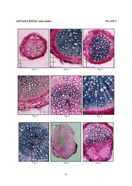
Conclusions The root passes quite early to the secondary structure, due to the activity of both lateral meristems. Only a few species present mechanic tissue, represented by isolated or grouped periphloemic sclerenchymatic fibers and a few weak collenchymatized elements in hypo<strong>de</strong>rmic position. Haustoria present a structure adapted to their function, bearing xylem vessels which facilitate the transport of the cru<strong>de</strong> sap from the host plant to the xylemic tissue of the parasite’s root. The stem presents a secondary structure resulted only from the cambium’s activity; the secondary conductive tissues are of annular type. The indumentums of leaves and bracts differs in all five species, being consi<strong>de</strong>red as very good taxonomic criteria. The mesophyll differs, too, in all five species, being correlated with the environment of the species. REFERENCES 1. BONNIER G., SABLON L., 1905 - Cours <strong>de</strong> Botanique 1. Librarie Génér<strong>ale</strong> <strong>de</strong> l’Enseignement, Paris 2. CIOCÎRLAN V., 2000 - Flora ilustrată a României. Bucureşti. Edit. Ceres 3. GORENFLOT R., 1994 - <strong>Biologie</strong> végét<strong>ale</strong>. Plantes supérieurs. Paris. Edit. Masson: 184-193 4. LECLERC DU SABLON, 1889 - Recherches sur les organes d’absorption <strong>de</strong>s plantes parasites. (Rhinanthées et Santalacées). Ann. <strong>de</strong>s Sci. Nat. Bot. 7 e série, 6: 91-117 5. METCALFE C. R., CHALK L., 1972 - Anatomy of the Dicotyledons. Oxford, Clarendon Press. 2:1184-1194 6. NAPP-ZINN KL., 1973, 1974, 1984 – Anatomie <strong>de</strong>s Blattes. II. Angiospermen. In Handbuch <strong>de</strong>r Pflanzenanatomie, Bd. VIII, 2 A1-2, 2 B1, Gebru<strong>de</strong>r Borntraeger, Berlin, Stuttgart 7. NICKRENT D. L., MUSSELMAN L. J., 2004 - Introduction to Parasitic Flowering Plants. A. P. S. Education Center, Boston 8. NIŢĂ MIHAELA, TUDOSE MIHAELA, GIUŞCĂ FL., 1995 - Contribuţii histo-anatomice referitoare la unele specii <strong>de</strong> Melampyrum L. Bul. Grăd. Bot. Iaşi. 5: 75-81 9. PAUCĂ ANA, NYARADY E. I., 1960 – Melampyrum L. în Flora R. P. Române, Edit. Acad. Rom., Bucureşti, 7: 622-639 10. TOMA C., 2002 - Strategii evolutive în regnul vegetal. Iaşi. Edit. Univ. „Al. I. Cuza” 11. TOMA C., GOSTIN IRINA, 2000 - Histologie vegetală. Iaşi. Edit. Univ. „Al. I. Cuza” Explanation of plates PLATE I Fig. 1 – Melampyrum arvense: lateral root (cross section) Fig. 2 – Melampyrum bihariense: lateral root (cross section) Fig. 3 – Melampyrum cristatum: lateral root (cross section) Fig. 4 – Melampyrum saxosum: main root (cross section) Fig. 5 – Melampyrum sylvaticum: main root (cross section) Fig. 6 – Melampyrum bihariense: main root (cross section) Fig. 7 – Melampyrum bihariense: main root (cross section) Fig. 8 – Melampyrum arvense: haustorul (cross section through the main root) Fig. 9 – Melampyrum sylvaticum: haustorul (cross section through the main root) PLATE II Fig. 10 – Melampyrum saxosum: haustorul (cross section through the main root) Fig. 11 – Melampyrum cristatum: haustorul (cross section through the main root) Fig. 12 – Melampyrum bihariense: tulpina (cross section) Fig. 13 – Melampyrum sylvaticum: stem (cross section) Fig. 14 – Melampyrum arvense: stem (cross section) Fig. 15 – Melampyrum bihariense: stem (cross section) Fig. 16 – Melampyrum arvense: stem (cross section) Fig. 17 – Melampyrum cristatum: tulpina (cross section) Fig. 18 – Melampyrum saxosum: peţiolul (cross section) Fig. 19 – Melampyrum bihariense: limbul (cross section) Fig. 20 – Melampyrum saxosum: limbul (cross section) 10
ASPAZIA BĂEŞU and colabs. PLATE I Fig. 1 Fig. 2 Fig. 3 Fig. 4 Fig. 5 Fig. 6 Fig. 7 Fig. 8 Fig. 9 11