Pfister-1973-The-psilopezioid-fungi-IV-Pachyella-0001 - ASCOfrance
Pfister-1973-The-psilopezioid-fungi-IV-Pachyella-0001 - ASCOfrance
Pfister-1973-The-psilopezioid-fungi-IV-Pachyella-0001 - ASCOfrance

Create successful ePaper yourself
Turn your PDF publications into a flip-book with our unique Google optimized e-Paper software.
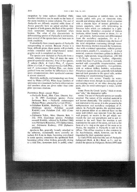
201 0 CAN. J. BOT. VOL. 51, <strong>1973</strong><br />
excipulum by some authors (Eckblad 1968).<br />
Another distinction can be made on the basis of<br />
the ascus reaction in iodine solution. <strong>The</strong> asci of<br />
<strong>Pachyella</strong> are diffusely amyloid while those of<br />
Peziza generally have an apical amyloid ring.<br />
In species of both genera, the flesh of the apothe-<br />
cium sometimes becomes discolored when<br />
broken. <strong>The</strong> value of this characteristic in<br />
differentiating species of <strong>Pachyella</strong> is not known,<br />
since several of the species have not been seen in<br />
fresh condition.<br />
Undoubtedly there remain species of <strong>Pachyella</strong><br />
masquerading as pezizas. Because Peziza is a<br />
large, difficult genus, these species will probably<br />
remain concealed until comprehensive mono-<br />
graphic work is completed on Peziza.<br />
<strong>The</strong> species now included in <strong>Pachyella</strong> form a<br />
closely related group. <strong>The</strong>y all share the same<br />
general apothecial anatomy. Four of the species,<br />
P. adnata (Berk. & Curt.) Pfist., P. clypeata<br />
(Schw.) Le Gal, P. megalosperma (Le Gal) Pfist.,<br />
and P. violaceonigra (Rehm) Pfist., are distin-<br />
guished from one another by differences in their<br />
spore ornamentations; their apothecial anatomy<br />
is almost identical.<br />
Materials, methods, and terminology are those<br />
used by <strong>Pfister</strong> (<strong>1973</strong>b). When large numbers of<br />
specimens were examined, herbaria abbreviations<br />
and numbers alone are given rather than com-<br />
plete specimen citations.<br />
PACHYELLA Boud. emend. <strong>Pfister</strong><br />
=<strong>Pachyella</strong> Boud., Hist. Class. Discom. Eur .<br />
p. 50. 1907. (Lectotype species: Peziza<br />
barlaeana Bres. = <strong>Pachyella</strong> violaceonigra<br />
(Rehm) <strong>Pfister</strong>, selected by Le Gal 1953b.)<br />
=Peltidium Kalchb., Hedwigia, 2: 58. 1862.<br />
(Holotype species : Peltidium oocardii<br />
Kalchb.) non Peltidium Zoll. 1820 (Compositae).<br />
= Pulvinaria Velen., Mon. Discom. Boh. 1 :<br />
3 32. 1934. (Lectotype species : Peltidium<br />
oocardii Kalchb., selected by Eckblad 1968)<br />
non Pulvinaria Bonorden, 195 1 (Sphaeriales),<br />
nec Pulvinaria Rodway, 19 18 (Sphaeropsidales).<br />
Apothecia flat, generally broadly attached to<br />
the substrate, occasionally more centrally attached,<br />
0.4-8 cm in diam, becoming convoluted<br />
in some species, sometimes the apothecial flesh<br />
becoming yellow when broken, generally drying<br />
to a thin film; hymenium dark to pallid, some-<br />
times with vinaceous or reddish tints; on the<br />
outside pallid with grey or vinaceous tints,<br />
smooth and shining when fresh. Ectal excipulum<br />
with a discrete layer of textura globulosa to<br />
textura angularis, outer cells terminating in<br />
flexuous hairs which are embedded in a gela-<br />
tinous matrix. Medullary exciplum of textura<br />
intricata, either loosely woven or dense, J+ or<br />
J- gel present. Subhymenium not differentiated<br />
from the medullary excipulum, J+ or J-.<br />
Margin not present as a distinct zone, the hairs of<br />
the ectal excipulurn continuing (though some-<br />
times becoming shorter) toward the hymenium.<br />
Asci with a terminal operculum, without promi-<br />
nent croziers, usually J+, sometimes J- in dried<br />
material, contents sometimes golden in Melzer's<br />
reagent, eight-spored, long-cylindrical, 250-500<br />
X 15-20 p.m. Ascospores hyaline, ellipsoidal,<br />
mostly less than 25 pm long, smooth or variously<br />
marked with cyanophilic ornamentation, outer<br />
spore wall usually cyanophilic, two-guttulate,<br />
with or without deBary bubbles, uninucleate.<br />
Paraphyses septate clavate, generally with definite<br />
internal dark granules in the apical cells, neither<br />
branching nor anastornosing frequently.<br />
SUBSTRATE AND RANGE: Usually on water-<br />
soaked rotten wood, though occasionally on soil<br />
surrounding wood, or on other decaying plant<br />
parts, also on wood submerged in water; world-<br />
wide.<br />
NAME: From the Greek "pachy" thick or stout,<br />
and "ella," the diminutive suffix.<br />
NOTES: <strong>The</strong> asci of <strong>Pachyella</strong> species are usually<br />
amyloid. <strong>The</strong> nature of the amyloid material is<br />
not known chemically. <strong>The</strong> amyloid reaction is<br />
not restricted to the ascus; it is also present in the<br />
subhymenium and medullary excipulum of P.<br />
adnata. In <strong>Pachyella</strong> species, the amyloid mate-<br />
rial is present either as an external layer on the<br />
ascus wall (which may separate from the ascus<br />
wall proper), or occurs in the gel which sur-<br />
rounds the asci and paraphyses and is not re-<br />
stricted to the wall. Since the reaction is not<br />
restricted to the apex of the ascus, nor is it in the<br />
form of a J+ ring at the apex of the ascus as in<br />
Peziza, the reaction in <strong>Pachyella</strong> is said to be<br />
diffuse. This diffuse reaction is also common in<br />
the Ascobolaceae, which, however, can be dis-<br />
tinguished by their eguttulate spores.<br />
Le Gal (1953b) described the asci of <strong>Pachyella</strong><br />
babingtonii (Berk. & Br.) Boud. as J- and in-<br />
cluded the species in Psilopezia. Rehm (1 8%) and