Oral Presentations - Arteriosclerosis, Thrombosis, and Vascular ...
Oral Presentations - Arteriosclerosis, Thrombosis, and Vascular ...
Oral Presentations - Arteriosclerosis, Thrombosis, and Vascular ...

Create successful ePaper yourself
Turn your PDF publications into a flip-book with our unique Google optimized e-Paper software.
A Role for C/EBP in Hepatic Transcription of the Gene Encoding TAFI<br />
Michael B Boffa, Jeffrey Hamill, Rebecca Dillon, Michael E Nesheim, Marlys L Koschinsky.<br />
Queen’s University, Kingston, ON, Canada<br />
P84<br />
Thrombin activable fibrinolysis inhibitor (TAFI) is a recently described plasma zymogen that<br />
appears to play a role in regulating the balance between the activities of the coagulation <strong>and</strong><br />
fibrinolytic cascades. Plasma concentrations of TAFI are largely genetically determined <strong>and</strong> vary<br />
in the population by almost an order of magnitude. Emerging data suggest that TAFI<br />
concentrations constitute a risk factor for thrombotic disorders <strong>and</strong> that TAFI is a positive acute<br />
phase reactant. Currently, no information exists concerning the mechanisms underlying control<br />
of TAFI gene expression. Using a computer algorithm to locate consensus transcription factor<br />
binding sites, we identified overlapping potential binding sites for the liver-enriched transcription<br />
factors C/EBP <strong>and</strong> hepatic leukemia factor (HLF) in the TAFI promoter. Based on the known<br />
sequence requirements for binding of the respective transcription factors, single nucleotide<br />
mutations were introduced into the TAFI promoter that would be expected to abolish either<br />
C/EBP or HLF binding or both. The mutant promoter sequences were used to construct<br />
luciferase reporter plasmids that were transiently transfected into HepG2 cells. Mutations that<br />
would be expected to abolish C/EBP binding alone or both C/EBP <strong>and</strong> HLF binding drastically<br />
reduced TAFI promoter activity (to 10 –20% of wild-type) while the mutation that would be<br />
expected to abolish HLF binding alone had no effect. Gel mobility shift assays <strong>and</strong> supershift<br />
assays revealed that this site was able to bind C/EBP present in nuclear extracts prepared from<br />
HepG2 cells <strong>and</strong> adult rat liver; C/EBP was the predominant binding isoform in the HepG2<br />
nuclear extracts (consistent with the fetal-like phenotype of this cell line) while both C/EBP<br />
<strong>and</strong> C/EBP bound in the adult rat liver nuclear extracts. Importantly, binding site mutations<br />
that decreased TAFI promoter activity also abolished C/EBP binding. These results were<br />
corroborated using nuclear extracts from a non-hepatic cell line (Baby Hamster Kidney (BHK))<br />
overexpressing C/EBP isoforms; these experiments also showed that C/EBP, which is induced<br />
in liver during the acute phase, also is capable of binding to the TAFI promoter C/EBP site.<br />
P85<br />
Fabry Disease in Mice Is Associated with Age-Dependent Susceptibility to<br />
<strong>Vascular</strong> <strong>Thrombosis</strong><br />
Peter F Bodary, Yuechen Shen, Susan R Wild, Akira Abe, James A Shayman, Daniel T<br />
Eitzman. University of Michigan, Ann Arbor, MI<br />
Fabry disease is an x-linked lysosomal storage disorder due to deficiency of -galactosidase<br />
A activity that results in widespread accumulation of neutral glycosphingolipids. Renal failure,<br />
neuropathy, premature myocardial infarction <strong>and</strong> stroke occur in patients with this condition<br />
primarily due to progressive deposition of glycosphingolipids in vascular endothelial cells. The<br />
clinical consequences of Fabry disease suggest that vascular thrombosis may play a prominent<br />
role in the pathogenesis of this disease, however the vasculopathy associated with Fabry<br />
disease has not been extensively studied. To determine if Fabry mice are susceptible to<br />
vascular thrombosis, mice deficient in -galactosidase A were studied in a photochemical<br />
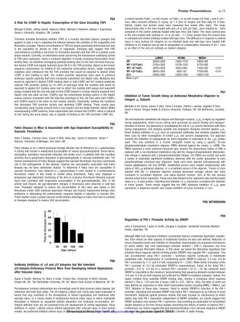
thrombosis model. In this model of carotid artery thrombosis, Fabry mice displayed a<br />
progressive age dependent shortening of the time to occlusive thrombosis following vascular<br />
injury that correlated with progressive accumulation of the glycosphingolipid, Gb3, in the<br />
arterial wall (Figure). No age-dependent changes in thrombosis were observed in wild-type<br />
mice. Therapies designed to reduce the accumulation of Gb3 were also tested in this<br />
thrombosis model. Both substrate deprivation therapy <strong>and</strong> enzyme replacement therapy were<br />
ineffective in attenuating the prothrombotic response despite a reduction in vascular Gb3.<br />
These studies reveal a potent vascular prothrombotic phenotype in Fabry mice that is resistant<br />
to therapies designed to reduce Gb3 accumulation.<br />
P86<br />
Antibody Inhibition of V <strong>and</strong> 3 Integrins but Not Inherited<br />
3-Integrin-Deficiency Protects Mice from Developing Intimal Hyperplasia<br />
after <strong>Vascular</strong> Injury<br />
Susan S Smyth, Hsiming Yu, Barry S Coller, Ernane Reis. University of North Carolina,<br />
Chapel Hill, NC; The Rockefeller University, NY, NY; Mount Sinai School of Medicine, NY, NY<br />
Percutaneous coronary interventions are increasingly used to treat coronary artery disease, but<br />
restenosis still limits their utility. The 3 integrins (IIb3 <strong>and</strong> V3) have been implicated in<br />
events that may contribute to the development of intimal hyperplasia <strong>and</strong> restenosis after<br />
vascular injury. In a mouse model of endoluminal femoral artery injury in which endothelial<br />
denudation is followed by sequential platelet deposition <strong>and</strong> leukocyte accumulation, 3integrin-deficient<br />
mice are not protected from the development of intimal hyperplasia. Since<br />
inhibitors of V3(IIb3) reduce intimal hyperplasia after vascular injury in other animal<br />
models, we performed bilateral arterial injury in wild-type Downloaded mice; in wild-type from<br />
mice treated with<br />
http://atvb.ahajournals.org/<br />
a control hamster F(ab)’ 2, an anti-mouse V F(ab)’ 2, or an anti-mouse 3 F(ab)’ 2; <strong>and</strong> in 3-/mice.<br />
Mice received antibody (2 mg/kg, ip) 1 h prior to surgery <strong>and</strong> then daily for 9 days.<br />
Intimal, medial <strong>and</strong> luminal areas were measured four weeks after injury. The mean<br />
intima/media ratio in the mice treated with anti-V or anti-3 F(ab)’ 2 were reduced by 60%<br />
compared to the control antibody-treated wild-type mice (see Table). The mean luminal area<br />
in the mice treated with antibody to V or3 was 1.5 times greater than the luminal area<br />
in untreated <strong>and</strong> control antibody-treated wild-type mice. The difference in response to arterial<br />
injury in mice lacking 3 integrins on an inherited basis <strong>and</strong> wild-type mice treated with<br />
inhibitors of 3 integrins may be due to upregulation of compensatory molecules in 3-/- mice<br />
or an effect of the anti-3 antibody on distinct integrins.<br />
Inhibition of Tumor Growth Using an Antisense Morpholino Oligomer to<br />
Integrin v Subunit<br />
Michelle A De Sousa, James D Bird, Carla A London, Patrick L Iversen, Gayathri R Devi,<br />
David H Farrell. Oregon Health & Science University, Portl<strong>and</strong>, OR; AVI BioPharma, Corvallis,<br />
OR<br />
The microvascular endothelial cell integrin <strong>and</strong> fibrinogen receptor, v 3, is highly up-regulated<br />
during angiogenesis, which occurs during such processes as wound healing <strong>and</strong> neovascularization<br />
of tumors. Our laboratory is investigating the role of v 3 <strong>and</strong> its interaction with fibrin<br />
during angiogenesis, <strong>and</strong> studying possible anti-angiogenic therapies directed against v 3.<br />
Direct binding inhibitors of v 3 such as monoclonal antibodies <strong>and</strong> synthetic peptides have<br />
been used by other investigators to inhibit v 3 <strong>and</strong> prevent angiogenesis. Our approach<br />
involved the inhibition of angiogenesis by down-regulation of expression of the v 3 protein<br />
itself. This was achieved in a murine xenograft model by using a specific 20-mer<br />
phosphorodiamidate morpholino oligomer (PMO) directed against the mouse v mRNA. The<br />
PMOs represent a novel antisense structural type, wherein the deoxyribose moiety of DNA is<br />
replaced with a six-membered morpholine ring <strong>and</strong> the charged phosphodiester internucleoside<br />
linkage is replaced with a phosphorodiamidate linkage. The PMOs are neutral <strong>and</strong> avoid<br />
a variety of potentially significant limitations observed with the earlier generation of ionic<br />
phosphorothioate structural type oligomers. Nude mice were injected subcutaneously with<br />
human glioblastoma cell line U87MG. Established tumors were treated intratumorally with<br />
saline or 300 g antisense oligomer, or a scrambled version of the oligomer. Tumors of mice<br />
injected with the v antisense oligomer showed decreased average volume <strong>and</strong> mass<br />
compared to scrambled oligomer- <strong>and</strong> saline-injected controls. 50% of the test animals<br />
showed actual tumor regression. Those mice that showed tumor regression also had the lowest<br />
ratios of v to total protein, indicating that inhibition of v expression correlated with inhibition<br />
of tumor growth. These results suggest that the PMO antisense inhibition of v 3 gene<br />
expression is sequence-specific <strong>and</strong> causes inhibition of tumor formation in vivo.<br />
Regulation of PAI-1 Promoter Activity by SREBP<br />
Poster <strong>Presentations</strong> a-15<br />
P87<br />
P88 WITHDRAWN<br />
John A Schoenhard, Layton H Smith, Douglas E Vaughan. V<strong>and</strong>erbilt University Medical<br />
Center, Nashville, TN<br />
Although HMG-CoA reductase inhibitors consistently improve endothelial-dependent vasodilation,<br />
their effects on other aspects of endothelial function are less well defined. Reduction of<br />
serum cholesterol levels <strong>and</strong> inhibition of intracellular isoprenylation are proposed mechanisms<br />
by which statins may limit plasminogen activator inhibitor-1 (PAI-1) expression <strong>and</strong> thus<br />
improve vascular fibrinolytic balance. In this study, we tested the alternative hypothesis that<br />
activated sterol response element binding protein (SREBP) may regulate PAI-1 expression. This<br />
was accomplished using PAI-1 promoter / luciferase reporter constructs in transfected<br />
endothelial cells. Overexpression of constitutively active SREBP1a induced -3.4 <strong>and</strong> -0.8 kb<br />
PAI-1 promoters by 11.5- <strong>and</strong> 9.5-fold, respectively (P 0.001). While further truncation of the<br />
PAI-1 promoter to -0.4 kb eliminated SREBP1a responsiveness, fusion of the distal PAI-1<br />
promoter (-0.8 to -0.4 kb) to a minimal PAI-1 promoter (-52 to 76 bp) conferred novel<br />
SREBP1a inducibility to this construct, demonstrating that sequence elements located between<br />
-0.8 <strong>and</strong> -0.4 kb are both required <strong>and</strong> sufficient for SREBP1a mediated activation. Indeed, this<br />
region contains three potential SREBP binding sites, namely a consensus sterol response<br />
element (-766 to -757) <strong>and</strong> two E-boxes (-680 to -675, -569 to -564), the latter of which we<br />
have defined as responsive to other bHLH transcription factors including BMAL1, BMAL2, <strong>and</strong><br />
TFE3. Mutation of these sites, however, failed to obviate SREBP1a induction of the PAI-1<br />
promoter, indicating that activated SREBP may induce PAI-1 expression by an indirect or novel<br />
mechanism. Balanced against previous studies that have focused on mechanisms by which<br />
statins may limit PAI-1 expression independent of SREBP activation, our results suggest that<br />
SREBP activation may enhance PAI-1 expression, thus providing an explanation for inconsistent<br />
results obtained so far from clinical studies concerning the effects of statins on plasma PAI-1.<br />
Furthermore, our results suggest that lipid lowering therapies more narrowly targeted to SREBP<br />
activation by mayguest not capture on April potential 4, fibrinolytic 2013 benefits of statin therapy.<br />
P89